15N NMR Relaxation Data Reveal Significant Chemical Exchange Broadening in the α-Domain of Human...
9
15 N NMR Relaxation Data Reveal Significant Chemical Exchange Broadening in the r-Domain of Human r-Lactalbumin † Gilles Bruylants ‡ and Christina Redfield* Department of Biochemistry, University of Oxford, South Parks Road, Oxford OX1 3QU, United Kingdom ‡ Current address: Molecular and Biomolecular Engineering, Service Mati eres et Mat eriaux, CP165/64, Universit e Libre de Bruxelles, 50 Avenue Franklin D. Roosevelt, 1050 Brussels, Belgium Received January 7, 2009. Revised Manuscript Received March 19, 2009 ABSTRACT: Human R-lactalbumin (R-LA), a 123-residue calcium-binding protein, has been studied using 15 N NMR relaxation methods in order to characterize backbone dynamics of the native state at the level of individual residues. Relaxation data were collected at three magnetic field strengths and analyzed using the model-free formalism of Lipari and Szabo. The order parameters derived from this analysis are generally high, indicating a rigid backbone. A total of 46 residues required an exchange contribution to T 2 ; 43 of these residues are located in the R-domain of the protein. The largest exchange contributions are observed in the A-, B-, D-, and C-terminal 3 10 -helices of the R-domain; these residues have been shown previously to form a highly stable core in the R-LA molten globule. The observed exchange broadening, along with previous hydrogen/deuterium amide exchange data, suggests that this part of the R-domain may undergo a local structural transition between the well- ordered native structure and a less-ordered molten-globule-like structure. Human R-lactalbumin (R-LA) 1 is a 123-residue two- domain calcium-binding protein (Figure 1a); the R-do- main, composed of residues 1-39 and 82-123, is largely R-helical, and the β-domain, composed of residues 40-81, contains a triple-stranded antiparallel β-sheet (1). This structure is stabilized by four disulfide bridges, two in the R-domain (Cys6-Cys120 and Cys28-Cys111), one in the β-domain (Cys61-Cys77), and one connecting these two domains (Cys73-Cys91). Human R-LA displays significant sequence (∼40%) and structural homology (rmsd of 1.26 A ˚ for 122 C R positions) with hen egg white lysozyme (HEWL) (1,2). However, despite these similarities human R-LA and HEWL differ in function but also in their stability with respect to pH and temperature (2-5). The native state of HEWL is stable at pH values ranging from 2 to 10. By contrast, R-LA adopts a native structure homo- logous to that of HEWL at neutral and alkaline pH but undergoes a partial unfolding transition to a molten globule at acidic pH. Native HEWL unfolds in denaturant or with temperature via a two-step transition whereas R-LA populates a molten globule prior to complete un- folding. The human R-LA molten globule is compact and is characterized by significant native-like helical secondary structure but lacks specific tertiary contacts. This molten globule has a bipartite structure; the R-domain retains native-like helical secondary structure while the β-domain is largely unstructured (6-9). A close similarity exists between the R-LA molten globule observed at equilibrium at low pH and that formed during the early stages of refolding (10-14). For this reason, the R-LA molten globule has been used as a model system for characterizing intermediate states in protein folding. The stable molten globule formed by R-LA at low pH or high temperature has been extensively studied by NMR spectroscopy (8,9,15-21) and by other biophysical and computational techniques (5,6,22-27). Fewer NMR stu- dies have focused on the native state of the protein (8,13,28-31). In particular, the dynamics of the native state have not been studied previously. Here, we have used 15 N NMR relaxation methods to characterize, at the level of individual amino acid residues, the backbone dynamics of human R-LA. By collecting 15 N longitudinal (T 1 ) and transverse (T 2 ) relaxation times and the { 1 H}- 15 N hetero- nuclear NOE values at multiple frequencies, it is possible to obtain a detailed picture of backbone dynamics over a † Funding from the Wellcome Trust is acknowledged (Grant 079440). G.B. thanks the Fondation Wiener-Anspach and the Belgian FRS-FNRS for postdoctoral fellowships. *Corresponding author. Tel: +44 1865 275330. Fax: +44 1865 613201. E-mail: [email protected]. 1 Abbreviations: R-LA, R-lactalbumin; rmsd, root-mean-square deviation; HEWL, hen egg white lysozyme; NMR, nuclear mag- netic resonance; TOCSY, total correlation spectroscopy; HSQC, heteronuclear single-quantum correlation; NOESY, nuclear Over- hauser enhancement spectroscopy; R-LA(β), a two-disulfide variant of human R-LA containing the Cys61-Cys77 and Cys73-Cys91 disulfides but lacking the Cys6-Cys120 and Cys28-Cys111 disul- fides. Biochemistry 2009, 48, 4031–4039 DOI:10.1021/bi900023m © 2009 American Chemical Society Published on Web 3/23/2009 pubs.acs.org/Biochemistry 4031
Transcript of 15N NMR Relaxation Data Reveal Significant Chemical Exchange Broadening in the α-Domain of Human...

15N NMR Relaxation Data Reveal Significant Chemical Exchange Broadeningin the r-Domain of Human r-Lactalbumin†
Gilles Bruylants‡ and Christina Redfield*
Department of Biochemistry, University of Oxford, South Parks Road, Oxford OX1 3QU, United Kingdom‡Current address: Molecular and Biomolecular Engineering, Service Mati�eres et Mat�eriaux, CP165/64,
Universit�e Libre de Bruxelles, 50 Avenue Franklin D. Roosevelt, 1050 Brussels, Belgium
Received January 7, 2009. Revised Manuscript Received March 19, 2009
ABSTRACT: Human R-lactalbumin (R-LA), a 123-residue calcium-binding protein, has been studiedusing 15NNMR relaxation methods in order to characterize backbone dynamics of the native state atthe level of individual residues. Relaxation data were collected at three magnetic field strengths andanalyzed using themodel-free formalism of Lipari and Szabo. The order parameters derived from thisanalysis are generally high, indicating a rigid backbone. A total of 46 residues required an exchangecontribution to T2; 43 of these residues are located in the R-domain of the protein. The largestexchange contributions are observed in the A-, B-, D-, and C-terminal 310-helices of the R-domain;these residues have been shown previously to form a highly stable core in the R-LA molten globule.The observed exchange broadening, along with previous hydrogen/deuterium amide exchange data,suggests that this part of the R-domain may undergo a local structural transition between the well-ordered native structure and a less-ordered molten-globule-like structure.
Human R-lactalbumin (R-LA)1 is a 123-residue two-domain calcium-binding protein (Figure 1a); the R-do-main, composed of residues 1-39 and 82-123, is largelyR-helical, and the β-domain, composed of residues 40-81,contains a triple-stranded antiparallel β-sheet (1). Thisstructure is stabilized by four disulfide bridges, two in theR-domain (Cys6-Cys120 and Cys28-Cys111), one in theβ-domain (Cys61-Cys77), and one connecting these twodomains (Cys73-Cys91).HumanR-LAdisplays significantsequence (∼40%) and structural homology (rmsd of 1.26A for 122 CR positions) with hen egg white lysozyme(HEWL) (1,2). However, despite these similarities humanR-LA and HEWL differ in function but also in theirstability with respect to pH and temperature (2-5). Thenative state ofHEWL is stable at pHvalues ranging from 2to 10. By contrast, R-LA adopts a native structure homo-logous to that of HEWL at neutral and alkaline pH but
undergoes a partial unfolding transition to a moltenglobule at acidic pH.NativeHEWLunfolds in denaturantor with temperature via a two-step transition whereasR-LA populates a molten globule prior to complete un-folding. The human R-LA molten globule is compact andis characterized by significant native-like helical secondarystructure but lacks specific tertiary contacts. This moltenglobule has a bipartite structure; the R-domain retainsnative-like helical secondary structure while the β-domainis largely unstructured (6-9). A close similarity existsbetween the R-LAmolten globule observed at equilibriumat low pH and that formed during the early stages ofrefolding (10-14). For this reason, the R-LA moltenglobule has been used as amodel system for characterizingintermediate states in protein folding.The stablemolten globule formed byR-LA at low pHor
high temperature has been extensively studied by NMRspectroscopy (8,9,15-21) and by other biophysical andcomputational techniques (5,6,22-27). Fewer NMR stu-dies have focused on the native state of the protein(8,13,28-31). In particular, the dynamics of the nativestate have not been studied previously. Here, we have used15N NMR relaxation methods to characterize, at the levelof individual amino acid residues, the backbone dynamicsof human R-LA. By collecting 15N longitudinal (T1) andtransverse (T2) relaxation times and the {1H}-15N hetero-nuclearNOEvalues atmultiple frequencies, it is possible toobtain a detailed picture of backbone dynamics over a
†Funding from the Wellcome Trust is acknowledged (Grant079440). G.B. thanks the Fondation Wiener-Anspach and theBelgian FRS-FNRS for postdoctoral fellowships.*Corresponding author. Tel: +44 1865 275330. Fax: +44 1865
613201. E-mail: [email protected]: R-LA, R-lactalbumin; rmsd, root-mean-square
deviation; HEWL, hen egg white lysozyme; NMR, nuclear mag-netic resonance; TOCSY, total correlation spectroscopy; HSQC,heteronuclear single-quantum correlation; NOESY, nuclear Over-hauser enhancement spectroscopy;R-LA(β), a two-disulfide variantof human R-LA containing the Cys61-Cys77 and Cys73-Cys91disulfides but lacking the Cys6-Cys120 and Cys28-Cys111 disul-fides.
Biochemistry 2009, 48, 4031–4039DOI:10.1021/bi900023m
© 2009 American Chemical Society Published on Web 3/23/2009 pubs.acs.org/Biochemistry
4031

large range of time scales, ranging from picoseconds tomilliseconds. In contrast to observations for hen and
human lysozymes (32,33), the NMR spectrum of native
human R-lactalbumin is characterized by a very largenumber of exchange-broadened peaks. The majority of
these arise from residues in the A-, B-, D-, and C-terminal
310-helices of the R-domain of the native protein. Thisobservation, along with previous hydrogen/deuterium
exchange data, suggests that this part of the R-domainmay undergo a local structural transition between the
well-ordered native structure and a less-ordered molten-
globule-like structure.
MATERIALS AND METHODS
Sample Preparation. Uniformly 15N-labeled recombi-nant human R-lactalbumin was expressed and purified asdescribed previously (8). NMR samples of R-LA wereprepared at a concentration of 1 mM at pH 6.3 in 95%H2O/5% D2O with 3 mM CaCl2. No significant concen-tration-dependent changes have been observed in NMRspectra of R-LA obtained at protein concentrations ran-ging from ∼0.3 to ∼3 mM. Pulse-gradient spin long-itudinal echo diffusion (PG-SLED) experiments havebeen used previously to determine the hydrodynamicradius (Rs) of the native, molten globule and unfoldedstates of human R-LA (34). The Rs value of 20.6 Adetermined for 1mMhumanR-LA at pH 6.2 is consistentwith the predicted value of ∼19.2 A for monomericprotein (35). In addition, solution X-ray scattering ex-periments have shown that native R-LA is monomeric atconcentrations of ∼1 mM (36).Assignment of the NMR Spectrum. NMR experiments
were performed on a home-built 600 MHz spectrometerequipped with an Oxford Instruments Co. magnet, ahome-built triple-resonance probe, and the GE/Omegadata acquisition computer and software. The NMR spec-trum was assigned using 3D 15N-edited TOCSY-HSQCand NOESY-HSQC data sets (37-39). The isotropic
mixing time in the 3D TOCSY experiment was24 ms, and a mixing time of 200 ms was used in the 3DNOESY experiment. These were collected with eightscans per increment using 128 complex t1 (1H) values,32 complex t2 (
15N) values, and 1024 complex points inthe acquisition dimension, t3 (1H). Sweep widths of10526.3, 8196.7, and 1730.1 Hz were used in F3, F1, andF2, respectively. A recycle delay of 1.2 s was used in allexperiments.
15N NMR Relaxation Experiments. NMR relaxationexperiments were performed at 18.2 �C on three home-built spectrometers equipped with triple-resonanceprobes with 1H operating frequencies of 500.1, 600.2,and 750.2MHz. The temperaturewas calibrated using thechemical shift difference between the proton resonancesin a neat [1H]methanol sample. Pulse sequences for themeasurement of the longitudinal (T1) and transverse (T2)relaxation times and the {1H}-15N heteronuclear NOEof the backbone 15N nuclei have been described pre-viously (40-42). T1 measurements were performed usinga series of nine experiments with relaxation delays ran-ging from 20 to 1106 ms at 500 MHz, from 20 to 1206 msat 600 MHz, and from 20 to 1506 ms at 750 MHz. T2
measurements were performed using a series of eightexperiments with relaxation delays ranging from 8.8 to211.0ms at 500 and 600MHzand from20.0 to 157.8ms at750 MHz. The CPMG delay (2τcp) for these experimentswas set to 1 ms. The {1H}-15N NOE experiments wererecorded at 500 and 750MHz; spectra were collected withand without 1H saturation for 3.5 s at 500MHz and 5 s at750MHz. Recycle delays were as follows: 2 s at 500MHz,4 s for T1 and 2 s for T2 at 600MHz, and 4 s for T1 and 3 sforT2 at 750MHz. The data sets were acquired using 110,128, and 150 complex t1 increments with 15N sweepwidths of 1091.7, 1308.9, and 1634.0 Hz at 15N frequen-cies of 50.69, 60.83, and 76.02 MHz, respectively. Com-plex data points (2K)were recorded in theF2 dimension at500 and 600 MHz with sweep widths of 10000.0 and12048.2 Hz, respectively. Complex data points (4K) wererecorded in F2 at 750MHz with a sweep width of 25000.0Hz.At least 16 scanswere collected per t1 increment forT1
and 32 scans per t1 increment forT2. A total of 128 and 96scans were collected for the {1H}-15N NOE experimentsat 500 and 750 MHz, respectively.Determination of T1, T2 and {1H}-15N NOE Ratios.
15N T1 and T2 values and {1H}-15N NOE ratios weremeasured for 106 of the 123 residues of R-LA. HSQCpeaks for Cys6,Gly51, Leu105, andLys108 have not beenassigned, Gly19, Leu52, Ser64, and Glu121 give rise tovery weak peaks, and Asn44, Asn71, Asp78, Lys79,Asp82, and Ala92 give rise to overlapping peaks. T1 andT2 were fitted as single-exponential decays to the peakintensities determined as a function of the eight or ninedelay times. The {1H}-15N NOE was calculated as theratio of the peak intensities in the spectra recorded withandwithout 1H saturation. Peak heights were determinedusing in-house peak-picking software. Uncertainties inthe T1, T2, and {1H}-15N NOE values were estimatedfrom 500 Monte Carlo simulations using the baselinenoise as a measure of the error in the peak heights.Repeats of the first time point in T1 and T2 experimentsconfirmed that baseline noise gave a good estimate of
FIGURE 1: (a) 3D structure of human R-LA rendered with UCSFChimera (69) (PDB code 1HML (47)). The elements of secondarystructure are shown: helices are labeled on the diagram, and the threestrands of the β-sheet are represented in red. The direction ofthe principal component of the diffusion tensor, D ), is indicated.(b) Residues requiring model 2 or 4, with an exchange contribution,Rex (S
-1), (Rex) toT2, are shown (0<Rex<1.5 in yellow, 1.5eRex<3.0 in orange, and Rex g 3.0 in red). (c) Schematic representation ofthe hydrogen/deuterium exchange protection factors determined bySchulman et al. (8). Residues with protection factors less that 1� 104
are shown in red, residueswith protection factors in the range 1� 104
to 1� 105 are shown in orange, and residues with protection factorsgreater than 1 � 105 are shown in yellow.
Biochemistry, Vol. 48, No. 19, 2009 Bruylants and Redfield4032
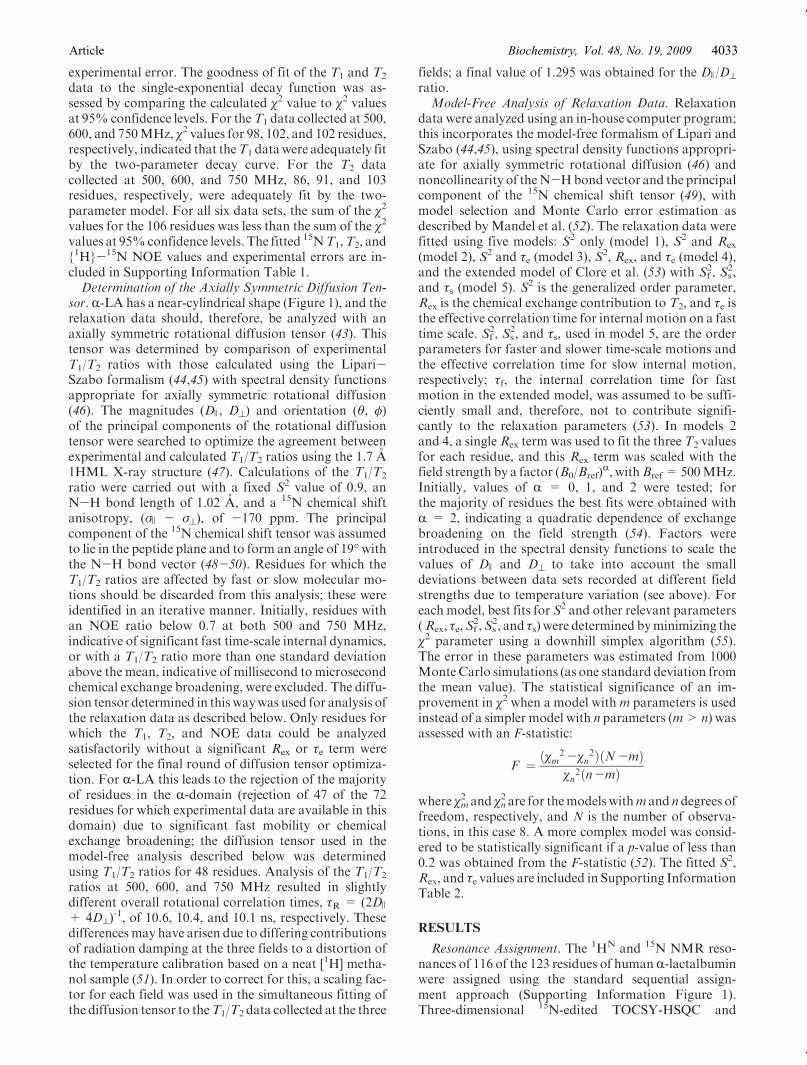
experimental error. The goodness of fit of the T1 and T2
data to the single-exponential decay function was as-sessed by comparing the calculated χ2 value to χ2 valuesat 95% confidence levels. For theT1 data collected at 500,600, and 750MHz, χ2 values for 98, 102, and 102 residues,respectively, indicated that theT1 datawere adequately fitby the two-parameter decay curve. For the T2 datacollected at 500, 600, and 750 MHz, 86, 91, and 103residues, respectively, were adequately fit by the two-parameter model. For all six data sets, the sum of the χ2
values for the 106 residues was less than the sum of the χ2
values at 95%confidence levels. The fitted 15NT1,T2, and{1H}-15N NOE values and experimental errors are in-cluded in Supporting Information Table 1.Determination of the Axially Symmetric Diffusion Ten-
sor. R-LA has a near-cylindrical shape (Figure 1), and therelaxation data should, therefore, be analyzed with anaxially symmetric rotational diffusion tensor (43). Thistensor was determined by comparison of experimentalT1/T2 ratios with those calculated using the Lipari-Szabo formalism (44,45) with spectral density functionsappropriate for axially symmetric rotational diffusion(46). The magnitudes (D ), D^) and orientation (θ, φ)of the principal components of the rotational diffusiontensor were searched to optimize the agreement betweenexperimental and calculated T1/T2 ratios using the 1.7 A1HML X-ray structure (47). Calculations of the T1/T2
ratio were carried out with a fixed S2 value of 0.9, anN-H bond length of 1.02 A, and a 15N chemical shiftanisotropy, (σ ) - σ^), of -170 ppm. The principalcomponent of the 15N chemical shift tensor was assumedto lie in the peptide plane and to form an angle of 19�withthe N-H bond vector (48-50). Residues for which theT1/T2 ratios are affected by fast or slow molecular mo-tions should be discarded from this analysis; these wereidentified in an iterative manner. Initially, residues withan NOE ratio below 0.7 at both 500 and 750 MHz,indicative of significant fast time-scale internal dynamics,or with a T1/T2 ratio more than one standard deviationabove the mean, indicative of millisecond to microsecondchemical exchange broadening, were excluded. The diffu-sion tensor determined in this waywas used for analysis ofthe relaxation data as described below. Only residues forwhich the T1, T2, and NOE data could be analyzedsatisfactorily without a significant Rex or τe term wereselected for the final round of diffusion tensor optimiza-tion. For R-LA this leads to the rejection of the majorityof residues in the R-domain (rejection of 47 of the 72residues for which experimental data are available in thisdomain) due to significant fast mobility or chemicalexchange broadening; the diffusion tensor used in themodel-free analysis described below was determinedusing T1/T2 ratios for 48 residues. Analysis of the T1/T2
ratios at 500, 600, and 750 MHz resulted in slightlydifferent overall rotational correlation times, τR = (2D )
+ 4D^)-1, of 10.6, 10.4, and 10.1 ns, respectively. These
differences may have arisen due to differing contributionsof radiation damping at the three fields to a distortion ofthe temperature calibration based on a neat [1H] metha-nol sample (51). In order to correct for this, a scaling fac-tor for each field was used in the simultaneous fitting ofthe diffusion tensor to theT1/T2 data collected at the three
fields; a final value of 1.295 was obtained for the D )/D^ratio.Model-Free Analysis of Relaxation Data. Relaxation
data were analyzed using an in-house computer program;this incorporates the model-free formalism of Lipari andSzabo (44,45), using spectral density functions appropri-ate for axially symmetric rotational diffusion (46) andnoncollinearity of theN-Hbond vector and the principalcomponent of the 15N chemical shift tensor (49), withmodel selection and Monte Carlo error estimation asdescribed by Mandel et al. (52). The relaxation data werefitted using five models: S2 only (model 1), S2 and Rex
(model 2), S2 and τe (model 3), S2, Rex, and τe (model 4),and the extended model of Clore et al. (53) with S2
f , S2s,
and τs (model 5). S2 is the generalized order parameter,Rex is the chemical exchange contribution to T2, and τe isthe effective correlation time for internal motion on a fasttime scale. S2
f , S2s, and τs, used in model 5, are the order
parameters for faster and slower time-scale motions andthe effective correlation time for slow internal motion,respectively; τf, the internal correlation time for fastmotion in the extended model, was assumed to be suffi-ciently small and, therefore, not to contribute signifi-cantly to the relaxation parameters (53). In models 2and 4, a singleRex term was used to fit the three T2 valuesfor each residue, and this Rex term was scaled with thefield strength by a factor (B0/Bref)
R, withBref= 500MHz.Initially, values of R = 0, 1, and 2 were tested; forthe majority of residues the best fits were obtained withR = 2, indicating a quadratic dependence of exchangebroadening on the field strength (54). Factors wereintroduced in the spectral density functions to scale thevalues of D ) and D^ to take into account the smalldeviations between data sets recorded at different fieldstrengths due to temperature variation (see above). Foreach model, best fits for S2 and other relevant parameters(Rex, τe,S
2f ,S
2s, and τs) were determined byminimizing the
χ2 parameter using a downhill simplex algorithm (55).The error in these parameters was estimated from 1000MonteCarlo simulations (as one standard deviation fromthe mean value). The statistical significance of an im-provement in χ2 when a model withm parameters is usedinstead of a simpler model with n parameters (m>n) wasassessed with an F-statistic:
F ¼ ðχm2 -χn2ÞðN-mÞ
χn2ðn-mÞ
where χm2 and χn
2 are for themodelswithm and n degrees offreedom, respectively, and N is the number of observa-tions, in this case 8. A more complex model was consid-ered to be statistically significant if a p-value of less than0.2 was obtained from the F-statistic (52). The fitted S2,Rex, and τe values are included in Supporting InformationTable 2.
RESULTS
Resonance Assignment. The 1HN and 15N NMR reso-nances of 116 of the 123 residues of humanR-lactalbuminwere assigned using the standard sequential assign-ment approach (Supporting Information Figure 1).Three-dimensional 15N-edited TOCSY-HSQC and
Article Vol. 48, No. 19, 2009Biochemistry, 4033
NOESY-HSQC spectra provided the necessary through-bond and through-space correlations. The assignment ofthe R-LA spectrum was significantly more difficult thanthat of the spectra of the homologous proteins hen andhuman lysozyme (56,57) because there are a large numberof broad resonances in the spectrumofR-LA, particularlyfor residues arising from the B- and D-helices of theR-domain (Supporting Information Figure 1). Cross-peaks arising from Leu105 and Lys108, located in theD-helix, have not been identified here and are presumablybroadened beyond detection. Resonances from theseresidues are also missing in the NMR spectra of bovineand guinea pig R-lactalbumin (13,30). The assignmentshave been deposited in the BioMagRes data bank(BMRB entry ID 15892), and the 1HN and 15N chemicalshifts are included in Supporting Information Table 3.Analysis of 15N Relaxation Data. 15N T1 and T2 values
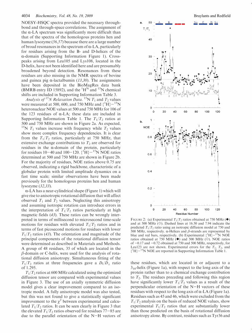
were measured at 500, 600, and 750 MHz and {1H}-15Nheteronuclear NOE values at 500 and 750MHz for 106 ofthe 123 residues of R-LA; these data are included inSupporting Information Table 1. The T1/T2 ratios at500 and 750 MHz are shown in Figure 2a. As expected,15N T1 values increase with frequency while T2 valuesshow more complex frequency dependencies. It is clearfrom the T1/T2 ratios, particularly at 750 MHz, thatextensive exchange contributions to T2 are observed forresidues in the R-domain of the protein, particularlyfor residues 10-40 and 100-120. {1H}-15N NOE ratiosdetermined at 500 and 750 MHz are shown in Figure 2b.For the majority of residues, NOE ratios above 0.75 areobserved, indicating a rigid backbone, characteristic of aglobular protein with limited amplitude dynamics on afast time scale; similar observations have been madepreviously for the homologous proteins hen and humanlysozyme (32,33).R-LA has a near-cylindrical shape (Figure 1) which will
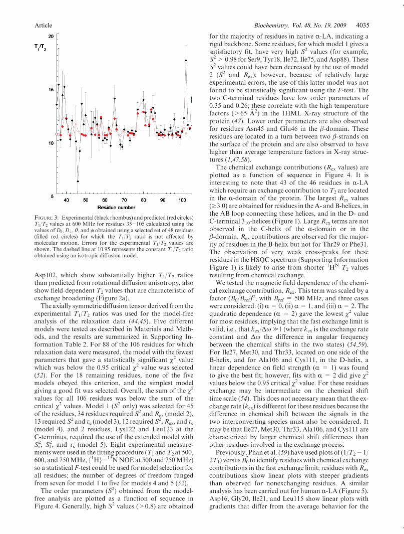
give rise to anisotropic rotational diffusion that will affectobserved T1 and T2 values. Neglecting this anisotropyand assuming isotropic rotation can introduce errors inthe interpretation of T1/T2 ratios particularly at highmagnetic fields (43). These ratios can be wrongly inter-preted in terms of millisecond to microsecond time-scalemotions for residues with elevated T1/T2 ratios and interms of fast picosecond motions for residues with lowerT1/T2 ratios (43). The orientation and magnitude of theprincipal components of the rotational diffusion tensorwere determined as described in Materials and Methods.A group of 48 residues, 35 of which are located in theβ-domain or C-helix, were used for the analysis of rota-tional diffusion anisotropy. Simultaneous fitting of theT1/T2 ratios at three frequencies gave a D )/D^ ratioof 1.295.T1/T2 ratios at 600MHz calculated using the optimized
diffusion tensor are compared with experimental valuesin Figure 3. The use of an axially symmetric diffusionmodel gives a clear improvement compared to an iso-tropic model. A fully anisotropic model was also tested,but this was not found to give a statistically significantimprovement to the χ2 between experimental and calcu-lated T1/T2 ratios. It can be seen clearly in Figure 3 thatthe elevated T1/T2 ratios observed for residues 77-85 aredue to the parallel orientation of the N-H vectors of
these residues, which are located in or adjacent to a310-helix (Figure 1a), with respect to the long axis of theprotein rather than to a chemical exchange contributionto T2. The residues preceding and following this regionhave significantly lower T1/T2 values as a result of theperpendicular orientation of the N-H vectors of theseresidues with respect to the long axis of R-LA (Figure 1a).Residues such as 45 and 46, which were excluded from theT1/T2 analysis on the basis of reduced NOE values, showexperimental T1/T2 ratios that are substantially lowerthan those predicted on the basis of rotational diffusionanisotropy alone. By contrast, residues such as Tyr36 and
FIGURE 2: (a) Experimental T1/T2 ratios obtained at 750 MHz ([)and at 500 MHz (]). Dashed lines at 16.50 and 7.94 indicate thepredicted T1/T2 ratio using an isotropic diffusion model at 750 and500 MHz, respectively. R-Helices and β-strands are represented byblue and red bars, respectively. (b) Experimental {1H}-15N NOEratios obtained at 750 MHz ([) and 500 MHz (]). NOE ratiosof -0.17 and -0.72 obtained at 750 and 500 MHz, respectively, forLeu123 are not shown. Experimental errors for the T1, T2, and{1H}-15N NOE are reported in Supporting Information Table 1.
Biochemistry, Vol. 48, No. 19, 2009 Bruylants and Redfield4034
Asp102, which show substantially higher T1/T2 ratiosthan predicted from rotational diffusion anisotropy, alsoshow field-dependent T2 values that are characteristic ofexchange broadening (Figure 2a).The axially symmetric diffusion tensor derived from the
experimental T1/T2 ratios was used for the model-freeanalysis of the relaxation data (44,45). Five differentmodels were tested as described in Materials and Meth-ods, and the results are summarized in Supporting In-formation Table 2. For 88 of the 106 residues for whichrelaxation data were measured, the model with the fewestparameters that gave a statistically significant χ2 valuewhich was below the 0.95 critical χ2 value was selected(52). For the 18 remaining residues, none of the fivemodels obeyed this criterion, and the simplest modelgiving a good fit was selected. Overall, the sum of the χ2
values for all 106 residues was below the sum of thecritical χ2 values. Model 1 (S2 only) was selected for 45of the residues, 34 residues required S2 andRex (model 2),13 requiredS2 and τe (model 3), 12 requiredS2,Rex, and τe(model 4), and 2 residues, Lys122 and Leu123 at theC-terminus, required the use of the extended model withS2s, S
2f , and τs (model 5). Eight experimental measure-
ments were used in the fitting procedure (T1 andT2 at 500,600, and 750MHz, {1H}-15NNOE at 500 and 750MHz)so a statistical F-test could be used for model selection forall residues; the number of degrees of freedom rangedfrom seven for model 1 to five for models 4 and 5 (52).The order parameters (S2) obtained from the model-
free analysis are plotted as a function of sequence inFigure 4. Generally, high S2 values (>0.8) are obtained
for the majority of residues in native R-LA, indicating arigid backbone. Some residues, for which model 1 gives asatisfactory fit, have very high S2 values (for example,S2> 0.98 for Ser9, Tyr18, Ile72, Ile75, andAsp88). TheseS2 values could have been decreased by the use of model2 (S2 and Rex); however, because of relatively largeexperimental errors, the use of this latter model was notfound to be statistically significant using the F-test. Thetwo C-terminal residues have low order parameters of0.35 and 0.26; these correlate with the high temperaturefactors (>65 A2) in the 1HML X-ray structure of theprotein (47). Lower order parameters are also observedfor residues Asn45 and Glu46 in the β-domain. Theseresidues are located in a turn between two β-strands onthe surface of the protein and are also observed to havehigher than average temperature factors in X-ray struc-tures (1,47,58).The chemical exchange contributions (Rex values) are
plotted as a function of sequence in Figure 4. It isinteresting to note that 43 of the 46 residues in R-LAwhich require an exchange contribution to T2 are locatedin the R-domain of the protein. The largest Rex values(g3.0) are obtained for residues in theA- andB-helices, inthe AB loop connecting these helices, and in the D- andC-terminal 310-helices (Figure 1). LargeRex terms are notobserved in the C-helix of the R-domain or in theβ-domain. Rex contributions are observed for the major-ity of residues in the B-helix but not for Thr29 or Phe31.The observation of very weak cross-peaks for theseresidues in the HSQC spectrum (Supporting InformationFigure 1) is likely to arise from shorter 1HN T2 valuesresulting from chemical exchange.We tested the magnetic field dependence of the chemi-
cal exchange contribution, Rex. This term was scaled by afactor (B0/Bref)
R, with Bref = 500 MHz, and three caseswere considered: (i) R=0, (ii) R=1, and (iii) R=2. Thequadratic dependence (R = 2) gave the lowest χ2 valuefor most residues, implying that the fast exchange limit isvalid, i.e., that kex/Δω.1 (where kex is the exchange rateconstant and Δω the difference in angular frequencybetween the chemical shifts in the two states) (54,59).For Ile27, Met30, and Thr33, located on one side of theB-helix, and for Ala106 and Cys111, in the D-helix, alinear dependence on field strength (R = 1) was foundto give the best fit; however, fits with R = 2 did give χ2
values below the 0.95 critical χ2 value. For these residuesexchange may be intermediate on the chemical shifttime scale (54). This does not necessary mean that the ex-change rate (kex) is different for these residues because thedifference in chemical shift between the signals in thetwo interconverting species must also be considered. Itmay be that Ile27,Met30, Thr33, Ala106, and Cys111 arecharacterized by larger chemical shift differences thanother residues involved in the exchange process.Previously, Phan et al. (59) have used plots of (1/T2- 1/
2T1) versusB02 to identify residueswith chemical exchange
contributions in the fast exchange limit; residues with Rex
contributions show linear plots with steeper gradientsthan observed for nonexchanging residues. A similaranalysis has been carried out for human R-LA (Figure 5).Asp16, Gly20, Ile21, and Leu115 show linear plots withgradients that differ from the average behavior for the
FIGURE 3: Experimental (black rhombus) and predicted (red circles)T1/T2 values at 600 MHz for residues 35-105 calculated using thevalues ofD ),D^, θ, and φ obtained using a selected set of 48 residues(filled red circles) for which the T1/T2 ratio is not affected bymolecular motion. Errors for the experimental T1/T2 values areshown. The dashed line at 10.95 represents the constant T1/T2 ratioobtained using an isotropic diffusion model.
Article Vol. 48, No. 19, 2009Biochemistry, 4035

residues that are not affected by chemical exchange. Thelinear dependence of these plots confirms the fast chemi-cal exchange limit for these residues (59).
DISCUSSION
Using 15N NMR relaxation methods, we have charac-terized thebackbonedynamics of thenative state of humanR-lactalbumin at the level of individual residues. Relaxa-tion data were collected at three magnetic field strengthsfor 106 residues and analyzed using the model-free form-alism of Lipari and Szabo (44,45) with spectral densityfunctions appropriate for axially symmetric rotationaldiffusion (46,49). Order parameters, S2, greater than 0.8are obtained for 100of 106 residues in nativeR-LA.Similarbehavior has been observed previously for the homologousproteins hen and human lysozyme (32,33). Lower orderparameters are observed for Asn45 and Glu46 in the β-domain and for the two C-terminal residues, Lys122 andLeu123; this is consistent with the higher B-factors ob-served for these residues inX-ray structures (47,58). Lowerorder parameters are observed for the homologous resi-dues in HEWL; for example, Thr47 of HEWL, whichcorresponds toAsn45 inR-LA,has anS2 value of 0.78 (32).The C-helix has the highest average S2 value (0.95 com-pared to 0.89 for the entire protein); in HEWL, this helixalso has the highest average S2 value (0.91 compared to0.87 for the entire protein) (32). Overall, the order para-meters determined for R-LA suggest a rigid polypeptide
backbone throughout the structure with only small ampli-tude fluctuations on a fast time scale.A total of 46 residues in R-LA required an exchange
contribution (Rex) to T2. This is in marked contrastto HEWL for which only 3 residues require an Rex termwhen axially symmetric rotational diffusion is considered(G. Bruylants and C. Redfield, unpublished results) (60).Therefore, although the fast time-scale dynamics of R-LAand HEWL appear to be quite similar, the slower milli-second to microsecond time-scale dynamics show a verysignificant difference. In R-LA, 43 of the 46 residues withexchange broadening are located in the R-domain ofthe protein. The 28 residues withRex values of greater than1.5 are all located in the R-domain and specifically in theA-, B-, D-, and C-terminal 310-helices and in the AB loop(connecting theA and B helices) (Figure 1). This clusteringof residues requiring an exchange term suggests a commonmechanism for exchange broadening in R-LA.Hydrogen/deuterium exchange protection factors have
been reported for the backbone amide protons of nativehuman R-LA at pH 6.3 and 15 �C (8). Protected amidesare observed for hydrogen-bonded residues located inboththe R-domain and the β-domain. The highest protectionfactors are observed for residues in the C-helix of theR-domain and close to disulfide bonds in the β-domain;protection factors of greater than 105 are observed for 4residues in the C-helix (Cys91, Ala92, Lys93, and Lys94)and for Cys61, Cys73, Cys77, and Phe80 in the β-domain.Lower levels of protection are observed for otherR-domain residues; the average protection factor for re-sidues in the A-helix is 1 � 104 (6 residues) and for theB-helix is 5 � 104 (5 residues). No protection is detectedfor the D- and C-terminal 310-helices of human R-LA (8).A similar pattern of protection is observed for the nativestate of bovine R-LA (13); protection factors of 105 andhigher are observed for many residues in the β-domain,and values greater than 107 are observed in the C-helix. Noprotection factors greater than 105 are observed for theA-, B-, D-, and C-terminal 310-helices of native bovineR-LA (13). The lower protection factors observed in the
FIGURE 4: (a)Rex and (b) S2 parameters obtained from the fitting of
the 15N relaxation data using the model-free approach, as describedin theMaterials andMethods section.Errors derived from theMonteCarlo analysis are shown. R-Helices and β-strands are represented in(b) by blue and red bars, respectively.
FIGURE 5: Representative plots of (1/T2- 1/2T1) versus B02 for four
selected residues illustrate the quadratic dependence of Rex on B0.Experimental values are shown for Asp16 (]), Gly20 (4), Ile21 (O),and Leu115 (0). Average values obtained for the 45 residues forwhich T1 and T2 are not affected by molecular motions are shown ascrosses. For clarity, and as done by Phan et al. (59), all the fitted datasets have been scaled to the same y-intercept (that of the fit for theaverage of the nonexchanging residues) so that only differences in theslope are shown.
Biochemistry, Vol. 48, No. 19, 2009 Bruylants and Redfield4036

A-, B-, D-, and C-terminal 310-helices of R-LA correlatewith the observation ofRex contributions for these regionsof the structure (Figure 1b,c).The pattern of hydrogen/deuterium exchange observed
in hen and human lysozymes differs from that observed forhuman and bovine R-LA. Both hen and human lysozymesshow more uniform protection factors throughout thestructure. For lysozyme, protection factors of greater than106 have been reported for residues in the A-, B-, andC-helices of the R-domain and in the β-domain; protectionfactors for amides in the C-helix and β-domain are notsignificantly higher than those observed in the A- andB-helices (61,62). No significant chemical exchange broad-ening is observed in either domain of HEWL.Human R-LA is known to crystallize in two forms; rod-
like crystals are obtained at room temperature at pH4.2 (LT form) and plate-like crystals are obtained at 37 �Cat pH 6.5 (HT form) (63). These two forms show nosignificant difference in structure for residues 1-95, butdifferences are observed for residues 96-123. The largestdifferences are observed for residues Trp104 to Cys111.These residues adopt an R-helical structure with His107buried in the pH 6.5 HT form whereas this region formsa loop with His107 exposed to solvent in the pH 4.2 LTform. At pH 6.5 at room temperature, conditions similarto those used in our NMR study, both crystal formsare found, suggesting that under these conditions the twoconformations interconvert in solution (63). This struc-tural transition, occurring on a millisecond to micro-second time scale, may explain the exchange broadeningobserved for Ala106, His107, Ala109, Leu110, andCys111and the absence of HSQC peaks for Leu105 and Lys108.It may also explain the lack of observed protection forD-helix amides; although the amides of Ala109, Leu110,and Cys111 are hydrogen bonded in the HT form, nohydrogen bonds exist in the LT form. Cys28 in the B-helixis disulfide bonded to Cys111, and several residuesin the B-helix make contacts with Trp104-Cys111. Thecarboxyl group of Glu25 has a polar contact with theside chain of His107 in the HT form but not in the LTform (63). Thus, exchange broadening of B-helix residuesmay arise from differences in chemical shifts arisingfrom changes in these long-range contacts in the twoconformations.The structural transition for residues 104-111 does not,
however, explain the more widespread exchange broad-ening observed for theA-helix and theC-terminal 310-helixor the reduced protection factors observed for amidesin the A- and B-helices. Residues in the A- and C-terminal310-helices do not make contacts with residues Trp104-Cys111 (Figure 1). In addition, no changes in hydrogenbondingor solvent accessibility are observed for theA- andB-helices in the two structures (63). It may be that insolution R-LA undergoes a more substantial structuralperturbation or samples a larger ensemble of conforma-tions in order to accommodate the transition between thetwo local structures for residues 104-111 that are trappedin the two crystal forms. This may involve significantchanges in the environments of the A-, B-, and C-terminal310-helices, in addition to residues in the D-helix, givingrise to more widespread chemical shift differences andenhanced amide exchange.
It is interesting to note that the A-, B-, D-, andC-terminal 310-helices form a stable core that is highlyresistant to denaturant-induced unfolding in the low pHmolten globule of human R-LA and that this core is stablein the absence of structure in the C-helix and β-domain (9).Studies of peptide models have identified residues 1-38linked by the Cys28-Cys111 disulfide to residues 101-120as the minimum core for a stable human R-LA moltenglobule (64); this peptide model encompasses the A-, B-,D-, and C-terminal 310-helices of R-LA. The conforma-tional transition in native R-LA, responsible for the ob-served exchange broadening described in this study, mayinvolve interconversion of the A-, B-, D-, and C-terminal310-helices between the well-ordered native structures ob-served in the HT and LT crystal structures and a partiallyfolded molten-globule-like structure. In future, 15N and1HN relaxation dispersion studies of native R-LA mayprovide chemical shift information that allows the natureof the interconverting structures to be described in moredetail (65).Harata and co-workers have suggested that the HT
form, crystallized at pH 6.5 and 37 �C, with an R-helicalconformation for Trp104-Cys111 represents the physio-logical state of R-LA (63). However, HSQC spectracollected for R-LA at pH 7 and 40 �C show broadening ofR-domain peaks similar to that observed at pH 6.3 and18.2 �C. This suggests that structural fluctuations withinthe A-, B-, D-, and C-terminal 310-helices of the R-domainare likely to be present in the physiological state of R-LA.The R-domain of R-LA is generally defined to include
residues 1-39 and 82-123, which encompass the A-, B-,C-, D-, and C-terminal 310-helices (1). An alternativedefinition of the domain structure of R-LA places theC-helix in the β-domain of the protein (R-domain residues1-37 and 105-123, β-domain 38-104) (66,67). The 15Nrelaxation data reported here for human R-LA, whichshowdifferent behavior for theC-helix from the remainderof the R-domain, and previous hydrogen/deuterium ex-change data, which show higher levels of protection forthe C-helix, would appear to correlate better with thelatter definition of the domain structure in the native stateof R-LA.In native R-LA, Asp87 and Asp88, near the N-terminus
of the C-helix, are involved in calcium binding along withLys79, Asp82, and Asp84 (1). Calcium binding to humanR-LA requires theCys61-Cys77andCys73-Cys91 disulfidebonds but does not require the R-domain disulfides,Cys6-Cys120 and Cys28-Cys111 (68). Human R-LA(β),a two-disulfide variant lacking the R-domain disulfides,has native-like levels of secondary structure in the presenceof calcium. This species has tighter side-chain packing inthe presence of calcium than in the molten globule formedin the absence of calcium but has substantially less well-ordered tertiary structure than the fully native state (68).This suggests that calcium binding to R-LA(β) results in astructured β-domain, calcium-binding loop, and C-helixwhile the remainder of the R-domain may be substan-tially more disordered (67,68). The presence of the twoR-domain disulfides in native R-LA leads to stabiliza-tion of well-ordered tertiary structure throughout theR-domain. Nevertheless, the 15N relaxation data presentedhere, and previous hydrogen/deuterium exchange data,
Article Vol. 48, No. 19, 2009Biochemistry, 4037
suggest that the A-, B-, D-, and C-terminal 310-helices ofthe R-domain may retain a propensity for transient un-folding to a molten-globule-like species even in the pre-sence of the R-domain disulfide bonds.
ACKNOWLEDGMENT
We thank Peter S. Kim and Brenda A. Schulman forproviding 15N-labeled recombinant human R-LA for thisstudy.
SUPPORTING INFORMATION AVAILABLE
A figure showing the assigned 750 MHz HSQC spec-trum of human R-LA at pH 6.3 and 18.2 �C, a tablesummarizing the 15N T1, T2, and {1H}-15N NOE datacollected at 500, 600, and 750 MHz, a table summarizingresults of the Lipari-Szabo model-free analysis (modelused, S2, Rex, τe), and a table summarizing backbone 1HN
and 15N chemical shifts for native human R-LA at pH 6.3.This material is available free of charge via the Internet athttp://pubs.acs.org.
REFERENCES
1. Acharya, K. R., Ren, J. S., Stuart, D. I., Phillips, D. C., and Fenna,R. E. (1991) Crystal structure of human R-lactalbumin at 1.7 Aresolution. J. Mol. Biol. 221, 571–581.
2. Permyakov, E.A. (2005) Alpha-Lactalbumin, NovaScience Publish-ers, New York.
3. Imoto, T., Johnson, L. N., North, A. C. T., Phillips, D. C., andRupley, J. A. (1972) Vertebrate Lysozymes, in The Enzymes (Boyer,P. D., Ed.) Vol. VII, pp 666-868, Academic Press, New York.
4. Kuwajima, K. (1989) The molten globule state as a clue for under-standing the folding and cooperativity of globular-protein structure.Proteins: Struct., Funct., Genet. 6, 87–103.
5. Yutani,K., Ogasahara,K., andKuwajima,K. (1992)Absence of thethermal transition in apo-R-lactalbumin in the molten globule state.A study by differential scanningmicrocalorimetry. J.Mol. Biol. 228,347–350.
6. Peng, Z. Y., and Kim, P. S. (1994) A protein dissection study ofa molten globule. Biochemistry 33, 2136–2141.
7. Wu, L. C., Peng, Z. Y., and Kim, P. S. (1995) Bipartite structureof the R-lactalbumin molten globule. Nat. Struct. Biol. 2, 281–286.
8. Schulman, B.A., Redfield, C., Peng, Z.-y.,Dobson,C.M., andKim,P. S. (1995) Different subdomains are most protected from hydro-gen exchange in the molten globule and native states of humanR-lactalbumin. J. Mol. Biol. 253, 651–657.
9. Schulman, B. A., Kim, P. S., Dobson, C.M., andRedfield, C. (1997)A residue-specific NMR view of the non-cooperative unfolding of amolten globule. Nat. Struct. Biol. 4, 630–634.
10. Kuwajima, K., Hiraoka, Y., Ikeguchi, M., and Sugai, S. (1985)Comparison of the transient folding intermediates in lysozyme andR-lactalbumin. Biochemistry 24, 874–881.
11. Ikeguchi, M., Kuwajima, K., Mitani, M., and Sugai, S. (1986)Evidence for identity between the equilibrium unfolding intermedi-ate and a transient folding intermediate: a comparative study of thefolding reactions of R-lactalbumin and lysozyme. Biochemistry 25,6965–6972.
12. Arai, M., and Kuwajima, K. (1996) Rapid formation of a moltenglobule intermediate in refolding of R-lactalbumin. Folding Des. 1,275–287.
13. Forge, V., Wijesinha, R. T., Balbach, J., Brew, K., Robinson, C. V.,Redfield, C., and Dobson, C. M. (1999) Rapid collapse andslow structural reorganisation during the refolding of bovineR-lactalbumin. J. Mol. Biol. 288, 673–688.
14. Arai, M., Ito, K., Inobe, T., Nakao, M., Maki, K., Kamagata, K.,Kihara, H., Amemiya, Y., and Kuwajima, K. (2002) Fast compac-tion of R-lactalbumin during folding studied by stopped-flow X-rayscattering. J. Mol. Biol. 321, 121–132.
15. Baum, J., Dobson, C. M., Evans, P. A., and Hanley, C. (1989)Characterization of a partly folded protein by NMR methods;Studies on the molten globule state of guinea-pig R-lactalbumin.Biochemistry 28, 7–13.
16. Ramboarina, S., and Redfield, C. (2003) Structural characterisationof the human R-lactalbumin molten globule at high temperature.J. Mol. Biol. 330, 1177–1188.
17. Wijesinha-Bettoni, R.,Gao,C., Jenkins, J. A.,Mackie,A.R.,Wilde,P. J., Mills, E. N. C., and Smith, L. J. (2007) Heat treatmentof bovine R-lactalbumin results in partially folded, disulfide bondshuffled states with enhanced surface activity. Biochemistry 46,9774–9784.
18. Chyan, C. L., Wormald, C., Dobson, C. M., Evans, P. A., andBaum, J. (1993) Structure and stability of themolten globule state ofguinea-pigR-lactalbumin: a hydrogen exchange study. Biochemistry32, 5681–5691.
19. Kim, S., Bracken, C., and Baum, J. (1999) Characterization ofmillisecond time-scale dynamics in the molten globule state ofR-lactalbumin by NMR. J. Mol. Biol. 294, 551–560.
20. Alexandrescu, A. T., Evans, P. A., Pitkeathly, M., Baum, J., andDobson, C.M. (1993) Structure and dynamics of the acid-denaturedmolten globule state of R-lactalbumin: a two-dimensional NMRstudy. Biochemistry 32, 1707–1718.
21. Ramboarina, S., and Redfield, C. (2008) Probing the effect oftemperature on the backbone dynamics of the humanR-lactalbuminmolten globule. J. Am. Chem. Soc. 130, 15318–15326.
22. Ewbank, J. J., and Creighton, T. E. (1993) Structural characteriza-tion of the disulfide folding intermediates of bovine R-lactalbumin.Biochemistry 32, 3694–3707.
23. Schulman, B. A., and Kim, P. S. (1996) Proline scanning mutagen-esis of a molten globule reveals non-cooperative formation of aprotein’s overall topology. Nat. Struct. Biol. 3, 682–687.
24. Wu, L. C., and Kim, P. S. (1998) A specific hydrophobic core in theR-lactalbumin molten globule. J. Mol. Biol. 280, 175–182.
25. Smith, L. J., Dobson, C. M., and van Gunsteren, W. F. (1999)Molecular dynamics simulations of human R-lactalbumin: changesto the structural and dynamical properties of the protein at low pH.Proteins 36, 77–86.
26. Troullier, A., Reinstadler, D., Dupont, Y., Naumann, D., andForge, V. (2000) Transient non-native secondary structures duringthe refolding of R-lactalbumin detected by infrared spectroscopy.Nat. Struct. Biol. 7, 78–86.
27. Vendruscolo, M., Paci, E., Karplus, M., and Dobson, C. M. (2003)Structures and relative free energies of partially folded states ofproteins. Proc. Natl. Acad. Sci. U.S.A. 100, 14817–14821.
28. Koga, K., and Berliner, L. J. (1985) Structural elucidation of ahydrophobic box in bovine R-lactalbumin by NMR: nuclear Over-hauser effects. Biochemistry 24, 7257–7262.
29. Wijesinha-Bettoni, R., Dobson, C. M., and Redfield, C. (2001)Comparison of the structural and dynamical properties of holoand apo bovine R-lactalbumin by NMR spectroscopy. J. Mol. Biol.307, 885–898.
30. Kim, S., and Baum, J. (1998) Electrostatic interactions in the aciddenaturation of R-lactalbumin determined by NMR. Protein Sci. 7,1930–1938.
31. Alexandrescu, A. T., Broadhurst, R. W., Wormald, C., Chyan, C.L., Baum, J., and Dobson, C. M. (1992) 1H-NMR assignments andlocal environments of aromatic residues in bovine, human andguinea pig variants ofR-lactalbumin. Eur. J. Biochem. 210, 699–709.
32. Buck, M., Boyd, J., Redfield, C., MacKenzie, D. A., Jeenes, D. J.,Archer, D. B., andDobson, C.M. (1995) Structural determinants ofprotein dynamics: analysis of 15N NMR relaxation measurementsfor main-chain and side-chain nuclei of hen egg white lysozyme.Biochemistry 34, 4041–4055.
33. Chamberlain, A. K., Receveur, V., Spencer, A., Redfield, C., andDobson, C. M. (2001) Characterization of the structure and dy-namics of amyloidogenic variants of human lysozyme by NMRspectroscopy. Protein Sci. 10, 2525–2530.
34. Redfield, C., Schulman, B. A., Milhollen, M. A., Kim, P. S., andDobson, C. M. (1999) R-Lactalbumin forms a compact moltenglobule in the absence of disulfide bonds. Nat. Struct. Biol. 6,948–952.
35. Wilkins, D. K., Grimshaw, S. B., Receveur, V., Dobson, C. M.,Jones, J. A., and Smith, L. J. (1999) Hydrodynamic radii of nativeand denatured proteins measured by pulse field gradient NMRtechniques. Biochemistry 38, 16424–16431.
36. Kataoka, M., Kuwajima, K., Tokunaga, F., and Goto, Y. (1997)Structural characterization of the molten globule of R-lactalbuminby solution X-ray scattering. Protein Sci. 6, 422–430.
37. Marion, D., Driscoll, P. C., Kay, L. E., Wingfield, P. T., Bax, A.,Gronenborn, A. M., and Clore, G. M. (1989) Overcoming theoverlap problem in the assignment of 1H NMR spectra of lar-ger proteins by use of three-dimensional heteronuclear 1H-15N
Biochemistry, Vol. 48, No. 19, 2009 Bruylants and Redfield4038

Hartmann-Hahn-multiple quantum coherence and nuclear Over-hauser-multiple quantum coherence spectroscopy: application tointerleukin 1 beta. Biochemistry 28, 6150–6156.
38. Driscoll, P. C., Clore, G. M., Marion, D., Wingfield, P. T., andGronenborn, A. M. (1990) Complete resonance assignment for thepolypeptide backbone of interleukin 1 beta using three-dimensionalheteronuclear NMR spectroscopy. Biochemistry 29, 3542–3556.
39. Ikura, M., Bax, A., Clore, G. M., and Gronenborn, A. M. (1990)Detection of nuclear Overhauser effects between degenerate amideproton resonances by heteronuclear three-dimensional NMR spec-troscopy. J. Am. Chem. Soc. 112, 9020–9022.
40. Kay, L. E., Torchia, D. A., and Bax, A. (1989) Backbone dynamicsof proteins as studied by nitrogen-15 inverse detected heteronu-clear NMR spectroscopy: application to staphylococcal nuclease.Biochemistry 28, 8972–8979.
41. Boyd, J.,Hommel,U., andCampbell, I.D. (1990) Influence of cross-correlation between dipolar and anisotropic chemical shift relaxa-tion mechanisms upon longitudinal relaxation rates of nitrogen-15in macromolecules. Chem. Phys. Lett. 175, 477–482.
42. Palmer, A. G.III, Skelton, N. J., Chazin, W. J., Wright, P. E., andRance, M. (1992) Suppression of the effects of cross correlationbetween dipolar and anisotropic chemical-shift relaxation mechan-isms in themeasurement of spin-spin relaxation rates. Mol. Phys. 75,699–711.
43. Tjandra, N., Feller, S. E., Pastor, R. W., and Bax, A. (1995)Rotational diffusion anisotropy of human ubiquitin from 15NNMR relaxation. J. Am. Chem. Soc. 117, 12562–12566.
44. Lipari, G., and Szabo, A. (1982) Model-free approach to theinterpretation of nuclear magnetic resonance relaxation in macro-molecules. 1. Theory and range of validity. J. Am. Chem. Soc. 104,4546–4559.
45. Lipari, G., and Szabo, A. (1982) Model-free approach to theinterpretation of nuclear magnetic resonance relaxation in macro-molecules. 2. Analysis of experimental results. J. Am. Chem. Soc.104, 4559–4570.
46. Abragam, A. (1961) The Principles of Nuclear Magnetism, Clar-endon Press, Oxford.
47. Ren, J., Stuart, D. I., and Acharya, K. R. (1993) R-Lactalbuminpossesses a distinct zinc binding site. J. Biol. Chem. 268, 19292–19298.
48. Cornilescu, G., and Bax, A. (2000) Measurement of proton, nitro-gen, and carbonyl chemical shielding anisotropies in a proteindissolved in a dilute liquid crystalline phase. J. Am. Chem. Soc.122, 10143–10154.
49. Boyd, J., and Redfield, C. (1998) Defining the orientation of the N-15 shielding tensor usingN-15 NMR relaxation data for a protein insolution. J. Am. Chem. Soc. 120, 9692–9693.
50. Boyd, J., and Redfield, C. (1999) Characterization of N-15 chemicalshift anisotropy from orientation-dependent changes to N-15chemical shifts in dilute bicelle solutions. J. Am. Chem. Soc. 121,7441–7442.
51. Findeisen,M., Brand, T., and Berger, S. (2007) A 1H-NMR thermo-meter suitable for cryoprobes. Magn. Reson. Chem. 45, 175–178.
52. Mandel, A. M., Akke, M., and Palmer, A. G.III (1995) Backbonedynamics of Escherichia coli ribonuclease HI: correlations withstructure and function in an active enzyme. J. Mol. Biol. 246,144–163.
53. Clore, G. M., Szabo, A., Bax, A., Kay, L. E., Driscoll, P. C., andGronenborn, A. M. (1990) Deviations from the simple two-para-meter model-free approach to the interpretation of nitrogen-15nuclear magnetic relaxation of proteins. J. Am. Chem. Soc. 112,4989–4991.
54. Millet, O., Loria, J. P., Kroenke, C. D., Pons,M., and Palmer, A. G.III (2000) The staticmagnetic field dependence of chemical exchangelinebroadening defines the NMR chemical shift time scale. J. Am.Chem. Soc. 122, 2867–2877.
55. Johnson, M. L., and Faunt, L. M. (1992) Parameter estimation byleast-squares methods. Methods Enzymol. 210, 1–37.
56. Redfield, C., and Dobson, C. M. (1988) Sequential proton NMRassignments and secondary structure of hen egg white lysozyme insolution. Biochemistry 27, 122–136.
57. Redfield, C., and Dobson, C. M. (1990) Proton NMR studies ofhuman lysozyme: spectral assignment and comparison with henlysozyme. Biochemistry 29, 7201–7214.
58. Harata, K., Abe, Y., and Muraki, M. (1999) Crystallogra-phic evaluation of internal motion of human R-lactalbuminrefined by full-matrix least-squares method. J. Mol. Biol. 287,347–358.
59. Phan, I. Q.H., Boyd, J., andCampbell, I. D. (1996)Dynamic studiesof a fibronectin type I module pair at three frequencies: anisotropicmodeling and direct determination of conformational exchange.J. Biomol. NMR 8, 369–378.
60. Buck,M., Bouguet-Bonnet, S., Pastor, R.W., andMacKerell, A. D.Jr. (2006) Importance of the CMAP correction to the CHARMM22protein force field: dynamics of hen lysozyme. Biophys. J. 90,L36–L38.
61. Radford, S. E., Buck, M., Topping, K. D., Dobson, C. M., andEvans, P. A. (1992) Hydrogen exchange in native and denaturedstates of hen egg-white lysozyme. Proteins 14, 237–248.
62. Canet, D., Last, A. M., Tito, P., Sunde, M., Spencer, A., Archer, D.B., Redfield, C., Robinson, C. V., and Dobson, C. M. (2002) Localcooperativity in the unfolding of an amyloidogenic variant of humanlysozyme. Nat. Struct. Biol. 9, 308–315.
63. Harata, K., and Muraki, M. (1992) X-ray structural evidence for alocal helix-loop transition in R-lactalbumin. J. Biol. Chem. 267,1419–1421.
64. Demarest, S. J., Boice, J. A., Fairman, R., and Raleigh, D. P. (1999)Defining the core structure of the R-lactalbumin molten globulestate. J. Mol. Biol. 294, 213–221.
65. Vallurupalli, P., Hansen, D. F., and Kay, L. E. (2008) Structures ofinvisible, excited protein states by relaxation dispersion NMRspectroscopy. Proc. Natl. Acad. Sci. U.S.A. 105, 11766–11771.
66. Siddiqui, A. S., and Barton, G. J. (1995) Continuous and discontin-uous domains: an algorithm for the automatic generation of reliableprotein domain definitions. Protein Sci. 4, 872–884.
67. Hendrix, T. M., Griko, Y., and Privalov, P. (1996) Energetics ofstructural domains in R-lactalbumin. Protein Sci. 5, 923–931.
68. Wu, L. C., Schulman, B. A., Peng, Z. Y., and Kim, P. S. (1996)Disulfide determinants of calcium-induced packing in R-Lactalbu-min. Biochemistry 35, 859–863.
69. Pettersen, E. F., Goddard, T. D., Huang, C. C., Couch, G. S.,Greenblatt, D. M., Meng, E. C., and Ferrin, T. E. (2004) UCSFChimera;A visualization system for exploratory research andanalysis. J. Comput. Chem. 25, 1605–1612.
Article Vol. 48, No. 19, 2009Biochemistry, 4039