1 Citric acid cycle Synthesis of heme. Hemoproteins © Department of Biochemistry (J.D.) 2013.
78
1 Citric acid cycle Synthesis of heme. Hemoproteins © Department of Biochemistry (J.D.) 2013
-
Upload
abigail-nichols -
Category
Documents
-
view
220 -
download
0
Transcript of 1 Citric acid cycle Synthesis of heme. Hemoproteins © Department of Biochemistry (J.D.) 2013.
1
Citric acid cycle
Synthesis of heme. Hemoproteins
© Department of Biochemistry (J.D.) 2013
2
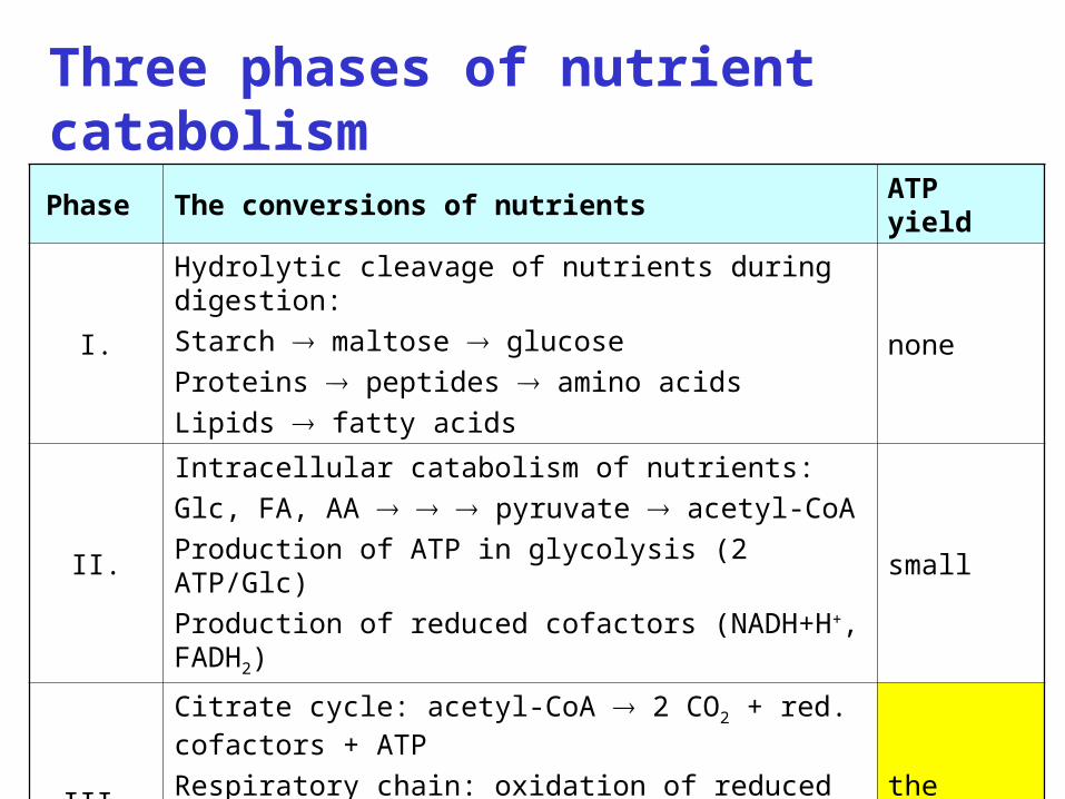
Phase The conversions of nutrients ATP yield
I.
Hydrolytic cleavage of nutrients during digestion:
Starch maltose glucose
Proteins peptides amino acids
Lipids fatty acids
none
II.
Intracellular catabolism of nutrients:
Glc, FA, AA pyruvate acetyl-CoA
Production of ATP in glycolysis (2 ATP/Glc)
Production of reduced cofactors (NADH+H+, FADH2)
small
III.
Citrate cycle: acetyl-CoA 2 CO2 + red. cofactors + ATP
Respiratory chain: oxidation of reduced cofactors
Aerobic phosphorylation: synthesis of ATP from ADP + Pi
the biggest
Three phases of nutrient catabolism
3
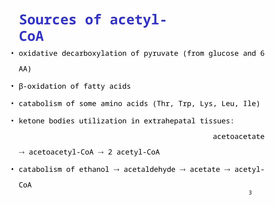
Sources of acetyl-CoA
• oxidative decarboxylation of pyruvate (from glucose and 6 AA)
• β-oxidation of fatty acids
• catabolism of some amino acids (Thr, Trp, Lys, Leu, Ile)
• ketone bodies utilization in extrahepatal tissues:
acetoacetate acetoacetyl-CoA 2 acetyl-CoA
• catabolism of ethanol acetaldehyde acetate acetyl-CoA
4
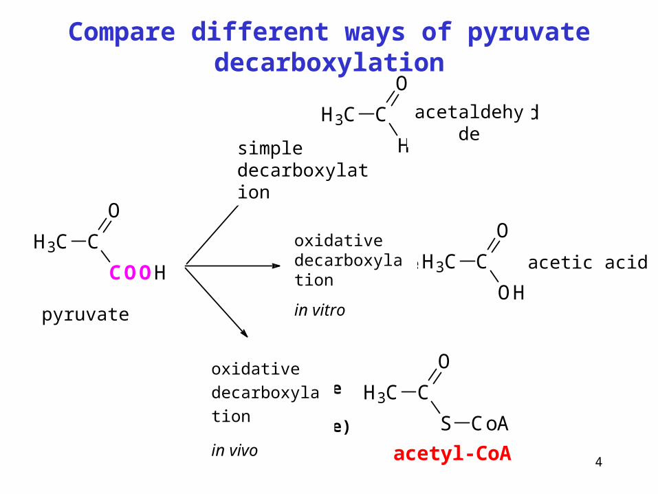
Compare different ways of pyruvate decarboxylation
H3C C
COOH
O
dekarboxylace
H3C C
H
O
acetaldehyd
H3C C
OH
Ooxidačnídekarboxylacein vitro
oxidačnídekarboxylacein vivo(mitochondrie)
H3C C
S
O
CoA
octová kyselina
acetyl-CoA
pyruvate
simple decarboxylation
acetaldehyde
acetic acidoxidative decarboxylation
in vitro
oxidative
decarboxylation
in vivo
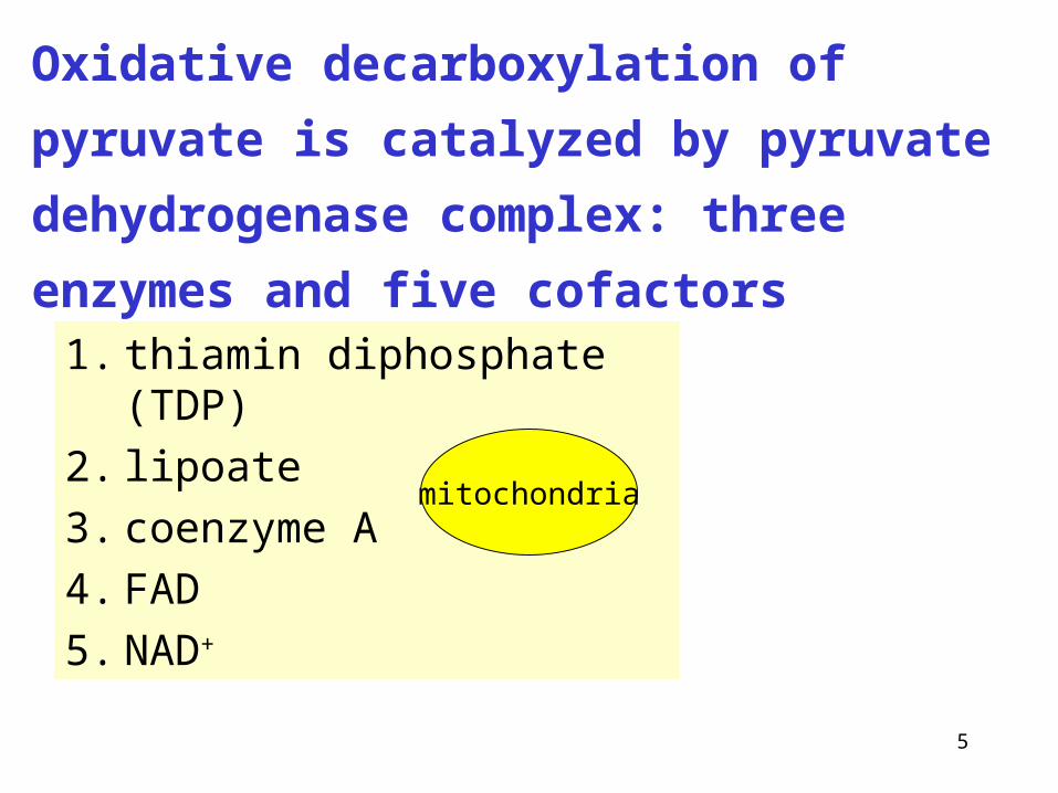
5
1. thiamin diphosphate (TDP)
2. lipoate
3. coenzyme A
4. FAD
5. NAD+
Oxidative decarboxylation of pyruvate is
catalyzed by pyruvate dehydrogenase complex:
three enzymes and five cofactors
mitochondria
6
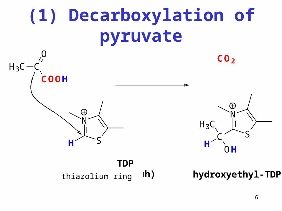
(1) Decarboxylation of pyruvate
N
SH
H3C CCOOH
O CO2
N
SCH
H3C
OH
hydroxyethyl-TDP TDP(thiazoliový kruh)
"aktivní acetaldehyd"
thiazolium ring
7
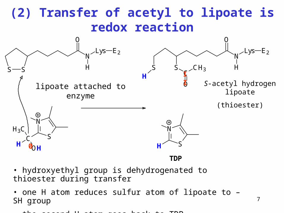
(2) Transfer of acetyl to lipoate is redox reaction
S S
N
O
Lys
H
E2
N
SCH
H3C
OH
lipoát vázaný na enzym
N
SH
N
O
Lys
H
E2
SSH C
O
CH3
S-acetylhydrogenlipoát (thioester)
TDP
• hydroxyethyl group is dehydrogenated to thioester during transfer
• one H atom reduces sulfur atom of lipoate to –SH group
• the second H atom goes back to TDP
II
0
lipoate attached to enzyme S-acetyl hydrogen lipoate
(thioester)
8
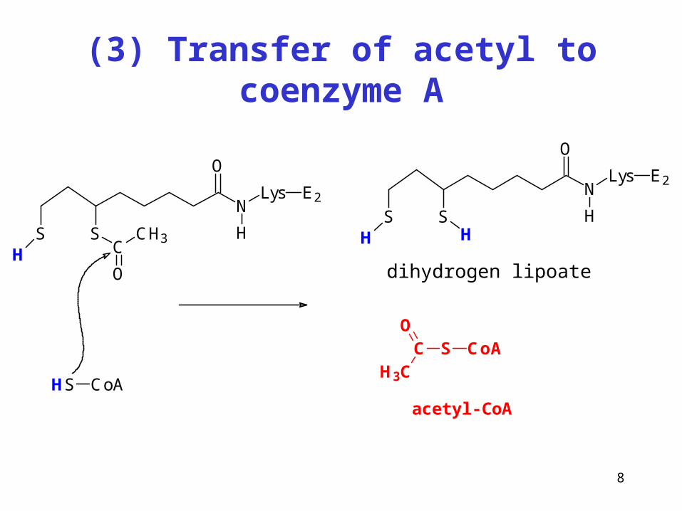
(3) Transfer of acetyl to coenzyme A
N
O
Lys
H
E2
SSH C
O
CH3
HS CoA
N
O
Lys
H
E2
SSH H
dihydrogenlipoát
S CoACO
H3C
acetyl-CoA
dihydrogen lipoate
9
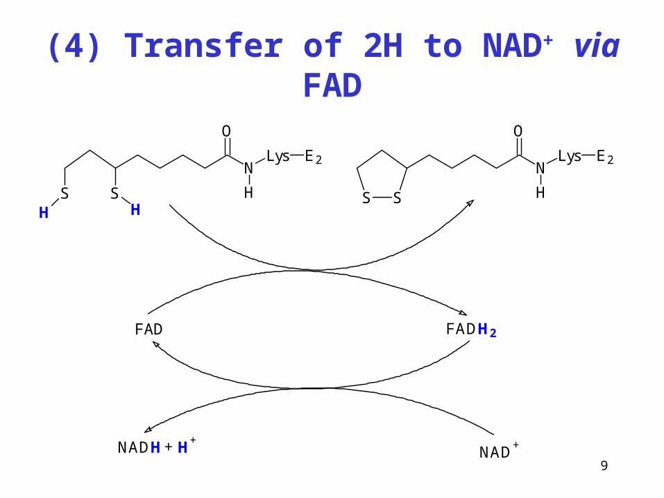
(4) Transfer of 2H to NAD+ via FAD
N
O
Lys
H
E2
SSH H
S S
N
O
Lys
H
E2
FAD FADH2
NAD+NADH H
++
10
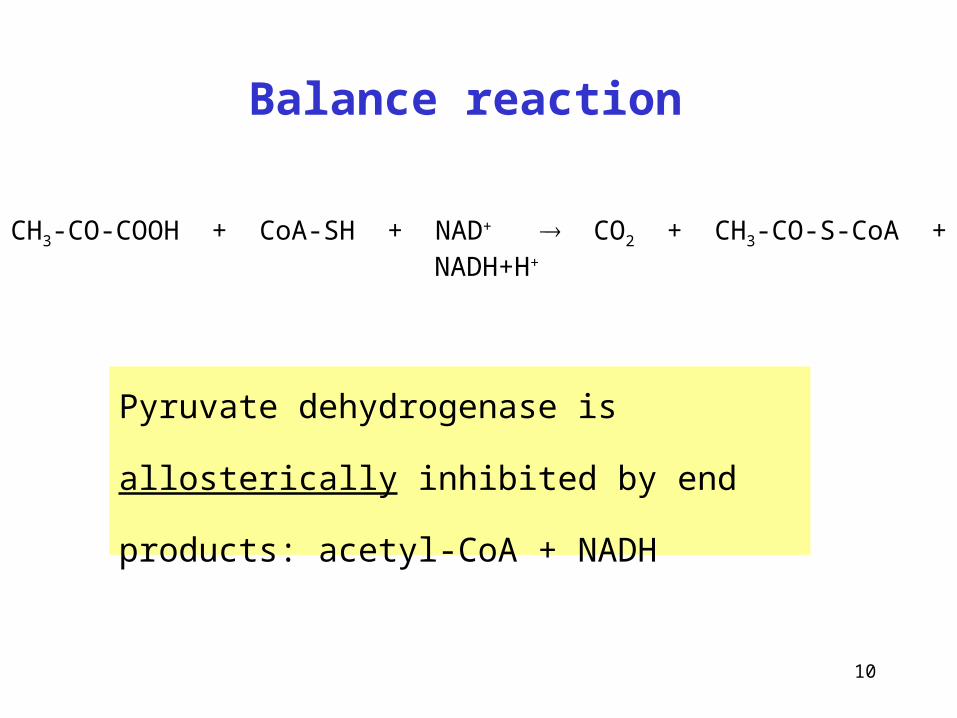
Balance reaction
Pyruvate dehydrogenase is allosterically inhibited
by end products: acetyl-CoA + NADH
CH3-CO-COOH + CoA-SH + NAD+ CO2 + CH3-CO-S-CoA + NADH+H+
11
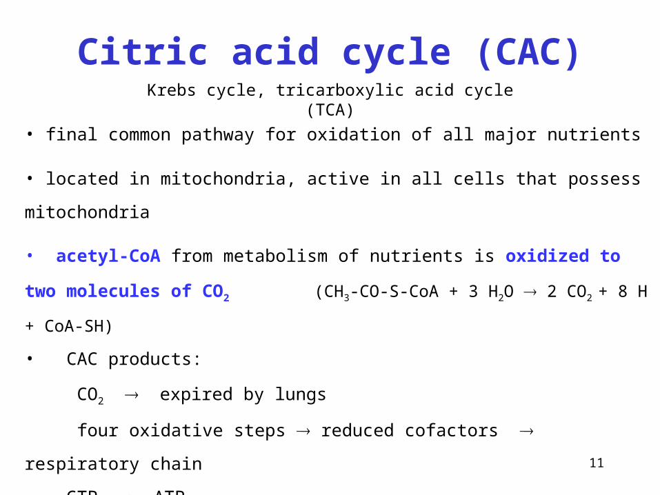
Citric acid cycle (CAC)Krebs cycle, tricarboxylic acid cycle (TCA)
• final common pathway for oxidation of all major nutrients
• located in mitochondria, active in all cells that possess mitochondria
• acetyl-CoA from metabolism of nutrients is oxidized to two molecules of CO2
(CH3-CO-S-CoA + 3 H2O 2 CO2 + 8 H + CoA-SH)
• CAC products:
CO2 expired by lungs
four oxidative steps reduced cofactors respiratory chain
GTP ATP
• most reactions are reversible, only three reactions are irreversible
12
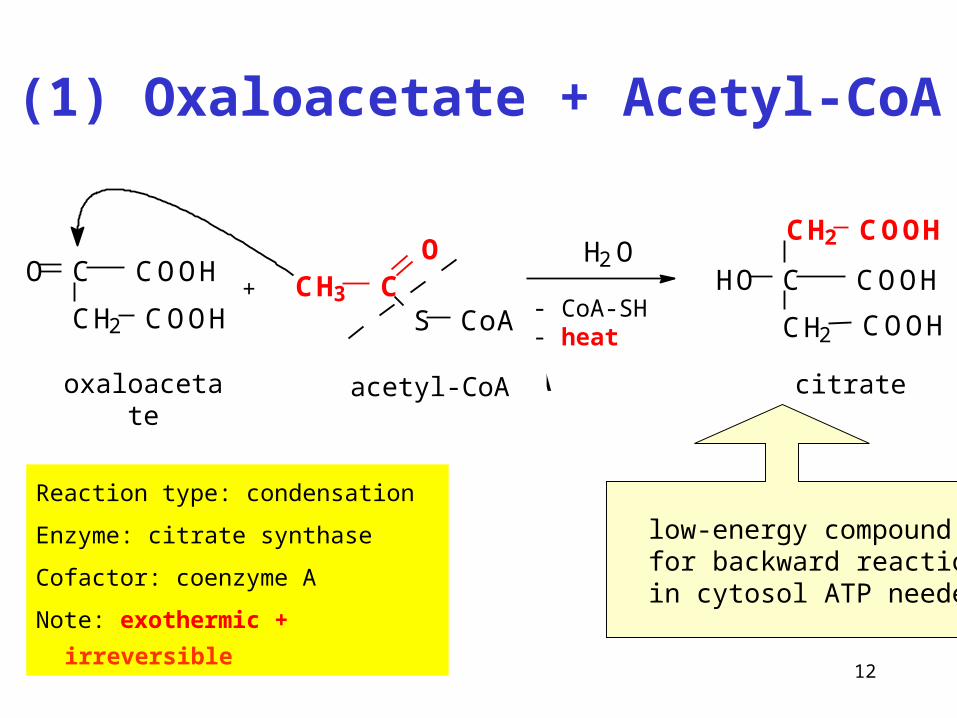
(1) Oxaloacetate + Acetyl-CoA
Reaction type: condensation
Enzyme: citrate synthase
Cofactor: coenzyme A
Note: exothermic + irreversible
C
CH2
COOHO
COOH+ CH3 C
O
S CoA
H2O
- CoA-SH- heat
oxalacetate acetyl-koenzym A citrate
C
CH2
COOH
COOH
HO
CH2 COOH
oxaloacetate acetyl-CoA
low-energy compound for backward reaction in cytosol ATP needed
13
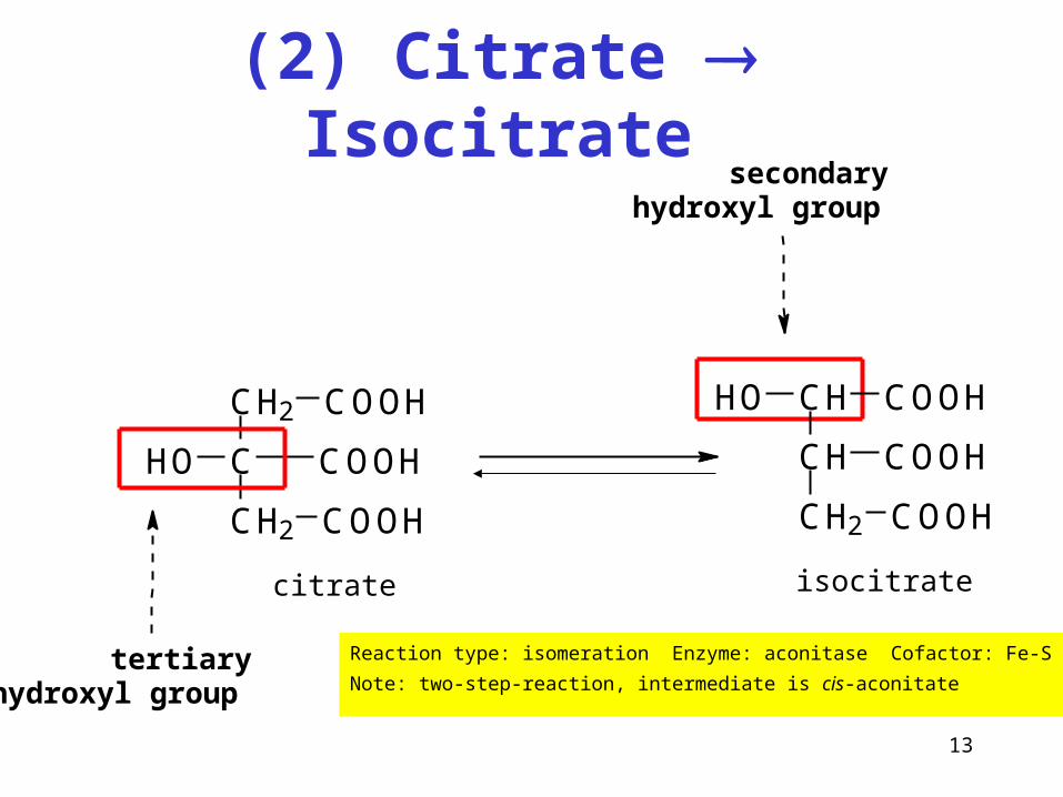
(2) Citrate Isocitrate
Reaction type: isomeration Enzyme: aconitase Cofactor: Fe-S
Note: two-step-reaction, intermediate is cis-aconitate
C
CH2
COOH
COOH
CH2
HO
COOH
CH
CH2 COOH
CH
COOH
HO COOH
tertiary hydroxyl group
secondary hydroxyl group
citrate isocitrate
14
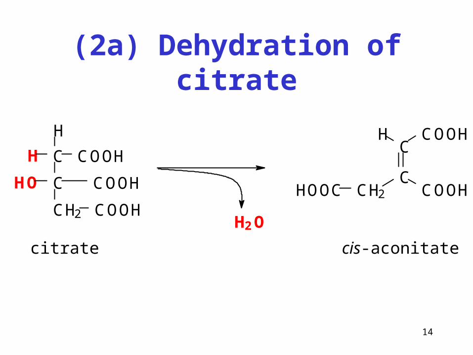
(2a) Dehydration of citrate
citrate
C
CH2
COOH
COOH
C
HO
COOH
H
H
H2O
C
C
COOHH
CH2 COOHHOOC
cis-aconitate
15
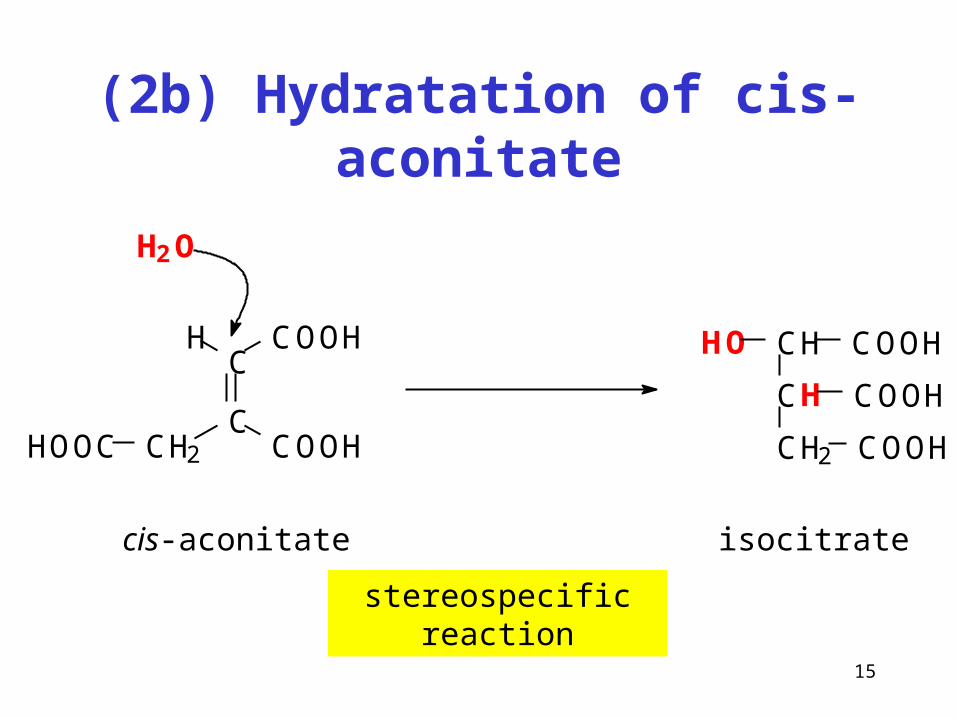
(2b) Hydratation of cis-aconitate
stereospecific reaction
CH
CH2 COOH
CH
COOH
HO COOHC
C
COOHH
CH2 COOHHOOC
cis-aconitate
H2O
isocitrate
16
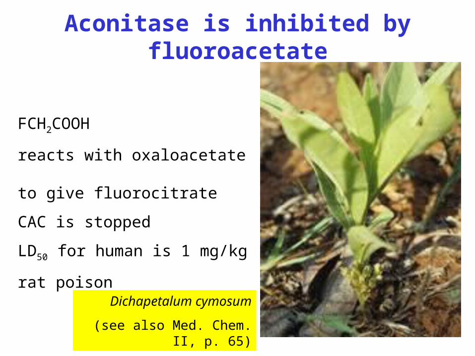
Aconitase is inhibited by fluoroacetate
Dichapetalum cymosum
(see also Med. Chem. II, p. 65)
FCH2COOH
reacts with oxaloacetate to give fluorocitrate
CAC is stopped
LD50 for human is 1 mg/kg
rat poison
17
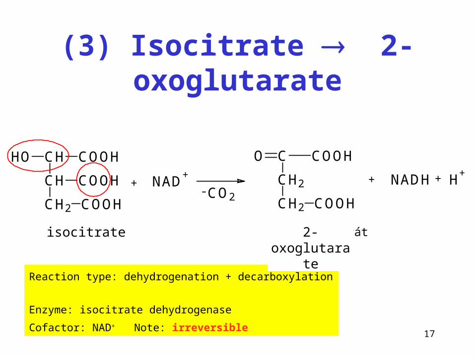
(3) Isocitrate 2-oxoglutarate
+ NAD+CH
CH2 COOH
CH
COOH
HO COOH
CH2
CH2 COOH
C COOHO
- CO2
NADH + H+
+
isocitrát 2-oxoglutarát
Reaction type: dehydrogenation + decarboxylation
Enzyme: isocitrate dehydrogenase
Cofactor: NAD+ Note: irreversible
isocitrate 2-oxoglutarate
18
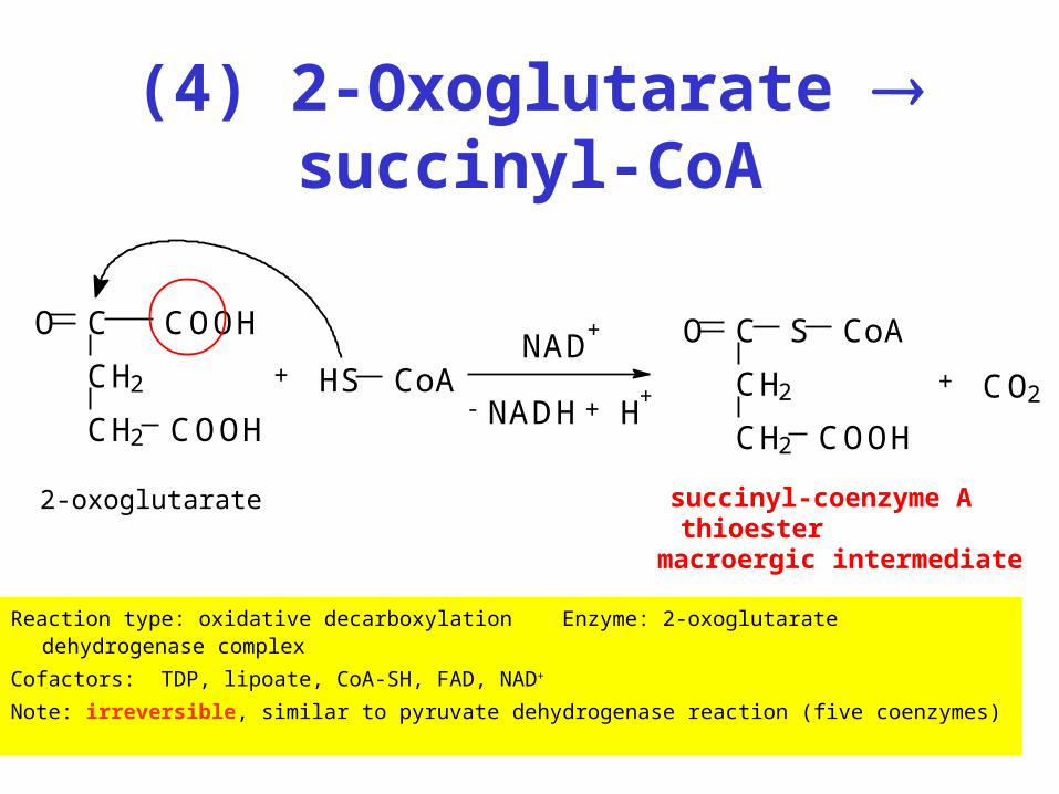
(4) 2-Oxoglutarate succinyl-CoA
Reaction type: oxidative decarboxylation Enzyme: 2-oxoglutarate dehydrogenase complex
Cofactors: TDP, lipoate, CoA-SH, FAD, NAD+
Note: irreversible, similar to pyruvate dehydrogenase reaction (five coenzymes)
CH2
CH2 COOH
C COOHO
+
NADH + H+
NAD+
-CH2
CH2 COOH
CO S CoA
+ CO2
2-oxoglutarate succinyl-coenzyme Athioester
macroergic intermediate
HS CoA
19
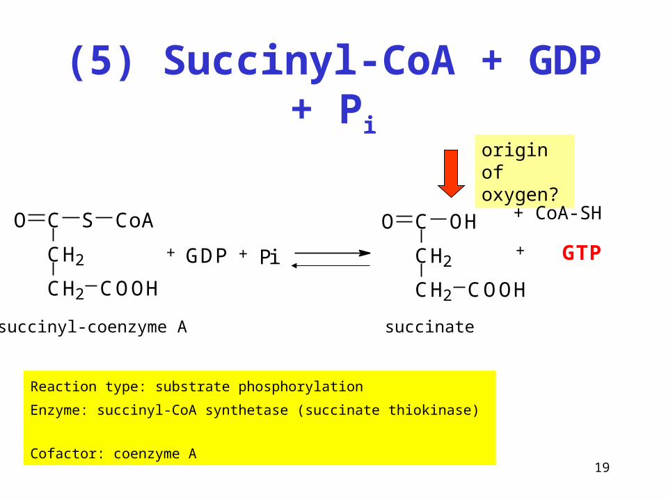
(5) Succinyl-CoA + GDP + Pi
Reaction type: substrate phosphorylation
Enzyme: succinyl-CoA synthetase (succinate thiokinase)
Cofactor: coenzyme A
+CH2
CH2 COOH
CO S CoA
+ +GDP Pi CH2
CH2 COOH
CO OH
GTP
succinyl-coenzyme A succinate
origin of oxygen?
+ CoA-SH
20
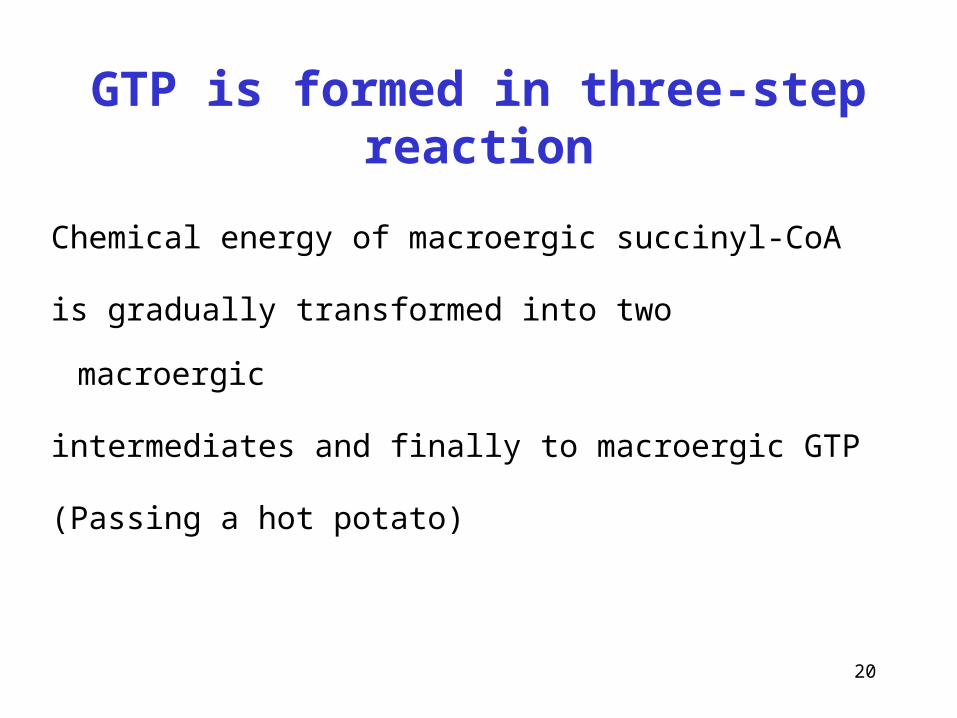
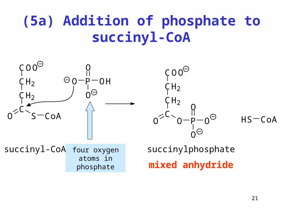
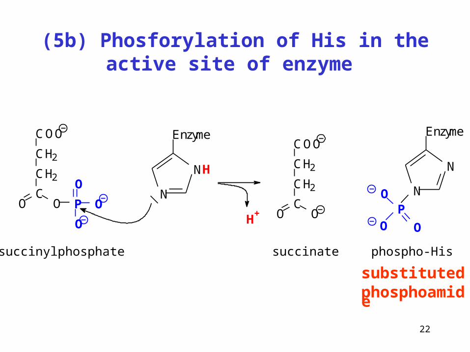
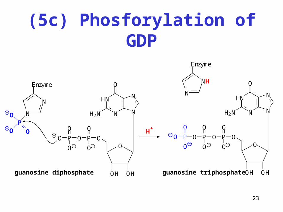
GTP is formed in three-step reaction
Chemical energy of macroergic succinyl-CoA
is gradually transformed into two macroergic
intermediates and finally to macroergic GTP
(Passing a hot potato)
21
(5a) Addition of phosphate to succinyl-CoA
mixed anhydride
four oxygen atoms in phosphate
P
O
O
O
OH
COO
CH2
CH2
CO S CoA HS CoA
COO
CH2
CH2
CO O P
O
O
O
succinyl-CoA succinylphosphate
22
(5b) Phosforylation of His in the active site of enzyme
substituted phosphoamide
N
NH
EnzymeCOO
CH2
CH2
CO O P
O
O
O
COO
CH2
CH2
CO O
succinylphosphate succinate phospho-His
N
N
Enzyme
P
O
O
OH+
23
(5c) Phosforylation of GDP
N
N
N
N
O
H2N
H
O
OH OH
OPO
O
O
P
O
O
O
N
N
Enzyme
P
O
O
O
N
N
N
N
O
H2N
H
O
OH OH
OPO
O
O
P
O
O
OP
O
O
O
guanosine diphosphate guanosine triphosphate
N
NH
Enzyme
H+
24
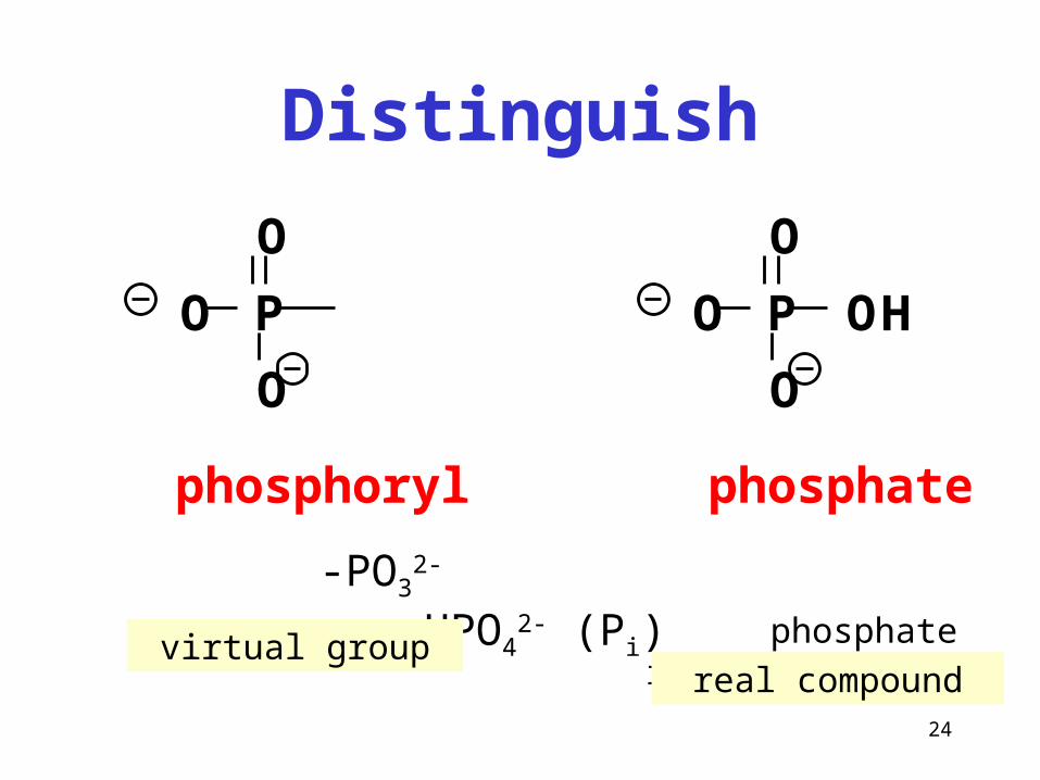
Distinguish
-PO32- HPO4
2- (Pi) phosphate inorganic
P
O
O
O
P
O
O
O
OH
phosphoryl phosphate
virtual groupreal compound
25
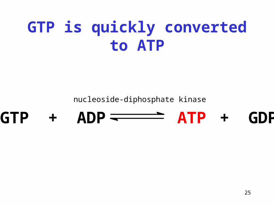
GTP is quickly converted to ATP
GTP + ADP ATP + GDPnucleoside-diphosphate kinase
26
(6) Succinate fumarate
Reaction type: dehydrogenation (-CH2-CH2- bond)
Enzyme: succinate dehydrogenase
Cofactor: FAD
COOH
CH2
CH2
COOH
+ FADC
C
COOHH
HOOC H
-II
-II-I
-I+ FADH2
succinate fumarate
27
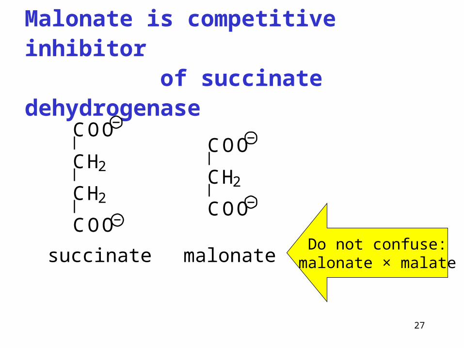
Malonate is competitive inhibitor of succinate dehydrogenase
Do not confuse:malonate × malate
COO
CH2
CH2
COO
COO
CH2
COO
succinate malonate
28
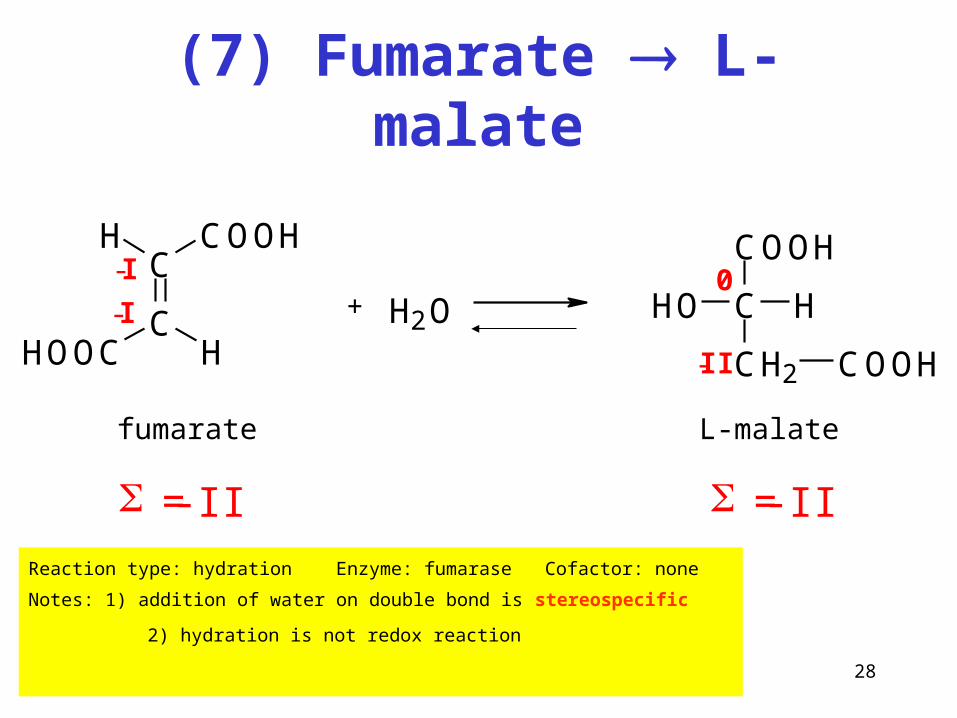
(7) Fumarate L-malate
Reaction type: hydration Enzyme: fumarase Cofactor: none
Notes: 1) addition of water on double bond is stereospecific
2) hydration is not redox reaction
-II
+ H2O
COOH
C H
CH2
HO
COOH
0
fumarate L-malate
= -II = -II
C
C
COOHH
HOOC H-I
-I
29
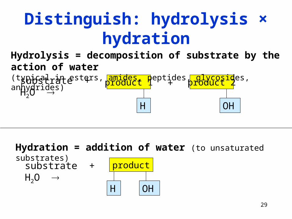
Distinguish: hydrolysis × hydration
substrate + H2O product 2
OH
substrate + H2O
+product 1
H
product
OHH
Hydrolysis = decomposition of substrate by the action of water (typical in esters, amides, peptides, glycosides, anhydrides)
Hydration = addition of water (to unsaturated substrates)
30
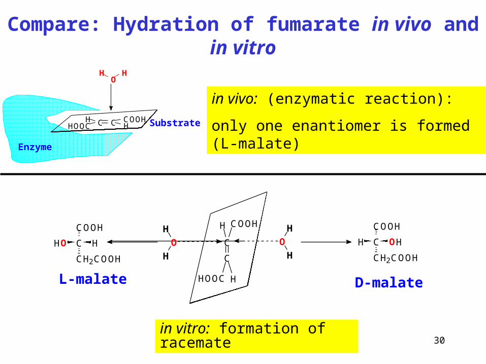
Compare: Hydration of fumarate in vivo and in vitro
in vivo: (enzymatic reaction):
only one enantiomer is formed (L-malate)
in vitro: formation of racemate
C C COOHH
HHOOC
HO
H
Enzyme
Substrate
C
C
COOHH
HOOC H
H
H
OH
H
O C
COOH
CH2COOH
OHHC
COOH
CH2COOH
HHO
L-malate D-malate
31
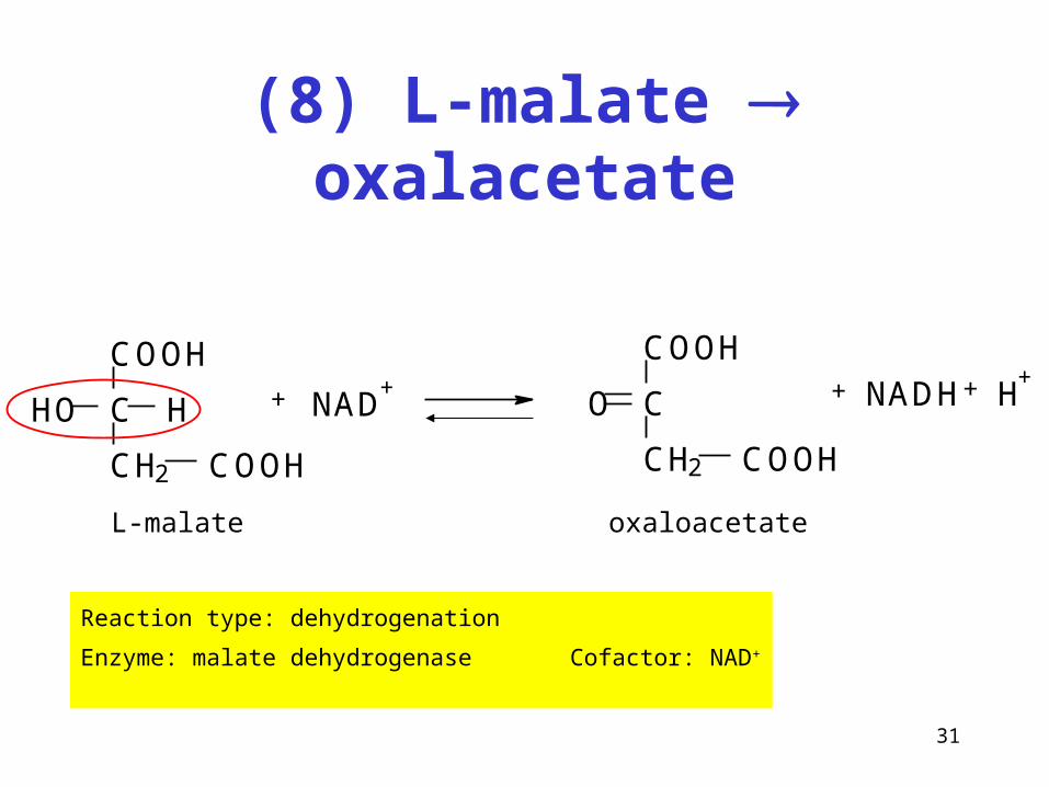
(8) L-malate oxalacetate
Reaction type: dehydrogenation
Enzyme: malate dehydrogenase Cofactor: NAD+
COOH
C H
CH2
HO
COOH
+ NAD+
COOH
C
CH2 COOH
O + NADH H++
L-malate oxaloacetate
32
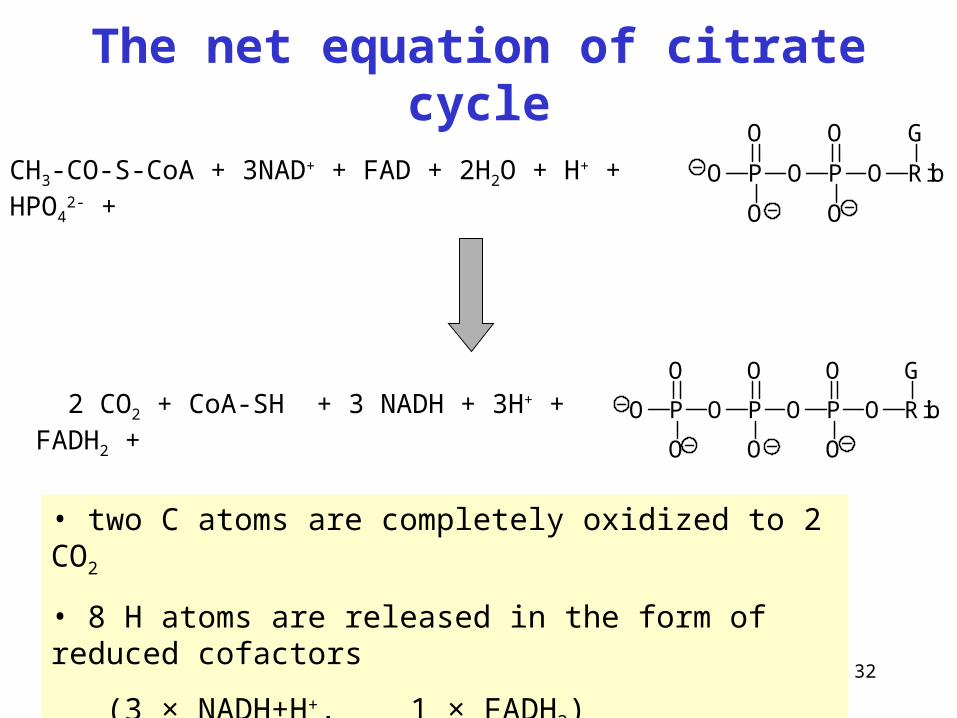
The net equation of citrate cycle
P
O
O
O O P
O
O
O Rib
G
CH3-CO-S-CoA + 3NAD+ + FAD + 2H2O + H+ + HPO42- +
2 CO2 + CoA-SH + 3 NADH + 3H+ + FADH2 +
• two C atoms are completely oxidized to 2 CO2
• 8 H atoms are released in the form of reduced cofactors
(3 × NADH+H+, 1 × FADH2)
P
O
O
O O P
O
O
O Rib
G
P
O
O
O
33
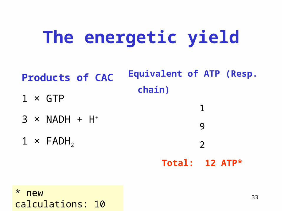
The energetic yield
Products of CAC
1 × GTP
3 × NADH + H+
1 × FADH2
Equivalent of ATP (Resp. chain)
1
9
2
Total: 12 ATP*
* new calculations: 10 ATP
34
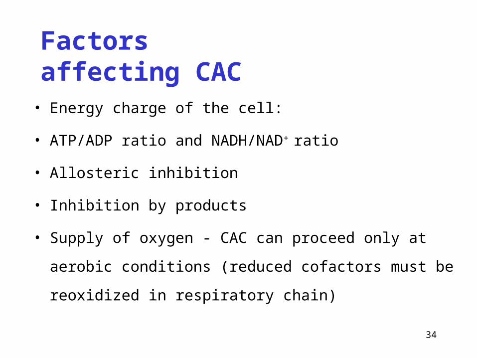
Factors affecting CAC
• Energy charge of the cell:
• ATP/ADP ratio and NADH/NAD+ ratio
• Allosteric inhibition
• Inhibition by products
• Supply of oxygen - CAC can proceed only at aerobic conditions
(reduced cofactors must be reoxidized in respiratory chain)
35
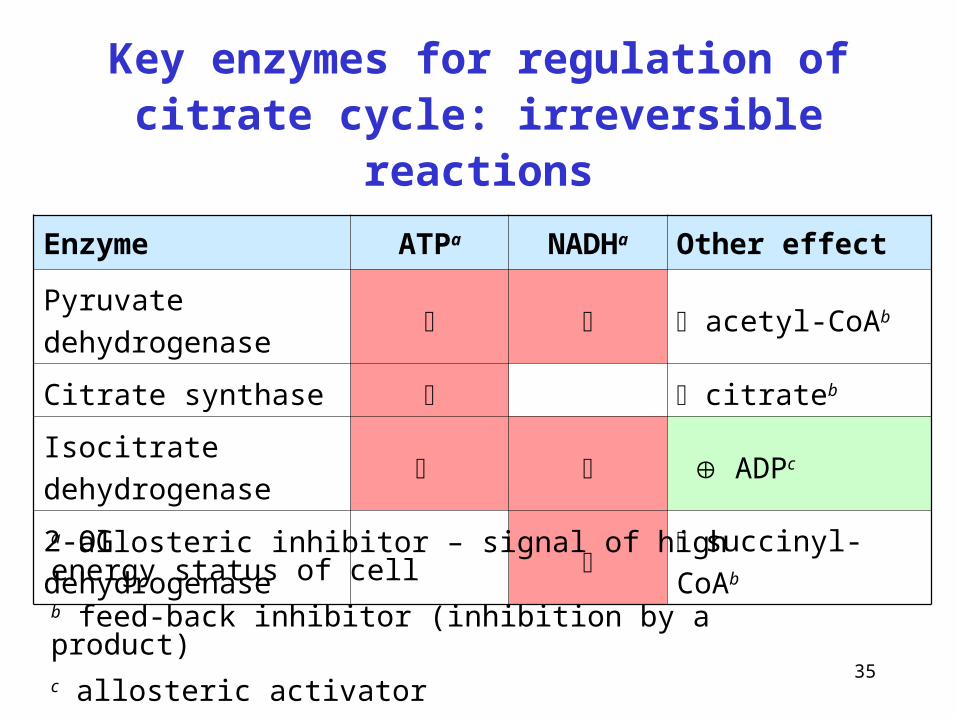
Key enzymes for regulation of citrate cycle: irreversible reactions
Enzyme ATPa NADHa Other effect
Pyruvate dehydrogenase acetyl-CoAb
Citrate synthase citrateb
Isocitrate dehydrogenase ADPc
2-OG dehydrogenase succinyl-CoAb
a allosteric inhibitor – signal of high energy status of cellb feed-back inhibitor (inhibition by a product)c allosteric activator
36
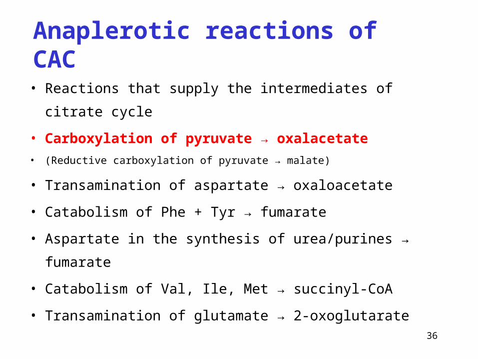
Anaplerotic reactions of CAC
• Reactions that supply the intermediates of citrate cycle
• Carboxylation of pyruvate → oxalacetate
• (Reductive carboxylation of pyruvate → malate)
• Transamination of aspartate → oxaloacetate
• Catabolism of Phe + Tyr → fumarate
• Aspartate in the synthesis of urea/purines → fumarate
• Catabolism of Val, Ile, Met → succinyl-CoA
• Transamination of glutamate → 2-oxoglutarate
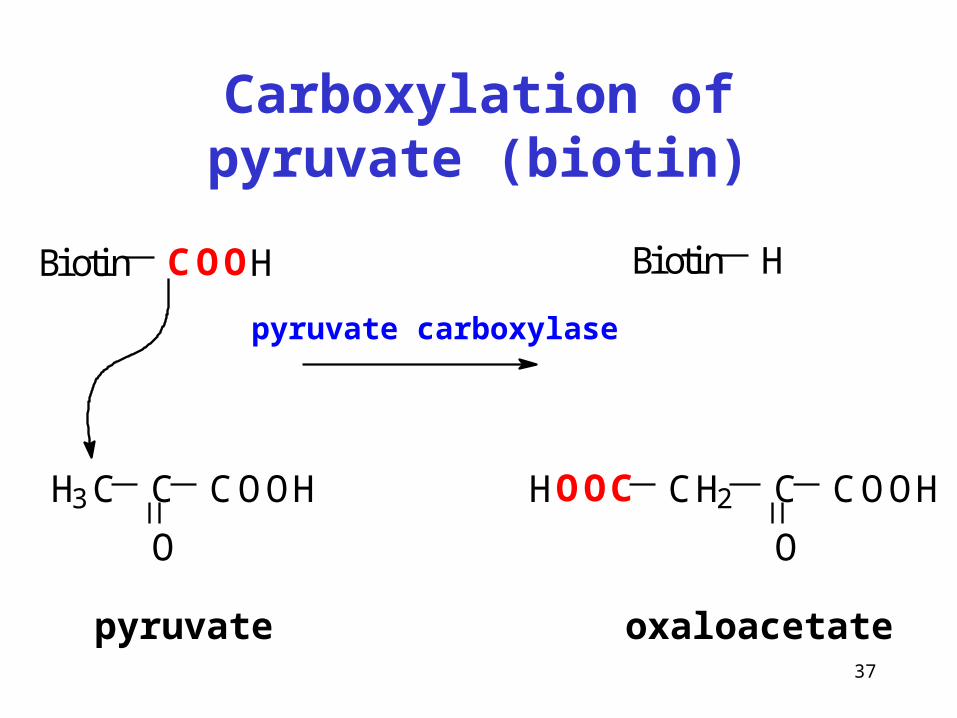
37
Carboxylation of pyruvate (biotin)
Biotin COOH
H3C C
O
COOH
Biotin H
CH2 C
O
COOHHOOC
pyruvate oxaloacetate
pyruvate carboxylase
38
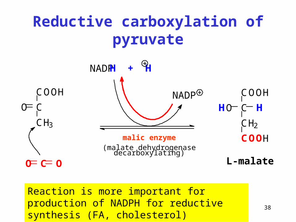
Reductive carboxylation of pyruvate
Reaction is more important for production of NADPH for reductive synthesis (FA, cholesterol)
malic enzyme(malate dehydrogenase decarboxylating)
COOH
C
CH3
O
CO O
NADPH + H
COOH
C
CH2
HO H
COOH
L-malate
NADP
39
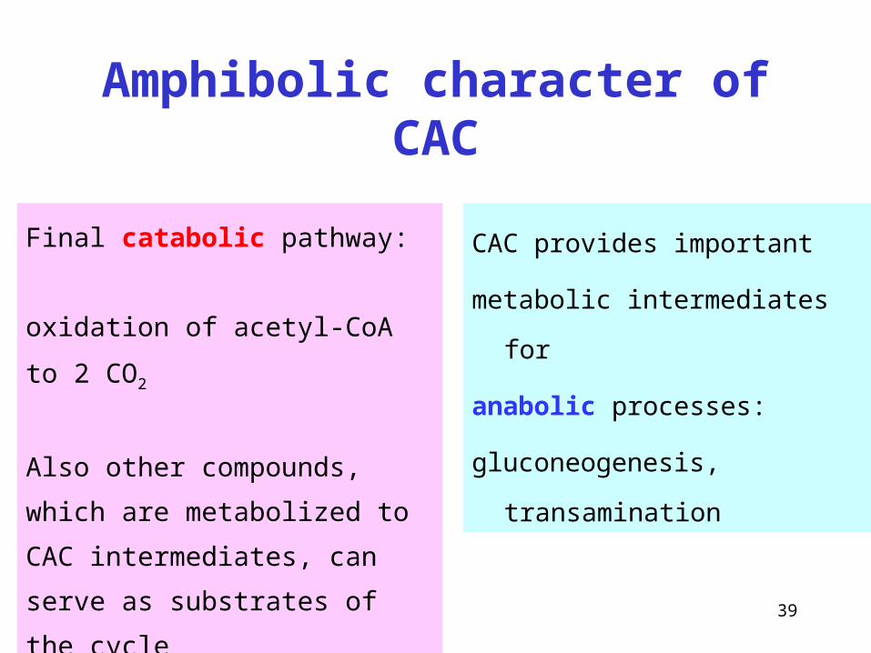
Amphibolic character of CAC
CAC provides important
metabolic intermediates for
anabolic processes:
gluconeogenesis, transamination
Final catabolic pathway:
oxidation of acetyl-CoA to 2 CO2
Also other compounds,
which are metabolized to CAC
intermediates, can serve as substrates
of the cycle
40
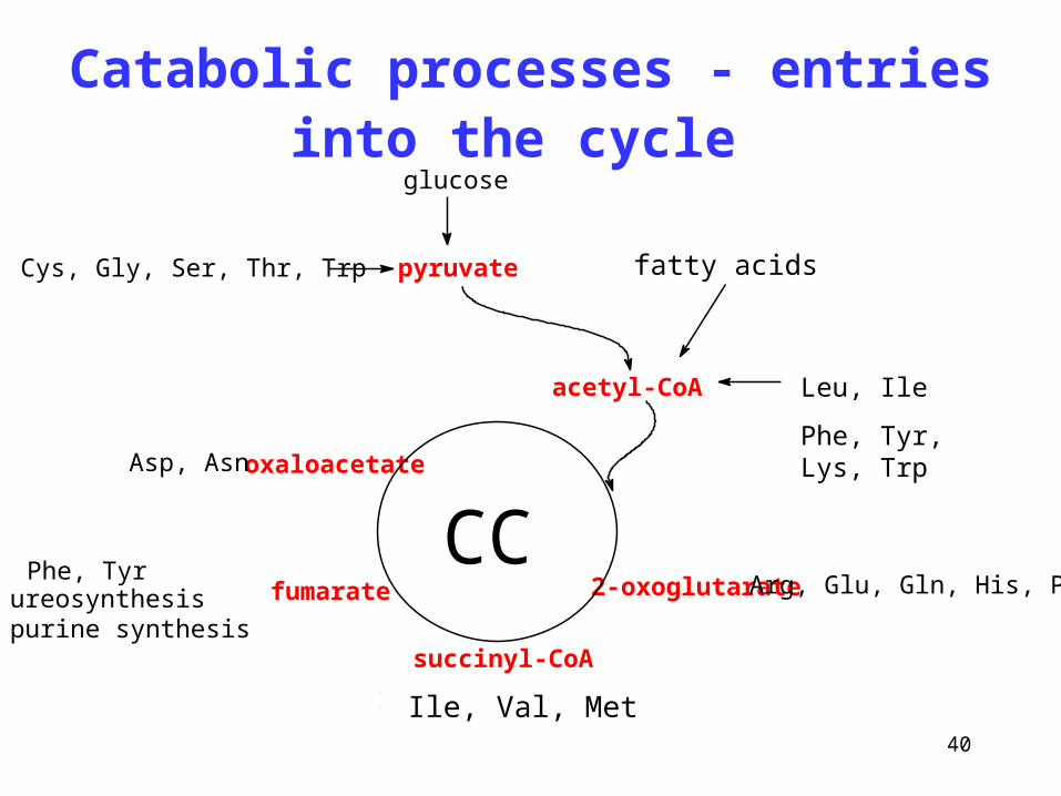
Catabolic processes - entries into the cycle
Leu, Ile
Phe, Tyr, Lys, Trpoxaloacetate
fumarate
succinyl-CoA
2-oxoglutarateCC
acetyl-CoA
Phe, Tyrureosynthesispurine synthesis
Ile, Val, Met, Thr
Arg, Glu, Gln, His, Pro
Asp, Asn
pyruvateAla, Cys, Gly, Ser, Thr, Trp fatty acids
glucose
Ile, Val, Met
41
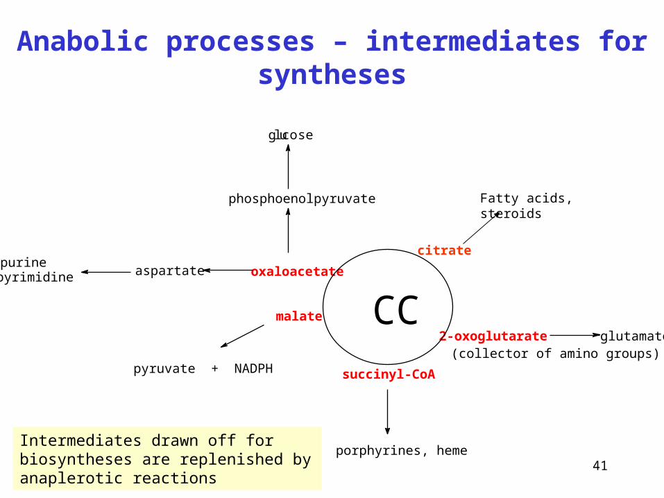
Anabolic processes – intermediates for syntheses
oxaloacetate
succinyl-CoA
2-oxoglutarateCCmalate
porphyrines, heme
(collector of amino groups)pyruvate + NADPH
aspartatepurinepyrimidine
phosphoenolpyruvate
gluco se
glutamate
citrate
Fatty acids, steroids
Intermediates drawn off for biosyntheses are replenished by anaplerotic reactions
42
CAC and the synthesis of lipids
ATP
TAG
malate
FA synthesis
CAC
mitochondriacitrate
cytosol
citrate
oxaloacetate acetyl-CoA
malic enzyme
CO2 NADPH H+
+ + +pyruvate
43
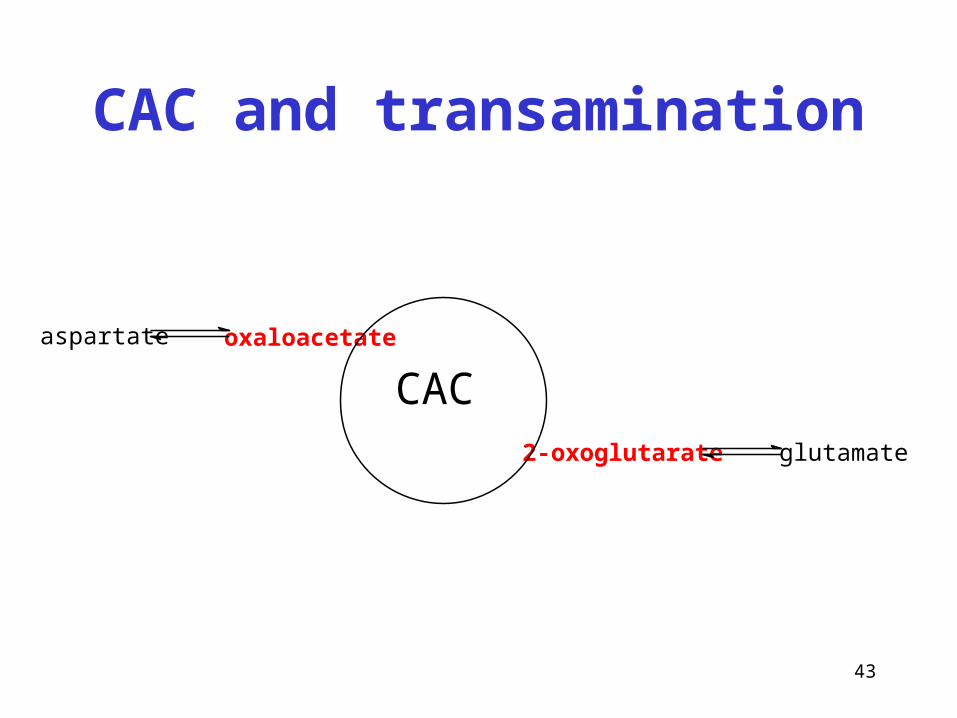
CAC and transamination
oxaloacetate
2-oxoglutarate
CACaspartate
glutamate
44
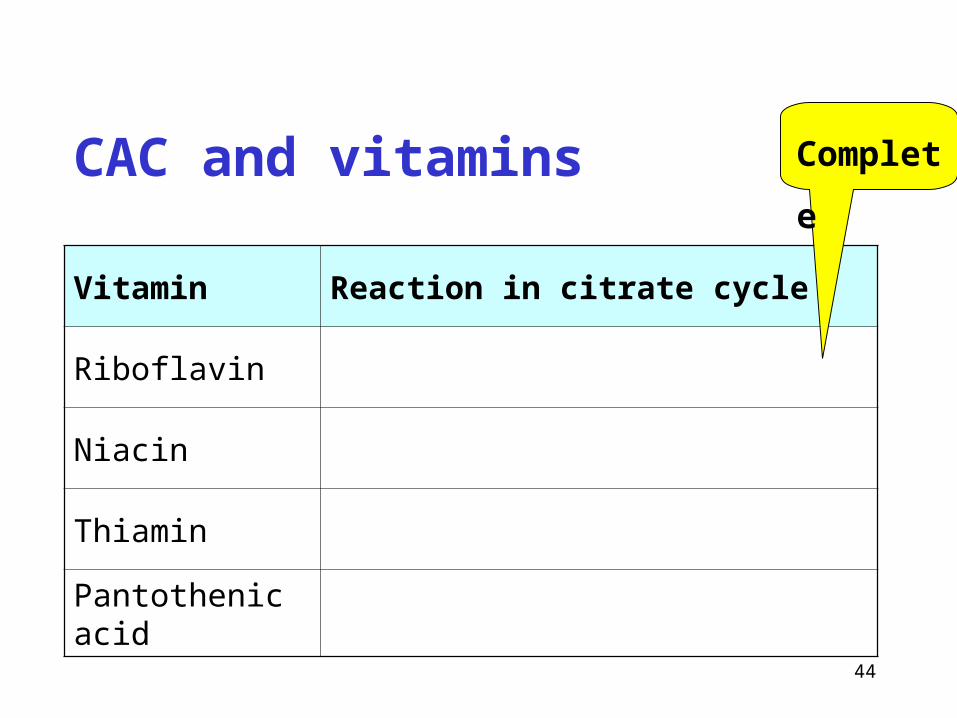
CAC and vitamins
Vitamin Reaction in citrate cycle
Riboflavin
Niacin
Thiamin
Pantothenic acid
Complete
45
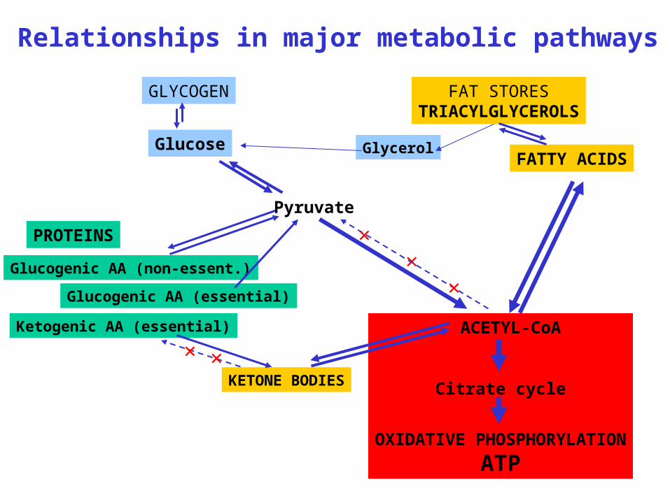
Relationships in major metabolic pathways
GLYCOGEN
Glucose
FAT STORESTRIACYLGLYCEROLS
FATTY ACIDS
PROTEINS
Glucogenic AA (non-essent.)
Glucogenic AA (essential)
Ketogenic AA (essential) ACETYL-CoA
Citrate cycle
OXIDATIVE PHOSPHORYLATION
ATP
KETONE BODIES
Glycerol
×
Pyruvate
×
×
× ×
46
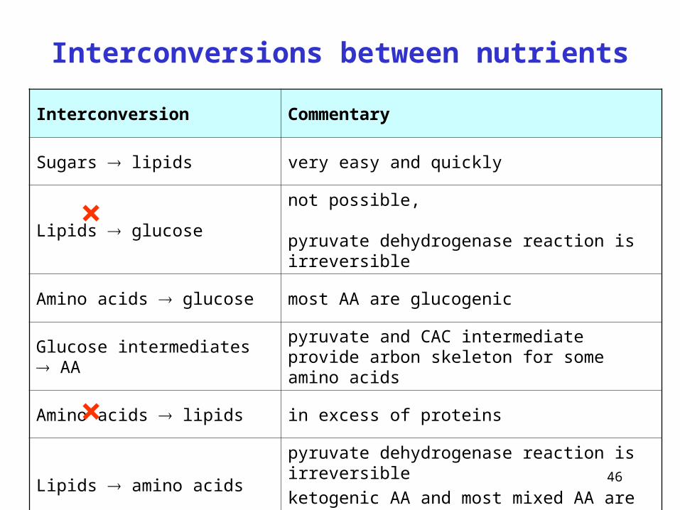
Interconversions between nutrients
Interconversion Commentary
Sugars lipids very easy and quickly
Lipids glucosenot possible, pyruvate dehydrogenase reaction is irreversible
Amino acids glucose most AA are glucogenic
Glucose intermediates AApyruvate and CAC intermediate provide arbon skeleton for some amino acids
Amino acids lipids in excess of proteins
Lipids amino acidspyruvate dehydrogenase reaction is irreversible
ketogenic AA and most mixed AA are essential
×
×
47
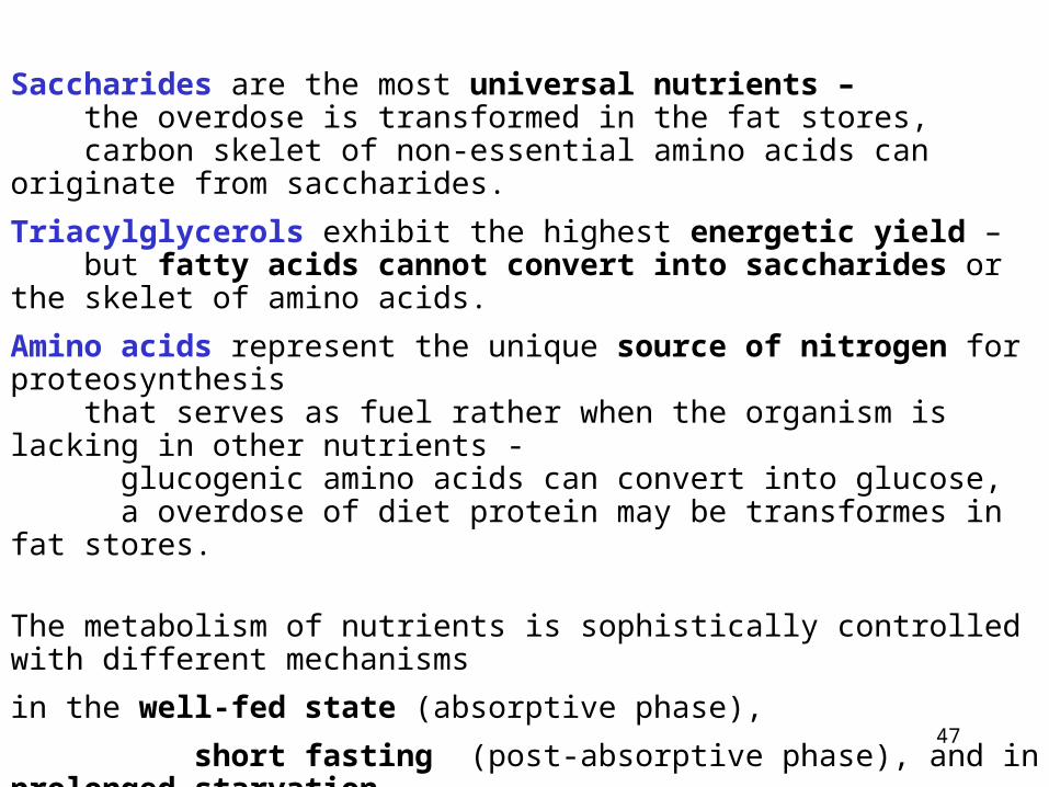
Saccharides are the most universal nutrients – the overdose is transformed in the fat stores, carbon skelet of non-essential amino acids can originate from saccharides.
Triacylglycerols exhibit the highest energetic yield – but fatty acids cannot convert into saccharides or the skelet of amino acids.
Amino acids represent the unique source of nitrogen for proteosynthesis that serves as fuel rather when the organism is lacking in other nutrients - glucogenic amino acids can convert into glucose, a overdose of diet protein may be transformes in fat stores.
The metabolism of nutrients is sophistically controlled with different mechanisms
in the well-fed state (absorptive phase),
short fasting (post-absorptive phase), and in prolonged starvation.
It also depends on energy expenditure (predominantly muscular work) – either of maximal intensity (anaerobic, of short duration only) or aerobic work of much lower intensity (long duration).
48
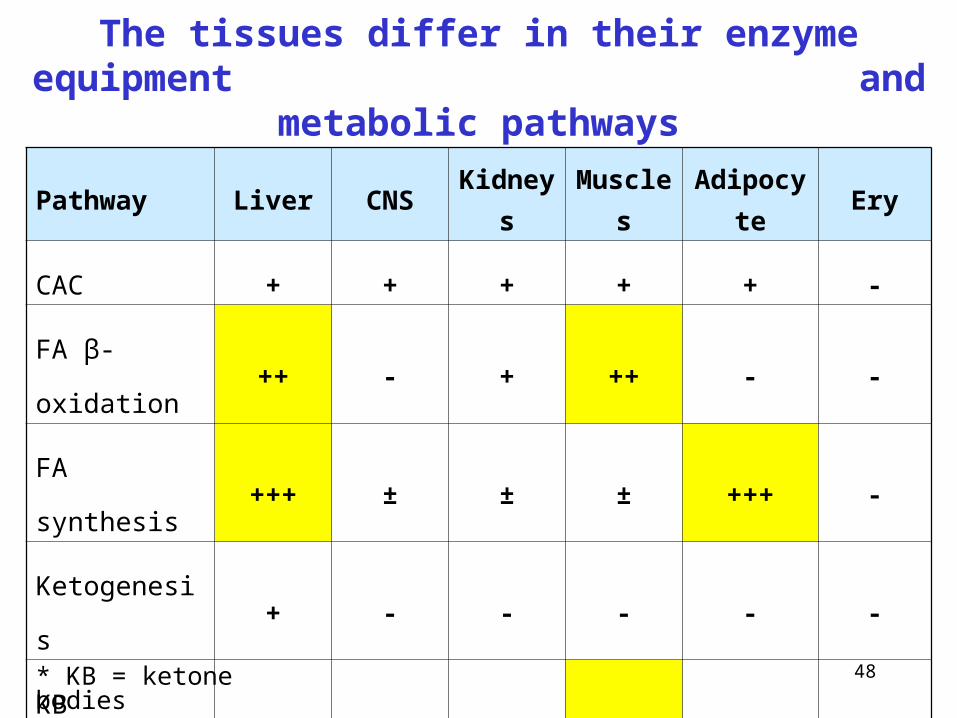
The tissues differ in their enzyme equipment and metabolic pathways
Pathway Liver CNS Kidneys Muscles Adipocyte Ery
CAC + + + + + -
FA β-oxidation ++ - + ++ - -
FA synthesis +++ ± ± ± +++ -
Ketogenesis + - - - - -
KB oxidation* - + + +++ + -
Glycolysis + +++ + +++ + +++
Gluconeogenesis +++ - + - - -
* KB = ketone bodies
49
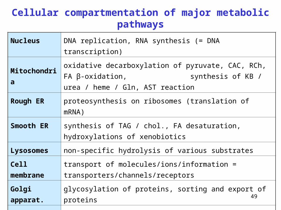
Cellular compartmentation of major metabolic pathways
Nucleus DNA replication, RNA synthesis (= DNA transcription)
Mitochondriaoxidative decarboxylation of pyruvate, CAC, RCh, FA β-oxidation,
synthesis of KB / urea / heme / Gln, AST reaction
Rough ER proteosynthesis on ribosomes (translation of mRNA)
Smooth ER synthesis of TAG / chol., FA desaturation, hydroxylations of xenobiotics
Lysosomes non-specific hydrolysis of various substrates
Cell membrane transport of molecules/ions/information = transporters/channels/receptors
Golgi apparat. glycosylation of proteins, sorting and export of proteins
Peroxisomes formation and decomposition of H2O2 and peroxides
Cytosol
glycolysis, gluconeogenesis, glycogen metabolism, pentose cycle,
transamination, synthesis of FA / urea / urate / heme;
ethanol acetaldehyde
50
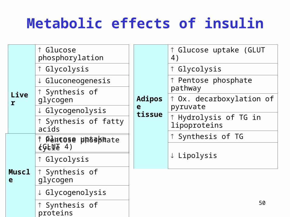
Liver
Glucose phosphorylation
Glycolysis
Gluconeogenesis
Synthesis of glycogen
Glycogenolysis
Synthesis of fatty acids
Pentose phosphate cycle
Adipose tissue
Glucose uptake (GLUT 4)
Glycolysis
Pentose phosphate pathway
Ox. decarboxylation of pyruvate
Hydrolysis of TG in lipoproteins
Synthesis of TG
Lipolysis
Muscle
Glucose uptake (GLUT 4)
Glycolysis
Synthesis of glycogen
Glycogenolysis
Synthesis of proteins
Metabolic effects of insulin
51
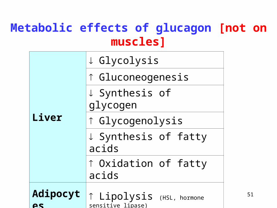
Liver
Glycolysis
Gluconeogenesis
Synthesis of glycogen
Glycogenolysis
Synthesis of fatty acids
Oxidation of fatty acids
Adipocytes Lipolysis (HSL, hormone sensitive lipase)
Metabolic effects of glucagon [not on muscles]
52
Biosynthesis of heme
~
Hemoproteins
53
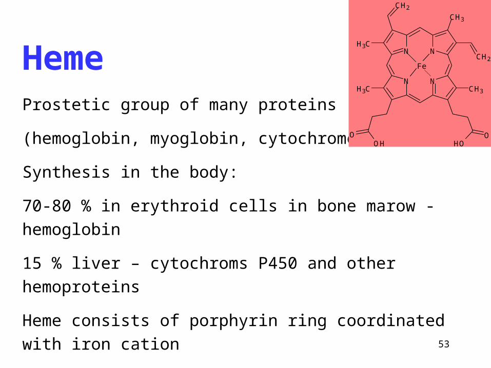
HemeProstetic group of many proteins
(hemoglobin, myoglobin, cytochromes)
Synthesis in the body:
70-80 % in erythroid cells in bone marow - hemoglobin
15 % liver – cytochroms P450 and other hemoproteins
Heme consists of porphyrin ring coordinated with iron cation
N
N
Fe
N
N
O O
CH2
CH2
CH3
CH3
OH OH
CH3
CH3
54
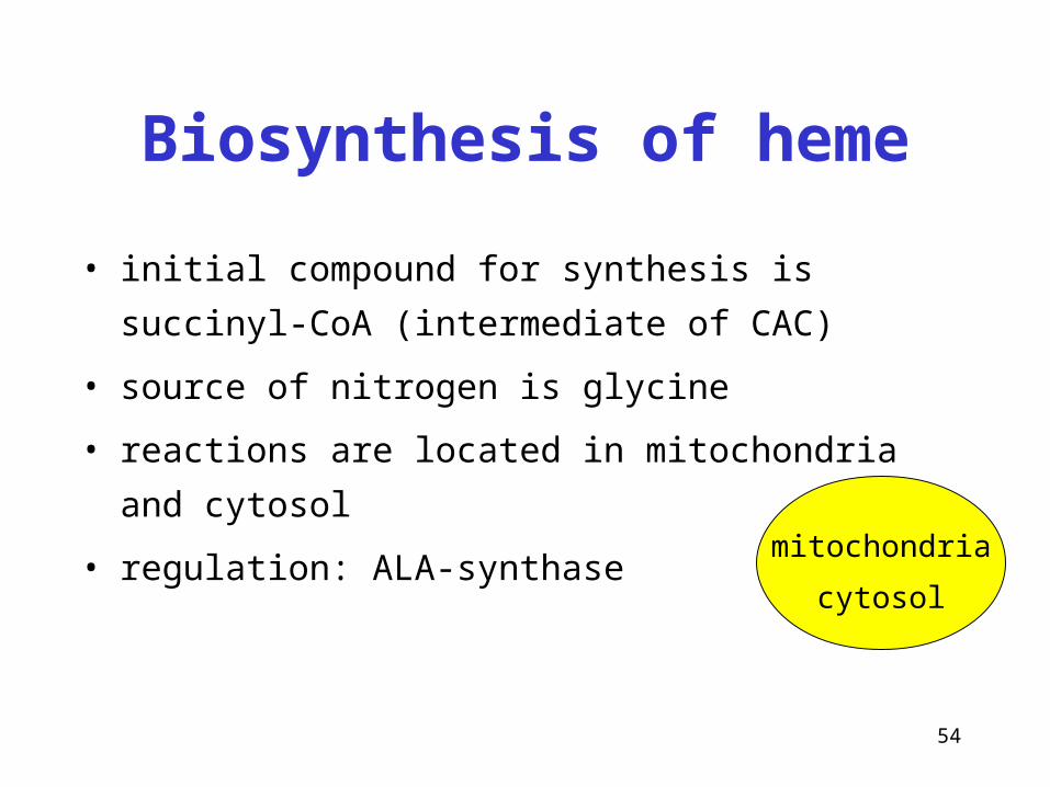
Biosynthesis of heme
• initial compound for synthesis is succinyl-CoA
(intermediate of CAC)
• source of nitrogen is glycine
• reactions are located in mitochondria and cytosol
• regulation: ALA-synthasemitochondria
cytosol
55
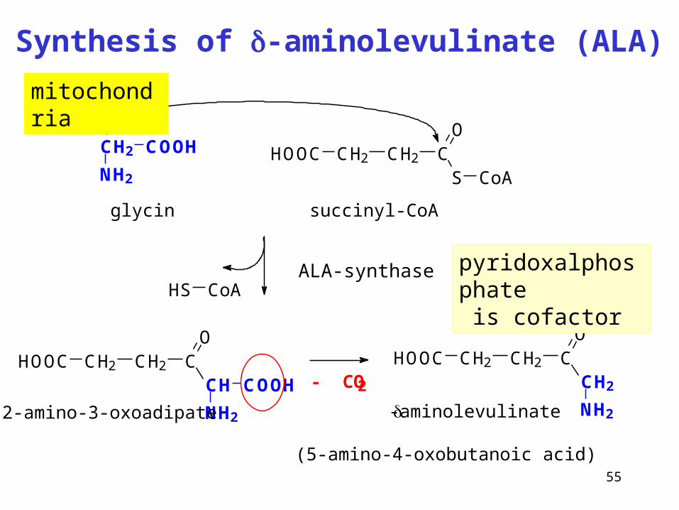
Synthesis of -aminolevulinate (ALA)
ALA-synthase
CH2
NH2
COOH HOOC CH2 CH2 CS
O
CoA
glycin succinyl-CoA
HOOC CH2 CH2 CO
CH2
NH2
HOOC CH2 CH2 CO
CH
NH2
COOH
2-amino-3-oxoadipate
HS CoA
- CO2
-aminolevulinate
(5-amino-4-oxobutanoic acid)
pyridoxalphosphate is cofactor
mitochondria
56
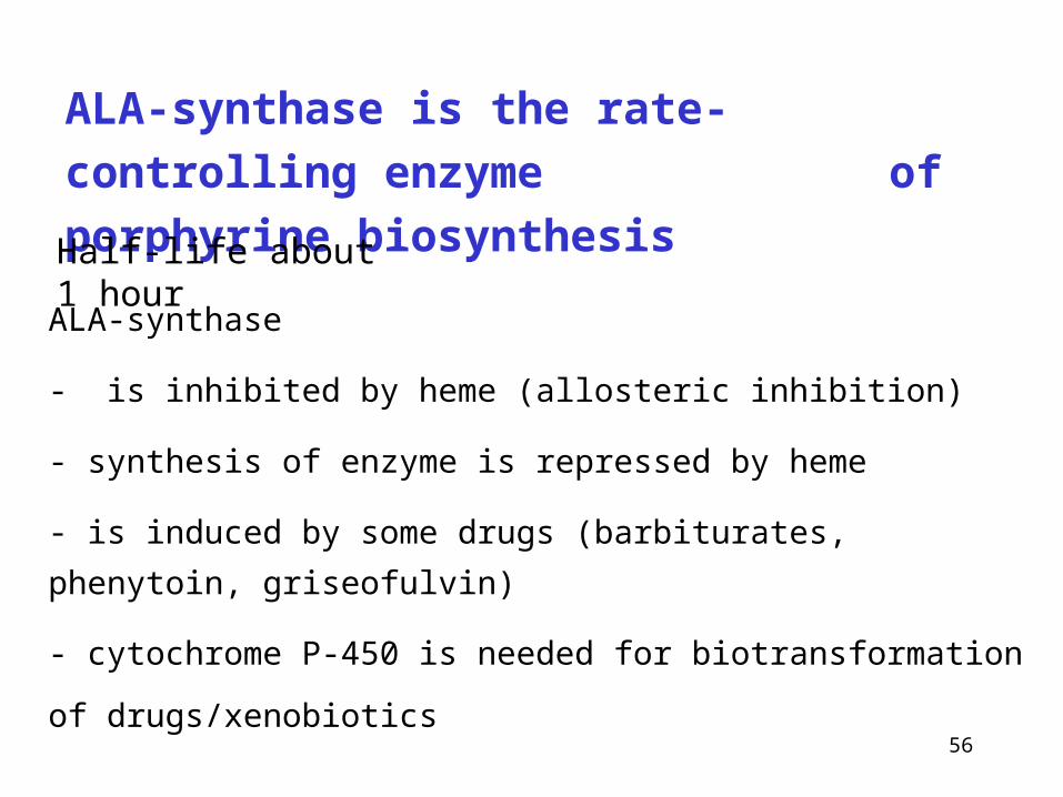
ALA-synthase is the rate-controlling enzyme
of porphyrine biosynthesis
ALA-synthase
- is inhibited by heme (allosteric inhibition)
- synthesis of enzyme is repressed by heme
- is induced by some drugs (barbiturates, phenytoin, griseofulvin)
- cytochrome P-450 is needed for biotransformation of drugs/xenobiotics
Half-life about 1 hour
57
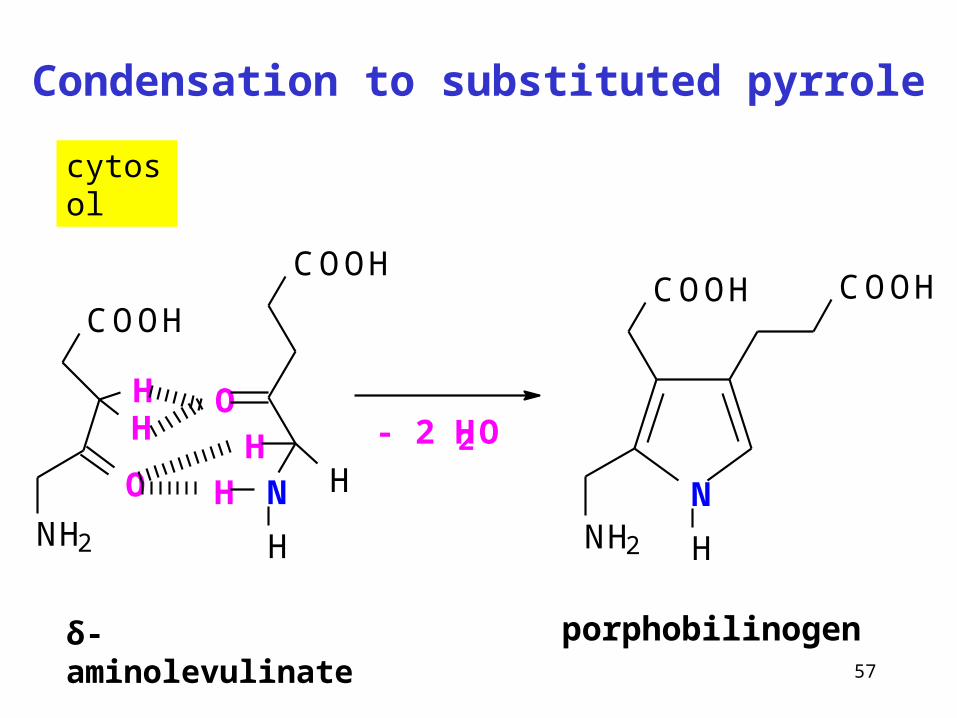
Condensation to substituted pyrrole
δ-aminolevulinate
cytosol
COOH
N
O
H
HH
HO
NH2
COOH
HH - 2 H2O
porphobilinogen
NNH2
COOHCOOH
H
58
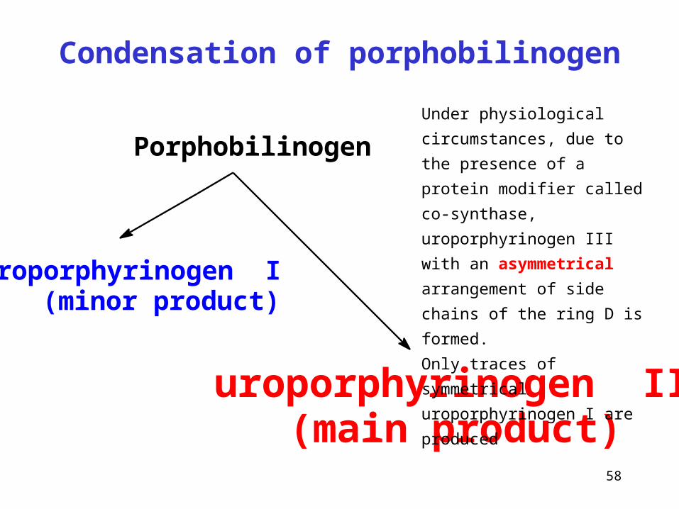
Condensation of porphobilinogen
Porphobilinogen
uroporphyrinogen I (minor product)
uroporphyrinogen III (main product)
Under physiological circumstances,
due to the presence of a protein
modifier called co-synthase,
uroporphyrinogen III with an
asymmetrical arrangement of side
chains of the ring D is formed.
Only traces of symmetrical
uroporphyrinogen I are produced
59
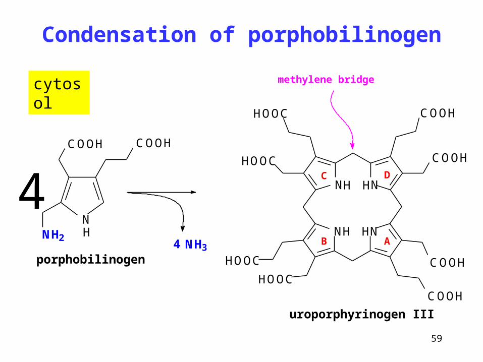
Condensation of porphobilinogen
NNH2
COOHCOOH
H
porphobilinogen
4NH34
H
NH N
NN
COOHHOOC
COOH
HOOC
HOOC
HOOC
COOH
COOH
H
H
uroporphyrinogen III
methylene bridge
AB
C D
cytosol
60
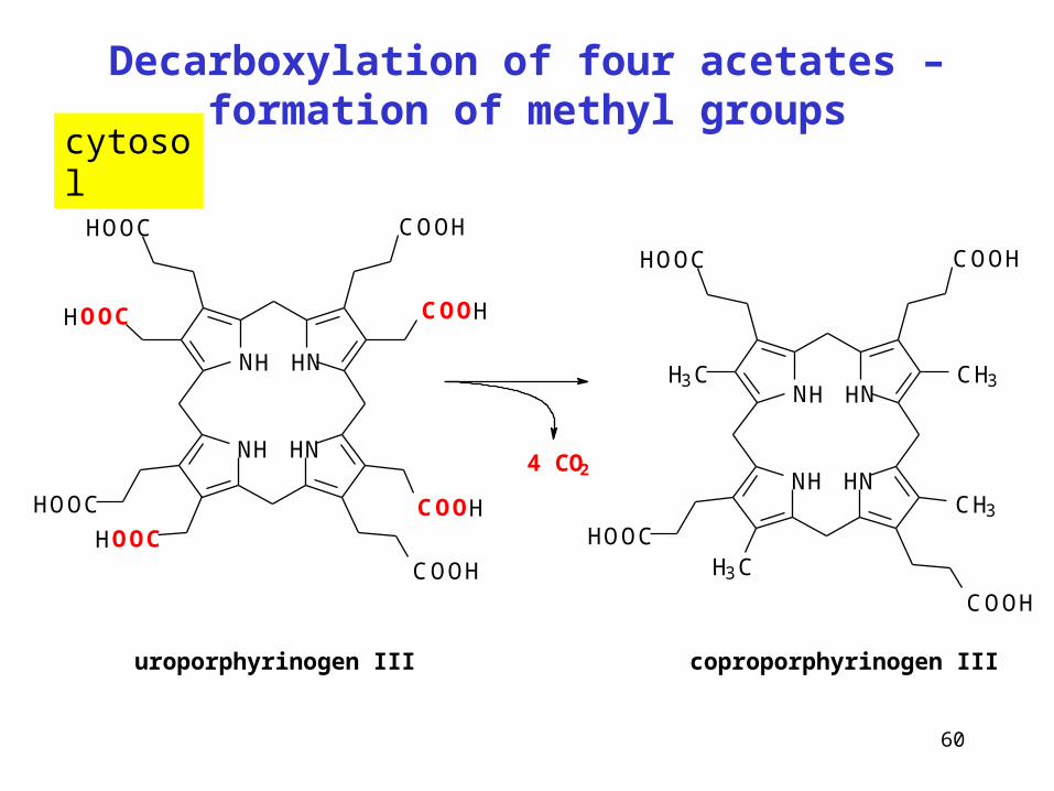
Decarboxylation of four acetates – formation of methyl groups
H
NH N
NN
COOHHOOC
COOH
HOOC
HOOC
HOOC
COOH
COOH
H
H
H
NH N
NNCH3H3C
COOHHOOC
H3CHOOC
CH3
COOH
H
H4 CO2
uroporphyrinogen III coproporphyrinogen III
cytosol
61
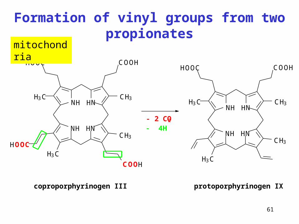
Formation of vinyl groups from two propionates
H
NH N
NNCH3H3C
COOHHOOC
H3CHOOC
CH3
COOH
H
H
coproporphyrinogen III
- 4H- 2 CO2
H
NH N
NNCH3H3C
COOHHOOC
H3C
CH3
H
H
protoporphyrinogen IX
mitochondria
62
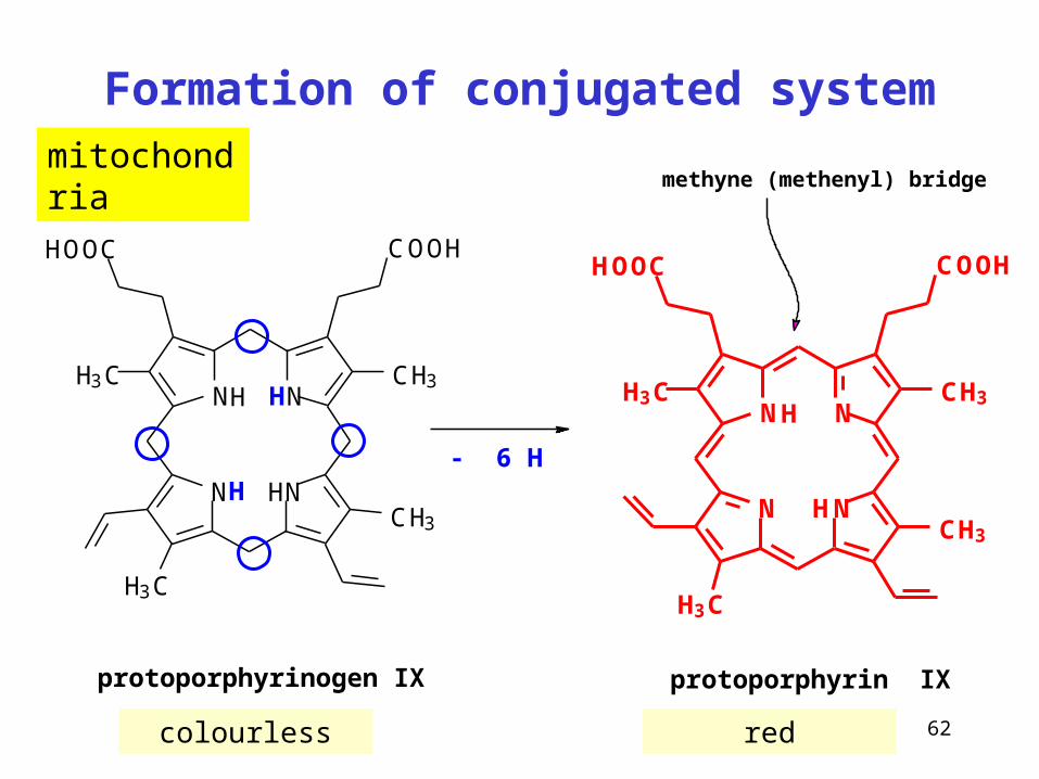
Formation of conjugated system
colourless red
H
NH N
NNCH3H3C
COOHHOOC
H3C
CH3
H
H
protoporphyrinogen IX
- 6 H
N N
NNCH3H3C
CH3
H3C
COOHHOOC
H
protoporphyrin IX
H
methyne (methenyl) bridgemitochondria
63
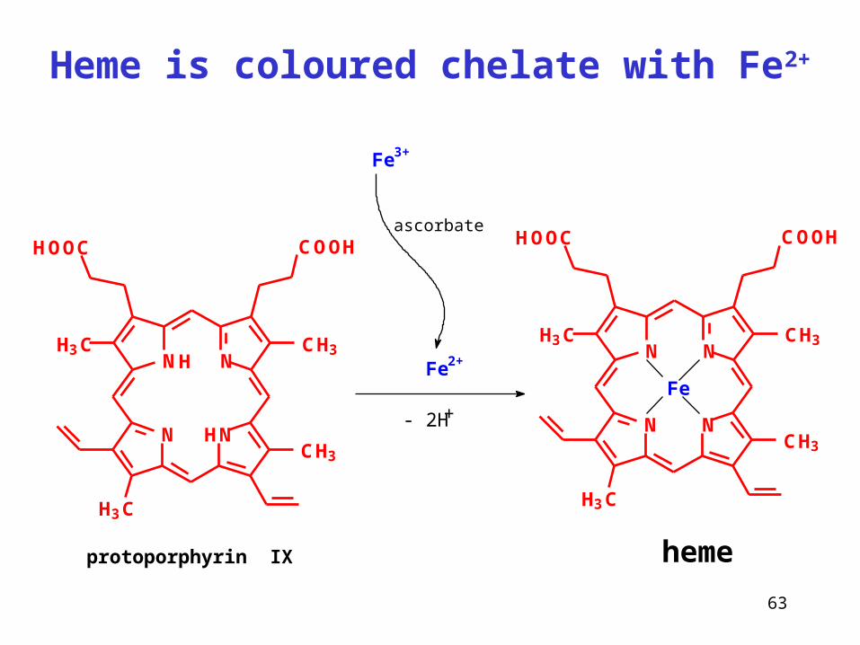
Heme is coloured chelate with Fe2+
protoporphyrin IX
Fe
heme
HN N
NNCH3H3C
CH3
H3C
COOHHOOC
H
ascorbate
Fe3+
- 2H+N N
NNCH3H3C
CH3
H3C
COOHHOOC
Fe2+
64
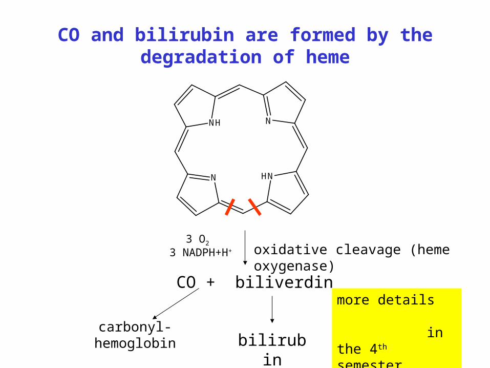
CO and bilirubin are formed by the degradation of heme
N HN
NNH
oxidative cleavage (heme oxygenase)
CO + biliverdin
bilirubincarbonyl-hemoglobin
3 O2 3 NADPH+H+
more details in the 4th semester
65
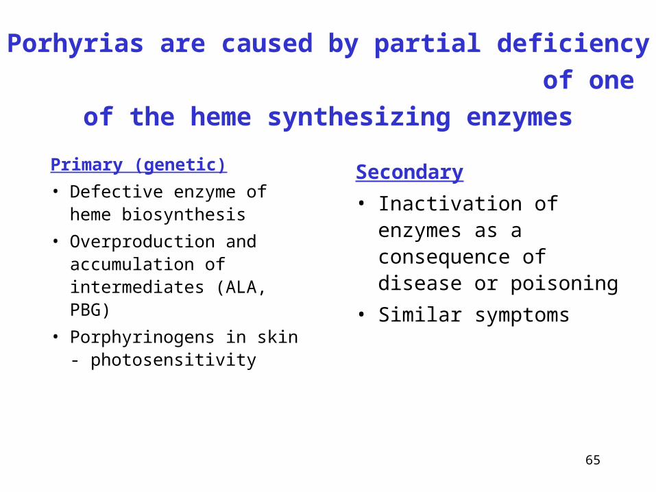
Porhyrias are caused by partial deficiency
of one of the heme synthesizing enzymes
Primary (genetic)
• Defective enzyme of heme biosynthesis
• Overproduction and accumulation of intermediates (ALA, PBG)
• Porphyrinogens in skin - photosensitivity
Secondary
• Inactivation of enzymes as a consequence of disease or poisoning
• Similar symptoms
66
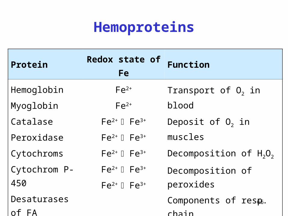
Hemoproteins
Protein Redox state of Fe Function
Hemoglobin
Myoglobin
Catalase
Peroxidase
Cytochroms
Cytochrom P-450
Desaturases of FA
Fe2+
Fe2+
Fe2+ Fe3+
Fe2+ Fe3+
Fe2+ Fe3+
Fe2+ Fe3+
Fe2+ Fe3+
Transport of O2 in blood
Deposit of O2 in muscles
Decomposition of H2O2
Decomposition of peroxides
Components of resp. chain
Hydroxylation
Desaturation of FA
67
Oxidation number of Fe in various hemes
Does not change
• Fe2+
• prosthetic group for O2
transport
• hemoglobin, myoglobin
• heme is hidden in hydrophobic pocket of globin
• oxidation of Fe2+ means the loss of function
Does change
• Fe2+ Fe3+
• cofactor of oxidoreductases
• cytochromes, heme enzymes
• heme is relatively exposed
• reversible redox change is the primary function = the transfer of one electron
68
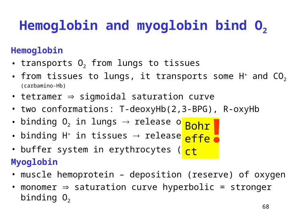
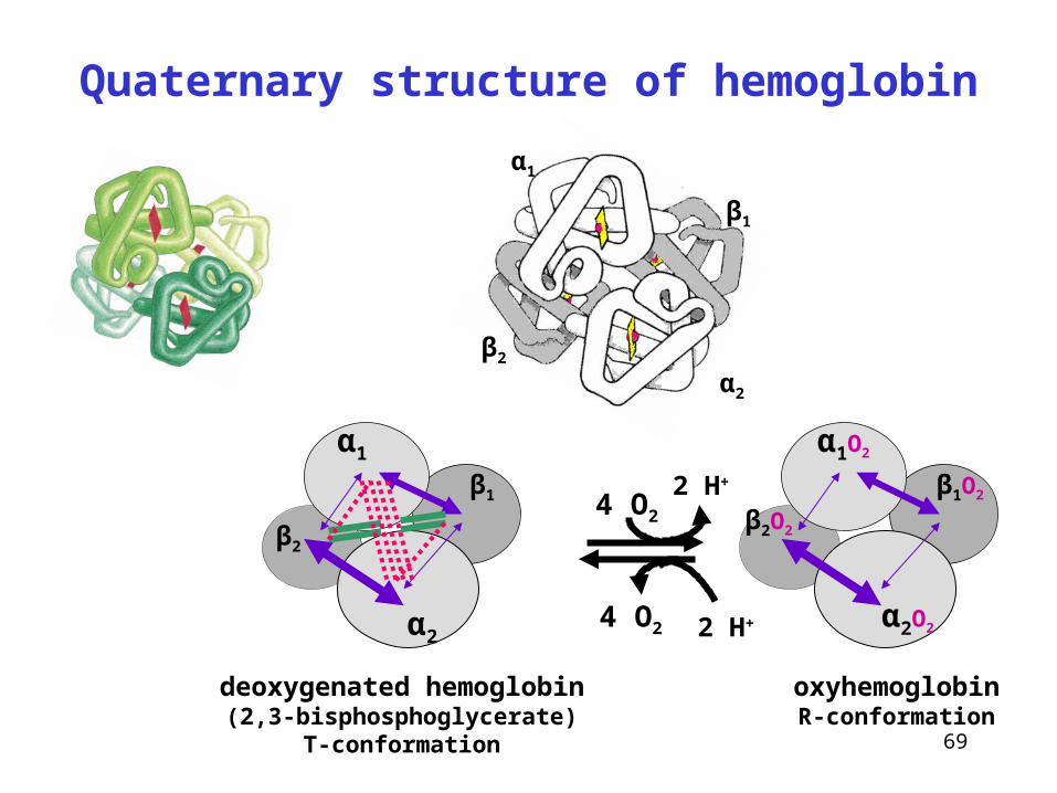
Hemoglobin and myoglobin bind O2
Hemoglobin
• transports O2 from lungs to tissues
• from tissues to lungs, it transports some H+ and CO2 (carbamino-Hb)
• tetramer sigmoidal saturation curve
• two conformations: T-deoxyHb(2,3-BPG), R-oxyHb
• binding O2 in lungs release of H+
• binding H+ in tissues release of O2
• buffer system in erythrocytes (His)
Myoglobin
• muscle hemoprotein – deposition (reserve) of oxygen
• monomer saturation curve hyperbolic = stronger binding O2
Bohr effect!
69
Quaternary structure of hemoglobin
α2
α1
β2
β1
4 O2
α1
α2
β1
β2
deoxygenated hemoglobin(2,3-bisphosphoglycerate)
T-conformation
oxyhemoglobinR-conformation
α1O2
α2O2
β1O2
β2O2
4 O2 2 H+
2 H+
70
Derivatives of hemoglobin
Carbonylhemoglobin
• CO has great affinity to Fe2+ in heme
• physiological level: 1 - 15 % from total Hb (environment, smokers etc.)
Glycated hemoglobin
• non-enzymatic reaction with free glucose, –NH2 group of Hb
(N-terminus, Lys) and aldehyde group of glucose
• physiological level: 2.8 – 4.0 % (from total Hb)
Methemoglobin (hemiglobin)
• oxidation of heme iron, Fe2+ Fe3+, physiol. level: 0.5 - 1.5 %
• oxidation agents: nitrites, alkyl nitrites, aromatic amines, nitro compounds
• Hb mutation: hemoglobin M (HbM), the replacement of F8HisTyr
• deficit of methemoglobin reductase
71
Language note: Methemoglobin
• it has nothing to do with methyl group !!!
• abbreviated from metahemoglobin
• the prefix meta- (from Greek) indicates
change, transformation, alteration
• other examples with the prefix meta:
metabolic (= catabolic +
anabolic)
metamorphosis, metazoan ...

72
Linguistic note:
How to express two redox states of iron
Fe2+ Fe3+
Infix -o -i
Biochemical names
hemoglobin,
ferroportin,
ferroxidase
hemiglobin = methemoglobin,
ferritin, transferrin, lactoferrin,
gastroferrin, ferric reductase
Chemical names
Latin ferrosi chloridum ferri chloridum
Old English ferrous chloride ferric chloride
New English iron(II) chloride iron(III) chloride
73
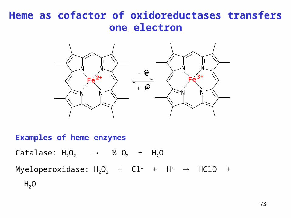
Heme as cofactor of oxidoreductases transfers one electron
N N
NN
Fe 2+
N N
NN
Fe 3+- e
+ e
Examples of heme enzymes
Catalase: H2O2 ½ O2 + H2O
Myeloperoxidase: H2O2 + Cl- + H+ HClO + H2O
74
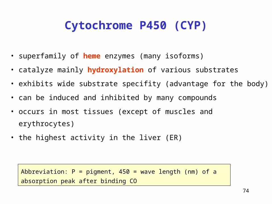
Cytochrome P450 (CYP)
• superfamily of heme enzymes (many isoforms)
• catalyze mainly hydroxylation of various substrates
• exhibits wide substrate specifity (advantage for the body)
• can be induced and inhibited by many compounds
• occurs in most tissues (except of muscles and erythrocytes)
• the highest activity in the liver (ER)
Abbreviation: P = pigment, 450 = wave length (nm) of a absorption peak after
binding CO
75
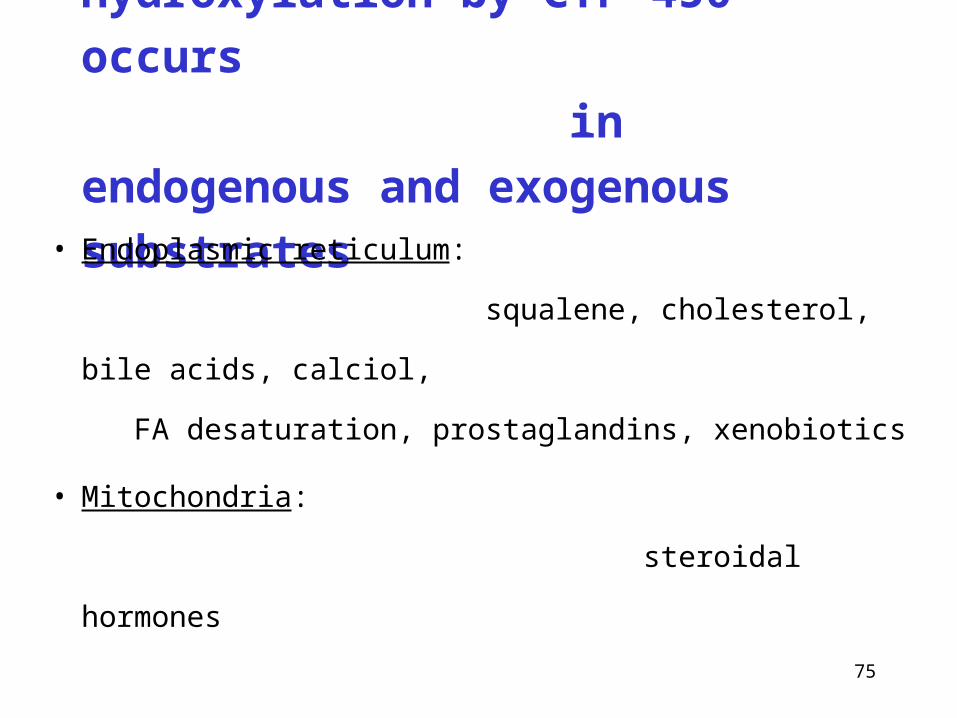
Hydroxylation by CYP 450 occurs
in endogenous and
exogenous substrates
• Endoplasmic reticulum:
squalene, cholesterol, bile acids, calciol,
FA desaturation, prostaglandins, xenobiotics
• Mitochondria:
steroidal hormones
76
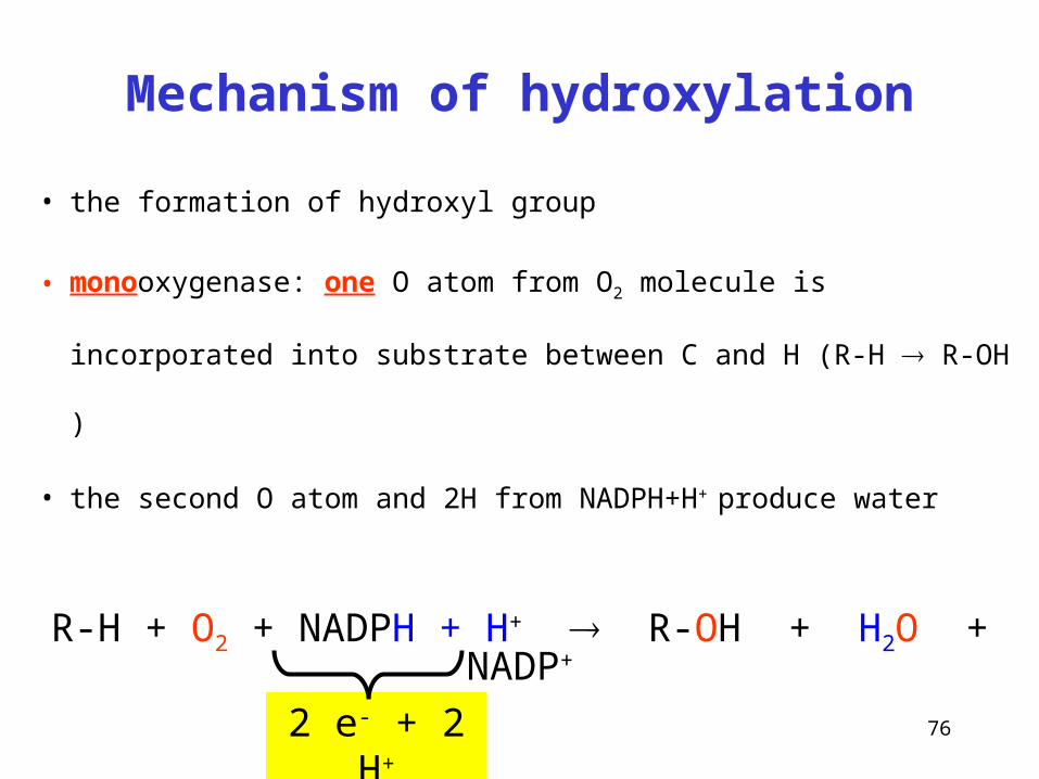
Mechanism of hydroxylation
• the formation of hydroxyl group
• monooxygenase: one O atom from O2 molecule is incorporated
into substrate between C and H (R-H R-OH )
• the second O atom and 2H from NADPH+H+ produce water
R-H + O2 + NADPH + H+ R-OH + H2O + NADP+
2 e- + 2 H+
77
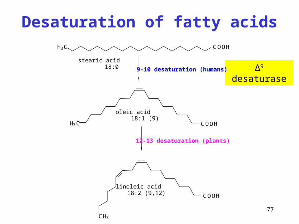
Desaturation of fatty acids
∆9 desaturase
H3C COOH
COOHH3C
COOH
CH3
9-10 desaturation (humans)
12-13 desaturation (plants)
stearic acid 18:0
oleic acid 18:1 (9)
linoleic acid 18:2 (9,12)
78
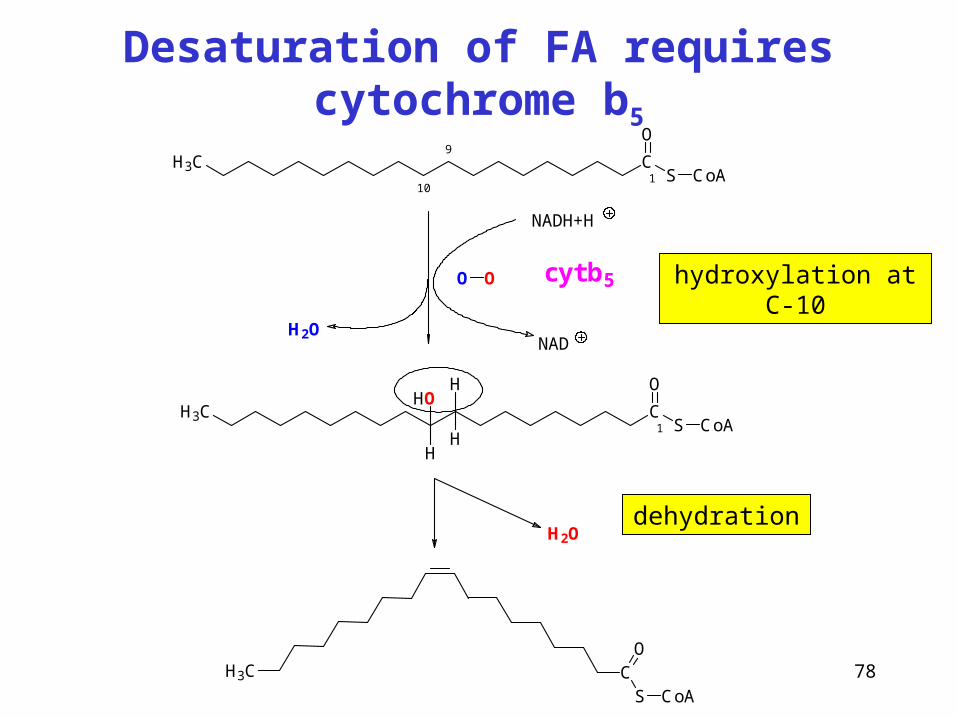
Desaturation of FA requires cytochrome b5
CS
O
CoA
H3C
NADH+H
NAD
cyt b5
H2O
H3C CS
O
CoA1
9
10
H3C CS
O
CoA
H
OH
H1
H
O O
H2O
hydroxylation at C-10
dehydration