Α -Synuclein occurs physiologically as a helically folded tetramer that resists aggregation Tim...
29
α-Synuclein occurs physiologically as a helically folded tetramer that resists aggregation Tim Bartels, Joanna G. Choi, & Dennis J. Selkoe Christopher O’Brien CHEM 645 Project Presentation 11/09/11
-
date post
22-Dec-2015 -
Category
Documents
-
view
215 -
download
0
Transcript of Α -Synuclein occurs physiologically as a helically folded tetramer that resists aggregation Tim...
α-Synuclein occurs physiologically as a helically folded tetramer that resists
aggregationTim Bartels, Joanna G. Choi, & Dennis J. Selkoe
Christopher O’BrienCHEM 645 Project Presentation
11/09/11
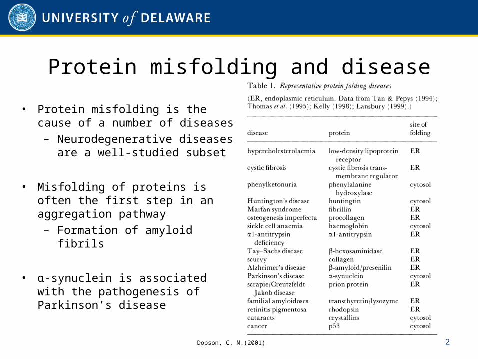
Protein misfolding and disease
• Protein misfolding is the cause of a number of diseases– Neurodegenerative diseases
are a well-studied subset
• Misfolding of proteins is often the first step in an aggregation pathway– Formation of amyloid fibrils
• α-synuclein is associated with the pathogenesis of Parkinson’s disease
2Dobson, C. M.(2001)
α-synuclein – role in disease
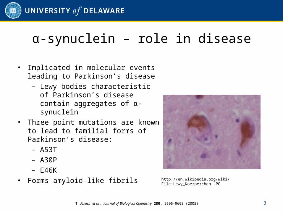
• Implicated in molecular events leading to Parkinson’s disease– Lewy bodies characteristic of
Parkinson’s disease contain aggregates of α-synuclein
• Three point mutations are known to lead to familial forms of Parkinson’s disease:– A53T– A30P– E46K
• Forms amyloid-like fibrils
3
http://en.wikipedia.org/wiki/File:Lewy_Koerperchen.JPG
T Ulmer et al. Journal of Biological Chemistry 280, 9595-9603 (2005)
α-synuclein – natively unfolded protein?
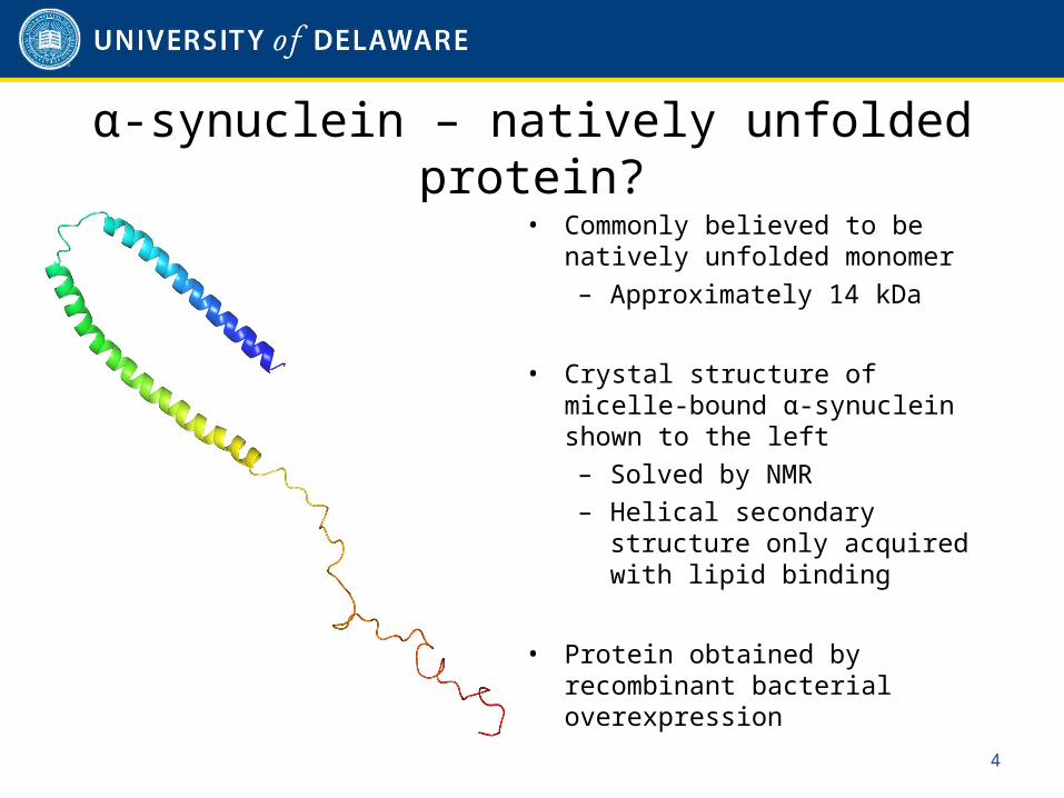
• Commonly believed to be natively unfolded monomer – Approximately 14 kDa
• Crystal structure of micelle-bound α-synuclein shown to the left– Solved by NMR– Helical secondary structure
only acquired with lipid binding
• Protein obtained by recombinant bacterial overexpression
4
New methods for determination of native state of α-synuclein
• Look at cell lines that endogenously express α-synuclein– M17D (dopaminergic human neuroblastoma)– HEK293– HeLa– COS-7
• Other sources of native α-synuclein– Frontal cortex of wild-type mice– Human red blood cells
• The above were examined using native gel electrophoresis
5
Native gel electrophoresis
• Non-denaturing conditions (no SDS)– Proteins expected to remain folded in native state
• Charged denaturing agent not used– Proteins may migrate in gel differently based on molecular
mass, charge in gel conditions, as well as hydrodynamic size and shape
• In this work, both BN-PAGE and CN-PAGE are used– CN-PAGE omits coomassie blue from sample preparation and
cathode buffer
6
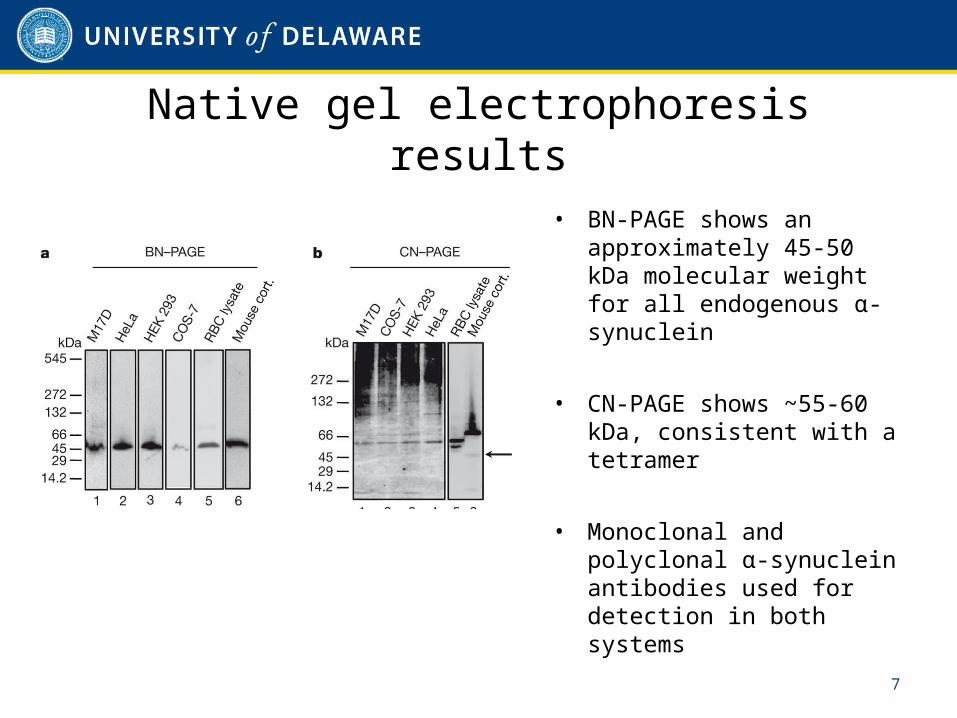
• BN-PAGE shows an approximately 45-50 kDa molecular weight for all endogenous α-synuclein
• CN-PAGE shows ~55-60 kDa, consistent with a tetramer
• Monoclonal and polyclonal α-synuclein antibodies used for detection in both systems
7
Native gel electrophoresis results
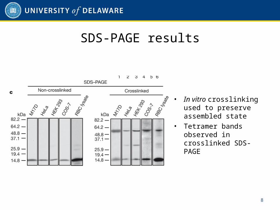
• In vitro crosslinking used to preserve assembled state
• Tetramer bands observed in crosslinked SDS-PAGE
8
SDS-PAGE results
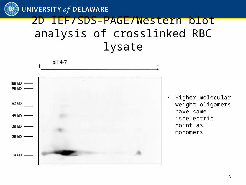
2D IEF/SDS-PAGE/Western blot analysis of crosslinked RBC lysate
• Higher molecular weight oligomers have same isoelectric point as monomers
9
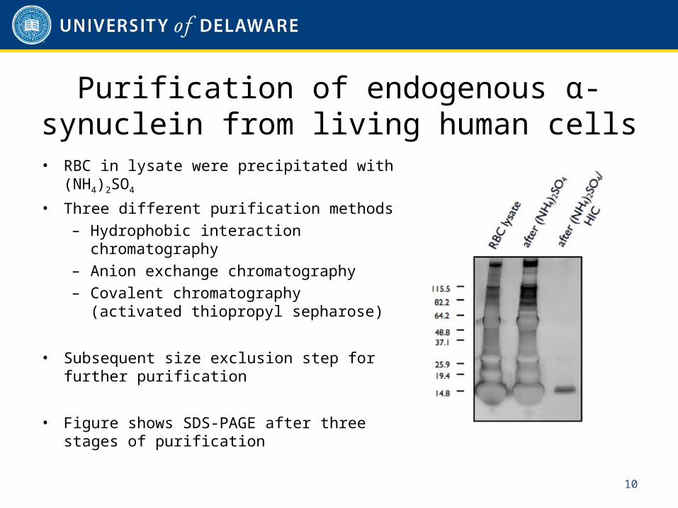
Purification of endogenous α-synuclein from living human cells
• RBC in lysate were precipitated with (NH4)2SO4
• Three different purification methods– Hydrophobic interaction
chromatography– Anion exchange chromatography– Covalent chromatography
(activated thiopropyl sepharose)
• Subsequent size exclusion step for further purification
• Figure shows SDS-PAGE after three stages of purification
10
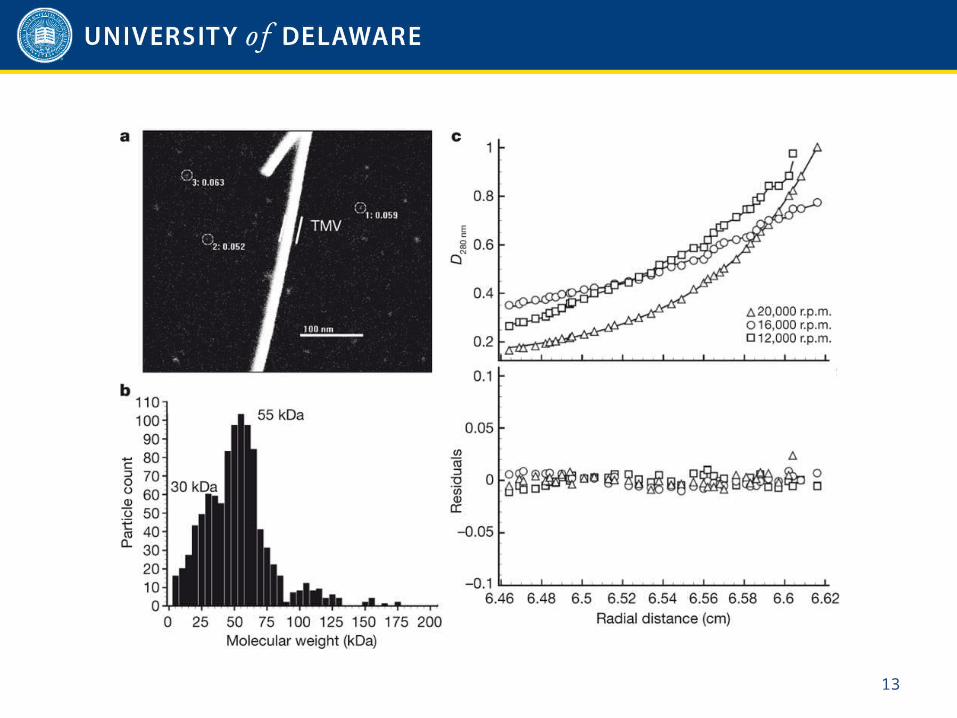
Scanning transmission electron microscopy (STEM)
• Type of TEM– Electrons pass through thin sample
• Electron beam focused onto single spot and scanned over sample• Tobacco mosaic virus (TMV) rods included during preparation as
internal sizing standard• Measurements carried out at the Brookhaven National Laboratory
11
Sedimentation equilibrium analytical ultracentrifugation (SE-AUC)
• Time-independent equilibrium concentration profile where sedimentation and diffusion are in equilibrium– Distributions are Boltzmann distributions– Insensitive to shape of molecules
• Results fitted using software SEDPHAT to calculate molecular weight– Determined from average of three concentrations– Error and standard deviation calculated using Monte-Carlo
simulations
12
13
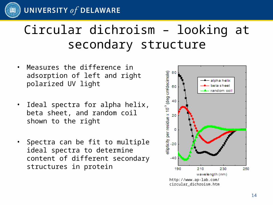
Circular dichroism – looking at secondary structure
• Measures the difference in adsorption of left and right polarized UV light
• Ideal spectra for alpha helix, beta sheet, and random coil shown to the right
• Spectra can be fit to multiple ideal spectra to determine content of different secondary structures in protein
14
http://www.ap-lab.com/circular_dichroism.htm
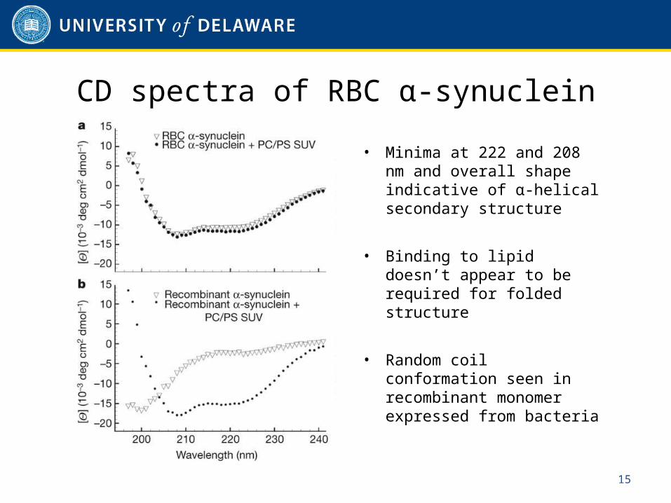
CD spectra of RBC α-synuclein
• Minima at 222 and 208 nm and overall shape indicative of α-helical secondary structure
• Binding to lipid doesn’t appear to be required for folded structure
• Random coil conformation seen in recombinant monomer expressed from bacteria
15
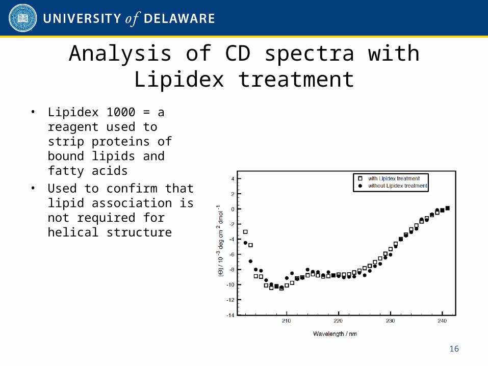
Analysis of CD spectra with Lipidex treatment
• Lipidex 1000 = a reagent used to strip proteins of bound lipids and fatty acids
• Used to confirm that lipid association is not required for helical structure
16
Quantitative elemental phosphate analysis
• Used to verify lack of a significant phospholipid presence• Involves incubation with ammonium molybdate(VI) tetrahydrate
and 10% ascorbic acid • Absorbance at 280nm measured and compared to calibration
curve of 7 standards
• Results indicated only 0.25mol phosphate per mole α-synuclein present– Significant presence of phospholipids on purified native α-
synuclein unlikely
17
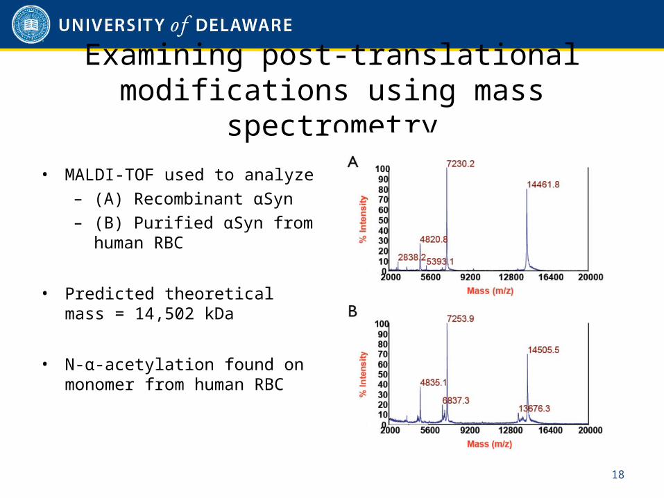
Examining post-translational modifications using mass spectrometry
• MALDI-TOF used to analyze– (A) Recombinant αSyn– (B) Purified αSyn from
human RBC
• Predicted theoretical mass = 14,502 kDa
• N-α-acetylation found on monomer from human RBC
18
Purification of stably transfected α-synuclein from human neuroblastoma
cells
• 3D5 cells, a M17D human neuroblastoma cell line was stably transfected to overexpress wild-type human α-synuclein– Endogenous α-synuclein from untransfected M17D cells also
analyzed• Cells were lysed by sonication• Similar purification performed as described for RBCs using anion
exchange chromatography
19
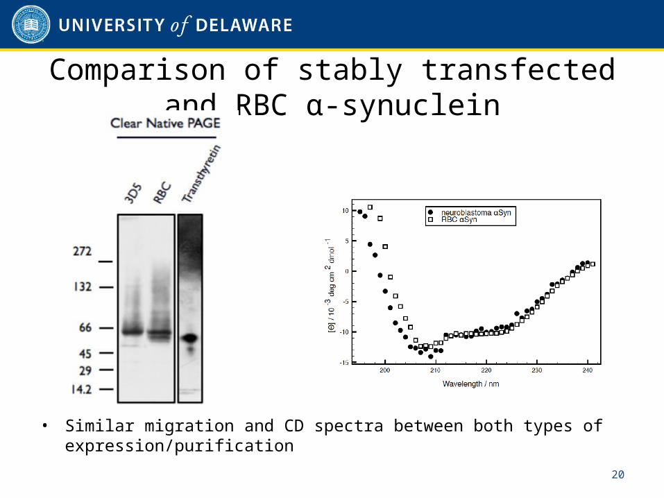
Comparison of stably transfected and RBC α-synuclein
• Similar migration and CD spectra between both types of expression/purification
20
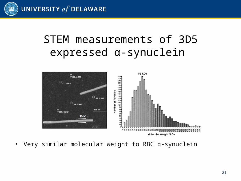
STEM measurements of 3D5 expressed α-synuclein
• Very similar molecular weight to RBC α-synuclein
21
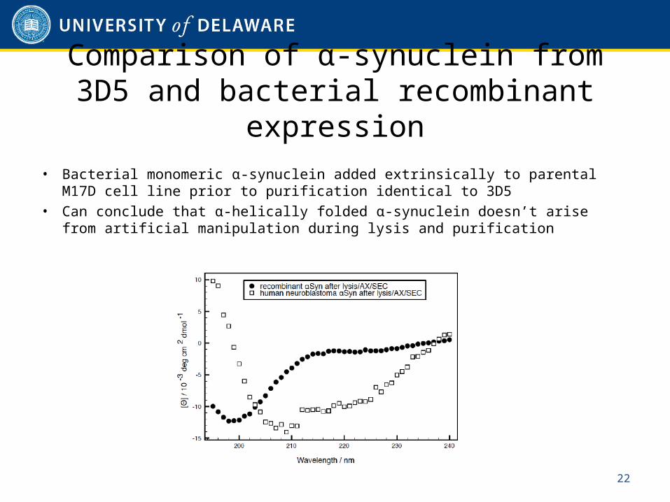
Comparison of α-synuclein from 3D5 and bacterial recombinant expression
• Bacterial monomeric α-synuclein added extrinsically to parental M17D cell line prior to purification identical to 3D5
• Can conclude that α-helically folded α-synuclein doesn’t arise from artificial manipulation during lysis and purification
22
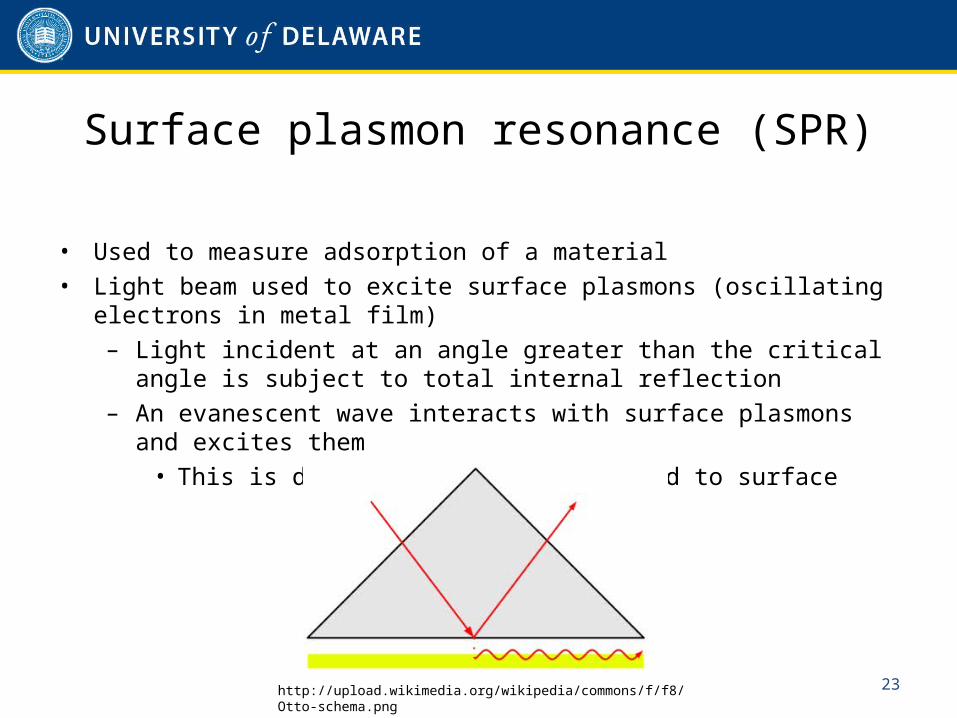
Surface plasmon resonance (SPR)
• Used to measure adsorption of a material• Light beam used to excite surface plasmons (oscillating electrons
in metal film)– Light incident at an angle greater than the critical angle is
subject to total internal reflection– An evanescent wave interacts with surface plasmons and
excites them• This is dependent on proteins bound to surface
23http://upload.wikimedia.org/wikipedia/commons/f/f8/Otto-schema.png
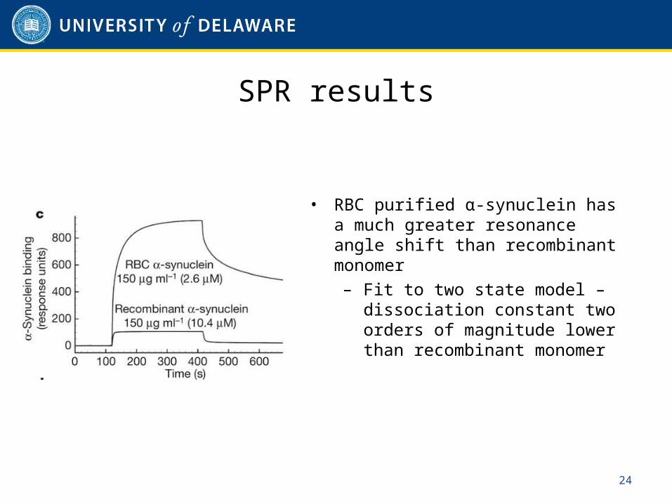
SPR results
• RBC purified α-synuclein has a much greater resonance angle shift than recombinant monomer– Fit to two state model –
dissociation constant two orders of magnitude lower than recombinant monomer
24
Thioflavin T (ThT) fluorescence
• Used to quantify formation of amyloid fibril growth– Binds to beta sheet-rich structures and displays enhanced
fluorescence with a red shift in its spectrum• Sample is incubated with ThT and fluorescence is measured from
460-550nm• Possibility of unwanted binding or spectroscopic change may
cause unreliable results in quantitative analysis
25
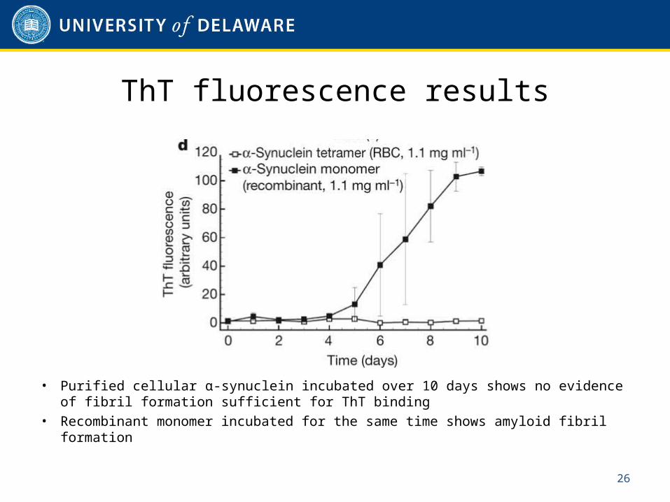
ThT fluorescence results
• Purified cellular α-synuclein incubated over 10 days shows no evidence of fibril formation sufficient for ThT binding
• Recombinant monomer incubated for the same time shows amyloid fibril formation
26
Conclusions
• Endogenous α-synuclein is natively an α-helical tetramer of about 58 kDa– This contradicts previously published work describing α-
synuclein as a natively unfolded 14 kDa monomer– Variable amounts of lower number oligomers also present in
cells• Based on the predominant native structure, it is likely that α-
synuclein tetramers undergo destabilization before non-native aggregation and fibrillar assemblies that are characteristic of Parkinson’s disease form– Consistent with the observation that partial unfolding of a
protein typically occurs before aggregation
27
Possible future directions
• Further investigation into the native tetramer conformation of α-synuclein can be performed now that a protocol for its expression and purification is published– Closer examination of its mechanism of destabilization and
aggregation• Therapeutic compounds that could kinetically stabilize native
tetramers could also be investigated– Could lead to novel treatments for Parkinson’s and similar
diseases
28

Questions?
29