ΝευροφυσιολογίακαιΑισθήσεις...
14
Biomedical Imaging & Applied Optics University of Cyprus Νευροφυσιολογία και Αισθήσεις Διάλεξη 8 The Eye (Το Μάτι) Biomedical Imaging and Applied Optics Laboratory 2 Introduction • Sensation ≠ Perception • Perception • Our understanding (conscious interpretation) of the physical world • An interpretation of the senses • Different from what is out there because • Our receptors detect limited number of existing energy forms • The information does not reach our brain unaltered. Some features are accentuated and some are suppressed • The brain interprets the information and often distorts it (“completes the picture” or “feels in the gaps”) to extract conclusions. • Interpretation is affected by cultural, social and personal experiences stored in our memory
Transcript of ΝευροφυσιολογίακαιΑισθήσεις...
Biomedical Imaging & Applied OpticsUniversity of Cyprus
Νευροφυσιολογία και Αισθήσεις
Διάλεξη 8
The Eye (Το Μάτι)
Biomedical Imaging and Applied Optics Laboratory22
Introduction
• Sensation ≠ Perception
• Perception• Our understanding (conscious
interpretation) of the physical world
• An interpretation of the senses
• Different from what is out there because
• Our receptors detect limited number of existing energy forms
• The information does not reach our brain unaltered. Some features are accentuated and some are suppressed
• The brain interprets the information and often distorts it (“completes the picture” or “feels in the gaps”) to extract conclusions.
• Interpretation is affected by cultural, social and personal experiences stored in our memory
Biomedical Imaging and Applied Optics Laboratory33
Properties of Light
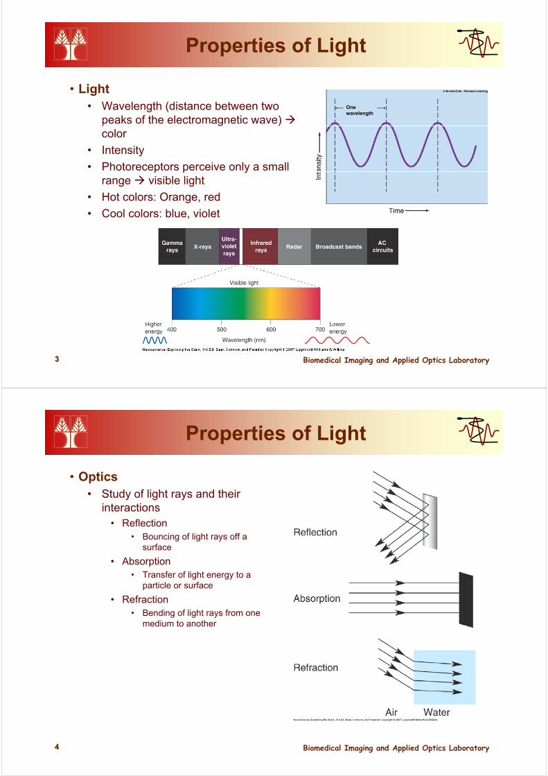
• Light• Wavelength (distance between two
peaks of the electromagnetic wave) color
• Intensity
• Photoreceptors perceive only a small range visible light
• Hot colors: Orange, red
• Cool colors: blue, violet
Onewavelength
Biomedical Imaging and Applied Optics Laboratory44
Properties of Light
• Optics• Study of light rays and their
interactions• Reflection
• Bouncing of light rays off a surface
• Absorption• Transfer of light energy to a
particle or surface
• Refraction• Bending of light rays from one
medium to another
Biomedical Imaging and Applied Optics Laboratory55
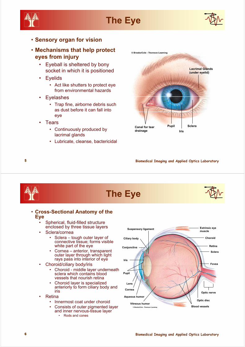
The Eye
• Sensory organ for vision
• Mechanisms that help protect eyes from injury
• Eyeball is sheltered by bony socket in which it is positioned
• Eyelids• Act like shutters to protect eye
from environmental hazards
• Eyelashes• Trap fine, airborne debris such
as dust before it can fall into eye
• Tears • Continuously produced by
lacrimal glands
• Lubricate, cleanse, bactericidal
Canal for teardrainage
Pupil
Iris
Sclera
Lacrimal Glands(under eyelid)
Biomedical Imaging and Applied Optics Laboratory66
The Eye
• Cross-Sectional Anatomy of the Eye
• Spherical, fluid-filled structure enclosed by three tissue layers
• Sclera/cornea• Sclera – tough outer layer of
connective tissue; forms visible white part of the eye
• Cornea – anterior, transparent outer layer through which light rays pass into interior of eye
• Choroid/ciliary body/iris• Choroid - middle layer underneath
sclera which contains blood vessels that nourish retina
• Choroid layer is specialized anteriorly to form ciliary body and iris
• Retina • Innermost coat under choroid• Consists of outer pigmented layer
and inner nervous-tissue layer• Rods and cones
Suspensory ligament
Ciliary body
Conjunctiva
Iris
Pupil
Lens
Cornea
Aqueous humor
Vitreous humorBlood vessels
Optic disc
Optic nerve
Fovea
Sclera
Retina
Choroid
Extrinsic eyemuscle
Biomedical Imaging and Applied Optics Laboratory77
The Eye
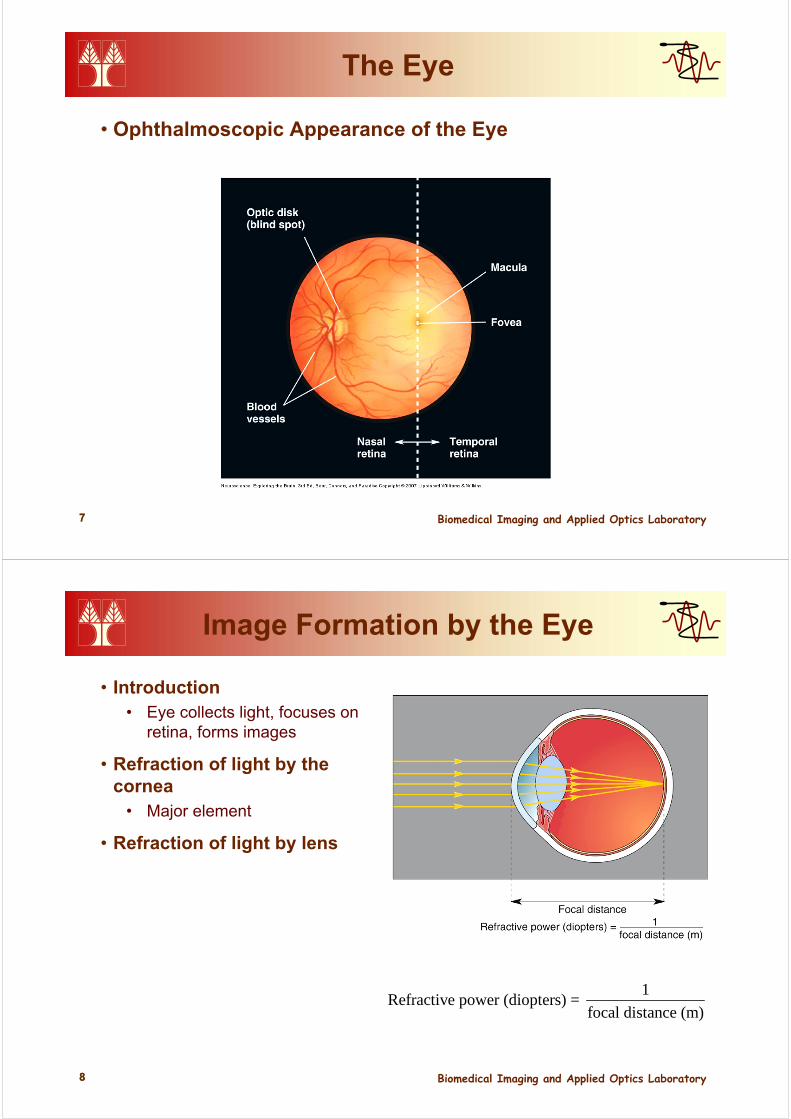
• Ophthalmoscopic Appearance of the Eye
Biomedical Imaging and Applied Optics Laboratory88
Image Formation by the Eye
• Introduction• Eye collects light, focuses on
retina, forms images
• Refraction of light by the cornea
• Major element
• Refraction of light by lens
1Refractive power (diopters) =
focal distance (m)
Biomedical Imaging and Applied Optics Laboratory99
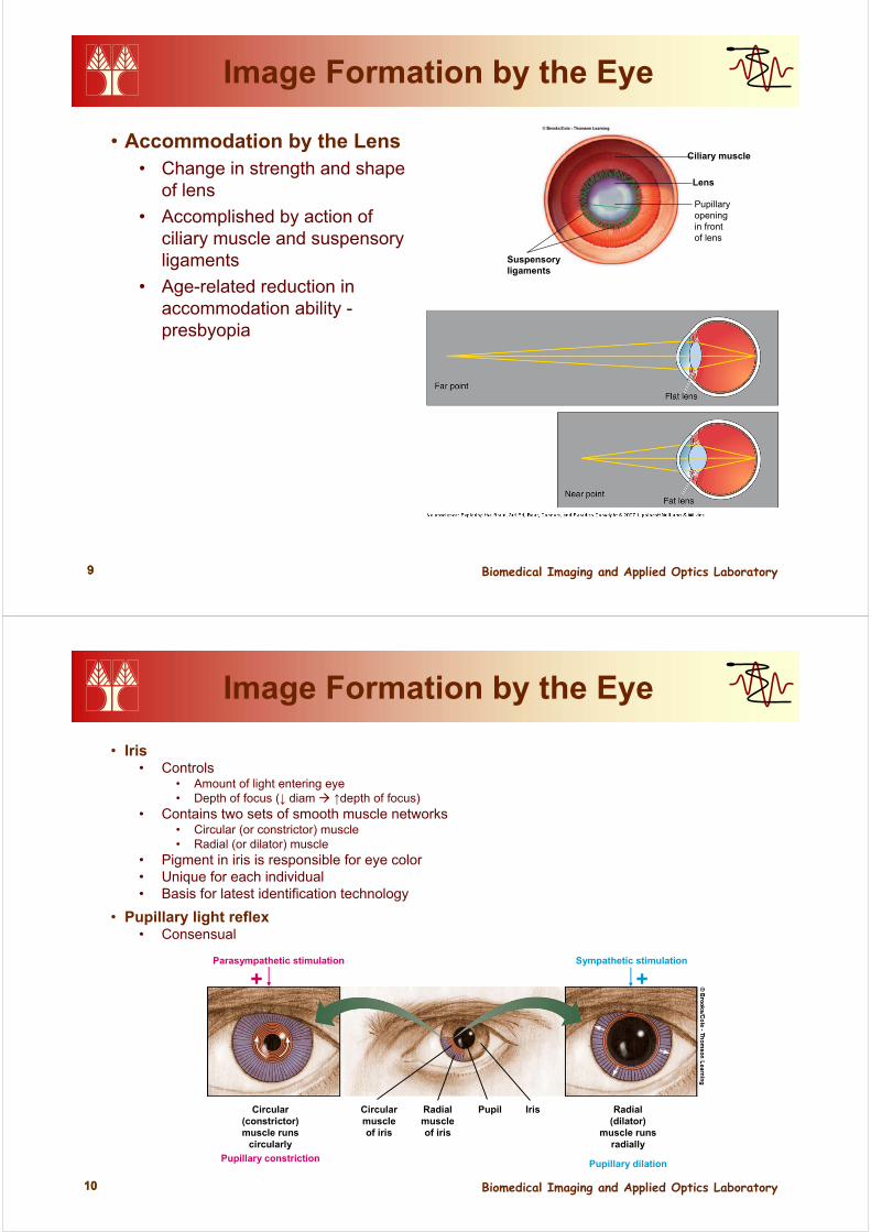
Image Formation by the Eye
• Accommodation by the Lens • Change in strength and shape
of lens
• Accomplished by action of ciliary muscle and suspensoryligaments
• Age-related reduction in accommodation ability -presbyopia
Suspensoryligaments
Ciliary muscle
Lens
Pupillaryopeningin frontof lens
Biomedical Imaging and Applied Optics Laboratory1010
Image Formation by the Eye
• Iris • Controls
• Amount of light entering eye• Depth of focus (↓ diam ↑depth of focus)
• Contains two sets of smooth muscle networks• Circular (or constrictor) muscle• Radial (or dilator) muscle
• Pigment in iris is responsible for eye color• Unique for each individual • Basis for latest identification technology
• Pupillary light reflex• Consensual
Parasympathetic stimulation
+
Pupillary constriction
Circular(constrictor)muscle runs
circularly
Radialmuscleof iris
Circularmuscleof iris
Pupil Iris Radial(dilator)
muscle runsradially
Pupillary dilation
Sympathetic stimulation
+
Biomedical Imaging and Applied Optics Laboratory1111
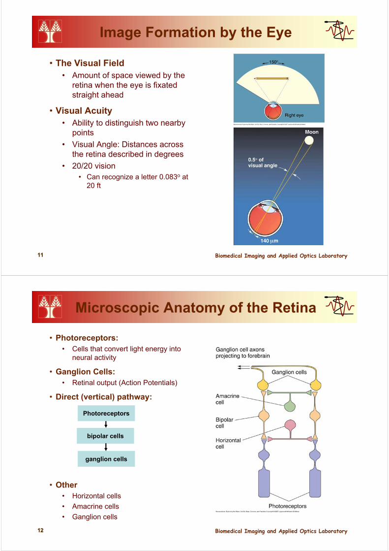
Image Formation by the Eye
• The Visual Field• Amount of space viewed by the
retina when the eye is fixated straight ahead
• Visual Acuity• Ability to distinguish two nearby
points
• Visual Angle: Distances across the retina described in degrees
• 20/20 vision• Can recognize a letter 0.083o at
20 ft
Biomedical Imaging and Applied Optics Laboratory1212
Microscopic Anatomy of the Retina
• Photoreceptors:• Cells that convert light energy into
neural activity
• Ganglion Cells:• Retinal output (Action Potentials)
• Direct (vertical) pathway:
• Other• Horizontal cells
• Amacrine cells
• Ganglion cells
bipolar cells
ganglion cells
Photoreceptors
Biomedical Imaging and Applied Optics Laboratory1313
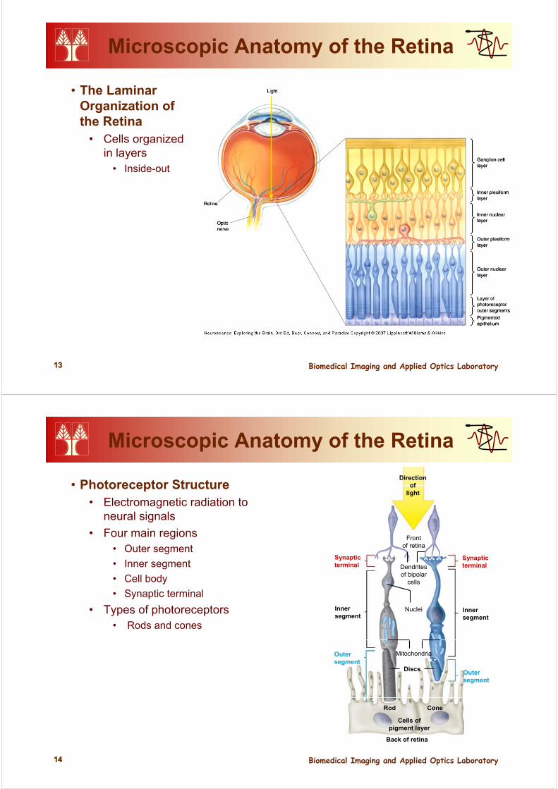
Microscopic Anatomy of the Retina
• The Laminar Organization of the Retina
• Cells organized in layers
• Inside-out
Biomedical Imaging and Applied Optics Laboratory1414
Microscopic Anatomy of the Retina
• Photoreceptor Structure• Electromagnetic radiation to
neural signals
• Four main regions• Outer segment
• Inner segment
• Cell body
• Synaptic terminal
• Types of photoreceptors• Rods and cones
Back of retina
Outersegment
Outersegment
Innersegment
Synapticterminal
Synapticterminal
Innersegment
Directionof
light
Cells ofpigment layer
ConeRod
Discs
Mitochondria
Nuclei
Dendritesof bipolar
cells
Frontof retina
Biomedical Imaging and Applied Optics Laboratory1515
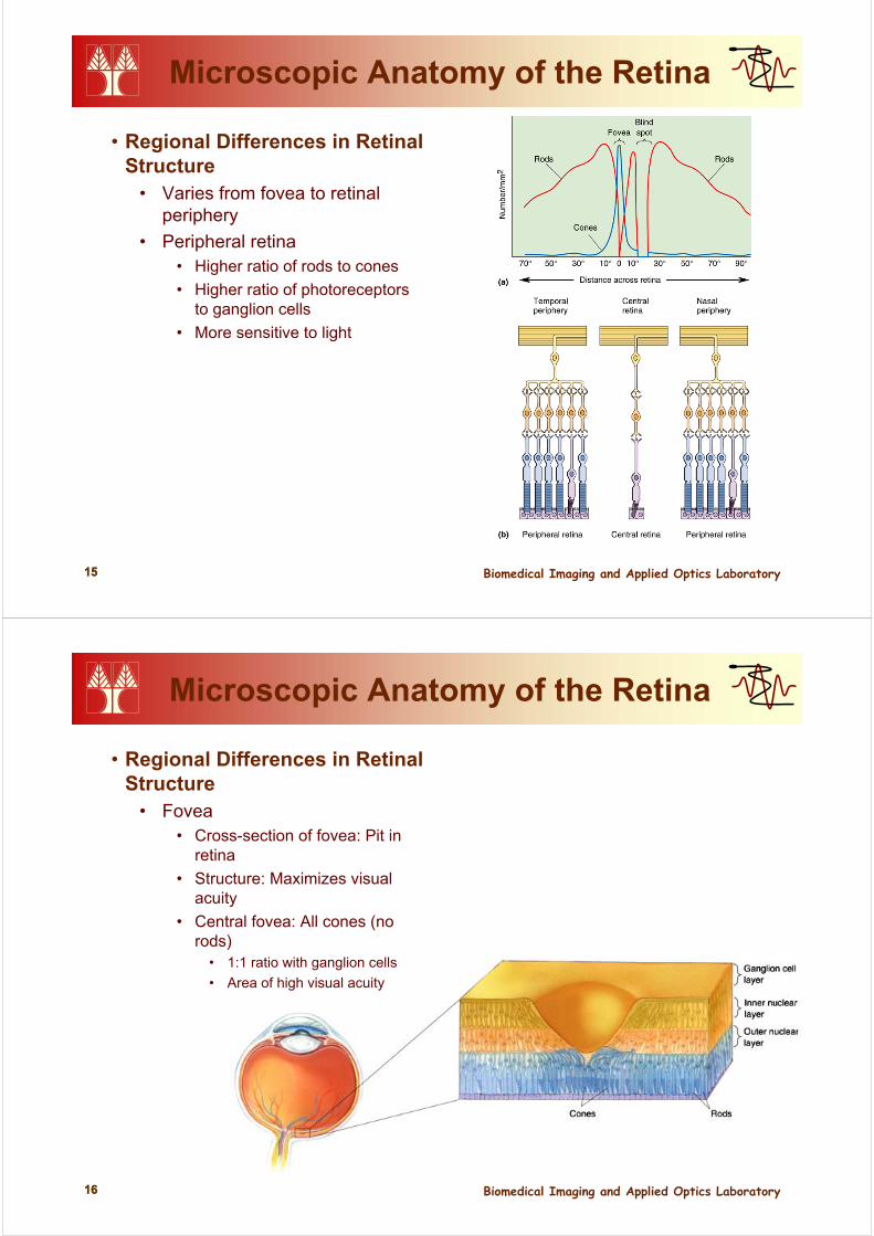
Microscopic Anatomy of the Retina
• Regional Differences in Retinal Structure
• Varies from fovea to retinal periphery
• Peripheral retina• Higher ratio of rods to cones
• Higher ratio of photoreceptors to ganglion cells
• More sensitive to light
Biomedical Imaging and Applied Optics Laboratory1616
Microscopic Anatomy of the Retina
• Regional Differences in Retinal Structure
• Fovea• Cross-section of fovea: Pit in
retina
• Structure: Maximizes visual acuity
• Central fovea: All cones (no rods)
• 1:1 ratio with ganglion cells
• Area of high visual acuity
Biomedical Imaging and Applied Optics Laboratory1717
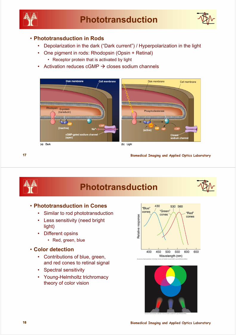
Phototransduction
• Phototransduction in Rods• Depolarization in the dark (“Dark current”) / Hyperpolarization in the light
• One pigment in rods: Rhodopsin (Opsin + Retinal)• Receptor protein that is activated by light
• Activation reduces cGMP closes sodium channels
Biomedical Imaging and Applied Optics Laboratory1818
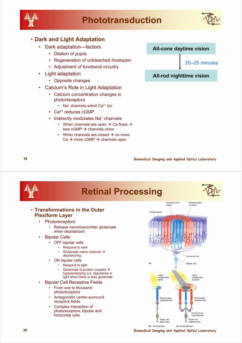
Phototransduction
• Phototransduction in Cones• Similar to rod phototransduction
• Less sensitivity (need bright light)
• Different opsins• Red, green, blue
• Color detection• Contributions of blue, green,
and red cones to retinal signal
• Spectral sensitivity
• Young-Helmholtz trichromacytheory of color vision
Biomedical Imaging and Applied Optics Laboratory1919
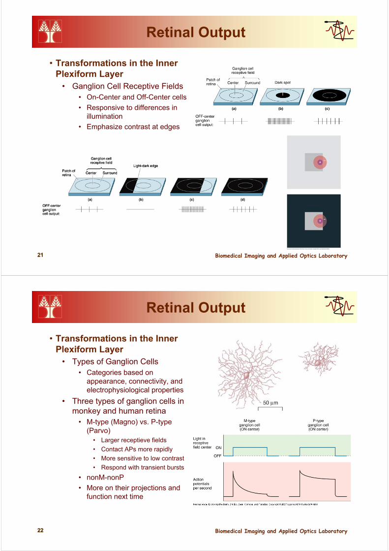
Phototransduction
• Dark and Light Adaptation• Dark adaptation—factors
• Dilation of pupils
• Regeneration of unbleached rhodopsin
• Adjustment of functional circuitry
• Light adaptation• Opposite changes
• Calcium’s Role in Light Adaptation• Calcium concentration changes in
photorteceptors• Na+ channels admit Ca2+ too
• Ca2+ reduces cGMP
• Indirectly modulates Na+ channels• When channels are open Ca flows
less cGMP channels close
• When channels are closed no more Ca more cGMP channels open
All-cone daytime vision
All-rod nighttime vision
20–25 minutes
Biomedical Imaging and Applied Optics Laboratory2020
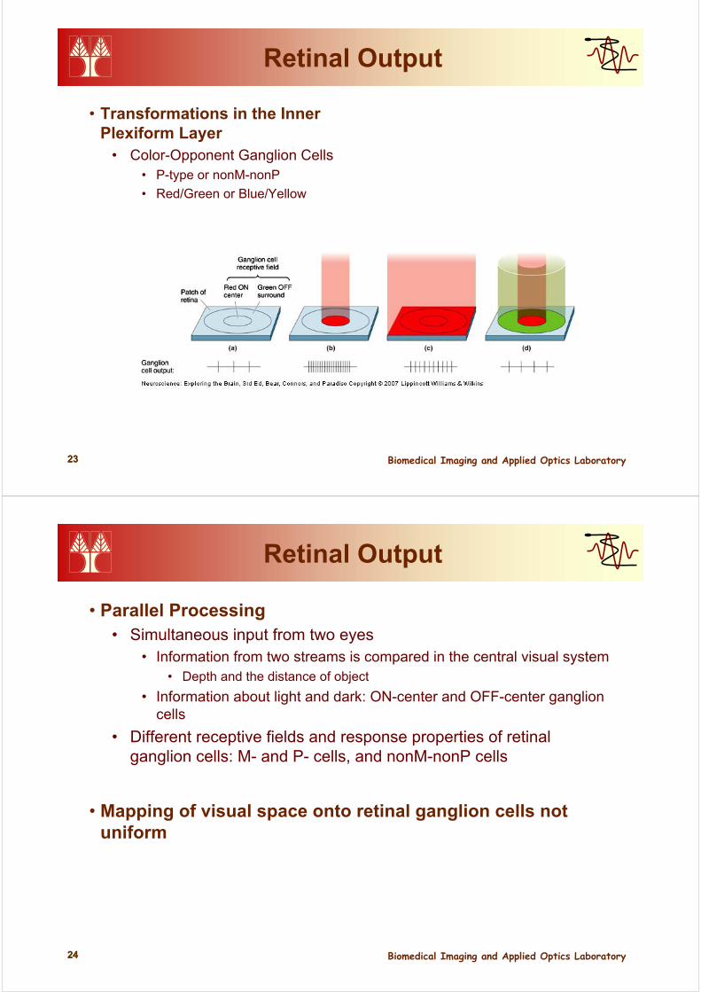
Retinal Processing
• Transformations in the Outer Plexiform Layer
• Photoreceptors• Release neurotransmitter glutamate
when depolarized
• Bipolar Cells• OFF bipolar cells
• Respond to dark• Glutamate cation channel
depolarizing
• ON bipolar cells• Respond to light• Glutamate G-protein coupled
hyperpolarizing (i.e. depolarize in light when there is less glutamate
• Bipolar Cell Receptive Fields• From one to thousand
photoreceptors• Antagonistic center-surround
receptive fields• Complex interaction of
photoreceptors, bipolar and horizontal cells
Biomedical Imaging and Applied Optics Laboratory2121
Retinal Output
• Transformations in the Inner Plexiform Layer
• Ganglion Cell Receptive Fields • On-Center and Off-Center cells
• Responsive to differences in illumination
• Emphasize contrast at edges
Biomedical Imaging and Applied Optics Laboratory2222
Retinal Output
• Transformations in the Inner Plexiform Layer
• Types of Ganglion Cells• Categories based on
appearance, connectivity, and electrophysiological properties
• Three types of ganglion cells in monkey and human retina
• M-type (Magno) vs. P-type (Parvo)
• Larger receptieve fields
• Contact APs more rapidly
• More sensitive to low contrast
• Respond with transient bursts
• nonM-nonP
• More on their projections and function next time
Biomedical Imaging and Applied Optics Laboratory2323
Retinal Output
• Transformations in the Inner Plexiform Layer
• Color-Opponent Ganglion Cells• P-type or nonM-nonP
• Red/Green or Blue/Yellow
Biomedical Imaging and Applied Optics Laboratory2424
Retinal Output
• Parallel Processing• Simultaneous input from two eyes
• Information from two streams is compared in the central visual system• Depth and the distance of object
• Information about light and dark: ON-center and OFF-center ganglion cells
• Different receptive fields and response properties of retinal ganglion cells: M- and P- cells, and nonM-nonP cells
• Mapping of visual space onto retinal ganglion cells not uniform
Biomedical Imaging and Applied Optics Laboratory2525
Επόμενη Διάλεξη …
Διάλεξη 9
The Central Visual System
(Το Κεντρικό Οπτικό Σύστημα)
Biomedical Imaging and Applied Optics Laboratory2626
Vision
Far source Near source
No accommodations Accommodations
Normal eye (Emmetropia)
Far source focused on retina withoutaccommodation
Near source focused on retina withaccommodation
No accommodations No accommodations
Nearsightedness (Myopia)–Eyeball too long or lens too strong
1. Uncorrected
Far source focused in front ofretina (where retina would be ineye of normal length)
No accommodations Accommodations
Focus
1.Imageout offocus
2.
Near source focused on retinawith accommodations
2. Corrected with concave lens,which diverges light rays beforethey reach the eye
Far source focused on retinawithout accommodations
Near source focused on retinawith accommodations
Biomedical Imaging and Applied Optics Laboratory2727
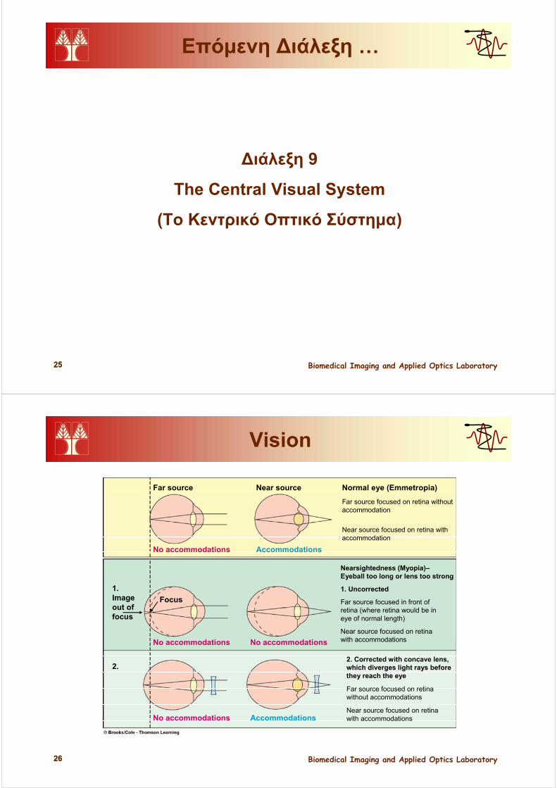
Vision
Far source Near source
No accommodations Accommodations
Normal eye (Emmetropia)
Far source focused on retina withoutaccommodation
Near source focused on retina withaccommodation
Farsightedness (Hyperopia)–Eyeball too short or lens too weak
1. Uncorrected
Far source focused on retinawith accommodations
No accommodations Accommodations
1.
2.
Near source focused behindretina even with accommodations
2. Corrected with cortex lens,which converges light rays before they reach the eye
Far source focused on retinawithout accommodations
Near source focused on retinawith accommodations
Accommodations Accommodations
Imageout offocus
Focus
Biomedical Imaging and Applied Optics Laboratory2828
Vision