β-Galactosidase from Lactobacillus acidophilus isolated...
5
Indian Journal of Biotechnology Vol 5, April 2006, pp 184-188 β-Galactosidase from Lactobacillus acidophilus isolated from fermented ragi (Eleusine coracana) S K Akolkar, A D Sajgure and S S Lele* Food and Fermentation Technology Department, Mumbai University Institute of Chemical Technology Matunga, Mumbai 400 019, India Received 2 August 2004; revised 6 May 2005; accepted 5 July 2005 Lactobacillus acidophilus was isolated from fermented millet, Eleusine coracana. It was characterized using several biochemical tests. This strain was found to be homofermentative, slime forming and β-galactosidase producer. Recovery and characterization of β-galactosidase were studied at laboratory scale. Since the enzyme was intracellular various cell lysis methods were studied to achieve maximum enzyme release from cell fragments. Homogenization at a pressure of 2000 psig two passes showed an enzyme release of 2050 U/mL and was found the best method for cell lysis. The cell extract was purified using ultrafiltration and gel permeation chromatography. Specific activity and fold purification of β-galactosidase was found to be 568.61 and 21.2, respectively. Kinetic parameters were determined using ONPG (o-nitrophenyl galactopyranoside) as a substrate. Michaeli’s Menten equation was found to fit the reaction of ONPG using β-galactosidase from L. acidophilus. Vmax and Km were calculated from the Lineweaver Burk plot. Vmax was found to be 4.94 min -1 and Km to be 0.11 mM. Using gel permeation chromatography, the molecular weight was found to be in the range of 450-500 kDa. Keywords: Lactobacillus acidophilus, Eleusine coracana, β-galactosidase IPC Code: Int. Cl. 8 C12N 19/38; C12R1:23 Introduction Ragi (Eleusine coracana Gaertn.) is a millet grown in India as a subsistence crop and is predominantly used by rural population for consumption 1,2 . Fermentation of ragi provides an environment conducive for the growth of lactic acid bacteria (LAB) along with yeast. These microorganisms ferment ragi at different stages. One of the LAB viz. lactobacilli are known to possess health-promoting attributes including anti-carcinogenic 3 . Lactobacilli are indispensable in the manufacture and preservation of many fermented food products and are involved in the protection of young animals against intestinal infections. Lactobacilli are reported to show diverse behaviour and, hence, attracted attention as a potential source of new applications as well as for enzyme production 3,4 . In this work, Lactobacillus was isolated from fermented ragi. The strain was found to be homofermentative, slime forming and produced β- galactosidase or Lactase (β-D-galactohydrolase EC 3.2.1.23) in significant quantities. β-Galactosidase is a hydrolytic enzyme, which hydrolyses the terminal non-reducing β-D-galactoside residues to form β- galactosides to glucose and galactose, which are simple monosaccharides. β-galactosidase is one of the important commercial enzymes having several applications in food and pharmaceutical industry. Lactose free milk for consumption by lactose intolerant persons is produced using this enzyme 5-7 . This enzyme is not commercially manufactured in India. Hence this work was undertaken. The present work focuses on the recovery and characterization of β-galactosidase that was obtained by laboratory scale fermentation of Lactobacillus acidophilus isolated from fermented ragi. Since the enzyme is intracellular, various cell lysis methods were tried to achieve maximum enzyme release from the cells. Molecular weight determination was done by gel permeation chromatography. Michaeli’s Menten equation and Lineweaver-Burk plot were applied to study the kinetic parameters of β- galactosidase. The β-galactosidase obtained in the present study was compared with a commercial sample of β-galactosidase from Kluveromyces fragilis. _______________ *Author for correspondence: Tel: 91-22-24145616, ext-345. Fax: 91-22-2414 5614 E-mail: [email protected]
Transcript of β-Galactosidase from Lactobacillus acidophilus isolated...
Indian Journal of Biotechnology Vol 5, April 2006, pp 184-188
β-Galactosidase from Lactobacillus acidophilus isolated from fermented ragi (Eleusine coracana)
S K Akolkar, A D Sajgure and S S Lele* Food and Fermentation Technology Department, Mumbai University Institute of Chemical Technology
Matunga, Mumbai 400 019, India
Received 2 August 2004; revised 6 May 2005; accepted 5 July 2005
Lactobacillus acidophilus was isolated from fermented millet, Eleusine coracana. It was characterized using several biochemical tests. This strain was found to be homofermentative, slime forming and β-galactosidase producer. Recovery and characterization of β-galactosidase were studied at laboratory scale. Since the enzyme was intracellular various cell lysis methods were studied to achieve maximum enzyme release from cell fragments. Homogenization at a pressure of 2000 psig two passes showed an enzyme release of 2050 U/mL and was found the best method for cell lysis. The cell extract was purified using ultrafiltration and gel permeation chromatography. Specific activity and fold purification of β-galactosidase was found to be 568.61 and 21.2, respectively. Kinetic parameters were determined using ONPG (o-nitrophenyl galactopyranoside) as a substrate. Michaeli’s Menten equation was found to fit the reaction of ONPG using β-galactosidase from L. acidophilus. Vmax and Km were calculated from the Lineweaver Burk plot. Vmax was found to be 4.94 min-1 and Km to be 0.11 mM. Using gel permeation chromatography, the molecular weight was found to be in the range of 450-500 kDa.
Keywords: Lactobacillus acidophilus, Eleusine coracana, β-galactosidase IPC Code: Int. Cl.8 C12N 19/38; C12R1:23
Introduction Ragi (Eleusine coracana Gaertn.) is a millet grown
in India as a subsistence crop and is predominantly used by rural population for consumption1,2. Fermentation of ragi provides an environment conducive for the growth of lactic acid bacteria (LAB) along with yeast. These microorganisms ferment ragi at different stages. One of the LAB viz. lactobacilli are known to possess health-promoting attributes including anti-carcinogenic3. Lactobacilli are indispensable in the manufacture and preservation of many fermented food products and are involved in the protection of young animals against intestinal infections. Lactobacilli are reported to show diverse behaviour and, hence, attracted attention as a potential source of new applications as well as for enzyme production3,4. In this work, Lactobacillus was isolated from fermented ragi. The strain was found to be homofermentative, slime forming and produced β-galactosidase or Lactase (β-D-galactohydrolase EC 3.2.1.23) in significant quantities. β-Galactosidase is a
hydrolytic enzyme, which hydrolyses the terminal non-reducing β-D-galactoside residues to form β-galactosides to glucose and galactose, which are simple monosaccharides. β-galactosidase is one of the important commercial enzymes having several applications in food and pharmaceutical industry. Lactose free milk for consumption by lactose intolerant persons is produced using this enzyme5-7. This enzyme is not commercially manufactured in India. Hence this work was undertaken.
The present work focuses on the recovery and characterization of β-galactosidase that was obtained by laboratory scale fermentation of Lactobacillus acidophilus isolated from fermented ragi. Since the enzyme is intracellular, various cell lysis methods were tried to achieve maximum enzyme release from the cells. Molecular weight determination was done by gel permeation chromatography. Michaeli’s Menten equation and Lineweaver-Burk plot were applied to study the kinetic parameters of β-galactosidase. The β-galactosidase obtained in the present study was compared with a commercial sample of β-galactosidase from Kluveromyces fragilis.
_______________ *Author for correspondence: Tel: 91-22-24145616, ext-345. Fax: 91-22-2414 5614 E-mail: [email protected]
AKOLKAR et al: β-GALACTOSIDASE FROM L. ACIDOPHILUS ISOLATED FROM FERMENTED RAGI
185
Materials and Methods Lactobacillus acidophilus isolated from fermented
ragi was initially characterized using standard biochemical tests8. Performing Gram staining whereas macroscopic identification involved catalase, nitrate reductase test, and fermentation of different sugars did microscopic identification. Peptone water fermentation test media: [1l contained, 10 g Peptone and 5 g NaCl] and 0.1 mL Andrade’s indicator 1.0 N NaOH added to 0.5% solution of acid fuschin until the colour just becomes yellow] were used for characterization. Fermentation for β-galactosidase Production
The strain isolated from fermented ragi was characterized as Lactobacillus acidophilus and used as inoculum for β-galactosidase production. Fermentation media was inoculated with 3% inoculum. The culture was maintained using MRS medium. Fermentation for β-galactosidase production was carried out in the modified (optimized) MRS medium (unpublished work) in 250 mL static flasks with 100 mL media at 37°C for 24 h. Determination of Enzyme Activity and Protein
Assay of β-galactosidase activity was carried out using o-nitrophenyl galacto-pyranoside (ONPG). ONPG solution (0.2 mL) was mixed with 1 mL enzyme solution of proper dilution. Reaction mixture was incubated at 50°C for 10 min. 1 mL of 1 M Na2CO3 solution was added to stop the reaction. Absorbance was noted at wavelength of 420 nm. One unit enzyme activity (EU) is defined as μmoles of o-nitrophenol formed per mL per min. Folin Lowry method was used for protein estimation using BSA as standard. Methods of Cell Lysis for Enzyme Recovery
Cells were subjected to cell lysis by ultrasonication, trituration, homogenization, detergent treatment, osmotic shock, and thermal shock. Samples were assayed for β-galactosidase activity. Optimization of pressure applied by high pressure homogenizer was done by lysing the cells at pressure 2000, 3000, 4000, 5000 psig and by giving single and double pass at each pressure. Enzyme Purification and Characterization
Purification of β-galactosidase was carried out using ultrafiltration and gel column chromatography. 25 mL of intracellular extract was subjected to
ultrafiltration with a 50 kDa cut off membrane and assayed for protein content and β-galactosidase activity. Retentate was used as a sample for gel column chromatography using Sephadex G200 (Column height 15 cm, Phosphate buffer I.P. pH-7.0. Flow rate 0.09 mL/min). Determination of Kinetic Constants
ONPG was used as a substrate in the concentration range of 0.6-5.4 μmoles/mL for determination of kinetic constants. Michaeli’s Menten constants Km and Vmax were calculated by using Lineweaver- Burk plot. Instruments
Spectrophotometric analysis was performed using Hitachi U 2001. Centrifugation was carried out using Beckmann Cold centrifuge. Branson sonifier was used for sonication. An Amicon stirred cell model (Amicon Corporation Scientific Systems Divisions, USA) with cellulose acetate membranes was used for ultrafiltration. Chromatographic experiments were carried out using peristaltic pump Alitea-xv, Sweden. Fraction collector ISCO- Retriever-II USA, online Perkin-Elmer UV-visible spectrophotometer (Model-lambda-3B) was used for chromatographic experiments. Results and Discussion
Four lactobacilli strains were isolated from fermented ragi and screened for the production of β-galactosidase. One of the strains was found to be β-galactosidase producing, It was characterized as Lactobacillus acidophilus by performing various biochemical tests8 and the results are shown in Table 1. Fermentation of this culture resulted in quantitative production of β-galactosidase. It is known that β-galactosidase is an intracellular enzyme, primarily bound to cell walls9. However, small quantities may be leached out in the broth. In order to assess enzyme activities obtained as extracellular, intracellular and cell bound fraction, preliminary separation experiments were conducted. Whole cell suspension showed activity in the range of 800-1000U/mL, whereas extracellular broth showed practically no activity. However, activity in the range of 250-300U/mL was observed in the intracellular extract. Hence, cell fragments were tested for the enzyme activity. These showed an activity of 1500-2000U/mL. It can be seen from these preliminary experiments that the cell fragments showed maximum
INDIAN J BIOTECHNOL, APRIL 2006
186
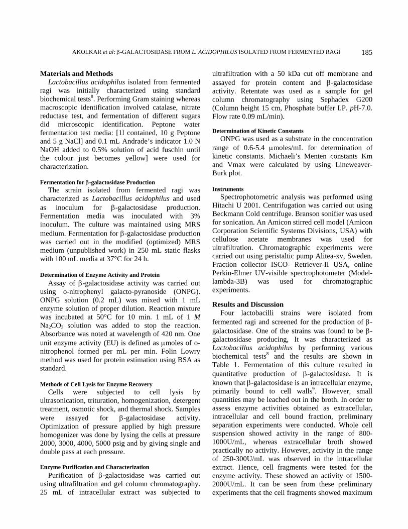
β-galactosidase activity confirming the intracellular-cell wall bound nature of the enzyme. In order to maximize the recovery of β-galactosidase, various cell lysis methods were used.
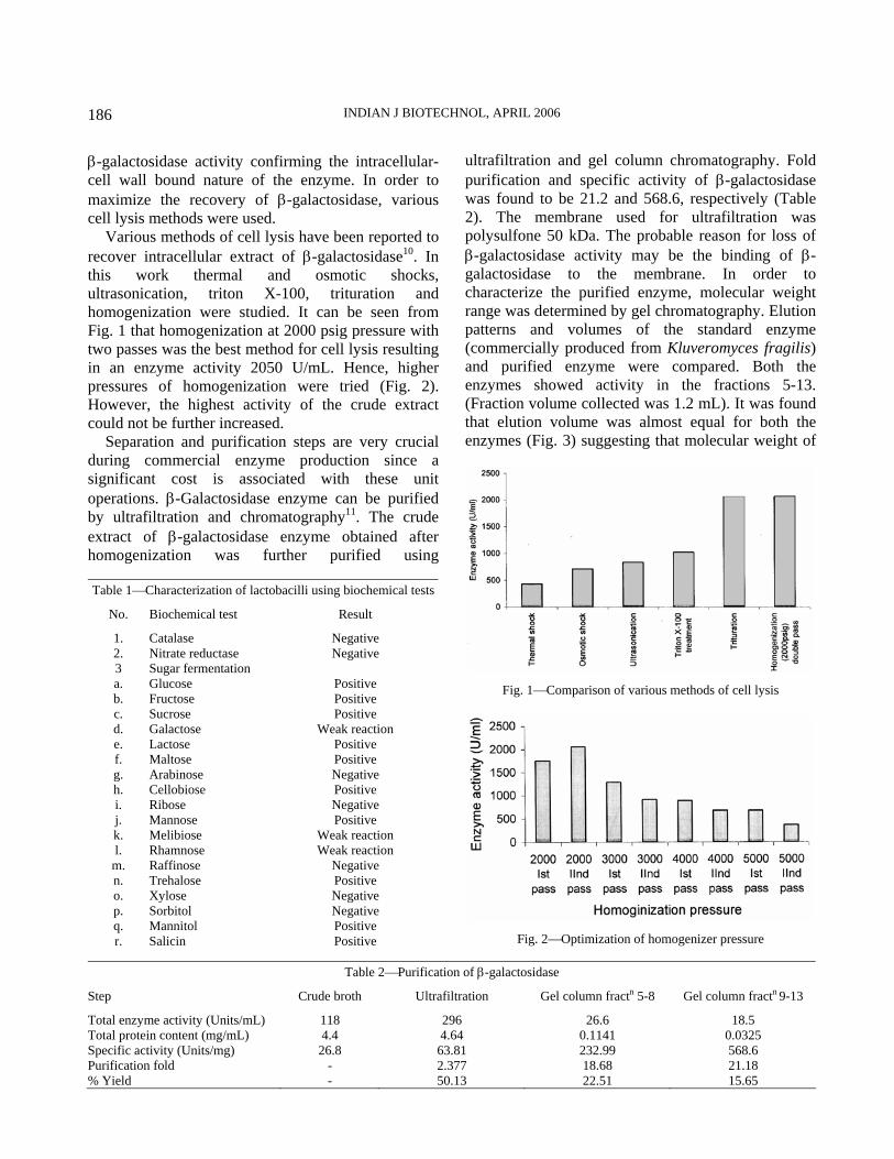
Various methods of cell lysis have been reported to recover intracellular extract of β-galactosidase10. In this work thermal and osmotic shocks, ultrasonication, triton X-100, trituration and homogenization were studied. It can be seen from Fig. 1 that homogenization at 2000 psig pressure with two passes was the best method for cell lysis resulting in an enzyme activity 2050 U/mL. Hence, higher pressures of homogenization were tried (Fig. 2). However, the highest activity of the crude extract could not be further increased.
Separation and purification steps are very crucial during commercial enzyme production since a significant cost is associated with these unit operations. β-Galactosidase enzyme can be purified by ultrafiltration and chromatography11. The crude extract of β-galactosidase enzyme obtained after homogenization was further purified using
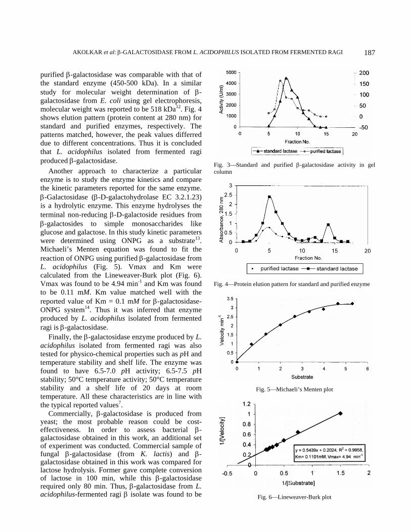
ultrafiltration and gel column chromatography. Fold purification and specific activity of β-galactosidase was found to be 21.2 and 568.6, respectively (Table 2). The membrane used for ultrafiltration was polysulfone 50 kDa. The probable reason for loss of β-galactosidase activity may be the binding of β-galactosidase to the membrane. In order to characterize the purified enzyme, molecular weight range was determined by gel chromatography. Elution patterns and volumes of the standard enzyme (commercially produced from Kluveromyces fragilis) and purified enzyme were compared. Both the enzymes showed activity in the fractions 5-13. (Fraction volume collected was 1.2 mL). It was found that elution volume was almost equal for both the enzymes (Fig. 3) suggesting that molecular weight of
Table 2⎯Purification of β-galactosidase
Step Crude broth Ultrafiltration Gel column fractn 5-8 Gel column fractn 9-13
Total enzyme activity (Units/mL) 118 296 26.6 18.5 Total protein content (mg/mL) 4.4 4.64 0.1141 0.0325 Specific activity (Units/mg) 26.8 63.81 232.99 568.6 Purification fold - 2.377 18.68 21.18 % Yield - 50.13 22.51 15.65
Table 1⎯Characterization of lactobacilli using biochemical tests
No. Biochemical test Result
1. Catalase Negative 2. Nitrate reductase Negative 3 Sugar fermentation a. Glucose Positive b. Fructose Positive c. Sucrose Positive d. Galactose Weak reaction e. Lactose Positive f. Maltose Positive g. Arabinose Negative h. Cellobiose Positive i. Ribose Negative j. Mannose Positive k. Melibiose Weak reaction l. Rhamnose Weak reaction m. Raffinose Negative n. Trehalose Positive o. Xylose Negative p. Sorbitol Negative q. Mannitol Positive r. Salicin Positive
Fig. 1⎯Comparison of various methods of cell lysis
Fig. 2⎯Optimization of homogenizer pressure
AKOLKAR et al: β-GALACTOSIDASE FROM L. ACIDOPHILUS ISOLATED FROM FERMENTED RAGI
187
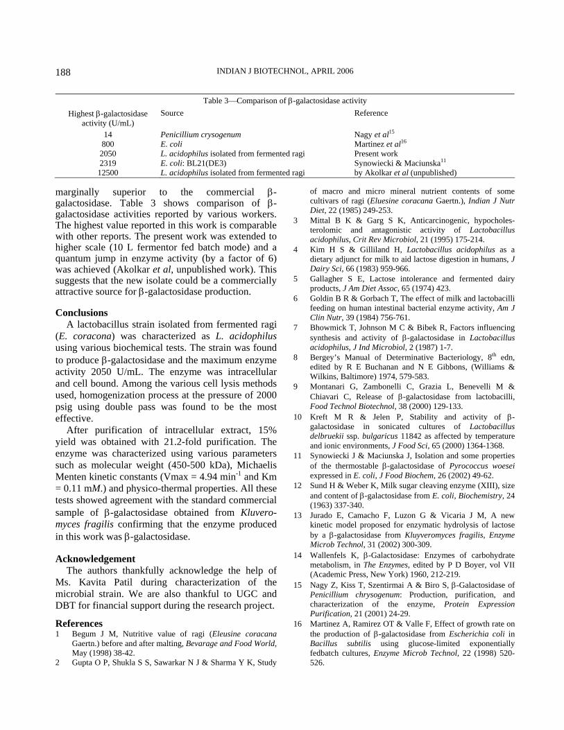
purified β-galactosidase was comparable with that of the standard enzyme (450-500 kDa). In a similar study for molecular weight determination of β-galactosidase from E. coli using gel electrophoresis, molecular weight was reported to be 518 kDa12. Fig. 4 shows elution pattern (protein content at 280 nm) for standard and purified enzymes, respectively. The patterns matched, however, the peak values differred due to different concentrations. Thus it is concluded that L. acidophilus isolated from fermented ragi produced β-galactosidase.
Another approach to characterize a particular enzyme is to study the enzyme kinetics and compare the kinetic parameters reported for the same enzyme. β-Galactosidase (β-D-galactohydrolase EC 3.2.1.23) is a hydrolytic enzyme. This enzyme hydrolyses the terminal non-reducing β-D-galactoside residues from β-galactosides to simple monosaccharides like glucose and galactose. In this study kinetic parameters were determined using ONPG as a substrate13. Michaeli’s Menten equation was found to fit the reaction of ONPG using purified β-galactosidase from L. acidophilus (Fig. 5). Vmax and Km were calculated from the Lineweaver-Burk plot (Fig. 6). Vmax was found to be 4.94 min-1 and Km was found to be 0.11 mM. Km value matched well with the reported value of Km = 0.1 mM for β-galactosidase-ONPG system14. Thus it was inferred that enzyme produced by L. acidophilus isolated from fermented ragi is β-galactosidase.
Finally, the β-galactosidase enzyme produced by L. acidophilus isolated from fermented ragi was also tested for physico-chemical properties such as pH and temperature stability and shelf life. The enzyme was found to have 6.5-7.0 pH activity; 6.5-7.5 pH stability; 50°C temperature activity; 50°C temperature stability and a shelf life of 20 days at room temperature. All these characteristics are in line with the typical reported values7.
Commercially, β-galactosidase is produced from yeast; the most probable reason could be cost-effectiveness. In order to assess bacterial β-galactosidase obtained in this work, an additional set of experiment was conducted. Commercial sample of fungal β-galactosidase (from K. lactis) and β-galactosidase obtained in this work was compared for lactose hydrolysis. Former gave complete conversion of lactose in 100 min, while this β-galactosidase required only 80 min. Thus, β-galactosidase from L. acidophilus-fermented ragi β isolate was found to be
Fig. 3⎯Standard and purified β-galactosidase activity in gel column
Fig. 4⎯Protein elution pattern for standard and purified enzyme
Fig. 5⎯Michaeli’s Menten plot
Fig. 6⎯Lineweaver-Burk plot
INDIAN J BIOTECHNOL, APRIL 2006
188
marginally superior to the commercial β-galactosidase. Table 3 shows comparison of β-galactosidase activities reported by various workers. The highest value reported in this work is comparable with other reports. The present work was extended to higher scale (10 L fermentor fed batch mode) and a quantum jump in enzyme activity (by a factor of 6) was achieved (Akolkar et al, unpublished work). This suggests that the new isolate could be a commercially attractive source for β-galactosidase production. Conclusions
A lactobacillus strain isolated from fermented ragi (E. coracona) was characterized as L. acidophilus using various biochemical tests. The strain was found to produce β-galactosidase and the maximum enzyme activity 2050 U/mL. The enzyme was intracellular and cell bound. Among the various cell lysis methods used, homogenization process at the pressure of 2000 psig using double pass was found to be the most effective.
After purification of intracellular extract, 15% yield was obtained with 21.2-fold purification. The enzyme was characterized using various parameters such as molecular weight (450-500 kDa), Michaelis Menten kinetic constants (Vmax = 4.94 min-1 and Km = 0.11 mM.) and physico-thermal properties. All these tests showed agreement with the standard commercial sample of β-galactosidase obtained from Kluvero-myces fragilis confirming that the enzyme produced in this work was β-galactosidase. Acknowledgement
The authors thankfully acknowledge the help of Ms. Kavita Patil during characterization of the microbial strain. We are also thankful to UGC and DBT for financial support during the research project.
References 1 Begum J M, Nutritive value of ragi (Eleusine coracana
Gaertn.) before and after malting, Bevarage and Food World, May (1998) 38-42.
2 Gupta O P, Shukla S S, Sawarkar N J & Sharma Y K, Study
of macro and micro mineral nutrient contents of some cultivars of ragi (Eluesine coracana Gaertn.), Indian J Nutr Diet, 22 (1985) 249-253.
3 Mittal B K & Garg S K, Anticarcinogenic, hypocholes-terolomic and antagonistic activity of Lactobacillus acidophilus, Crit Rev Microbiol, 21 (1995) 175-214.
4 Kim H S & Gilliland H, Lactobacillus acidophilus as a dietary adjunct for milk to aid lactose digestion in humans, J Dairy Sci, 66 (1983) 959-966.
5 Gallagher S E, Lactose intolerance and fermented dairy products, J Am Diet Assoc, 65 (1974) 423.
6 Goldin B R & Gorbach T, The effect of milk and lactobacilli feeding on human intestinal bacterial enzyme activity, Am J Clin Nutr, 39 (1984) 756-761.
7 Bhowmick T, Johnson M C & Bibek R, Factors influencing synthesis and activity of β-galactosidase in Lactobacillus acidophilus, J Ind Microbiol, 2 (1987) 1-7.
8 Bergey’s Manual of Determinative Bacteriology, 8th edn, edited by R E Buchanan and N E Gibbons, (Williams & Wilkins, Baltimore) 1974, 579-583.
9 Montanari G, Zambonelli C, Grazia L, Benevelli M & Chiavari C, Release of β-galactosidase from lactobacilli, Food Technol Biotechnol, 38 (2000) 129-133.
10 Kreft M R & Jelen P, Stability and activity of β-galactosidase in sonicated cultures of Lactobacillus delbruekii ssp. bulgaricus 11842 as affected by temperature and ionic environments, J Food Sci, 65 (2000) 1364-1368.
11 Synowiecki J & Maciunska J, Isolation and some properties of the thermostable β-galactosidase of Pyrococcus woesei expressed in E. coli, J Food Biochem, 26 (2002) 49-62.
12 Sund H & Weber K, Milk sugar cleaving enzyme (XIII), size and content of β-galactosidase from E. coli, Biochemistry, 24 (1963) 337-340.
13 Jurado E, Camacho F, Luzon G & Vicaria J M, A new kinetic model proposed for enzymatic hydrolysis of lactose by a β-galactosidase from Kluyveromyces fragilis, Enzyme Microb Technol, 31 (2002) 300-309.
14 Wallenfels K, β-Galactosidase: Enzymes of carbohydrate metabolism, in The Enzymes, edited by P D Boyer, vol VII (Academic Press, New York) 1960, 212-219.
15 Nagy Z, Kiss T, Szentirmai A & Biro S, β-Galactosidase of Penicillium chrysogenum: Production, purification, and characterization of the enzyme, Protein Expression Purification, 21 (2001) 24-29.
16 Martinez A, Ramirez OT & Valle F, Effect of growth rate on the production of β-galactosidase from Escherichia coli in Bacillus subtilis using glucose-limited exponentially fedbatch cultures, Enzyme Microb Technol, 22 (1998) 520-526.
Table 3⎯Comparison of β-galactosidase activity Highest β-galactosidase
activity (U/mL) Source Reference
14 Penicillium crysogenum Nagy et al15 800 E. coli Martinez et al16 2050 L. acidophilus isolated from fermented ragi Present work 2319 E. coli: BL21(DE3) Synowiecki & Maciunska11
12500 L. acidophilus isolated from fermented ragi by Akolkar et al (unpublished)