β-Defensin 1 Is Prominent in the Liver and Induced During ...
Acta Histochem. Cytochem. 46 (1): 25–34, 2013doi:10.1267/ahc.12033
© 2013 The Japan Society of Histochemistry and Cytochemistry
AHCActa Histochemica et Cytochemica0044-59911347-5800Japan Society of Histochemistry and CytochemistryTokyo, JapanAHC1203310.1267/ahc.12033Regular Article
Induction of β-Defensin Expression by Porphyromonas gingivalis-Infected Human
Gingival Graft Transplanted in nu/nu Mouse Subdermis
Masahiro To1, Yohei Kamata2, Juri Saruta1, Tomoko Shimizu1,2, Takenori Sato3,
Yusuke Kondo1, Takashi Hayashi1, Nobushiro Hamada3 and Keiichi Tsukinoki1
1Department of Environmental Pathology and Research Institute of Salivary Gland Health Medicine, Kanagawa Dental College
Postgraduate School, 82 Inaoka-cho, Yokosuka, Kanagawa 238–8580, Japan, 2Department of Comprehensive Dentistry,
Yokohama Clinic, Kanagawa Dental College, 3–31–6 Tsuruya-cho, Kanagawa, Yokohama, Kanagawa 221–0835, Japan and 3Department of Infection Control, Division of Microbiology, Kanagawa Dental College Postgraduate School, 82 Inaoka-cho,
Yokosuka, Kanagawa 238–8580, Japan
Correspondence to: Keiichi Tsukinoki, D.D.S., Ph.D., Department of
Environmental Pathology, Kanagawa Dental College Postgraduate
School, 82 Inaoka-cho, Yokosuka, Kanagawa 238–8580, Japan.
E-mail: [email protected]
00 Received September 18, 2012; accepted November 20, 2012; published online January 11, 2013
© 2013 The Japan Society of Histochemistry andIt is important to understand the onset of periodontal disease in terms of bacterial infection
and host factors. Host-bacteria interactions can be elicited in human cultured cells and animal
models, but these models provide only limited biological information about human host
reactions against bacterial attacks. Development of an in vivo model using human gingival
tissue is needed. We established an in vivo model using nu/nu mice and evaluated host
defense following bacterial infection in human gingiva. Human gingival samples were collected
from periodontitis patients and transplanted in nu/nu mouse subdermis. After 2 weeks, human
characteristics were confirmed by positive immunohistochemical reactions for human-specific
markers. We used this model to investigate human β-defensin-2 (hBD-2), an antimicrobial
peptide that contributes to initial defense against bacterial invasion. Using real-time
polymerase chain reaction, in situ hybridization, and immunohistochemistry, we investigated
whether hBD-2 expression was induced in human gingiva as a response to Porphyromonas
gingivalis as a periodontal pathogen. Two hours after infection with bacteria, we detected
increased expression of hBD-2 mRNA, which was localized in the epithelium of human
gingiva. Using our in vivo model, we concluded that increased hBD-2 may play an important
role in early defense from bacterial infection in human gingival epithelium.
Key words: human β-defensin-2 (hBD-2), human gingiva, in vivo model, periodontal disease, nude mice
I. Introduction
Periodontal diseases are highly prevalent and include
gingivitis and chronic periodontitis [16]. Chronic perio-
dontitis is caused by pathogenic oral bacteria such as
Porphyromonas gingivalis. P. gingivalis is a Gram-
negative, black-pigmented anaerobe that induces chronic
inflammation in periodontal tissues such as the gingiva,
cementum, periodontal ligament, and alveolar bone [4, 15].
Gingival tissue of stratified squamous epithelium is directly
attacked by bacteria. This tissue is not only a barrier but
is also part of the innate immune system, because host
responses also cause bacteria exclusion [6, 21]. Research
into human host responses is important in the analysis of
periodontal diseases [21]. So far, these biological responses
have been evaluated in human cultured cells and various
animal models [15, 24]. However, the information obtained
from such in vitro models about the host response is
insufficient. In addition, experiments using animals some-
times show differences in the reaction between humans and
animals. To solve these problems, immunodeficient nu/nu

To et al.26
mice or scid/scid mice have been widely used as in vivo
models with human characteristics because various human
tissues, including oral mucosa, can be transplanted into
these mice [8, 10, 22, 31]. In oral mucosa, transplanted
grafts frequently show cystic formation [10]. The native
condition of human mucosal tissue was attenuated by this
phenomenon. Tsukinoki et al. [27] established a transplan-
tation technique for normal human oral mucosa that does
not exhibit cystic changes using immunodeficient nu/scid
mice, which were derived from a cross of nu/nu mice and
scid/scid mice [9, 26]. However, the transplantation of
gingival tissue from periodontal diseases has yet to be
established in common immunodeficient nu/nu or scid/scid
mice. The development of such a model will be useful
for investigating relationships between host responses of
gingival squamous epithelium and pathogen attacks.
Expression of antimicrobial peptides as a host response
has been detected in all human epithelium, including oral
epithelium, and is an important part of epithelial function
[3]. These antimicrobial peptides have a broad spectrum of
activity against both Gram-positive and Gram-negative
bacteria as well as against yeast and viruses [3]. One type
of antimicrobial peptide that plays an important role in
host defense is defensins [1, 6]. Defensins are small,
cationic antimicrobial peptides with a structure that contains
disulfide bonds. They are classified into two subfamilies,
α- and β-defensins, and the spectrum of antibacterial activ-
ity varies individually [6]. Among these defensins, human
β-defensin 2 (hBD-2) is expressed in epithelium, including
skin, lung, vagina, and oral mucosa, and exhibits potent
antimicrobial activity against Gram-negative bacteria and
fungi [1]. hBD-2 is typically produced by epithelial tissues
after stimulation with microorganisms and proinflammatory
mediators [4, 12], and contributes to initial defense in innate
immune response. hBD-2 is expressed in the gingival epi-
thelium in periodontal diseases in human biopsy samples
(in vivo) and in vitro studies [2, 4, 24, 28]. However, the
relationship between induction of hBD-2 in gingival epithe-
lium and bacterial infection by P. gingivalis is not well
known. In addition, recent data have demonstrated that
hBD-2 is upregulated in the inflamed mucosa of patients
with ulcerative colitis [30]. These studies indicated that
hBD-2 is regulated by Toll-like receptor (TLR) 2 and TLR4
signaling in human intestinal epithelial cells.
The purpose of this study was to establish an in vivo
experimental model for investigation of the host-bacteria
interaction using conventional immunodeficient mice. We
examined the expression profile of hBD-2 as a host
response to P. gingivalis infection in human gingiva using
this novel model. We also investigated whether TLR
expression is associated with hBD-2 in gingival epithelium.
II. Materials and Methods
Animals
Six- to 8-week-old male BALB/c scid/scid and nu/nu
mice, each weighing 20–25 g (CLEA Japan, Inc., Tokyo,
Japan), were used in this study. Mice were kept in a specific
pathogen-free room and had free access to autoclaved food
and sterile water.
Human tissue
After obtaining informed consent, gingival tissues
were collected from 25 patients (10 men and 15 women;
mean age, 58±14.5 years) with chronic marginal perio-
dontitis during periodontal surgery at Yokohama Clinic,
Kanagawa Dental College (Yokohama, Japan). The Com-
mittee of Ethics of the Kanagawa Dental College approved
the present study (Approval No. 104-20090803). Patients
received preoperative initial preparation. Tissue was col-
lected from the external marginal epithelium in all patients.
Grafts from 8 cases were transplanted into scid/scid mice,
and grafts from 17 cases were transplanted into nu/nu mice.
Transplantation methods
We modified the transplantation method of Tsukinoki
et al. [27]. The collected tissue was immediately transferred
to a mouse on a clean bench as follows. The tissues were
immersed in tetracycline hydrochloride, and the size was
adjusted to 3×2 mm before transplantation. In addition,
subepithelial connective tissue was removed. Each mouse
was anesthetized with sodium pentobarbital (50 mg/kg,
i.p.), and a flap was formed on the dorsal epidermis. The
human gingival tissue was sutured into the flap so that the
connective tissue side of the graft was placed onto the
murine subepithelial tissue (Fig. 1). The flap was returned
to its original position and sutured. The surgical area was
covered with Tegaderm® (Sumitomo 3M, Ltd., Tokyo,
Japan), and the mice were kept in a specific pathogen-free
room for 2 weeks.
Bacterial conditions and infection methods
The bacterial strain used was P. gingivalis, which was
obtained from the American Type Culture Collection 33277
(ATCC 33277; Manassas, VA, USA). P. gingivalis strains
were grown at 37°C for 18 hr in brain heart infusion broth
(BHI; Difco Laboratories, Sparks, MD, USA) containing 5
mg/ml yeast extract, 5 μg/ml hemin, and 10 μg/ml vitamin
K1 in an anaerobic chamber with an atmosphere of 85%
N2, 10% H2, and 5% CO2. In the present study, the trans-
planted gingival tissues were exposed to 7.1×10⁸ cells/ml
P. gingivalis ATCC 33277 in BHI 1.0 ml medium by
subcutaneous injection for 2 hr (Fig. 1). Control trans-
planted grafts were treated with BHI medium. The experi-
mental protocol used in this study was reviewed and
approved by the Committee of Ethics on Animal Experi-
ments of Kanagawa Dental College and was carried out in
accordance with the Guidelines for Animal Experimentation
of Kanagawa Dental College.
Histological and immunohistochemical analysis
Collected or transplanted gingival tissues were fixed in
4% paraformaldehyde for 24 hr and embedded in paraffin
or OCT compound (Sakura Finetek Japan Co., Ltd., Tokyo,

β-Defensin Expression in Human Gingiva 27
Japan). To confirm morphological changes in gingival
tissue, hematoxylin and eosin staining was performed
according to standard routine techniques.
Subsequently, immunohistochemical analysis was per-
formed using a Histofine staining kit (Nichirei Biosciences
Inc., Tokyo, Japan), according to the instruction manual. To
activate antigen for involucrin, Ki-67, and vimentin, slides
were autoclaved at 121°C for 5 min in 10 mM citrate buffer
(pH 6.0). Sections were then preincubated in 3% H2O2 for
15 min. After washing in phosphate-buffered saline, sec-
tions were incubated with anti-human involucrin polyclonal
antibody (1:100; Harbor Bio-Products, Norwood, MA,
USA), anti-human Ki-67 monoclonal antibody (MIB-1,
1:50; Dako Denmark A/S, Glostrup, Denmark), anti-human
vimentin monoclonal antibody (Nichirei), or anti-human
CD34 monoclonal antibody (Nichirei) for 1 hr at room
temperature. To confirm that human-specific antibodies did
not react with murine tissue, we also investigated immuno-
reactivity in normal murine skin.
To observe localization of hBD-2, TLR2, and TLR4
in the grafts, the tissue was embedded in OCT compound.
Four-micrometer-thick frozen tissue sections were incu-
bated with anti-human hBD-2 polyclonal antibody (1:200;
Peptide Institute, Inc., Osaka, Japan), anti-human TLR2
polyclonal antibody (TL2.1, 1:200; Santa Cruz Bio-
technology, Inc., Santa Cruz, CA, USA), or anti-human
TLR4 polyclonal antibody (H-80, 1:200; Santa Cruz Bio-
technology) overnight at 4°C.
Subsequently, sections were treated with horseradish
peroxidase-labeled secondary antibody (Nichirei) for 30
min at room temperature. The chromogen used was 3,3'-
diaminobenzidine-tetrahydrochloride containing 0.003%
H2O2 in phosphate-buffered saline. The sections were
counterstained with hematoxylin or methyl blue. As a
negative control, phosphate-buffered saline was used
instead of primary antibody.
RNA isolation and real-time polymerase chain reaction
(PCR)
Total RNA was isolated from gingival tissue using
the ISOGEN reagent (Nippon Gene Co., Ltd., Toyama,
Japan), according to the manufacturer’s instructions. The
quality of RNA was judged from the pattern of ribosomal
RNA after electrophoresis through a 1.5% agarose gel
containing ethidium bromide and visualization by UV illu-
mination. RNA concentrations were determined with a
NanoDrop 1000 (Thermo Fisher Scientific Inc., Waltham,
MA, USA). cDNA was synthesized from total RNA using
a first-strand cDNA synthesis kit (Roche Diagnostics Ltd.,
Lewes, UK) according to the manufacturer’s instructions.
Real-time PCR was performed using a LightCycler system
(Roche) according to the manufacturer’s instructions. The
primer sequences used to amplify hBD-2 were 5'-TCT TCT
CGT TCC TCT TCA TA-3' (forward) and 5'-TGT TTA
TAC CTT CTA GGG CA-3' (reverse; PCR product: 127
bp) as designed and synthesized by Nihon Gene Research
Laboratory (Sendai, Miyagi, Japan). Other target sequences
were amplified using LightCycler Primer sets (Search-LC,
Heidelberg, Germany). The PCR reactions were 95°C for
10 min followed by 35 cycles of 95°C for 10 sec, 58°C for
20 sec, and 72°C for 10 sec for hBD-2, and 95°C for 10
min followed by 35 cycles of 95°C for 10 sec, 68°C for 20
sec, and 72°C for 16 sec for TLR2 and TLR4. β-actin was
used as a housekeeping gene (95°C for 10 min followed by
40 cycles of 95°C for 10 sec, 62°C for 10 sec, and 72°C for
10 sec). Gene expression is given as the ratio of the copy
number of individual gene mRNA to β-actin mRNA for
each sample.
In situ hybridization
Complementary RNA (cRNA) probes were produced
by in vitro transcription of linearized pGEM-T Easy Vector
(Promega Co., Madison, WI, USA). The two probes were
chemically synthesized 120-mers of sense and antisense
oligonucleotides specific for hBD-2 (nucleotides 56–175
and 217–336 of the hBD-2 coding sequence, Accession
number: NM_004942). Digoxigenin (DIG)-11-UTP-labeled
single-stranded cRNA probes for hBD-2 were prepared
using a DIG labeling kit SP6/T7 (Roche) according to
Fig. 1. Experimental designs used for investigating the host
response against P. gingivalis in human gingiva. Schema for trans-
plantation, infection experiments, and sampling. Human gingival
samples were collected from periodontitis patients and transplanted
into immunodeficient mice. After 2 weeks, the grafts were infected
with P. gingivalis by subcutaneous injection in the mice. The grafts
exposed to P. gingivalis for 2 hr were analyzed in mRNA and
protein levels.

To et al.28
the manufacturer’s instructions. Procedures for in situ
hybridization were as described previously [11, 20]. Four-
micrometer-thick paraffin sections were digested with 1 μg/
ml proteinase K for 20 min at 37°C. Hybridization was
performed at 50°C for 17 hr using DIG-11-UTP-labeled
single-stranded cRNA probes dissolved in hybridization
medium (Wako Pure Chemical Industries, Ltd., Tokyo,
Japan). After hybridization, mRNA was detected colorimet-
rically using a DIG-non-radioactive nucleic acid detection
kit (Roche).
Statistical analysis
Statistical analyses were carried out using SPSS
Version 17.0 (SPSS, Inc., Chicago, IL, USA) statistics
programs. Mean mRNA levels were compared with the
Mann-Whitney U test. All measurements are expressed as
the group mean SEM. P values <0.05 were considered
statistically significant.
III. Results
Transplantation success rate and histological findings of
grafts
Parakeratosis, mild epithelial spike, and mild inflam-
matory cell infiltration were observed in the dissected
gingival tissue before transplantation (Fig. 2A).
Transplantation into scid/scid mice was performed
with tissue from 8 cases. All cases showed strong fibrosis
with inflammatory cell infiltration (asterisk), focal abscess
formation, and fragmentation of epithelium (Fig. 2B). The
normal structures of human gingival tissue cannot be main-
tained by postoperative infection (0/8: 0%), further analysis
was not performed.
In contrast, all grafts (17 cases) into nu/nu mice
maintained normal structure, including the absence of cystic
transformation and strong fibrosis in the epithelium
(Fig. 2C, D). The unstimulated grafts transplanted into the
Fig. 2. Histological findings of human gingiva with hematoxylin and eosin staining. (A) Human gingival tissue showed parakeratosis, mild
epithelial spike, and mild inflammatory cell infiltration before transplantation (Bar=50 μm). (B) Tissue grafted into scid/scid mice showed
strong fibrosis with inflammatory cell infiltration (*), focal abscess formation, and fragmentation of the epithelium (arrow: murine skin
epithelium, Bar=200 μm). (C, D) Tissue grafted into nu/nu mice showed retention of the basal layer in the epithelium and no changes in the
connective tissue. The unstimulated grafts showed little inflammatory cell infiltration (C, Bar=50 μm), whereas stimulated grafts showed
marked infiltration of inflammatory cells in subepithelial tissue and mild intraepithelial infiltration (D, Bar=50 μm).
β-Defensin Expression in Human Gingiva 29
mice showed little inflammatory cell infiltration, and the
presence of bacteria was not noted (Fig. 2C). We observed
a tendency towards a decrease in inflammatory cell infiltra-
tion into the grafts compared with before transplantation.
Transplanted grafts stimulated with P. gingivalis showed
marked infiltration of inflammatory cells in subepithelial
tissues and mild intraepithelial infiltration (Fig. 2D).
Moreover, the basal layer of the epithelium was maintained
and the connective tissue exhibited no abnormal arrange-
ment or thickness of collagen fibers. These histological
findings in transplanted grafts into nu/nu mice showed
maintenance of the normal structure of epithelial tissue
(17/17, 100%) and subepithelial connective tissue, similar
to those before transplantation. These findings suggest that
nu/nu mice are suitable for transplantation rather than
scid/scid mice. Therefore, the experimental data shown are
from nu/nu mice.
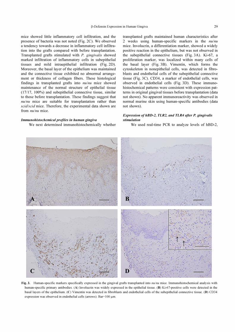
Immunohistochemical profiles in human gingiva
We next determined immunohistochemically whether
transplanted grafts maintained human characteristics after
2 weeks using human-specific markers in the nu/nu
mice. Involucrin, a differentiation marker, showed a widely
positive reaction in the epithelium, but was not observed in
the subepithelial connective tissues (Fig. 3A). Ki-67, a
proliferation marker, was localized within many cells of
the basal layer (Fig. 3B). Vimentin, which forms the
cytoskeleton in nonepithelial cells, was detected in fibro-
blasts and endothelial cells of the subepithelial connective
tissue (Fig. 3C). CD34, a marker of endothelial cells, was
observed in endothelial cells (Fig. 3D). These immuno-
histochemical patterns were consistent with expression pat-
terns in original gingival tissues before transplantation (data
not shown). No apparent immunoreactivity was observed in
normal murine skin using human-specific antibodies (data
not shown).
Expression of hBD-2, TLR2, and TLR4 after P. gingivalis
stimulation
We used real-time PCR to analyze levels of hBD-2,
Fig. 3. Human-specific markers specifically expressed in the gingival grafts transplanted into nu/nu mice. Immunohistochemical analysis with
human-specific primary antibodies. (A) Involucrin was widely expressed in the epithelial tissue. (B) Ki-67-positive cells were detected in the
basal layers of the epithelium. (C) Vimentin was detected in fibroblasts and endothelial cells of the subepithelial connective tissue. (D) CD34
expression was observed in endothelial cells (arrows). Bar=100 μm.
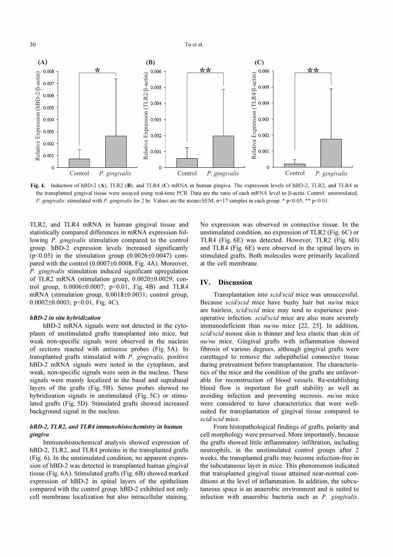
To et al.30
TLR2, and TLR4 mRNA in human gingival tissue and
statistically compared differences in mRNA expression fol-
lowing P. gingivalis stimulation compared to the control
group. hBD-2 expression levels increased significantly
(p<0.05) in the stimulation group (0.0026±0.0047) com-
pared with the control (0.0007±0.0008, Fig. 4A). Moreover,
P. gingivalis stimulation induced significant upregulation
of TLR2 mRNA (stimulation group, 0.0020±0.0029; con-
trol group, 0.0006±0.0007; p<0.01, Fig. 4B) and TLR4
mRNA (stimulation group, 0.0018±0.0031; control group,
0.0002±0.0003; p<0.01, Fig. 4C).
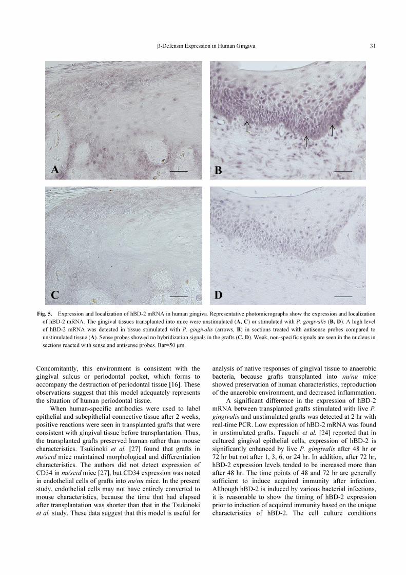
hBD-2 in situ hybridization
hBD-2 mRNA signals were not detected in the cyto-
plasm of unstimulated grafts transplanted into mice, but
weak non-specific signals were observed in the nucleus
of sections reacted with antisense probes (Fig. 5A). In
transplanted grafts stimulated with P. gingivalis, positive
hBD-2 mRNA signals were noted in the cytoplasm, and
weak, non-specific signals were seen in the nucleus. These
signals were mainly localized in the basal and suprabasal
layers of the grafts (Fig. 5B). Sense probes showed no
hybridization signals in unstimulated (Fig. 5C) or stimu-
lated grafts (Fig. 5D). Stimulated grafts showed increased
background signal in the nucleus.
hBD-2, TLR2, and TLR4 immunohistochemistry in human
gingiva
Immunohistochemical analysis showed expression of
hBD-2, TLR2, and TLR4 proteins in the transplanted grafts
(Fig. 6). In the unstimulated condition, no apparent expres-
sion of hBD-2 was detected in transplanted human gingival
tissue (Fig. 6A). Stimulated grafts (Fig. 6B) showed marked
expression of hBD-2 in spinal layers of the epithelium
compared with the control group. hBD-2 exhibited not only
cell membrane localization but also intracellular staining.
No expression was observed in connective tissue. In the
unstimulated condition, no expression of TLR2 (Fig. 6C) or
TLR4 (Fig. 6E) was detected. However, TLR2 (Fig. 6D)
and TLR4 (Fig. 6E) were observed in the spinal layers in
stimulated grafts. Both molecules were primarily localized
at the cell membrane.
IV. Discussion
Transplantation into scid/scid mice was unsuccessful.
Because scid/scid mice have bushy hair but nu/nu mice
are hairless, scid/scid mice may tend to experience post-
operative infection. scid/scid mice are also more severely
immunodeficient than nu/nu mice [22, 25]. In addition,
scid/scid mouse skin is thinner and less elastic than skin of
nu/nu mice. Gingival grafts with inflammation showed
fibrosis of various degrees, although gingival grafts were
curettaged to remove the subepithelial connective tissue
during pretreatment before transplantation. The characteris-
tics of the mice and the condition of the grafts are unfavor-
able for reconstruction of blood vessels. Re-establishing
blood flow is important for graft stability as well as
avoiding infection and preventing necrosis. nu/nu mice
were considered to have characteristics that were well-
suited for transplantation of gingival tissue compared to
scid/scid mice.
From histopathological findings of grafts, polarity and
cell morphology were preserved. More importantly, because
the grafts showed little inflammatory infiltration, including
neutrophils, in the unstimulated control groups after 2
weeks, the transplanted grafts may become infection-free in
the subcutaneous layer in mice. This phenomenon indicated
that transplanted gingival tissue attained near-normal con-
ditions at the level of inflammation. In addition, the subcu-
taneous space is an anaerobic environment and is suited to
infection with anaerobic bacteria such as P. gingivalis.
Fig. 4. Induction of hBD-2 (A), TLR2 (B), and TLR4 (C) mRNA in human gingiva. The expression levels of hBD-2, TLR2, and TLR4 in
the transplanted gingival tissue were assayed using real-time PCR. Data are the ratio of each mRNA level to β-actin. Control: unstimulated,
P. gingivalis: stimulated with P. gingivalis for 2 hr. Values are the mean±SEM; n=17 samples in each group. * p<0.05, ** p<0.01.
β-Defensin Expression in Human Gingiva 31
Concomitantly, this environment is consistent with the
gingival sulcus or periodontal pocket, which forms to
accompany the destruction of periodontal tissue [16]. These
observations suggest that this model adequately represents
the situation of human periodontal tissue.
When human-specific antibodies were used to label
epithelial and subepithelial connective tissue after 2 weeks,
positive reactions were seen in transplanted grafts that were
consistent with gingival tissue before transplantation. Thus,
the transplanted grafts preserved human rather than mouse
characteristics. Tsukinoki et al. [27] found that grafts in
nu/scid mice maintained morphological and differentiation
characteristics. The authors did not detect expression of
CD34 in nu/scid mice [27], but CD34 expression was noted
in endothelial cells of grafts into nu/nu mice. In the present
study, endothelial cells may not have entirely converted to
mouse characteristics, because the time that had elapsed
after transplantation was shorter than that in the Tsukinoki
et al. study. These data suggest that this model is useful for
analysis of native responses of gingival tissue to anaerobic
bacteria, because grafts transplanted into nu/nu mice
showed preservation of human characteristics, reproduction
of the anaerobic environment, and decreased inflammation.
A significant difference in the expression of hBD-2
mRNA between transplanted grafts stimulated with live P.
gingivalis and unstimulated grafts was detected at 2 hr with
real-time PCR. Low expression of hBD-2 mRNA was found
in unstimulated grafts. Taguchi et al. [24] reported that in
cultured gingival epithelial cells, expression of hBD-2 is
significantly enhanced by live P. gingivalis after 48 hr or
72 hr but not after 1, 3, 6, or 24 hr. In addition, after 72 hr,
hBD-2 expression levels tended to be increased more than
after 48 hr. The time points of 48 and 72 hr are generally
sufficient to induce acquired immunity after infection.
Although hBD-2 is induced by various bacterial infections,
it is reasonable to show the timing of hBD-2 expression
prior to induction of acquired immunity based on the unique
characteristics of hBD-2. The cell culture conditions
Fig. 5. Expression and localization of hBD-2 mRNA in human gingiva. Representative photomicrographs show the expression and localization
of hBD-2 mRNA. The gingival tissues transplanted into mice were unstimulated (A, C) or stimulated with P. gingivalis (B, D). A high level
of hBD-2 mRNA was detected in tissue stimulated with P. gingivalis (arrows, B) in sections treated with antisense probes compared to
unstimulated tissue (A). Sense probes showed no hybridization signals in the grafts (C, D). Weak, non-specific signals are seen in the nucleus in
sections reacted with sense and antisense probes. Bar=50 μm.

To et al.32
Fig. 6. Immunohistochemical localization of hBD-2, TLR2, and TLR4 in human gingiva. Representative photomicrographs show the immuno-
histochemical localization of hBD-2 (A, B), TLR2 (C, D), and TLR4 (E, F) protein. The transplanted gingival tissues were unstimulated (A, C,
and E) or stimulated with P. gingivalis (B, D, and F). hBD-2 was detected in the spinal layers of the epithelium in stimulated grafts. hBD-2
exhibited cell membrane and intracellular distribution. TLR2 and TLR4 were localized at the cell membrane in the spinal layers. Bar=50 μm.

β-Defensin Expression in Human Gingiva 33
reported by Taguchi et al. may lead to increased basal levels
of hBD-2 expression in the control group, which showed
hBD-2 expression beginning at 3 hr [24]. In contrast, our
novel in vivo experimental model showed that induction
of hBD-2 occurred in the early stages following live
P. gingivalis infection. Thus, hBD-2 may play a role in the
initial defense system in gingival epithelial cells following
bacterial infection.
Dommisch et al. [7] reported no statistically significant
difference among healthy control, gingivitis, and chronic
periodontitis groups in terms of hBD-2 expression in gingi-
val tissue samples. Moreover, Vardar-Sengul et al. [28]
reported that hBD-2 is significantly lower in the gingivitis
group, but higher in the chronic periodontitis group relative
to healthy controls. Thus, expression levels of hBD-2 in
gingival specimens with periodontal diseases remain con-
troversial. Interestingly, gingival epithelium synthesizes
hBD-2, even in the healthy state [2, 7]. Therefore, basal
levels of hBD-2 may be high in control healthy gingival
epithelium. However, our results clearly demonstrated
that hBD-2 levels in stimulated grafts were increased by
P. gingivalis infection compared with control grafts. In
patients with periodontitis that was caused by P. gingivalis,
expression of hBD-2 may be higher than in non-inflamed
gingival tissue.
We also found that hBD-2 mRNA was detected in
the suprabasal layers, whereas the protein was localized in
spinal layers of gingival epithelium. Dale et al. [5] reported
that the distribution between mRNA and protein differs in
human gingival epithelium. More importantly, we found
that hBD-2 mRNA and protein were higher in the grafts
stimulated with P. gingivalis than in unstimulated grafts.
Thus, the expression of hBD-2 mRNA and protein may
be induced by bacterial infection in human gingival
epithelium.
TLRs recognize specific pathogen-associated molecu-
lar patterns [13, 17, 29] and play an important role in the
induction of proinflammatory cytokines and antimicrobial
peptides, which trigger innate immunity and acquired
immunity [14, 18, 23]. In our study, the expression of TLR2
and TLR4 mRNA was upregulated in stimulated grafts.
Previously, an in vitro study demonstrated that TLR
signaling is related to induced expression of hBD-2 [13, 14,
19]. To determine whether this interaction occurs in vivo,
we investigated changes in TLR2 and TLR4 expression
following bacterial stimulation. The induction of TLR we
observed was similar to that reported in past studies [17,
19, 23, 29], and the immunohistochemical localization
was also consistent with that of hBD-2. Therefore, TLR
may be involved in induced expression of hBD-2 following
bacterial infection. Collectively, our results show that hBD-
2 expression is actively induced in response to bacterial
infection and TLR may regulate hBD-2 expression in
human gingival epithelium.
In conclusion, to confirm associations between human
gingival responses and bacterial infections, we established
a novel in vivo experimental model using nu/nu mice.
This model demonstrated that expression of hBD-2 was
induced soon after infections and was increased in gingival
epithelium that was infected with P. gingivalis. hBD-2
expression in gingival epithelium may play an important
role in protection against P. gingivalis. Further examination
of TLR signaling as a possible mechanism of induction of
expression of hBD-2 will be necessary in the future.
V. Acknowledgments
This research was supported in part by a Grant-in Aid
for Scientific Research (B, #23390420).
VI. References
1. Abiko, Y., Saitoh, M., Nishimura, M., Yamazaki, M., Sawamura,
D. and Kaku, T. (2007) Role of β-defensins in oral epithelial
health and disease. Med. Mol. Morphol. 40; 179–184.
2. Bissell, J., Joly, S., Johnson, G. K., Organ, C. C., Dawson, D.,
McCray, P. B. Jr. and Guthmiller, J. M. (2004) Expression of β-
defensins in gingival health and in periodontal disease. J. Oral
Pathol. Med. 33; 278–285.
3. Cederlund, A., Gudmundsson, G. H. and Agerberth, B. (2011)
Antimicrobial peptides important in innate immunity. FEBS J.
278; 3942–3951.
4. Chung, W. O., Hansen, S. R., Rao, D. and Dale, B. A. (2004)
Protease-activated receptor signaling increases epithelial anti-
microbial peptide expression. J. Immunol. 173; 5165–5170.
5. Dale, B. A., Kimball, J. R., Krisanaprakornkit, S., Roberts, F.,
Robinovitch, M., O’Neal, R., Valore, E. V., Ganz, T., Anderson,
G. M. and Weinberg, A. (2001) Localized antimicrobial peptide
expression in human gingiva. J. Periodontal Res. 36; 285–294.
6. Dale, B. A. and Krisanaprakornkit, S. (2001) Defensin anti-
microbial peptides in the oral cavity. J. Oral Pathol. Med. 30;
321–327.
7. Dommisch, H., Açil, Y., Dunsche, A., Winter, J. and Jepsen, S.
(2005) Differential gene expression of human β-defensins (hBD-
1, -2, -3) in inflammatory gingival diseases. Oral Microbiol.
Immunol. 20; 186–190.
8. Harasawa, M., Yasuda, M., Hirasawa, T., Miyazawa, M., Shida,
M., Muramatsu, T., Douguchi, K., Matsui, N., Takekoshi, S.,
Kajiwara, H., Osamura, R.Y. and Mikami, M. (2011) Analysis of
mTOR inhibition-involved pathway in ovarian clear cell adeno-
carcinoma. Acta Histochem. Cytochem. 44; 113–118.
9. Hioki, K., Kuramochi, T., Endoh, S., Terada, E., Ueyama, Y. and
Ito, M. (2001) Lack of B cell leakiness in BALB/cA-nu, scid
double mutant mice. Exp. Anim. 50; 67–72.
10. Holmstrup, P., Stoltze, K., Bretlau, P. and Dabelsteen, E. (1981)
Human buccal mucosa transplants in nude mice. Scand. J. Dent.
Res. 89; 89–96.
11. Kondo, Y., Saruta, J., To, M., Shiiki, N., Sato, C. and Tsukinoki,
K. (2010) Expression and role of the BDNF receptor-TrkB in rat
adrenal gland under acute immobilization stress. Acta Histochem.
Cytochem. 43; 139–147.
12. Krisanaprakornkit, S., Kimball, J. R., Weinberg, A., Darveau, R.
P., Bainbridge, B. W. and Dale, B. A. (2000) Inducible expression
of human β-defensin 2 by Fusobacterium nucleatum in oral
epithelial cells: multiple signaling pathways and role of
commensal bacteria in innate immunity and the epithelial barrier.
Infect. Immun. 68; 2907–2915.
13. Mahanonda, R., Sa-Ard-Iam, N., Eksomtramate, M., Rerkyen, P.,
Phairat, B., Schaecher, K. E., Fukuda, M. M. and Pichyangkul, S.
(2009) Cigarette smoke extract modulates human β-defensin-2
and interleukin-8 expression in human gingival epithelial cells.
To et al.34
J. Periodontal Res. 44; 557–564.
14. Nagy, I., Pivarcsi, A., Koreck, A., Széll, M., Urbán, E. and
Kemény, L. (2005) Distinct strains of Propionibacterium acnes
induce selective human β-defensin-2 and interleukin-8 expression
in human keratinocytes through toll-like receptors. J. Invest.
Dermatol. 124; 931–938.
15. Nakajima, K., Hamada, N., Takahashi, Y., Sasaguri, K.,
Tsukinoki, K., Umemoto, T. and Sato, S. (2006) Restraint stress
enhances alveolar bone loss in an experimental rat model. J.
Periodontal Res. 41; 527–534.
16. Pihlstrom, B. L., Michalowicz, B. S. and Johnson, N. W. (2005)
Periodontal diseases. Lancet 366; 1809–1820.
17. Pivarcsi, A., Bodai, L., Réthi, B., Kenderessy-Szabó, A., Koreck,
A., Széll, M., Beer, Z., Bata-Csörgoo, Z., Magócsi, M.,
Rajnavölgyi, E., Dobozy, A. and Kemény, L. (2003) Expression
and function of Toll-like receptors 2 and 4 in human keratino-
cytes. Int. Immunol. 15; 721–730.
18. Pivarcsi, A., Nagy, I., Koreck, A., Kis, K., Kenderessy-Szabo, A.,
Szell, M., Dobozy, A. and Kemeny, L. (2005) Microbial com-
pounds induce the expression of pro-inflammatory cytokines,
chemokines and human β-defensin-2 in vaginal epithelial cells.
Microbes Infect. 7; 1117–1127.
19. Romano-Carratelli, C., Mazzola, N., Paolillo, R., Sorrentino, S.
and Rizzo, A. (2009) Toll-like receptor-4 (TLR4) mediates
human β-defensin-2 (HBD-2) induction in response to Chlamydia
pneumoniae in mononuclear cells. FEMS Immunol. Med.
Microbiol. 57; 116–124.
20. Saruta, J., Fujino, K., To, M. and Tsukinoki, K. (2012) Expression
and localization of brain-derived neurotrophic factor (BDNF)
mRNA and protein in human submandibular gland. Acta
Histochem. Cytochem. 45; 211–218.
21. Schenkein, H. A. (2006) Host responses in maintaining perio-
dontal health and determining periodontal disease. Periodontol.
2000 40; 77–93.
22. Sugai, J., Iizuka, M., Kawakubo, Y., Ozawa, A., Ohkido, M.,
Ueyama, Y., Tamaoki, N., Inokuchi, S. and Shimamura, K. (1998)
Histological and immunocytochemical studies of human psoriatic
lesions transplanted onto SCID mice. J. Dermatol. Sci. 17; 85–92.
23. Sun, Y., Shu, R., Zhang, M. Z. and Wu, A. P. (2008) Toll-like
receptor 4 signaling plays a role in triggering periodontal infec-
tion. FEMS Immunol. Med. Microbiol. 52; 362–369.
24. Taguchi, Y. and Imai, H. (2006) Expression of β-defensin-2 in
human gingival epithelial cells in response to challenge with
Porphyromonas gingivalis in vitro. J. Periodontal Res. 41; 334–
339.
25. Takizawa, Y., Saida, T., Tokuda, Y., Dohi, S., Ikegawa, S. and
Ueyama, Y. (1995) Engraftment of precursor lesions of human
cutaneous neoplasms onto C.B-17 SCID mice: a useful in vivo
experimental model of carcinogenesis in human skin. Arch.
Dermatol. Res. 287; 237–241.
26. Takizawa, Y., Saida, T., Tokuda, Y., Dohi, S., Wang, Y. L., Urano,
K., Hioki, K. and Ueyama, Y. (1997) New immunodeficient
(nude-scid, beige-scid) mice as excellent recipients of human skin
grafts containing intraepidermal neoplasms. Arch. Dermatol. Res.
289; 213–218.
27. Tsukinoki, K., Miyoshi, Y., Aoki, T., Karakida, K., Ohta, Y.,
Kaneko, A., Ueyama, Y. and Watanabe, Y. (2007) In vivo experi-
mental model of human gingival mucosa using immunodeficient
mice. J. Periodontal Res. 42; 294–299.
28. Vardar-Sengul, S., Demirci, T., Sen, B. H., Erkizan, V., Kurulgan,
E. and Baylas, H. (2007) Human b defensin-1 and -2 expression
in the gingiva of patients with specific periodontal diseases. J.
Periodontal Res. 42; 429–437.
29. Varoga, D., Klostermeier, E., Paulsen, F., Wruck, C., Lippross, S.,
Brandenburg, L. O., Tohidnezhad, M., Seekamp, A., Tillmann, B.
and Pufe, T. (2009) The antimicrobial peptide HBD-2 and the
Toll-like receptors-2 and -4 are induced in synovial membranes in
case of septic arthritis. Virchows Arch. 454; 685–694.
30. Vora, P., Youdim, A., Thomas, L. S., Fukata, M., Tesfay, S. Y.,
Lukasek, K., Michelsen, K. S., Wada, A., Hirayama, T., Arditi,
M. and Abreu, M. T. (2004) β-defensin-2 expression is regulated
by TLR signaling in intestinal epithelial cells. J. Immunol. 173;
5398–5405.
31. Zhang, Z., Jin, L., Champion, G., Seydel, K. B. and Stanley, S. L.
Jr. (2001) Shigella infection in a SCID mouse-human intestinal
xenograft model: role for neutrophils in containing bacterial
This is an open access article distributed under the Creative Commons Attribu-tion License, which permits unrestricted use, distribution, and reproduction inany medium, provided the original work is properly cited.
dissemination in human intestine. Infect. Immun. 69; 3240–3247.