Human TNF-α High Sensitivity ELISAtools.thermofisher.com/content/sfs/manuals/223HS.pdf · 4 Human...
23
Human TNF-α High Sensitivity ELISA PRODUCT INFORMATION & MANUAL Human TNF-α High Sensitivity ELISA PRODUCT INFORMATION & MANUAL Enzyme-linked Immunosorbent Assay for quantitative detection of human TNF-α. BMS223HS
Transcript of Human TNF-α High Sensitivity ELISAtools.thermofisher.com/content/sfs/manuals/223HS.pdf · 4 Human...

Human TNF-α High Sensitivity ELISA PRODUCT INFORMATION & MANUAL
Human TNF-α High Sensitivity ELISA PRODUCT INFORMATION & MANUAL
Enzyme-linked Immunosorbent Assay for quantitative detection of human TNF-α.
BMS223HS
Information in this document is subject to change without notice.
DISCLAIMER
TO THE EXTENT ALLOWED BY LAW, THERMO FISHER SCIENTIFIC AND/OR ITS AFFILIATE(S) WILL NOT BE LIABLE FOR SPECIAL, INCIDENTAL, INDIRECT, PUNITIVE, MULTIPLE OR CONSEQUENTIAL DAMAGES IN CONNECTION WITH OR ARISING FROM THIS DOCUMENT, INCLUDING YOUR USE OF IT.
Important Licensing Information
These products may be covered by one or more Limited Use Label Licenses. By use of these products, you accept the terms and conditions of all applicable Limited Use Label Licenses.
Corporate entity
Life Technologies | Carlsbad, CA 92008 USA | Toll free in USA 1.800.955.6288
Trademarks
All trademarks are the property of Thermo Fisher Scientific and its subsidiaries unless otherwise specified. All other trademarks are the property of their respective owners.
© 2017 Thermo Fisher Scientific Inc. All rights reserved.
Bender MedSystems GmbH Campus Vienna Biocenter 2 1030 Vienna, Austria.
2 Human TNF-α High Sensitivity ELISA PRODUCT INFORMATION & MANUAL
TABLE OF CONTENTS 1 Intended Use ................................................................................................................................ 3
2 Summary ..................................................................................................................................... 3
3 Principles of the Test ..................................................................................................................... 4
4 Principle of Amplification Reaction ................................................................................................ 6
5 Reagents Provided ........................................................................................................................ 6
6 Storage Instructions – ELISA Kit .................................................................................................... 7
7 Specimen Collection and Storage Instructions .................................................................................. 7
8 Materials Required But Not Provided .............................................................................................. 7
9 Precautions for Use ....................................................................................................................... 8
10 Preparation of Reagents ............................................................................................................... 8
11 Test Protocol ............................................................................................................................ 11
12 Calculation of Results ................................................................................................................ 13
13 Limitations ............................................................................................................................... 15
14 Performance Characteristics ....................................................................................................... 16
15 Reagent Preparation Summary .................................................................................................... 17
16 Test Protocol Summary .............................................................................................................. 18
Documentation and support ............................................................................................................ 20

Human TNF-α High Sensitivity ELISA PRODUCT INFORMATION & MANUAL 3
1 Intended Use The human TNFα ELISA is an enzyme-linked immunosorbent assay for the quantitative detection of human TNFα.
The human TNFα ELISA is for research use only. Not for diagnostic or therapeutic procedures.
2 Summary TNF-α is a multifunctional cytokine involved in many different pathways, in homeostasis and pathophysiology of
mammals. It can show opposing biological effects suggesting complex regulatory mechanisms.
TNF-α, also known as cachectin, was first detected as a cytotoxic factor inducing lysis of certain tumor cells. The
TNF-α gene is member 2 of the TNF-superfamily (consisting of at least 20 distinct members).
TNF-α release is mainly triggered by viral infections and endotoxins, lipopolysaccharides or other bacterial
components, by tissue injury, DNA-damage and by IL-1, PDFG and TNF-α itself. It is primarily expressed in
macrophages, but also in monocytes, neutrophils,
NK-cells, mast-cells, endothelial cells and activated lymphocytes. TNF-α expression in endothelial cells and
fibroblasts can be induced by IL-17. The expression of other cytokines, chemokines, reactive oxygen intermediates,
nitric oxide and prostaglandins is stimulated by TNF-α.
The initially membrane bound TNF-α is enzymatically cleaved by TACE (= ADAM17). The soluble monomers
aggregate to homotrimers and are secreted into blood and other biological fluids. The membrane bound and the soluble
form are biologically active and bind to the TNF- receptors TNFR1 ( = TNFRSF1A, p55-60) and TNFR2 ( =
TNFRSF1B, TNFBR2, p75-80).
Upon ligand binding, receptors form trimers leading to conformational changes, protein dissociation (SODD = silencer
of death domains, BAG4, Bcl2-associated athanogene 4) and association (TRADD = TNF-R1 associated death domain
protein) and yielding the following biological activities:
- transcription of anti-apoptotic factors and proteins involved in cell proliferation and inflammation via binding of
TRAF2 (TNF-R associated factor 2) and RIPK1 (TNF-R interacting serine-threonine kinase 1) and activation of the
transcription factor NF-κB.
- cell proliferation, differentiation but also apoptotisis via TRAF2 binding, kinases activation, activation of c-Jun and
ATF2 (JNK-MAPK-pathway).
- apoptosis via the binding of FADD (Fas associating protein with death domain) to TRADD and activation of
caspases (including caspase 8 = FLICE).
- necrosis, a caspase independent cell death, mediated by NADPH oxidases, which form a complex with TRADD and
RIPK1, leading to the generation of oxygen species.
TNF-R2 contains no DD (death domain), but exhibits its function via direct TRAF binding.
Thus the multiple biological functions of TNF-α comprise cellular proliferation and differentiation, tumorigenesis,
apoptotic or necrotic cell death (including certain tumor cell lines), immunoregulatory activities, lipid metabolism,
coagulation and endothelial function. It promotes local or systemic inflammation (TNF-α is a potent pyrogen) and
stimulates the acute phase response. Very high expressions of TNF-α after infection can lead to septic shock (TNF-α is
highly cytotoxic.), whereas sustained low levels induce cachexia and inflammation.
Dysregulation of TNF-α is involved in many diseases:
Cancer: Diverse roles of TNF-α in cancer have been described, depending on the tumor type, tumor microenvironment
and on overall TNF-α expression levels and expression kinetics. A model was proposed, in which single very high
TNF-α levels lead to tumor regression and that chronic low-dose levels are associated with tumor progression.
Systemic lupus erythematosus (SLE): Murine models of SLE show contradictory effects of TNF-α: anti-autoimmune
effect at low TNF-α levels (TNF-α administration leads to an attenuation of symptoms and TNF-α blockade to
generation of SLE-symptoms), pro-inflammatory effect at high TNF-α levels (TNF-α blockade damped the disease).
Chronic intestinal inflammation (Crohn´s disease (CD), Inflammatory bowel disease (IBD), Ulcerative Colitis (UC)):
An essential effect of TNF-α/TNF-R1 activity on induction of chronic intestinal inflammation was shown. In IBD
overexpression of TNF-α in monocytes/macrophages of gut and intestinal tissue has been described.
Psoriasis: In psoriatic patients TNF-α was shown to be increased systemically and in cutaneous tissue. TNF-α
expression in peripheral blood mononuclear cells (PBMCs) was highly elevated in patients in active phase of the
disease and elevated in chronic psoriasis. In an animal model it was shown that TNF-α dependent activation of
resident T cells was essential for Psoriasis development.
Pulmanory disorders (Cystic Fibrosis (CF), Asthma): In cystic fibrosis high levels of TNF-α were observed. TNF-α, is
over-expressed in persistent severe asthma. In allergic asthma, under low antigen exposure, TNF-α contributes to
elevated histamine release. In a mouse model for asthma, airway inflammation was caused by TNF-α mediated
activation of phospholipase A2.
4 Human TNF-α High Sensitivity ELISA PRODUCT INFORMATION & MANUAL
Rheumathoid arthritis (RA), Ankylosing spondylitis (AS): TNF-α has stimulating effects on matrix degrading
proteases (matrix metallo- proteinases, MMPs), tissue remodeling and osteoclasts, causing bone resorption leading to
joint erosions. In a mouse model for RA, elevated levels of TNF-α were observed in bone marrow. Cytokines,
synthesized by TNF-α activated synovial cells, were shown to induce RA. In AS patients, TNF-α concentration was
shown to be elevated in the sacroiliac joint.
Transplantation (Graft-versus-host disease, Allograft rejection): Measurement of TNF-α levels was shown to be useful
in transplantation research. TNF-α was reported to be markedly elevated in renal allograft rejection. Evidence has been
presented on increased TNF-α levels in Bone Marrow Transplants (BMT). BMT patients with major transplant related
complications such as interstitial pneumonitis and severe acute graft-versus-host disease showed significantly
increased TNF-α levels.
Atherosclerosis, arterial calcification: Increased risk of recurrent myocardial infarction, atherosclerotic thickening of
carotid intima-media, disturbances in triglyceride and glucose homeostasis and age-related atherosclerosis have been
connected to circulating TNF-α levels. TNF-α induced mechanisms lead to increased aortic calcium accumulation in
animals with type 2 diabetes mellitus.
Insulin Resistance (IR) and Obesity: In adipose tissue of rodent models for obesity and in adipose individuals
increased TNF-α expression was observed. In adipose tissue and in IR chronic low-grade inflammation was shown.
Increased TNF-α levels affect insulin pathway regulation.
Neurodegenerative diseases (Multiple sclerosis (MS), Alzheimer´s Disease, Prion Disease, Parkinson): TNF-α is
produced from activated microglial cells and leads to neuronal degeneration, apoptosis of neuronal tissue and
increased inflammation. TNF-α administration caused oligodendrocytes cell death - a symptom of MS – in coculture
with astrocytes and microglia.
For literature update refer to www.Thermo Fisher.com
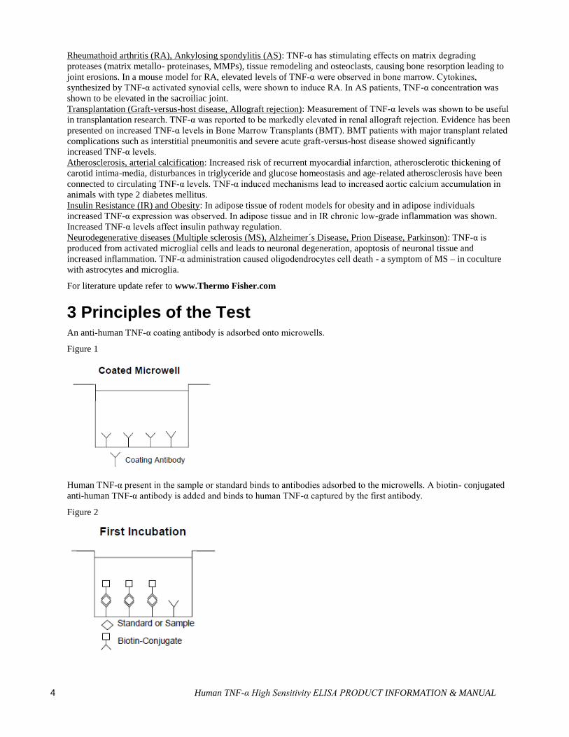
3 Principles of the Test An anti-human TNF-α coating antibody is adsorbed onto microwells.
Figure 1
Human TNF-α present in the sample or standard binds to antibodies adsorbed to the microwells. A biotin- conjugated
anti-human TNF-α antibody is added and binds to human TNF-α captured by the first antibody.
Figure 2

Human TNF-α High Sensitivity ELISA PRODUCT INFORMATION & MANUAL 5
Following incubation unbound biotin- conjugated anti-human TNF-α antibody is removed during a wash step.
Streptavidin-HRP is added and binds to the biotin-conjugated anti- human TNF-α antibody.
Figure 3
Following incubation unbound Streptavidin-HRP is removed during a wash step, and amplification reagent I (Biotinyl-
Tyramide) is added to the wells.
Figure 4
Following incubation unbound amplification reagent I is removed during a wash step and amplification reagent II
(Streptavidin-HRP) is added.
Figure 5

6 Human TNF-α High Sensitivity ELISA PRODUCT INFORMATION & MANUAL
Following incubation unbound amplification reagent II is removed during a wash step and substrate solution reactive
with HRP is added.
Figure 6
A coloured product is formed in proportion to the amount of human TNF-α present in the sample or standard. The
reaction is terminated by addition of acid and absorbance is measured at 450 nm. A standard curve is prepared from 7
human TNF-α standard dilutions and human TNF-α sample concentration determined.
Figure 7
4 Principle of Amplification Reaction The amplification reaction is based upon PerkinElmer Life Sciences’ TSA (Tyramide Signal Amplification)
technology (see 15, References 1 and 2).
Amplification reagent I contains biotinyl-tyramide. HRP converts multiple biotinyl-tyramide molecules into highly
reactive derivates (free radicals). These free radicals bind covalently to any protein in the well.
Thus, the amount of reacted biotinyl-tyramide is proportional to the amount of HRP in the well.
Following incubation unbound biotinyl-tyramide is removed during a wash step. Amplification reagent II contains
Streptavidin-HRP, which binds to the biotin sites created during the biotinyl-tyramide reaction, thus multiplying the
HRP molecules available at the surface for the substrate reaction.
5 Reagents Provided 1 aluminium pouch with a Microwell Plate coated with monoclonal antibody to human TNF-α
1 vial (100 µl) Biotin-Conjugate anti-human TNF-α polyclonal antibody
1 vial (150 µl) Streptavidin-HRP
2 vials human TNF-α Standard lyophilized, 1 ng/ml upon reconstitution
1 vial Control high, lyophilized
1 vial Control low, lyophilized

Human TNF-α High Sensitivity ELISA PRODUCT INFORMATION & MANUAL 7
1 bottle (12 ml) Sample Diluent
1 vial (5 ml) Assay Buffer Concentrate 20x (PBS with 1% Tween 20 and 10% BSA)
1 vial (7 ml) Amplification Diluent Concentrate (2x)
1 vial (75 µl) Amplification Reagent I*
1 vial (90 µl) Amplification Reagent II
2 bottles (50 ml) Wash Buffer Concentrate 20x (PBS with 1% Tween 20)
1 vial (15 ml) Substrate Solution (tetramethyl-benzidine)
1 vial (15 ml) Stop Solution (1M Phosphoric acid)
8 Adhesive Films
* reagent contains ethyl alcohol
6 Storage Instructions – ELISA Kit Store kit reagents between 2° and 8°C except controls. Store lyophilized controls at -20°C.
Immediately after use remaining reagents should be returned to cold storage (2° to 8°C), controls to -20°C,
respectively. Expiry of the kit and reagents is stated on labels.
Expiry of the kit components can only be guaranteed if the components are stored properly, and if, in case of repeated
use of one component, this reagent is not contaminated by the first handling.
7 Specimen Collection and Storage Instructions Cell culture supernatant, serum and plasma (EDTA, heparin, citrate) were tested with this assay. Other biological
samples might be suitable for use in the assay. Remove serum or plasma from the clot or cells as soon as possible after
clotting and separation.
Samples containing a visible precipitate must be clarified prior to use in the assay. Do not use grossly hemolyzed or
lipemic specimens.
Samples should be aliquoted and must be stored frozen at -20°C to avoid loss of bioactive human TNF-α. If samples
are to be run within 24 hours, they may be stored at 2° to 8°C (for sample stability refer to 14.5).
Avoid repeated freeze-thaw cycles. Prior to assay, the frozen sample should be brought to room temperature slowly
and mixed gently.
8 Materials Required But Not Provided 5 ml and 10 ml graduated pipettes
5 µl to 1000 µl adjustable single channel micropipettes with disposable tips
50 µl to 300 µl adjustable multichannel micropipette with disposable tips
Multichannel micropipette reservoir
Beakers, flasks, cylinders necessary for preparation of reagents
Device for delivery of wash solution (multichannel wash bottle or automatic wash system)
Microplate shaker
Microwell strip reader capable of reading at 450 nm (620 nm as optional reference wave length)
Glass-distilled or deionized water
Statistical calculator with program to perform regression analysis
8 Human TNF-α High Sensitivity ELISA PRODUCT INFORMATION & MANUAL
9 Precautions for Use All chemicals should be considered as potentially hazardous. We therefore recommend that this product is
handled only by those persons who have been trained in laboratory techniques and that it is used in accordance
with the principles of good laboratory practice. Wear suitable protective clothing such as laboratory overalls,
safety glasses and gloves. Care should be taken to avoid contact with skin or eyes. In the case of contact with skin
or eyes wash immediately with water. See material safety data sheet(s) and/or safety statement(s) for specific
advice.
Reagents are intended for research use only and are not for use in diagnostic or therapeutic procedures.
Do not mix or substitute reagents with those from other lots or other sources.
Do not use kit reagents beyond expiration date on label.
Do not expose kit reagents to strong light during storage or incubation.
Do not pipette by mouth.
Do not eat or smoke in areas where kit reagents or samples are handled.
Avoid contact of skin or mucous membranes with kit reagents or specimens.
Rubber or disposable latex gloves should be worn while handling kit reagents or specimens.
Avoid contact of substrate solution with oxidizing agents and metal.
Avoid splashing or generation of aerosols.
In order to avoid microbial contamination or cross-contamination of reagents or specimens which may invalidate
the test use disposable pipette tips and/or pipettes.
Use clean, dedicated reagent trays for dispensing the conjugate and substrate reagent.
Exposure to acid inactivates the conjugate.
Glass-distilled water or deionized water must be used for reagent preparation.
Substrate solution must be at room temperature prior to use.
Decontaminate and dispose specimens and all potentially contaminated materials as they could contain infectious
agents. The preferred method of decontamination is autoclaving for a minimum of 1 hour at 121.5°C.
Liquid wastes not containing acid and neutralized waste may be mixed with sodium hypochlorite in volumes such
that the final mixture contains 1.0% sodium hypochlorite. Allow 30 minutes for effective decontamination. Liquid
waste containing acid must be neutralized prior to the addition of sodium hypochlorite.
10 Preparation of Reagents Buffer concentrates should be brought to room temperature and should be diluted before starting the test procedure.
If crystals have formed in the Buffer Concentrates, warm them gently until they have completely dissolved.
10.1 Wash Buffer (1x) Pour entire contents (50 ml) of the Wash Buffer Concentrate (20x) into a clean 1000 ml graduated cylinder. Bring to
final volume of 1000 ml with glass-distilled or deionized water. Mix gently to avoid foaming.
Transfer to a clean wash bottle and store at 2° to 25°C. Please note that Wash Buffer (1x) is stable for 30 days.
Wash Buffer (1x) may also be prepared as needed according to the following table:
Human TNF-α High Sensitivity ELISA PRODUCT INFORMATION & MANUAL 9
Number of Strips Wash Buffer Concentrate (20x) (ml)
Distilled Water (ml)
1 - 6 25 475
1 - 12 50 950
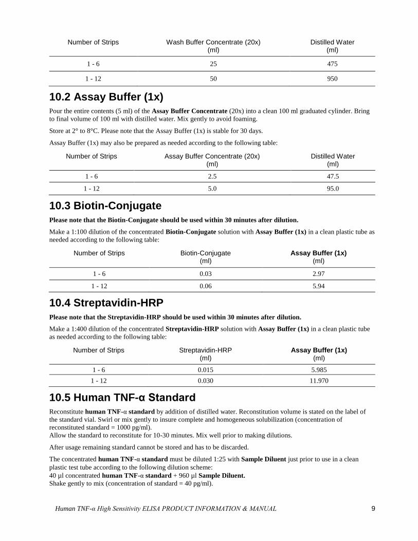
10.2 Assay Buffer (1x) Pour the entire contents (5 ml) of the Assay Buffer Concentrate (20x) into a clean 100 ml graduated cylinder. Bring
to final volume of 100 ml with distilled water. Mix gently to avoid foaming.
Store at 2° to 8°C. Please note that the Assay Buffer (1x) is stable for 30 days.
Assay Buffer (1x) may also be prepared as needed according to the following table:
Number of Strips Assay Buffer Concentrate (20x) (ml)
Distilled Water (ml)
1 - 6 2.5 47.5
1 - 12 5.0 95.0
10.3 Biotin-Conjugate
Please note that the Biotin-Conjugate should be used within 30 minutes after dilution.
Make a 1:100 dilution of the concentrated Biotin-Conjugate solution with Assay Buffer (1x) in a clean plastic tube as
needed according to the following table:
Number of Strips Biotin-Conjugate (ml)
Assay Buffer (1x) (ml)
1 - 6 0.03 2.97
1 - 12 0.06 5.94
10.4 Streptavidin-HRP
Please note that the Streptavidin-HRP should be used within 30 minutes after dilution.
Make a 1:400 dilution of the concentrated Streptavidin-HRP solution with Assay Buffer (1x) in a clean plastic tube
as needed according to the following table:
Number of Strips Streptavidin-HRP (ml)
Assay Buffer (1x) (ml)
1 - 6 0.015 5.985
1 - 12 0.030 11.970
10.5 Human TNF-α Standard Reconstitute human TNF-α standard by addition of distilled water. Reconstitution volume is stated on the label of
the standard vial. Swirl or mix gently to insure complete and homogeneous solubilization (concentration of
reconstituted standard = 1000 pg/ml).
Allow the standard to reconstitute for 10-30 minutes. Mix well prior to making dilutions.
After usage remaining standard cannot be stored and has to be discarded.
The concentrated human TNF-α standard must be diluted 1:25 with Sample Diluent just prior to use in a clean
plastic test tube according to the following dilution scheme:
40 µl concentrated human TNF-α standard + 960 µl Sample Diluent.
Shake gently to mix (concentration of standard = 40 pg/ml).
10 Human TNF-α High Sensitivity ELISA PRODUCT INFORMATION & MANUAL
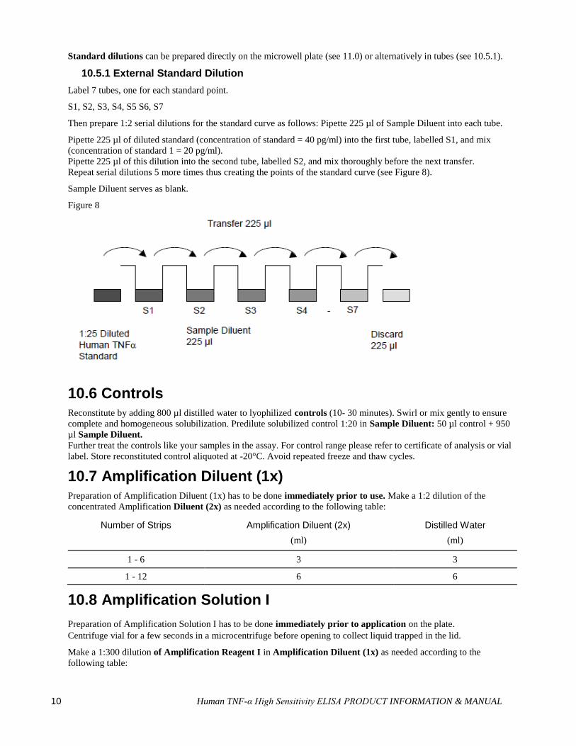
Standard dilutions can be prepared directly on the microwell plate (see 11.0) or alternatively in tubes (see 10.5.1).
10.5.1 External Standard Dilution
Label 7 tubes, one for each standard point.
S1, S2, S3, S4, S5 S6, S7
Then prepare 1:2 serial dilutions for the standard curve as follows: Pipette 225 µl of Sample Diluent into each tube.
Pipette 225 µl of diluted standard (concentration of standard = 40 pg/ml) into the first tube, labelled S1, and mix
(concentration of standard 1 = 20 pg/ml).
Pipette 225 µl of this dilution into the second tube, labelled S2, and mix thoroughly before the next transfer.
Repeat serial dilutions 5 more times thus creating the points of the standard curve (see Figure 8).
Sample Diluent serves as blank.
Figure 8
10.6 Controls Reconstitute by adding 800 µl distilled water to lyophilized controls (10- 30 minutes). Swirl or mix gently to ensure
complete and homogeneous solubilization. Predilute solubilized control 1:20 in Sample Diluent: 50 µl control + 950
µl Sample Diluent.
Further treat the controls like your samples in the assay. For control range please refer to certificate of analysis or vial
label. Store reconstituted control aliquoted at -20°C. Avoid repeated freeze and thaw cycles.
10.7 Amplification Diluent (1x) Preparation of Amplification Diluent (1x) has to be done immediately prior to use. Make a 1:2 dilution of the
concentrated Amplification Diluent (2x) as needed according to the following table:
Number of Strips Amplification Diluent (2x) Distilled Water
(ml) (ml)
1 - 6 3 3
1 - 12 6 6
10.8 Amplification Solution I
Preparation of Amplification Solution I has to be done immediately prior to application on the plate.
Centrifuge vial for a few seconds in a microcentrifuge before opening to collect liquid trapped in the lid.
Make a 1:300 dilution of Amplification Reagent I in Amplification Diluent (1x) as needed according to the
following table:

Human TNF-α High Sensitivity ELISA PRODUCT INFORMATION & MANUAL 11
Number of Strips Amplification Reagent I
(ml)
Amplification Diluent (1x)
(ml)
1 - 6 0.02 5.98
1 - 12 0.04 11.96
Discard immediately any prediluted Amplification Solution I after usage.
10.9 Amplification Solution II Preparation of Amplification Solution II has to be done immediately prior to application on the plate.
Centrifuge vial for a few seconds in a microcentrifuge before opening to collect liquid trapped in the lid.
Make a 1:200 dilution of Amplification Reagent II in Assay Buffer (1x) as needed according to the following
scheme:
Number of Strips Amplification Reagent II
(ml)
Assay Buffer (1x) (ml)
1 - 6 0.03 5.97
1 - 12 0.06 11.94
Discard immediately any prediluted Amplification Solution II after usage.
11 Test Protocol As this ELISA is a high sensitive system it is extremely important to stick exactly to the manual (washing
procedure; chronology of / and preparation of solutions; incubation time) to obtain optimal test performance!
Soaking is highly recommended between the washes to obtain a good test performance!
Please note: Amplification Solutions have to be prepared immediately prior to application on the plate! It is
extremely important to wash the wells properly to obtain a good test performance!
1. Determine the number of microwell strips required to test the desired number of samples plus appropriate number
of wells needed for running blanks and standards. Each sample, standard, blank and optional control sample
should be assayed in duplicate. Remove extra microwell strips from holder and store in foil bag with the desiccant
provided at 2°-8°C sealed tightly.
2. Wash the microwell strips twice with exactly 400 µl Wash Buffer per well with thorough aspiration of microwell
contents between washes. Allow the Wash Buffer to sit in the wells for about 10 – 15 seconds before aspiration.
Soaking is highly recommended between the washes to obtain a good test performance! Take care not to scratch
the surface of the microwells.
After the last wash step, empty wells and tap microwell strips on absorbent pad or paper towel to remove excess
Wash Buffer. Use the microwell strips immediately after washing. Do not allow wells to dry.
Standard dilution on the microwell plate (Alternatively the standard dilution can be prepared in tubes -see 10.5.1.):
Add 100 µl of Sample Diluent in duplicate to all standard wells. Pipette 100 µl of prepared standard (see
Preparation of Standard 10.5, concentration = 40 pg/ml) in duplicate into well A1 and A2 (see Table 1). Mix the
contents of wells A1 and A2 by repeated aspiration and ejection (concentration of standard 1, S1 = 20 pg/ml), and
transfer 100 µl to wells B1 and B2, respectively (see Figure 9). Take care not to scratch the inner surface of the
microwells. Continue this procedure 5 times, creating two rows of human TNF-α standard dilutions ranging from
20.00 to 0.31 pg/ml. Discard 100 µl of the contents from the last microwells (G1, G2) used.

12 Human TNF-α High Sensitivity ELISA PRODUCT INFORMATION & MANUAL
Figure 9
In case of an external standard dilution (see 10.5.1.), pipette 100 µl of these standard dilutions (S1 - S7) in the
standard wells according to Table 1.
Table 1 Table depicting an example of the arrangement of blanks, standards and samples in the microwell strips:
1 2 3 4
A Standard 1
(20.00 pg/ml)
Standard 1
(20.00 pg/ml)
Sample 1 Sample 1
B Standard 2
(10.00 pg/ml)
Standard 2
(10.00 pg/ml)
Sample 2 Sample 2
C Standard 3
(5.00 pg/ml)
Standard 3
(5.00 pg/ml)
Sample 3 Sample 3
D Standard 4
(2.50 pg/ml)
Standard 4
(2.50 pg/ml)
Sample 4 Sample 4
E Standard 5
(1.25 pg/ml)
Standard 5
(1.25 pg/ml)
Sample 5 Sample 5
F Standard 6
(0.63 pg/ml)
Standard 6
(0.63 pg/ml)
Sample 6 Sample 6
G Standard 7
(0.31 pg/ml)
Standard 7
(0.31 pg/ml)
Sample 7 Sample 7
H Blank Blank Sample 8 Sample 8
3. Add 100 µl of Sample Diluent in duplicate to the blank wells.
4. Add 50 µl of Sample Diluent to the sample wells.
5. Add 50 µl of each sample in duplicate to the sample wells.
6. Prepare Biotin-Conjugate (see Preparation of Biotin-Conjugate 10.3).
7. Add 50 µl of Biotin-Conjugate to all wells.
8. Cover with an adhesive film and incubate at room temperature (18° to 25°C) for 2 hours on a microplate shaker
set at 400 rpm. (Shaking is absolutely necessary for an optimal test performance.)
9. Prepare Streptavidin-HRP (refer to Preparation of Streptavidin-HRP 10.4).

Human TNF-α High Sensitivity ELISA PRODUCT INFORMATION & MANUAL 13
10. Remove adhesive film and empty wells. Wash microwell strips 6 times according to point b. of the test protocol.
Proceed immediately to the next step.
11. Add 100 µl of diluted Streptavidin-HRP to all wells, including the blank wells.
12. Cover with an adhesive film and incubate at room temperature (18° to 25°C) for 1 hour on a microplate shaker set
at 400 rpm. (Shaking is absolutely necessary for an optimal test performance.)
13. Prepare Amplification Solution I diluted in Amplification Diluent (1x) (see Preparation of Amplification
Solution I 10.8) immediately prior to use.
14. Remove adhesive film and empty wells. Wash microwell strips 6 times according to point b. of the test protocol.
Proceed immediately to the next step.
15. Add 100 µl of Amplification Solution I to all wells, including the blank wells.
16. Cover with an adhesive film and incubate at room temperature (18° to 25°C) for exactly 15 minutes on a
microplate shaker set at 400 rpm. (Shaking is absolutely necessary for an optimal test performance.)
17. Prepare Amplification Solution II diluted in Assay Buffer (see Preparation of Amplification Solution II 10.9)
immediately prior to use.
18. Remove adhesive film and empty wells. Wash microwell strips 6 times according to point b. of the test protocol.
Proceed immediately to the next step.
19. Add 100 µl of Amplification Solution II to all wells, including the blank wells.
20. Cover with an adhesive film and incubate at room temperature (18° to 25°C) for exactly 30 minutes on a
microplate shaker set at 400 rpm. (Shaking is absolutely necessary for an optimal test performance.)
21. Remove adhesive film and empty wells. Wash microwell strips 6 times according to point b. of the test protocol.
Proceed immediately to the next step.
22. Pipette 100 µl of TMB Substrate Solution to all wells.
23. Incubate the microwell strips at room temperature (18° to 25°C) for about 10-20 min. Avoid direct exposure to
intense light.
The colour development on the plate should be monitored and the substrate reaction stopped (see next
point of this protocol) before positive wells are no longer properly recordable. Determination of the ideal
time period for colour development has to be done individually for each assay.
It is recommended to add the stop solution when the highest standard has developed a dark blue colour.
Alternatively the colour development can be monitored by the ELISA reader at 620 nm. The substrate reaction
should be stopped as soon as Standard 1 has reached an OD of 0.9 – 0.95.
24. Stop the enzyme reaction by quickly pipetting 100 µl of Stop Solution into each well. It is important that the Stop
Solution is spread quickly and uniformly throughout the microwells to completely inactivate the enzyme. Results
must be read immediately after the Stop Solution is added or within one hour if the microwell strips are stored at 2
- 8°C in the dark.
25. Read absorbance of each microwell on a spectro-photometer using 450 nm as the primary wave length (optionally
620 nm as the reference wave length; 610 nm to 650 nm is acceptable). Blank the plate reader according to the
manufacturer's instructions by using the blank wells. Determine the absorbance of both the samples and the
standards.
Note: In case of incubation without shaking the obtained O.D. values may be lower than indicated below. Nevertheless the results are still valid.
12 Calculation of Results Calculate the average absorbance values for each set of duplicate standards and samples. Duplicates should be
within 20 per cent of the mean value.
Create a standard curve by plotting the mean absorbance for each standard concentration on the ordinate against
the human TNF-α concentration on the abscissa. Draw a best fit curve through the points of the graph (a 5-
parameter curve fit is recommended).
14 Human TNF-α High Sensitivity ELISA PRODUCT INFORMATION & MANUAL
To determine the concentration of circulating human TNF-α for each sample, first find the mean absorbance value
on the ordinate and extend a horizontal line to the standard curve. At the point of intersection, extend a vertical
line to the abscissa and read the corresponding human TNF-α concentration.
If instructions in this protocol have been followed samples have been diluted 1:2 (50 µl sample + 50 µl
Sample Diluent) and controls 1:40 (50 µl of 1:20 prediluted control + 50 µl Sample Diluent). Thus
concentrations read from the standard curve must be multiplied by the dilution factor (x 2 for samples, x
40 for controls).
Calculation of samples with a concentration exceeding standard 1 may result in incorrect, low human TNF-
α levels. Such samples require further external predilution according to expected human TNF-α values with
Sample Diluent in order to precisely quantitate the actual human TNF-α level.
It is suggested that each testing facility establishes a control sample of known human TNF-α concentration and
runs this additional control with each assay. If the values obtained are not within the expected range of the control,
the assay results may be invalid.
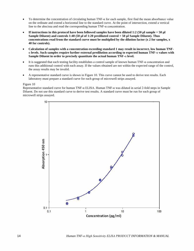
A representative standard curve is shown in Figure 10. This curve cannot be used to derive test results. Each
laboratory must prepare a standard curve for each group of microwell strips assayed.
Figure 10
Representative standard curve for human TNF-α ELISA. Human TNF-α was diluted in serial 2-fold steps in Sample
Diluent. Do not use this standard curve to derive test results. A standard curve must be run for each group of
microwell strips assayed.
Human TNF-α High Sensitivity ELISA PRODUCT INFORMATION & MANUAL 15
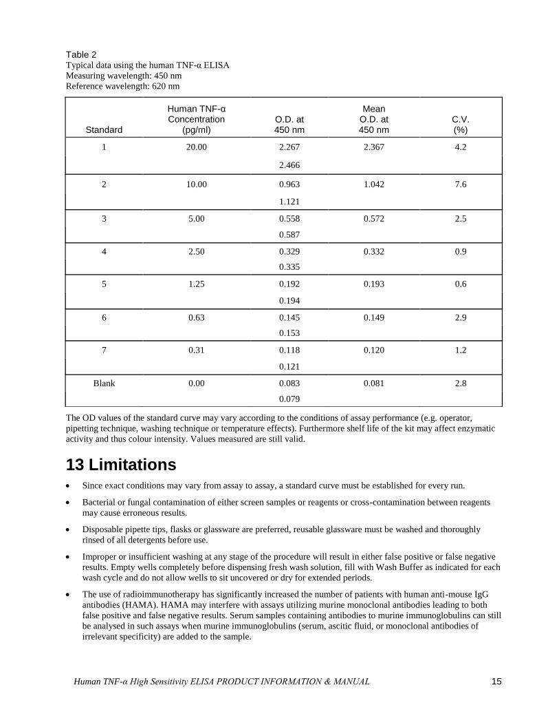
Table 2 Typical data using the human TNF-α ELISA
Measuring wavelength: 450 nm
Reference wavelength: 620 nm
Standard
Human TNF-α Concentration
(pg/ml) O.D. at 450 nm
Mean O.D. at 450 nm
C.V. (%)
1 20.00 2.267 2.367 4.2
2.466
2 10.00 0.963 1.042 7.6
1.121
3 5.00 0.558 0.572 2.5
0.587
4 2.50 0.329 0.332 0.9
0.335
5 1.25 0.192 0.193 0.6
0.194
6 0.63 0.145 0.149 2.9
0.153
7 0.31 0.118 0.120 1.2
0.121
Blank 0.00 0.083 0.081 2.8
0.079
The OD values of the standard curve may vary according to the conditions of assay performance (e.g. operator,
pipetting technique, washing technique or temperature effects). Furthermore shelf life of the kit may affect enzymatic
activity and thus colour intensity. Values measured are still valid.
13 Limitations Since exact conditions may vary from assay to assay, a standard curve must be established for every run.
Bacterial or fungal contamination of either screen samples or reagents or cross-contamination between reagents
may cause erroneous results.
Disposable pipette tips, flasks or glassware are preferred, reusable glassware must be washed and thoroughly
rinsed of all detergents before use.
Improper or insufficient washing at any stage of the procedure will result in either false positive or false negative
results. Empty wells completely before dispensing fresh wash solution, fill with Wash Buffer as indicated for each
wash cycle and do not allow wells to sit uncovered or dry for extended periods.
The use of radioimmunotherapy has significantly increased the number of patients with human anti-mouse IgG
antibodies (HAMA). HAMA may interfere with assays utilizing murine monoclonal antibodies leading to both
false positive and false negative results. Serum samples containing antibodies to murine immunoglobulins can still
be analysed in such assays when murine immunoglobulins (serum, ascitic fluid, or monoclonal antibodies of
irrelevant specificity) are added to the sample.
16 Human TNF-α High Sensitivity ELISA PRODUCT INFORMATION & MANUAL
14 Performance Characteristics
14.1 Sensitivity
The limit of detection of human TNF-α defined as the analyte concentration resulting in an absorbance significantly
higher than that of the dilution medium (mean plus 2 standard deviations) was determined to be 0.13 pg/ml (mean of 6
independent assays).
14.2 Reproducibility
14.2.1 Intra-assay
Reproducibility within the assay was evaluated in 3 independent experiments. Each assay was carried out with 6
replicates of 6 serum samples containing different concentrations of human TNF-α. 2 standard curves were run on
each plate. The calculated overall intra- assay coefficient of variation was 8.5%.
14.2.2 Inter-assay
Assay to assay reproducibility within one laboratory was evaluated in 3 independent experiments. Each assay was
carried out with 6 replicates of 6 serum samples containing different concentrations of human TNF- α. 2 standard
curves were run on each plate. The calculated overall inter-assay coefficient of variation was 9.8%.
14.3 Spike Recovery
The spike recovery was evaluated by spiking recombinant human TNF- α into human serum samples. Recoveries were
determined in 3 independent experiments with 8 serum samples. Recoveries ranged from 84% to 97% with an overall
mean recovery of 91%.
14.4 Dilution Parallelism 4 serum samples with different levels of human TNF-α were analysed at serial 2 fold dilutions with 4 replicates each.
The recovery ranged from 81% to 101% with an overall recovery of 95%.
14.5 Sample Stability
14.5.1 Freeze-Thaw Stability
Aliquots of serum samples (spiked or unspiked) were stored at -20°C and thawed 5 times, and the human TNF-α levels
determined. There was a slight loss of human TNF-α immunoreactivity detected by freezing and thawing, which
therefore should be avoided.
14.5.2 Storage Stability
Aliquots of serum and cell culture samples (spiked or unspiked) were stored at -20°C, 2-8°C, room temperature (RT)
and at 37°C, and the human TNF-α level determined after 24 h. There was no significant loss of human TNF-α
immunoreactivity detected during storage at -20°C and 2-8°C.
A slight loss of human TNF-α immunoreactivity was detected during storage at RT and 37°C after 24 h, which
therefore should be avoided.
14.6 Comparison of Serum and Plasma From several individuals, serum as well as EDTA, citrate and heparin plasma obtained at the same time, were
evaluated. Human TNF-α concentrations were not significantly different and therefore all these body fluids are
suitable for the assay. It is nevertheless highly recommended to assure the uniformity of blood preparations.
14.7 Specificity The interference of circulating factors of the immune systeme was evaluated by spiking these proteins at
physiologically relevant concentrations into a human TNF-α positive serum.
There was no crossreactivity detected, notably not with TNF-β.
14.8 Expected Values

Human TNF-α High Sensitivity ELISA PRODUCT INFORMATION & MANUAL 17
A panel of 8 sera from apparently healthy blood donors (males and females) was tested for human TNF-α. The
detected human TNF-α levels ranged between not detectable and 3.22 pg/ml with a mean level of 1.59 pg/ml. The
normal levels measured may vary with the sample collective used.
14.9 Calibration
This immunoassay is calibrated with highly purified recombinant human TNF-α, which has been evaluated against the
International Reference Standard NIBSC 87/650 and has been shown to be equivalent.
NIBSC 87/650 is quantitated in International Units (IU), 1IU corresponding to 25 pg human TNF-α.
15 Reagent Preparation Summary
15.1 Wash Buffer (1x) Add Wash Buffer Concentrate 20x (50 ml) to 950 ml distilled water.
15.2 Assay Buffer (1x) Add Assay Buffer Concentrate 20x (5 ml) to 95 ml distilled water.
Number of Strips Assay Buffer Concentrate (ml) Distilled Water (ml)
1 - 6 2.5 47.5
1 - 12 5.0 95.0
15.3 Biotin-Conjugate
Make a 1:100 dilution of Biotin-Conjugate in Assay Buffer (1x):
Number of Strips Biotin-Conjugate (ml) Assay Buffer (1x) (ml)
1 - 6 0.03 2.97
1 - 12 0.06 5.94
15.4 Streptavidin-HRP Make a 1:400 dilution of Streptavidin-HRP in Assay Buffer (1x):
Number of Strips Streptavidin-HRP (ml) Assay Buffer (1x) (ml)
1 - 6 0.015 5.985
1 - 12 0.030 11.970
15.5 Human TNF-α Standard Reconstitute lyophilized human TNF-α standard with distilled water. (Reconstitution volumαe is stated on
the label of the standard vial.) The concentrated human TNF-α standard must be diluted 1:25 with Sample
Diluent.
18 Human TNF-α High Sensitivity ELISA PRODUCT INFORMATION & MANUAL
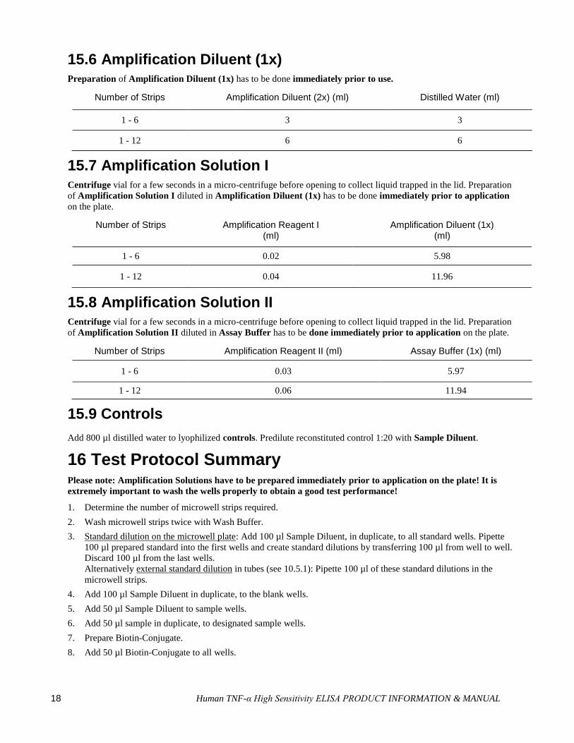
15.6 Amplification Diluent (1x) Preparation of Amplification Diluent (1x) has to be done immediately prior to use.
Number of Strips Amplification Diluent (2x) (ml) Distilled Water (ml)
1 - 6 3 3
1 - 12 6 6
15.7 Amplification Solution I Centrifuge vial for a few seconds in a micro-centrifuge before opening to collect liquid trapped in the lid. Preparation
of Amplification Solution I diluted in Amplification Diluent (1x) has to be done immediately prior to application
on the plate.
Number of Strips Amplification Reagent I (ml)
Amplification Diluent (1x) (ml)
1 - 6 0.02 5.98
1 - 12 0.04 11.96
15.8 Amplification Solution II Centrifuge vial for a few seconds in a micro-centrifuge before opening to collect liquid trapped in the lid. Preparation
of Amplification Solution II diluted in Assay Buffer has to be done immediately prior to application on the plate.
Number of Strips Amplification Reagent II (ml) Assay Buffer (1x) (ml)
1 - 6 0.03 5.97
1 - 12 0.06 11.94
15.9 Controls
Add 800 µl distilled water to lyophilized controls. Predilute reconstituted control 1:20 with Sample Diluent.
16 Test Protocol Summary Please note: Amplification Solutions have to be prepared immediately prior to application on the plate! It is
extremely important to wash the wells properly to obtain a good test performance!
1. Determine the number of microwell strips required.
2. Wash microwell strips twice with Wash Buffer.
3. Standard dilution on the microwell plate: Add 100 µl Sample Diluent, in duplicate, to all standard wells. Pipette
100 µl prepared standard into the first wells and create standard dilutions by transferring 100 µl from well to well.
Discard 100 µl from the last wells.
Alternatively external standard dilution in tubes (see 10.5.1): Pipette 100 µl of these standard dilutions in the
microwell strips.
4. Add 100 µl Sample Diluent in duplicate, to the blank wells.
5. Add 50 µl Sample Diluent to sample wells.
6. Add 50 µl sample in duplicate, to designated sample wells.
7. Prepare Biotin-Conjugate.
8. Add 50 µl Biotin-Conjugate to all wells.
Human TNF-α High Sensitivity ELISA PRODUCT INFORMATION & MANUAL 19
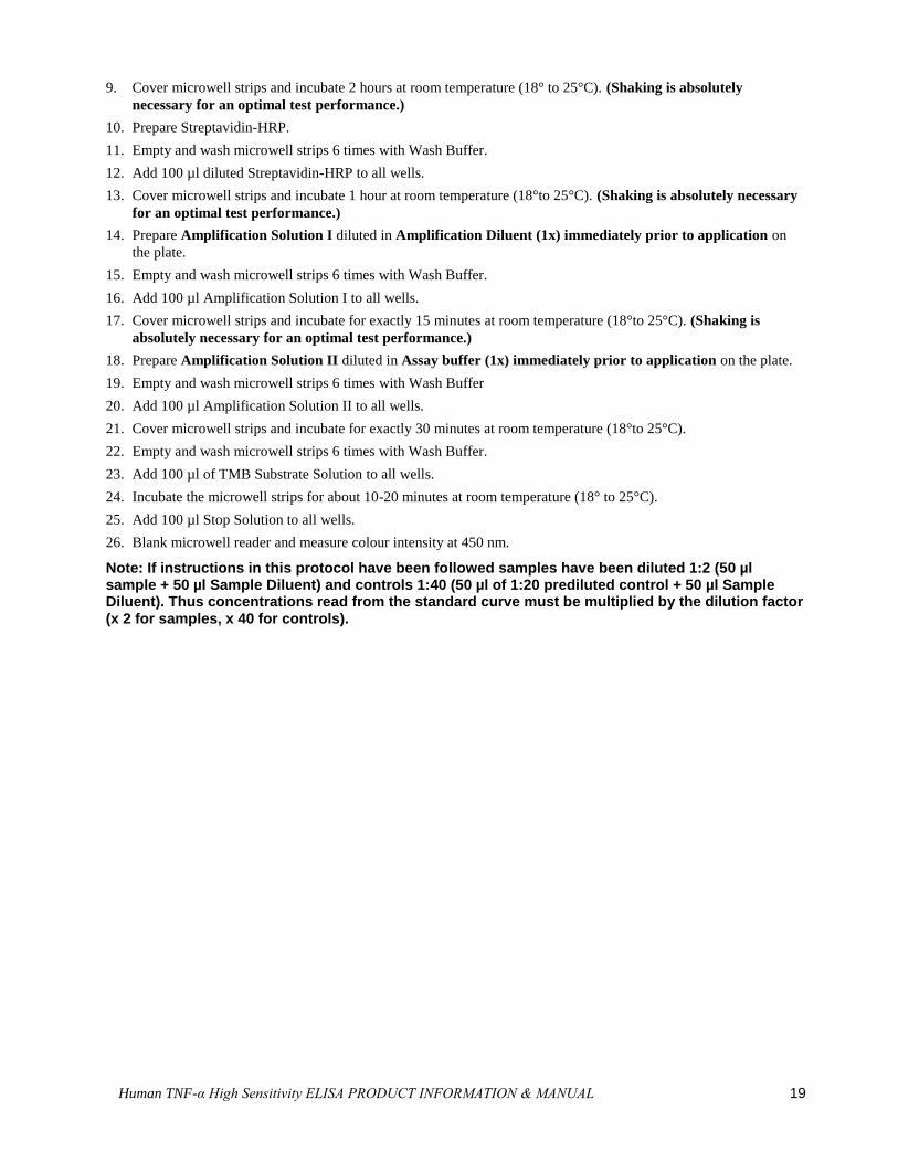
9. Cover microwell strips and incubate 2 hours at room temperature (18° to 25°C). (Shaking is absolutely
necessary for an optimal test performance.)
10. Prepare Streptavidin-HRP.
11. Empty and wash microwell strips 6 times with Wash Buffer.
12. Add 100 µl diluted Streptavidin-HRP to all wells.
13. Cover microwell strips and incubate 1 hour at room temperature (18°to 25°C). (Shaking is absolutely necessary
for an optimal test performance.)
14. Prepare Amplification Solution I diluted in Amplification Diluent (1x) immediately prior to application on
the plate.
15. Empty and wash microwell strips 6 times with Wash Buffer.
16. Add 100 µl Amplification Solution I to all wells.
17. Cover microwell strips and incubate for exactly 15 minutes at room temperature (18°to 25°C). (Shaking is
absolutely necessary for an optimal test performance.)
18. Prepare Amplification Solution II diluted in Assay buffer (1x) immediately prior to application on the plate.
19. Empty and wash microwell strips 6 times with Wash Buffer
20. Add 100 µl Amplification Solution II to all wells.
21. Cover microwell strips and incubate for exactly 30 minutes at room temperature (18°to 25°C).
22. Empty and wash microwell strips 6 times with Wash Buffer.
23. Add 100 µl of TMB Substrate Solution to all wells.
24. Incubate the microwell strips for about 10-20 minutes at room temperature (18° to 25°C).
25. Add 100 µl Stop Solution to all wells.
26. Blank microwell reader and measure colour intensity at 450 nm.
Note: If instructions in this protocol have been followed samples have been diluted 1:2 (50 µl sample + 50 µl Sample Diluent) and controls 1:40 (50 µl of 1:20 prediluted control + 50 µl Sample Diluent). Thus concentrations read from the standard curve must be multiplied by the dilution factor (x 2 for samples, x 40 for controls).
20 Human TNF-α High Sensitivity ELISA PRODUCT INFORMATION & MANUAL
Documentation and support
Obtaining support
Technical support For the latest services and support information for all locations, visit
www.thermofisher.com.
At the website, you can:
Access worldwide telephone and fax numbers to contact Technical Support and Sales
facilities
Search through frequently asked questions (FAQs)
Submit a question directly to Technical Support (thermofisher.com/support)
Search for user documents, SDSs, vector maps and sequences, application notes,
formulations, handbooks, certificates of analysis, citations, and other product support
documents
Obtain information about customer training
Download software updates and patches
Safety Data Sheets (SDS)
Safety Data Sheets (SDSs) are available at thermofisher.com/support.
Limited product warranty
Life Technologies Corporation and/or its affiliate(s) warrant their products as set forth in
the Life Technologies’ General Terms and Conditions of Sale found on Life
Technologies’ website at www.thermofisher.com/us/en/home/global/terms-and-
conditions.html. If you have any questions, please contact Life Technologies at
www.thermofisher.com/support.


For support visit thermofisher.com/support or email [email protected]
thermofisher.com
23 January 2017