Allosteric signaling Biochemistry Direct negative feedback Indirect feedback Cyclic processes.
An unaltered orthosteric site and a network of long-range allosteric interactions for PNU-120596 in α7 nicotinic acetylcholine receptors
Christopher B. Marotta1, Henry A. Lester2, and Dennis A. Dougherty1
1Division of Chemistry and Chemical Engineering, California Institute of Technology, Pasadena, CA, 91125, USA
2Division of Biology and Biological Engineering, California Institute of Technology, Pasadena, CA, 91125, USA
Summary
Nicotinic acetylcholine receptors (nAChRs) are vital to neuronal signaling, are implicated in
important processes such as learning and memory, and are therapeutic targets for neural diseases.
The α7 nAChR has been implicated in Alzheimer’s disease and schizophrenia, and allosteric
modulators have become one focus of drug development efforts. We investigate the mode of
action of the α7-selective positive allosteric modulator, PNU-120596, and show that the higher
potency of acetylcholine in the presence of PNU-120596 is not due to an altered agonist binding
site. In addition, we propose several residues in the gating interface and transmembrane region
that are functionally important to transduction of allosteric properties and link PNU-120596, the
acetylcholine binding region, and the receptor’s gate. These results suggest global protein
stabilization from a communication network through several key residues that alter the gating
equilibrium of the receptor while leaving the agonist binding properties unperturbed.
Graphical abstract
Contact: Dennis A Dougherty, Division of Chemistry and Chemical Engineering, California Institute of Technology, 1200 E California Blvd., Pasadena, CA 91125. Tel.: 626-395-6089; Fax: 626-564-9297; [email protected].
Publisher's Disclaimer: This is a PDF file of an unedited manuscript that has been accepted for publication. As a service to our customers we are providing this early version of the manuscript. The manuscript will undergo copyediting, typesetting, and review of the resulting proof before it is published in its final citable form. Please note that during the production process errors may be discovered which could affect the content, and all legal disclaimers that apply to the journal pertain.
HHS Public AccessAuthor manuscriptChem Biol. Author manuscript; available in PMC 2016 August 20.
Published in final edited form as:Chem Biol. 2015 August 20; 22(8): 1063–1073. doi:10.1016/j.chembiol.2015.06.018.
Author M
anuscriptA
uthor Manuscript
Author M
anuscriptA
uthor Manuscript
Introduction
Nicotinic acetylcholine receptors (nAChRs) are pentameric ion channels that are part of the
Cys-loop superfamily of ligand-gated ion channels, which includes receptors gated by other
neurotransmitters such as glycine, serotonin, and GABA. The α7 nAChR displays a large
and dispersed presence throughout the central nervous system (CNS) (Millar and Gotti,
2009). It is comprised of five identical subunits, an uncommon arrangement for nAChRs,
and each subunit contains an extracellular domain, transmembrane domain, and a gating
interface (Figure 1a) (Dougherty, 2008; Lemoine, et al., 2012; Unwin, 2005).
The concepts of allostery, including cooperative transitions between two states of
multisubunit proteins (Monod, et al., 1965), have been applied to nAChRs in two ways.
First, the nAChR itself has been identified as a protein containing two distinct domains, a
binding site for agonists and a conducting pathway (Edelstein and Changeux, 1998; Karlin,
1967).
Second, and more relevant to the present study, compounds have been identified that do not
produce activation on their own, yet modulate activation and desensitization, and bind at
sites distinct from both the agonist site (the “orthosteric” site) and the channel pore. These
are allosteric ligands. At α7 nAChRs, positive allosteric modulators (PAMs) are especially
well studied, and two classes can be distinguished. Type I PAMs increase agonist-induced
activation. Type II PAMs, such as PNU-120596 (Figure 1b), increase agonist-induced
activation and also vastly prolong the waveform of agonist-induced currents; in the usual
interpretation, PAMs favor the active states at the expense of the desensitized states (Figure
1c) (Bertrand and Gopalakrishnan, 2007; Faghih, et al., 2008; Gronlien, et al., 2007; Hurst,
et al., 2005; Szabo, et al., 2014; Williams, et al., 2011). The existence of one or more
additional, desensitized, states was recognized early on (Heidmann and Changeux, 1986;
Katz and Thesleff, 1957).
Marotta et al. Page 2
Chem Biol. Author manuscript; available in PMC 2016 August 20.
Author M
anuscriptA
uthor Manuscript
Author M
anuscriptA
uthor Manuscript
Inherent to models of allostery is the notion of action at a distance, and it is of interest to ask
whether the orthosteric binding site, and/or the channel pore, is affected by the presence of
an allosteric modulator. Unfortunately no atomic-scale structural information is available for
full α7 nAChRs in any state, let alone all three states in the presence of either an agonist or
allosteric modulator. However, the high functional resolution of electrophysiological data
allows other approaches to this question. For example, the structurally unrelated allosteric
modulator 4PB-TQS has been shown to change the kinetics of gating as well as single-
channel conductance of α7 nAChRs (Pałczyńska, et al., 2012), indicating that an allosteric
modulator can change the structure of the conducting pore.
This study begins by determining to what extent, if any, an allosteric modulator changes the
orthosteric site (Figure 2). Previous data suggested that an allosteric modulator can affect
residues within the extracellular domain, but outside the orthosteric site itself (Barron, et al.,
2009). Non-canonical amino acid mutagenesis provides high-resolution data that
complement those from X-ray crystallography. The key binding interactions at the agonist
binding site of nAChRs – a cation-π interaction and two hydrogen bonds – can be probed in
ways that would be sensitive to ligand displacements of <1 Å (Dougherty and Van Arnam,
2014; Tavares, et al., 2012; Van Arnam, et al., 2013). We have therefore applied non-
canonical amino acid mutagenesis to ask whether the presence of a PAM in any way
modulates these binding interactions at the orthosteric site.
Our next goal was to map out the functional coupling pathway from the orthosteric site to
the allosteric binding site of PNU-120596 – which is thought to be in the transmembrane
region (Bertrand, et al., 2008; Young, et al., 2008) – and/or from the allosteric site to the
channel gate. We term our strategy, which again uses functional measurements, “double
perturbation cycle analysis” (see Figure 4 below). In this analysis, the first perturbation is
mutation of the protein (with conventional or non-canonical amino acids); and the second
perturbation is not a mutation, but the addition of PNU-120596. Non-additivity of the two
perturbations indicates that the protein mutation differentially impacts receptor function
depending on whether PNU-120596 is or is not present, suggesting that the residue under
study plays an important role in allosteric modulation (Daeffler, et al., 2012; Gleitsman, et
al., 2009; Miles, et al., 2012). The analysis parallels that of the common double mutant cycle
analysis, including the notion of an interaction energy, designated as ΔΔG. From these
studies, we have identified several residues with significant ΔΔG values, suggesting a
potential pathway of communication from the agonist binding site to the PAM binding site
and then on to the receptor gate.
Results
Methodology for Interpretation of Functional Coupling Comparisons
As noted in the Introduction, we sought to identify mutations of the receptor that
differentially impact function when PNU-120596 is or is not present. Since we wish to
evaluate a large number of sites throughout the protein, our metric is EC50, rather than more
tedious single-channel methods. We fully appreciate the composite nature of EC50, and have
in fact used it to our advantage in evaluating an allosteric modulator.
Marotta et al. Page 3
Chem Biol. Author manuscript; available in PMC 2016 August 20.
Author M
anuscriptA
uthor Manuscript
Author M
anuscriptA
uthor Manuscript
As noted above, for a ligand-gated ion channel one can envision two limiting modes of
allosteric activation. Binding of the modulator could induce a conformational change in the
protein that propagates to the orthosteric site, altering the innate affinity of that site for the
natural agonist. Alternatively, the allosteric modulator could impact the gating transition of
the receptor, by binding essentially at the “gate” or, again, by action through a distance.
We have developed a strategy to distinguish these two possibilities and, in so doing, have
removed ambiguities associated with EC50 measurements. Over the past 20 years, we have
developed methods for probing structure-function relationships at the agonist binding site of
nicotinic receptors and related proteins with unprecedented precision (Dougherty and Van
Arnam, 2014). Using non-canonical amino acids, we can reveal key drug-receptor contacts.
We can identify cation-π interactions using fluorination, and we can evaluate potential
hydrogen bonding interactions using backbone mutagenesis. Importantly, both approaches
provide information on the magnitude of the non-covalent interaction between drug and
receptor. As such, we can detect subtle changes in the agonist binding site that would
enhance (or diminish) agonist binding in ways that are just not possible with conventional
approaches. As described below, we find no evidence of alteration of the agonist binding
site on addition of PNU-120596.
EC50 describes a composite of several equilibria, some involving agonist binding, some
involving channel gating. Since we can rule out alteration of binding equilibria, we can
conclude that changes in EC50 induced by PNU-120596 reflect changes in the gating
equilibria of the receptor. We have used such analyses before, removing the innate
ambiguity in EC50 by eliminating one component of the measurement (Lummis, et al., 2005;
Xiu, et al., 2009).
Again, our goal is to identify residues that play a role in the allosteric modulation provided
by PNU-120596. To do this, we compare the impact of a mutation on wild type function vs.
function when PNU-120596 is present. If the mutation does not affect PNU-120596 in any
way, the mutation’s impact should be the same whether PNU-120596 is present or not.
Stated differently, the impacts on wild type EC50 of the mutation and of PNU-120596
should simply have additive energies. Alternatively, if a mutation alters PNU-120596
function, then the effect of PNU-120596 on receptor function should be different from wild
type when the mutation is present. That is, the side chain mutation and the impact of
PNU-120596 should be non-additive. By analogy to conventional double mutant cycle
analysis, the non-additivity can be expressed as a ΔΔG value (Figure 4), which if markedly
different from zero signifies the presence of non-local conformational effects on a large and
complex membrane bound protein.
The Orthosteric Site: Binding Interactions are Unaffected by PNU-120596
Previous studies of the α7 nAChR show that acetylcholine makes a single cation-π
interaction with TyrA (Figure 2), which is one of five aromatic residues at the orthosteric
binding site (Puskar, et al., 2011). One possible way in which the allosteric binding of
PNU-120596 could impact receptor function is by influencing the shape of the aromatic box,
such that the strength of the cation-π interaction to acetylcholine could change, or the site of
the cation-π interaction could move to another aromatic residue instead of or in cooperation
Marotta et al. Page 4
Chem Biol. Author manuscript; available in PMC 2016 August 20.
Author M
anuscriptA
uthor Manuscript
Author M
anuscriptA
uthor Manuscript
with TyrA. Other studies show that mutations outside the agonist binding site can reshape
the binding site and significantly alter agonist-receptor contacts (Xiu, et al., 2009). One can
identify cation-π interactions using progressive fluorination of the aromatic groups that
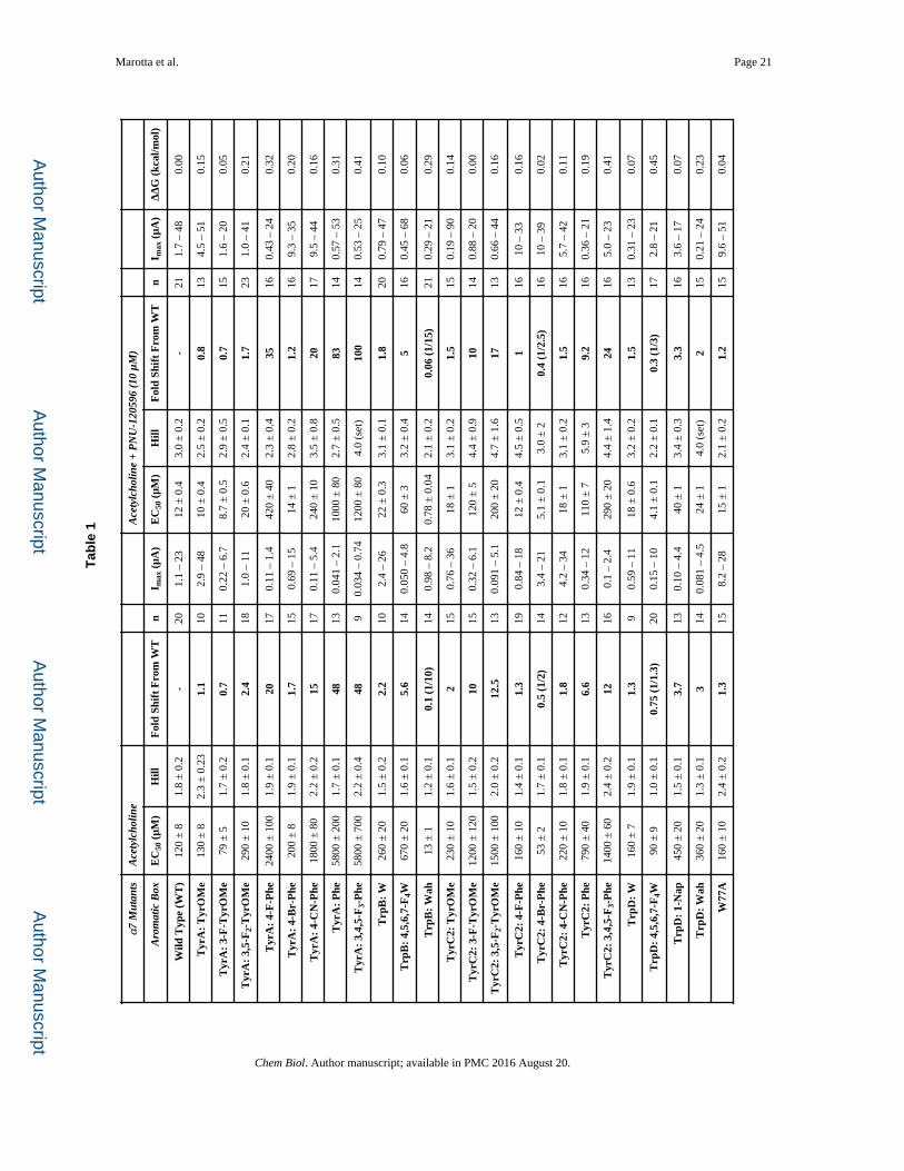
contribute the π electrons (Figure S1). We were able to probe four of the five aromatic box
residues for acetylcholine cation-π interactions in the absence and presence of PNU-120596
(Table 1). TyrC1 could not be probed because of the large loss of function for any
substitution made at this residue; TyrC1 has never been implicated in a cation-π interaction
in the dozens of studies of Cys-loop receptors we have performed (Dougherty and Van
Arnam, 2014).
The first observation is that acetylcholine continues to make a cation-π interaction with
TyrA in the presence of PNU-120596 (Fig. 3a). In addition, the slopes of the two
fluorination plots (with and without PNU-120596) are not meaningfully different, which
indicates that the strength of the cation-π interaction was also unaltered (Dougherty and Van
Arnam, 2014). Since the interaction with TyrA was unchanged, TyrC2 was probed next,
because previous data show that a higher sensitivity agonist – epibatidine – makes a cation-π
interaction with TyrC2 in addition to TyrA in the α7 nAChR (Puskar, et al., 2011). As seen
in Figure 3b, a cation-π interaction still does not exist between TyrC2 and acetylcholine in
the presence of PNU-120596. Two additional observations can be made regarding
interactions with the TyrC2 residue. The near wild type receptor response for bulky
substituent groups (4-CN-Phe, 4-Br-Phe and TyrOMe) and severe loss of function for
small/no substituents (4-F-Phe and Phe) suggest a large substituent is needed at the 4-
position in the aromatic ring to maintain proper receptor function. In addition, the receptor
cannot tolerate substitutions at the 3- or 5-position in the ring system, as indicated by the
large loss of function for 3-F-TyrOMe and 3,5-F2-TyrOMe residues. These results suggest a
tight steric environment for TyrC2 at the orthosteric site. Of more relevance here, however,
is the fact that the pattern of responses to substitution at TyrC2 is unaltered by the presence
of PNU-120596.
Since PNU-120596 did not influence the two critical Tyr residues in the α7 nAChR, the Trp
residues were also studied. Note that for most combinations of agonist and nAChR, the
cation-π interaction is to TrpB; α7 is unusual in employing TyrA. For both TrpB and TrpD
in the presence and absence of PNU-120596, there was not a meaningful shift in function
when we express the highly perturbing residue 4,5,6,7-F4-Trp (Table 1) (Figure S1). This
indicates that acetylcholine is not making a cation-π interaction with either of these residues.
Previous studies showed that a much more dramatic mutation of TrpD – to Ala – converted
PNU-120596 into a partial agonist, while maintaining its function as a positive allosteric
modulator (Barron, et al., 2009; Papke, et al., 2014). We find that this mutation shows a
ΔΔG of essentially zero (Table 1).
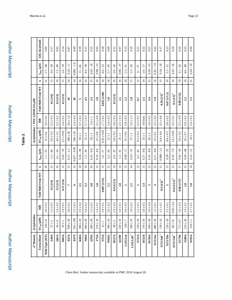
We also applied a previously described strategy for evaluating the two key H-bonding
interactions at the agonist binding site (Tavares, et al., 2012; Van Arnam, et al., 2013).
Briefly, α-hydroxy analogues of α-amino acids are incorporated in ways that are known to
strongly modulate the hydrogen bonding ability of the protein backbone (Figure S1). We
found that perturbation of the hydrogen bond acceptor (L141) or the hydrogen bond donor
(S172) had little impact on PNU-120596 modulation (Figure 2) (Table 2).
Marotta et al. Page 5
Chem Biol. Author manuscript; available in PMC 2016 August 20.
Author M
anuscriptA
uthor Manuscript
Author M
anuscriptA
uthor Manuscript
Overall, the high-precision methodology of non-canonical amino acid mutagenesis allows us
to conclude that the presence PNU-120596 does not measurably alter the key binding
interactions to ACh at the orthosteric site.
A Double Perturbation Cycle Analysis to Identify Residues Critical to PNU-120596 Function
We assessed numerous residues throughout the α7 subunit to determine whether they play a
role in the allosteric modulation by PNU-120596. Previous studies have emphasized how
mutations impact the potentiation produced by PNU-120596, an approach that has produced
useful insights such as identification of the PNU-120596 binding site in the transmembrane
region. We extended previous data by adopting the double perturbation cycle analysis,
which is appropriate for identifying long-range communication between two sites of interest
(described in Figure 4). Results are tabulated for all residues studied in Tables 1, 2, and 3. If
the absolute value of the calculated ΔΔG is ≥ 0.5 kcal/mol, we consider the protein residue
to be functionally important to the allosteric modulatory activity of PNU-120596. This
approach may minimize possible complications arising from changes to the receptor that do
not influence the allosteric communication pathway. (Gleitsman, et al., 2009).
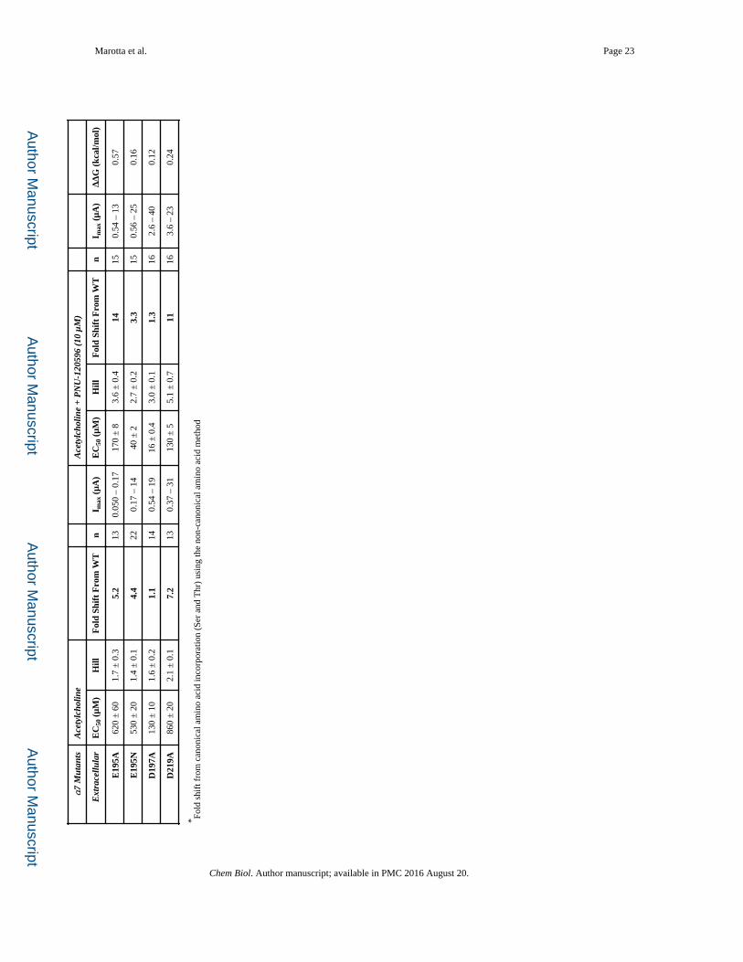
Previous experiments probing agonist-binding interactions in α7 nAChRs were aided by the
inclusion of a pore mutation (T6’S) that produces a modest gain of function and slows α7
desensitization, allowing more precise waveform analysis (Puskar, et al., 2011; Van Arnam,
et al., 2013). However, this mutation is coupled to PNU-120596, with a ΔΔG of 0.68 kcal/
mol. As such, this mutation was not employed except in two cases where the introduced
mutations generated a non-functional receptor that was recoverable through the introduction
of a gain-of-function pore mutation (Zhang, et al., 2011).
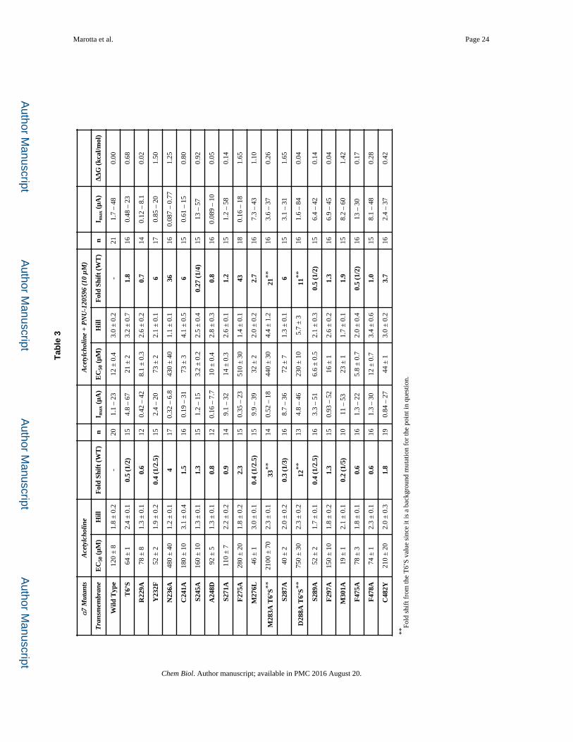
Measuring the Coupling at the Proposed PNU-120596 Binding Site
Several studies of the impact of mutations on PNU-120596 potentiation of acetylcholine
response suggested that PNU-120596 binds in the transmembrane region, across α-helices
M1 (S245 & A248), M2 (M276), and M4 (F478 & C482) of a single subunit (Figure 5)
(Collins, et al., 2011; Young, et al., 2008). Since the goal of the present work was to map out
the functional coupling pathway between the binding site for PNU-120596 and the agonist
binding site, we mutated a large number of residues throughout the receptor (Tables 1, 2, 3).
For purposes of discussion, we will begin at the “bottom” and work our way up to the
agonist binding site.
Of the five residues thought to contribute to the PNU-120596 binding site, only two show a
meaningful functional coupling. However, several nearby residues did show meaningful
coupling (Table 3). Interestingly, these residues generally lie “above” the three residues that
were previously implicated in binding but show no coupling (A248, F478, C482). Further
exploration of the area around this region yielded several other residues – C241 (M1), F275
(M2), and M301 (M3) – that resulted in a large coupling, as reflected in the ΔΔG values. As
seen in Figure 5, these residues lie outside the previously proposed PNU-120596 binding
pocket (Williams, et al., 2011).
Some of these mutations produced altered response waveforms. F275A showed responses
that – regardless of the addition of PNU-120596 – resembled the examples of wild type α7
Marotta et al. Page 6
Chem Biol. Author manuscript; available in PMC 2016 August 20.
Author M
anuscriptA
uthor Manuscript
Author M
anuscriptA
uthor Manuscript
acetylcholine waveforms in Figure 1 (referred to as Type A in Table 4). In contrast, M276L
(example in Figure 1) and M301A markedly lengthened the waveform for application of
ACh alone (Type B).
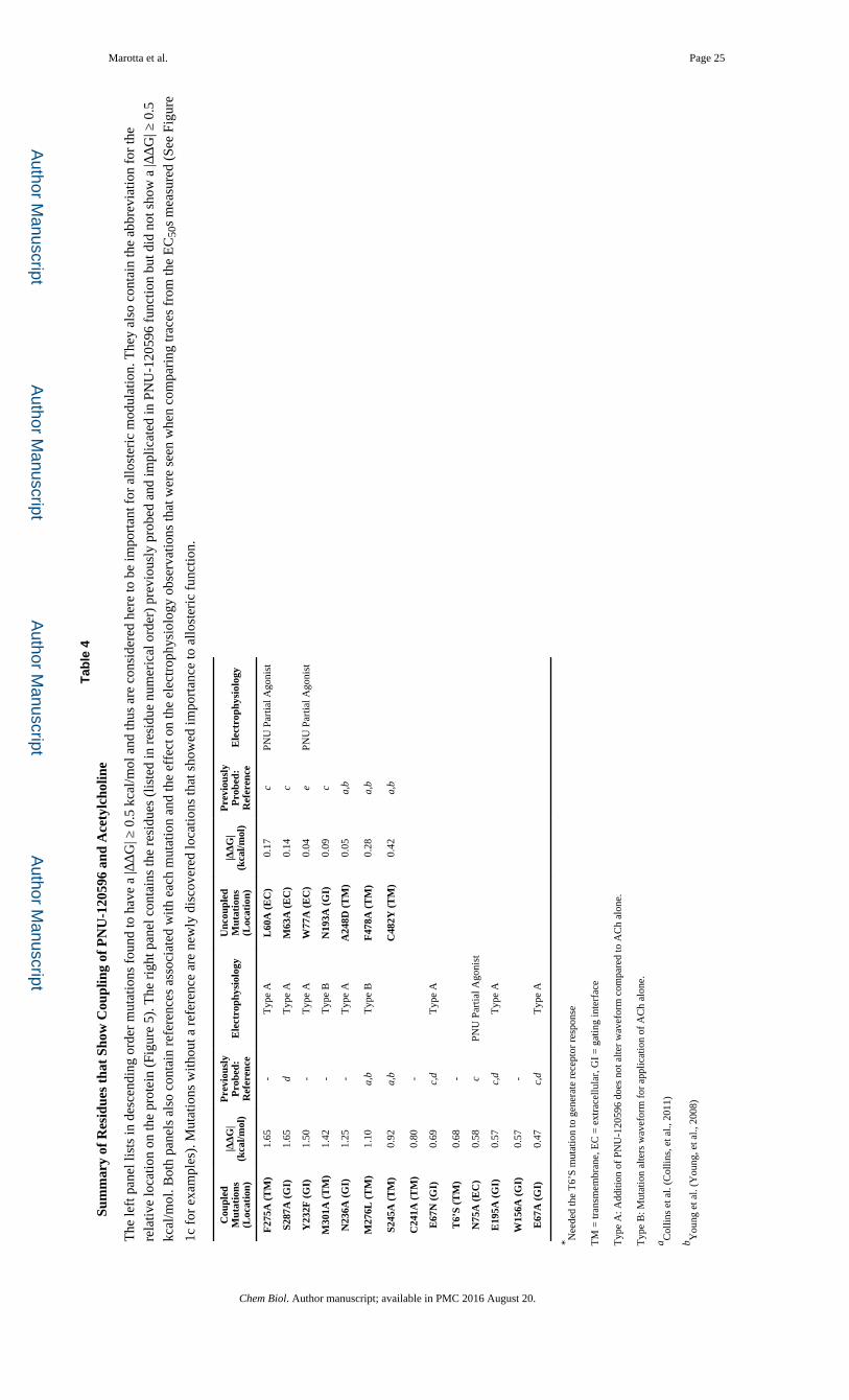
Important Residues in the Gating Interface and the Extracellular Domain
Previous cysteine labeling experiments identified several extracellular residues positioned at
the interface of two adjacent subunits that underwent conformational changes in the α7
nAChR when exposed to either PNU-120596 or acetylcholine (Barron, et al., 2009). These
residues (L60, M63, E67, N75, N193, and E195) were evaluated along with several others in
the surrounding region (Table 2). Figure 5 shows the residues in the gating interface with |
ΔΔG| ≥ 0.5 kcal/mol (Hanek, et al., 2008; Tillman, et al., 2014). Tillman et al. showed
through chimera analysis that specific loops and linkers were necessary for PNU-120596
potentiation of α7 receptors (Tillman, et al., 2014). Here, we were able to isolate specific
residues on some of these identified regions: in Loop 2, E67; in Loop 9, E195; and in the
M2-M3 Linker, S287. Again, we observed distinct response waveforms. Results for
E67A/N, E195A, Y232F, N236A, and S287A all resembled α7 wild type acetylcholine
waveforms, even in the presence of PNU-120596 (Figure 1 and Table 4).
All the mutations that had |ΔΔG| ≥ 0.5 kcal/mol, or that rendered PNU-120596 an agonist,
define a network of residues necessary for propagation of the PNU-120596 effects
throughout the full receptor (Figure 5). These results suggest a conformational wave of
movement upon activation and give insight to the molecular motions that potentially take
place between the closed and open forms of the receptor.
Discussion
The present work aims to evaluate the influences of the positive allosteric modulator
PNU-120596 on α7 nAChRs. Through the use of non-canonical amino acids, the binding
region of acetylcholine was probed for potential changes in interactions with acetylcholine
when PNU-120596 was introduced. These studies produced three key results: 1) the cation-π
interaction with the TyrA residue is not perturbed by the introduction of PNU-120596, 2) no
other cation-π interactions are gained when PNU-120596 is present, and 3) the overall shape
of the agonist binding site seems unperturbed. The aromatic fluorination series and
introduction of α-hydroxy acid residues give compelling evidence that no significant
rearrangements propagate to the orthosteric binding site from the allosteric binding site of
PNU-120596.
Since no distinct change to the agonist binding motif was observed, we suggest that
PNU-120596 does not alter the binding step of the agonist to the receptor and instead exerts
its effects only on the gating equilibrium. As explained above, this result effectively
removes the ambiguity in comparing EC50 values for various mutations. Since the agonist
binding site is unperturbed, it is reasonable to assume that PNU-120596 primarily, if not
solely, influences receptor function by perturbing gating equilibria. The binding of
PNU-120596 apparently impacts important residues required for signal transduction from
the agonist binding site to the channel gate. In evaluating long-range interactions between
residues, functional coupling comparisons based on a double perturbation cycle analysis
Marotta et al. Page 7
Chem Biol. Author manuscript; available in PMC 2016 August 20.
Author M
anuscriptA
uthor Manuscript
Author M
anuscriptA
uthor Manuscript
provide an appropriate and rigorous method (Daeffler, et al., 2012; Gleitsman, et al., 2009;
Miles, et al., 2012). The functional coupling comparisons were used to probe for mutations
that coupled acetylcholine and PNU-120596 together, thus allowing for identification of
residues necessary for proper PAM function and influence. Table 4 summarizes the major
results of this study. From these data, several observations and conclusions can be drawn to
elicit new information on allosteric modulation of α7 nAChRs by PNU-120596.
An interesting observation concerned the mutations M276L and M301A, which changed the
decay current rate of acetylcholine-only waveforms (Table 4). This effect is quite similar to
that seen with the T6’S mutation. Coupling was seen in the double perturbation cycle
analysis for all three residues, because all of these mutations apparently alter the gating
equilibrium and thus diminish the total effect PNU-120596 can exert on the system. M301A
(adjacent to the putative PNU-120596 binding site) and M276L (on the M2 pore-lining
helix) most likely contribute to structural rearrangements in the transmembrane region. In
addition, the mutation L60 shows a large gain of function for acetylcholine alone, which
suggests a possible restructuring coupled to the agonist binding site. Thus, the allosteric
propagation between acetylcholine and PNU-120596 is disrupted, and PNU-120596
becomes a weak agonist.
Another surprising result seen here is that several residues previously implicated in
PNU-120596 binding do not show functional coupling with respect to transmitting the
effects of PNU-120596. Previously, the binding pocket had been proposed to lie in the
transmembrane region and to interact with residues on the M1, M2, and M4 α-helices
(Collins, et al., 2011; Young, et al., 2008). Even though these residues were implicated in
comprising the PNU-120596 binding, they may not contribute to allosteric propagation in
the protein – which is analogous to the residues critical for acetylcholine binding shown
above. Residues adjacent to the proposed binding pocket were implicated in the
communication pathway: N75 lies on the β2 strand connected to loop 2; C241 & S245 lie on
the M1 helix and F275 on the M2 helix. Remarkably, both of these edges of the binding sites
seem to be oriented for interaction with residues at the gating interface. The residues E195
(Loop 9), Y232 (M1), and N236 (M1) of one subunit are located in the vicinity of E67
(Loop 2), W156 (Loop 7), and S287 (M2-3 Linker) of the adjacent subunit. This suggests
the presence of a collection of residues in the gating interface region that communicates the
allosteric potentiation between the extracellular and transmembrane regions (Figure 5). This
interpretation is reinforced by the fact that five of these six residues (excluding W156) show
nearly α7 wild type response waveforms even when PNU-120596 is present (Table 4). Even
though the residue N193 is not included in this complex despite its proximity, a possible
explanation for the effects previously seen by Barron et al. is that its physical location can be
reorienting because the above-mentioned complex movements change the solvent exposure
(Barron, et al., 2009).
PNU-120596 thus exerts positive allosteric modulation by binding in the transmembrane
region, which stabilizes the gating interface and changes the gating equilibrium of the
receptor, allowing for a lower concentration of the agonist to open the channel. Also,
stabilizing this interaction prolongs channel activation, presumably by decelerating
desensitization of the α7 receptor, which is the most rapidly desensitizing nAChR known
Marotta et al. Page 8
Chem Biol. Author manuscript; available in PMC 2016 August 20.
Author M
anuscriptA
uthor Manuscript
Author M
anuscriptA
uthor Manuscript
(Zhang, et al., 2011). Naturally this suggests a global interaction involving a complex
change in the stability of the wild type receptor after agonist binding. Here we have
provided a quantitative analysis for identification of residues necessary for proper
propagation of allosteric effects.
Significance
Nicotinic acetylcholine receptors (nAChRs) are critical contributors to neuronal
communication, which also implicates them in vital normal brain processes as well as neural
diseases. The α7 nAChR in particular has been implicated in Alzheimer’s disease and
schizophrenia; thus the molecular understanding of how compounds affect this receptor has
attracted much interest (Narla, et al., 2013; Pandya and Yakel, 2013; Parri, et al., 2011;
Tong, et al., 2011; Young and Geyer, 2013). Attempts to design small molecules that are
specific to α7 have yielded numerous agonists with some therapeutic success (Horenstein, et
al., 2008). As an alternative to selective agonist design, ongoing research has targeted the
development and understanding of allosteric modulators – which have the potential to be
more target specific and thus produce fewer side effects (Christopoulos, 2002; Williams, et
al., 2011). In this study, the use of non-canonical amino acids allowed individual chemical
interactions of the agonist binding to the protein to be probed in the presence of the α7-
specific positive allosteric modulator PNU-120596. The conclusion from this analysis is that
PNU-120596 does not alter the agonist binding pocket. To further probe the molecular basis
of the properties of PNU-120596, conventional mutagenesis throughout the receptor was
performed. Several gating interface residues as well as transmembrane residues were
identified as vital for propagating PNU-120596 properties throughout the receptor. This
network of residues links the agonist binding site to the PNU-120596 binding site and to the
channel gate in the pore of the receptor, influencing the global stabilization of the gating
equilibria.
Methods
Residue Numbering & Protein Modeling
Residue numbering was based on the full-length protein containing the signaling sequence
as found on the Ligand Gated Ion Channel Database (http://lenoverelab.org/LGICdb/
LGICdb.php). The figures were generated using PyMOL and a homology model (generated
via MODELLER) of the rat α7 receptor based on the GluCl crystal structure (PDB 3RHW)
(Hibbs and Gouaux, 2011).
Molecular Biology
The QuikChange protocol (Stratagene) was used for site-directed mutagenesis of the rat
nAChR α7 subunit (pAMV vector). NotI was used to linearize the circular plasmid. The
DNA was purified (Qiagen) and in vitro transcription of mRNA from the linearized DNA
templates was performed using the T7 mMessage Machine kit (Ambion). The resulting
mRNA was purified and isolated using Qiagen’s RNeasy RNA purification kit. The same
linearization and mRNA synthesis protocols were used for the human Ric3 (pAMV)
accessory protein.
Marotta et al. Page 9
Chem Biol. Author manuscript; available in PMC 2016 August 20.
Author M
anuscriptA
uthor Manuscript
Author M
anuscriptA
uthor Manuscript
For non-canonical amino acid incorporation, the amber (UAG) stop codon was used for all
α7 subunit incorporation. The 74-nucleotide THG73 tRNA and 76-nucleotide THG73 tRNA
were in vitro transcribed using the MEGAshortscript T7 (Ambion) kit and isolated using
Chroma Spin DEPC-H2O columns (Clontech). Synthesized non-canonical amino acids
coupled to the dinucleotide dCA were enzymatically ligated to the 74-nucleotide tRNA as
previously described (Nowak, et al., 1998; Xiu, et al., 2009).
ND96 media was used for all experimental running/wash buffers (96 mM NaCl, 1.8mM
CaCl2, 2 mM KCl, 1 mM MgCl2, 5 mM HEPES at pH 7.5). ND96+ media was used for
oocyte storage media (2.5mM sodium pyruvate and 6.7mM theophylline added). No
gentamicin was added to the ND96+ storage media in order to avoid distorting ACh-induced
responses (Amici, et al., 2005).
Oocyte Preparation and Injection
Xenopus laevis stage V and VI oocytes were harvested via standard protocols (Nowak, et al.,
1998). For conventional mutagenesis, mRNA mixtures of α7 and Ric3 (Ben-Ami, et al.,
2005; Castillo, et al., 2005; Cheng, et al., 2005; Halevi, et al., 2002; Williams, et al., 2005)
were mixed a ratio of 1:1 by weight to a final concentration of 0.8 ng/nL. Each oocyte
received a 50 nL injection for a 40 ng total mRNA mass delivery. Oocytes were incubated at
18° C for 24–48 h. For non-canonical amino acid incorporation, mRNA mixtures of α7 and
Ric3 were made in a 1:1 ratio to a final concentration of 1.6 ng/nL. These mRNA mixtures
were then mixed in a 1:1 volume ratio with de-protected (photolysis) tRNA, and 50 nL were
injected into each oocyte. Oocytes were incubated at 18° C for 24 h. For non-canonical
amino acids that showed no response after 24 h, the oocyte was subjected to a second
injection and incubation following the aforementioned procedure. Read-through/
reaminoacylation tests (76-nucleotide THG73 tRNA) were performed to confirm non-
canonical amino acid incorporation (Van Arnam, et al., 2013).
Chemical Preparation
Acetylcholine chloride (Sigma-Aldrich) was dissolved to 1 M stock solutions in ND96
buffer. PNU-120596 (Selleckchem) was dissolved in DMSO to 150 mM stock solutions.
Further dilution was performed to make a 10 μM and 0.1% v/v DMSO solution for
experimentation.
Electrophysiology
The two-electrode voltage clamp mode of an OpusXpress 6000A (Axon Instruments) was
used. A holding potential of −60 mV and ND96 media for running buffer were used.
For acetylcholine EC50 measurements, 2-fold and 2.5-fold acetylcholine concentration steps
were applied over several orders of magnitude for dose-response measurements. Drug
applications consisted of applying 1 mL of solution over 8 s. Then the oocytes were washed
with buffer for 3 min at a rate of 3 mL min−1 before the next application of drug. For the
acetylcholine and PNU-120596 EC50 measurements, a similar protocol was used.
PNU-120596 at a 10 μM concentration was pumped in at a rate of 3 mL min−1 for 30 s (1.5
mL total volume per oocyte) prior to the co-application of acetylcholine and 10 μM
Marotta et al. Page 10
Chem Biol. Author manuscript; available in PMC 2016 August 20.
Author M
anuscriptA
uthor Manuscript
Author M
anuscriptA
uthor Manuscript

PNU-120596. The co-application dose was 1 mL of solution over 15 s followed by an
additional 15 s pause to allow each response to reach its maximum value. Then the oocytes
were washed with buffer for 5 min at a rate of 3 mL min−1 before the next co-application of
drug. Again, 2-fold and 2.5-fold acetylcholine concentration steps were used over several
orders of magnitude.
Data were sampled at 50 Hz and then low-passed filtered at 5 Hz. Data were normalized on
a per cell basis, response was averaged on a per concentration basis, and then fit to a single
Hill term to generate EC50 and Hill coefficient (nH) values. Error bars represent standard
error of the mean (SEM) values.
Supplementary Material
Refer to Web version on PubMed Central for supplementary material.
Acknowledgments
We thank Matt Rienzo and Noah Duffy for their work in making the dCA-coupled fluorinated-OMe-Tyrosines and tRNA, and Emily Blythe for developing the α7 homology model. Support for this work came from the NIH (NS 34407).
References
Amici M, Eusebi F, Miledi R. Effects of the antibiotic gentamicin on nicotinic acetylcholine receptors. Neuropharmacology. 2005; 49:627–637. [PubMed: 15936782]
Barron SC, McLaughlin JT, See JA, Richards VL, Rosenberg RL. An allosteric modulator of α7 nicotinic receptors, N-(5-Chloro-2,4-dimethoxyphenyl)-N′-(5-methyl-3-isoxazolyl)-urea (PNU-120596), causes conformational changes in the extracellular ligand binding domain similar to those caused by acetylcholine. Molecular pharmacology. 2009; 76:253–263. [PubMed: 19411608]
Ben-Ami HC, Yassin L, Farah H, Michaeli A, Eshel M, Treinin M. RIC-3 affects properties and quantity of nicotinic acetylcholine receptors via a mechanism that does not require the coiled-coil domains. The Journal of biological chemistry. 2005; 280:28053–28060. [PubMed: 15932871]
Bertrand D, Bertrand S, Cassar S, Gubbins E, Li J, Gopalakrishnan M. Positive allosteric modulation of the α7 nicotinic acetylcholine receptor: ligand interactions with distinct binding sites and evidence for a prominent role of the M2-M3 segment. Molecular pharmacology. 2008; 74:1407–1416. [PubMed: 18678621]
Bertrand D, Gopalakrishnan M. Allosteric modulation of nicotinic acetylcholine receptors. Biochemical pharmacology. 2007; 74:1155–1163. [PubMed: 17707779]
Castillo M, Mulet J, Gutierrez LM, Ortiz JA, Castelan F, Gerber S, Sala S, Sala F, Criado M. Dual role of the RIC-3 protein in trafficking of serotonin and nicotinic acetylcholine receptors. The Journal of biological chemistry. 2005; 280:27062–27068. [PubMed: 15927954]
Cheng A, McDonald NA, Connolly CN. Cell surface expression of 5-hydroxytryptamine type 3 receptors is promoted by RIC-3. The Journal of biological chemistry. 2005; 280:22502–22507. [PubMed: 15809299]
Christopoulos A. Allosteric binding sites on cell-surface receptors: novel targets for drug discovery. Nature reviews Drug discovery. 2002; 1:198–210. [PubMed: 12120504]
Collins T, Young GT, Millar NS. Competitive binding at a nicotinic receptor transmembrane site of two α7-selective positive allosteric modulators with differing effects on agonist-evoked desensitization. Neuropharmacology. 2011; 61:1306–1313. [PubMed: 21820451]
Daeffler KN, Lester HA, Dougherty DA. Functionally important aromatic-aromatic and sulfur-π interactions in the D2 dopamine receptor. Journal of the American Chemical Society. 2012; 134:14890–14896. [PubMed: 22897614]
Marotta et al. Page 11
Chem Biol. Author manuscript; available in PMC 2016 August 20.
Author M
anuscriptA
uthor Manuscript
Author M
anuscriptA
uthor Manuscript
Dougherty DA. Cys-Loop Neuroreceptors: Structure to the Rescue? Chemical reviews. 2008; 108:1642–1653. [PubMed: 18447378]
Dougherty DA, Van Arnam EB. In vivo incorporation of non-canonical amino acids by using the chemical aminoacylation strategy: a broadly applicable mechanistic tool. Chembiochem: a European journal of chemical biology. 2014; 15:1710–1720. [PubMed: 24990307]
Edelstein SJ, Changeux JP. Allosteric transitions of the acetylcholine receptor. Adv Protein Chem. 1998; 51:121–184. [PubMed: 9615170]
Faghih R, Gopalakrishnan M, Briggs CA. Allosteric Modulators of the α7 Nicotinic Acetylcholine Receptor. Journal of Medicinal Chemistry. 2008; 51:701–712. [PubMed: 18198823]
Gleitsman KR, Shanata JA, Frazier SJ, Lester HA, Dougherty DA. Long-range coupling in an allosteric receptor revealed by mutant cycle analysis. Biophysical journal. 2009; 96:3168–3178. [PubMed: 19383461]
Gronlien JH, Hakerud M, Ween H, Thorin-Hagene K, Briggs CA, Gopalakrishnan M, Malysz J. Distinct profiles of α7 nAChR positive allosteric modulation revealed by structurally diverse chemotypes. Molecular pharmacology. 2007; 72:715–724. [PubMed: 17565004]
Halevi S, McKay J, Palfreyman M, Yassin L, Eshel M, Jorgensen E, Treinin M. The C. elegans ric-3 gene is required for maturation of nicotinic acetylcholine receptors. EMBO J. 2002; 21:1012–1020. [PubMed: 11867529]
Hanek AP, Lester HA, Dougherty DA. A Stereochemical Test of a Proposed Structural Feature of the Nicotinic Acetylcholine Receptor. Journal of the American Chemical Society. 2008; 130:13216–13218. [PubMed: 18781739]
Heidmann T, Changeux JP. Characterization of the transient agonist-triggered state of the acetylcholine receptor rapidly labeled by the noncompetitive blocker [3H]chlorpromazine: additional evidence for the open channel conformation. Biochemistry. 1986; 25:6109–6113. [PubMed: 3790508]
Hibbs RE, Gouaux E. Principles of activation and permeation in an anion-selective Cys-loop receptor. Nature. 2011; 474:54–60. [PubMed: 21572436]
Horenstein NA, Leonik FM, Papke RL. Multiple pharmacophores for the selective activation of nicotinic α7-type acetylcholine receptors. Molecular pharmacology. 2008; 74:1496–1511. [PubMed: 18768388]
Hurst RS, Hajos M, Raggenbass M, Wall TM, Higdon NR, Lawson JA, Rutherford-Root KL, Berkenpas MB, Hoffmann WE, Piotrowski DW, et al. A novel positive allosteric modulator of the α7 neuronal nicotinic acetylcholine receptor: in vitro and in vivo characterization. The Journal of neuroscience: the official journal of the Society for Neuroscience. 2005; 25:4396–4405. [PubMed: 15858066]
Karlin A. On the application of “a plausible model” of allosteric proteins to the receptor for acetylcholine. Journal of Theoretical Biology. 1967; 16:306–320. [PubMed: 6048545]
Katz B, Thesleff S. A study of ‘desensitization’ produced by acetylcholine at the motor end-plate. J Physiol (Lond). 1957; 138:63–80. [PubMed: 13463799]
Lemoine D, Jiang R, Taly A, Chataigneau T, Specht A, Grutter T. Ligand-gated ion channels: new insights into neurological disorders and ligand recognition. Chemical reviews. 2012; 112:6285–6318. [PubMed: 22988962]
Lummis SC, Beene DL, Lee LW, Lester HA, Broadhurst RW, Dougherty DA. Cis-trans isomerization at a proline opens the pore of a neurotransmitter-gated ion channel. Nature. 2005; 438:248–252. [PubMed: 16281040]
Miles TF, Bower KS, Lester HA, Dougherty DA. A coupled array of noncovalent interactions impacts the function of the 5-HT3A serotonin receptor in an agonist-specific way. ACS chemical neuroscience. 2012; 3:753–760. [PubMed: 23077719]
Millar NS, Gotti C. Diversity of vertebrate nicotinic acetylcholine receptors. Neuropharmacology. 2009; 56:237–246. [PubMed: 18723036]
Monod J, Wyman J, Changeux JP. On the nature of allosteric transitions: A plausible model. J Mol Biol. 1965; 12:88–118. [PubMed: 14343300]
Marotta et al. Page 12
Chem Biol. Author manuscript; available in PMC 2016 August 20.
Author M
anuscriptA
uthor Manuscript
Author M
anuscriptA
uthor Manuscript
Narla S, Klejbor I, Birkaya B, Lee YW, Morys J, Stachowiak EK, Terranova C, Bencherif M, Stachowiak MK. α7 nicotinic receptor agonist reactivates neurogenesis in adult brain. Biochemical pharmacology. 2013; 86:1099–1104. [PubMed: 23933384]
Nowak M, Gallivan JP, Silverman S, Labarca CG, Dougherty DA, Lester HA. In Vivo Incorporation of Unnatural Amino Acids into Ion Channels in Xenopus Oocyte Expression System. Methods Enzymol. 1998; 293:504–529. [PubMed: 9711626]
Pałczyńska MM, Jindrichova M, Gibb AJ, Millar NS. Activation of α7 Nicotinic Receptors by Orthosteric and Allosteric Agonists: Influence on Single-Channel Kinetics and Conductance. Molecular pharmacology. 2012; 82:910–917. [PubMed: 22874415]
Pandya AA, Yakel JL. Effects of neuronal nicotinic acetylcholine receptor allosteric modulators in animal behavior studies. Biochemical pharmacology. 2013; 86:1054–1062. [PubMed: 23732296]
Papke RL, Horenstein NA, Kulkarni AR, Stokes C, Corrie LW, Maeng CY, Thakur GA. The activity of GAT107, an allosteric activator and positive modulator of α7 nicotinic acetylcholine receptors (nAChR), is regulated by aromatic amino acids that span the subunit interface. The Journal of biological chemistry. 2014; 289:4515–4531. [PubMed: 24362025]
Parri HR, Hernandez CM, Dineley KT. Research update: α7 nicotinic acetylcholine receptor mechanisms in Alzheimer’s disease. Biochemical pharmacology. 2011; 82:931–942. [PubMed: 21763291]
Puskar NL, Xiu X, Lester HA, Dougherty DA. Two neuronal nicotinic acetylcholine receptors, α4β4 and α7, show differential agonist binding modes. The Journal of biological chemistry. 2011; 286:14618–14627. [PubMed: 21343288]
Szabo AK, Pesti K, Mike A, Vizi ES. Mode of action of the positive modulator PNU-120596 on α7 nicotinic acetylcholine receptors. Neuropharmacology. 2014; 81:42–54. [PubMed: 24486377]
Tavares XD, Blum AP, Nakamura DT, Puskar NL, Shanata JA, Lester HA, Dougherty DA. Variations in binding among several agonists at two stoichiometries of the neuronal, α4β2 nicotinic receptor. Journal of the American Chemical Society. 2012; 134:11474–11480. [PubMed: 22716019]
Tillman TS, Seyoum E, Mowrey DD, Xu Y, Tang P. ELIC- α7 Nicotinic acetylcholine receptor (α7nAChR) chimeras reveal a prominent role of the extracellular-transmembrane domain interface in allosteric modulation. The Journal of biological chemistry. 2014; 289:13851–13857. [PubMed: 24695730]
Tong M, Arora K, White MM, Nichols RA. Role of key aromatic residues in the ligand-binding domain of α7 nicotinic receptors in the agonist action of β-amyloid. The Journal of biological chemistry. 2011; 286:34373–34381. [PubMed: 21828053]
Unwin N. Refined structure of the nicotinic acetylcholine receptor at 4Å resolution. Journal of molecular biology. 2005; 346:967–989. [PubMed: 15701510]
Van Arnam EB, Blythe EE, Lester HA, Dougherty DA. An unusual pattern of ligand-receptor interactions for the α7 nicotinic acetylcholine receptor, with implications for the binding of varenicline. Molecular pharmacology. 2013; 84:201–207. [PubMed: 23680636]
Williams DK, Wang J, Papke RL. Investigation of the molecular mechanism of the α7 nicotinic acetylcholine receptor positive allosteric modulator PNU-120596 provides evidence for two distinct desensitized states. Molecular pharmacology. 2011; 80:1013–1032. [PubMed: 21885620]
Williams DK, Wang J, Papke RL. Positive allosteric modulators as an approach to nicotinic acetylcholine receptor-targeted therapeutics: advantages and limitations. Biochemical pharmacology. 2011; 82:915–930. [PubMed: 21575610]
Williams ME, Burton B, Urrutia A, Shcherbatko A, Chavez-Noriega LE, Cohen CJ, Aiyar J. Ric-3 promotes functional expression of the nicotinic acetylcholine receptor α7 subunit in mammalian cells. The Journal of biological chemistry. 2005; 280:12571263.
Xiu X, Puskar NL, Shanata JA, Lester HA, Dougherty DA. Nicotine binding to brain receptors requires a strong cation-π interaction. Nature. 2009; 458:534–537. [PubMed: 19252481]
Young GT, Zwart R, Walker AS, Sher E, Millar NS. Potentiation of α7 nicotinic acetylcholine receptors via an allosteric transmembrane site. Proceedings of the National Academy of Sciences of the United States of America. 2008; 105:14686–14691. [PubMed: 18791069]
Marotta et al. Page 13
Chem Biol. Author manuscript; available in PMC 2016 August 20.
Author M
anuscriptA
uthor Manuscript
Author M
anuscriptA
uthor Manuscript
Young JW, Geyer MA. Evaluating the role of the α7 nicotinic acetylcholine receptor in the pathophysiology and treatment of schizophrenia. Biochemical pharmacology. 2013; 86:1122–1132. [PubMed: 23856289]
Zhang J, Xue F, Whiteaker P, Li C, Wu W, Shen B, Huang Y, Lukas RJ, Chang Y. Desensitization of α7 nicotinic receptor is governed by coupling strength relative to gate tightness. The Journal of biological chemistry. 2011; 286:25331–25340. [PubMed: 21610071]
Marotta et al. Page 14
Chem Biol. Author manuscript; available in PMC 2016 August 20.
Author M
anuscriptA
uthor Manuscript
Author M
anuscriptA
uthor Manuscript
Highlights
• PNU-120596 does not alter acetylcholine binding interactions
• Functional residues in allosteric communication identified
• Global network stabilization rather than adjusted agonist binding domain
Marotta et al. Page 15
Chem Biol. Author manuscript; available in PMC 2016 August 20.
Author M
anuscriptA
uthor Manuscript
Author M
anuscriptA
uthor Manuscript
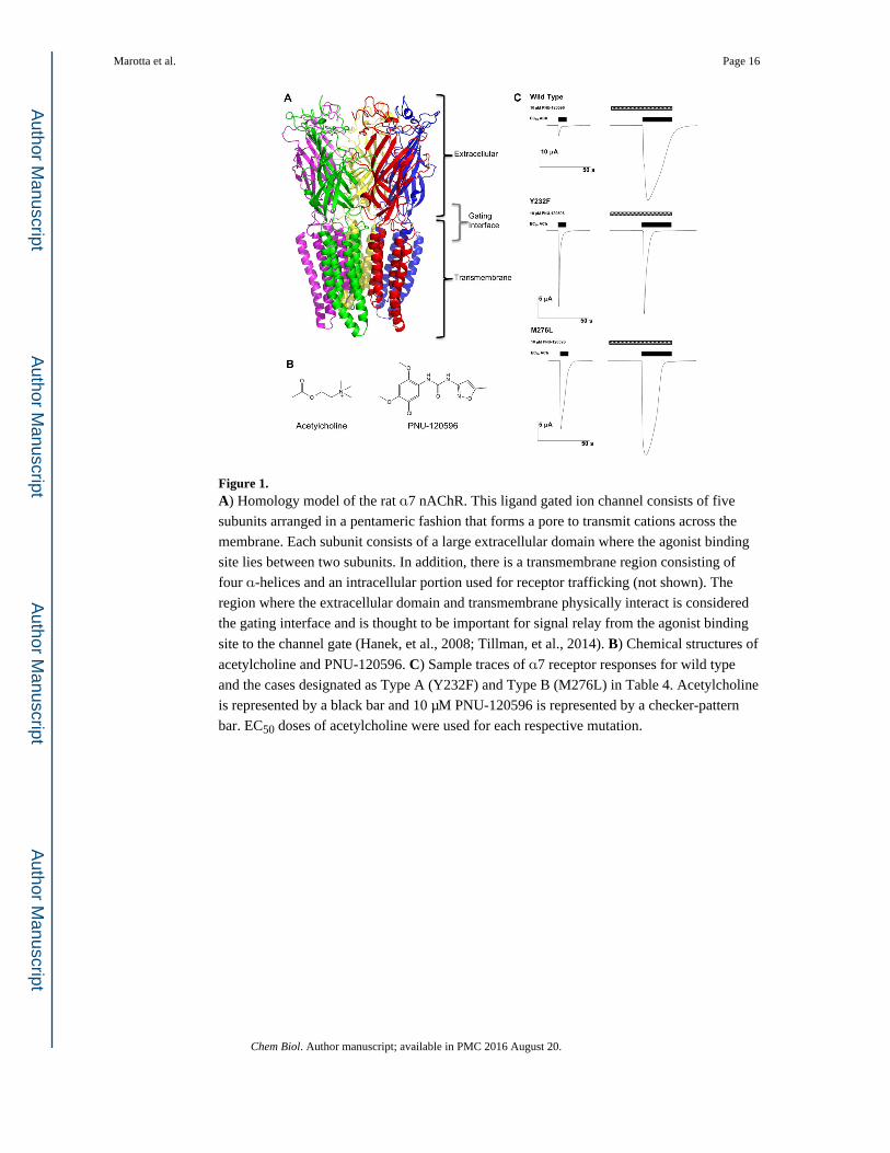
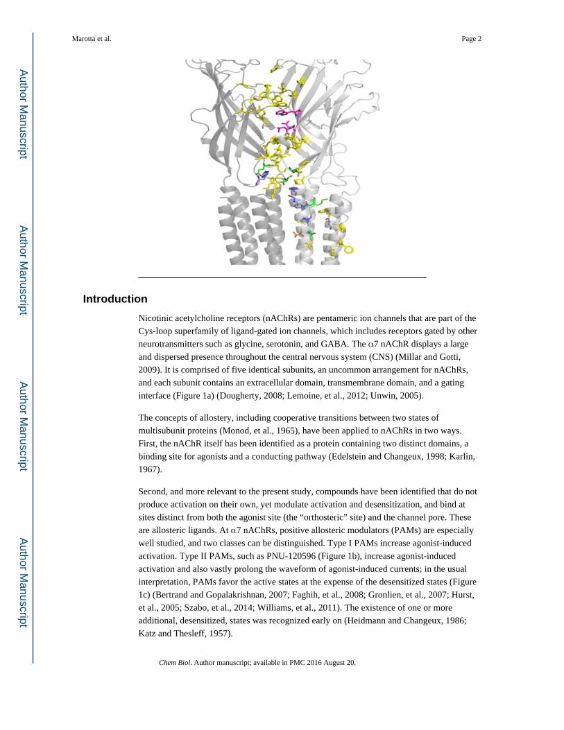
Figure 1. A) Homology model of the rat α7 nAChR. This ligand gated ion channel consists of five
subunits arranged in a pentameric fashion that forms a pore to transmit cations across the
membrane. Each subunit consists of a large extracellular domain where the agonist binding
site lies between two subunits. In addition, there is a transmembrane region consisting of
four α-helices and an intracellular portion used for receptor trafficking (not shown). The
region where the extracellular domain and transmembrane physically interact is considered
the gating interface and is thought to be important for signal relay from the agonist binding
site to the channel gate (Hanek, et al., 2008; Tillman, et al., 2014). B) Chemical structures of
acetylcholine and PNU-120596. C) Sample traces of α7 receptor responses for wild type
and the cases designated as Type A (Y232F) and Type B (M276L) in Table 4. Acetylcholine
is represented by a black bar and 10 μM PNU-120596 is represented by a checker-pattern
bar. EC50 doses of acetylcholine were used for each respective mutation.
Marotta et al. Page 16
Chem Biol. Author manuscript; available in PMC 2016 August 20.
Author M
anuscriptA
uthor Manuscript
Author M
anuscriptA
uthor Manuscript
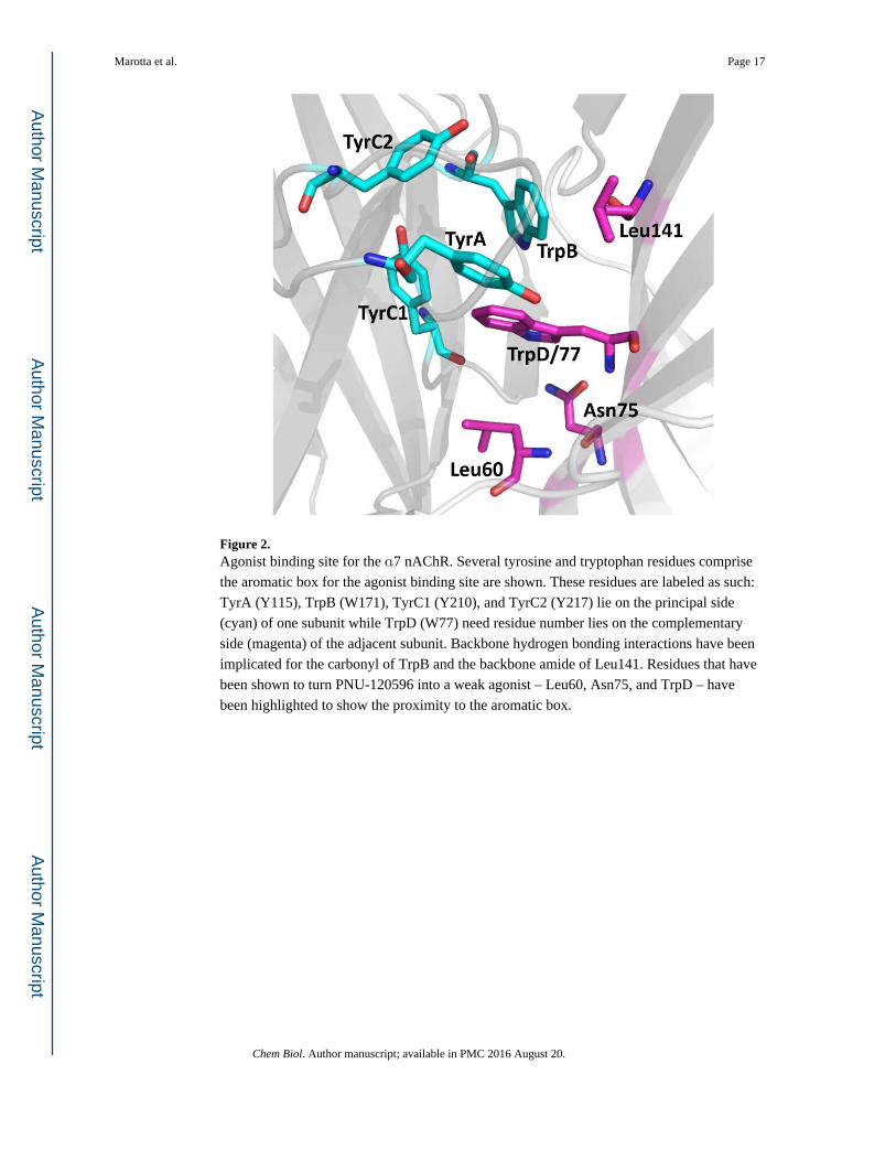
Figure 2. Agonist binding site for the α7 nAChR. Several tyrosine and tryptophan residues comprise
the aromatic box for the agonist binding site are shown. These residues are labeled as such:
TyrA (Y115), TrpB (W171), TyrC1 (Y210), and TyrC2 (Y217) lie on the principal side
(cyan) of one subunit while TrpD (W77) need residue number lies on the complementary
side (magenta) of the adjacent subunit. Backbone hydrogen bonding interactions have been
implicated for the carbonyl of TrpB and the backbone amide of Leu141. Residues that have
been shown to turn PNU-120596 into a weak agonist – Leu60, Asn75, and TrpD – have
been highlighted to show the proximity to the aromatic box.
Marotta et al. Page 17
Chem Biol. Author manuscript; available in PMC 2016 August 20.
Author M
anuscriptA
uthor Manuscript
Author M
anuscriptA
uthor Manuscript
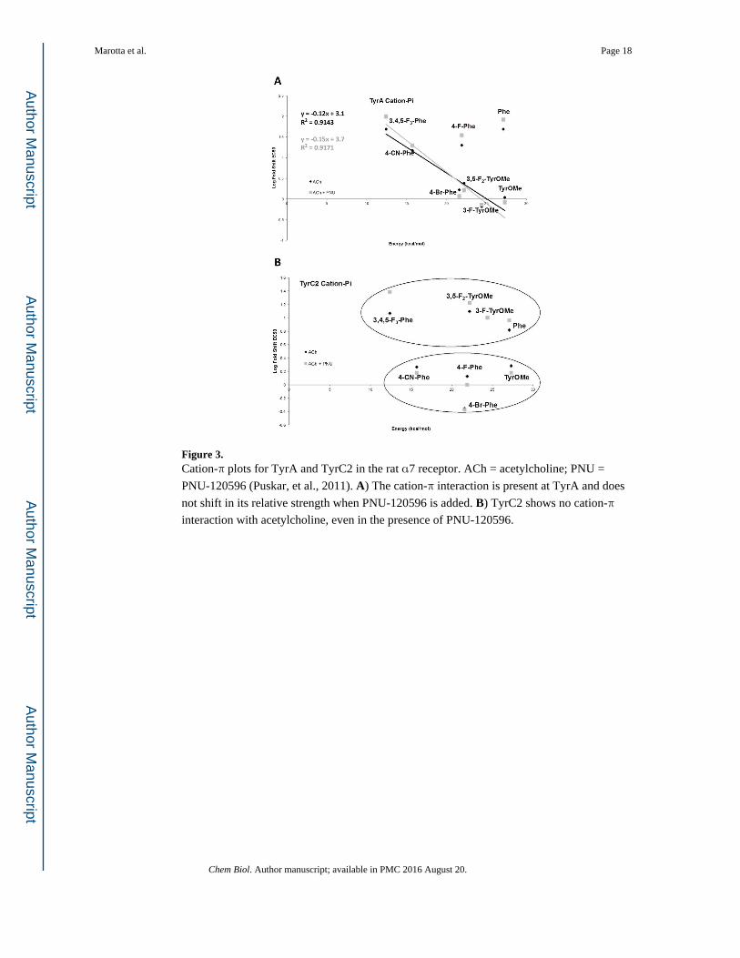
Figure 3. Cation-π plots for TyrA and TyrC2 in the rat α7 receptor. ACh = acetylcholine; PNU =
PNU-120596 (Puskar, et al., 2011). A) The cation-π interaction is present at TyrA and does
not shift in its relative strength when PNU-120596 is added. B) TyrC2 shows no cation-π
interaction with acetylcholine, even in the presence of PNU-120596.
Marotta et al. Page 18
Chem Biol. Author manuscript; available in PMC 2016 August 20.
Author M
anuscriptA
uthor Manuscript
Author M
anuscriptA
uthor Manuscript
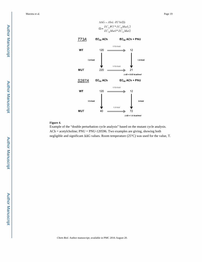
Figure 4. Example of the “double perturbation cycle analysis” based on the mutant cycle analysis.
ACh = acetylcholine; PNU = PNU-120596. Two examples are giving, showing both
negligible and significant ΔΔG values. Room temperature (25°C) was used for the value, T.
Marotta et al. Page 19
Chem Biol. Author manuscript; available in PMC 2016 August 20.
Author M
anuscriptA
uthor Manuscript
Author M
anuscriptA
uthor Manuscript
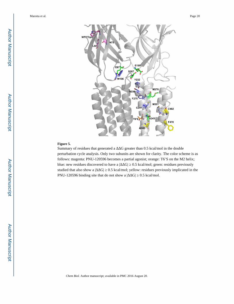
Figure 5. Summary of residues that generated a ΔΔG greater than 0.5 kcal/mol in the double
perturbation cycle analysis. Only two subunits are shown for clarity. The color scheme is as
follows: magenta: PNU-120596 becomes a partial agonist; orange: T6’S on the M2 helix;
blue: new residues discovered to have a |ΔΔG| ≥ 0.5 kcal/mol; green: residues previously
studied that also show a |ΔΔG| ≥ 0.5 kcal/mol; yellow: residues previously implicated in the
PNU-120596 binding site that do not show a |ΔΔG| ≥ 0.5 kcal/mol.
Marotta et al. Page 20
Chem Biol. Author manuscript; available in PMC 2016 August 20.
Author M
anuscriptA
uthor Manuscript
Author M
anuscriptA
uthor Manuscript
Author M
anuscriptA
uthor Manuscript
Author M
anuscriptA
uthor Manuscript
Marotta et al. Page 21
Tab
le 1
α7
Mut
ants
Ace
tylc
holin
eA
cety
lcho
line
+ P
NU
-120
596
(10
μM
)
Aro
mat
ic B
oxE
C50
(μM
)H
illF
old
Shif
t F
rom
WT
nI m
ax (
μA)
EC
50 (
μM)
Hill
Fol
d Sh
ift
Fro
m W
Tn
I max
(μA
)Δ
ΔG
(kc
al/m
ol)
Wild
Typ
e (W
T)
120
± 8
1.8
± 0
.2-
201.
1 –
2312
± 0
.43.
0 ±
0.2
-21
1.7
– 48
0.00
Tyr
A:
Tyr
OM
e13
0 ±
82.
3 ±
0.2
31.
110
2.9
– 48
10 ±
0.4
2.5
± 0
.20.
813
4.5
– 51
0.15
Tyr
A:
3-F
-Tyr
OM
e79
± 5
1.7
± 0
.20.
711
0.22
– 6
.78.
7 ±
0.5
2.9
± 0
.50.
715
1.6
– 20
0.05
Tyr
A:
3,5-
F2-
Tyr
OM
e29
0 ±
10
1.8
± 0
.12.
418
1.0
– 11
20 ±
0.6
2.4
± 0
.11.
723
1.0
– 41
0.21
Tyr
A:
4-F
-Phe
2400
± 1
001.
9 ±
0.1
2017
0.11
– 1
.442
0 ±
40
2.3
± 0
.435
160.
43 –
24
0.32
Tyr
A:
4-B
r-P
he20
0 ±
81.
9 ±
0.1
1.7
150.
69 –
15
14 ±
12.
8 ±
0.2
1.2
169.
3 –
350.
20
Tyr
A:
4-C
N-P
he18
00 ±
80
2.2
± 0
.215
170.
11 –
5.4
240
± 1
03.
5 ±
0.8
2017
9.5
– 44
0.16
Tyr
A:
Phe
5800
± 2
001.
7 ±
0.1
4813
0.04
1 –
2.1
1000
± 8
02.
7 ±
0.5
8314
0.57
– 5
30.
31
Tyr
A:
3,4,
5-F
3-P
he58
00 ±
700
2.2
± 0
.448
90.
034
– 0.
7412
00 ±
80
4.0
(set
)10
014
0.53
– 2
50.
41
Trp
B:
W26
0 ±
20
1.5
± 0
.22.
210
2.4
– 26
22 ±
0.3
3.1
± 0
.11.
820
0.79
– 4
70.
10
Trp
B:
4,5,
6,7-
F4W
670
± 2
01.
6 ±
0.1
5.6
140.
050
– 4.
860
± 3
3.2
± 0
.45
160.
45 –
68
0.06
Trp
B:
Wah
13 ±
11.
2 ±
0.1
0.1
(1/1
0)14
0.98
– 8
.20.
78 ±
0.0
42.
1 ±
0.2
0.06
(1/
15)
210.
29 –
21
0.29
Tyr
C2:
Tyr
OM
e23
0 ±
10
1.6
± 0
.12
150.
76 –
36
18 ±
13.
1 ±
0.2
1.5
150.
19 –
90
0.14
Tyr
C2:
3-F
-Tyr
OM
e12
00 ±
120
1.5
± 0
.210
150.
32 –
6.1
120
± 5
4.4
± 0
.910
140.
88 –
20
0.00
Tyr
C2:
3,5
-F2-
Tyr
OM
e15
00 ±
100
2.0
± 0
.212
.513
0.09
1 –
5.1
200
± 2
04.
7 ±
1.6
1713
0.66
– 4
40.
16
Tyr
C2:
4-F
-Phe
160
± 1
01.
4 ±
0.1
1.3
190.
84 –
18
12 ±
0.4
4.5
± 0
.51
1610
– 3
30.
16
Tyr
C2:
4-B
r-P
he53
± 2
1.7
± 0
.10.
5 (1
/2)
143.
4 –
215.
1 ±
0.1
3.0
± 2
0.4
(1/2
.5)
1610
– 3
90.
02
Tyr
C2:
4-C
N-P
he22
0 ±
10
1.8
± 0
.11.
812
4.2
– 34
18 ±
13.
1 ±
0.2
1.5
165.
7 –
420.
11
Tyr
C2:
Phe
790
± 4
01.
9 ±
0.1
6.6
130.
34 –
12
110
± 7
5.9
± 3
9.2
160.
36 –
21
0.19
Tyr
C2:
3,4
,5-F
3-P
he14
00 ±
60
2.4
± 0
.212
160.
1 –
2.4
290
± 2
04.
4 ±
1.4
2416
5.0
– 23
0.41
Trp
D:
W16
0 ±
71.
9 ±
0.1
1.3
90.
59 –
11
18 ±
0.6
3.2
± 0
.21.
513
0.31
– 2
30.
07
Trp
D:
4,5,
6,7-
F4W
90 ±
91.
0 ±
0.1
0.75
(1/
1.3)
200.
15 –
10
4.1
± 0
.12.
2 ±
0.1
0.3
(1/3
)17
2.8
– 21
0.45
Trp
D:
1-N
ap45
0 ±
20
1.5
± 0
.13.
713
0.10
– 4
.440
± 1
3.4
± 0
.33.
316
3.6
– 17
0.07
Trp
D:
Wah
360
± 2
01.
3 ±
0.1
314
0.08
1 –
4.5
24 ±
14.
0 (s
et)
215
0.21
– 2
40.
23
W77
A16
0 ±
10
2.4
± 0
.21.
315
8.2
– 28
15 ±
12.
1 ±
0.2
1.2
159.
6 –
510.
04
Chem Biol. Author manuscript; available in PMC 2016 August 20.
Author M
anuscriptA
uthor Manuscript
Author M
anuscriptA
uthor Manuscript
Marotta et al. Page 22
Tab
le 2
α7
Mut
ants
Ace
tylc
holin
eA
cety
lcho
line
+ P
NU
-120
596
(10
μM
)
Ext
race
llula
rE
C50
(μM
)H
illF
old
Shif
t F
rom
WT
nI m
ax (
μA)
EC
50 (
μM)
Hill
Fol
d Sh
ift
Fro
m W
Tn
I max
(μA
)Δ
ΔG
(kc
al/m
ol)
Wild
Typ
e (W
T)
120
± 8
1.8
± 0
.2-
201.
1 –
2312
± 0
.43.
0 ±
0.2
-21
1.7
– 48
0.00
L60
A27
± 2
1.3
± 0
.10.
2 (1
/4)
151.
2 –
442.
0 ±
0.2
2.3
± 0
.30.
2 (1
/6)
153.
9 –
500.
17
Q61
A44
± 4
1.4
± 0
.20.
3 (1
/3)
164.
1 –
344.
0 ±
0.1
3.2
± 0
.20.
3 (1
/3)
181.
3 –
890.
05
M63
A32
± 1
1.9
± 0
.10.
27 (
1/4)
130.
42 –
19
2.5
± 0
.03
3.4
± 0
.20.
2 (1
/5)
152.
6 –
390.
14
E67
A83
0 ±
30
1.8
± 0
.17
150.
13 –
1.1
190
± 3
03.
6 ±
1.6
1624
0.24
– 1
10.
47
E67
N72
0 ±
100
1.5
± 0
.36
100.
027
– 0.
4024
0 ±
20
2.4
± 0
.420
140.
085
– 1.
60.
69
K68
A30
0 ±
20
1.9
± 0
.22.
518
0.24
– 3
959
± 1
3.7
± 0
.25
1611
– 4
40.
39
N69
A40
0 ±
30
1.8
± 0
.23.
316
1.3
– 39
52 ±
32.
7 ±
0.3
4.3
164.
4 –
380.
15
Q70
A45
0 ±
30
1.3
± 0
.13.
820
0.10
– 6
.829
± 1
5.4
± 1
2.4
150.
56 –
18
0.25
T73
A21
0 ±
10
1.4
± 0
.11.
813
0.19
– 8
.622
± 0
.83.
5 ±
0.4
1.8
160.
98 –
23
0.03
N75
A8
± 1
1.0
± 0
.10.
067
(1/1
5)15
1.6
– 27
0.29
± 0
.02
1.7
± 0
.20.
025
(1/4
0)16
4.6
– 25
0.58
P10
3G18
0 ±
10
1.9
± 0
.21.
515
2.3
– 26
21 ±
12.
4 ±
0.3
1.8
165.
7 –
450.
09
H12
7A48
± 3
1.9
± 0
.20.
4 (1
/2.5
)14
2.4
– 35
5.8
± 0
.23.
0 ±
0.3
0.5
(1/2
)16
3.8
– 42
0.11
Q13
9F22
0 ±
10
1.8
± 0
.11.
814
1.3
– 35
25 ±
12.
5 ±
0.3
2.1
160.
96 –
37
0.07
L14
1:L
eu12
0 ±
71.
9 ±
0.2
115
0.48
– 1
715
± 0
.62.
8 ±
0.2
1.2
161.
3 –
410.
13
L14
1:L
ah*
180
± 1
01.
9 ±
0.2
1.5
140.
23 –
31
27 ±
15.
0 ±
1.3
1.8
151.
5 –
260.
10
Y15
1A12
0 ±
20
1.2
± 0
.11
153.
2 –
438.
3 ±
0.3
2.3
± 0
.20.
712
11 –
37
0.21
D15
3A17
0 ±
10
1.8
± 0
.21.
415
0.27
– 9
.625
± 1
4.0
(set
)2.
114
1.6
– 17
0.22
W15
6A24
0 ±
30
1.9
± 0
.42
120.
16 –
8.8
65 ±
31.
9 ±
0.2
5.4
150.
18 –
23
0.57
S172
:Ser
480
± 2
01.
3 ±
0.1
415
0.21
– 4
.823
± 0
.53.
2 ±
0.2
1.9
160.
30 –
7.8
0.42
S172
:Sah
*13
0 ±
10
1.2
± 0
.10.
3 (1
/3)*
150.
088
– 3.
18.
4 ±
0.6
3.5
± 0
.80.
36 (
1/3)
*15
0.54
– 1
80.
17
S172
:Thr
120
± 1
01.
2 ±
0.1
115
0.28
– 3
58.
0 ±
0.5
2.8
± 0
.50.
67 (
1/1.
5)16
5.3
– 31
0.23
S172
:Tah
*48
± 1
00.
93 ±
0.2
0.4
(1/2
.5)*
130.
18 –
7.9
1.6
± 0
.05
3.5
± 0
.20.
2 (1
/5)*
160.
33 –
26
0.39
G17
5K7.
5 ±
0.7
1.6
± 0
.20.
06 (
1/17
)17
0.42
– 3
01.
0 ±
0.2
2.5
± 0
.90.
08 (
1/12
)19
2.1
– 42
0.16
S189
A21
0 ±
20
1.4
± 0
.11.
815
0.20
– 4
816
± 0
.72.
9 ±
0.2
1.3
164.
6 –
490.
15
N19
3A21
0 ±
51.
7 ±
0.1
1.8
140.
22 –
12
18 ±
12.
4 ±
0.2
1.5
210.
18 –
18
0.09
Chem Biol. Author manuscript; available in PMC 2016 August 20.
Author M
anuscriptA
uthor Manuscript
Author M
anuscriptA
uthor Manuscript
Marotta et al. Page 23
α7
Mut
ants
Ace
tylc
holin
eA
cety
lcho
line
+ P
NU
-120
596
(10
μM
)
Ext
race
llula
rE
C50
(μM
)H
illF
old
Shif
t F
rom
WT
nI m
ax (
μA)
EC
50 (
μM)
Hill
Fol
d Sh
ift
Fro
m W
Tn
I max
(μA
)Δ
ΔG
(kc
al/m
ol)
E19
5A62
0 ±
60
1.7
± 0
.35.
213
0.05
0 –
0.17
170
± 8
3.6
± 0
.414
150.
54 –
13
0.57
E19
5N53
0 ±
20
1.4
± 0
.14.
422
0.17
– 1
440
± 2
2.7
± 0
.23.
315
0.56
– 2
50.
16
D19
7A13
0 ±
10
1.6
± 0
.21.
114
0.54
– 1
916
± 0
.43.
0 ±
0.1
1.3
162.
6 –
400.
12
D21
9A86
0 ±
20
2.1
± 0
.17.
213
0.37
– 3
113
0 ±
55.
1 ±
0.7
1116
3.6
– 23
0.24
* Fold
shi
ft f
rom
can
onic
al a
min
o ac
id in
corp
orat
ion
(Ser
and
Thr
) us
ing
the
non-
cano
nica
l am
ino
acid
met
hod
Chem Biol. Author manuscript; available in PMC 2016 August 20.
Author M
anuscriptA
uthor Manuscript
Author M
anuscriptA
uthor Manuscript
Marotta et al. Page 24
Tab
le 3
α7
Mut
ants
Ace
tylc
holin
eA
cety
lcho
line
+ P
NU
-120
596
(10
μM
)
Tra
nsm
embr
ane
EC
50 (
μM)
Hill
Fol
d Sh
ift
(WT
)n
I max
(μA
)E
C50
(μM
)H
illF
old
Shif
t (W
T)
nI m
ax (
μA)
ΔΔ
G (
kcal
/mol
)
Wild
Typ
e12
0 ±
81.
8 ±
0.2
-20
1.1
– 23
12 ±
0.4
3.0
± 0
.2-
211.
7 –
480.
00
T6’
S64
± 1
2.4
± 0
.10.
5 (1
/2)
154.
8 –
6721
± 2
3.2
± 0
.71.
816
0.48
– 2
30.
68
R22
9A78
± 8
1.3
± 0
.10.
612
0.42
– 4
28.
1 ±
0.3
2.6
± 0
.20.
714
0.12
– 8
.10.
02
Y23
2F52
± 2
1.9
± 0
.20.
4 (1
/2.5
)15
2.4
– 20
73 ±
22.
1 ±
0.1
617
0.85
– 2
01.
50
N23
6A48
0 ±
40
1.2
± 0
.14
170.
32 –
6.8
430
± 4
01.
1 ±
0.1
3616
0.08
7 –
0.77
1.25
C24
1A18
0 ±
10
3.1
± 0
.41.
516
0.19
– 3
173
± 3
4.1
± 0
.56
150.
61 –
15
0.80
S245
A16
0 ±
10
1.3
± 0
.11.
315
1.2
– 15
3.2
± 0
.22.
5 ±
0.4
0.27
(1/
4)15
13 –
57
0.92
A24
8D92
± 5
1.3
± 0
.10.
812
0.16
– 7
.710
± 0
.42.
8 ±
0.3
0.8
160.
089
– 10
0.05
S271
A11
0 ±
72.
2 ±
0.2
0.9
149.
1 –
3214
± 0
.32.
6 ±
0.1
1.2
151.
2 –
580.
14
F27
5A28
0 ±
20
1.8
± 0
.22.
315
0.35
– 2
351
0 ±
30
1.4
± 0
.143
180.
16 –
18
1.65
M27
6L46
± 1
3.0
± 0
.10.
4 (1
/2.5
)15
9.9
– 39
32 ±
22.
0 ±
0.2
2.7
167.
3 –
431.
10
M28
3A T
6’S*
*21
00 ±
70
2.3
± 0
.133
**14
0.52
– 1
844
0 ±
30
4.4
± 1
.221
**16
3.6
– 37
0.26
S287
A40
± 2
2.0
± 0
.20.
3 (1
/3)
168.
7 –
3672
± 7
1.3
± 0
.16
153.
1 –
311.
65
D28
8A T
6’S*
*75
0 ±
30
2.3
± 0
.212
**13
4.8
– 46
230
± 1
05.
7 ±
311
**16
1.6
– 84
0.04
S289
A52
± 2
1.7
± 0
.10.
4 (1
/2.5
)16
3.3
– 51
6.6
± 0
.52.
1 ±
0.3
0.5
(1/2
)15
6.4
– 42
0.14
F29
7A15
0 ±
10
1.8
± 0
.21.
315
0.93
– 5
216
± 1
2.6
± 0
.21.
316
6.9
– 45
0.04
M30
1A19
± 1
2.1
± 0
.10.
2 (1
/5)
1011
– 5
323
± 1
1.7
± 0
.11.
915
8.2
– 60
1.42
F47
5A78
± 3
1.8
± 0
.10.
616
1.3
– 22
5.8
± 0
.72.
0 ±
0.4
0.5
(1/2
)16
13 –
30
0.17
F47
8A74
± 1
2.3
± 0
.10.
616
1.3
– 30
12 ±
0.7
3.4
± 0
.61.
015
8.1
– 48
0.28
C48
2Y21
0 ±
20
2.0
± 0
.31.
819
0.84
– 2
744
± 1
3.0
± 0
.23.
716
2.4
– 37
0.42
**Fo
ld s
hift
fro
m th
e T
6’S
valu
e si
nce
it is
a b
ackg
roun
d m
utat
ion
for
the
poin
t in
ques
tion.
Chem Biol. Author manuscript; available in PMC 2016 August 20.
Author M
anuscriptA
uthor Manuscript
Author M
anuscriptA
uthor Manuscript
Marotta et al. Page 25
Tab
le 4
Sum
mar
y of
Res
idue
s th
at S
how
Cou
plin
g of
PN
U-1
2059
6 an
d A
cety
lcho
line
The
left
pan
el li
sts
in d
esce
ndin
g or
der
mut
atio
ns f
ound
to h
ave
a |Δ
ΔG
| ≥ 0
.5 k
cal/m
ol a
nd th
us a
re c
onsi
dere
d he
re to
be
impo
rtan
t for
allo
ster
ic m
odul
atio
n. T
hey
also
con
tain
the
abbr
evia
tion
for
the
rela
tive
loca
tion
on th
e pr
otei
n (F
igur
e 5)
. The
rig
ht p
anel
con
tain
s th
e re
sidu
es (
liste
d in
res
idue
num
eric
al o
rder
) pr
evio
usly
pro
bed
and
impl
icat
ed in
PN
U-1
2059
6 fu
nctio
n bu
t did
not
sho
w a
|ΔΔ
G| ≥
0.5
kcal
/mol
. Bot
h pa
nels
als
o co
ntai
n re
fere
nces
ass
ocia
ted
with
eac
h m
utat
ion
and
the
effe
ct o
n th
e el
ectr
ophy
siol
ogy
obse
rvat
ions
that
wer
e se
en w
hen
com
pari
ng tr
aces
fro
m th
e E
C50
s m
easu
red
(See
Fig
ure
1c f
or e
xam
ples
). M
utat
ions
with
out a
ref
eren
ce a
re n
ewly
dis
cove
red
loca
tions
that
sho
wed
impo
rtan
ce to
allo
ster
ic f
unct
ion.
Cou
pled
Mut
atio
ns(L
ocat
ion)
|ΔΔ
G|
(kca
l/mol
)
Pre
viou
sly
Pro
bed:
Ref
eren
ceE
lect
roph
ysio
logy
Unc
oupl
edM
utat
ions
(Loc
atio
n)
|ΔΔ
G|
(kca
l/mol
)
Pre
viou
sly
Pro
bed:
Ref
eren
ceE
lect
roph
ysio
logy
F27
5A (
TM
)1.
65-
Typ
e A
L60
A (
EC
)0.
17c
PNU
Par
tial A
goni
st
S287
A (
GI)
1.65
dT
ype
AM
63A
(E
C)
0.14
c
Y23
2F (
GI)
1.50
-T
ype
AW
77A
(E
C)
0.04
ePN
U P
artia
l Ago
nist
M30
1A (
TM
)1.
42-
Typ
e B
N19
3A (
GI)
0.09
c
N23
6A (
GI)
1.25
-T
ype
AA
248D
(T
M)
0.05
a,b
M27
6L (
TM
)1.
10a,
bT
ype
BF
478A
(T
M)
0.28
a,b
S245
A (
TM
)0.
92a,
bC
482Y
(T
M)
0.42
a,b
C24
1A (
TM
)0.
80-
E67
N (
GI)
0.69
c,d
Typ
e A
T6’
S (T
M)
0.68
-
N75
A (
EC
)0.
58c
PNU
Par
tial A
goni
st
E19
5A (
GI)
0.57
c,d
Typ
e A
W15
6A (
GI)
0.57
-
E67
A (
GI)
0.47
c,d
Typ
e A
* Nee
ded
the
T6’
S m
utat
ion
to g
ener
ate
rece
ptor
res
pons
e
TM
= tr
ansm
embr
ane,
EC
= e
xtra
cellu
lar,
GI
= g
atin
g in
terf
ace
Typ
e A
: Add
ition
of
PNU
-120
596
does
not
alte
r w
avef
orm
com
pare
d to
AC
h al
one.
Typ
e B
: Mut
atio
n al
ters
wav
efor
m f
or a
pplic
atio
n of
AC
h al
one.
a Col
lins
et a
l. (C
ollin
s, e
t al.,
201
1)
b You
ng e
t al.
(You
ng, e
t al.,
200
8)
Chem Biol. Author manuscript; available in PMC 2016 August 20.
Author M
anuscriptA
uthor Manuscript
Author M
anuscriptA
uthor Manuscript
Marotta et al. Page 26c B
arro
n et
al.
(Bar
ron,
et a
l., 2
009)
d Till
man
et a
l. (T
illm
an, e
t al.,
201
4)
e Papk
e et
al.
(Pap
ke, e
t al.,
201
4)
Chem Biol. Author manuscript; available in PMC 2016 August 20.


![Biochimica et Biophysica Acta - COnnecting REpositoriesby volatile anesthetics [20,21]. Reliable structures for individual sub-types of nAChRs, especially their TM domains, are also](https://static.fdocument.org/doc/165x107/60f824a6246e9522bd1db7e7/biochimica-et-biophysica-acta-connecting-repositories-by-volatile-anesthetics.jpg)