Glucose-induced βcell production of IL-1 βcontributes to ... · PDF fileType 2...
10
Introduction Type 2 diabetes mellitus results from an inadequate adaptation of the functional pancreatic β cell mass in the face of insulin resistance. In turn, hyperglycemia by itself has secondary adverse effects on β cells. Indeed, several studies have shown that chronic eleva- tion of blood glucose concentration impairs β cell function, leading to the concept of glucotoxicity (1–7). Moreover, elevated glucose concentrations induce β cell apoptosis in cultured islets from diabetes-prone Psammomys obesus (8) and from humans (9, 10); some- what higher concentrations of glucose are required to induce β cell apoptosis in rodent islets (8, 11, 12). Var- ious molecular mechanisms have been proposed to underlie glucose-induced β cell dysfunction, including formation of advanced glycation end products (13), direct impairment of insulin gene transcription and proinsulin biosynthesis (14, 15), and reduced binding activity of pancreatic duodenal homeobox 1 (PDX-1) (7). Recently, we proposed a mechanism underlying glucose-induced β cell apoptosis in human islets that involves upregulation of Fas receptors by elevated glu- cose levels (9). However, the mediator of glucose- induced Fas expression and its role in glucotoxicity remains unknown. IL-1β has been proposed to mediate both impaired function and destruction of pancreatic β cells during the development of autoimmune type 1 diabetes (16). In keeping with this, treatment of rodent islets with IL-1β results in a potent inhibition of insulin secretion followed by islet destruction (17–23). In human islets, IL-1β has been shown to impair insulin release and to induce Fas expression, enabling Fas-triggered apopto- sis (9, 24–28). Finally, activation of the nuclear tran- scription factor NF-κB is required for IL-1β–induced Fas expression (29–31). Part of these IL-1β effects are reminiscent of the toxic effects of elevated glucose con- centrations. Together, the above findings led us to pos- tulate that glucose may induce IL-1β secretion from β cells in the absence of an autoimmune process. We now The Journal of Clinical Investigation | September 2002 | Volume 110 | Number 6 851 Glucose-induced β cell production of IL-1β contributes to glucotoxicity in human pancreatic islets Kathrin Maedler, 1 Pavel Sergeev, 1 Frédéric Ris, 2, 3 José Oberholzer, 3 Helen I. Joller-Jemelka, 4 Giatgen A. Spinas, 1 Nurit Kaiser, 5 Philippe A. Halban, 2 and Marc Y. Donath 1 1 Division of Endocrinology and Diabetes, University Hospital, Zurich, Switzerland 2 Louis-Jeantet Research Laboratories, University of Geneva Medical Center, Geneva, Switzerland 3 Division of Surgical Research, Department of Surgery, University of Geneva Medical Center, Geneva, Switzerland 4 Division of Clinical Immunology, University Hospital, Zurich, Switzerland 5 Department of Endocrinology and Metabolism, Hebrew University — Hadassah Medical Center, Jerusalem, Israel In type 2 diabetes, chronic hyperglycemia is suggested to be detrimental to pancreatic β cells, caus- ing impaired insulin secretion. IL-1β is a proinflammatory cytokine acting during the autoimmune process of type 1 diabetes. IL-1β inhibits β cell function and promotes Fas-triggered apoptosis in part by activating the transcription factor NF-κB. Recently, we have shown that increased glucose con- centrations also induce Fas expression and β cell apoptosis in human islets. The aim of the present study was to test the hypothesis that IL-1β may mediate the deleterious effects of high glucose on human β cells. In vitro exposure of islets from nondiabetic organ donors to high glucose levels result- ed in increased production and release of IL-1β, followed by NF-κB activation, Fas upregulation, DNA fragmentation, and impaired β cell function. The IL-1 receptor antagonist protected cultured human islets from these deleterious effects. β cells themselves were identified as the islet cellular source of glucose-induced IL-1β. In vivo, IL-1β–producing β cells were observed in pancreatic sections of type 2 diabetic patients but not in nondiabetic control subjects. Similarly, IL-1β was induced in β cells of the gerbil Psammomys obesus during development of diabetes. Treatment of the animals with phlo- rizin normalized plasma glucose and prevented β cell expression of IL-1β. These findings implicate an inflammatory process in the pathogenesis of glucotoxicity in type 2 diabetes and identify the IL-1β/NF-κB pathway as a target to preserve β cell mass and function in this condition. J. Clin. Invest. 110:851–860 (2002). doi:10.1172/JCI200215318. Received for publication February 20, 2002, and accepted in revised form July 16, 2002. Address correspondence to: Marc Y. Donath, Division of Endocrinology and Diabetes, Department of Medicine, University Hospital, CH-8091 Zurich, Switzerland. Phone: 41-1-255-3625; Fax: 41-1-255-4447; E-mail: [email protected]. Pavel Sergeev and Frédéric Ris contributed equally to this work. Conflict of interest: No conflict of interest has been declared. Nonstandard abbreviations used: IL-1 receptor antagonist (IL-1Ra); Fas ligand (FasL); pyrrolidinedithiocarbamate (PDTC); inducible nitric oxide synthase (iNOS); inhibitory κB (I-κB).
Transcript of Glucose-induced βcell production of IL-1 βcontributes to ... · PDF fileType 2...
IntroductionType 2 diabetes mellitus results from an inadequateadaptation of the functional pancreatic β cell mass inthe face of insulin resistance. In turn, hyperglycemiaby itself has secondary adverse effects on β cells.Indeed, several studies have shown that chronic eleva-tion of blood glucose concentration impairs β cellfunction, leading to the concept of glucotoxicity (1–7).Moreover, elevated glucose concentrations induce βcell apoptosis in cultured islets from diabetes-pronePsammomys obesus (8) and from humans (9, 10); some-what higher concentrations of glucose are required toinduce β cell apoptosis in rodent islets (8, 11, 12). Var-ious molecular mechanisms have been proposed to
underlie glucose-induced β cell dysfunction, includingformation of advanced glycation end products (13),direct impairment of insulin gene transcription andproinsulin biosynthesis (14, 15), and reduced bindingactivity of pancreatic duodenal homeobox 1 (PDX-1)(7). Recently, we proposed a mechanism underlyingglucose-induced β cell apoptosis in human islets thatinvolves upregulation of Fas receptors by elevated glu-cose levels (9). However, the mediator of glucose-induced Fas expression and its role in glucotoxicityremains unknown.
IL-1β has been proposed to mediate both impairedfunction and destruction of pancreatic β cells duringthe development of autoimmune type 1 diabetes (16).In keeping with this, treatment of rodent islets with IL-1β results in a potent inhibition of insulin secretionfollowed by islet destruction (17–23). In human islets,IL-1β has been shown to impair insulin release and toinduce Fas expression, enabling Fas-triggered apopto-sis (9, 24–28). Finally, activation of the nuclear tran-scription factor NF-κB is required for IL-1β–inducedFas expression (29–31). Part of these IL-1β effects arereminiscent of the toxic effects of elevated glucose con-centrations. Together, the above findings led us to pos-tulate that glucose may induce IL-1β secretion from βcells in the absence of an autoimmune process. We now
The Journal of Clinical Investigation | September 2002 | Volume 110 | Number 6 851
Glucose-induced β cell production of IL-1β contributes to glucotoxicity in human pancreatic islets
Kathrin Maedler,1 Pavel Sergeev,1 Frédéric Ris,2, 3 José Oberholzer,3
Helen I. Joller-Jemelka,4 Giatgen A. Spinas,1 Nurit Kaiser,5 Philippe A. Halban,2
and Marc Y. Donath1
1Division of Endocrinology and Diabetes, University Hospital, Zurich, Switzerland2Louis-Jeantet Research Laboratories, University of Geneva Medical Center, Geneva, Switzerland3Division of Surgical Research, Department of Surgery, University of Geneva Medical Center, Geneva, Switzerland4Division of Clinical Immunology, University Hospital, Zurich, Switzerland5Department of Endocrinology and Metabolism, Hebrew University — Hadassah Medical Center, Jerusalem, Israel
In type 2 diabetes, chronic hyperglycemia is suggested to be detrimental to pancreatic β cells, caus-ing impaired insulin secretion. IL-1β is a proinflammatory cytokine acting during the autoimmuneprocess of type 1 diabetes. IL-1β inhibits β cell function and promotes Fas-triggered apoptosis in partby activating the transcription factor NF-κB. Recently, we have shown that increased glucose con-centrations also induce Fas expression and β cell apoptosis in human islets. The aim of the presentstudy was to test the hypothesis that IL-1β may mediate the deleterious effects of high glucose onhuman β cells. In vitro exposure of islets from nondiabetic organ donors to high glucose levels result-ed in increased production and release of IL-1β, followed by NF-κB activation, Fas upregulation, DNAfragmentation, and impaired β cell function. The IL-1 receptor antagonist protected cultured humanislets from these deleterious effects. β cells themselves were identified as the islet cellular source ofglucose-induced IL-1β. In vivo, IL-1β–producing β cells were observed in pancreatic sections of type2 diabetic patients but not in nondiabetic control subjects. Similarly, IL-1β was induced in β cells ofthe gerbil Psammomys obesus during development of diabetes. Treatment of the animals with phlo-rizin normalized plasma glucose and prevented β cell expression of IL-1β. These findings implicatean inflammatory process in the pathogenesis of glucotoxicity in type 2 diabetes and identify the IL-1β/NF-κB pathway as a target to preserve β cell mass and function in this condition.
J. Clin. Invest. 110:851–860 (2002). doi:10.1172/JCI200215318.
Received for publication February 20, 2002, and accepted in revised formJuly 16, 2002.
Address correspondence to: Marc Y. Donath, Division ofEndocrinology and Diabetes, Department of Medicine,University Hospital, CH-8091 Zurich, Switzerland. Phone: 41-1-255-3625; Fax: 41-1-255-4447; E-mail: [email protected] Sergeev and Frédéric Ris contributed equally to this work.Conflict of interest: No conflict of interest has been declared.Nonstandard abbreviations used: IL-1 receptor antagonist (IL-1Ra); Fas ligand (FasL); pyrrolidinedithiocarbamate (PDTC);inducible nitric oxide synthase (iNOS); inhibitory κB (I-κB).

identify β cells as the cellular source of glucose-inducedIL-1β in cultured human islets and confirm this usingtissue sections from the pancreas of type 2 diabeticpatients and of Psammomys obesus. The role of suchendogenously produced IL-1β in β cell glucotoxicitywas also explored.
MethodsIslet isolation and culture. Islets were isolated from pan-creata of 11 organ donors at the Department ofSurgery, University of Geneva Medical Center, asdescribed (32–34). Islet purity was greater than 95% asjudged by dithizone staining (if this degree of puritywas not achieved by routine isolation, islets were handpicked). The donors, aged 40–70 years, were heart-beat-ing cadaver organ donors, and none had a previous his-tory of diabetes or metabolic disorders. For long-termin vitro studies, the islets were cultured on extracellu-lar matrix–coated plates derived from bovine cornealendothelial cells (Novamed Ltd., Jerusalem, Israel),allowing the cells to attach to the dishes and spread,preserving their functional integrity (7, 35). Islets werecultured in CMRL 1066 medium (hereafter referred toas culture medium) containing 100 U/ml penicillin,100 µg/ml streptomycin, and 10% FCS (Invitrogen Ltd.,Carlsbad, California, USA). Two days after plating,when most islets were attached and began to flatten,the medium was changed to culture medium contain-ing 5.5, 11.1, or 33.3 mM glucose. In some experiments,islets were additionally cultured with 2 ng/ml recom-binant human IL-1β, 1,000 U/ml recombinant humanIFN-γ (ReproTech EC Ltd., London, United Kingdom),500 ng/ml IL-1 receptor antagonist (IL-1Ra; R&D Sys-tems Inc., Minneapolis, Minnesota, USA), 1 ng/mlmembrane-bound Fas ligand (FasL; Upstate Biotech-nology Inc., Lake Placid, New York, USA) (36), or with100 µM pyrrolidinedithiocarbamate (PDTC) for2 hours for every 2 days in culture (Sigma-Aldrich, St.Louis, Missouri, USA).
Animals. Psammomys obesus of both sexes (age 2.0–3.5months) from the diabetes-prone and diabetes-resist-ant lines of the Hebrew University colonies were origi-nally obtained from Harlan Laboratories Ltd.(Jerusalem, Israel). After weaning, diabetes-pronePsammomys obesus were maintained on a low-energy dietcontaining 2.38 kcal/g (Koffolk Ltd., Petach Tikva,Israel) until the start of the experiments, whereas dia-betes-resistant Psammomys obesus were maintained on ahigh-energy diet containing 2.93 kcal/g (WeizmannInstitute of Science, Rehovot, Israel) to identify animalsthat develop diabetes and exclude them from the study(∼30–40% of the animals in the diabetes-resistantcolony). All nonfasted animals with random blood glu-cose concentrations below 7.8 mmol/l (tested with theGlucometer Elite from Bayer Corp., Elkhart, Indiana,USA) were considered nondiabetic. Diabetes-pronePsammomys obesus switched to a high-energy dietreceived an injection of 0.4 g/kg phlorizin (Sigma-Aldrich) or solvent (40% propylene glycol) every 12
hours and were killed after 8 days. Psammomys obesuswere anesthetized with ketamine (Ketalar; Parke-Davis& Co., Gwent, United Kingdom) and exsanguinated bycardiac puncture. The pancreas was rapidly removedand immersion-fixed in 10% phosphate-buffered for-malin. The animal studies were approved by the Insti-tutional Animal Care and Use Committee of HebrewUniversity and the Hadassah Medical Organization.
Detection of IL-1β–expressing β cells. Pancreata fromroutine necropsies and from Psammomys obesus wereimmersion-fixed in formalin, followed by paraffinembedding. Sections were deparaffinized and rehy-drated, and endogenous peroxidase was blocked bysubmersion in 0.3% H2O2 for 15 minutes. Sectionswere then incubated in methanol for 4 minutes. Afterwashing with PBS, cultured islets and isolated β cellswere fixed in 4% paraformaldehyde (30 minutes atroom temperature) followed by permeabilization with0.5% Triton X-100 (4 minutes at room temperature).Both tissue sections and cultured cells were double-labeled for IL-1β and insulin by 1 hour of exposure to10% BSA followed by incubation for 1 hour at 37°Cwith mouse anti–IL-1β antibody (1:30 dilution; R&DSystems Inc.). Detection was performed using donkeyanti-mouse Cy3-conjugated antibody (1:100 dilution;Jackson ImmunoResearch Laboratories, West Grove,Pennsylvania, USA). Subsequently, specimens wereincubated for 30 minutes at 37°C with guinea piganti-insulin antibody diluted 1:50 (DAKO Corp.,Carpinteria, California, USA), followed by a 30-minuteincubation with a 1:20 dilution of fluorescein-conju-gated rabbit anti–guinea pig antibody (DAKO Corp.).For positive control of IL-1β staining, humanmononuclear cells were isolated as described previ-ously (37) and exposed for 2 hours at 37°C to 1 µg/mlLPS (Becton, Dickinson and Co., Franklin Lakes, NewJersey, USA). Coverslips were air-dried and mountedonto slides, then fixed and permeabilized for 5 min-utes at room temperature in 1:1 acetone/methanoland stained for IL-1β as described.
For mRNA in situ hybridization of IL-1β, DNA tem-plates were generated by PCR with incorporation of aT3 or a T7 promoter into the antisense or sense primer.The following primers were used: T3, 5′-AAGCGCG-CAATTAACCCTCACTAAAGGGTCAGCACCTCTCAAGCA-GAA-3′ and T7, 5′-GGCCAGTAATTGTAATACGACTCAC-TATAGGGAGGCGGCCCTGAAAGGAGAGAGCTGA-3′.Purification of PCR product was performed withNucleospin Extract 2 in 1 (Macherey-Nagel GmbH,Düren, Germany) according to the manufacturer’sinstructions. After phenol/chloroform purification,digoxigenin-labeled RNA probes were prepared usingRNA T7- and T3-polymerase and RNA digoxigeninlabeling mix (Roche Diagnostics GmbH, Mannheim,Germany). Tissue sections were treated with 20 µg/mlproteinase K (Roche Diagnostics GmbH) and prehy-bridized for 2 hours at 55°C in hybridization buffercontaining 50% formamide, 5× sodium chloride–sodi-um citrate, 50 µg/ml salmon sperm (Sigma-Aldrich),
852 The Journal of Clinical Investigation | September 2002 | Volume 110 | Number 6
1× Denhardt solution, and 250 µg/ml RNA type IVfrom calf liver (Sigma-Aldrich). Hybridization was per-formed overnight at 52°C in 100 µl hybridizationbuffer containing 30 ng of digoxigenin-labeled RNAprobe. Sections were then blocked with 5% milk pow-der at room temperature and incubated for 1 hour at37°C with anti-digoxigenin–rhodamine Fab fragment(20 µg/ml; Roche Diagnostics GmbH), followed byinsulin immunostaining as described above.
After staining, samples were embedded in Kaiser’sglycerol gelatin (Merck KGaA, Darmstadt, Germany)and analyzed by light and fluorescence microscopy(Axiolab; Carl Zeiss Jena GmbH, Jena, Germany).
Western blot analysis. Islets were maintained in culturemedium in nonadherent plastic dishes. One day afterisolation, medium was changed and groups of 200islets were incubated for 44 hours in culture mediumcontaining 5.5 or 33.3 mM glucose without or with2 ng/ml IL-1β or 500 ng/ml IL-1Ra. At the end of theincubations, islets were washed in PBS, suspended in50 µl sample buffer containing 125 mM Tris-HCl (pH6.8), 4% SDS, 10% glycerol, 0.3% bromophenol blue,and 1.8% β-mercaptoethanol and boiled for 5 minutes.Equivalent amounts of each treatment group were runon 15% SDS polyacrylamide gels. Proteins were electri-cally transferred to nitrocellulose filters and incubatedwith rabbit anti-Fas (C20; Santa Cruz Biotechnology
Inc., Santa Cruz, California, USA), mouse anti–IL-1β(R&D Systems Inc.), rabbit anti–IL-1β (recognizing pre-cursor and cleaved forms of human IL-1β; New Eng-land BioLabs Inc., Beverly, Massachusetts, USA), mouseanti–NF-κB (p65) (Active Motif LLC, Carlsbad, Cali-fornia, USA), mouse anti–NOS-2 antibody C-11 (SantaCruz Biotechnology Inc.) recognizing mouse, rat, andhuman origin of inducible nitric oxide synthase(iNOS), or mouse anti-actin antibody (C-2; Santa CruzBiotechnology Inc.), followed by incubation withhorseradish peroxidase–linked anti-mouse or anti-rab-bit IgG’s (Santa Cruz Biotechnology Inc.). The emittedlight was captured on x-ray film after adding LumiGLOreagent (Phototope-HRP Western blot detection kit;
The Journal of Clinical Investigation | September 2002 | Volume 110 | Number 6 853
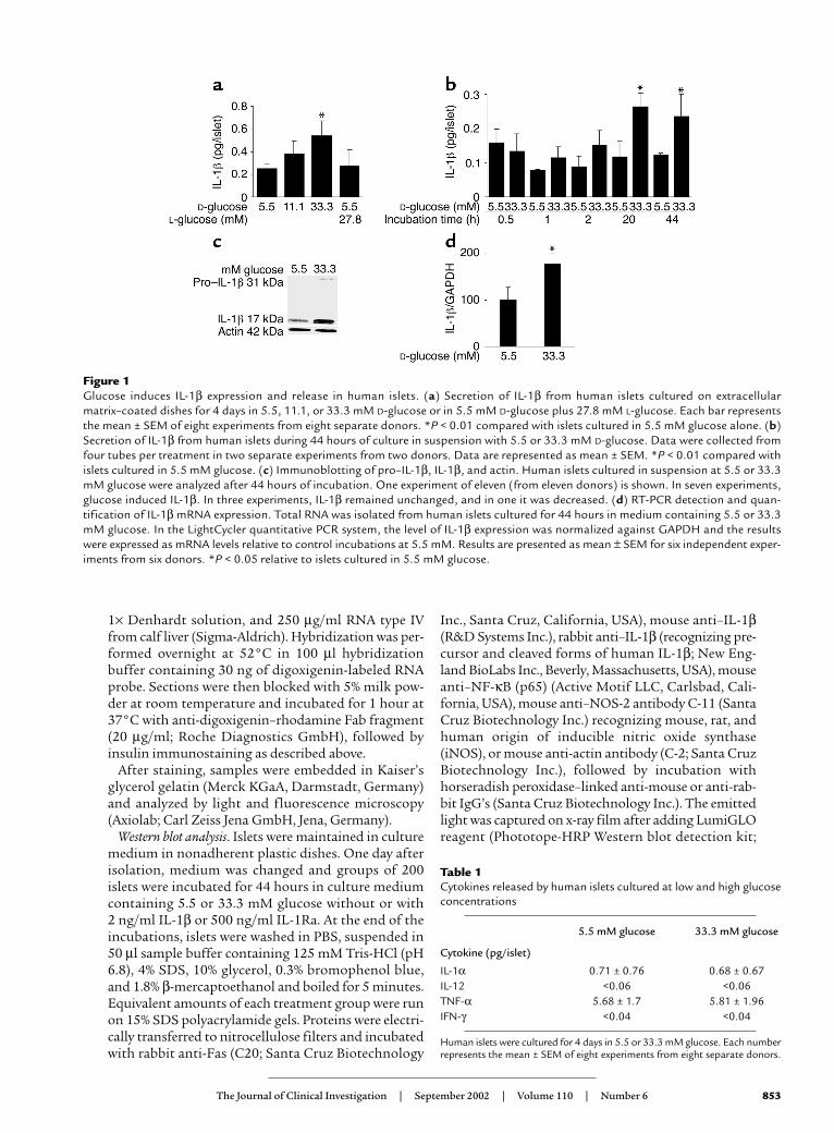
Figure 1Glucose induces IL-1β expression and release in human islets. (a) Secretion of IL-1β from human islets cultured on extracellularmatrix–coated dishes for 4 days in 5.5, 11.1, or 33.3 mM D-glucose or in 5.5 mM D-glucose plus 27.8 mM L-glucose. Each bar representsthe mean ± SEM of eight experiments from eight separate donors. *P < 0.01 compared with islets cultured in 5.5 mM glucose alone. (b)Secretion of IL-1β from human islets during 44 hours of culture in suspension with 5.5 or 33.3 mM D-glucose. Data were collected fromfour tubes per treatment in two separate experiments from two donors. Data are represented as mean ± SEM. *P < 0.01 compared withislets cultured in 5.5 mM glucose. (c) Immunoblotting of pro–IL-1β, IL-1β, and actin. Human islets cultured in suspension at 5.5 or 33.3mM glucose were analyzed after 44 hours of incubation. One experiment of eleven (from eleven donors) is shown. In seven experiments,glucose induced IL-1β. In three experiments, IL-1β remained unchanged, and in one it was decreased. (d) RT-PCR detection and quan-tification of IL-1β mRNA expression. Total RNA was isolated from human islets cultured for 44 hours in medium containing 5.5 or 33.3mM glucose. In the LightCycler quantitative PCR system, the level of IL-1β expression was normalized against GAPDH and the resultswere expressed as mRNA levels relative to control incubations at 5.5 mM. Results are presented as mean ± SEM for six independent exper-iments from six donors. *P < 0.05 relative to islets cultured in 5.5 mM glucose.
Table 1Cytokines released by human islets cultured at low and high glucoseconcentrations
5.5 mM glucose 33.3 mM glucose
Cytokine (pg/islet)
IL-1α 0.71 ± 0.76 0.68 ± 0.67IL-12 <0.06 <0.06TNF-α 5.68 ± 1.7 5.81 ± 1.96IFN-γ <0.04 <0.04
Human islets were cultured for 4 days in 5.5 or 33.3 mM glucose. Each numberrepresents the mean ± SEM of eight experiments from eight separate donors.
New England BioLabs Inc.). As a marker, biotinylatedprotein molecular weight standard (New England Bio-Labs Inc.) was run in parallel. Between incubations,nitrocellulose membranes were stripped for 30 minutesat 50°C in 40 ml of a solution containing 280 µl β-mer-captoethanol, 5 ml 0.5 M Tris-HCl (pH 6.8), and 10%SDS, and then washed for 1 hour in Tris-buffered
saline containing 0.1% Tween-20. Intensity of bandswas analyzed using MultiAnalyst (Bio-Rad Laborato-ries Inc., Hercules, California, USA).
NF-κB activation. Islets were cultured in suspension asdescribed above and washed with PBS. Activation ofNF-κB complex was quantified with an ELISA-basedkit (Trans-AM NF-κB; Active Motif LLC) usingattached oligonucleotides binding to an NF-κB con-sensus site and detected by an anti-p65 or p50 subunitantibody according to the manufacturer’s instructions.In parallel, islets were fixed in Bouin solution for 15minutes and resuspended in 40 µl of 2% melted agarosein PBS (40°C), followed by rapid centrifugation andparaffin embedding. Sections were deparaffinized andrehydrated, endogenous peroxidase blocked by sub-mersion in 0.3% H2O2 for 15 minutes, and incubated inmethanol for 4 minutes. Sections were incubated witha 1:50 dilution of either mouse anti–NF-κB (p65) orrabbit anti–inhibitory κB (I-κB) (C-21; Santa CruzBiotechnology Inc.) antibodies, detected by donkeyanti-mouse or anti-rabbit Cy3-conjugated antibodies,and double stained for insulin as described above.
RNA extraction, RT-PCR, and sequencing of RT-PCR prod-uct. Islets were cultured in suspension as describedabove. Total RNA was extracted using an RNeasy minikit (Qiagen Inc., Basel, Switzerland), and RT-PCR wasperformed using the Superscript II RNase H– reversetranscriptase kit and oligo(dT) (24) (Life TechnologiesInc., Gaithersburg, Maryland, USA) according to theinstructions from the manufacturers. The primerswere: human IL-1β,
5′-AAGCTGATGGCCCTAAACAG-3′ and 5′-AGGTG-CATCGTGCACATAAG-3′; human Fas, 5′-GCATCTGGAC-CCTCCTACCT-3′ and 5′-CAGTCTGGTTCATCCCCATT-3′;and human iNOS, 5′-ACGTGCGTTACTCCACCAACA-3′and 5′-CATAGCGGATGAGCTGAGCATT-3′.
PCR conditions for IL-1β and Fas were: denaturationfor 30 seconds at 94°C, annealing for 30 seconds at60°C, and elongation for 30 seconds at 72°C, followedfor real-time PCR by quantification 5 seconds at 80°C;repetition for 45 cycles. Saturation of PCR productoccurred between 22 and 28 cycles. The size of theamplification product was 250 bp. The purified PCRproducts were sequenced to confirm amplification ofthe correct gene. The conditions for PCR amplifica-tion of iNOS were: denaturation for 30 seconds at94°C, annealing for 40 seconds at 55°C, and elonga-tion for 30 seconds at 72°C; repetition for 35 cycles.For quantitative analysis, we used the LightCyclerquantitative PCR system (Roche Diagnostics GmbH)and performed quantitative PCR with a commercialkit (LightCycler–DNA Master SYBR Green I; RocheDiagnostics GmbH). The amounts of Fas and IL-1βmRNA were standardized against GAPDH (5′-AACAGCGACACCCACTCCTC-3′ and 5′-GGAGGGGA-GATTCAGTGTGGT-3′).
β cell apoptosis. The free 3′-OH strand breaks resultingfrom DNA degradation were detected using theTUNEL technique (38). After washing with PBS,
854 The Journal of Clinical Investigation | September 2002 | Volume 110 | Number 6
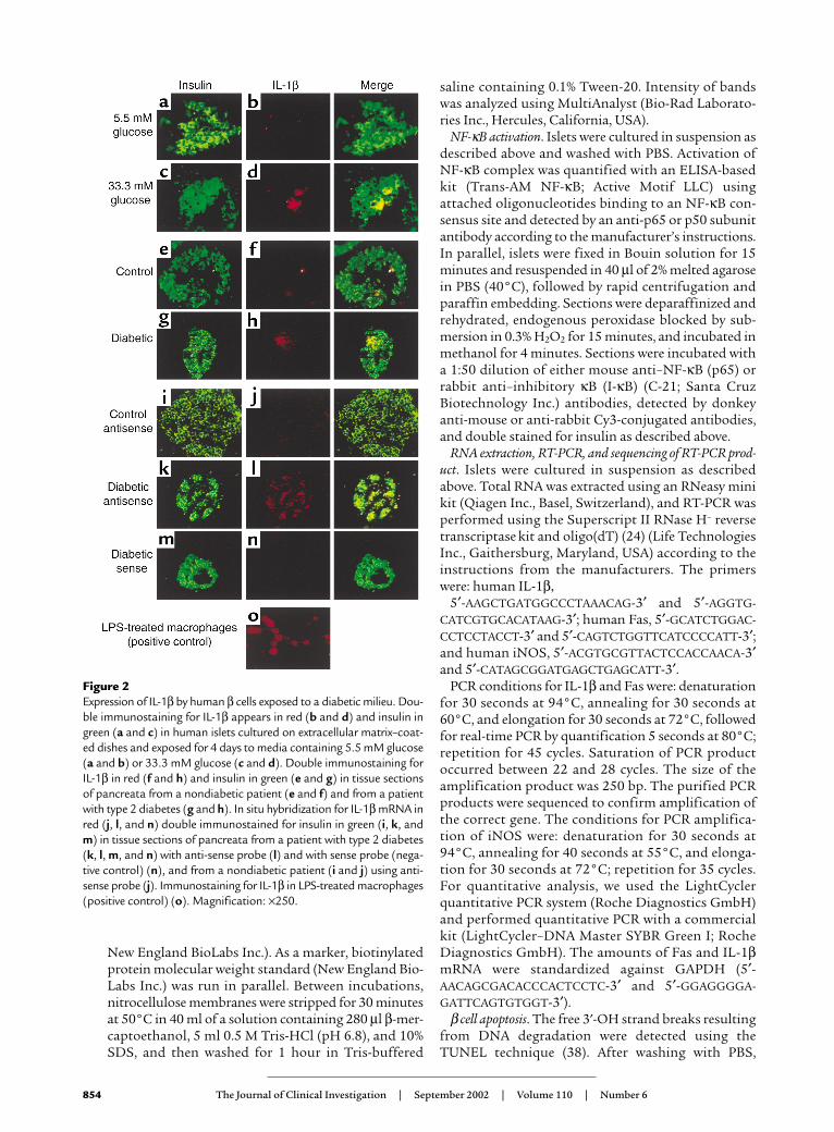
Figure 2Expression of IL-1β by human β cells exposed to a diabetic milieu. Dou-ble immunostaining for IL-1β appears in red (b and d) and insulin ingreen (a and c) in human islets cultured on extracellular matrix–coat-ed dishes and exposed for 4 days to media containing 5.5 mM glucose(a and b) or 33.3 mM glucose (c and d). Double immunostaining forIL-1β in red (f and h) and insulin in green (e and g) in tissue sectionsof pancreata from a nondiabetic patient (e and f) and from a patientwith type 2 diabetes (g and h). In situ hybridization for IL-1β mRNA inred (j, l, and n) double immunostained for insulin in green (i, k, andm) in tissue sections of pancreata from a patient with type 2 diabetes(k, l, m, and n) with anti-sense probe (l) and with sense probe (nega-tive control) (n), and from a nondiabetic patient (i and j) using anti-sense probe (j). Immunostaining for IL-1β in LPS-treated macrophages(positive control) (o). Magnification: ×250.
cultured islets were fixed in 4% paraformaldehyde for30 minutes at room temperature, followed by perme-abilization with 0.5% Triton X-100 for 4 minutes atroom temperature. The TUNEL assay was performedaccording to the manufacturer’s instructions (In SituCell Death Detection Kit, AP; Roche Molecular Bio-chemicals, Mannheim, Germany). The preparationswere then rinsed with Tris-buffered saline and incu-bated for 10 minutes at room temperature with the 5-bromo-4-chloro-3-indolyl phosphate/nitroblue tetra-zolium (BCIP/NBT) liquid substrate system (Sigma-Aldrich). Thereafter, islets were incubated with a guineapig anti-insulin antibody as above, followed by detec-tion using the streptavidin-biotin-horseradish peroxi-dase complex (Zymed Laboratories Inc., South SanFrancisco, California, USA). In parallel to the TUNELreaction, we used the DNA-binding dye propidiumiodide (Sigma-Aldrich) to assess the effects of glucoseon necrosis. Cultured islets were washed with PBS(without paraformaldehyde fixation), incubated for 10minutes on ice with 10 µg/ml propidium iodide in PBS,washed with PBS, and embedded in fluorescentmounting medium (DAKO Corp.). The samples wereimmediately evaluated by fluorescence microscopy forpositively stained necrotic nuclei.
Cytokine release. Cytokine release was evaluated in theculture medium collected before the termination ofeach experiment. The following ELISA kits were used:human IL-1α, IL-1β, and IL-12 (R&D Systems Inc.),human TNF-α (Endogen Inc., Boston, Massachusetts,USA), and human IFN-γ (Invitrogen Ltd.).
Insulin release and content. To determine acute insulinrelease in response to glucose stimulation, islets werewashed in RPMI 1640 medium with 10% FCS and con-taining 3.3 mM glucose, and preincubated for 1 hourin the same medium. The medium was then discardedand replaced with fresh medium containing 3.3 mMglucose for 1 hour for basal secretion, followed by anadditional 1 hour incubation in medium containing16.7 mM glucose. Supernatants were collected andfrozen for insulin assays. Thereafter islets were washedwith PBS and extracted with HCl (0.18 N) in 70%ethanol for 24 hours at 4°C. The acid-ethanol extractswere collected for determination of insulin content.Insulin was determined using a human insulin RIA kit(CIS Bio International, Gif-sur-Yvette, France).
Evaluation and statistical analysis. Samples were evalu-ated in a randomized manner by a single investigator(K. Maedler) who was blinded to the treatment con-ditions. Care was taken to score islets of similar size.Some larger islets did not completely spread and wereseveral cells thick. Such islets were excluded because amonolayer is a prerequisite for single-cell evaluation.The mean surface area of the evaluated islet mono-layers was 0.031 ± 0.012 mm2 and 0.029 ± 0.011 mm2
in islets cultured at 5.5 and 33.3 mM glucose, respec-tively (no significant difference). Thus, the exclusionof larger islets occurred to a similar extent in eachdish regardless of the treatment. Saisam software
(Microvision Instruments, Evry, France) was used tomeasure area. Data were analyzed by Student t test orby ANOVA with a Bonferroni correction for multiplegroup comparisons.
ResultsGlucose induces IL-1β production and release in human islets.Human islets were exposed to elevated glucose con-centrations for 4 days. Measurement of IL-1β releasedin the culture medium revealed a 2.2-fold increase inislets cultured at 33.3 mM compared with islets in 5.5mM glucose (Figure 1a). To exclude a nonspecific effectof this high concentration of D-glucose, osmolarity wascorrected with 27.8 mM L-glucose together with 5.5mM D-glucose, resulting in a similar release of IL-1β tothat observed with 5.5 mM D-glucose alone. No IL-1βwas detectable in unused culture medium. The speci-ficity of IL-1β release was assessed by comparison withthe release of other cytokines. Limited amounts of IL-1α and TNF-α were found, but neither was regulat-ed by glucose and no significant amount of IFN-γ or IL-12 was detectable (Table 1). The time-course effectof 33.3 mM glucose on IL-1β secretion became signifi-cant only after 20 hours of exposure to high glucose,and persisted after 44 hours (Figure 1b). Western blotanalysis and quantitative RT-PCR measurement of IL-1β production in human islets revealed that elevat-ed glucose concentration induces not only IL-1β releasebut also IL-1β protein and RNA synthesis (Figure 1, cand d). However, Western blot analysis gave quite
The Journal of Clinical Investigation | September 2002 | Volume 110 | Number 6 855
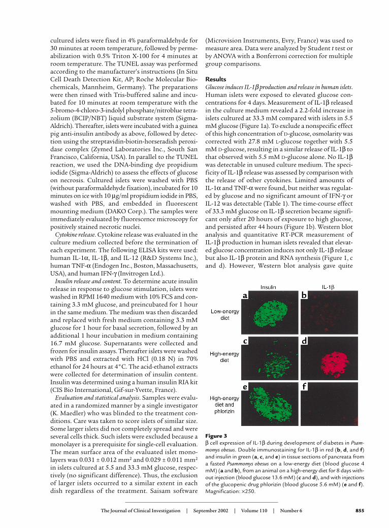
Figure 3β cell expression of IL-1β during development of diabetes in Psam-momys obesus. Double immunostaining for IL-1β in red (b, d, and f)and insulin in green (a, c, and e) in tissue sections of pancreata froma fasted Psammomys obesus on a low-energy diet (blood glucose 4mM) (a and b), from an animal on a high-energy diet for 8 days with-out injection (blood glucose 13.6 mM) (c and d), and with injectionsof the glucopenic drug phlorizin (blood glucose 5.6 mM) (e and f).Magnification: ×250.
856 The Journal of Clinical Investigation | September 2002 | Volume 110 | Number 6
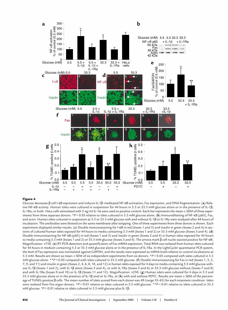
Figure 4Glucose decreases β cell I-κB expression and induces IL-1β–meditated NF-κB activation, Fas expression, and DNA fragmentation. (a) Rela-tive NF-κB activity. Human islets were cultured in suspension for 44 hours in 5.5 or 33.3 mM glucose alone or in the presence of IL-1β, IL-1Ra, or both. HeLa cells stimulated with 5 ng/ml IL-1α were used as positive control. Each bar represents the mean ± SEM of three exper-iments from three separate donors. *P < 0.05 relative to islets cultured in 5.5 mM glucose alone. (b) Immunoblotting of NF-κB (p65), Fas,and actin. Human islets cultured in suspension at 5.5 or 33.3 mM glucose with and without IL-1β or IL-1Ra were analyzed after 44 hours ofincubation. The antibodies were blotted on the same membrane after stripping. One of three experiments from three donors is shown. Eachexperiment displayed similar results. (c) Double immunostaining for I-κB in red (boxes 1 and 3) and insulin in green (boxes 2 and 4) in sec-tions of cultured human islets exposed for 44 hours to media containing 5.5 mM (boxes 1 and 2) or 33.3 mM glucose (boxes 3 and 4). (d)Double immunostaining for NF-κB (p65) in red (boxes 1 and 3) and insulin in green (boxes 2 and 4) in human islets exposed for 44 hoursto media containing 5.5 mM (boxes 1 and 2) or 33.3 mM glucose (boxes 3 and 4). The arrows mark β cell nuclei stained positive for NF-κB.Magnification: ×750. (e) RT-PCR detection and quantification of Fas mRNA expression. Total RNA was isolated from human islets culturedfor 44 hours in medium containing 5.5 or 33.3 mM glucose alone or in the presence of IL-1Ra. In the LightCycler quantitative PCR system,the level of Fas expression was normalized against GAPDH, and the results were expressed as mRNA levels relative to control incubations at5.5 mM. Results are shown as mean ± SEM of six independent experiments from six donors. *P < 0.05 compared with islets cultured in 5.5mM glucose alone. **P < 0.05 compared with islets cultured in 33.3 mM glucose. (f) Double immunostaining for Fas in red (boxes 1, 3, 5,7, 9, and 11) and insulin in green (boxes 2, 4, 6, 8, 10, and 12) in human islets exposed for 4 days to media containing 5.5 mM glucose with-out IL-1β (boxes 1 and 2), with IL-1β alone (boxes 3 and 4), or with IL-1Ra (boxes 5 and 6) or 33.3 mM glucose without (boxes 7 and 8)and with IL-1Ra (boxes 9 and 10) or IL-1β (boxes 11 and 12). Magnification: ×250. (g) Human islets were cultured for 4 days in 5.5 and33.3 mM glucose alone or in the presence of IL-1β and/or IL-1Ra, or (h) with and without PDTC. Results are mean ± SEM of the percent-age of TUNEL-positive β cells. The mean number of islets scored from each donor was 49 (range 35–63) for each treatment condition. Isletswere isolated from five organ donors. *P < 0.01 relative to islets cultured in 5.5 mM glucose. **P < 0.01 relative to islets cultured in 33.3mM glucose. +P < 0.01 relative to islets cultured in 5.5 mM glucose plus IL-1β.
variable results in terms of increased IL-1β in responseto glucose. This may be due to varying numbers of non-endocrine cells (including most notably macrophages)in the different islet preparations. IL-1β productionfrom such accompanying cells is not stimulated by glu-cose. Therefore, the higher the contribution in total IL-1β production, the lower the expected glucose effect.
Identification of the islet cellular source of glucose-dependentIL-1β production. We next identified the islet cells pro-ducing IL-1β. Exposure of cultured human islets to33.3 mM glucose for 4 days induced IL-1β expressionin clusters of β cells, as determined by doubleimmunostaining of islets plated on extracellularmatrix–coated dishes with anti–IL-1β and anti-insulinantibodies. Figure 2, a–d, shows representative imagesfrom one experiment of six from six separate donors.In each experiment, IL-1β–positive β cells wereobserved in islets cultured at 33.3 mM glucose. Toexclude false-positive results due to IL-1β secreted byother cells binding to β cell membranes, islets weretreated with IL-1Ra. Under these conditions, IL-1Rashould antagonize the interaction of IL-1β with its sur-face receptors on β cells. Coincubation with IL-1Ra dis-played similar results, confirming β cell production ofIL-1β (not shown).
IL-1β is not expressed in normal human pancreaticislets (28). However, based on the in vitro studies, it wasanticipated that it might be expressed in islets ofpatients with type 2 diabetes, as a result of hyper-glycemia. Expression of IL-1β was therefore studied insections of pancreata from five poorly controlled type2 diabetic patients, all with documented fasting bloodglucose higher than 8 mM. Double immunostaining ofthe pancreatic sections for IL-1β and insulin revealedlocalization of IL-1β in clusters of β cells in all pancre-ata. Clusters of IL-1β–expressing β cells were exhibitedin 22.5% ± 3.4% of the islets in each pancreas (represen-tative images are sown in Figure 2, g–h). The presenceof IL-1β mRNA transcripts was verified by in situhybridization in β cells of diabetic patients (Figure 2, kand l). IL-1β expression could not be detected in β cellsof nondiabetic controls (Figure 2, e, f, i, and j) or in theexocrine pancreas. A digoxigenin-labeled sense probewas used as control and gave no signal (Figure 2n).
β cell expression of IL-1β during development of diabetes inPsammomys obesus is glucose-dependent. To examinewhether induction of IL-1β in vivo is also regulated byglucose, three diabetes-resistant and eight diabetes-prone Psammomys obesus fed a low- or high-energy dietwere studied. The hyperglycemic animals were furthertreated with phlorizin, which corrects hyperglycemia byinhibiting renal tubular reabsorption of glucose. No IL-1β–expressing β cells were observed in islets of dia-betes-resistant (not shown) or in fasted diabetes-pronePsammomys obesus (Figure 3, a and b). After 8 days on ahigh-energy diet, islets of severely hyperglycemic dia-betes-prone Psammomys obesus exhibited IL-1β expres-sion in most β cells, which barely expressed insulin(Figure 3, c and d). Normalization of blood glucose by
injection of phlorizin in animals fed a high-energy dietrestored insulin stores and prevented IL-1β expression(Figure 3, e and f).
IL-1β mediates glucose-induced NF-κB activation, Fas expres-sion, and β cell apoptosis. The functional role of glucose-induced IL-1β was tested using IL-1Ra as an inhibitor.In human islets, elevated glucose concentrationsinduced a 1.9-fold increase in NF-κB activity (Figure 4a).This was prevented by IL-1Ra. Moreover, high glucoseinduced the expression of the p65 subunit (Figure 4b).NF-κB is bound in the cytoplasm to I-κB proteins (39).Exposure of human islets to 33 mM glucose decreasedI-κB expression (Figure 4c), leading to apparition of NF-κB (p65) in β cell nuclei (Figure 4d). Glucose-dependent induction of Fas protein and mRNA werealso hindered by IL-1Ra (Figure 4, b, e, and f).
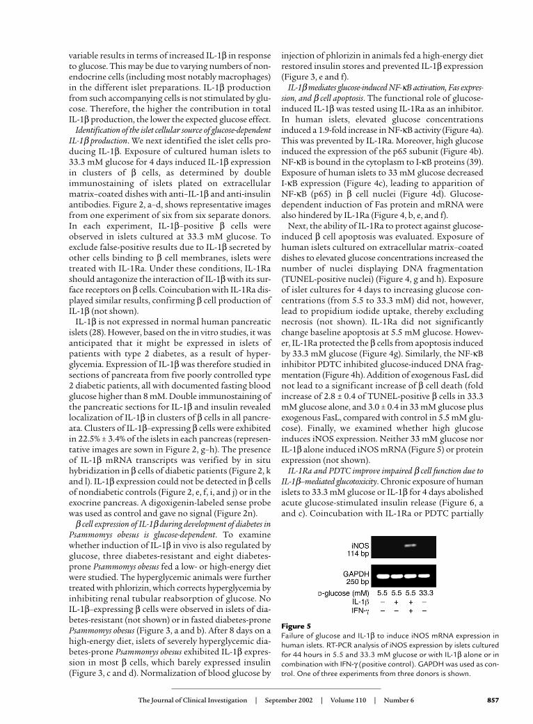
Next, the ability of IL-1Ra to protect against glucose-induced β cell apoptosis was evaluated. Exposure ofhuman islets cultured on extracellular matrix–coateddishes to elevated glucose concentrations increased thenumber of nuclei displaying DNA fragmentation(TUNEL-positive nuclei) (Figure 4, g and h). Exposureof islet cultures for 4 days to increasing glucose con-centrations (from 5.5 to 33.3 mM) did not, however,lead to propidium iodide uptake, thereby excludingnecrosis (not shown). IL-1Ra did not significantlychange baseline apoptosis at 5.5 mM glucose. Howev-er, IL-1Ra protected the β cells from apoptosis inducedby 33.3 mM glucose (Figure 4g). Similarly, the NF-κBinhibitor PDTC inhibited glucose-induced DNA frag-mentation (Figure 4h). Addition of exogenous FasL didnot lead to a significant increase of β cell death (foldincrease of 2.8 ± 0.4 of TUNEL-positive β cells in 33.3mM glucose alone, and 3.0 ± 0.4 in 33 mM glucose plusexogenous FasL, compared with control in 5.5 mM glu-cose). Finally, we examined whether high glucoseinduces iNOS expression. Neither 33 mM glucose norIL-1β alone induced iNOS mRNA (Figure 5) or proteinexpression (not shown).
IL-1Ra and PDTC improve impaired β cell function due toIL-1β–mediated glucotoxicity. Chronic exposure of humanislets to 33.3 mM glucose or IL-1β for 4 days abolishedacute glucose-stimulated insulin release (Figure 6, aand c). Coincubation with IL-1Ra or PDTC partially
The Journal of Clinical Investigation | September 2002 | Volume 110 | Number 6 857
Figure 5Failure of glucose and IL-1β to induce iNOS mRNA expression inhuman islets. RT-PCR analysis of iNOS expression by islets culturedfor 44 hours in 5.5 and 33.3 mM glucose or with IL-1β alone or incombination with IFN-γ (positive control). GAPDH was used as con-trol. One of three experiments from three donors is shown.
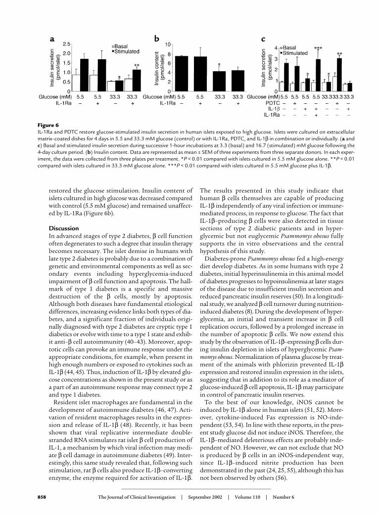
restored the glucose stimulation. Insulin content ofislets cultured in high glucose was decreased comparedwith control (5.5 mM glucose) and remained unaffect-ed by IL-1Ra (Figure 6b).
DiscussionIn advanced stages of type 2 diabetes, β cell functionoften degenerates to such a degree that insulin therapybecomes necessary. The islet demise in humans withlate type 2 diabetes is probably due to a combination ofgenetic and environmental components as well as sec-ondary events including hyperglycemia-inducedimpairment of β cell function and apoptosis. The hall-mark of type 1 diabetes is a specific and massivedestruction of the β cells, mostly by apoptosis.Although both diseases have fundamental etiologicaldifferences, increasing evidence links both types of dia-betes, and a significant fraction of individuals origi-nally diagnosed with type 2 diabetes are cryptic type 1diabetics or evolve with time to a type 1 state and exhib-it anti–β cell autoimmunity (40–43). Moreover, apop-totic cells can provoke an immune response under theappropriate conditions, for example, when present inhigh enough numbers or exposed to cytokines such asIL-1β (44, 45). Thus, induction of IL-1β by elevated glu-cose concentrations as shown in the present study or asa part of an autoimmune response may connect type 2and type 1 diabetes.
Resident islet macrophages are fundamental in thedevelopment of autoimmune diabetes (46, 47). Acti-vation of resident macrophages results in the expres-sion and release of IL-1β (48). Recently, it has beenshown that viral replicative intermediate double-stranded RNA stimulates rat islet β cell production ofIL-1, a mechanism by which viral infection may medi-ate β cell damage in autoimmune diabetes (49). Inter-estingly, this same study revealed that, following suchstimulation, rat β cells also produce IL-1β–convertingenzyme, the enzyme required for activation of IL-1β.
The results presented in this study indicate thathuman β cells themselves are capable of producing IL-1β independently of any viral infection or immune-mediated process, in response to glucose. The fact thatIL-1β–producing β cells were also detected in tissuesections of type 2 diabetic patients and in hyper-glycemic but not euglycemic Psammomys obesus fullysupports the in vitro observations and the centralhypothesis of this study.
Diabetes-prone Psammomys obesus fed a high-energydiet develop diabetes. As in some humans with type 2diabetes, initial hyperinsulinemia in this animal modelof diabetes progresses to hypoinsulinemia at later stagesof the disease due to insufficient insulin secretion andreduced pancreatic insulin reserves (50). In a longitudi-nal study, we analyzed β cell turnover during nutrition-induced diabetes (8). During the development of hyper-glycemia, an initial and transient increase in β cellreplication occurs, followed by a prolonged increase inthe number of apoptotic β cells. We now extend thisstudy by the observation of IL-1β–expressing β cells dur-ing insulin depletion in islets of hyperglycemic Psam-momys obesus. Normalization of plasma glucose by treat-ment of the animals with phlorizin prevented IL-1βexpression and restored insulin expression in the islets,suggesting that in addition to its role as a mediator ofglucose-induced β cell apoptosis, IL-1β may participatein control of pancreatic insulin reserves.
To the best of our knowledge, iNOS cannot beinduced by IL-1β alone in human islets (51, 52). More-over, cytokine-induced Fas expression is NO-inde-pendent (53, 54). In line with these reports, in the pres-ent study glucose did not induce iNOS. Therefore, theIL-1β–mediated deleterious effects are probably inde-pendent of NO. However, we can not exclude that NOis produced by β cells in an iNOS-independent way,since IL-1β–induced nitrite production has beendemonstrated in the past (24, 25, 55), although this hasnot been observed by others (56).
858 The Journal of Clinical Investigation | September 2002 | Volume 110 | Number 6
Figure 6IL-1Ra and PDTC restore glucose-stimulated insulin secretion in human islets exposed to high glucose. Islets were cultured on extracellularmatrix–coated dishes for 4 days in 5.5 and 33.3 mM glucose (control) or with IL-1Ra, PDTC, and IL-1β in combination or individually. (a andc) Basal and stimulated insulin secretion during successive 1-hour incubations at 3.3 (basal) and 16.7 (stimulated) mM glucose following the4-day culture period. (b) Insulin content. Data are represented as mean ± SEM of three experiments from three separate donors. In each exper-iment, the data were collected from three plates per treatment. *P < 0.01 compared with islets cultured in 5.5 mM glucose alone. **P < 0.01compared with islets cultured in 33.3 mM glucose alone. ***P < 0.01 compared with islets cultured in 5.5 mM glucose plus IL-1β.
IL-1β stimulates I-κB degradation and NF-κB nuclearlocalization in a time-dependent manner that is maxi-mal following 20–30 minutes of exposure (31). There-fore, it is not clear how NF-κB activation remainsdetectable following a 44-hour incubation in high glu-cose. The prolonged period of activation may be relat-ed to the interplay of all factors required for NF-κBactivation. Indeed, we show that high glucose inducedp65 expression and decreased I-κB. Moreover, othermodulators of IL-1β–induced NF-κB activation, suchas endogenous IL-1Ra, may interfere. All these factorsmay influence the time course of glucose-induced NF-κB activation. Regardless, at least 20 hours of expo-sure to high glucose is required to induce IL-1β, lead-ing to NF-κB activation, Fas expression, and β celldeath. This is in line with glucotoxic effects, whichappear only after prolonged exposure to high glucose.
Inhibition of NF-κB activation by an adenoviral vec-tor encoding the repressor I-κB protects human isletsfrom Fas-triggered apoptosis and results in normalinsulin response in the presence of IL-1β (55). Simi-larly, in purified rat β cells, inhibition of cytokine-induced NF-κB activation prevents β cell apoptosis(57). Thus, the present finding that glucose decreasesI-κB expression and induces NF-κB activation via IL-1β opens the path to prevent glucotoxic effects byinhibition of NF-κB activation.
So far, IL-1β production and release by islets hasbeen considered to be limited to type 1 diabetes. Herewe demonstrate that high concentrations of glucoseinduce IL-1β production and secretion in human βcells, leading to Fas receptor upregulation, NF-κBactivation, β cell apoptosis, and dysfunction. More-over, we observed IL-1β–producing β cells in diabeticpatients and diabetic Psammomys obesus. The pathwayby which hyperglycemia causes impairment and lossof insulin-producing cells thus shares features withimmune-mediated processes. It follows that theproinflammatory cytokine IL-1β may be a crucial fac-tor contributing to β cell glucotoxicity in the patho-genesis of type 2 diabetes.
AcknowledgmentsThis work was supported by Swiss National ScienceFoundation grants 3200-067049.01 (M.Y. Donath) and3200-061776.00 (P.A. Halban), and by Juvenile Dia-betes Research Foundation grants 4-1999-844 (P.A.Halban) and I-1998-9 (N. Kaiser). M.Y. Donath is sup-ported by the Max Cloetta Foundation. We thank G.Schoedon for helpful suggestions, P.U. Heitz, P. Kom-minoth, and P. Saremaslani for providing the sectionsof human pancreas, and G. Siegfried-Kellenberger andC.S. Manzano for technical assistance.
1. Unger, R.H., and Grundy, S. 1985. Hyperglycaemia as an inducer as wellas a consequence of impaired islet cell function and insulin resistance:implications for the management of diabetes. Diabetologia. 28:119–121.
2. Kaiser, N., Corcos, A.P., Sarel, I., and Cerasi, E. 1991. Monolayer cultureof adult rat pancreatic islets on extracellular matrix: modulation of B-cell function by chronic exposure to high glucose. Endocrinology.129:2067–2076.
3. Leahy, J.L., Cooper, H.E., Deal, D.A., and Weir, G.C. 1986. Chronic hyper-glycemia is associated with impaired glucose influence on insulin secre-tion. A study in normal rats using chronic in vivo glucose infusions. J. Clin. Invest. 77:908–915.
4. Robertson, R.P. 1989. Type II diabetes, glucose “non-sense,” and isletdesensitization. Diabetes. 38:1501–1505.
5. Rossetti, L., Giaccari, A., and DeFronzo, R.A. 1990. Glucose toxicity. Diabetes Care. 13:610–630.
6. Eizirik, D.L., Korbutt, G.S., and Hellerstrom, C. 1992. Prolonged expo-sure of human pancreatic islets to high glucose concentrations in vitroimpairs the beta-cell function. J. Clin. Invest. 90:1263–1268.
7. Marshak, S., et al. 1999. Impaired beta-cell functions induced by chron-ic exposure of cultured human pancreatic islets to high glucose. Diabetes.48:1230–1236.
8. Donath, M.Y., Gross, D.J., Cerasi, E., and Kaiser, N. 1999. Hyperglycemia-induced beta-cell apoptosis in pancreatic islets of Psammomys obesusduring development of diabetes. Diabetes. 48:738–744.
9. Maedler, K., et al. 2001. Glucose induces b-cell apoptosis via upregula-tion of the Fas-receptor in human islets. Diabetes. 50:1683–1690.
10. Federici, M., et al. 2001. High glucose causes apoptosis in culturedhuman pancreatic islets of Langerhans: a potential role for regulation ofspecific Bcl family genes toward an apoptotic cell death program. Diabetes. 50:1290–1301.
11. Efanova, I.B., et al. 1998. Glucose and tolbutamide induce apoptosis inpancreatic beta-cells. A process dependent on intracellular Ca2+ con-centration. J. Biol. Chem. 273:33501–33507.
12. Maedler, K., et al. 2001. Distinct effects of saturated and monounsatu-rated fatty acids on beta-cell turnover and function. Diabetes. 50:69–76.
13. Tajiri, Y., Moller, C., and Grill, V. 1997. Long-term effects of aminoguani-dine on insulin release and biosynthesis: evidence that the formation ofadvanced glycosylation end products inhibits B cell function. Endocrinol-ogy. 138:273–280.
14. Robertson, R.P., Zhang, H.J., Pyzdrowski, K.L., and Walseth, T.F. 1992.Preservation of insulin mRNA levels and insulin secretion in HIT cellsby avoidance of chronic exposure to high glucose concentrations. J. Clin.Invest. 90:320–325.
15. Robertson, R.P., Olson, L.K., and Zhang, H.J. 1994. Differentiating glu-cose toxicity from glucose desensitization: a new message from theinsulin gene. Diabetes. 43:1085–1089.
16. Mandrup-Poulsen, T. 1996. The role of interleukin-1 in the pathogene-sis of IDDM. Diabetologia. 39:1005–1029.
17. Mandrup-Poulsen, T., Bendtzen, K., Nielsen, J.H., Bendixen, G., andNerup, J. 1985. Cytokines cause functional and structural damage to iso-lated islets of Langerhans. Allergy. 40:424–429.
18. Bendtzen, K., et al. 1986. Cytotoxicity of human pI 7 interleukin-1 forpancreatic islets of Langerhans. Science. 232:1545–1547.
19. Spinas, G.A., et al. 1987. Interleukin 1 dose-dependently affects thebiosynthesis of (pro)insulin in isolated rat islets of Langerhans. Dia-betologia. 30:474–480.
20. Mandrup-Poulsen, T., et al. 1986. Affinity-purified human interleukin Iis cytotoxic to isolated islets of Langerhans. Diabetologia. 29:63–67.
21. Spinas, G.A., et al. 1986. Low concentrations of interleukin-1 stimulateand high concentrations inhibit insulin release from isolated rat isletsof Langerhans. Acta Endocrinol. (Copenh.). 113:551–558.
22. Yamada, K., et al. 1996. Mouse islet cell lysis mediated by interleukin-1-induced Fas. Diabetologia. 39:1306–1312.
23. Corbett, J.A., Lancaster, J.R., Jr., Sweetland, M.A., and McDaniel, M.L.1991. Interleukin-1 beta-induced formation of EPR-detectable iron-nitrosyl complexes in islets of Langerhans. Role of nitric oxide in inter-leukin-1 beta-induced inhibition of insulin secretion. J. Biol. Chem.266:21351–21354.
24. Giannoukakis, N., et al. 1999. Adenoviral gene transfer of the inter-leukin-1 receptor antagonist protein to human islets prevents IL-1beta-induced beta-cell impairment and activation of islet cell apoptosis invitro. Diabetes. 48:1730–1736.
25. Giannoukakis, N., et al. 2000. Prevention of beta cell dysfunction andapoptosis activation in human islets by adenoviral gene transfer of theinsulin-like growth factor I. Gene Ther. 7:2015–2022.
26. Loweth, A.C., Williams, G.T., James, R.F., Scarpello, J.H., and Morgan,N.G. 1998. Human islets of Langerhans express Fas ligand and undergoapoptosis in response to interleukin-1beta and Fas ligation. Diabetes.47:727–732.
27. Rabinovitch, A., Sumoski, W., Rajotte, R.V., and Warnock, G.L. 1990.Cytotoxic effects of cytokines on human pancreatic islet cells in mono-layer culture. J. Clin. Endocrinol. Metab. 71:152–156.
28. Stassi, G., et al. 1997. Nitric oxide primes pancreatic beta cells for Fas-mediated destruction in insulin-dependent diabetes mellitus. J. Exp. Med.186:1193–1200.
29. Kwon, G., Corbett, J.A., Rodi, C.P., Sullivan, P., and McDaniel, M.L. 1995.Interleukin-1 beta-induced nitric oxide synthase expression by rat pan-creatic beta-cells: evidence for the involvement of nuclear factor kappaB in the signaling mechanism. Endocrinology. 136:4790–4795.
The Journal of Clinical Investigation | September 2002 | Volume 110 | Number 6 859
30. Darville, M.I., and Eizirik, D.L. 2001. Cytokine induction of Fas geneexpression in insulin-producing cells requires the transcription factorsNF-kappaB and C/EBP. Diabetes. 50:1741–1748.
31. Flodstrom, M., Welsh, N., and Eizirik, D.L. 1996. Cytokines activate thenuclear factor kappa B (NF-kappa B) and induce nitric oxide productionin human pancreatic islets. FEBS Lett. 385:4–6.
32. Linetsky, E., et al. 1997. Improved human islet isolation using a newenzyme blend, liberase. Diabetes. 46:1120–1123.
33. Oberholzer, J., et al. 2000. Human islet transplantation: lessons from 13autologous and 13 allogeneic transplantations. Transplantation.69:1115–1123.
34. Ricordi, C., Lacy, P.E., Finke, E.H., Olack, B.J., and Scharp, D.W. 1988.Automated method for isolation of human pancreatic islets. Diabetes.37:413–420.
35. Kaiser, N., Corcos, A.P., Sarel, I., and Cerasi, E. 1991. Monolayer cultureof adult rat pancreatic islets on extracellular matrix: modulation of B-cell function by chronic exposure to high glucose. Endocrinology.129:2067–2076.
36. Jodo, S., et al. 2001. Apoptosis-inducing membrane vesicles. A novelagent with unique properties. J. Biol. Chem. 276:39938–39944.
37. Schneemann, M., et al. 1993. Nitric oxide synthase is not a constituentof the antimicrobial armature of human mononuclear phagocytes. J. Infect. Dis. 167:1358–1363.
38. Gavrieli, Y., Sherman, Y., and Ben-Sasson, S.A. 1992. Identification ofprogrammed cell death in situ via specific labeling of nuclear DNA frag-mentation. J. Cell. Biol. 119:493–501.
39. Karin, M. 1999. How NF-kappaB is activated: the role of the IkappaBkinase (IKK) complex. Oncogene. 18:6867–6874.
40. Mathis, D., Vence, L., and Benoist, C. 2001. beta-Cell death during pro-gression to diabetes. Nature. 414:792–798.
41. Pietropaolo, M., Barinas-Mitchell, E., Pietropaolo, S.L., Kuller, L.H., andTrucco, M. 2000. Evidence of islet cell autoimmunity in elderly patientswith type 2 diabetes. Diabetes. 49:32–38.
42. Rowley, M.J., Mackay, I.R., Chen, Q.Y., Knowles, W.J., and Zimmet, P.Z.1992. Antibodies to glutamic acid decarboxylase discriminate majortypes of diabetes mellitus. Diabetes. 41:548–551.
43. Wilkin, T.J. 2001. The accelerator hypothesis: weight gain as the missinglink between Type I and Type II diabetes. Diabetologia. 44:914–922.
44. Bellone, M., et al. 1997. Processing of engulfed apoptotic bodies yields Tcell epitopes. J. Immunol. 159:5391–5399.
45. Trudeau, J.D., et al. 2000. Neonatal beta-cell apoptosis: a trigger forautoimmune diabetes? Diabetes. 49:1–7.
46. Lacy, P.E., and Finke, E.H. 1991. Activation of intraislet lymphoid cellscauses destruction of islet cells. Am. J. Pathol. 138:1183–1190.
47. Lacy, P.E. 1994. The intraislet macrophage and type I diabetes. Mt. SinaiJ. Med. 61:170–174.
48. Arnush, M., Scarim, A.L., Heitmeier, M.R., Kelly, C.B., and Corbett, J.A.1998. Potential role of resident islet macrophage activation in the initi-ation of autoimmune diabetes. J. Immunol. 160:2684–2691.
49. Heitmeier, M.R., Arnush, M., Scarim, A.L., and Corbett, J.A. 2001. Pan-creatic beta-cell damage mediated by beta-cell production of IL-1: a novelmechanism for virus-induced diabetes. J. Biol. Chem. 276:11151–11158.
50. Gadot, M., et al. 1994. Hyperproinsulinemia and insulin deficiency inthe diabetic Psammomys obesus. Endocrinology. 135:610–616.
51. Chen, M.C., Proost, P., Gysemans, C., Mathieu, C., and Eizirik, D.L. 2001.Monocyte chemoattractant protein-1 is expressed in pancreatic isletsfrom prediabetic NOD mice and in interleukin-1 beta-exposed humanand rat islet cells. Diabetologia. 44:325–332.
52. Eizirik, D.L., and Darville, M.I. 2001. beta-cell apoptosis and defensemechanisms: lessons from type 1 diabetes. Diabetes. 50:S64–S69.
53. Liu, D., et al. 2000. Cytokines induce apoptosis in beta-cells isolatedfrom mice lacking the inducible isoform of nitric oxide synthase(iNOS–/–). Diabetes. 49:1116–1122.
54. Zumsteg, U., Frigerio, S., and Hollander, G.A. 2000. Nitric oxide pro-duction and Fas surface expression mediate two independent pathwaysof cytokine-induced murine beta-cell damage. Diabetes. 49:39–47.
55. Giannoukakis, N., Rudert, W.A., Trucco, M., and Robbins, P.D. 2000. Pro-tection of human islets from the effects of interleukin-1beta by adenovi-ral gene transfer of an Ikappa B repressor. J. Biol. Chem. 275:36509–36513.
56. Corbett, J.A., Sweetland, M.A., Wang, J.L., Lancaster, J.R., Jr., andMcDaniel, M.L. 1993. Nitric oxide mediates cytokine-induced inhibitionof insulin secretion by human islets of Langerhans. Proc. Natl. Acad. Sci.USA. 90:1731–1735.
57. Heimberg, H., et al. 2001. Inhibition of cytokine-induced NF-kappaBactivation by adenovirus-mediated expression of a NF-kappaB super-repressor prevents beta-cell apoptosis. Diabetes. 50:2219–2224.
860 The Journal of Clinical Investigation | September 2002 | Volume 110 | Number 6