Chapter 5: Amino Acids, Peptides & Proteins Proteins ...faculty.tamucc.edu/plarkin/4401folder/Slide...
8
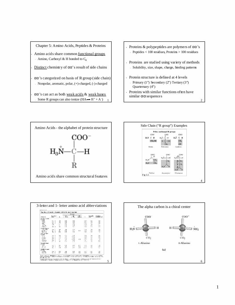
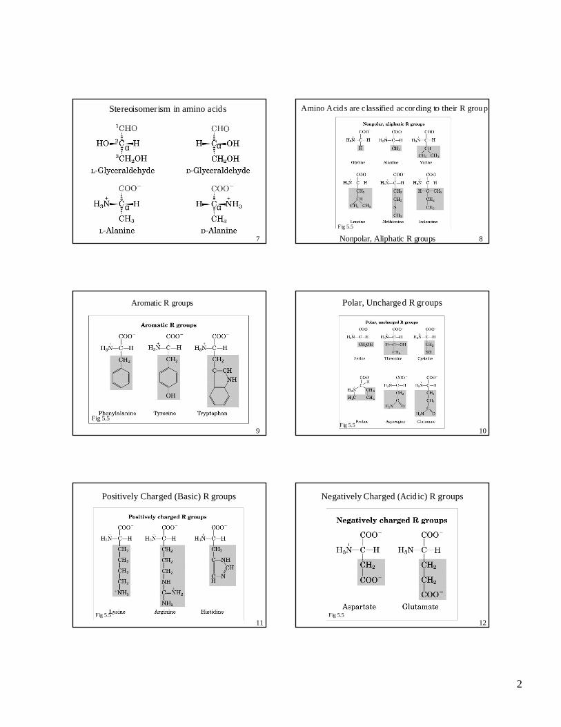
1 1 Chapter 5: Amino Acids, Peptides & Proteins • Amino a cid s share common functional groups – Amino, Carb oxyl & H bonded to C α • Distinct chemistry of αα’s result of side chains • αα ’s categorized on basi s of R group (side chain) – Nonpolar, aromatic, polar, (+)-charged, (-)-charged • αα ’s can act as both wea k a cid s & wea k bases – Some R groups can also ionize (HA H + + A - ) 2 • Proteins & polypeptides are polymers of αα ’s – Peptides < 100 r esi dues, Prot ein s > 100 resi dues • Proteins are stud ied using variety of methods – Solubility, size, shape, ch arge, b inding pat tern s • Protein structure is defined at 4 levels – Primary (1° ) S econ dary (2 °) Tertiary (3 ° ) Quarternary (4°) • Proteins with similar functions often have similar αα sequence s Amino Acids - the alphabet of protein structure Amino a cid s share common structural features 4 Side Chain (“R group”) Examples β γ δ α Fig 5.5 5 3-letter and 1- letter amino acid abbre viations 6 The alpha carbon is a chiral center
Transcript of Chapter 5: Amino Acids, Peptides & Proteins Proteins ...faculty.tamucc.edu/plarkin/4401folder/Slide...
1
1
Chapter 5: Amino Acids, Peptides & Proteins
• Amino acids share common functional groups– Amino, Carboxyl & H bonded to Cα
• Distinct chemistry of αα’s result of side chains
• αα’s categorized on basis of R group (side chain)
– Nonpolar, aromatic, polar, (+)-charged, (-)-charged
• αα’s can act as both weak acids & weak bases– Some R groups can also ionize (HA H + + A -) 2
• Proteins & polypeptides are polymers of αα’s– Peptides < 100 residues, Proteins > 100 residues
• Proteins are studied using variety of methods– Solubility, size, shape, charge, binding patterns
• Protein structure is defined at 4 levels– Primary (1°) Secondary (2°) Tertiary (3°)
Quarternary (4°)
• Proteins with similar functions often havesimilar αα sequences
Amino Acids - the alphabet of protein structure
Amino acids share common structural features4
Side Chain (“R group”) Examples
βγδ
α
Fig 5.5
5
3-letter and 1- letter amino acid abbreviations
6
The alpha carbon is a chiral center
2
7
Stereoisomerism in amino acids
α
α
α
α
8Nonpolar, Aliphatic R groups
Amino Acids are c lassified according to their R grou p
Fig 5.5
9
Aromatic R groups
Fig 5.5
10
Polar, Uncharged R groups
Fig 5.5
11
Positively Charged (Basic) R groups
Fig 5.5
12
Negatively Charged (Acidic) R groups
Fig 5.5
3
13
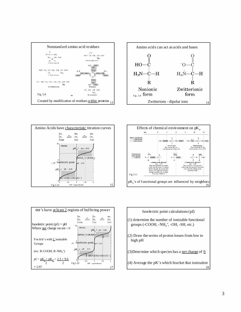
Nonstandard amino acid residues
Created by modification of residues within proteins
Fig. 5.8
14Zwitterions - dipolar ions
Amino acids can act as acids and bases
Fig . 5.9
15
Amino Acids have characteristic titration curves
pKa =
pKa =
Isoelectric point
pKa =
pKa =
Isoelectric point
[RC OOH]=[RCOO - ]
pKa =
pKa =
Isoelectric point
[RC OOH]=[RCOO - ]
[RNH 3+] =[RNH2]
F ig 5.10 16
Effects of chemical environment on pKa
pKa’s of f unctional groups are influenced by neighbors
Fig 5.11
17
αα’s have at least 2 regions of buffer ing power
pKa =
pKa =
Isoelectric point
1
2[RNH 3
+] =[RNH2]
[RC OOH]=[RCOO - ]
Isoeletric point (pI) = pH Where net charge on αα = 0
For αα’s with 2 ionizable Groups
(ex: R-COOH, R-NH3+)
pI = pK1 + pK2 = 2.3 + 9.6 2 2
= 5.97Fig 5.10
18
Isoelectric point calculations (pI)
(1) determine the number of ionizable functionalgroups (-COOH, -NH3
+, -OH, -SH, etc.)
(2) Draw the series of proton losses from low tohigh pH
(3)Determine which species has a net charge of 0
(4) Average the pK’s which bracket that ionization
4
19
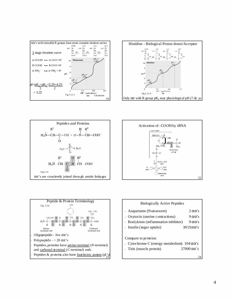
αα’s with ionizable R groups have more complex titration curves
Glu tamateFig 5.12 a
3 stage titration curve
α-CO OH α-CO O-+H+
δ-COOH δ-CO O-+H+
α-NH3+ α-NH2 + H+
pI=pK1+pK2=2.19+4.25 2 2
= 3.22
20
Histidine - Biological Proton donor/Acceptor
Only αα with R group pKa near physiological pH (7.4)
Fig 5.12 b
Peptides and Proteins
αα’s are covalently joined through amide linkages
Fig 5.13
22
Activation of -COOH by tRNA
23
Peptide & Protein Terminology
• Oligopeptide - few αα’s
• Polypeptide - > 20 αα’s
• Peptides, proteins have amino terminal (N-termina l)and carboxyl-te rminal (C-termina l) ends
• Peptides & proteins a lso have Isoe lectric points (pI ’s)
Fig . 5.14
24
Biologically Active Peptides
• Asapartame (Nutrasweet) 2 αα’s
• Oxytocin (uterine contractions) 9 αα’s
• Bradykinin (inflammation inhibitor) 9 αα’s
• Insulin (sugar uptake) 30/21αα’s
Compare to proteins:
• Cytochrome C (energy metabolism) 104 αα’s
• Titin (muscle protein) 27000 αα’s
5
25
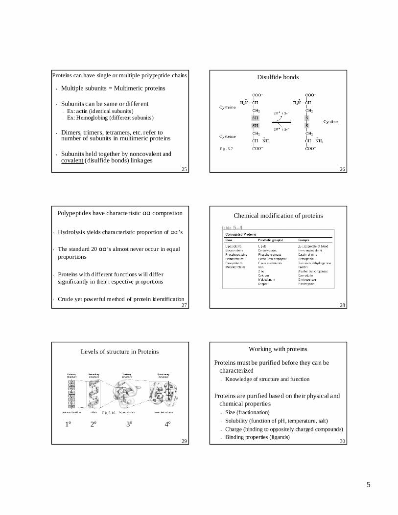
Proteins can have single or multiple polypeptide chains
• Multiple subunits = Multimeric proteins
• Subunits can be same or different– Ex: actin (identical subunits)– Ex: Hemoglobing (different subunits)
• Dimers, trimers, tetramers, etc. refer tonumber of subunits in multimeric proteins
• Subunits held together by noncovalent andcovalent (disulfide bonds) linkages
26
Disulfide bonds
Fig . 5.7
27
Polypeptides have characteristic αα compostion
• Hydrolysis yields characte ristic proportion of αα’s
• The standard 20 αα’s almost never occur in equalproportions
• Proteins w ith d ifferent fu nctions w ill d iffe rsignificantly in their r espective proportions
• Crude yet powerful method of protein identification28
Chemical modification of proteins
29
Levels of structure in Proteins
1° 2° 3° 4°
Fig 5.16
30
Working with proteins
Proteins must be purified before they can becharacterized– Knowledge of structure and function
Proteins are purified based on their physical andchemical properties– Size (fractionation)
– Solubility (function of pH, temperature, salt)
– Charge (binding to oppositely charged compounds)– Binding properties (ligands)
6
31
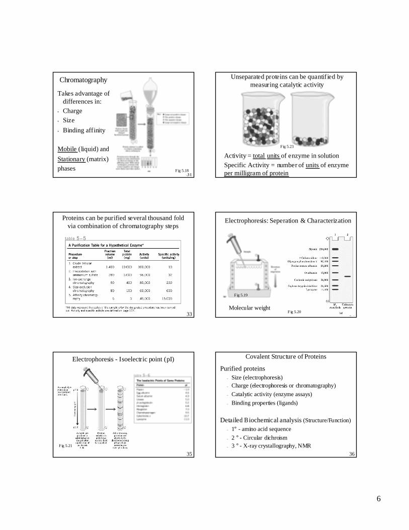
Chromatography
Takes advantage ofdifferences in:
• Charge
• Size
• Binding affinity
Mobile (liquid) and
Stationary (matrix)
phases Fig 5.18
Unseparated proteins can be quantified bymeasuring catalytic activity
Activity = total units of enzyme in solution
Specific Activity = number of units of enzymeper milligram of protein
Same activity as (a), but higher specific activ ity
(a) (b)
Fig 5.23
33
Proteins can be purified several thousand foldvia combination of chromatography steps
34
Electrophoresis: Seperation & Characterization
Molecular weight
Gel serves as a Molecula r “sie ve”
Fig 5.20
Fig 5.19
35
Electrophoresis - Isoelectric point (pI)
Fig 5.21
36
Covalent Structure of Proteins
Purified proteins– Size (electrophoresis)– Charge (electrophoresis or chromatography)
– Catalytic activity (enzyme assays)
– Binding properties (ligands)
Detailed Biochemical analysis (Structure/Function)
– 1° - amino acid sequence
– 2 ° - Circular dichroism– 3 ° - X-ray crystallography, NMR
7
37
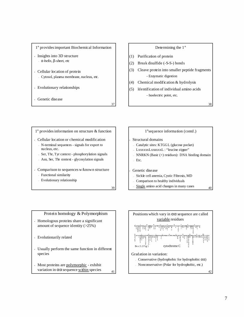
1° provides important Biochemical Information
• Insights into 3D structure– α-helix, β-sheet, etc
• Cellular location of protein– Cytosol, plasma membrane, nucleus, etc.
• Evolutionary relationships
• Genetic disease38
Determining the 1°
(1) Purification of protein
(2) Break disulfide (-S-S-) bonds
(3) Cleave protein into smaller peptide fragments
- Enzymatic digestion
(4) Chemical modification & hydrolysis
(5) Identification of individual amino acids
- Isoelectric point, etc.
39
1° provides information on structure & function
• Cellular location or chemical modification– N-terminal sequences - signals for export to
nucleus, etc.
– Ser, Thr, Tyr context - phosphorylation signals
– Asn, Ser, Thr context - glycosylation signals
• Comparison to sequences w/known structure– Functional similarity
– Evolutionary relationship
40
1°sequence information (contd .)
• Structural domains– Catalytic sites: KTGGL (glucose pocket)– LxxxxxxLxxxxxxL : “leucine zipper”
– NNRKN (Basic (+) residues): DNA binding domain
– Etc.
• Genetic disease– Sickle cell anemia, Cystic Fibrosis, MD
– Comparison to healthy individuals
– Single amino acid changes in many cases
41
Protein homology & Polymorphism
• Homologous proteins share a significantamount of sequence identity (>25%)
• Evolutionarily related
• Usually perform the same function in differentspecies
• Most proteins are polymorphic - exhibitvariation in αα sequence within species 42
Positions which vary in αα sequence are calledvariable residues
cytochrome CBo x 5 .2 F ig 1
Gradation in variation:– Conservative (hydrophobic for hydrophobic αα)
– Nonconservative (Polar for hydrophobic, etc.)
8
43
Biomedical & related benefits
• Evolutionary relationships– Anthropology
• Forensic Science– Identification of individuals, populations
• Proteomics– Examination of the expression of all proteins in a
cell
– Comparison of healthy/diseased states - whatproteins are (not) expressed?
– Provide targets for drug development44
Chapter 5 - Summary
• 20 Standard αα’s found in proteins– α-COOH group
– α-NH2 group
– Distinctive side chain (R group)
• α-carbon (central carbon) is asymmetric in allαα’s (except glycine)
– 2 stereoisomeric forms (D- and L-)– Only L- form in proteins
45
Summary (contd .)• αα’s classified on the basis of polarity & charge
– Nonpolar aliphatic
– Aromatic– Polar uncharged
– Acidic (-) charged
– Basic (+) charged
• αα’s ionize in aqueous solution– α-COOH α-COO -+ H+
– α-NH3+ α-NH2 + H+
– R Group ionization 46
Summary (contd .)
• αα’s are often characterized by their isoelectricpoint (pI) - pH where they have no net charge
• αα’s are covalently joined through peptide bonds– Amino (N-) and Carboxy (C-) terminal ends
• Proteins are often conjugated to other molecules– Metal ions– Lipids– Carbohydrates– Etc.
47
Summary (contd .)
• 4 levels of protein structure– Primary, secondary, tertiary & quarternary
• Protein structural & functional analysis– Solubility (Precipitation with salts)
– Chromatography (Size, Charge, Binding affinity)
– Electrophoresis (Size and charge)
• 1° provides important Biochemical Information– 3D structure, active sites, targeting signals, etc.
– Protein homology and polymorphism