![Stability of the human polymerase δ holoenzyme …thesis (TLS)] so that pol δ may resume synthesis (9–12). How-ever, studies on the human pol δ holoenzyme are lacking, and hence,](https://static.fdocument.org/doc/165x107/5ecf5ac71e33ba350c72b907/stability-of-the-human-polymerase-holoenzyme-thesis-tls-so-that-pol-may.jpg)
1 DNA Binding Properties of Human Pol γB José A. Carrodeguas ...
34
1 DNA Binding Properties of Human Pol γB José A. Carrodeguas*, Kevin G. Pinz and Daniel F. Bogenhagen ‡ Department of Pharmacological Sciences State University of New York at Stony Brook, Stony Brook, NY 11794-8651 Corresponding author Telephone: 631-444-3068 Fax: 631-444-3218 email: [email protected] Running title: DNA Binding by DNA Pol γB * Present Address, Laboratory of Neurobiology, Dept. of Anatomy, Embryology and Genetics, University of Zaragoza, Zaragoza, Spain. ‡ This work was supported by Grants NIGMS R01-GM296801 and NIEHS P01-04068. Copyright 2002 by The American Society for Biochemistry and Molecular Biology, Inc. JBC Papers in Press. Published on October 11, 2002 as Manuscript M207030200 by guest on April 6, 2018 http://www.jbc.org/ Downloaded from
Transcript of 1 DNA Binding Properties of Human Pol γB José A. Carrodeguas ...
1
DNA Binding Properties of Human Pol γB
José A. Carrodeguas*, Kevin G. Pinz and Daniel F. Bogenhagen‡
Department of Pharmacological Sciences
State University of New York at Stony Brook, Stony Brook, NY 11794-8651
Corresponding author Telephone: 631-444-3068
Fax: 631-444-3218 email: [email protected]
Running title: DNA Binding by DNA Pol γB
* Present Address, Laboratory of Neurobiology, Dept. of Anatomy, Embryology and
Genetics, University of Zaragoza, Zaragoza, Spain.
‡This work was supported by Grants NIGMS R01-GM296801 and NIEHS P01-04068.
Copyright 2002 by The American Society for Biochemistry and Molecular Biology, Inc.
JBC Papers in Press. Published on October 11, 2002 as Manuscript M207030200 by guest on A
pril 6, 2018http://w
ww
.jbc.org/D
ownloaded from
2
SUMMARY
We have recently reported the crystal structure of the accessory subunit of
mitochondrial DNA polymerase, pol γB, and identified a region of the protein involved in
DNA binding. The DNA employed in previous studies was presumed to be single-
stranded since it was generated by single-sided PCR. Further characterization of this
DNA indicated that, due to a strand transfer event during synthesis by single-sided
PCR, the DNA adopts a double-stranded hairpin conformation under native conditions.
We used a series of double and single-stranded oligonucleotides of different lengths to
confirm that human pol γB prefers to bind double-stranded DNA longer than 40 bp with
little apparent sequence specificity. Site-specific deletion mutagenesis identified clusters
of basic residues in two surface loops required for DNA binding located on opposite
sides of the symmetrical pol γB dimer. A heterodimer of pol γB that contains one mutant
and one wild-type DNA binding region was shown to be unable to bind double-stranded
DNA, suggesting that a single DNA molecule must contact both DNA binding sites in the
pol γB dimer. The ability to bind double-stranded DNA is not essential for pol γB
stimulation of pol γA activity in vitro, but may play a role in DNA replication or repair.
by guest on April 6, 2018
http://ww
w.jbc.org/
Dow
nloaded from
3
INTRODUCTION
Mitochondrial DNA is replicated by DNA polymerase γ, an enzyme with a
catalytic subunit, pol γA, containing both 5’���� �������� � �� ������ �� ������
activities, and an accessory subunit, pol γB, that affects a number of key properties of
the catalytic subunit (1,2). Pol γB is related both in primary sequence and structure to
class IIa prokaryotic aminoacyl-tRNA synthetases (aaRSs) (1,3). Apart from its role in
the stimulation of pol γ, pol γB has DNA binding activity that may reflect properties of
aaRSs. All aaRSs bind specific RNAs, although one, phenylalanyl tRNA synthetase,
has been shown to bind specifically to double-stranded DNA using an atypical helix-
turn-helix domain (4). The so-called b5 domain that mediates this DNA binding is not
found in most aaRSs. In preliminary experiments, we found that pol γB was able to bind
to a DNA substrate generated by single-sided PCR that was presumed to have a mostly
single-stranded conformation (3) similar to the H-strand region that serves as origin for
lagging strand mtDNA replication (OL) (5). These observations provided support for
models suggesting that the DNA binding ability of pol γB might play a role in initiation of
mtDNA replication (6). This sort of model has been suggested for Drosophila pol γ as
well (7), although this enzyme appears to have a simple heterodimer structure with
extensive contacts between the A and B subunits (8).
In this report we present the results of further experiments to characterize the
nucleic acid binding properties of mammalian pol γB, by studying binding to a variety of
single-stranded and double-stranded DNAs and by exploring the effects of amino acid
changes on nucleic acid binding. The results show that wild type pol γB binds only to
one of two major DNA species generated by single-sided PCR extending through OL.
by guest on April 6, 2018
http://ww
w.jbc.org/
Dow
nloaded from
4
Pol γB prefers to bind to an aberrant PCR product that is substantially double-stranded
due to a strand transfer event at the hairpin structure at OL. Binding titrations with a
variety of single-stranded and double-stranded DNAs of different lengths confirmed that
pol γB prefers to bind double-stranded DNA. We further show that clustered point
mutations that convert basic residues to alanine residues in two nucleic acid binding
loops alter the DNA binding properties of the protein. The pol γB dimer contains two
DNA binding sites on opposite sides of the protein. We constructed a pol γB
heterodimer containing one mutant and one wild-type DNA binding site and found that
this heterodimer was unable to bind double-stranded DNA. Thus, we conclude that both
sites are required for high affinity DNA binding, suggesting that an individual DNA
molecule must wrap around pol γB to interact simultaneously with both sites.
EXPERIMENTAL PROCEDURES
DNA clones and oligonucleotides
The DNA template used for the synthesis of 129-mer and other single-stranded DNAs
was first cloned by PCR from HeLa mitochondrial DNA using primers containing Xba I
(HOL1) and Xho I (HOL2) restriction sites. This clone, containing human mitochondrial
DNA sequences from position 5495 to 5920, spanning OL, was named pJAC64.
Sequencing revealed a point mutation, G to A, at position 5773.
Oligonucleotides were obtained from Operon. The sequences of oligonucleotides
used to generate fragments of mtDNA for binding assays are as follows:
HOL1: 5’-TCTAGATATACTAATAATCTTAT-3’. Used with HOL2 to make clone pJAC64.
HOL2: 5’-TCTGAGCAACGGTCGGCGAACAT-3’.
HOL3: 5’-CCGAGGTGATTTTCATATTG-3’. Used to make 129-mer and 117-mer.
by guest on April 6, 2018
http://ww
w.jbc.org/
Dow
nloaded from
5
HOL5: 5’-GAATTGCAAATTCGAAGAAG-3’. Used to make 98-mer and 83-mer.
HOL6: 5’-CCCTAATCAACTGGCTTC-3’. Used with HOL2 to make a double-stranded
221 bp DNA by PCR that was used as template for synthesis of the 98-mer (5’ end of
HOL6 matches 3’ end of 98-mer).
HOL8: 5’-TTCAATCTACTTCTCCCG-3’. Used with HOL2 to make a double-stranded
206 bp DNA by PCR that was used as template to prepare the 83-mer (5’ end of HOL8
matches 3’ end of 83-mer).
The following forward oligonucleotides (F) were used as single-stranded DNA in
EMSA. Each forward oligonucleotide was annealed with its corresponding complement
(R) to prepare double-stranded oligonucleotides:
32F: 5’-GCCGAGATGGAGCAGCAAATGTGGTTCCTTGT-3’.
32R: 5’-ACAAGGAACCACATTTGCTGCTCCATCTCGGC-3’.
40F: 5’-AGAGAACTCCTTTACAGCAGCAGCAAATCTTCATAGAAAG-3’.
40R: 5’-CTTTCTATGAAGATTTGCTGCTGCTGTAAAGGAGTTCTCT-3’.
47F: 5’-TATATCCAAATTAAAAGCATTTTTGATTGCATATATATCATCAGCTA-3’.
47R: 5’-TAGCTGATGATATATATGCAATCAAAAATGCTTTTAATTTGGATATA-3’.
The 32F and 32R oligonucleotides were also used for site-specific mutagenesis to
create mutant P1; the 40F and 40R oligonucleotides were used to create mutant P2.
Oligonucleotides used to produce the Not I/Xho I cassette encoding the calmodulin
binding protein tag were
CBPF: 5’-ATAAGAATGCGGCCGCAAAGCGACGATGGAAAAAG and
CBPR: 5’-ACCGCTCGAGTCATGCCCCGGAGGATGAGAT.
Labeling, purification and annealing of oligonucleotides
by guest on April 6, 2018
http://ww
w.jbc.org/
Dow
nloaded from
6
Oligonucleotides used for PCR and EMSA were gel-purified before labeling and again
after labeling as described above. Concentrations of unlabeled oligonucleotides were
calculated based on UV absorption. Labeling was carried out with polynucleotide kinase
(NEB) and [γ32P]-ATP, under standard conditions. Fractions of known amounts of
labeled oligonucleotides were spotted onto DE-81 paper (Whatman), washed with 250
mM potassium phosphate and counted in a scintillation counter to determine specific
activities. To generate double-stranded oligonucleotides, equal amounts of
complementary single-stranded oligonucleotides were mixed in a buffer containing 100
mM NaCl, 10 mM Tris, pH 8.0, 1 mM EDTA, heated at 90°C and then cooled slowly to
room temperature.
Synthesis of single-stranded DNA
The general approach used for synthesis of single-stranded DNA by PCR has been
described (3). To generate the 129-mer, the 228 bp double-stranded DNA used as a
template was excised by restriction digestion with enzymes Xho I and Hinc II from clone
pJAC64. Primer HOL3 was used in a standard PCR reaction using either Taq DNA
polymerase (Fisher) or Pfu Turbo DNA polymerase (Stratagene) with buffers supplied
by the manufacturer. To prepare other single-stranded DNA species, the template was
obtained by PCR using pJAC64 DNA as template and the primers described above. 25
or 30 cycles were carried out at 94°C for 45 sec, 54°C for 45 sec and 72°C for 20 sec.
In order to incorporate internal label, reactions were carried out using 100 ng of
template DNA, 20 pmol of unlabeled primer and 5 µCi of [α32P]-dATP. For 5’ end
labeled products, 5 pmol of kinased HOL3 were used in a PCR reaction under the same
conditions as above. The PCR products were precipitated with ethanol, collected by
by guest on April 6, 2018
http://ww
w.jbc.org/
Dow
nloaded from
7
centrifugation, and the pellet resuspended in formamide loading buffer, boiled and run in
8-13% polyacrylamide-8M urea sequencing gels. Bands were identified by
autoradiography, excised from the gel and crush-eluted by rotating end over end
overnight in a buffer containing 0.3 M sodium acetate, 10 mM Tris, pH 8, 1 mM EDTA.
After brief centrifugation in a microfuge the supernatant was filtered through 0.22 µm
Ultrafree-MC filters (Millipore). DNA was precipitated with ethanol and resuspended in
10 mM Tris, pH 8, 1 mM EDTA.
The concentration of internally-labeled PCR products was calculated by
measuring the incorporated radioactivity and the specific radioactivity of the precursors
in the PCR mix, taking into account the sequence of the DNA. For 5’ end labeled PCR
products, the final concentration was estimated using the known specific radioactivity of
the labeled primer.
Generation of mutants and purification of recombinant proteins
Human pol γB mutants HGB P1 (pJAC81) and HGB P2 (pJAC82) were generated using
the Quick Change method (Stratagene), using pJAC44 DNA (6) as template and
primers 32F, 32R (P1) and 40F, 40R (P2). Recombinant proteins were expressed and
purified essentially as described (6), with the following changes: expression was carried
out at room temperature, with addition of extra ampicillin (50 µg/ml) every hour after
induction, for a total of 3 hours. Frequent addition of fresh ampicillin helped prevent loss
of the plasmid by the BL21(DE3) cells and substantially increased yield of recombinant
protein. C-terminal his-tagged recombinant proteins were purified by Ni-NTA (Qiagen)
affinity chromatography (6) except that the wash buffer was adjusted to contain 1 M
NaCl instead of 300 mM NaCl to reduce contamination by bacterial proteins.
by guest on April 6, 2018
http://ww
w.jbc.org/
Dow
nloaded from
8
In order to generate pol γB heterodimers with differential affinity tags on the two
monomers, a derivative of pET22b(+) was generated with a C-terminal calmodulin
binding protein tag in place of the His tag. Oligonucleotide CBPF containing a Not I
recognition site and oligonucleotide CBPR containing an Xho I recognition site were
used for PCR on plasmid pSH6 (9) to produce a 105-bp product encoding the
calmodulin binding peptide tag sequence AAAKRRWKKNFIAVSAANRFKKISSSG.
Following cleavage of the PCR product with Not I and Xho I, the resulting fragment was
cloned into pET22b(+) vector cut with the same restriction enzymes. The resulting
ampicillin-resistant vector was named pET22b(+)/CBP. This vector was digested with
Nde I and Not I to permit it to accept Nde I/Not I DNA fragments encoding wild-type
human pol γB or the P2 mutant. To permit selection for heterdimeric pol γB, Nde I/Not I
cassettes encoding wild-type and P1 mutant human pol γB were inserted into the
kanamycin-resistant vector pET29a(+), which supports synthesis of his-tagged proteins.
Co-transfection of E. coli BL21(DE3) with HGB P1 in pET29a(+) and HGB P2 in
pET22b(+)/CBP and selection for both ampicillin and kanamycin resistance generated a
strain capable of expressing both proteins. As a control, the two wild-type HGB clones
in both pET22b(+)/CBP and pET29a(+) were also co-expressed. In each case, co-
expression was expected to produce three forms of dimeric protein, the His-tagged and
CBP-tagged homodimers and the heterodimer bearing both His- and CBP-tags.
Purification of the His-/CBP-tagged heterodimers was carried out first on a
calmodulin column (Stratagene) and then on Ni-NTA (Qiagen). Bacterial cells were
sonicated in lysis buffer containing 50 mM Tris, pH 7.4, 1 mM DTT, 150 mM NaCl and
0.1% TX-100, 0.2 mM PMSF, 5 µg/ml leupeptin, 1 µM pepstatin, 1 mM imidazole, 1 mM
by guest on April 6, 2018
http://ww
w.jbc.org/
Dow
nloaded from
9
MgCl2, and 4 mM CaCl2. The homogenate was clarified by centrifugation and the
supernatant was incubated with calmodulin affinity resin on a rotator for 2 hr at 4°C. The
beads were washed extensively with lysis buffer and bound protein was step-eluted with
the same buffer lacking CaCl2 and MgCl2 but containing 2 mM EGTA. The eluate was
concentrated by ultrafiltration using a Centricon 30 and adjusted to 25 mM NaPi, pH 8.0,
300 mM NaCl, 20% glycerol, 2 mM BME and 0.2 mM PMSF. Additional purification by
Ni-NTA affinity chromatography was performed as described (6). Quantitation of
recombinant proteins was carried out by UV absorbance or by densitometry of
Coomassie blue-stained SDS-PAGE gels using commercial glutamate dehydrogenase
as a standard.
Electrophoretic mobility shift assays (EMSA)
Reactions were carried out in 10 µl volumes containing 10 mM Tris, pH 8, 2.5 mM DTT,
1 mM EDTA, 150 µg/ml BSA, 10 % glycerol and 70 mM NaCl. Each reaction contained
a total of 2 µl of either protein or dialysis buffer (6), which supplied the glycerol and 60
mM salt to the reaction. 1 µl DNA was used, containing 100 mM NaCl, which was
responsible for 10 mM salt in the reaction. Reactions were incubated at 30°C for 10 min
and run in native polyacrylamide gels. Gels contained 6% acrylamide, 0.1% bis-
acrylamide, 20 mM HEPES, pH 8, 0.1 mM EDTA. Running buffer was 20 mM HEPES,
pH 8, 0.1 mM EDTA. Gels were pre-run at room temperature for 1 h at 80V, using the
miniprotean II system (Bio-Rad), and run under the same conditions. Gels were dried on
DEAE paper (Whatman) and imaged using either Kodak XAR5 film or a
phosphorimager screen.
Purine ladders
by guest on April 6, 2018
http://ww
w.jbc.org/
Dow
nloaded from
10
30 fmol (10000 cpm) of end-labeled DNA was mixed with 10 µg tRNA carrier in a total
volume of 20 µl. 4 µl of 4% pyridinium formate, pH 2, was added and the mix was
incubated at 48°C for 15 minutes. Following ethanol precipitation, the DNA was
resuspended in 20 µl of water, 80 µl of 10% piperidine was added and the mix was
incubated at 90°C for 10 minutes. After ethanol precipitation, the DNA was resuspended
in formamide loading buffer, boiled and run in 8% polyacrylamide-8M urea sequencing
gels. Gels were fixed in a solution containing 10% acetic acid, 12% methanol, dried on
Whatman 3MM paper and an autoradiogram was obtained.
RESULTS
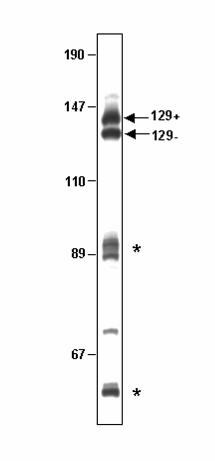
Single-sided PCR through OL produces two major DNA species, only one of
which binds Pol γB.
We have previously suggested that the relationship of pol γB with some aminoacyl-tRNA
synthetases could indicate a role in binding tRNA-like structures present at the
mitochondrial origins of replication. To test this model we attempted to synthesize a
single-stranded DNA spanning the light strand origin of replication (OL). This heavy
strand DNA fragment was expected to contain the stem-loop structure known to be
required for the initiation of light strand synthesis. We chose single-sided PCR as a
quick method to generate single-stranded DNA. This method involves the use of PCR to
extend an oligonucleotide primer using double-stranded DNA as template, generating a
run-off product. The product, labeled by incorporation of radioactive dAMP, was then
separated from the template DNA using denaturing electrophoresis in polyacrylamide-
urea gels. Two closely migrating species were observed near the position expected for
the single-stranded DNA (Fig. 1) in addition to other shorter extension products that
by guest on April 6, 2018
http://ww
w.jbc.org/
Dow
nloaded from
11
appeared to terminate at secondary structures in the template. We assumed that the
largest species was the expected run-off product of 129 nucleotides and the shorter one
was produced by the polymerase stalling before reaching the end of the template. We
refer to the slower migrating species as 129(+) and to the faster migrating one as 129(-).
129(+) was used as the presumed single-stranded DNA in previous binding studies (3).
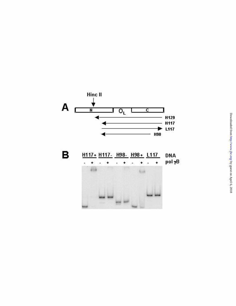
In our attempts to delimit the region in the 129-mer necessary for pol γB binding,
we generated shorter products using the same single-sided PCR technique, changing
the primer and/or DNA template used for synthesis. In this way, we synthesized a 117-
mer, a 98-mer and an 83-mer (Fig. 2A). In each case we obtained two major DNA
species around the expected size (data not shown), resembling products we obtained
for the 129-mer. Experiments employing the 83-mer are not included in this report.
EMSA experiments revealed that pol γB bound the (+) species in every case, but
not the (–) species (Fig. 2B). Unexpectedly, in the native gels used for mobility shift
assays, the (+) and (–) species interchanged their mobilities with respect to denaturing
gels, with 129(+), 117(+), and 98(+) now migrating faster than 129(-), 117(-) and 98(-),
respectively. This unusual electrophoretic mobility prompted further investigation of the
nature of the pairs of PCR products.
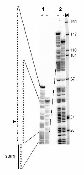
We synthesized 5’-end labeled 129(+) and 129(-) species using 5’ end-labeled
primers to permit chemical sequencing to determine whether they represented different
conformations of the same DNA fragment or differed somehow in sequence. Following
purification of the DNAs in denaturing gels, we carried out partial DNA sequencing by
generating purine ladders for each of the DNA species. The ladders obtained with 129
(-) species matched the sequence expected for the 129-mer. The ladder obtained with
by guest on April 6, 2018
http://ww
w.jbc.org/
Dow
nloaded from
12
the 129(+) species showed the identical sequence up to a point corresponding to the
“descending” side of the OL stem, in the direction of synthesis. From that point on, the
sequence was divergent (Fig. 3). Equivalent results were obtained with end-labeled
98(+) and 98(-) PCR products, with the 98(-) ladder matching the expected sequence
for the 98-mer and the 98(+) one diverging at the same point seen with 129(+) (data not
shown).
As shown in Fig. 1, when the 129-mer was synthesized, a significant fraction of
PCR products appeared to be aborted at a size around 89 nucleotides (upper asterisk).
This is the point at which 129(+) and 129(-) sequences diverge, coinciding with the
descending half of the stem at OL, in the direction of synthesis. We reasoned that the 3’
region of those aborted molecules should be able to fold in a stem-loop structure,
resembling the OL. The 3’ end of this stem could then prime synthesis by the
polymerase, extending the size of the double-stranded stem back to the 5’ end of the
primer (see Fig. 4B). This would generate a large stem structure with a loop
corresponding to the OL loop, i.e., a 70 bp stem with a 12 nt loop. In denaturing gels,
this DNA species would behave as a single-stranded DNA of 152 nts, which agrees well
with the mobility of the 129(+) species seen in Fig. 3. The relative mobility of the hairpin
PCR products varied somewhat with the gel temperature and the quantity loaded on
gels containing 8M urea. It is well-known that urea is a rather weak denaturing agent
that is not able to completely disrupt secondary structures (10). Analysis of the purine
ladder generated from the 129(+) species beyond the point of sequence divergence in
Fig. 3 showed that it matched the expected sequences for the 152-mer DNA predicted
by the mechanism shown in Fig. 4. The 129(+) and 98(+) DNAs would be expected to
by guest on April 6, 2018
http://ww
w.jbc.org/
Dow
nloaded from
13
behave as double-stranded DNAs of approximately half the size under native
conditions. This agrees with the faster mobility of the (+) species in the native gels used
for EMSA (see Fig. 2B).
We also probed the structure of these PCR products by digestion with the
restriction enzyme Alu I. This enzyme should produce a labeled 42 nucleotide fragment
from 5’ end-labeled 129(+), when analyzed in denaturing gels (Fig. 4B). Alu I should not
cut the single-stranded 129(-) species. This is exactly what we observed (data not
shown). We also confirmed that when internally labeled 129(-) is annealed to a
complementary single strand, it can be cut by Alu I, generating the expected products
shown in Fig. 4A (data not shown).
The reason why the polymerase stalls in the region corresponding to the
descending half of the OL stem loop is not obvious. The polymerase might be expected
to stall preferentially when entering the stem structure (assuming such a structure exists
during synthesis), but the products seen during synthesis due to stalling at this site are
only minor species. Instead, the polymerase appears to stall and to engage in hairpin
replication at the base of the loop at OL. Interestingly, the hairpin product was only
observed when the light strand was the template, not when the polymerase was moving
in the opposite direction using the heavy strand as template. There may be some
unusual structure resulting from the G-rich tract at the base of the OL loop that facilitates
hairpin replication by the polymerase. We have obtained different ratios of 129(+) and
129(-) species using different polymerases (Taq, Pfu) and different concentrations of
nucleotides, suggesting that different conditions could produce different amounts of the
hairpin species. We tested whether pol γ is similarly prone to hairpin formation at this
by guest on April 6, 2018
http://ww
w.jbc.org/
Dow
nloaded from
14
site, but did not find evidence of hairpin products (K. P. unpublished observation).
Collectively, these results demonstrated that the reported binding by pol γB to a single-
stranded 129-mer (3) represented binding to a mostly double-stranded DNA of 70 bp,
with a 12 nt loop at one end.
Pol γB binding to synthetic double-stranded DNA oligonucleotides
The data presented in Fig. 2B indicates that pol γB binds more tightly to double-
stranded (H117(+), H98(+)) than to single-stranded DNA (H117(-), H98(-), L117). In
order to confirm the double strand DNA binding preference of pol γB, we used a series
of complementary oligonucleotides of different lengths. Oligonucleotides of 32, 40, 47
and 65 nts of unrelated sequences were annealed to their complementary
oligonucleotides to generate double-stranded DNA or were used alone as single
strands. Binding assays proved that pol γB prefers to bind double-stranded instead of
single-stranded DNA (Fig. 5). More avid binding is observed with DNAs of 47 bp or
larger, which indicates an approximate minimum DNA size requirement for pol γB
binding. Also, the fact that pol γB was able to bind a variety of DNA sequences indicates
that there is little or no sequence specificity for this reaction, although this aspect has
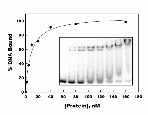
not been studied in detail. In order to calculate an approximate Kd for double-stranded
DNA binding by pol γB we measured the disappearance of free DNA as the protein
concentration was increased since more than one complex can be seen. A binding
titration with 1 nM ds47 DNA as shown in Fig. 6 was analyzed as a simple binding 1:1
interaction between the pol γB dimer and DNA, provided an apparent Kd of 8.6 +/- 1.5
nM.
Identification of residues in pol γB necessary for DNA binding
by guest on April 6, 2018
http://ww
w.jbc.org/
Dow
nloaded from
15
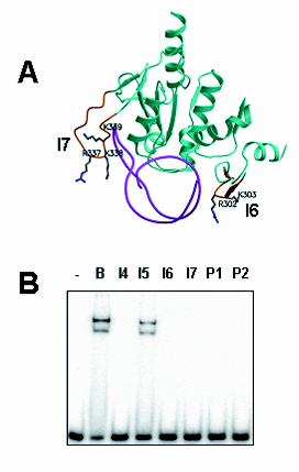
We previously used deletion mutagenesis to identify two protein loops in pol γB
required for DNA binding (3). We refer to these surface loops as loop I6, between β
strands 10 and 11, and I7, between β strands 13 and 14. These two loops are closely
apposed in the dimeric protein structure (3) and contain clusters of basic residues. The
corresponding regions in threonyl-tRNA synthetase contribute to the RNA binding site
for anticodon recognition (11). To identify residues in these loops necessary for DNA
binding by pol γB, we generated alanine-replacement mutants. In mutant P1, two basic
residues in the I6 loop R302K303, were replaced with alanines; in mutant P2, three
residues in the I7 loop, R337K338K339, were replaced with alanines (see Fig. 7A).
We studied the abilities of the new mutants, HGB P1 and P2, as well as other
mutants of pol γB, to bind the double-stranded 47-mer, ds47. Fig. 7B shows the EMSA
results obtained with wild type pol γB, deletion mutants I4, I5, I6, I7 (described
previously (3)) and point mutants, P1 and P2. These results indicate that the basic
residues in loops I6 and I7 are required for pol γB binding to double-stranded DNA. The
P1 and P2 mutants are able to stimulate pol γA activity in vitro (not shown), as has been
shown for the I6 and I7 deletions (3), indicating that pol γB binding to double-stranded
DNA is not necessary for stimulation of pol γA activity.
Double-stranded DNA interacts with pol γB on two opposite sides of the protein
The results presented above suggest a working model to describe the DNA
binding of pol γB whereby an I6 loop from one monomer and an I7 loop from the other
create a binding site for double-stranded DNA. Since pol γB is a dimer, the complex
would be expected to contain two potential I6/I7 binding sites on either side of the
protein. When either the I6 or I7 loop is mutated, binding sites on both sides in the pol
by guest on April 6, 2018
http://ww
w.jbc.org/
Dow
nloaded from
16
γB dimer are affected. Thus, the foregoing results do not permit us to determine whether
one I6/I7 binding site is sufficient for binding to double-stranded DNA.
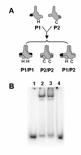
To test this model directly, we generated a pol γB heterodimer containing a
mutated I6 loop (P1) in one monomer and a mutated I7 loop (P2) in the other as
described in Experimental Procedures. The strategy to accomplish this was to
coexpress two forms of pol γB with different C-terminal affinity tags in the same E. coli
cells, as shown in Fig. 8A. We reasoned that successive chromatography on two
different affinity matrices would permit purification of heterodimers containing one
monomer with each type of affinity tag. We employed a C-terminal calmodulin binding
protein (CBP) tag for this experiment. This is a convenient affinity tag, since the protein
can be adsorbed to a calmodulin affinity column in the presence of calcium and
desorbed by the replacement of calcium with the chelator EGTA (9). To provide a
positive control, wild-type pol γB was cloned in the same two vectors and expressed
under the same conditions. In each case, three types of dimers are expected to form:
His-tagged homodimers, CBP-tagged homodimers and heterodimers containing one
His-tagged subunit and one CBP-tagged subunit. Only the heterodimers are retained on
both types of affinity matrices; this was confirmed by MALDI-TOF mass spectrometry
(data not shown). For the case of the P1/P2 heterodimer, the pol γB should have
mutated DNA binding loops I6 and I7 on one side of the protein dimer and wild type
ones on the opposite side. Dimeric wild-type and P1/P2 mutant pol γB proteins were
purified by chromatography on calmodulin affinity resin followed by Ni-NTA as described
in Materials and Methods. As shown in Fig. 8B, the P1-His/P2-CBP heterodimer was
unable to bind double-stranded DNA, although the wild-type heterodimer control was
by guest on April 6, 2018
http://ww
w.jbc.org/
Dow
nloaded from

17
fully active in DNA binding. These results suggest that two wild type I6 and I7 loops are
necessary for DNA binding by pol γB and that DNA must loop around the protein in
some fashion to permit this interaction.
DISCUSSION
The accessory subunit of DNA pol γ, pol γB, stimulates the activity of the catalytic
subunit under physiological buffer conditions (1). The finding that vertebrate pol γB is
related to prokaryotic aminoacyl-tRNA synthetases (aaRSs) suggested that the nucleic
acid binding properties of a tRNA synthetase might contribute to the function of the
accessory factor. Since the sequences surrounding both origins of mtDNA replication
have a high potential for forming complex secondary structures, we speculated that pol
γB could be involved in the recognition of such structures, directing the polymerase to
the origins of replication (6). As a first approach to test this hypothesis we studied pol γB
binding to a DNA fragment derived from a region of the mitochondrial genome that
contains the light strand origin of replication (OL). We initially documented binding of pol
γB to a DNA substrate synthesized by single sided PCR (3). In this paper we show that
the PCR product that bound tightly to pol γB in these experiments was, in fact, a largely
duplex hairpin generated by fold-back priming during PCR. To confirm the double-
stranded DNA binding preference of pol γB, we used a series of oligonucleotides, either
in single-stranded or double-stranded form. The data in Figs. 5 and 6 show that pol γB
has a poor ability to bind short duplex oligonucleotides, but is able to bind with high
affinity (Kd of ~8.6 nM) to a 47-mer oligonucleotide. Our measurement of the absolute
affinity of this interaction is subject to technical limitations of the EMSA assay and we
are working to develop independent measurements of this affinity using other methods.
by guest on April 6, 2018
http://ww
w.jbc.org/
Dow
nloaded from
18
Lim et al. (12) have previously observed binding of pol γB to a 34:38-mer primer-
template, but the lowest protein concentration used in their experiments, 2 pmol in a 20
µl binding reaction, did not permit determination of the KD for this interaction. Our results
suggest that the presence of a 5’ overhang in the primer-template used by Lim et al.
(1999) probably did not influence the binding. Pol γB has shown a similar ability to bind
other duplex fragments longer than ds47 (data not shown), suggesting that DNAs must
exceed a minimal size between 38 to 47 base pairs in order to bind. Pol γB binds very
poorly to single-stranded DNA, such that only a few percent of input DNA is bound by
150 nM protein (Fig. 5). To date, we have identified no specific sequences that
preferentially bind to pol γB. However, the finding that a single DNA molecule appears to
interact with binding sites on both sides of the pol γB dimer, suggests that DNA
sequences with an intrinsic bend may be bound more avidly.
The binding of pol γB to double-stranded DNA provides a contrast to the binding
of folded single-stranded RNA by tRNA synthetases. The affinity of pol γB for double-
stranded DNA is much higher than that previously observed for the phenylalanyl-tRNA
synthetase from T. thermophilus, which has been estimated to have a binding constant
of 400 nM. This interaction also requires a longer minimal DNA size of approximately 80
base pairs and does not employ the same regions of the protein required for tRNA
binding (4,13). Thus, it appears that there are significant differences between the DNA
binding reported for phenylalanyl-tRNA synthetase and that reported here for pol γB.
DNA binding by mammalian pol γB depends on the dimeric structure of the
protein and on two superficial loops initially identified by deletion analysis, I6 and I7 (3).
The corresponding regions of threonyl-tRNA synthetase are involved in binding to the
by guest on April 6, 2018
http://ww
w.jbc.org/
Dow
nloaded from
19
anticodon of tRNA, as depicted in Fig. 7. Both loops in pol γB contain basic lysine and
arginine residues that appeared to be good candidates to play a role in DNA binding.
Site directed mutagenesis to convert these residues to alanines confirmed this model
(Fig. 7). We conclude that the basic residues in the I6/I7 region are essential for the
double-stranded DNA binding activity of pol γB. Since we observed that DNA binding
requires a rather long segment of DNA, approximately 38 to 47 base pairs (Fig. 5), we
sought to test the model that a single DNA duplex must interact simultaneously with the
I6/I7 loops on both faces of the pol γB dimer protein. We produced a heterodimer
containing one pol γB polypeptide with point mutations in I6 and a second with point
mutations in I7. The results shown in Fig. 8 revealed that this heterodimer was not able
to bind DNA. The use of a dual-tagged control wild-type protein ruled out the trivial
possibility that this deficiency was due to the nature of the tags employed in purification.
Thus, we conclude that a single DNA molecule must contact basic residues on both
sides of pol γB for stable binding.
The structural basis for the action of pol γB on the catalytic subunit is poorly
understood. This reflects the fact that the structure of the catalytic subunit has not been
determined and the interactions between the large and small subunits have not been
defined precisely. Both the mammalian pol γB, which has a dimeric structure, and its
Drosophila homolog, which binds as a monomer to its cognate pol γA, resemble tRNA
synthetases. Recently, Drosophila pol γB has been shown to make extensive contacts
with the catalytic subunit (8). These extensive contacts may be critically important for
the activity of the small subunit as a processivity factor (14). Among processivity factors,
the ability of pol γB to bind duplex DNA is unusual, but not unprecedented. The toroidal
by guest on April 6, 2018
http://ww
w.jbc.org/
Dow
nloaded from
20
“sliding clamp” processivity factors like PCNA and E. coli DNA pol III β subunit do not
possess intrinsic DNA binding activity and must be loaded onto DNA by additional
factors. However, the herpes virus UL42 protein does bind DNA non-specifically with
high affinity (15). In this case, the non-specific DNA binding activity of UL42 appears to
contribute to the ability of the herpes virus DNA polymerase holoenzyme to conduct a
one-dimensional scan along DNA to identify primer-template binding sites (16). Indeed,
mutations in UL42 that abrogate non-specific DNA binding also impair the ability of the
protein to function as a processivity factor. This provides an interesting contrast to pol
γB, where mutants deficient in DNA binding, point mutants PI and P2 and the related
deletion mutants I6 and I7, are not impaired in their ability to stimulate in vitro DNA
synthesis by the catalytic subunit on a poly(dA):oligo(dT) template:primer ((3) and data
not shown). Thus, the role, if any, that is played in mtDNA maintenance by this double-
stranded DNA binding of pol γB remains to be established.
ACKNOWLEDGMENTS
We thank Brian Donohue for assistance in preparation of the P1 and P2 mutants and
Karsten Theis and Caroline Kisker for their comments on the manuscript.
REFERENCES
1. Carrodeguas, J. A., Kobayashi, R., Lim, S. E., Copeland, W. C., and
Bogenhagen, D. F. (1999) Mol. Cell. Biol. 19, 4039-4046
2. Johnson, A., Tsai, Y., Graves, S., and Johnson, K. (2000) Biochem 39, 1702-
1708
3. Carrodeguas, J. A., Theis, K., Bogenhagen, D. F., and Kisker, C. (2001) Mol. Cell
7, 43-54
by guest on April 6, 2018
http://ww
w.jbc.org/
Dow
nloaded from
21
4. Lechler, A., and Kreutzer, R. (1998) J. Mol. Biol. 278, 897-901
5. Clayton, D. A. (1982) Cell 28, 693-705
6. Carrodeguas, J. A., and Bogenhagen, D. F. (2000) Nucleic Acids Res. 28, 1237-
1244
7. Fan, L., Sanschagrin, P. C., Kaguni, L. S., and Kuhn, L. A. (1999) Proc. Natl.
Acad. Sci. USA 96, 9527-9532
8. Fan, L., and Kaguni, L. S. (2001) Biochemistry 40, 4780-4791
9. Honey, S., Schneider, B., Schieltz, D., Yates, J., and Futcher, B. (2001) Nucleic
Acids Res 29, e24
10. Maniatis, T., Jeffrey, A., and vandeSande, H. (1975) Biochemistry 14, 3787-3793
11. Sankaranarayanan, R., Dock-Bregeon, A. C., Romby, P., Caillet, J., Springer, M.,
Rees, B., Ehresmann, C., Ehresmann, B., and Moras, D. (1999) Cell 97, 371-381
12. Lim, S. E., Longley, M. J., and Copeland, W. C. (1999) J. Biol. Chem. 274,
38197-38203
13. Dou, X., Limmer, S., and Kreuzer, R. (2001) J. Mol. Biol. 305, 451-458
14. Breyer, W. A., and Matthews, B. W. (2001) Protein Sci 10, 1699-1711
15. Weisshart, K., Chow, C. S., and Coen, D. M. (1999) J. Virol. 73, 55-66
16. Randell, J. C., and Coen, D. M. (2001) Molecular Cell 8, 911-920
17. Kraulis, P. J. (1991) J. Appl. Crystallogr. 24, 946-950
18. Merritt, E. A., and Murphy, M. E. P. (1994) Acta Crystallographica Section D. 50,
869-873
by guest on April 6, 2018
http://ww
w.jbc.org/
Dow
nloaded from
22
FIGURE LEGENDS
FIG. 1. Products obtained by single-sided PCR through the OL region. DNA
synthesis was carried out by single-sided PCR as described in an attempt to synthesize
a 129-mer containing a segment of the H-strand surrounding OL (3). Products were
separated by electrophoresis in polyacrylamide-urea sequencing gels. The bands
corresponding to the 129(+) and 129(-) products described in the text are indicated. The
asterisks indicate major pause sites during DNA synthesis. The sizes in nucleotides of
markers run in parallel are indicated on the left.
FIG. 2. Pol γB binding to different DNA species containing human OL sequences.
A, Scheme showing the design for several DNA species that we attempted to
synthesize from the human OL region. H refers to heavy strand DNAs (synthesized from
right to left, as indicated by arrows), L refers to light strand DNAs (made from left to
right). PCR reactions generating L-strand sequences did not produce the sort of doublet
“+” and “-“ species observed for H-strand sequences. tRNA genes surrounding the OL
region are indicated with their one letter code (N, C). H129, H117, and H98 coincide
with the minus species mentioned in the text [129(-), 117(-), and 98(-)]. B, Results of pol
γB binding to H117(+), H117(-), H98(+), H98(-) and L117 using EMSA. Note that L117 is
the complementary strand of H117(-). Binding reaction conditions were as described (3)
and included 1 nM DNA and 20 nM protein (calculated as dimer).
FIG. 3. Purine ladder sequencing of 129(+) and 129(-) DNA species. 30 fmol of 5’
end labeled 129(+) and 129(-) DNAs synthesized by PCR were treated with pH 2 buffer
to partially depurinate the DNA, then treated with alkali to break DNA at abasic sites as
described under Experimental Procedures. The fragments were separated by
by guest on April 6, 2018
http://ww
w.jbc.org/
Dow
nloaded from
23
electrophoresis in a denaturing 8% polyacrylamide-8 M urea gel. The sample was
loaded twice at different times to resolve the upper (1) and lower (2) regions of the
purine ladders . The lane labels “+” and “–“ refer to 129(+) and 129(-), respectively. DNA
size markers in lane M, run in parallel on the right, were applied with the second
loading. The sequence corresponding to each of the purine ladders is indicated on the
left. Note that the sequence between 129(+) and 129(-) diverges after the stem (stem
loop of OL). Lines indicate the correspondence of the sequence with the bands. An
arrowhead indicates a mutation in our sequence with respect to the reported human
mtDNA sequence (G to A, position 5773).
FIG. 4. Scheme showing the expected structures of 129(+) and 129(-). A, expected
129-mer, which corresponds to 129(-), showing the location of restriction sites and
expected restriction fragments after annealing to a complementary strand and digesting
with Alu I. This DNA is expected to fold with a stem-loop structure comprising the OL as
shown. B, model for the formation of the 129(+) species. The 89-mer product can fold
back and prime synthesis by the polymerase (dashed line). This product would behave
as a 152-mer under denaturing conditions. The double-stranded DNA region can be cut
by Alu I to generate the indicated single-stranded fragments (two 42-mers and one 68-
mer).
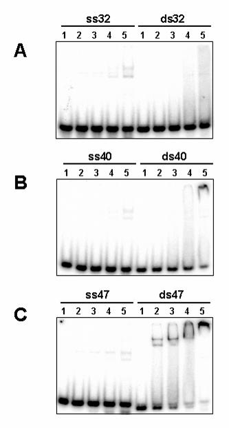
FIG. 5. Pol γB binding to single-stranded and double-stranded oligonucleotides of
different lengths. Reactions were performed as in Fig.2, but contained 1 nM DNA and
0, 5, 15, 50 or 150 nM protein (lanes 1 through 5, respectively). ss stands for single-
stranded and ds, for double-stranded DNA, followed by the size of the DNAs in
by guest on April 6, 2018
http://ww
w.jbc.org/
Dow
nloaded from
24
nucleotides or base pairs. Note that ds47 in panel C contains a minor portion of ss47
that failed to anneal to its complement and did not bind pol γB.
FIG. 6. Titration of the binding of pol γB to duplex oligonucleotide ds47. Binding
reactions were conducted under standard conditions with 1 nM ds47 DNA and 0, 2.5, 5,
10, 20, 40, 80 and 160 nM dimeric pol γB. The fraction of DNA bound to protein was
estimated by the removal of free DNA from the unbound position. The smooth curve
drawn through the data points was fit to the data using the Langmuir isotherm,
DNAbound/DNAtotal=[protein]/(KD+[protein]), and SigmaPlot software, resulting in the KD of
8.6 +/- 1.5 nM. The inset shows the phosphorimage of the gel analysis. Note that ds47
contains a minor portion of ss47 that failed to anneal to its complement and did not bind
pol γB.
FIG. 7. Binding of pol γB mutants to double-stranded 47-mer. A, domain 3 of one
monomer and loop I6 of domain 1 of the other monomer of pol γB were superimposed
over the equivalent region of threonyl-tRNA synthetase from Thermus thermophilus
complexed with its cognate tRNA. The thrRS protein regions were then removed from
the figure to illustrate how the I6 and I7 loops of pol γB might contact nucleic acid. Only
the tRNA from the aaRS-tRNA complex is shown (cyan), along with pol γB structures
(green). Domains I6 and I7 are shown in gold indicating the positions of mutated
residues (R302, R303 in loop I6, domain 1; R337, K338, K339 in loop I7, domain 3).
Side chains are shown only for the mutated residues. B, Binding of pol γB (B) and
mutants (I4-I7, P1 and P2) to 47-mer dsDNA, using 1 nM DNA and 10 nM protein.
Figure 7a was generated using Molscript (17) and Raster 3D (18).
by guest on April 6, 2018
http://ww
w.jbc.org/
Dow
nloaded from
25
FIG. 8. Double-stranded DNA binding by pol γB requires two sites on opposite
sides of the protein. A, Pol γB constructs were prepared containing point mutations P1
(His-tagged (H)) or P2 (CBP-tagged (C)) in loops I6 and I7 required for binding DNA.
The proteins were co-expressed in bacteria and the P1P2 heterodimer was purified by
chromatography on two affinity columns as described in the text. A heterodimer
containing wild-type pol γB with both tags was prepared as a positive control. B, The
ability of pol γB variants to bind 47-mer dsDNA was tested by EMSA. Binding reactions
contained 1 nM DNA alone (lane 1) or with 10 nM of His-tagged wild-type pol γB (lane 2)
or dual-tagged heterodimer constructs of wild-type pol γB (lane 3) or the P1P2 mutant
(lane 4).
by guest on April 6, 2018
http://ww
w.jbc.org/
Dow
nloaded from
Jose A. Carrodeguas, Kevin G. Pinz and Daniel F. BogenhagenBγDNA binding properties of human Pol
published online October 11, 2002J. Biol. Chem.
10.1074/jbc.M207030200Access the most updated version of this article at doi:
Alerts:
When a correction for this article is posted•
When this article is cited•
to choose from all of JBC's e-mail alertsClick here
by guest on April 6, 2018
http://ww
w.jbc.org/
Dow
nloaded from