


γλώσσες
Σελίδες
Νομικός

Vitamin B6 suppresses NLRP3 inflammasome
1
Vitamin B6 prevents IL-1β production by inhibiting NLRP3 inflammasome activation*
Peipei Zhang,ǂ Kohsuke Tsuchiya,ǂ Takeshi Kinoshita,ǂ Hiroko Kushiyama,ǂ Sofya Suidasari,§ Mizuki
Hatakeyama,¶ Hisanori Imura,¶ Norihisa Kato,§ and Takashi Sudaǂ1
From the ǂDepartment of Immunology and Molecular Biology, Cancer Research Institute, Kanazawa
University, Kanazawa, 920-1192, Japan, the §Graduate School of Biosphere Science, Hiroshima University,
Higashi-Hiroshima 739-8528, Japan, and the ¶Division of Material Sciences, Graduate School of Natural
Science and Technology, Kanazawa University, Kanazawa, 920-1192, Japan
Running title: Vitamin B6 suppresses NLRP3 inflammasome
To whom correspondence should be addressed: Prof. Takashi Suda; Cancer Research Institute, Kanazawa
University, Kakumamachi, Kanazawa, Ishikawa 920-1192, Japan. Telephone: 81-76-264-6720; fax: 81-76-
234-4525; e-mail address: [email protected].
Keywords: Vitamin B6, NF-κB, inflammasome, caspase-1, IL-1β, macrophage, mouse,
ABSTRACT Vitamin B6 includes six water-soluble vitamers:
pyridoxal (PL), pyridoxamine (PM), pyridoxine
(PN), and their phosphorylated forms. Pyridoxal 5'-
phosphate (PLP) is an important cofactor for many
metabolic enzymes. Several lines of evidence
demonstrate that blood levels of PLP are
significantly lower in patients with inflammation
than in control subjects, and that vitamin B6 has
anti-inflammatory effects, with therapeutic
potential for a variety of inflammatory diseases.
Although one of our group (NK) previously
demonstrated that PL inhibits the NF-κB pathway,
the molecular mechanism by which vitamin B6
suppresses inflammation is not well understood.
Here, we showed that both PL and PLP suppressed
the expression of cytokine genes in macrophages by
inhibiting TLR-mediated TAK1 phosphorylation
and the subsequent NF-κB and JNK activation.
Furthermore, PL and PLP abolished NLRP3-
dependent caspase-1 processing and the subsequent
secretion of mature IL-1β and IL-18 in LPS-primed
macrophages. In contrast, PM and PN had little
effect on IL-1β production. PLP, but not PL,
markedly reduced the production of mitochondrial
reactive oxygen species (ROS) in peritoneal
macrophages. Importantly, PL and PLP reduced the
IL-1β production induced by LPS and ATP, or by
LPS alone, in mice. Moreover, PL and PLP
protected mice from lethal endotoxic shock.
Collectively, these findings reveal novel anti-
inflammatory activities for vitamin B6, and suggest
its potential for preventing inflammatory diseases
driven by the NLRP3 inflammasome.
Vitamin B6 is ingested from a variety of foods,
and can also be taken as a dietary supplement or
clinical drug. The B6 vitamer Pyridoxal 5'-
phosphate (PLP)2 is an essential cofactor for many
enzymes involved in amino acid metabolism. There
is growing evidence that vitamin B6 has anti-
inflammatory activity. Epidemiological evidence
indicates that patients with inflammation have
significantly lower blood levels of PLP than control
subjects (1,2). In patients with rheumatoid arthritis,
high-dose vitamin B6 supplementation
(100 mg/day) suppresses plasma IL-6 and TNF-α
levels (3). Both human and animal studies have
demonstrated an inverse relationship between
vitamin B6 and colon cancer (4,5). Recent clinical
trials found that in Alzheimer’s patients, B-vitamin
supplementation (folic acid, vitamin B6, and
vitamin B12) slowed the shrinkage of the whole
brain and decreased atrophy in specific regions of
the brain (6). A low vitamin B6 intake is associated
with an increased risk of Parkinson’s disease (7).
Since inflammatory mechanisms are implicated in
these diseases, vitamin B6 may be useful in
preventing inflammatory diseases. Notably, B6
vitamer pyridoxal (PL) inhibits the LPS-induced
activation of NF-κB, which is an important
transcription factor for many inflammation-related
http://www.jbc.org/cgi/doi/10.1074/jbc.M116.743815The latest version is at JBC Papers in Press. Published on October 12, 2016 as Manuscript M116.743815
Copyright 2016 by The American Society for Biochemistry and Molecular Biology, Inc.
by guest on March 15, 2019
http://ww
w.jbc.org/
Dow
nloaded from
Vitamin B6 suppresses NLRP3 inflammasome
2
genes, in mouse macrophage RAW264.7 cells (8).
However, the anti-inflammatory mechanisms of
vitamin B6 are poorly understood.
The pleiotropic inflammatory cytokine IL-1β,
which is primarily produced by myeloid cells such
as monocytes and macrophages, induces the
proliferation and/or production of other
inflammatory cytokines, leukocyte adhesion
molecules, or acute-phase proteins in leukocytes,
myeloid cells, endothelial cells, hepatocytes, and so
forth (9). IL-1β is synthesized as a precursor form
(proIL-1β) that has to be proteolytically processed
into a mature form to gain biological activity (10).
The former step (proIL-1β synthesis) is mainly
regulated by a cytoplasmic signaling pathway that
activates NF-κB (signal 1), which is triggered, for
example, by TLRs. The latter step (IL-1β
processing) can be catalyzed by several proteases;
of these, caspase 1 is the most important, as shown
by the severe defects in mature IL-1β production in
caspase-1-deficient mice (11,12). Caspase 1 is also synthesized as an inactive
precursor, and is fully activated by autoprocessing.
The cytoplasmic signaling pathway that activates
caspase 1 (signal 2) has been extensively studied
recently, revealing that the inflammasome, which is
a cytoplasmic multiprotein complex consisting of
sensor proteins (such as NLRP3, NLRC4, or AIM2),
adaptor proteins (ASC), and caspase 1, forms a
platform to activate caspase 1 (10,13). Of the sensor
proteins, NLRP3 has been studied most intensively
because it responds (directly or indirectly) not only
to pathogen-associated molecules (such as bacterial
ionophores, pore-forming toxins, and bacterial and
viral RNA), but also to a variety of environmental
and endogenous inflammatory substances
(including asbestos, silica, ATP, urate crystals, β-
amyloids, cholesterol crystals, and even fatty acids)
(14-22). Accordingly, NLRP3 has been implicated
in a variety of inflammatory diseases, including
inflammatory bowel disease, gout, Alzheimer’s
disease, arteriosclerosis, and diabetes (19,21,23-28).
In addition, NLRP3 mutations are known to cause
autoinflammatory syndromes that are collectively
called cryopyrin-associated periodic syndrome, or
CAPS (29).
In this study, we demonstrate a novel role of
vitamin B6 in suppressing IL-1β production by
inhibiting the activation of the NLRP3
inflammasome. Furthermore, we showed that
vitamin B6 prevented LPS-induced endotoxic
shock in vivo, suggesting that the NLRP3
inflammasome is an important target for vitamin B6
anti-inflammatory activity.
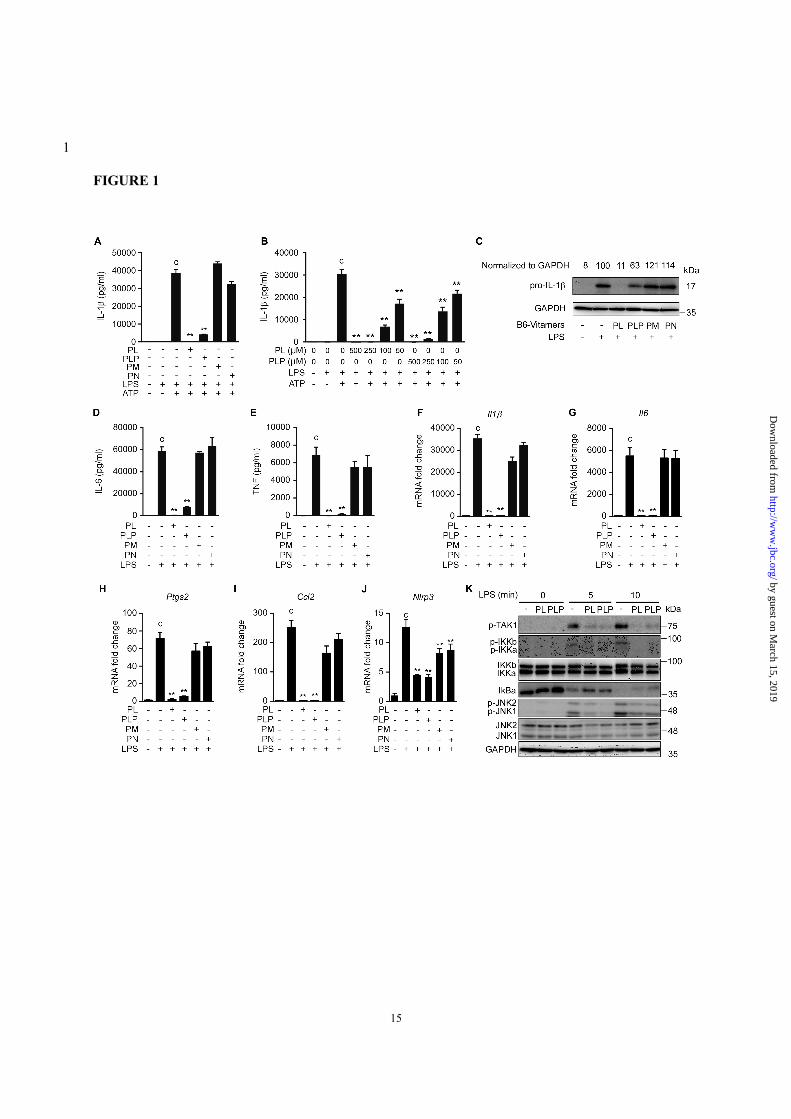
RESULTS Vitamin B6 inhibited LPS-induced NF-κB and
JNK activation and gene expression—We initially
investigated the overall effect of vitamin B6 on IL-
1β secretion from macrophages stimulated with
LPS plus ATP. To this end, thioglycollate-induced
peritoneal macrophages were cultured for 24 h with
or without B6 vitamer supplementation (500 µM),
and sequentially stimulated with LPS (TLR4
agonist) and ATP (NLRP3 activator) to induce IL-
1β secretion. Under these conditions, IL-1β
secretion was strongly inhibited by PL or PLP but
not by pyridoxamine (PM) or pyridoxine (PN) (Fig.
1A). Titration experiments indicated that as little as
50 µM PL or PLP significantly suppressed IL-1β
secretion (Fig. 1B). The LPS-induced intracellular
accumulation of proIL-1β and secretion of IL-6 and
TNF-α were also inhibited by PL or PLP, but not
by PM or PN (Fig. 1, C-E). Furthermore, the LPS-
induced expression of Il1b, Il6, Ptgs2, and Ccl2
mRNAs in peritoneal macrophages was inhibited
by PL or PLP, but not by PM or PN (Fig. 1, F-I).
PL and PLP, and although to a lesser extent PM and
PN suppressed Nlrp3 mRNA expression. (Fig. 1J).
These results were consistent with the previous
findings that PL inhibited LPS-induced NF-κB
activation and expression of NF-κB-target genes
(Nos2 and Ptgs2) in the Raw264.7 mouse
macrophage cell line (8). Actually, Western blot
analyses indicated that events upstream of LPS-
induced NF-κB activation, including the
phosphorylation of TAK1 and IκB kinases and the
degradation of IκBα, were severely suppressed by
PL or PLP (Fig. 1K). PL and PLP also inhibited the
LPS-induced JNK phosphorylation (Fig. 1K),
which occurs downstream of TAK1 and contributes
to IL-1β gene expression (30,31).
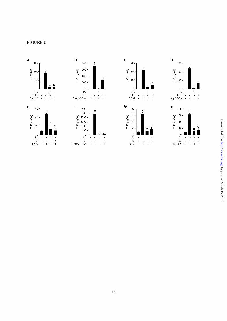
In addition, PL and PLP inhibited the IL-6 and
TNF-α production induced by other TLR ligands,
including the TLR3 ligand poly(I:C), the TLR2
ligand Pam3CSK4, the TLR7 ligand R837, and
CpG oligodeoxynucleotides, which are TLR9
ligands (Fig. 2). Taken together, our results
demonstrate that not only PL, but also PLP
negatively regulate the TLR-mediated activation of
by guest on March 15, 2019
http://ww
w.jbc.org/
Dow
nloaded from
Vitamin B6 suppresses NLRP3 inflammasome
3
NF-κB and MAPK pathways by inhibiting Tak1
phosphorylation, and thereby inhibit the expression
of cytokines genes including Il1b in primary
macrophages.
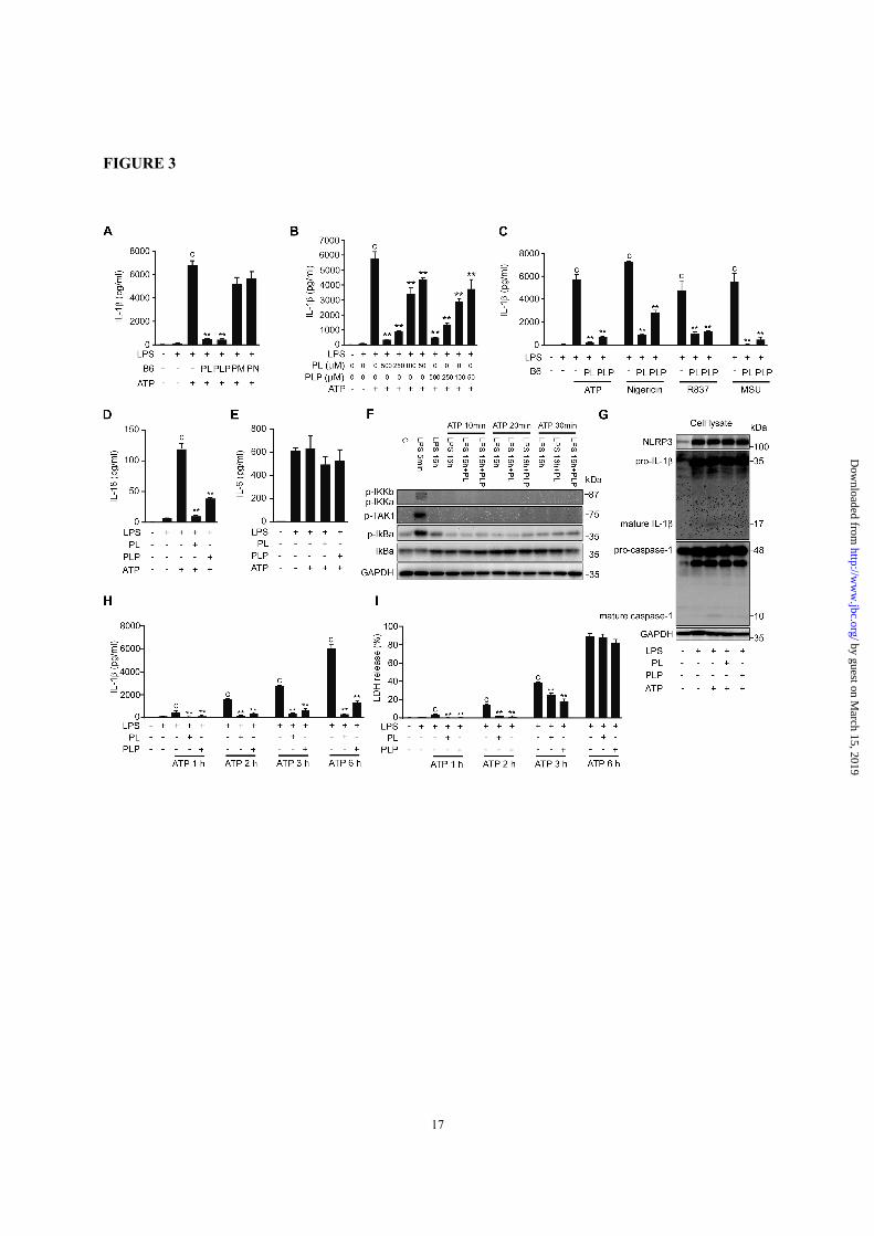
Vitamin B6 suppressed NLRP3 inflammasome
activation—To further investigate the effect of
vitamin B6 on signal 2, which leads to the
proteolytic maturation of IL-1β, peritoneal
macrophages were cultured with LPS for 16 h to
fully induce intracellular proIL-1β accumulation,
then treated with B6 vitamers, and stimulated with
ATP for 6 h to activate the NLRP3 inflammasome.
Under these conditions, IL-1β secretion was again
inhibited by as little as 50 µM PL or PLP, but not
by 500 µM PM or PN (Fig. 3, A and B). The IL-1β
secretion induced by other NLRP3 activators such
as nigericin, R837, and monosodium urate (MSU)
crystals was also inhibited by PL and PLP (Fig. 3C).
The secretion of IL-18, which, like IL-1β
undergoes proteolytic maturation catalyzed by
caspase-1, was also suppressed by PL and PLP (Fig.
3D). In contrast, neither PL nor PLP added after
LPS treatment affected the secretion of IL-6, an
NF-κB-dependent but caspase-1-independent
cytokine (Fig. 3E). In addition, ATP treatment after
LPS priming induced no more phosphorylation of
TAK1 and IκB kinases and degradation of IκBα,
and PL and PLP did not affect these events (Fig.
3F). These data indicated that the suppression of
IL-1β secretion by PL and PLP under these
conditions was not due to the inhibition of NF-κB.
Western blot analyses indicated that PL and PLP
inhibited the generation of mature IL-1β (p17) and
of the p10 fragment of mature caspase-1, when
these vitamers were added after LPS treatment and
before ATP stimulation (Fig. 3G). However, the
LPS-induced NLRP3 and proIL-1β expression and
the constitutive caspase-1 expression at both
mRNA and protein levels were not inhibited by PL
or PLP (data not shown and Fig. 3G), consistent
with the notion that NF-κB-dependent gene
expression was not inhibited under these conditions.
Inflammasome activation induces pyroptosis, a
caspase-1-dependent programmed cell death.
Pyroptotic cells rupture rapidly, releasing lactate
dehydrogenase (LDH) and other cytoplasmic
contents. Pyroptosis would also facilitate the IL-1β
release from macrophages. PL and PLP inhibited
the LDH release from macrophages at 1 h, 2 h and
3 h, but not 6 h after ATP stimulation (Fig. 3I).
These results indicate that PL and PLP delayed
pyroptosis; however, their suppression of IL-1β
secretion at 6 h was not due to the inhibition of
pyroptosis (Fig. 3H).
Taken together, these results indicate that PL
and PLP can inhibit the signal 2 mediated by the
NLRP3 inflammasome.
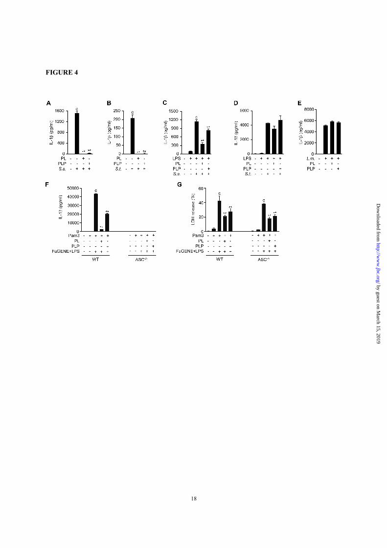
Vitamin B6 did not affect the signal 2 mediated
by the NLRC4 and AIM2 inflammasomes—The
NLRP3, NLRC4, and AIM2 inflammasomes can be
specifically activated by different bacterial species
under certain conditions. For example,
Staphylococcus (S). aureus and Salmonella (S.)
typhimurium at the logarithmic growth phase
activate mainly the NLRP3 and NLRC4
inflammasome, respectively (32-34). In contrast,
the infection of unprimed macrophages with
Listeria (L.) monocytogenes followed by penicillin
G treatment, which causes intracellular releases of
bacterial DNA, induces the AIM2-dependent
secretion of IL-1β (35).
To investigate whether PL and PLP inhibit the
IL-1β production induced by S. aureus and S.
typhimurium, unprimed macrophages and LPS-
primed macrophages were treated with PL or PLP
and then infected with the bacteria. In unprimed
macrophages, the signal 1 for proIL-1β production
depended on the bacterial infection. Under such
conditions, PL and PLP inhibited the IL-1β
production induced by either of these bacterial
species (Fig. 4, A and B). However, in LPS-primed
macrophages (in which proIL-1β had already been
produced), PL and PLP inhibited the IL-1β
production induced by S. aureus but not by S.
typhimurium (Fig. 4, C and D).
To further investigate the effect of PL and PLP
on the AIM2 inflammasome, macrophages were
infected with L. monocytogenes, which provides
Signal 1 through TLR2-dependent recognition of
cell wall components (36). The infected cells were
cultured with PL or PLP for 2h, and then treated
with penicillin G, leading to bacterial cell lysis and
DNA release into the cytoplasm followed by
inflammasome activation that largely depends on
AIM2 (35). Under these conditions, PL and PLP
did not affect the secretion of IL-1β (Fig. 4E).
by guest on March 15, 2019
http://ww
w.jbc.org/
Dow
nloaded from
Vitamin B6 suppresses NLRP3 inflammasome
4
Vitamin B6 suppressed noncanonical IL-1β
secretion and pyroptosis induced by LPS
transfection—We also investigated the effect of PL,
and PLP on noncanonical NLRP3 inflammasome-
dependent IL-1β secretion induced by LPS
transfection in Pam3CSK4-primed peritoneal
macrophages. Both PL and PLP suppressed IL-1β
secretion (Fig. 4F) under these conditions. As this
response requires NLRP3-inflammasome (37), IL-
1β production was not observed in ASC-deficient
macrophages (Fig. 4F). Under the same conditions,
pyroptosis is induced by caspase-11-dependent but
NLRP3-inflammasome-independent manner (37).
Consistently, ASC-/- macrophages released LDH to
a similar extent as wild-type macrophages (Fig. 4G).
Importantly, PL and PLP reduced the LDH release,
suggesting that PL and PLP could inhibit caspase-
11-dependent pyroptosis (Fig. 4G).
These results indicate that PL and PLP
commonly inhibit the signal 1 induced by different
bacteria, but specifically inhibit the signal 2
mediated by the NLRP3 inflammasome and not by
the NLRC4 or the AIM2 inflammasome.
Vitamin B6 inhibited signal 1 and signal 2 for
the IL-1β production in human cells—To test the
inhibitory effects of vitamin B6 on the IL-1β
secretion requiring signal 1 and signal 2 and on the
TNF-α secretion that requires only signal 1 in
human cells, we used macrophagic cells
differentiated from the THP-1 human monocytic
cell line by PMA treatment (THP-1 macrophages).
The THP-1 macrophages were treated with PL or
PLP before or after LPS treatment, and then
stimulated with nigericin to activate the NLRP3
inflammasome. As expected, potent IL-1β
secretion was observed after sequential stimulation
with LPS and nigericin, while TNF-α secretion was
fully induced by LPS stimulation alone; nigericin
did not affect the TNF-α production (Fig. 5, A-D).
When the THP-1 macrophages were treated with
PL or PLP before LPS was added, the secretion of
both IL-1β and TNF-α was inhibited (Fig. 5, A and
B). In contrast, when PL or PLP were added after
LPS treatment, only the IL-1β secretion was
inhibited; TNF-α secretion was unaffected (Fig. 5,
C and D). These results, which were consistent with
our results using mouse peritoneal macrophages,
suggest that PL and PLP inhibit both signal 1 and
signal 2 in human cells.
Vitamin B6 suppressed ASC speck formation
and oligomerization—ASC forms large aggregates
called ‘specks’ when inflammasomes are activated.
ASC speck formation is suggested to be involved in
efficient caspase-1 activation and IL-1β processing.
Therefore, we investigated whether PL and PLP
inhibited ASC speck formation. ASC specks
formed when LPS-primed peritoneal macrophages
were stimulated with nigericin (Fig. 6A, upper
panels, 6B). However, treating LPS-primed
macrophages with PL or PLP prior to nigericin
stimulation strongly inhibited the ASC speck
formation (Fig. 6A, middle and lower panels, 6B).
Consistently, PL and PLP inhibited ASC
oligomerization under the same conditions (Fig.
6C). Taken with our observation that PL and PLP
selectively inhibit the NLRP3 inflammasome, these
results suggest that PL and PLP inhibit signal 2 by
targeting NLRP3 or upstream events that induce the
NLRP3 inflammasome.
Mitochondrial ROS production was inhibited by
PLP but not by PL—Because the NLRP3
inflammasome is activated by structurally diverse
molecules, it has been postulated that different
activators induce common intracellular events that
eventually cause NLRP3 inflammasomes to form.
The efflux of K+ and the resulting decrease in
intracellular K+ concentration has been proposed as
a common upstream event in NLRP3
inflammasome formation (10,38). Therefore, we
measured the cellular potassium levels using
inductively coupled plasma mass spectrometry.
Consistent with previous findings (38), treating
LPS-primed macrophages with ATP or nigericin
decreased the cellular potassium level. This event
was not affected by PL or PLP treatment (Fig. 6D).
Mitochondrial reactive oxygen species (ROS)
generation has also been proposed as a common
upstream event in NLRP3 activation (39).
Consistently, treating LPS-primed macrophages
with ATP enhanced the MitoSox Red fluorescence,
indicating elevated mitochondrial ROS generation
(Fig. 6E). Furthermore, Mito-TEMPO, a
mitochondria-targeted antioxidant that inhibited
ATP-induced mitochondrial ROS elevation
suppressed IL-1β production in LPS-primed
macrophages (Fig. 6, E and F). Mito-temp also
inhibited IL-1β production induced by other
NLRP3 activators (Fig. 6F). Because vitamin B6
by guest on March 15, 2019
http://ww
w.jbc.org/
Dow
nloaded from
Vitamin B6 suppresses NLRP3 inflammasome
5
also acts as an antioxidant (40), we examined
whether PL and PLP could inhibit mitochondrial
ROS generation. Interestingly, this event was
markedly inhibited by PLP but not by PL (Fig. 6G).
These results indicate that PLP has a potential to
suppress mitochondrial ROS generation, which can,
at least in part, explain the PLP’s activity to inhibit
NLRP3-dependent IL-1β production.
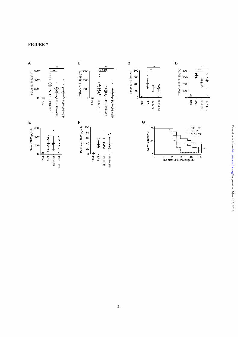
Vitamin B6 inhibited IL-1β production in mice
and protected mice against LPS-induced endotoxic
shock-Finally, we examined the in vivo effects of
PL and PLP. We induced IL-1β production in ICR
mice by i.p. injections of a low dose of LPS (2
µg/kg-bw) followed by ATP (50 µmole/kg-bw)
(41,42), or in C57BL/6 mice by a high dose of LPS
(20 mg/kg-bw) alone (43). In both experimental
systems, the serum and/or peritoneal IL-1β levels
were suppressed by injecting 20 mg/kg-bw PL or
PLP (Fig. 7, A-D). In contrast, PL or PLP did not
significantly suppress the serum or peritoneal TNF-
α levels (Fig. 7, E and F).
Injecting a high dose of LPS induces lethal
endotoxic shock in mice. Components of the
NLRP3 inflammasome (i.e., NLRP3 and ASC) play
essential roles in this disease model (44-46),
although IL-1β and IL-18 are dispensable (47,48).
To test whether PL and PLP can rescue mice from
lethal endotoxic shock, C57BL/6 mice pretreated
with PBS (control), PL, or PLP were given an
injection of 50 mg/kg-bw LPS. Mice pretreated
with PBS (n=15) died within 2 days after LPS
injection; notably, the survival was improved in
mice pretreated with PL or PLP (n=15 each group)
(Fig. 7G).
Taken together, these results suggest that PL and
PLP may inhibit the activity of the NLRP3
inflammasome in vivo.
DISCUSSION
In the present study, we showed that the B6
vitamers PL and PLP inhibit both TLR-induced
NF-κB activation (signal 1) and NLRP3-mediated
caspase-1 activation (signal 2), thereby abolishing
IL-1β production in macrophages. PLP is a
required cofactor for many metabolic enzymes.
However, the PLP concentration necessary to
inhibit IL-1β production was higher than its
physiological concentrations in humans and mice,
suggesting that PLP’s inhibition of IL-1β
production is likely to be pharmacological effect.
The inhibitory effect of PL and PLP on signal 1
was not TLR4-specific, since PL and PLP inhibited
the IL-6 and TNF-α production induced by ligands
for other TLRs (Fig. 2). While TLR4 activates both
the MyD88-dependent and TRIF-dependent NF-
κB-activation pathways, TLR2, TLR7, and TLR9
specifically activate the MyD88-dependent
pathway, and TLR3 only activates the TRIF-
dependent pathway (49). Thus, it is likely that the
target of PL and PLP in the NF-κB pathway is a
common downstream component of the MyD88-
and TRIF-dependent pathways. In this context, it is
worth noting that these pathways commonly induce
the phosphorylation cascade of TAK1-IKK-IκBα,
thereby degrading IκBα and activating NF-κB (49).
TAK1 also activates MAP kinases such as JNK and
p38, which in turn activate the AP-1 transcription
factor involved in IL-1β gene expression together
with NF-κB (49). Our results (Fig. 1K) indicate that
PL and PLP inhibited the LPS-induced TAK1
phosphorylation, IKK-IκBα pathway, and JNK
phosphorylation. Taken together, it is likely that PL
and PLP inhibit signal 1 by targeting TAK1 or a
molecule upstream of TAK1 but commonly found
downstream of TLRs. Further study is required to
determine whether PL and PLP directly target
TAK1 itself, or a molecule upstream of TAK1.
Our experiments using bacterial species that
selectively activate NLRP3, NLRC4, or AIM2
suggested that PL and PLP specifically inhibited
the NLRP3 inflammasome. Thus, it is likely that PL
and PLP target NLRP3 or a more upstream event in
signal 2. Consistent with this possibility, PL and
PLP sharply inhibited ASC speck formation, which
occurs immediately downstream of NLRP3
activation. K+ efflux and mitochondrial ROS
generation have been suggested as common
upstream events of the activation of NLRP3 by
various activators. Our results indicated that PLP
inhibited the ATP-induced mitochondrial ROS
generation, which may contribute to PLP’s
inhibition of signal 2. However, PL did not affect
mitochondrial ROS generation. In addition, neither
PL nor PLP inhibited the ATP- and nigericin-
induced K+ efflux.
Because it was previously reported that NLRP3
is recruited to mitochondria upon activation (50),
we investigated whether PL and PLP affect the
by guest on March 15, 2019
http://ww
w.jbc.org/
Dow
nloaded from
Vitamin B6 suppresses NLRP3 inflammasome
6
mitochondrial localization of NLRP3 in peritoneal
macrophages treated with LPS and/or ATP. In our
hands, a portion of NLRP3 localized at
mitochondria after induction of NLRP3 expression
by LPS, and ATP stimulation did not change the
amount of mitochondrial NLRP3. Furthermore,
neither PL nor PLP affected the amount of
mitochondrial NLRP3, when PL or PLP was added
after LPS stimulation but before ATP stimulation
(data not shown).
We also sought to determine whether PL and
PLP affected the interaction of NLRP3 and ASC or
post-translational modification of NLRP3 such as
ubiquitination or tyrosine phosphorylation (51).
However, in our hands, these responses of
endogenous NLRP3 in peritoneal macrophages
were hardly or barely detectable levels (data not
shown) so that we could not obtain conclusive
answers to these questions at this moment. Thus,
further study is required to determine the direct
target of PL in inhibiting signal 2.
Importantly, vitamin B6 suppressed the IL-1β
production in vivo and protected mice from LPS-
induced endotoxic shock. In our experiments on
LPS-induced IL-1β production, PL and PLP were
administered with the LPS injection. However, PL
and PLP did not significantly suppress the TNF-α
production, which requires only signal 1,
suggesting that PL and PLP suppressed the in vivo
IL-1β production primarily by inhibiting signal 2.
In addition, it has been demonstrated that
components of the NLRP3 inflammasome play
important roles in LPS toxicity (44-46), while IL-
1β and IL-18 are dispensable for it (47,48).
HMGB1, an alarmin released from dead cells, was
recently revealed to play an important role in LPS
toxicity (52). Because HMGB1 is released by
pyroptosis, the delay of pyroptosis by PL and PLP
might also have played a role in protecting mice
from LPS toxicity. Finally, because NLRP3 has
been suggested to play pathological roles in various
inflammation-related diseases, PL and PLP might
have clinical value in treating these diseases.
EXPERIMENTAL PROCEDURES Mice—ICR mice and C57BL/6J mice were
purchased from Japan SLC (Shizuoka, Japan). All
protocols for animal studies were approved by the
Kanazawa University Committee on Animal
Welfare.
Reagents—PL hydrochloride, PN hydrochloride,
PLP (Nacalai Tesque, Kyoto, Japan), PM
dihydrochloride (Calbiochem La Jolla, CA, USA),
LPS from E. coli K235 and from E. coli 0111:B4,
ATP, nigericin, poly(I:C) (Sigma-Aldrich, St.
Louis, MO), Pam3CSK4, R837 (InvivoGen, San
Diego, CA), CpG oligodeoxynucleotides (Genset
Oligos, La Jolla, CA), and MSU (Wako, Osaka
Japan) were purchased.
Macrophage preparation and stimulation—
C57BL/6J mice aged 8-12 weeks were injected i.p.
with 3% thioglycollate solution, and peritoneal
exudate cells were collected 4 d later. The cells
(5×10⁴ cells/well) were cultured in a 96-well plate
for 3 h, after which non-adherent cells were
removed by aspiration to enrich for macrophages.
Human THP-1 cells were treated with 100 nM
PMA for 3 h. The PMA-treated THP-1 cells were
seeded in a 96-well plate (5×10⁴ cells/well) and
cultured overnight without PMA to allow them to
differentiate into macrophagic cells. Cells were
cultured in RPMI 1640 medium supplemented with
10% FCS, 100 U/ml penicillin, and 100 µg/ml
streptomycin (Meiji Seika, Tokyo, Japan).
To test vitamin B6’s inhibition of signal 1,
mouse macrophages were cultured with a B6
vitamer (500 µM unless otherwise specified) for 24
h and then with LPS (from E. coli K235, 0.5 µg/ml
for all in vitro stimulations) for 16 h. To test the
inhibitory activity of vitamin B6 on other TLR
ligands, mouse macrophages were cultured with a
B6 vitamer for 2 h and then with various TLR
agonists for 6 h. THP-1 macrophages were treated
sequentially with a B6 vitamer for 2 h, LPS for 4 h,
and nigericin (5 µM) for 30 min.
To test the inhibitory activity of vitamin B6 on
signal 2, mouse macrophages were treated with
LPS for 16 h, with a B6 vitamer for 2 h, and with
various inflammasome activators for 6 h. THP-1
macrophages were treated with LPS for 4 h, with a
B6 vitamer for 2 h, and with nigericin for 30 min.
To induce noncanonical NLRP3-inflammasome
formation, peritoneal macrophages were primed
with 1 μg/ml Pam3CSK4 for 5 h, washed once with
fresh medium, and then treated with or without 500
μM PL or PLP for 1h. Finally, cells were
transfected with LPS (2 μg/ml) using FuGene HD
(Promega, Madison WI) and cultured for 18 h.
Bacterial Infection—S. aureus (Smith strain,
kindly provided by Dr. Nakanishi, Kanazawa
University, Kanazawa, Ishikawa, Japan) and S.
by guest on March 15, 2019
http://ww
w.jbc.org/
Dow
nloaded from
Vitamin B6 suppresses NLRP3 inflammasome
7
typhimurium (ATCC 14028) in the log phase were
used for infection. L. monocytogenes (EGD,
serovar 1/2a) was cultured in brain-heart infusion
broth (Eiken Chemical, Tokyo, Japan), collected in
the log phase, washed with PBS, suspended in PBS
supplemented with 10% glycerol, and stored in
aliquots at -80°C. Bacterial stocks were thawed and
diluted in RPMI 1640 medium just prior to
infecting macrophages (35).
The peritoneal macrophages were placed in
antibiotic-free medium in 96-well plates and
infected with the bacteria. The plates were briefly
centrifuged to improve interactions between the
cells and bacteria. Penicillin G (100 U/ml),
streptomycin (100 µg/ml), and gentamycin (50
µg/ml, Thermo Fisher Scientific, Waltham, MA)
were added 1 h after infection with S. aureus or S.
typhimurium, and the cells were further cultured for
5 h. To activate AIM2, peritoneal macrophages
were infected with L. monocytogenes. Finally,
penicillin G (100U/ml) was added to the culture to
facilitate intracellular bacterial DNA release, and
the cells were further cultured for 3 h.
Real—time PCR—Total RNA was purified from
mouse macrophages using TRIZOL reagent
(Thermo Fisher Scientific), and cDNA was
synthesized using the First Strand cDNA Synthesis
kit (Toyobo, Osaka, Japan). Real-time PCR was
performed using the StepOne Real-Time PCR
System (Thermo Fisher Scientific) with the
THUNDERBIRD SYBR qPCR Mix (Toyobo) and
the following primers: Il1b, 5’-
TGGGCCTCAAAGGAAAGA-3’ and 5’-
GGTGCTGATGTACCAGTT-3’; Il6, 5’-
AGACAAAGCCAGAGTCCTTCAG-3’ and 5’-
TGCCGAGTAGATCTCAAAGTGA-3’; Ccl2, 5’-
GGTCCCTGTCATGCTTCTGG-3’ and 5’-
CCTTCTTGGGGTCAGCACAG-3’; Ptgs2, 5’-
GCCAGGCTGAACTTCGAAACA-3’ and 5’-
GCTCACGAGGCCACTGATACCTA-3’; Nlrp3,
5’-GTGGTGACCCTCTGTGAGGT-3’ and 5’-
TCTTCCTGGAGCGCTTCTAA-3’; and Gapdh,
5’-CAATGACCCCTTCATTGACC-3’ and 5'-
TGGAAGATGGTGATGGGATT-3’.
ELISA and LDH analysis—The concentrations
of mouse and human IL-1β, IL-6, and TNF-α in
culture supernatants were determined using
OptEIA ELISA kits (BD Pharmingen, San Diego,
CA) according to the manufacturer’s protocols. The
mouse IL-18 ELISA kit was purchased from MBL
(Nagoya, Japan). Cell death was determined by
measuring LDH activity in the culture medium
using the CytoTox96 NonRadioactive Cytotoxicity
Assay (Promega).
Western blot analysis—Cells were lysed in Tris-
buffered saline containing 1% NP-40 and complete
protease inhibitor cocktail (Roche Diagnostics
GmbH, Mannheim, Germany) for 30 min on ice.
Lysates were centrifuged at 12,000 × g for 10 min
to remove debris and then boiled in Laemmli
sample buffer for 5 min. Proteins were separated by
SDS–PAGE and transferred to PVDF membranes.
Membranes were incubated for 1 h in a blocking
buffer (5% skim milk, 0.05% Tween 20 in Tris-
buffered saline) or in Blocking One-P (for
phosphoprotein detection, Nacalai Tesque),
followed by the addition of a primary antibody
against IL-1β (Santa Cruz Biotechnology, Dallas,
TX), caspase-1 (Santa Cruz Biotechnology, Santa
Cruz, CA), NLRP3 (Enzo Life Science,
Villeurbanne, France), IκBα, phospho-TAK1, JNK,
phospho-JNK, IKKα, IKKβ, phospho-IKKα/β
(Cell Signaling, Danvers, MA), or GAPDH (Novus
Biologicals, Littleton, CO). Antibodies were
detected by a horseradish-peroxidase-conjugated
goat anti-mouse or anti-rabbit secondary antibody
(Jackson Immunoresearch Laboratories, West
Grove, PA) using enhanced chemiluminescence
(Thermo Fisher Scientific).
To detect ASC oligomerization, cells were lysed
with Tris-buffered saline (TBS) containing 0.5%
Triton X-100, and centrifuged at 6,000 × g at 4 °C
for 15 min. Supernatants (lysates) were transferred
to new tubes. The pellets were washed with TBS
twice and then crosslinked at 37 °C for 45 min by
disuccinimidyl suberate (DSS, 2mM, Thermo
Fisher Scientific). The crosslinked pellets were
spun down at 6,000 × g for 15 min, dissolved in
SDS-containing sample buffer, and subjected to
SDS-PAGE and Western blot using anti-ASC
antibody (Santa Cruz Biotechnology, Santa Cruz,
CA).
Immunofluorescence confocal microscopy—
Mouse ASC was detected using a rat mAb (46)
followed by FITC-conjugated goat anti-rat IgG
(American Qualex, San Clemente, CA). Nuclei
were stained by DAPI (Dojindo, Kumamoto,
Japan). The stained cells were examined under a
laser scanning microscope (LSM 510 META with
EC Plan-Neofluar 40x/0.75; Carl Zeiss, Jena,
Germany) equipped with 488 nm argon, 543 nm
HeNe, 633 nm HeNe, and Blue Diode 405 lasers.
by guest on March 15, 2019
http://ww
w.jbc.org/
Dow
nloaded from
Vitamin B6 suppresses NLRP3 inflammasome
8
Images were acquired and analyzed using Zen2009
software.
Determination of intracellular potassium
levels—Cells (6 x 10⁵ cells/well in a 24-well plate)
were lysed in 3% ultrapure HNO3 (600 µl) for 30
min on ice. Lysates were diluted 10 times with
ultrapure H2O, and potassium concentrations were
determined by inductively coupled plasma mass
spectrometry (SPQ-9000, Seiko Instruments, Chiba,
Japan) using KCl solution as a standard.
Determination of mitochondrial ROS levels—
Mitochondrial ROS levels were measured using
MitoSox Red (Thermo Fisher Scientific) according
to the manufacturer’s protocol. Data were acquired
with a FACSCanto II (BD Biosciences, San Jose,
CA) and analyzed with FlowJo software (FlowJo,
Ashland, OR).
In vivo experiments—The Food and Nutrition
Board of the Institute of Medicine proposed a
tolerable upper limit of 100 mg/day of vitamin B6
for adult humans (53). Based on this and the
conversion between human doses and animal-
equivalent doses according to body surface area
(54), we estimated a tolerable vitamin B6 dose of
20 mg/kg-body weight (bw)/day for mice; this was
the dose used for the in vivo experiments in the
present study. ICR mice were i.p. injected first with
PL or PLP, 2 h later with LPS (from E. coli 0111:B4,
2 µg/kg-bw), and 90 min afterward with 10 ml/kg-
bw of 5 mM ATP (50 µmole/kg-bw). Serum and
peritoneal lavage samples were collected 1 h after
the ATP injection. C57BL/6 mice were i.p. injected
with LPS (20 mg/kg-bw) with or without PL or PLP,
and serum and peritoneal lavage samples were
collected 3 h later. Lethal endotoxic shock was
induced in C57BL/6 mice by i.p. LPS injection (50
mg/kg-bw, from E. coli 0111:B4, Sigma-Aldrich).
Statistical analysis—Data were analyzed using
GraphPad Prism 6.05 (GraphPad Software, La Jolla,
CA). Difference between a control and an
experimental group was examined by one-way
ANOVA and Dunnett’s test. Difference between
mouse survival curves was evaluated by the log-
rank (Mantel-Cox) test. P < 0.05 was considered
significant.
Acknowledgments: We thank Dr. Nakanishi from Kanazawa University for providing S. aureus (Smith
strain) used in bacterial infection experiments.
Conflicts of interest: The authors declare no competing financial interests.
Author Contributions: P.Z. contributed to the experimental design, performed most of the experiments
and write the manuscript; K.T. performed or supervised experiments involving bacterial infection; T.K. and
H.K. provided technical assistance; S.S. helped collect samples; M.H. and H.I. performed and supervised
inductively coupled plasma mass spectrometry analyses; N.K. contributed to the experimental design and
critical review of the manuscript; T.S. designed and supervised the research project and edited the
manuscript. All authors reviewed the results and approved the final version of the manuscript.
REFERENCES
1. Saibeni, S., Cattaneo, M., Vecchi, M., Zighetti, M. L., Lecchi, A., Lombardi, R., Meucci,
G., Spina, L., and de Franchis, R. (2003) Low vitamin B(6) plasma levels, a risk factor
for thrombosis, in inflammatory bowel disease: role of inflammation and correlation with
acute phase reactants. Am. J. Gastroenterol. 98, 112-117
2. Sakakeeny, L., Roubenoff, R., Obin, M., Fontes, J. D., Benjamin, E. J., Bujanover, Y.,
Jacques, P. F., and Selhub, J. (2012) Plasma pyridoxal-5-phosphate is inversely
associated with systemic markers of inflammation in a population of U.S. adults. J. Nutr.
142, 1280-1285
3. Huang, S. C., Wei, J. C., Wu, D. J., and Huang, Y. C. (2010) Vitamin B(6)
supplementation improves pro-inflammatory responses in patients with rheumatoid
arthritis. Eur. J. Clin. Nutr. 64, 1007-1013
by guest on March 15, 2019
http://ww
w.jbc.org/
Dow
nloaded from
Vitamin B6 suppresses NLRP3 inflammasome
9
4. Komatsu, S. I., Watanabe, H., Oka, T., Tsuge, H., Nii, H., and Kato, N. (2001) Vitamin
B-6-supplemented diets compared with a low vitamin B-6 diet suppress azoxymethane-
induced colon tumorigenesis in mice by reducing cell proliferation. J. Nutr. 131, 2204-
2207
5. Galluzzi, L., Vacchelli, E., Michels, J., Garcia, P., Kepp, O., Senovilla, L., Vitale, I., and
Kroemer, G. (2013) Effects of vitamin B6 metabolism on oncogenesis, tumor progression
and therapeutic responses. Oncogene 32, 4995-5004
6. Douaud, G., Refsum, H., de Jager, C. A., Jacoby, R., Nichols, T. E., Smith, S. M., and
Smith, A. D. (2013) Preventing Alzheimer's disease-related gray matter atrophy by B-
vitamin treatment. Proc. Natl. Acad. Sci. U. S. A. 110, 9523-9528
7. Murakami, K., Miyake, Y., Sasaki, S., Tanaka, K., Fukushima, W., Kiyohara, C., Tsuboi,
Y., Yamada, T., Oeda, T., Miki, T., Kawamura, N., Sakae, N., Fukuyama, H., Hirota, Y.,
Nagai, M., and Group, F. K. P. s. D. S. (2010) Dietary intake of folate, vitamin B-6,
vitamin B-12 and riboflavin and risk of Parkinson's disease: a case-control study in
Japan. Br. J. Nutr. 104, 757-764
8. Yanaka, N., Koyama, T. A., Komatsu, S., Nakamura, E., Kanda, M., and Kato, N. (2005)
Vitamin B6 suppresses NF-kappaB activation in LPS-stimulated mouse macrophages.
Int. J. Mol. Med. 16, 1071-1075
9. Garlanda, C., Dinarello, C. A., and Mantovani, A. (2013) The interleukin-1 family: back
to the future. Immunity. 39, 1003-1018
10. Franchi, L., Munoz-Planillo, R., and Nunez, G. (2012) Sensing and reacting to microbes
through the inflammasomes. Nat. Immunol. 13, 325-332
11. Li, P., Allen, H., Banerjee, S., Franklin, S., Herzog, L., Johnston, C., McDowell, J.,
Paskind, M., Rodman, L., Salfeld, J., and et al. (1995) Mice deficient in IL-1 beta-
converting enzyme are defective in production of mature IL-1 beta and resistant to
endotoxic shock. Cell 80, 401-411
12. Kayagaki, N., Warming, S., Lamkanfi, M., Vande Walle, L., Louie, S., Dong, J., Newton,
K., Qu, Y., Liu, J., Heldens, S., Zhang, J., Lee, W. P., Roose-Girma, M., and Dixit, V. M.
(2011) Non-canonical inflammasome activation targets caspase-11. Nature 479, 117-121
13. Schroder, K., and Tschopp, J. (2010) The inflammasomes. Cell. 140, 821-832
14. Mariathasan, S., Weiss, D. S., Newton, K., McBride, J., O'Rourke, K., Roose-Girma, M.,
Lee, W. P., Weinrauch, Y., Monack, D. M., and Dixit, V. M. (2006) Cryopyrin activates
the inflammasome in response to toxins and ATP. Nature. 440, 228-232
15. Craven, R. R., Gao, X., Allen, I. C., Gris, D., Bubeck Wardenburg, J., McElvania-
Tekippe, E., Ting, J. P., and Duncan, J. A. (2009) Staphylococcus aureus alpha-
hemolysin activates the NLRP3-inflammasome in human and mouse monocytic cells.
PLoS One 4, e7446
16. Harder, J., Franchi, L., Munoz-Planillo, R., Park, J. H., Reimer, T., and Nunez, G. (2009)
Activation of the Nlrp3 inflammasome by Streptococcus pyogenes requires streptolysin
O and NF-kappa B activation but proceeds independently of TLR signaling and P2X7
receptor. J. Immunol. 183, 5823-5829
17. Kanneganti, T. D., Ozoren, N., Body-Malapel, M., Amer, A., Park, J. H., Franchi, L.,
Whitfield, J., Barchet, W., Colonna, M., Vandenabeele, P., Bertin, J., Coyle, A., Grant, E.
P., Akira, S., and Nunez, G. (2006) Bacterial RNA and small antiviral compounds
activate caspase-1 through cryopyrin/Nalp3. Nature. 440, 233-236
by guest on March 15, 2019
http://ww
w.jbc.org/
Dow
nloaded from
Vitamin B6 suppresses NLRP3 inflammasome
10
18. Dostert, C., Petrilli, V., Van Bruggen, R., Steele, C., Mossman, B. T., and Tschopp, J.
(2008) Innate immune activation through Nalp3 inflammasome sensing of asbestos and
silica. Science. 320, 674-677
19. Martinon, F., Petrilli, V., Mayor, A., Tardivel, A., and Tschopp, J. (2006) Gout-
associated uric acid crystals activate the NALP3 inflammasome. Nature. 440, 237-241
20. Halle, A., Hornung, V., Petzold, G. C., Stewart, C. R., Monks, B. G., Reinheckel, T.,
Fitzgerald, K. A., Latz, E., Moore, K. J., and Golenbock, D. T. (2008) The NALP3
inflammasome is involved in the innate immune response to amyloid-beta. Nat. Immunol.
9, 857-865
21. Duewell, P., Kono, H., Rayner, K. J., Sirois, C. M., Vladimer, G., Bauernfeind, F. G.,
Abela, G. S., Franchi, L., Nunez, G., Schnurr, M., Espevik, T., Lien, E., Fitzgerald, K. A.,
Rock, K. L., Moore, K. J., Wright, S. D., Hornung, V., and Latz, E. (2010) NLRP3
inflammasomes are required for atherogenesis and activated by cholesterol crystals.
Nature. 464, 1357-1361
22. Wen, H., Gris, D., Lei, Y., Jha, S., Zhang, L., Huang, M. T., Brickey, W. J., and Ting, J.
P. (2011) Fatty acid-induced NLRP3-ASC inflammasome activation interferes with
insulin signaling. Nat. Immunol. 12, 408-415
23. Villani, A. C., Lemire, M., Fortin, G., Louis, E., Silverberg, M. S., Collette, C., Baba, N.,
Libioulle, C., Belaiche, J., Bitton, A., Gaudet, D., Cohen, A., Langelier, D., Fortin, P. R.,
Wither, J. E., Sarfati, M., Rutgeerts, P., Rioux, J. D., Vermeire, S., Hudson, T. J., and
Franchimont, D. (2009) Common variants in the NLRP3 region contribute to Crohn's
disease susceptibility. Nat. Genet. 41, 71-76
24. Schoultz, I., Verma, D., Halfvarsson, J., Torkvist, L., Fredrikson, M., Sjoqvist, U.,
Lordal, M., Tysk, C., Lerm, M., Soderkvist, P., and Soderholm, J. D. (2009) Combined
polymorphisms in genes encoding the inflammasome components NALP3 and CARD8
confer susceptibility to Crohn's disease in Swedish men. Am. J. Gastroenterol. 104, 1180-
1188
25. Bauer, C., Duewell, P., Mayer, C., Lehr, H. A., Fitzgerald, K. A., Dauer, M., Tschopp, J.,
Endres, S., Latz, E., and Schnurr, M. (2010) Colitis induced in mice with dextran sulfate
sodium (DSS) is mediated by the NLRP3 inflammasome. Gut 59, 1192-1199
26. Heneka, M. T., Kummer, M. P., Stutz, A., Delekate, A., Schwartz, S., Vieira-Saecker, A.,
Griep, A., Axt, D., Remus, A., Tzeng, T. C., Gelpi, E., Halle, A., Korte, M., Latz, E., and
Golenbock, D. T. (2013) NLRP3 is activated in Alzheimer's disease and contributes to
pathology in APP/PS1 mice. Nature. 493, 674-678
27. Masters, S. L., Dunne, A., Subramanian, S. L., Hull, R. L., Tannahill, G. M., Sharp, F.
A., Becker, C., Franchi, L., Yoshihara, E., Chen, Z., Mullooly, N., Mielke, L. A., Harris,
J., Coll, R. C., Mills, K. H., Mok, K. H., Newsholme, P., Nunez, G., Yodoi, J., Kahn, S.
E., Lavelle, E. C., and O'Neill, L. A. (2010) Activation of the NLRP3 inflammasome by
islet amyloid polypeptide provides a mechanism for enhanced IL-1beta in type 2
diabetes. Nat. Immunol. 11, 897-904
28. Stienstra, R., Joosten, L. A., Koenen, T., van Tits, B., van Diepen, J. A., van den Berg, S.
A., Rensen, P. C., Voshol, P. J., Fantuzzi, G., Hijmans, A., Kersten, S., Muller, M., van
den Berg, W. B., van Rooijen, N., Wabitsch, M., Kullberg, B. J., van der Meer, J. W.,
Kanneganti, T., Tack, C. J., and Netea, M. G. (2010) The inflammasome-mediated
caspase-1 activation controls adipocyte differentiation and insulin sensitivity. Cell Metab
12, 593-605
by guest on March 15, 2019
http://ww
w.jbc.org/
Dow
nloaded from
Vitamin B6 suppresses NLRP3 inflammasome
11
29. Kuemmerle-Deschner, J. B. (2015) CAPS--pathogenesis, presentation and treatment of
an autoinflammatory disease. Semin. Immunopathol. 37, 377-385
30. Lee, J., Mira-Arbibe, L., and Ulevitch, R. J. (2000) TAK1 regulates multiple protein
kinase cascades activated by bacterial lipopolysaccharide. J. Leukoc. Biol. 68, 909-915
31. Sato, S., Sanjo, H., Takeda, K., Ninomiya-Tsuji, J., Yamamoto, M., Kawai, T.,
Matsumoto, K., Takeuchi, O., and Akira, S. (2005) Essential function for the kinase
TAK1 in innate and adaptive immune responses. Nat. Immunol. 6, 1087-1095
32. Munoz-Planillo, R., Franchi, L., Miller, L. S., and Nunez, G. (2009) A critical role for
hemolysins and bacterial lipoproteins in Staphylococcus aureus-induced activation of the
Nlrp3 inflammasome. J. Immunol. 183, 3942-3948
33. Miao, E. A., Alpuche-Aranda, C. M., Dors, M., Clark, A. E., Bader, M. W., Miller, S. I.,
and Aderem, A. (2006) Cytoplasmic flagellin activates caspase-1 and secretion of
interleukin 1beta via Ipaf. Nat. Immunol. 7, 569-575
34. Franchi, L., Amer, A., Body-Malapel, M., Kanneganti, T. D., Ozoren, N., Jagirdar, R.,
Inohara, N., Vandenabeele, P., Bertin, J., Coyle, A., Grant, E. P., and Nunez, G. (2006)
Cytosolic flagellin requires Ipaf for activation of caspase-1 and interleukin 1beta in
salmonella-infected macrophages. Nat. Immunol. 7, 576-582
35. Tsuchiya, K., Hara, H., Kawamura, I., Nomura, T., Yamamoto, T., Daim, S., Dewamitta,
S. R., Shen, Y., Fang, R., and Mitsuyama, M. (2010) Involvement of absent in melanoma
2 in inflammasome activation in macrophages infected with Listeria monocytogenes. J.
Immunol. 185, 1186-1195
36. Flo, T. H., Halaas, O., Lien, E., Ryan, L., Teti, G., Golenbock, D. T., Sundan, A., and
Espevik, T. (2000) Human toll-like receptor 2 mediates monocyte activation by Listeria
monocytogenes, but not by group B streptococci or lipopolysaccharide. J. Immunol. 164,
2064-2069
37. Kayagaki, N., Stowe, I. B., Lee, B. L., O'Rourke, K., Anderson, K., Warming, S.,
Cuellar, T., Haley, B., Roose-Girma, M., Phung, Q. T., Liu, P. S., Lill, J. R., Li, H., Wu,
J., Kummerfeld, S., Zhang, J., Lee, W. P., Snipas, S. J., Salvesen, G. S., Morris, L. X.,
Fitzgerald, L., Zhang, Y., Bertram, E. M., Goodnow, C. C., and Dixit, V. M. (2015)
Caspase-11 cleaves gasdermin D for non-canonical inflammasome signalling. Nature
526, 666-671
38. Munoz-Planillo, R., Kuffa, P., Martinez-Colon, G., Smith, B. L., Rajendiran, T. M., and
Nunez, G. (2013) K+ Efflux Is the Common Trigger of NLRP3 Inflammasome
Activation by Bacterial Toxins and Particulate Matter. Immunity. 38, 1142-1153
39. Zhou, R., Yazdi, A. S., Menu, P., and Tschopp, J. (2011) A role for mitochondria in
NLRP3 inflammasome activation. Nature. 469, 221-225
40. Mooney, S., Leuendorf, J. E., Hendrickson, C., and Hellmann, H. (2009) Vitamin B6: a
long known compound of surprising complexity. Molecules 14, 329-351
41. Sun, S., Xia, S., Ji, Y., Kersten, S., and Qi, L. (2012) The ATP-P2X7 signaling axis is
dispensable for obesity-associated inflammasome activation in adipose tissue. Diabetes
61, 1471-1478
42. Griffiths, R. J., Stam, E. J., Downs, J. T., and Otterness, I. G. (1995) ATP induces the
release of IL-1 from LPS-primed cells in vivo. J. Immunol. 154, 2821-2828
43. Yan, Y. Q., Jiang, W., Liu, L., Wang, X. Q., Ding, C., Tian, Z. G., and Zhou, R. B.
(2015) Dopamine Controls Systemic Inflammation through Inhibition of NLRP3
Inflammasome. Cell. 160, 62-73
by guest on March 15, 2019
http://ww
w.jbc.org/
Dow
nloaded from
Vitamin B6 suppresses NLRP3 inflammasome
12
44. Mariathasan, S., Newton, K., Monack, D. M., Vucic, D., French, D. M., Lee, W. P.,
Roose-Girma, M., Erickson, S., and Dixit, V. M. (2004) Differential activation of the
inflammasome by caspase-1 adaptors ASC and Ipaf. Nature. 430, 213-218
45. Sutterwala, F. S., Ogura, Y., Szczepanik, M., Lara-Tejero, M., Lichtenberger, G. S.,
Grant, E. P., Berlin, J., Coyle, A. J., Galan, J. E., Askenase, P. W., and Flavell, R. A.
(2006) Critical role for NALP3/CIAS1/cryopyrin in innate and adaptive immunity
through its regulation of caspase-1. Immunity. 24, 317-327
46. Imamura, R., Wang, Y., Kinoshita, T., Suzuki, M., Noda, T., Sagara, J., Taniguchi, S.,
Okamoto, H., and Suda, T. (2010) Anti-inflammatory activity of PYNOD and its
mechanism in humans and mice. J. Immunol. 184, 5874-5884
47. Shornick, L. P., De Togni, P., Mariathasan, S., Goellner, J., Strauss-Schoenberger, J.,
Karr, R. W., Ferguson, T. A., and Chaplin, D. D. (1996) Mice deficient in IL-1beta
manifest impaired contact hypersensitivity to trinitrochlorobenzone. J. Exp. Med. 183,
1427-1436
48. Sakao, Y., Takeda, K., Tsutsui, H., Kaisho, T., Nomura, F., Okamura, H., Nakanishi, K.,
and Akira, S. (1999) IL-18-deficient mice are resistant to endotoxin-induced liver injury
but highly susceptible to endotoxin shock. Int. Immunol. 11, 471-480
49. Kawai, T., and Akira, S. (2007) Signaling to NF-kappaB by Toll-like receptors. Trends
Mol. Med. 13, 460-469
50. Subramanian, N., Natarajan, K., Clatworthy, M. R., Wang, Z., and Germain, R. N. (2013)
The adaptor MAVS promotes NLRP3 mitochondrial localization and inflammasome
activation. Cell 153, 348-361
51. Juliana, C., Fernandes-Alnemri, T., Kang, S., Farias, A., Qin, F., and Alnemri, E. S.
(2012) Non-transcriptional priming and deubiquitination regulate NLRP3 inflammasome
activation. J. Biol. Chem. 287, 36617-36622
52. Alves, J. N., Pires, K. M., Lanzetti, M., Barroso, M. V., Benjamim, C. F., Costa, C. A.,
Resende, A. C., Santos, J. C., Ribeiro, M. L., Porto, L. C., and Valenca, S. S. (2013)
Critical role for CCR2 and HMGB1 in induction of experimental endotoxic shock. Arch.
Biochem. Biophys. 537, 72-81
53. Institute of Medicine, US. (1998) Vitamin B6. in Dietary Reference Intakes for Thiamin,
Riboflavin, Niacin, Vitamin B6, Folate, Vitamin B12, Pantothenic Acid, Biotin, and
Choline, National Academies Press (US), Washington (DC). pp 150-195
54. CDER, USFDA. (2005) Guidance for industry: estimating the maximum safe starting
dose in initial clinical trials for therapeutics in adult healthy volunteers., US Department
of Health and Human Services Food and Drug Administration Center for Drug
Evaluation and Research, Rockville
FOOTNOTES
*This work was supported by a Grant-in-Aid for Challenging Exploratory Research (15K15078), and
Grant-in-Aid for Scientific Research on Innovative Areas (26110002) from the Japan Society for the
Promotion of Science.
1To whom correspondence should be addressed: Department of Immunology and Molecular Biology,
Cancer Research Institute, Kanazawa University, Kanazawa, 920-1192, Japan. Tel.: 81-76-264-6720; E-
mail: [email protected].
by guest on March 15, 2019
http://ww
w.jbc.org/
Dow
nloaded from
Vitamin B6 suppresses NLRP3 inflammasome
13
2Abbreviations used in this article: DSS, disuccinimidyl suberate; L. monocytogenes, Listeria
monocytogenes; LDH, lactate dehydrogenase; MOI, multiplicity of infection; MSU, monosodium urate; PL,
pyridoxal; PLP, pyridoxal 5'-phosphate; PM, pyridoxamine; PN, pyridoxine; ROS, reactive oxygen species;
S. typhimurium, Salmonella typhimurium; S. aureus, Staphylococcus aureus
FIGURE LEGENDS
FIGURE 1. PL and PLP suppress signal 1. A and B, Peritoneal macrophages were treated with the
indicated B6 vitamers (500 µM in A, and the indicated concentrations in B) for 24 h, then stimulated with
LPS for 16 h, and finally exposed to 5 mM ATP for 6 h. The IL-1β concentration in culture supernatants
was determined by ELISA. C-E, Peritoneal macrophages were treated with B6 vitamers for 24 h and then
stimulated with LPS for 16 h. ProIL-1β and GAPDH (loading control) were detected by Western blot (C).
The IL-6 and TNF-α concentrations in culture supernatants were determined by ELISA (D and E). F-J,
Peritoneal macrophages were treated with B6 vitamers for 24 h and then stimulated with LPS for 16 h, after
which the Il1b, Il6, Ptgs2, Ccl2 and Nlrp3 mRNAs were quantified by real-time PCR. K, Peritoneal
macrophages were treated with PL or PLP for 2 h and then stimulated with LPS for the indicated times.
The total and/or phosphorylated forms of TAK1, IKKs, IkBα, and JNKs were detected by Western blot. A,
B and D-J, Data show mean + SD; n=3. Asterisks indicate significant differences (**p < 0.01) from the
control group (c). All experiments were repeated at least three times, and representative data are shown.
FIGURE 2. PL and PLP suppress the IL-6 and TNF-αααα production induced by TLR ligands. A-H,
Peritoneal macrophages were incubated with PL or PLP for 2 h and then stimulated with poly I:C (20 µg/ml),
Pam3CSK4 (10 µg/ml), R837 (10 µg/ml), or CpG oligodeoxynucleotides for 6 h (1 µM). IL-6 and TNF
were measured by ELISA. Data show mean + SD; n=3. Asterisks indicate significant differences (**p <
0.01) from the control group (c). All experiments were repeated at least three times, and representative data
are shown.
FIGURE 3. PL and PLP suppress signal 2. A-E, Peritoneal macrophages were sequentially incubated with
LPS for 16 h, B6 vitamers for 2 h, and ATP (5 mM), nigericin (5 µM), R837 (10 µg/ml), or MSU (150
µg/ml) for 6 h. IL-1β, IL-18, and IL-6 in culture supernatants were quantified by ELISA. F, Mouse
peritoneal macrophages were exposed to LPS for 16 h, then incubated with PL or PLP for 2 h, and finally
stimulated with ATP for 10, 20, or 30 min). Whole cell lysates were collected and subjected to Western
blot using antibodies against phosphorylated forms of IKKα/β, TAK1, and IκBα, and total IκBα. GAPDH
serves as a loading control. G, Peritoneal macrophages were treated with LPS for 16 h, then incubated with
PL or PLP for 2 h, and finally exposed to ATP (5 mM) for 30 min. Pro-IL-1β, mature IL-1β, caspase-1, and
GAPDH in cell lysates were detected by Western blot. H and I, Peritoneal macrophages were sequentially
incubated with LPS for 16 h, B6 vitamers for 2 h, and ATP (5 mM) for 1 h, 2 h, 3 h, or 6 h. IL-1β in culture
supernatants were quantified by ELISA (H). Cell death was evaluated by assaying LDH release (I). A-E, H
and I, Data show mean + SD; n=3. Asterisks indicate significant differences (**p < 0.01) from the control
group (c). All experiments were repeated at least three times, and representative data are shown.
FIGURE 4. PL and PLP selectively suppress the NLRP3 inflammasome. A and B, Peritoneal
macrophages were incubated with PL or PLP for 24 h and then infected with S. aureus (S.a.) with a
multiplicity of infection (MOI) of 100 or S. typhimurium (S.t.) with a MOI of 20. C and D, Peritoneal
macrophages were primed with LPS for 16 h, then incubated with PL or PLP for 2 h, and finally infected
with S. aureus (MOI 50) or S. typhimurium (MOI 20). E, Peritoneal macrophages were infected with L.
monocytogenes (L.m., MOI 2) for 4 h and then incubated with PL or PLP for 2 h, after which penicillin G
(100U/ml) was added to facilitate intracellular bacterial DNA release. F and G, wild-type (WT) and ASC-
/- peritoneal macrophages were primed with Pam3CSK4 (1µg/ml) for 5h, then incubated with PL or PLP
for 1 h, and finally transfected with LPS (2 µg/ml) using FuGene HD for 18 h. IL-1β in culture supernatants
by guest on March 15, 2019
http://ww
w.jbc.org/
Dow
nloaded from
Vitamin B6 suppresses NLRP3 inflammasome
14
was quantified by ELISA (A-F). Cell death was evaluated by assaying LDH release (G). Data show mean
+ SD; n=3. Asterisks indicate significant differences (**p < 0.01) from the control group (c). All
experiments were repeated at least three times, and representative data are shown.
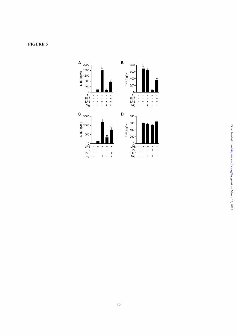
FIGURE 5. PL and PLP suppress signal 1 and signal 2 in human cells. A and B, THP-1 macrophages
were treated with PL or PLP for 2 h, then incubated with LPS for 4 h, and finally stimulated with nigericin
(5 µM) for 0.5 h. C and D, THP-1 macrophages were primed with LPS for 4 h, then treated with PL or PLP
for 2 h, and finally stimulated with nigericin (5 µM) for 0.5 h. A-D, IL-1β and TNF-α in culture supernatants
were measured by ELISA. Data show mean + SD; n=3. Asterisks indicate significant differences (**p <
0.01) from the control group (c). All experiments were repeated at least three times, and representative data
are shown.
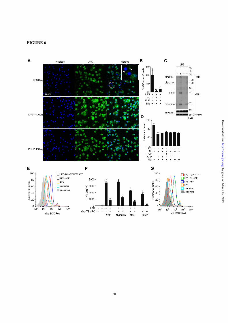
FIGURE 6. Effects of PL and PLP in ASC speck formation, ASC oligomerization, K+ efflux, and
mitochondrial ROS generation. A and B, Peritoneal macrophages cultured on glass coverslips in 24-well
plates were primed with LPS for 16 h, then treated with PL or PLP for 2 h, and finally exposed to nigericin
(5 µM) for 1 h. ASC specks (FITC) and nuclei (DAPI) were visualized using a confocal fluorescence
microscope (A). Total cells and cells with ASC aggregates (specks) were counted in 4 microscope fields
per group, and data show mean + SD (B). C, Peritoneal macrophages were primed with LPS for 16 h, then
treated with PL or PLP for 2 h, and finally exposed to nigericin (5 µM) for 1 h. Cells were lysed with 0.5%
Triton X-100 in TBS, and centrifuged. The supernatants (lysates) and DSS-crosslinked pellets were
subjected to Western blot using antibodies against ASC and GAPDH. D, Peritoneal macrophages were
primed with LPS for 16 h, then treated with PL or PLP for 2 h, and finally exposed to ATP (5 mM) or
nigericin (5 µM) for 0.5 h. Intracellular K+ levels relative to those in cells treated with LPS only were
determined using inductively coupled plasma mass spectrometry. E and G, Peritoneal macrophages primed
with LPS for 16 h were detached from the culture plate, and the single-cell suspension was treated with
Mito-TEMPO (500 µM, E) or PL or PLP (G) for 2 h, after which the cells were exposed to ATP (5 mM) in
the presence of MitoSOX (5 µM) for 10 min. Fluorescence intensities were analyzed by flow cytometry. F,
Peritoneal macrophages were primed with LPS for 16 h, then incubated with Mito-TEMPO (500 µM) for
2 h, and finally exposed to ATP (5 mM), 5 µM Nigericin, 150 µg/ml MSU, or 10 µg/ml R837 for 6 h.
Supernatant IL-1β content was measured by ELISA. B, D and F, Data show mean + SD; n=3. Asterisks
indicate significant differences (**p < 0.01) from the control group (c). All experiments were repeated at
least three times, and representative data are shown.
FIGURE 7. PL and PLP suppress IL-1β production in vivo. A and B, ICR mice aged 8 weeks were i.p.
injected first with PBS (solvent control), PL, or PLP; then 2 h later with PBS or LPS (2 µg/kg-bw); and 90
min after that with PBS or ATP (50 µmole/kg-bw). After 1 h, serum and peritoneal lavage samples were
collected, and the IL-1β was quantified by ELISA (n=11 mice for the PBS group; n=16-18 mice for the
other groups). C-F, C57BL/6 mice aged 10 weeks were i.p. injected with PBS or LPS (20 mg/kg-bw) alone
or with PL or PLP; serum and peritoneal lavage samples were collected 3 h later and analyzed by ELISA
for IL-1β (C and D) or TNF-α (E and F); n=4 mice for the PBS group; n=9 mice for the other groups. G,
C57BL/6 mice aged 8 weeks were i.p. injected with PBS, PL, or PLP, and challenged with LPS (50 mg/kg-
bw) 2 h later (n=15 mice for each group). A-G, *p < 0.05; **p < 0.01. All experiments were repeated at
least three times, and cumulative data are show.
by guest on March 15, 2019
http://ww
w.jbc.org/
Dow
nloaded from
Suidasari, Mizuki Hatakeyama, Hisanori Imura, Norihisa Kato and Takashi SudaPeipei Zhang, Kohsuke Tsuchiya, Takeshi Kinoshita, Hiroko Kushiyama, Sofya
activation production by inhibiting NLRP3 inflammasomeβVitamin B6 prevents IL-1
published online October 12, 2016J. Biol. Chem.
10.1074/jbc.M116.743815Access the most updated version of this article at doi:
Alerts:
When a correction for this article is posted•
When this article is cited•
to choose from all of JBC's e-mail alertsClick here
by guest on March 15, 2019
http://ww
w.jbc.org/
Dow
nloaded from
Top Related