


γλώσσες
Σελίδες
Νομικός

1Scientific RepoRts | 5:13564 | DOi: 10.1038/srep13564
www.nature.com/scientificreports
Binding interface between the Salmonella σS/RpoS subunit of RNA polymerase and Crl: hints from bacterial species lacking crlPaola Cavaliere1,2, Christina Sizun3, Fabienne Levi-Acobas4,5, Mireille Nowakowski4,5, Véronique Monteil1,2, François Bontems3, Jacques Bellalou4,5, Claudine Mayer5,6,7 & Françoise Norel1,2
In many Gram-negative bacteria, including Salmonella enterica serovar Typhimurium (S. Typhimurium), the sigma factor RpoS/σS accumulates during stationary phase of growth, and associates with the core RNA polymerase enzyme (E) to promote transcription initiation of genes involved in general stress resistance and starvation survival. Whereas σ factors are usually inactivated upon interaction with anti-σ proteins, σS binding to the Crl protein increases σS activity by favouring its association to E. Taking advantage of evolution of the σS sequence in bacterial species that do not contain a crl gene, like Pseudomonas aeruginosa, we identified and assigned a critical arginine residue in σS to the S. Typhimurium σS-Crl binding interface. We solved the solution structure of S. Typhimurium Crl by NMR and used it for NMR binding assays with σS and to generate in silico models of the σS-Crl complex constrained by mutational analysis. The σS-Crl models suggest that the identified arginine in σS interacts with an aspartate of Crl that is required for σS binding and is located inside a cavity enclosed by flexible loops, which also contribute to the interface. This study provides the basis for further structural investigation of the σS-Crl complex.
In bacteria, a primary housekeeping sigma factor and one or more alternative σ factors associate with the catalytically active RNA polymerase (RNAP) core enzyme (α 2β β ’ω , E), to form the holoenzyme Eσ , and direct transcription initiation of specific subsets of genes1,2. In many Gram-negative bacteria, σ S/RpoS is produced during late exponential phase, or in response to stress, to modify global gene transcription and to allow stationary phase survival and general stress resistance3–5. In the wide host-range pathogen S. Typhimurium, σ S is not only required for general stress resistance, but also for virulence, biofilm for-mation and development of the red dry and rough (rdar) morphotype, a colony morphology caused by the production of amyloid fibers (curli) and cellulose6–8.
The efficiency of formation of the housekeeping and alternative Eσ can be modulated by regula-tory factors that bind E and/or σ 5,9. So far, Crl is the only known σ S-dedicated regulatory factor that enhances σ S activity through a direct interaction, favouring Eσ S formation7,10–15. Analyses of sequenced bacterial genomes revealed that crl is less widespread and less conserved at the sequence level than
1Institut Pasteur, Laboratoire Systèmes Macromoléculaires et Signalisation, Département de Microbiologie, 25 rue du Docteur Roux, 75015 Paris, France. 2CNRS ERL3526, rue du Docteur Roux, 75015 Paris, France. 3Institut de Chimie des Substances Naturelles, CNRS UPR2301, 91190 Gif-sur-Yvette, France. 4Institut Pasteur, Plate-forme de Protéines Recombinantes, Département de Biologie Structurale et Chimie, 25 rue du Docteur Roux, 75015 Paris, France. 5CNRS UMR 3528, rue du Dr. Roux, 75015 Paris, France. 6Institut Pasteur, Unité de Microbiologie Structurale, Département de Biologie Structurale et Chimie, 25 rue du Docteur Roux, 75015 Paris, France. 7Université Paris Diderot, Sorbonne Paris Cité, Paris, France. Correspondence and requests for materials should be addressed to F.N. (email: [email protected])
Received: 30 March 2015
accepted: 30 July 2015
Published: 04 September 2015
OPEN
www.nature.com/scientificreports/
2Scientific RepoRts | 5:13564 | DOi: 10.1038/srep13564
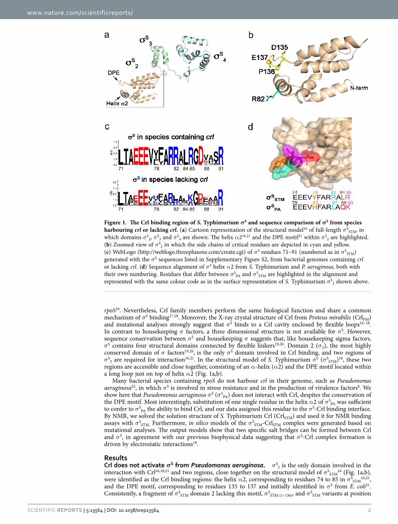
rpoS16. Nevertheless, Crl family members perform the same biological function and share a common mechanism of σ S binding17,18. Moreover, the X-ray crystal structure of Crl from Proteus mirabilis (CrlPM) and mutational analyses strongly suggest that σ S binds to a Crl cavity enclosed by flexible loops16–18. In contrast to housekeeping σ factors, a three dimensional structure is not available for σ S. However, sequence conservation between σ S and housekeeping σ suggests that, like housekeeping sigma factors, σ S contains four structural domains connected by flexible linkers19,20. Domain 2 (σ 2), the most highly conserved domain of σ factors19,20, is the only σ S domain involved in Crl binding, and two regions of σ S2 are required for interaction16,21. In the structural model of S. Typhimurium σ S (σ SSTM)16, these two regions are accessible and close together, consisting of an α -helix (α 2) and the DPE motif located within a long loop just on top of helix α 2 (Fig. 1a,b).
Many bacterial species containing rpoS do not harbour crl in their genome, such as Pseudomonas aeruginosa22, in which σ S is involved in stress resistance and in the production of virulence factors8. We show here that Pseudomonas aeruginosa σ S (σ SPA) does not interact with Crl, despites the conservation of the DPE motif. Most interestingly, substitution of one single residue in the helix α 2 of σ SPA was sufficient to confer to σ SPA the ability to bind Crl, and our data assigned this residue to the σ S-Crl binding interface. By NMR, we solved the solution structure of S. Typhimurium Crl (CrlSTM) and used it for NMR binding assays with σ SSTM. Furthermore, in silico models of the σ SSTM-CrlSTM complex were generated based on mutational analyses. The output models show that two specific salt bridges can be formed between Crl and σ S, in agreement with our previous biophysical data suggesting that σ S-Crl complex formation is driven by electrostatic interactions18.
ResultsCrl does not activate σS from Pseudomonas aeruginosa. σ S2 is the only domain involved in the interaction with Crl16,18,21 and two regions, close together on the structural model of σ SSTM
16 (Fig. 1a,b), were identified as the Crl binding regions: the helix α 2, corresponding to residues 74 to 85 in σ SSTM
16,21, and the DPE motif, corresponding to residues 135 to 137 and initially identified in σ S from E. coli21. Consistently, a fragment of σ SSTM domain 2 lacking this motif, σ SSTM (1–136), and σ SSTM variants at position
Figure 1. The Crl binding region of S. Typhimurium σS and sequence comparison of σS from species harbouring crl or lacking crl. (a) Cartoon representation of the structural model16 of full-length σ SSTM, in which domains σ S2, σ S3 and σ S4 are shown. The helix α 216,21 and the DPE motif21 within σ S2 are highlighted. (b) Zoomed view of σ S2 in which the side chains of critical residues are depicted in cyan and yellow. (c) WebLogo (http://weblogo.threeplusone.com/create.cgi) of σ S residues 71–91 (numbered as in σ SSTM) generated with the σ S sequences listed in Supplementary Figure S2, from bacterial genomes containing crl or lacking crl. (d) Sequence alignment of σ S helix α 2 from S. Typhimurium and P. aeruginosa, both with their own numbering. Residues that differ between σ SPA and σ SSTM are highlighted in the alignment and represented with the same colour code as in the surface representation of S. Typhimurium σ S2 shown above.
www.nature.com/scientificreports/
3Scientific RepoRts | 5:13564 | DOi: 10.1038/srep13564
D135 and E137, were not able to interact with Crl in bacterial two hybrid (BACTH) assays (Supplementary Fig. S1), confirming that the DPE motif in σ SSTM is involved in Crl binding.
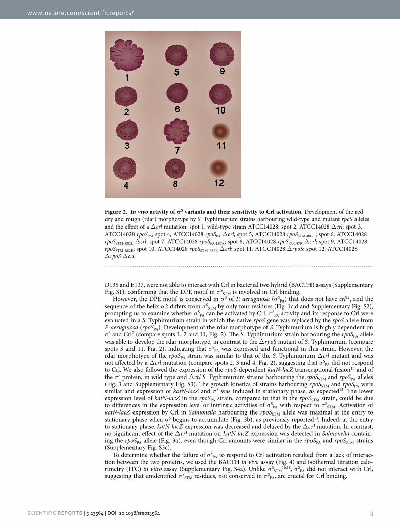
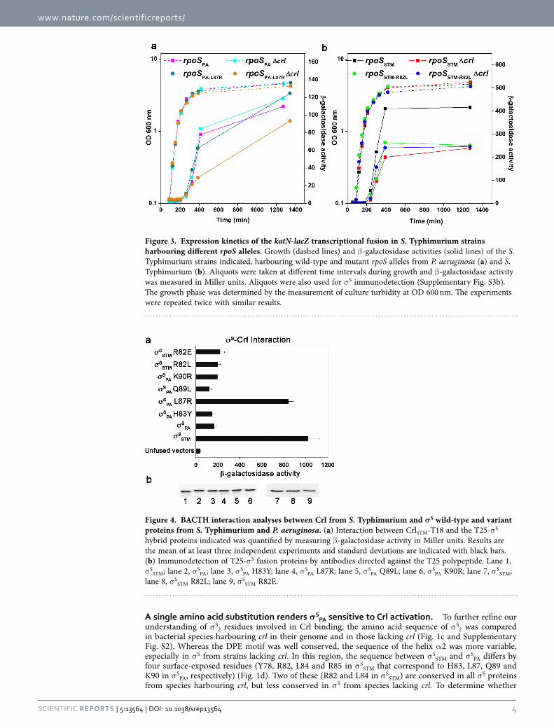
However, the DPE motif is conserved in σ S of P. aeruginosa (σ SPA) that does not have crl22, and the sequence of the helix α 2 differs from σ SSTM by only four residues (Fig. 1c,d and Supplementary Fig. S2), prompting us to examine whether σ SPA can be activated by Crl. σ SPA activity and its response to Crl were evaluated in a S. Typhimurium strain in which the native rpoS gene was replaced by the rpoS allele from P. aeruginosa (rpoSPA). Development of the rdar morphotype of S. Typhimurium is highly dependent on σ S and Crl7 (compare spots 1, 2 and 11, Fig. 2). The S. Typhimurium strain harbouring the rpoSPA allele was able to develop the rdar morphotype, in contrast to the Δ rpoS mutant of S. Typhimurium (compare spots 3 and 11, Fig. 2), indicating that σ SPA was expressed and functional in this strain. However, the rdar morphotype of the rpoSPA strain was similar to that of the S. Typhimurium Δ crl mutant and was not affected by a Δ crl mutation (compare spots 2, 3 and 4, Fig. 2), suggesting that σ SPA did not respond to Crl. We also followed the expression of the rpoS-dependent katN-lacZ transcriptional fusion13 and of the σ S protein, in wild type and Δ crl S. Typhimurium strains harbouring the rpoSSTM and rpoSPA alleles (Fig. 3 and Supplementary Fig. S3). The growth kinetics of strains harbouring rpoSSTM and rpoSPA were similar and expression of katN-lacZ and σ S was induced in stationary phase, as expected13. The lower expression level of katN-lacZ in the rpoSPA strain, compared to that in the rpoSSTM strain, could be due to differences in the expression level or intrinsic activities of σ SPA with respect to σ SSTM. Activation of katN-lacZ expression by Crl in Salmonella harbouring the rpoSSTM allele was maximal at the entry to stationary phase when σ S begins to accumulate (Fig. 3b), as previously reported13. Indeed, at the entry to stationary phase, katN-lacZ expression was decreased and delayed by the Δ crl mutation. In contrast, no significant effect of the Δ crl mutation on katN-lacZ expression was detected in Salmonella contain-ing the rpoSPA allele (Fig. 3a), even though Crl amounts were similar in the rpoSPA and rpoSSTM strains (Supplementary Fig. S3c).
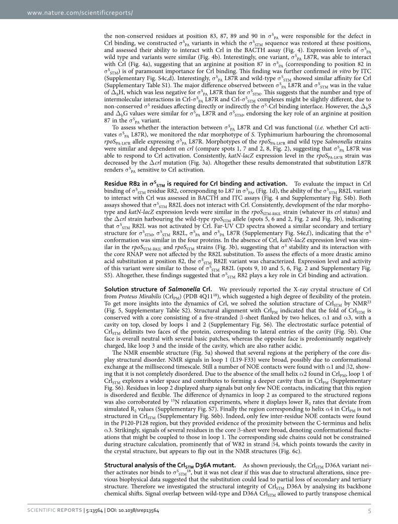
To determine whether the failure of σ SPA to respond to Crl activation resulted from a lack of interac-tion between the two proteins, we used the BACTH in vivo assay (Fig. 4) and isothermal titration calo-rimetry (ITC) in vitro assay (Supplementary Fig. S4a). Unlike σ SSTM
16,18, σ SPA did not interact with Crl, suggesting that unidentified σ SSTM residues, not conserved in σ SPA, are crucial for Crl binding.
Figure 2. In vivo activity of σS variants and their sensitivity to Crl activation. Development of the red dry and rough (rdar) morphotype by S. Typhimurium strains harbouring wild-type and mutant rpoS alleles and the effect of a Δ crl mutation: spot 1, wild-type strain ATCC14028; spot 2, ATCC14028 Δ crl; spot 3, ATCC14028 rpoSPA; spot 4, ATCC14028 rpoSPA Δ crl; spot 5, ATCC14028 rpoSSTM-R82L; spot 6, ATCC14028 rpoSSTM-R82L Δ crl; spot 7, ATCC14028 rpoSPA-L87R; spot 8, ATCC14028 rpoSPA-L87R Δ crl; spot 9, ATCC14028 rpoSSTM-R82E; spot 10, ATCC14028 rpoSSTM-R82E Δ crl; spot 11, ATCC14028 Δ rpoS; spot 12, ATCC14028 Δ rpoS Δ crl.
www.nature.com/scientificreports/
4Scientific RepoRts | 5:13564 | DOi: 10.1038/srep13564
A single amino acid substitution renders σSPA sensitive to Crl activation. To further refine our
understanding of σ S2 residues involved in Crl binding, the amino acid sequence of σ S2 was compared in bacterial species harbouring crl in their genome and in those lacking crl (Fig. 1c and Supplementary Fig. S2). Whereas the DPE motif was well conserved, the sequence of the helix α 2 was more variable, especially in σ S from strains lacking crl. In this region, the sequence between σ SSTM and σ SPA differs by four surface-exposed residues (Y78, R82, L84 and R85 in σ SSTM that correspond to H83, L87, Q89 and K90 in σ SPA, respectively) (Fig. 1d). Two of these (R82 and L84 in σ SSTM) are conserved in all σ S proteins from species harbouring crl, but less conserved in σ S from species lacking crl. To determine whether
Figure 3. Expression kinetics of the katN-lacZ transcriptional fusion in S. Typhimurium strains harbouring different rpoS alleles. Growth (dashed lines) and β -galactosidase activities (solid lines) of the S. Typhimurium strains indicated, harbouring wild-type and mutant rpoS alleles from P. aeruginosa (a) and S. Typhimurium (b). Aliquots were taken at different time intervals during growth and β -galactosidase activity was measured in Miller units. Aliquots were also used for σ S immunodetection (Supplementary Fig. S3b). The growth phase was determined by the measurement of culture turbidity at OD 600 nm. The experiments were repeated twice with similar results.
Figure 4. BACTH interaction analyses between Crl from S. Typhimurium and σS wild-type and variant proteins from S. Typhimurium and P. aeruginosa. (a) Interaction between CrlSTM-T18 and the T25-σ S hybrid proteins indicated was quantified by measuring β -galactosidase activity in Miller units. Results are the mean of at least three independent experiments and standard deviations are indicated with black bars. (b) Immunodetection of T25-σ S fusion proteins by antibodies directed against the T25 polypeptide. Lane 1, σ SSTM; lane 2, σ SPA; lane 3, σ SPA H83Y; lane 4, σ SPA L87R; lane 5, σ SPA Q89L; lane 6, σ SPA K90R; lane 7, σ SSTM; lane 8, σ SSTM R82L; lane 9, σ SSTM R82E.
www.nature.com/scientificreports/
5Scientific RepoRts | 5:13564 | DOi: 10.1038/srep13564
the non-conserved residues at position 83, 87, 89 and 90 in σ SPA were responsible for the defect in Crl binding, we constructed σ SPA variants in which the σ SSTM sequence was restored at these positions, and assessed their ability to interact with Crl in the BACTH assay (Fig. 4). Expression levels of σ SPA wild type and variants were similar (Fig. 4b). Interestingly, one variant, σ SPA L87R, was able to interact with Crl (Fig. 4a), suggesting that an arginine at position 87 in σ SPA (corresponding to position 82 in σ SSTM) is of paramount importance for Crl binding. This finding was further confirmed in vitro by ITC (Supplementary Fig. S4c,d). Interestingly, σ SPA L87R and wild-type σ SSTM showed similar affinity for Crl (Supplementary Table S1). The major difference observed between σ SPA L87R and σ SSTM was in the value of Δ bH, which was less negative for σ SPA L87R than for σ SSTM. This suggests that the number and type of intermolecular interactions in Crl-σ SPA L87R and Crl-σ SSTM complexes might be slightly different, due to non-conserved σ S residues affecting directly or indirectly the σ S-Crl binding interface. However, the Δ bS and Δ bG values were similar for σ SPA L87R and σ SSTM, endorsing the key role of an arginine at position 87 in the σ SPA variant.
To assess whether the interaction between σ SPA L87R and Crl was functional (i.e. whether Crl acti-vates σ SPA L87R), we monitored the rdar morphotype of S. Typhimurium harbouring the chromosomal rpoSPA-L87R allele expressing σ SPA L87R. Morphotypes of the rpoSPA-L87R and wild type Salmonella strains were similar and dependent on crl (compare spots 1, 7 and 2, 8, Fig. 2), suggesting that σ SPA L87R was able to respond to Crl activation. Consistently, katN-lacZ expression level in the rpoSPA-L87R strain was decreased by the Δ crl mutation (Fig. 3a). Altogether these results demonstrated that substitution L87R renders σ SPA sensitive to Crl activation.
Residue R82 in σSSTM is required for Crl binding and activation. To evaluate the impact in Crl
binding of σ SSTM residue R82, corresponding to L87 in σ SPA, (Fig. 1d), the ability of the σ SSTM R82L variant to interact with Crl was assessed in BACTH and ITC assays (Fig. 4 and Supplementary Fig. S4b). Both assays showed that σ SSTM R82L does not interact with Crl. Consistently, development of the rdar morpho-type and katN-lacZ expression levels were similar in the rpoSSTM-R82L strain (whatever its crl status) and the Δ crl strain harbouring the wild-type rpoSSTM allele (spots 5, 6 and 2, Fig. 2 and Fig. 3b), indicating that σ SSTM R82L was not activated by Crl. Far-UV CD spectra showed a similar secondary and tertiary structure for σ SSTM, σ SSTM R82L, σ SPA and σ SPA L87R (Supplementary Fig. S4e,f), indicating that the σ S conformation was similar in the four proteins. In the absence of Crl, katN-lacZ expression level was sim-ilar in the rpoSSTM-R82L and rpoSSTM strains (Fig. 3b), suggesting that σ S stability and its interaction with the core RNAP were not affected by the R82L substitution. To assess the effects of a more drastic amino acid substitution at position 82, the σ SSTM R82E variant was characterized. Expression level and activity of this variant were similar to those of σ SSTM R82L (spots 9, 10 and 5, 6, Fig. 2 and Supplementary Fig. S5). Altogether, these findings suggested that σ SSTM R82 plays a key role in Crl binding and activation.
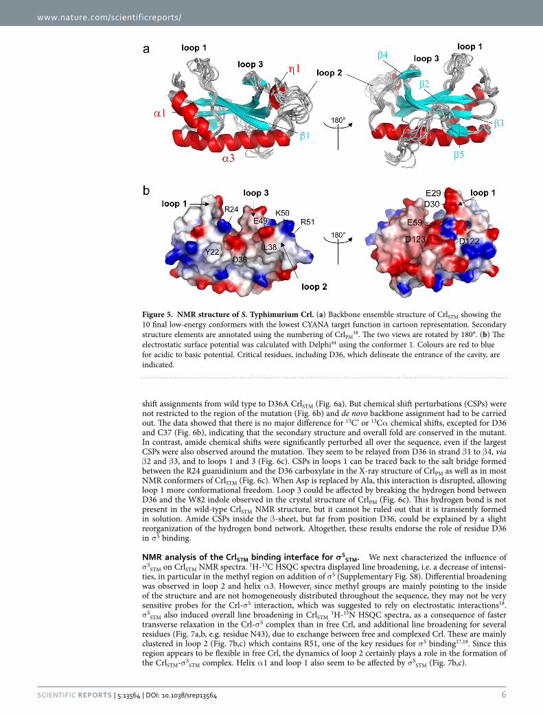
Solution structure of Salmonella Crl. We previously reported the X-ray crystal structure of Crl from Proteus Mirabilis (CrlPM) (PDB 4Q1118), which suggested a high degree of flexibility of the protein. To get more insights into the dynamics of Crl, we solved the solution structure of CrlSTM by NMR23 (Fig. 5, Supplementary Table S2). Structural alignment with CrlPM indicated that the fold of CrlSTM is conserved with a core consisting of a five-stranded β -sheet flanked by two helices, α 1 and α 3, with a cavity on top, closed by loops 1 and 2 (Supplementary Fig. S6). The electrostatic surface potential of CrlSTM delimits two faces of the protein, corresponding to lateral entries of the cavity (Fig. 5b). One face is overall neutral with several basic patches, whereas the opposite face is predominantly negatively charged, like loop 3 and the inside of the cavity, which are also rather acidic.
The NMR ensemble structure (Fig. 5a) showed that several regions at the periphery of the core dis-play structural disorder. NMR signals in loop 1 (L19-F33) were broad, possibly due to conformational exchange at the millisecond timescale. Still a number of NOE contacts were found with α 1 and β 2, show-ing that it is not completely disordered. Due to the absence of the small helix α 2 found in CrlPM, loop 1 of CrlSTM explores a wider space and contributes to forming a deeper cavity than in CrlPM (Supplementary Fig. S6). Residues in loop 2 displayed sharp signals but only few NOE contacts, indicating that this region is disordered and flexible. The difference of dynamics in loop 2 as compared to the structured regions was also corroborated by 15N relaxation experiments, where it displays lower R2 rates that deviate from simulated R2 values (Supplementary Fig. S7). Finally the region corresponding to helix α 4 in CrlPM is not structured in CrlSTM (Supplementary Fig. S6b). Indeed, only few inter-residue NOE contacts were found in the P120-P128 region, but they provided evidence of the proximity between the C-terminus and helix α 3. Strikingly, signals of several residues in the core β -sheet were broad, denoting conformational fluctu-ations that might be coupled to those in loop 1. The corresponding side chains could not be constrained during structure calculation, prominently that of W82 in strand β 4, which points towards the cavity in the crystal structure, but appears to flip out in the NMR structures (Fig. 6c).
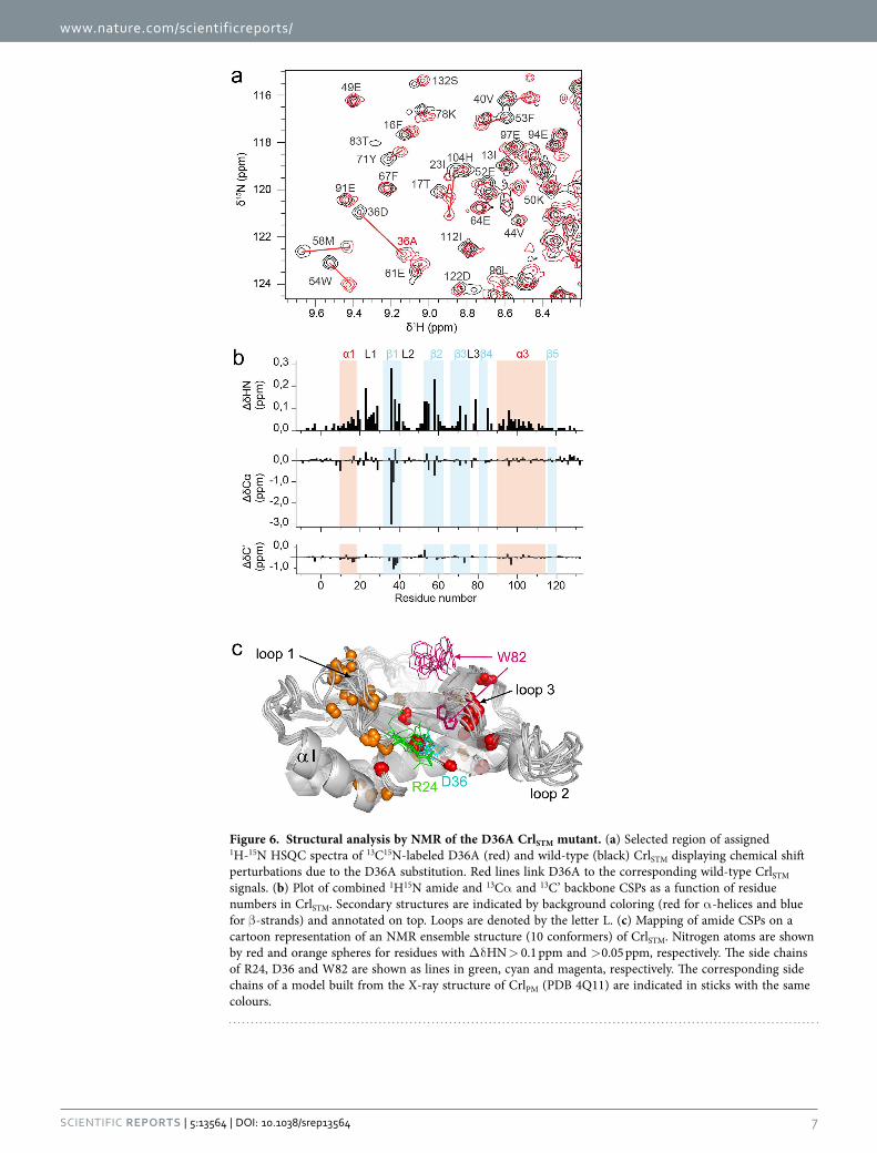
Structural analysis of the CrlSTM D36A mutant. As shown previously, the CrlSTM D36A variant nei-ther activates nor binds to σ SSTM
18, but it was not clear if this was due to structural alterations, since pre-vious biophysical data suggested that the substitution could lead to partial loss of secondary and tertiary structure. Therefore we investigated the structural integrity of CrlSTM D36A by analysing its backbone chemical shifts. Signal overlap between wild-type and D36A CrlSTM allowed to partly transpose chemical
www.nature.com/scientificreports/
6Scientific RepoRts | 5:13564 | DOi: 10.1038/srep13564
shift assignments from wild type to D36A CrlSTM (Fig. 6a). But chemical shift perturbations (CSPs) were not restricted to the region of the mutation (Fig. 6b) and de novo backbone assignment had to be carried out. The data showed that there is no major difference for 13C’ or 13Cα chemical shifts, excepted for D36 and C37 (Fig. 6b), indicating that the secondary structure and overall fold are conserved in the mutant. In contrast, amide chemical shifts were significantly perturbed all over the sequence, even if the largest CSPs were also observed around the mutation. They seem to be relayed from D36 in strand β 1 to β 4, via β 2 and β 3, and to loops 1 and 3 (Fig. 6c). CSPs in loops 1 can be traced back to the salt bridge formed between the R24 guanidinium and the D36 carboxylate in the X-ray structure of CrlPM as well as in most NMR conformers of CrlSTM (Fig. 6c). When Asp is replaced by Ala, this interaction is disrupted, allowing loop 1 more conformational freedom. Loop 3 could be affected by breaking the hydrogen bond between D36 and the W82 indole observed in the crystal structure of CrlPM (Fig. 6c). This hydrogen bond is not present in the wild-type CrlSTM NMR structure, but it cannot be ruled out that it is transiently formed in solution. Amide CSPs inside the β -sheet, but far from position D36, could be explained by a slight reorganization of the hydrogen bond network. Altogether, these results endorse the role of residue D36 in σ S binding.
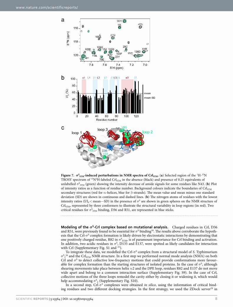
NMR analysis of the CrlSTM binding interface for σSSTM. We next characterized the influence of
σ SSTM on CrlSTM NMR spectra. 1H-13C HSQC spectra displayed line broadening, i.e. a decrease of intensi-ties, in particular in the methyl region on addition of σ S (Supplementary Fig. S8). Differential broadening was observed in loop 2 and helix α 3. However, since methyl groups are mainly pointing to the inside of the structure and are not homogeneously distributed throughout the sequence, they may not be very sensitive probes for the Crl-σ S interaction, which was suggested to rely on electrostatic interactions18. σ SSTM also induced overall line broadening in CrlSTM 1H-15N HSQC spectra, as a consequence of faster transverse relaxation in the Crl-σ S complex than in free Crl, and additional line broadening for several residues (Fig. 7a,b, e.g. residue N43), due to exchange between free and complexed Crl. These are mainly clustered in loop 2 (Fig. 7b,c) which contains R51, one of the key residues for σ S binding17,18. Since this region appears to be flexible in free Crl, the dynamics of loop 2 certainly plays a role in the formation of the CrlSTM-σ SSTM complex. Helix α 1 and loop 1 also seem to be affected by σ SSTM (Fig. 7b,c).
Figure 5. NMR structure of S. Typhimurium Crl. (a) Backbone ensemble structure of CrlSTM showing the 10 final low-energy conformers with the lowest CYANA target function in cartoon representation. Secondary structure elements are annotated using the numbering of CrlPM
18. The two views are rotated by 180°. (b) The electrostatic surface potential was calculated with Delphi44 using the conformer 1. Colours are red to blue for acidic to basic potential. Critical residues, including D36, which delineate the entrance of the cavity, are indicated.
www.nature.com/scientificreports/
7Scientific RepoRts | 5:13564 | DOi: 10.1038/srep13564
Figure 6. Structural analysis by NMR of the D36A CrlSTM mutant. (a) Selected region of assigned 1H-15N HSQC spectra of 13C15N-labeled D36A (red) and wild-type (black) CrlSTM displaying chemical shift perturbations due to the D36A substitution. Red lines link D36A to the corresponding wild-type CrlSTM signals. (b) Plot of combined 1H15N amide and 13Cα and 13C’ backbone CSPs as a function of residue numbers in CrlSTM. Secondary structures are indicated by background coloring (red for α -helices and blue for β -strands) and annotated on top. Loops are denoted by the letter L. (c) Mapping of amide CSPs on a cartoon representation of an NMR ensemble structure (10 conformers) of CrlSTM. Nitrogen atoms are shown by red and orange spheres for residues with Δ δ HN > 0.1 ppm and > 0.05 ppm, respectively. The side chains of R24, D36 and W82 are shown as lines in green, cyan and magenta, respectively. The corresponding side chains of a model built from the X-ray structure of CrlPM (PDB 4Q11) are indicated in sticks with the same colours.
www.nature.com/scientificreports/
8Scientific RepoRts | 5:13564 | DOi: 10.1038/srep13564
Modeling of the σS-Crl complex based on mutational analysis. Charged residues in Crl, D36 and R51, were previously found to be essential for σ S binding18. The results above corroborate the hypoth-esis that the Crl-σ S complex formation is likely driven by electrostatic interactions by demonstrating that one positively charged residue, R82 in σ SSTM, is of paramount importance for Crl binding and activation. In addition, two acidic residues in σ S, D135 and E137, were spotted as likely candidates for interaction with Crl (Supplementary Fig. S1 and 21).
To integrate these data, we modelled the Crl-σ S complex from a structural model of S. Typhimurium σ S2
16 and the CrlSTM NMR structure. In a first step we performed normal mode analysis (NMA) on both Crl and σ S to detect collective low-frequency motions that could provide conformations more favour-able for complex formation than the starting structures of isolated proteins. In the case of σ S, although shearing movements take place between helix α 2 and the DPE loop, residues R82 and E137 do not move wide apart and belong to a common interaction surface (Supplementary Fig. S9). In the case of Crl, collective motions of the three loops remodel the cavity either by closing it or widening it, which would help accommodating σ S2 (Supplementary Fig. S10).
In a second step, Crl-σ S complexes were obtained in silico, using the information of critical bind-ing residues and two different docking strategies. In the first strategy, we used the ZDock server24 in
Figure 7. σSSTM-induced perturbations in NMR spectra of CrlSTM. (a) Selected region of the 1H-15N
TROSY spectrum of 15N2H-labeled CrlSTM in the absence (black) and presence of 0.25 equivalents of unlabelled σ SSTM (green) showing the intensity decrease of amide signals for some residues like N43. (b) Plot of intensity ratios as a function of residue number. Background colours indicate the boundaries of CrlSTM secondary structures (red for α -helices, blue for β -strands). The mean value and mean minus one standard deviation (SD) are shown in continuous and dashed lines. (b) The nitrogen atoms of residues with the lowest intensity ratios (I/I0 < mean—SD) in the presence of σ S are shown in green spheres on the NMR structure of CrlSTM, represented by three conformers to illustrate the structural variability in loop regions (in red). Two critical residues for σ SSTM binding, D36 and R51, are represented in blue sticks.
www.nature.com/scientificreports/
9Scientific RepoRts | 5:13564 | DOi: 10.1038/srep13564
combination with refinement on the RosettaDock server25, that do not take into account conforma-tional changes and flexibility of proteins (Models A to E, Supplementary Fig. S11). The second strategy used the Haddock Webserver26,27 to integrate the high degree of flexibility of the NMR structure of Crl (Supplementary Fig. S12).
In models A and B (Supplementary Fig. S11), σ S R82 interacts with the Crl residues E25 or E102, respectively. These residues are not conserved in Crl family members22 and the Crl E25A and E102A variants interacted with σ SSTM in the same manner as wild type Crl (Supplementary Fig. S1) indicating that E25 and E102 are not required for σ S binding. It is noteworthy that in these two models the DPE motif does not have any interacting partner. Altogether these data suggest that models A and B do not represent the Crl-σ S interface. Models C and D are also unlikely since E137 in σ S interacts with R24 in Crl, a residue dispensible for σ S binding18. Furthermore, in model C, R82 in σ S interacts with E25 in Crl, which is not involved in σ S binding (Supplementary Fig. S1) and in both models R51 in Crl does not have any possible charged interacting partner in σ S.
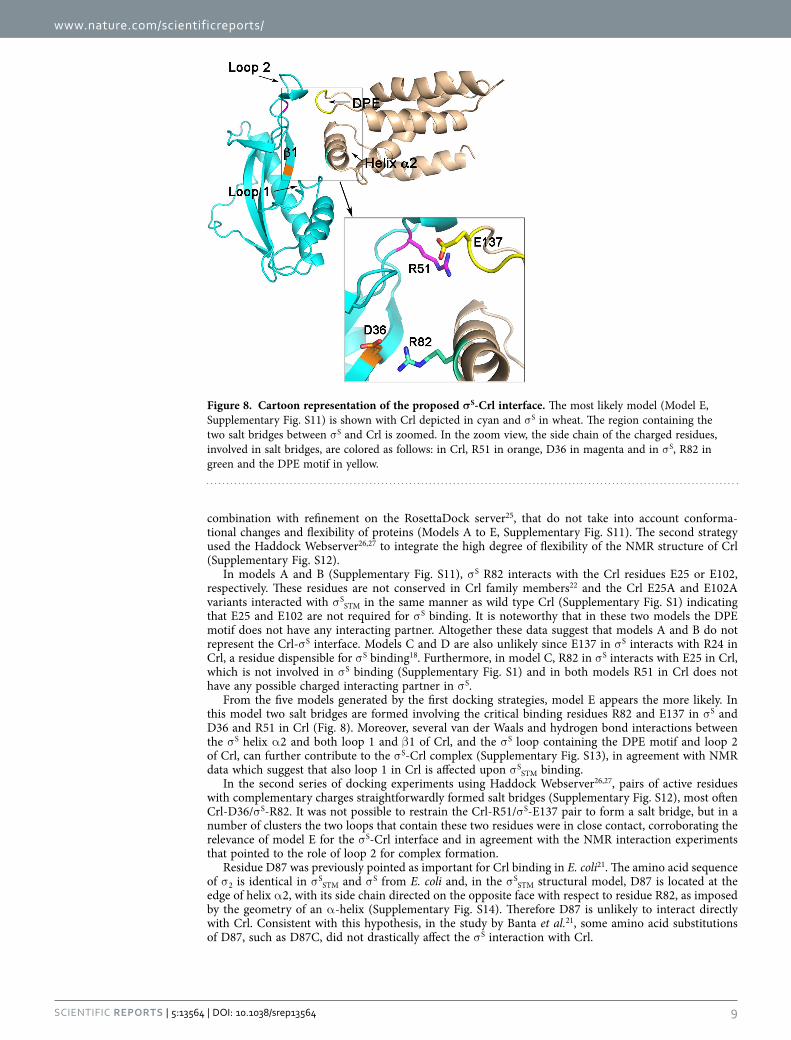
From the five models generated by the first docking strategies, model E appears the more likely. In this model two salt bridges are formed involving the critical binding residues R82 and E137 in σ S and D36 and R51 in Crl (Fig. 8). Moreover, several van der Waals and hydrogen bond interactions between the σ S helix α 2 and both loop 1 and β 1 of Crl, and the σ S loop containing the DPE motif and loop 2 of Crl, can further contribute to the σ S-Crl complex (Supplementary Fig. S13), in agreement with NMR data which suggest that also loop 1 in Crl is affected upon σ SSTM binding.
In the second series of docking experiments using Haddock Webserver26,27, pairs of active residues with complementary charges straightforwardly formed salt bridges (Supplementary Fig. S12), most often Crl-D36/σ S-R82. It was not possible to restrain the Crl-R51/σ S-E137 pair to form a salt bridge, but in a number of clusters the two loops that contain these two residues were in close contact, corroborating the relevance of model E for the σ S-Crl interface and in agreement with the NMR interaction experiments that pointed to the role of loop 2 for complex formation.
Residue D87 was previously pointed as important for Crl binding in E. coli21. The amino acid sequence of σ 2 is identical in σ SSTM and σ S from E. coli and, in the σ SSTM structural model, D87 is located at the edge of helix α 2, with its side chain directed on the opposite face with respect to residue R82, as imposed by the geometry of an α -helix (Supplementary Fig. S14). Therefore D87 is unlikely to interact directly with Crl. Consistent with this hypothesis, in the study by Banta et al.21, some amino acid substitutions of D87, such as D87C, did not drastically affect the σ S interaction with Crl.
Figure 8. Cartoon representation of the proposed σS-Crl interface. The most likely model (Model E, Supplementary Fig. S11) is shown with Crl depicted in cyan and σ S in wheat. The region containing the two salt bridges between σ S and Crl is zoomed. In the zoom view, the side chain of the charged residues, involved in salt bridges, are colored as follows: in Crl, R51 in orange, D36 in magenta and in σ S, R82 in green and the DPE motif in yellow.
www.nature.com/scientificreports/
1 0Scientific RepoRts | 5:13564 | DOi: 10.1038/srep13564
DiscussionIn many Gram-negative bacteria, σ S/RpoS is the master regulator of gene expression in stress conditions and during stationary phase. σ S is exquisitely and tightly regulated by many mechanisms that keep its production level and activity under strict control3–5. Crl is a unique regulatory factor, specifically ded-icated to σ S, which enhances its activity, helping the association of σ S with E15. Nevertheless, there are some rpoS-containing species, including P. aeruginosa, that do not harbour a crl gene16 and in which σ S activity may be controlled by alternative mechanisms or functional homologs of Crl.
The strong sequence conservation of σ S2, the only σ S domain that binds Crl16,18,21, prompted us to assess possible activation of σ SPA by Crl. We show here that σ SPA is not activated by Crl due to its inabil-ity to interact with Crl. Taking advantage of the evolution of the σ S sequence in P. aeruginosa and other species lacking crl, we identified residues conserved in σ S sequences from crl proficient species, and potentially implicated in Crl recognition. Among these, a surface-exposed arginine in σ SSTM, R82, was assigned to the σ S-Crl interface. This residue is not conserved in σ SPA, which instead contains a leucine. Importantly, substitution of this leucine by an arginine rendered σ SPA sensitive to Crl activation. It is noteworthy that, in some σ S proteins from species that do not harbour crl, the arginine residue is con-served (Supplementary Fig. S2). It would be interesting to determine whether these σ S proteins interact with Crl, and if not, whether they could be used to identify additional σ S residues involved in Crl binding by the strategy described in this study for σ SPA.
The in silico models of the σ S-Crl complex show that salt bridges can indeed be formed for the two pairs of residues Crl-D36/σ S-R82 and Crl-R51/σ S-E137. In some models they can be formed simultane-ously. This leads to a picture of an ideal binding interface in which helix α 2 of σ S, containing R82, would dock into the cavity of Crl containing D36, disrupting the intermolecular R24-D36 contact, and the DPE motif and loop 2 of Crl would make contact on the outside, driven by electrostatic interactions between Crl-R51 and σ S-D135/E137 (Fig. 8).
What renders the σ S-Crl system very intriguing is its transitory and dynamic binding mechanism, which is unclear so far. Our NMR data together with the in silico modelling shed some light on how σ S and Crl may interact and form a transient complex. The chemical shift perturbations in the NMR spec-trum of Crl in the presence of σ S indicate that loop 2 senses the presence of σ S, but extend beyond the region directly involved in σ S binding, including helix α 1, loop 1 and helix α 3. These findings suggest that local structural rearrangements might take place in the flexible loops that allow breathing of the cavity as indicated by normal mode analysis of the Crl structure. Such rearrangements might contribute not only to the formation of the σ S-Crl complex, but also to its dissociation, once Crl has accomplished its work. Moreover, in free Crl, residue D36 is involved in an intramolecular interaction with R24. To form a new salt bridge with σ S-R82, the first one has to be broken. The perturbations observed in the NMR spectra of the Crl D36A variant show how the disruption of this network is sensed by the whole Crl structure, in particular by loop 1. It is tempting to speculate that this variant mimics the molecular processes that Crl undergoes upon σ S binding, as we previously hypothesized18.
How does Crl binding to σ S increase the σ S association rate with E? Why is the σ S-Crl interaction so transient? These questions are still open. One possibility is that Crl triggers a conformational change in σ S favouring its association with E. There is no high resolution 3D structure for free σ factors, but several biochemical and structural studies using the housekeeping σ 70 have shown that σ factors undergo pro-nounced conformational changes upon E binding, allowing domains σ 2 and σ 4 to be spaced correctly for promoter binding2,20. These findings have led to the proposal that σ factors must be in a more compact conformation when free in the cell than in the Eσ complex. Consistent with this hypothesis, free σ are not able to bind promoters efficiently. This concept was further supported by the results obtained with engineered cysteine mutants of σ 28, which showed that this σ factor has a compact conformation when free in solution28.
Modulation of the free σ S conformation might be a common way to regulate both the stability and activity of σ S. σ S is degraded by the ATP-dependent complex ClpXP protease3–5. However, σ S binding by the RssB protein is required for delivery to ClpXP3–5. It has been postulated that RssB binding triggers a conformational opening of σ S that exposes a ClpXP binding site, that is otherwise occluded in a closed conformation of free σ S5. Therefore, if the conformation of σ S in the cell is rather compact, Crl binding to σ S2 may alleviate intramolecular interactions between σ S2 and other σ S domains, favouring an open conformation for just the time required for σ S to bind E, but transiently enough to avoid σ S degrada-tion by ClpXP. Further investigation of the structure of the σ S-Crl complex, for which a starting base is provided in the present study, and of the free σ S conformation will assess the relevance of this scenario.
MethodsBacterial strains, bacteriophage, plasmids and growth conditions. Strains and plasmids used for this work are listed in Supplementary Table S5. Bacteriophage P22HT105/1int was used to trans-fer mutations and the katN-lacZ fusion between Salmonella strains by transduction29. Green plates, for P22-infected cells or lysogens screening, were prepared as described previously30. Strains were grown in Luria-Bertani (LB) medium31 at 37°C under aeration. Development of the rdar morphotype was mon-itored on CR plates (LB agar without NaCl supplemented with Congo red 40 μ g/ml and Coomassie brilliant blue R250 20 μ g/ml), at 28°C as described7. Antibiotics were used at the following concen-trations: ampicillin (Ap) 100 μ g/mL; carbenicillin (Cb) 100 μ g/mL; chloramphenicol (Cm) 15 μ g/mL

www.nature.com/scientificreports/
1 1Scientific RepoRts | 5:13564 | DOi: 10.1038/srep13564
for the chromosomal resistance gene and 30 μ g/mL for the plasmid resistance gene; kanamycin (Km) 50 μ g/mL; and tetracycline (Tet) 20 μ g/mL.
rpoS allelic exchange in Salmonella. Allelic exchange of rpoS in S. Typhimurium ATCC14028 was achieved with a two-step Red-recombinase-based recombineering procedure32–35. The procedure involves replacement of the tetRA module of strain VFC326 by PCR-amplified DNA fragments of the rpoS allele from pVFC629, pVFD410, pVFD412 and pVFD399 (Supplementary Table S5 and S6) through posi-tive selection of tetracycline-sensitive recombinants. All strains were confirmed to contain the expected mutation by DNA sequencing.
Protein production and BACTH assays. The N-terminal (his)6-tagged σ SPA wild type and vari-ant L87R, σ SSTM R82L variant and CrlSTM were produced in E. coli strain BL21 (DE3) harbouring plas-mid derivatives of pETM11 (Supplementary Table S5). Production and purification of the proteins were carried out as previously described18. 15N-, 13C15N- or 15N2H-labeled wild type (his)6- CrlSTM and 15N13C-labeled CrlSTM (his)6-D36A protein samples for NMR experiments were produced in minimum M9 medium31 supplemented with 15NH4Cl and unlabelled or 13C- or 2H-labeled glucose following the same protocol as18. Samples were subsequently dialyzed into NMR buffer (50 mM sodium or potassium phosphate, 300 mM NaCl or KCl, 2 mM dithiotreitol, at pH 8 or 7.5).
For bacterial adenylate cyclase-based two hybrid assay, the E. coli cya strain DHT1 was transformed with derivatives of plasmids pKT25 and pUT18 encoding σ S and Crl proteins fused to the C-terminal part of T25 and the N-terminal part of T18, respectively (Supplementary Table S5). Co-transformants were plated onto MacConkey maltose plates supplemented with Cb, Km, and 0.5 mM IPTG to assess the Mal phenotype and on LB plates supplemented with X-Gal (40 μ g/ml) Cb, Km, and IPTG (0.5 mM) to assess the Lac phenotype. Plates were incubated at 30 °C for 2 days and then isolated colonies were grown in LB supplemented with Cb, Km, and IPTG, at 30 °C for 20 hours. β -galactosidase activities were measured as described by Miller and are expressed in Miller units36.
NMR experiments. NMR measurements were carried out at 293 K on a Bruker Avance III spec-trometer with a magnetic field of 18.8 T (800 MHz 1H frequency) equipped with a cryogenic TCI probe. The magnetic field was locked with 7% or 100% 2H2O. Spectra were processed with Topspin 3.1 (Bruker Biospin) or NMRPipe37 and analysed with CCPNMR 2.2 software38. Chemical shift assignments of CrlSTM are reported elsewhere23. For structure determination, 2D 1H-15N HSQC and 3D 1H-15N NOESY-HSQC spectra were recorded from a 300 μ M 15N-Crl sample, and 1H-13C HSQC, 2D 1H-1H NOESY and 3D 1H-13C NOESY-HSQC spectra from a 250 μ M 13C15N-Crl sample in deuterium oxide buffer. NOESY mixing times were 80 ms.
Sequential backbone assignment of CrlSTM D36A (380 μ M, 293 K) was carried out with a minimal set of triple resonance experiments: HNCA and HN(CO)CA were recorded at 14.1 T, CBCA(CO)NH and HNCO) at 18.8 T. Chemical shift perturbations induced by the D36A mutation were calculated as combined 1H and 15N perturbations Δ δ HN for a given residue i:
HN ppm H H N N110i D A i WT i D A i WT i36
22 36
2δ δ δ δ δΔ ( ) = ( − ) + ( − ), , , ,
The scaling factor 1/10 corresponds to the gyromagnetic ratio difference between 15N and 1H.
NMR structure calculation. NMR structures of wild-type CrlSTM were calculated using torsion angle dynamics in CYANA 2.239. Backbone torsion angle restraints were generated with TALOS-N40 using CrlSTM backbone chemical shifts. Ambiguous distance restraints were collected from three sets of NOESY spectra and purged from 3D peaks without possible assignments in the 1H dimension bound to a het-eroatom. The disordered N-terminal His-tag (His(− 20)-His(0)) was excluded from structure calculation. Structure statistics were obtained from the Protein Structure Validation Server, version 1.5 (http://psvs-1_5-dev.nesg.org/) (Supplementary Table S2). Normal Mode Analysis was performed on single conform-ers on the ElNémo webserver41.
Protein-protein docking. Rigid-body docking was carried out first on the ZDock server24, which employs a fast Fourier transform (FFT) algorithm, to generate the initial models (about 100) for the σ S-Crl complex with CrlSTM as the receptor and a homology model of S. Typhimurium σ S2 as the ligand16. Five models selected from ZDock were further refined using RosettaDock25, which performs a searching for the lowest-energy binding interface structures giving as ouput the 10 best-scoring models from 1000 total models. The presence of a ‘docking funnel’ was verified, considering that at least three of the first five lowest-energy binding interface models have a value of I_rmsd < 4 Å42 (Supplementary Table S3).
Flexible docking was carried out on the guru interface of the Haddock Webserver26,27 using single conformers from the NMR structure ensemble of CrlSTM and the homology model of S. Typhimurium σ S2. D36 and R51 in Crl and R82, D135 and E137 in σ S were defined as active residues. Passive residues were automatically defined around active residues. Loops 1 (E25-R32) and 2 (N43-E52) in Crl and the DPE loop (K133-F142) in σ S were defined as fully flexible. 1000 initial structures were generated. 200
www.nature.com/scientificreports/
1 2Scientific RepoRts | 5:13564 | DOi: 10.1038/srep13564
final structures were refined in water and clustered according to RMSD criterion. Statistics for clusters obtained for the conformers 1 and 2 are given in Supplementary Table S4.
Illustrations. Visualization and graphic rendering of protein structures were prepared with PyMOL43.
Other methods. Methods for DNA manipulation, immunoblot analysis of proteins and CD and ITC experiments are described in Supplementary Methods.
References1. Gruber, T. M. & Gross, C. A. Multiple sigma subunits and the partitioning of bacterial transcription space. Annu Rev Microbiol
57, 441–66 (2003).2. Feklistov, A., Sharon, B. D., Darst, S. A. & Gross, C. A. Bacterial sigma factors: a historical, structural, and genomic perspective.
Annu Rev Microbiol 68, 357–76 (2014).3. Klauck, E., Typas, A. & Hengge, R. The sigmaS subunit of RNA polymerase as a signal integrator and network master regulator
in the general stress response in Escherichia coli. Sci Prog 90, 103–27 (2007).4. Battesti, A., Majdalani, N. & Gottesman, S. The RpoS-mediated general stress response in Escherichia coli. Annu Rev Microbiol
65, 189–213 (2011).5. Hengge, R. The general stress response in Gram-negative bacteria. Bacterial Stress Responses (Second Edition, eds. G. Storz & R.
Hengge), ASM Press, Washington D.C., 251–289 (2011).6. Romling, U. Characterization of the rdar morphotype, a multicellular behaviour in Enterobacteriaceae. Cell Mol Life Sci 62,
1234–46 (2005).7. Robbe-Saule, V. et al. Crl activates transcription initiation of RpoS-regulated genes involved in the multicellular behavior of
Salmonella enterica serovar Typhimurium. J Bacteriol 188, 3983–94 (2006).8. Dong, T. & Schellhorn, H. E. Role of RpoS in virulence of pathogens. Infect Immun 78, 887–97 (2010).9. Osterberg, S., del Peso-Santos, T. & Shingler, V. Regulation of alternative sigma factor use. Annu Rev Microbiol 65, 37–55 (2011).
10. Pratt, L. A. & Silhavy, T. J. Crl stimulates RpoS activity during stationary phase. Mol Microbiol 29, 1225–36 (1998).11. Bougdour, A., Lelong, C., Geiselmann, J. Crl, a low temperature-induced protein in Escherichia Coli that binds directly to the
stationary phase σ subunit of RNA polymerase J Biol Chem 279, 19540–19550 (2004).12. Gaal, T., Mandel, M. J., Silhavy, T. J. & Gourse, R. L. Crl facilitates RNA polymerase holoenzyme formation. J Bacteriol 188,
7966–70 (2006).13. Robbe-Saule, V., Lopes, M. D., Kolb, A. & Norel, F. Physiological effects of Crl in Salmonella are modulated by sigmaS level and
promoter specificity. J Bacteriol 189, 2976–87 (2007).14. Typas, A., Barembruch, C., Possling, A. & Hengge, R. Stationary phase reorganisation of the Escherichia coli transcription
machinery by Crl protein, a fine-tuner of σ S activity and levels. EMBO J 26, 1569–1578 (2007).15. England, P. et al. Binding of the unorthodox transcription activator, Crl, to the components of the transcription machinery. J Biol
Chem 283, 33455–64 (2008).16. Monteil, V. et al. Crl binds to domain 2 of sigma(S) and confers a competitive advantage on a natural rpoS mutant of Salmonella
enterica serovar Typhi. J Bacteriol 192, 6401–10 (2010).17. Banta, A. B. et al. Structure of the RNA polymerase assembly factor Crl and identification of its interaction surface with sigma
S. J Bacteriol 196, 3279–88 (2014).18. Cavaliere, P. et al. Structural and functional features of Crl proteins and identification of conserved surface residues required for
interaction with the RpoS/sigmaS subunit of RNA polymerase. Biochem J 463, 215–24 (2014).19. Lonetto, M., Gribskov, M. & Gross, C. A. The sigma 70 family: sequence conservation and evolutionary relationships. J Bacteriol
174, 3843–9 (1992).20. Murakami, K. S. & Darst, S. A. Bacterial RNA polymerases: the wholo story. Curr Opin Struct Biol 13, 31–9 (2003).21. Banta, A. B. et al. Key features of sigmaS required for specific recognition by Crl, a transcription factor promoting assembly of
RNA polymerase holoenzyme. Proc Natl Acad Sci USA 110, 15955–15960 (2013).22. Monteil, V., Kolb, A., D’Alayer, J., Beguin, P. & Norel, F. Identification of conserved amino acid residues of the Salmonella sigmaS
chaperone Crl involved in Crl-sigmaS interactions. J Bacteriol 192, 1075–87 (2010).23. Cavaliere, P., Norel, F. & Sizun, C. 1H, 13C and 15N resonance assignments of σ S activating protein Crl from Salmonella enterica
serovar Typhimurium. Biomol NMR Assign Epub ahead of print (2015) doi: 10.1007/s12104-015-9617-z.24. Pierce, B. G. et al. ZDOCK server: interactive docking prediction of protein-protein complexes and symmetric multimers.
Bioinformatics 30, 1771–3 (2014).25. Lyskov, S. et al. Serverification of molecular modeling applications: the Rosetta Online Server that Includes Everyone (ROSIE).
PLoS One 8, e63906 (2013).26. de Vries, S. J., van Dijk, M. & Bonvin, A. M. The HADDOCK web server for data-driven biomolecular docking. Nat Protoc 5,
883–97 (2010).27. Wassenaar, T. A. et al. WeNMR: Structural Biology on the Grid. J Grid Comp 10, 743–767 (2012).28. Sorenson, M. K. & Darst, S. A. Disulfide cross-linking indicates that FlgM-bound and free sigma28 adopt similar conformations.
Proc Natl Acad Sci USA 103, 16722–7 (2006).29. Schmieger, H. Phage P22-mutants with increased or decreased transduction abilities. Mol Gen Genet 119, 75–88 (1972).30. Sternberg, N. L. & Maurer, R. Bacteriophage-mediated generalized transduction in Escherichia coli and Salmonella typhimurium.
Methods Enzymol 204, 18–43 (1991).31. Sambrook, J., Fritsch, E. F. & Maniatis, T. Molecular cloning: a laboratory manual, 2nd ed. Cold Spring Harbor Laboratory Press,
Cold Spring Harbor, NY. (1989).32. Bochner, B. R., Huang, H. C., Schieven, G. L. & Ames, B. N. Positive selection for loss of tetracycline resistance. J Bacteriol 143,
926–33 (1980).33. Datsenko, K. A. & Wanner, B. L. One-step inactivation of chromosomal genes in Escherichia coli K-12 using PCR products. Proc
Natl Acad Sci USA 97, 6640–5 (2000).34. Gerlach, R. G., Jackel, D., Holzer, S. U. & Hensel, M. Rapid oligonucleotide-based recombineering of the chromosome of
Salmonella enterica. Appl Environ Microbiol 75, 1575–80 (2009).35. Levi-Meyrueis, C. et al. Repressor activity of the RpoS/sigmaS-dependent RNA polymerase requires DNA binding. Nucleic Acids
Res 43, 1456–68 (2015).36. Miller, J. H. Experiments in molecular genetics. Cold Spring Harbor Laboratory Press, Cold Spring Harbor, NY. (1972).37. Delaglio, F. et al. NMRPipe: a multidimensional spectral processing system based on UNIX pipes. J Biomol NMR 6, 277–93
(1995).
www.nature.com/scientificreports/
13Scientific RepoRts | 5:13564 | DOi: 10.1038/srep13564
38. Vranken, W. F. et al. The CCPN data model for NMR spectroscopy: development of a software pipeline. Proteins 59, 687–96 (2005).
39. Guntert, P. Automated NMR structure calculation with CYANA. Methods Mol Biol 278, 353–78 (2004).40. Shen, Y. & Bax, A. Protein backbone and sidechain torsion angles predicted from NMR chemical shifts using artificial neural
networks. Journal of biomolecular NMR 56, 227–41 (2013).41. Suhre, K. & Sanejouand, Y. H. ElNemo: a normal mode web server for protein movement analysis and the generation of templates
for molecular replacement. Nucleic acids research 32, W610–4 (2004).42. Chaudhury, S. et al. Benchmarking and analysis of protein docking performance in Rosetta v3.2. PLoS One 6, e22477 (2011).43. Schrodinger, L. L. C. The PyMOL Molecular Graphics System, Version 1.3 (2010). Available at: http://pymol.sourceforge.net
(19/08/10).44. Rocchia, W., Alexov, E. & Honig, B. Extending the applicability of the nonlinear Poisson-Boltzmann equation: Multiple dielectric
constants and multivalent ions. Journal of Physical Chemistry B 105, 6507–6514 (2001).
AcknowledgementsWe thank O. Francetic, A. Pugsley and all members of the laboratory for their kind support. We thank G. André-Leroux for advice regarding the docking programs. We are also grateful to the collection of the Institut Pasteur for providing P. aeruginosa and D. Ladant for the T25 antibody. Funding: This work was supported by the French National Research Agency [grant ANR- 11-BSV3-009], the IR-RMN-THC (CNRS FR3050) for access to the 950 MHz spectrometer at Gif-sur-Yvette for preliminary experiments and by grants from the Institut Pasteur and the Centre National de la Recherche Scientifique. The WeNMR project (European FP7 e-Infrastructure grant, contract no. 261572, www.wenmr.eu), supported by the European Grid Initiative (EGI) through the national GRID Initiatives of Belgium, France, Italy, Germany, the Netherlands, Poland, Portugal, Spain, UK, South Africa, Malaysia, Taiwan, the Latin America GRID infrastructure via the Gisela project and the US Open Science Grid (OSG) are acknowledged for the use of web portals, computing and storage facilities.
Author ContributionsDesigned the study: P.C., C.S., J.B., C.M. and F.N. Performed the experiments: P.C., C.S., F.L.A., M.N., V.M. and F.B. Analysed the data: P.C., C.S., J.B., C.M. and F.N. Wrote the manuscript: P.C., C.S., C.M. and F.N.
Additional InformationSupplementary information accompanies this paper at http://www.nature.com/srepCompeting financial interests: The authors declare no competing financial interests.Coordinates for the NMR structure of S. Typhimurium were deposited at the Protein Data Bank under accession code 2MZ8.How to cite this article: Cavaliere, P. et al. Binding interface between the Salmonella σS/RpoS subunit of RNA polymerase and Crl: hints from bacterial species lacking crl. Sci. Rep. 5, 13564; doi: 10.1038/srep13564 (2015).
This work is licensed under a Creative Commons Attribution 4.0 International License. The images or other third party material in this article are included in the article’s Creative Com-
mons license, unless indicated otherwise in the credit line; if the material is not included under the Creative Commons license, users will need to obtain permission from the license holder to reproduce the material. To view a copy of this license, visit http://creativecommons.org/licenses/by/4.0/
Top Related