


γλώσσες
Σελίδες
Νομικός

PROPERTIES OF AMINO ACIDS
Dr Arun Kumar K
Alpha aminogroup (-NH2)
Alpha carboxylgroup (-COOH)
Side chain (-R).
L, α AMINO ACIDGeneral formula of

Properties
The chemical and physical properties
of amino acids are essentially of the
- amino,
- carboxyl and
- the R functional groups.
3
Ionic states of amino acids• All the amino acids contain at least two dissociable
hydrogens. .
• an acidic group (COOH) and a basic group (NH3+)
attached to the α carbon.• Acidic and basic amino acids have additional
charged group in their side chains
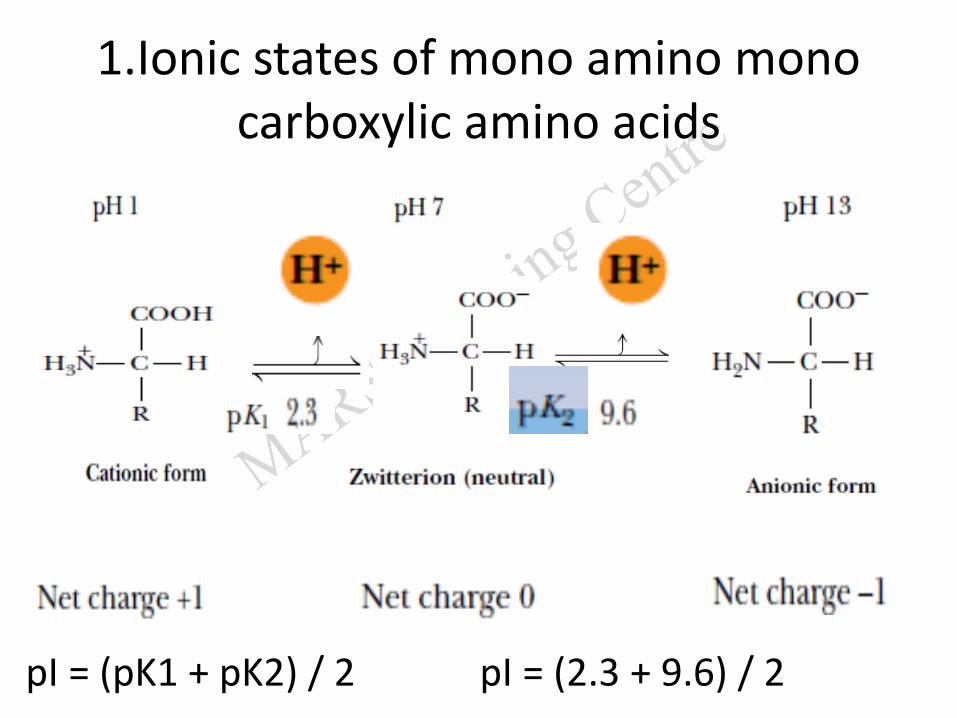
1.Ionic states of mono amino mono carboxylic amino acids
pI = (pK1 + pK2) / 2 pI = (2.3 + 9.6) / 2
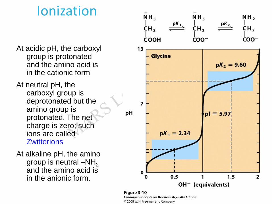
Ionization
At acidic pH, the carboxyl group is protonated and the amino acid is in the cationic form
At neutral pH, the carboxyl group is deprotonated but the amino group is protonated. The net charge is zero; such ions are called Zwitterions
At alkaline pH, the amino group is neutral –NH2
and the amino acid is in the anionic form.
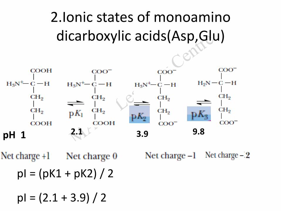
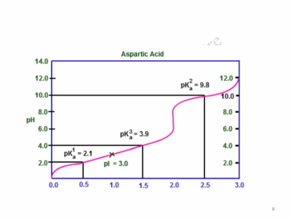
2.Ionic states of monoamino dicarboxylic acids(Asp,Glu)
2.1 3.9 9.8pH 1
pI = (pK1 + pK2) / 2
pI = (2.1 + 3.9) / 2
8
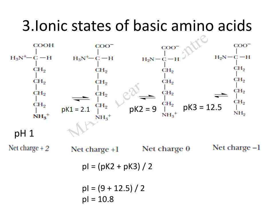
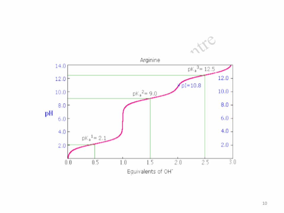
3.Ionic states of basic amino acids
pK2 = 9 pK3 = 12.5
pI = (pK2 + pK3) / 2
pI = (9 + 12.5) / 2pI = 10.8
pH 1
10
Isoelectric pH(PI)
• The isoelectric point (PI) is the pH at which an amino acid is electrically neutral
• that is, the sum of the positive charges equals the sum of the negative charges.
• At this point, the net charge is zero
– AA is least soluble in water
– AA does not migrate in electric field
1. For a mono amino mono carboxylic amino acid,
PI the is the average of pK1 and pK2 (around 6)
2. For a monoamino dicarboxylic amino acid,
PI the is the average of pK1 and pK2 (around 3)
3.For a diamino monocarboxylic acid,
PI the is the average of pK2 and pK3
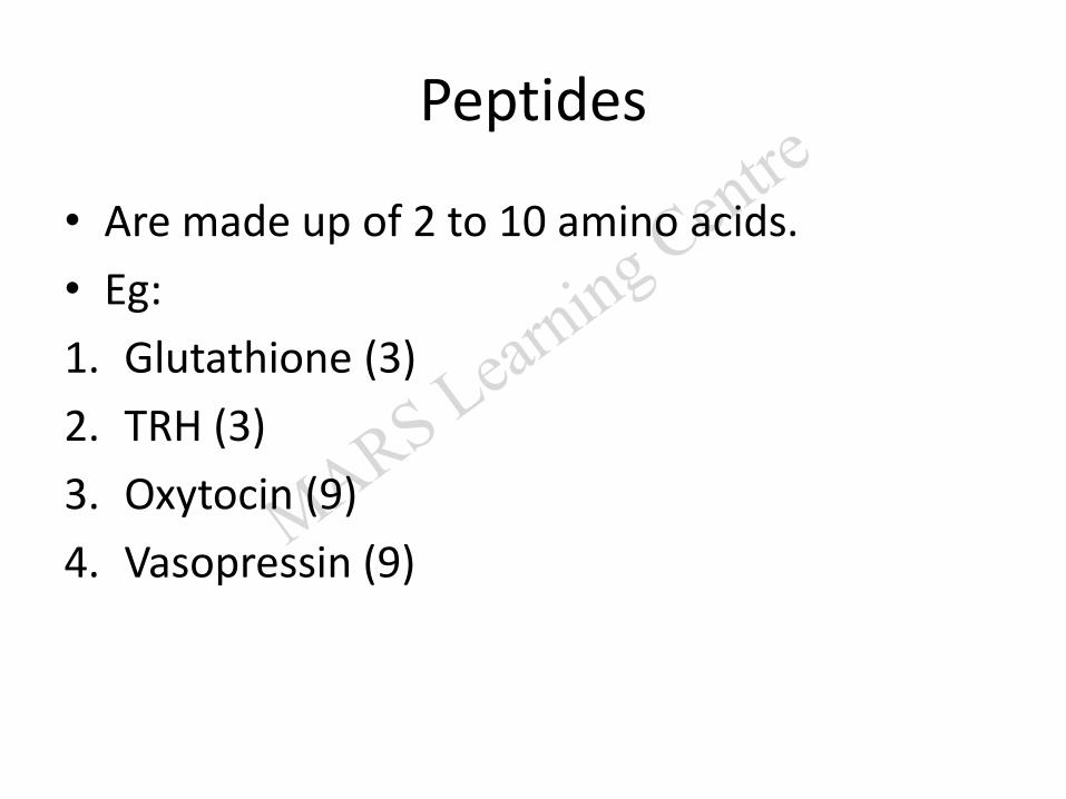
Peptides
• Are made up of 2 to 10 amino acids.
• Eg:
1. Glutathione (3)
2. TRH (3)
3. Oxytocin (9)
4. Vasopressin (9)
Proteins
• Biological Polymers Are
- Formed by the Removal of Water and
- Broken Down by the Addition of Water
Structure of proteins
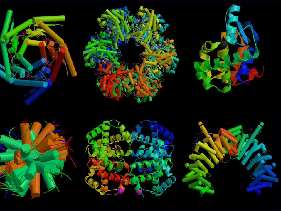
Protein Structure
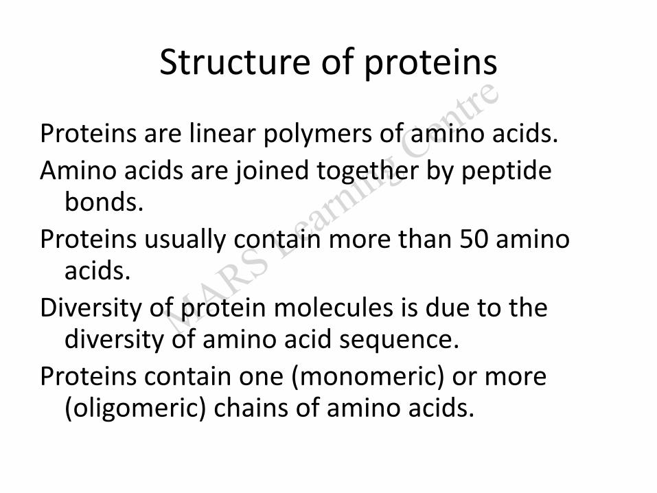
Structure of proteins
Proteins are linear polymers of amino acids.
Amino acids are joined together by peptide bonds.
Proteins usually contain more than 50 amino acids.
Diversity of protein molecules is due to the diversity of amino acid sequence.
Proteins contain one (monomeric) or more (oligomeric) chains of amino acids.
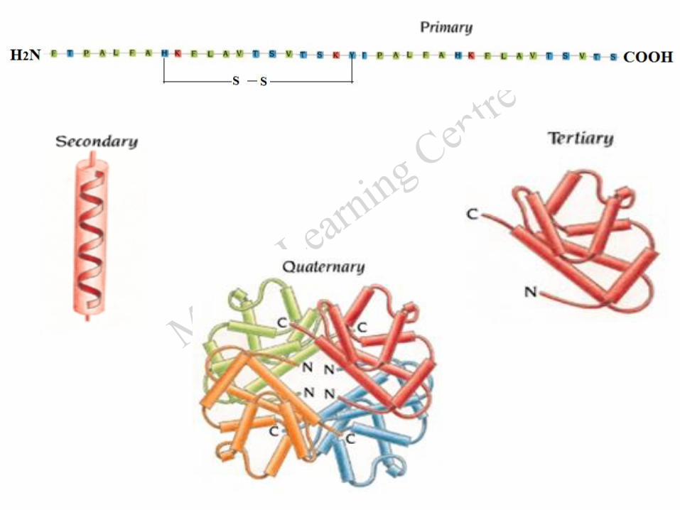
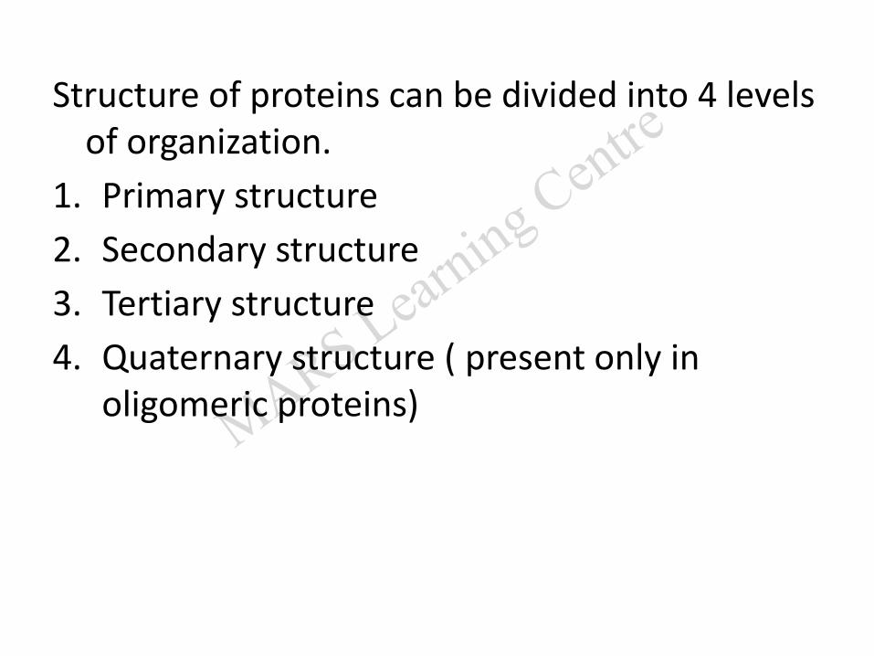
Structure of proteins can be divided into 4 levels of organization.
1. Primary structure
2. Secondary structure
3. Tertiary structure
4. Quaternary structure ( present only in oligomeric proteins)
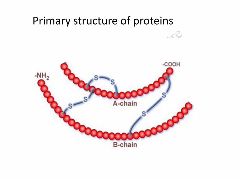
Primary structure of proteins
1.Primary structure
The primary structure denotes the number and sequence of amino acids present in the protein.
The higher levels of organizations are decided by the primary structure.
The primary structure is stabilized by
1. Peptide bond
2. Disulfide bond
1.Peptide bond
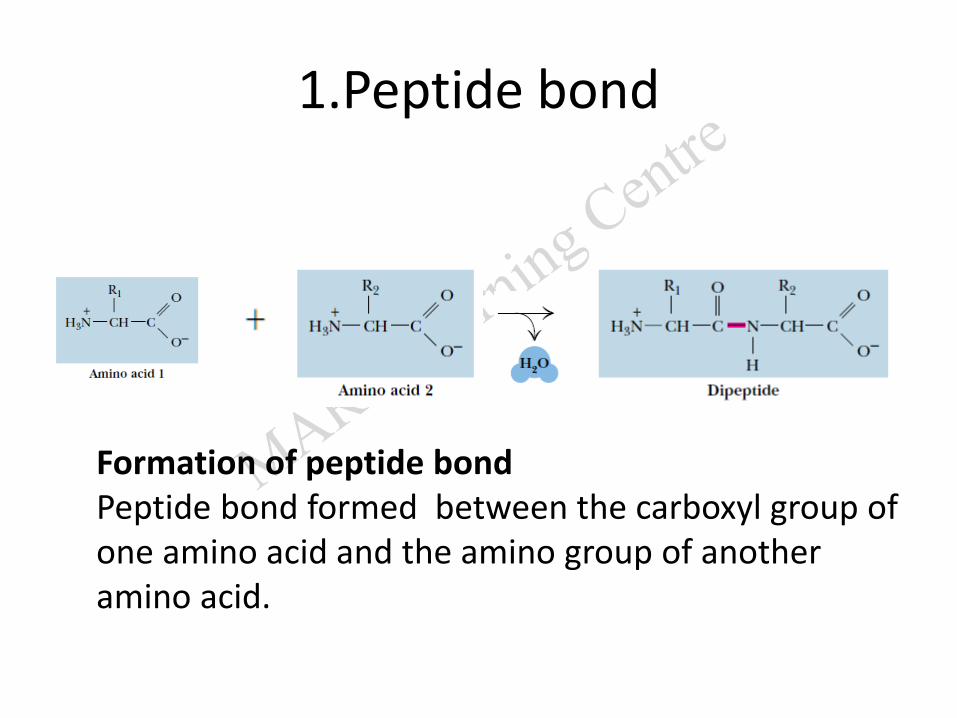
Formation of peptide bondPeptide bond formed between the carboxyl group of one amino acid and the amino group of another amino acid.
Characteristics of peptide bond
1. It is an amide bond.
2. It is coplanar and rigid.
3.It has partial double-bond character (bond length 1.32 Å).
C-N bond length is 1.49 Å
C=N bond length is 1.27 Å
4. Exists in Trans configuration.
5. It is a covalent bond, stabilizes primary structure.
Disulfide bond
It is formed between -SH groups of two cysteine residues.
It stabilizes primary structure of proteins.
It is a covalent bond.
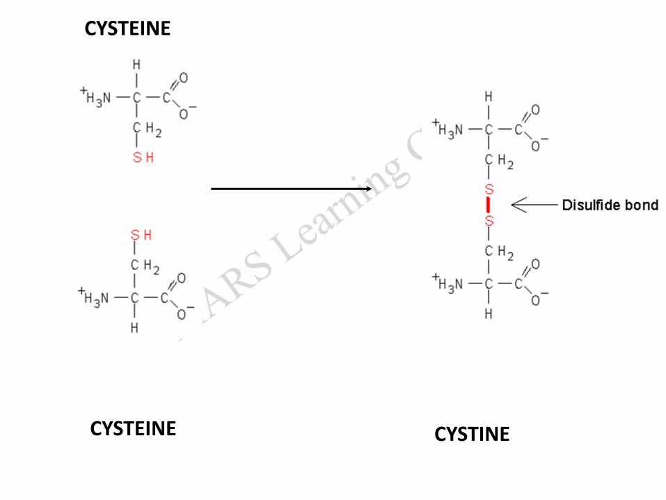
CYSTEINE
CYSTEINE CYSTINE
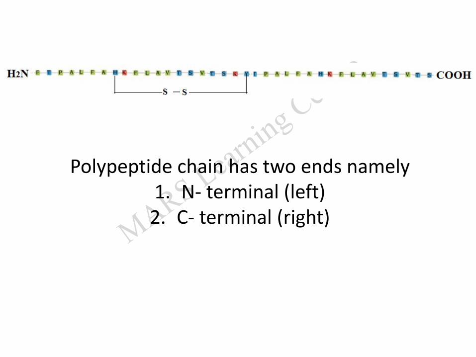
Polypeptide chain has two ends namely1. N- terminal (left)2. C- terminal (right)
Primary structure of insulin(by Frederick Sanger in 1953)
Insulin is synthesized by beta cells of islets of pancreas as a prohormone proinsulin.
Proinsulin is a single polypeptide with 86 amino acids.
Biologically active insulin is formed by the removal of the central portion containing 35 amino acids called C –peptide.
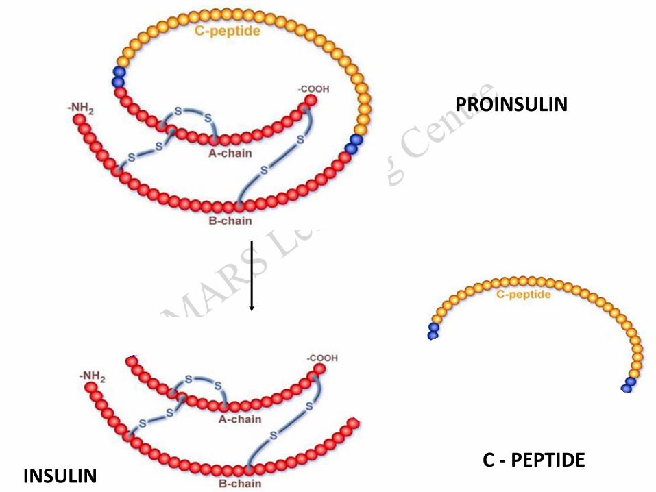
PROINSULIN
INSULINC - PEPTIDE
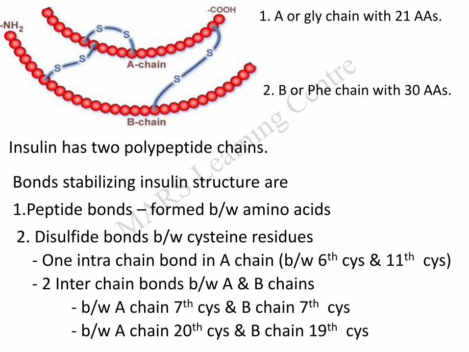
Insulin has two polypeptide chains.
1. A or gly chain with 21 AAs.
2. B or Phe chain with 30 AAs.
Bonds stabilizing insulin structure are
1.Peptide bonds – formed b/w amino acids
2. Disulfide bonds b/w cysteine residues
- One intra chain bond in A chain (b/w 6th cys & 11th cys)
- 2 Inter chain bonds b/w A & B chains
- b/w A chain 7th cys & B chain 7th cys
- b/w A chain 20th cys & B chain 19th cys

2.Secondary structure of proteins
Secondary structure denotes
- the configurational relationship between amino acid residues
- which are close to each other (about 3 to 4 amino acids apart)
- in the linear polypeptide chain.
It is stabilized by H bonds formed between peptide bonds.
2.Secondary structure of proteins
• Two types
(a) alpha helix
(b) beta pleated sheet.
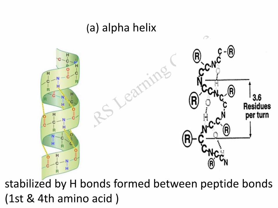
(a) alpha helix
stabilized by H bonds formed between peptide bonds (1st & 4th amino acid )
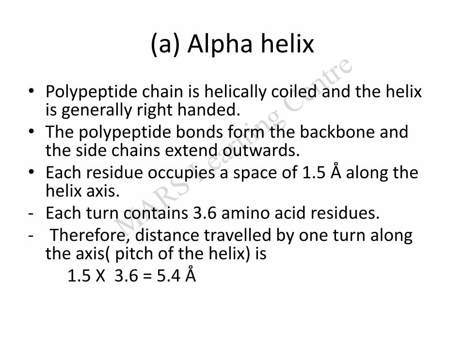
(a) Alpha helix
• Polypeptide chain is helically coiled and the helix is generally right handed.
• The polypeptide bonds form the backbone and the side chains extend outwards.
• Each residue occupies a space of 1.5 Å along the helix axis.
- Each turn contains 3.6 amino acid residues.- Therefore, distance travelled by one turn along
the axis( pitch of the helix) is 1.5 X 3.6 = 5.4 Å
The alpha helix is stabilized by hydrogen bonds formed between the NH and CO groups of peptide bond.
Alpha helix is destabilized by
• Proline
• It inserts a kink (bend) in the chain.
• Large numbers of charged amino acids (for example, glutamate,aspartate, histidine, lysine, or arginine).
• Large number of amino acids with bulky side chains (tryptophan, valine or isoleucine).
• Example:• Keratin - entirely helical• myosin, • tropomyosin,• Fibrin
• Hemoglobin,
• Myoglobin,
• Ferritin.
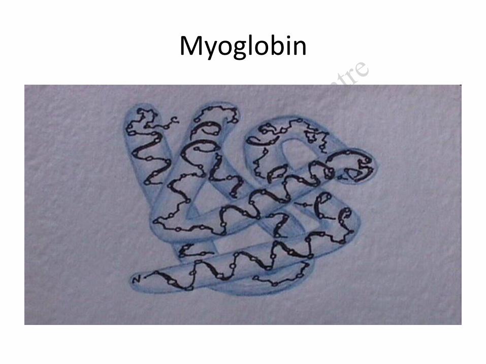
Myoglobin
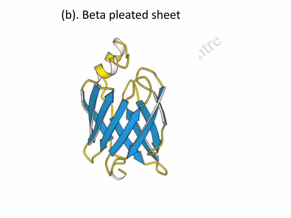
(b). Beta pleated sheet
(b) Beta pleated sheet.
• Polypeptide chain is arranged like a sheet.
• Formed by linking of two or more segments of polypeptide chains.
• The distance between adjacent amino acids is approximately 3.5 Å.
• Stabilized by hydrogen bonds.
• The structure is destabilized by the presence of proline.
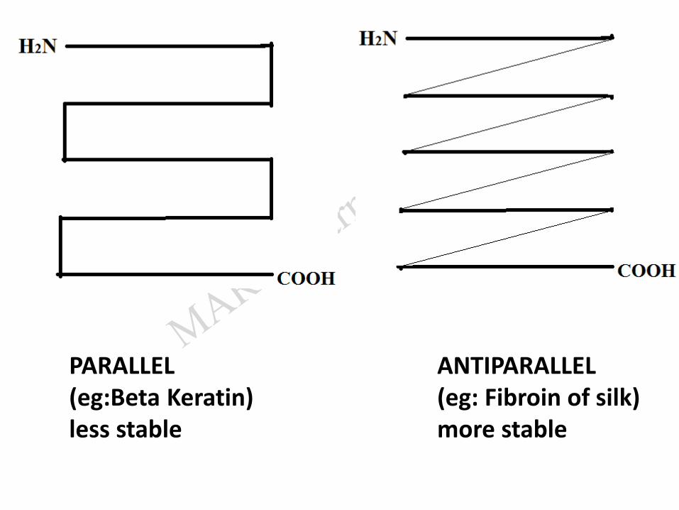
PARALLEL(eg:Beta Keratin)less stable
ANTIPARALLEL(eg: Fibroin of silk)more stable
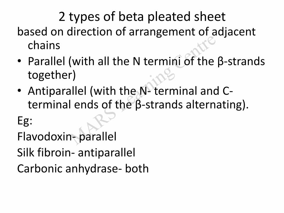
2 types of beta pleated sheetbased on direction of arrangement of adjacent
chains
• Parallel (with all the N termini of the β-strands together)
• Antiparallel (with the N- terminal and C-terminal ends of the β-strands alternating).
Eg:
Flavodoxin- parallel
Silk fibroin- antiparallel
Carbonic anhydrase- both
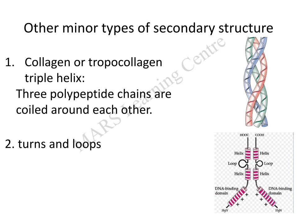
Other minor types of secondary structure
1. Collagen or tropocollagentriple helix:
Three polypeptide chains are coiled around each other.
2. turns and loops
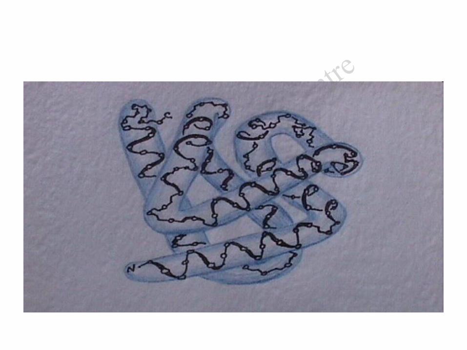
3. Tertiary structure of proteins
It denotes steric relationship of amino acids
- which are far apart from each other
- in the linear polypeptide chain.
• The polypeptide chain therefore folds so that
- its hydrophobic side chains are buried and
- its polar, charged chains are on the surface.
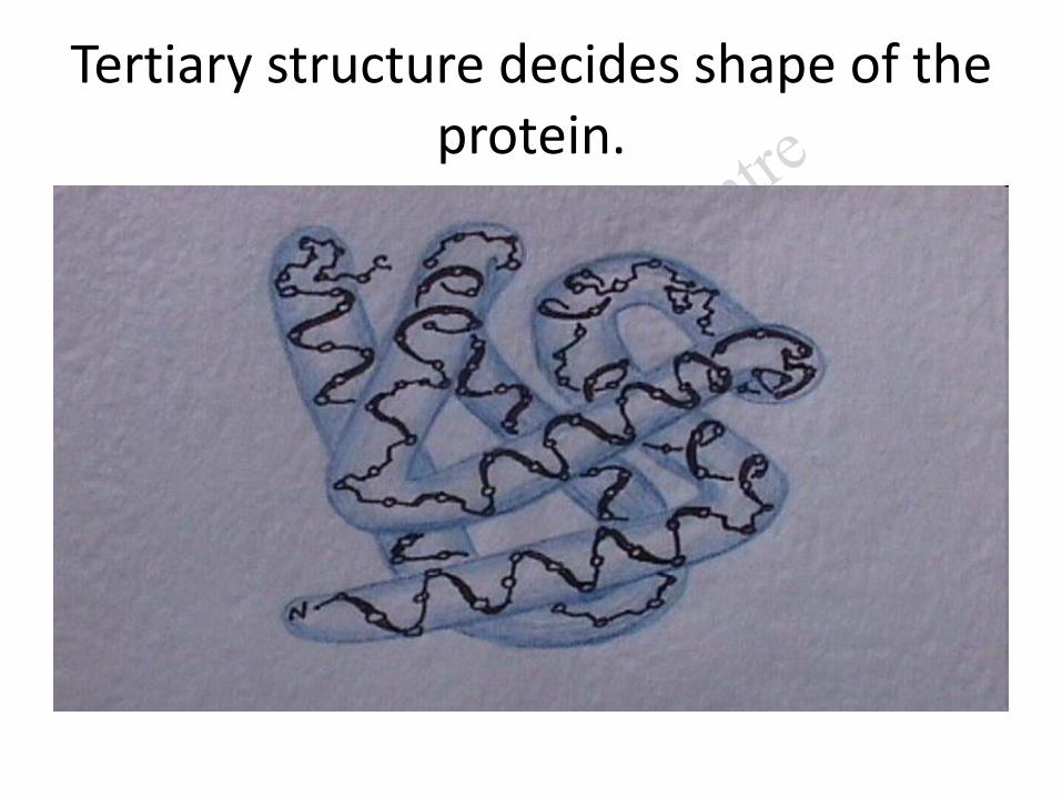
Tertiary structure decides shape of the protein.
Domains
• Polypeptide chain folds into two or more compact regions called domains.
• Range from about 30 to 400 amino acid residues.
• Domain is a functional unit of a protein
Intra-chain bonds
It is stabilized by interactions between "sidechains" including:
1.hydrogen bonds,
2.ionic interactions (electrostatic),
3. hydrophobic interactions, and
4.Van der waal’s forces.
All these are intra-chain bonds
Quaternary Structure
It is seen in proteins containing more than one polypeptide chain.
• Quaternary structure denotes the spatial arrangement of individual polypeptide chains with each other.
• Each polypeptide chain in an oligomeric protein is called a subunit.
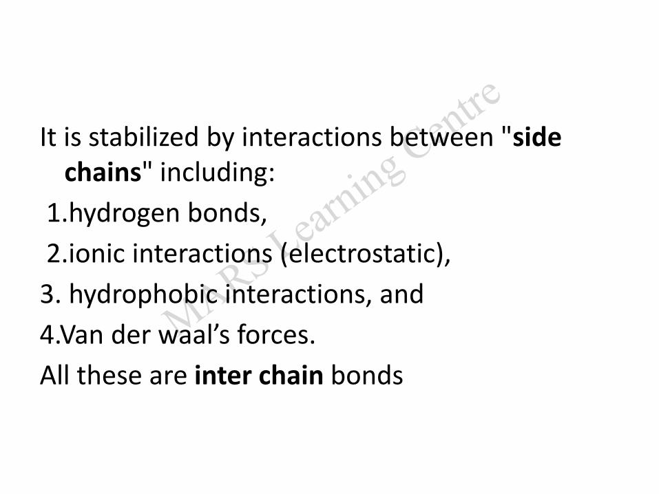
It is stabilized by interactions between "sidechains" including:
1.hydrogen bonds,
2.ionic interactions (electrostatic),
3. hydrophobic interactions, and
4.Van der waal’s forces.
All these are inter chain bonds
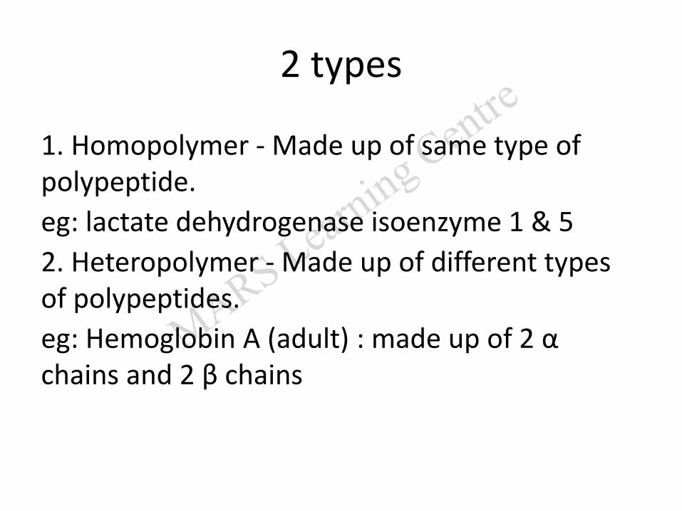
2 types
1. Homopolymer - Made up of same type of polypeptide.
eg: lactate dehydrogenase isoenzyme 1 & 5
2. Heteropolymer - Made up of different types of polypeptides.
eg: Hemoglobin A (adult) : made up of 2 αchains and 2 β chains

Denaturation
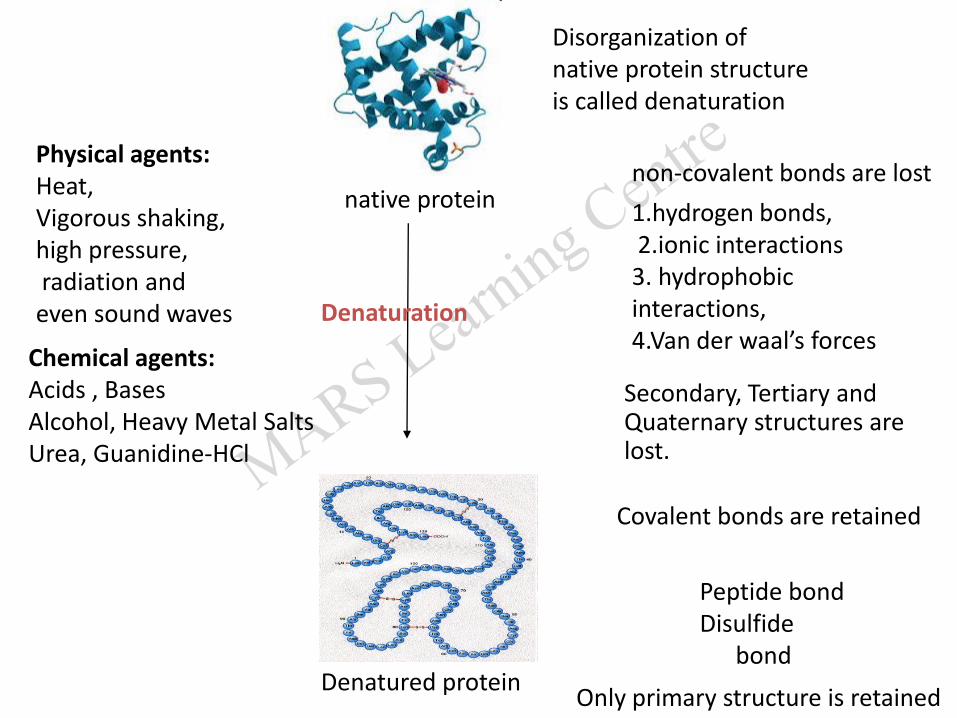
Disorganization of native protein structure is called denaturation.
native protein
Secondary, Tertiary and Quaternary structures are lost.
Denatured protein
non-covalent bonds are lost
Covalent bonds are retained
Only primary structure is retained
Chemical agents:Acids , BasesAlcohol, Heavy Metal Salts Urea, Guanidine-HCl
Physical agents:Heat,Vigorous shaking,high pressure,radiation and even sound waves
1.hydrogen bonds, 2.ionic interactions3. hydrophobic interactions, 4.Van der waal’s forces
Peptide bondDisulfide
bond
Denaturation
Disorganization of native protein structure is called denaturation
Isoelectric pH(PI) of proteins
• The isoelectric point (PI) is the pH at which a protein is electrically neutral
• that is, the sum of the positive charges equals the sum of the negative charges.
• Example: casein 4.6, Albumin 4.7, insulin 5.4.
• At this point, the net charge is zero.
• At isoelectric the pH protein
– is least soluble in water (precipitability is max)
– does not migrate in electric field

Questions
57
1.An aminoacid under certain conditions
have both positive and negative charges
simultaneously in the same molecule.
Such a form of aminoacid is called
a. Acidic form
b. Basic form
c. Aromatic form
d. Zwitter ionic form
In the primary structure of protein
(a) Left end represents → 1st amino acid (C-terminal amino acid)
(b) Right end represents → Last amino acid (N terminal amino acid)
(c) Left end represents → 1st amino acid (N-terminal amino acid)
(d) Right end represents → 1st amino acid (C-terminal amino acid)
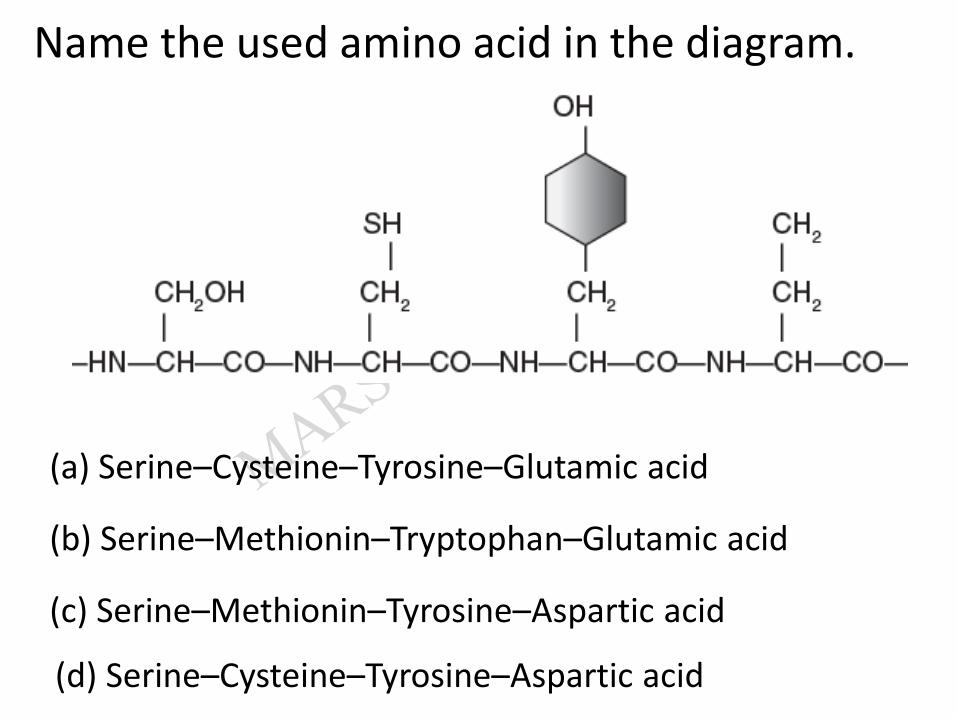
Name the used amino acid in the diagram.
(a) Serine–Cysteine–Tyrosine–Glutamic acid
(b) Serine–Methionin–Tryptophan–Glutamic acid
(c) Serine–Methionin–Tyrosine–Aspartic acid
(d) Serine–Cysteine–Tyrosine–Aspartic acid
Which of the following is correct about secondary structure?
(a) Helix is a primary structure.
(b) In proteins left handed helices are observed.
(c) In proteins right handed helices are observed.
(d) None of these

Which structure is absolutely necessary for the many biological activities of proteins?
(a) 1°
(b) 3°
(c) 2°
(d) 4°
When an assembly of more than one polypeptide occurs then it is known as _____ structure of protein.
(a) 1°
(b) 2°
(c) 3°
(d) 4°
Which of the following is correct about human Haemoglobin (Hb)?
(a) Made up to 2-a and 2-b subunits
(b) Present in RBC
(c) Use to carry O2 and CO2
(d) All of these
Select the incorrect statement from the following:
(a) Most of the enzymes are protein.
(b) ‘Hb’ is an example of quaternary structure of protein.
(c) In the primary structure of protein, the left hand is N-terminal and the right hand is C-terminal.
(d) In protein or polypeptide, the amino acids are linked by glycosidic bond.
Which of the following is the least likely to be involved in stabilizing the three-dimensional
folding of most proteins? [NEET - II, 2016]
(a) Electrostatic interaction
(b) Hydrophobic interaction
(c) Ester bonds
(d) Hydrogen bonds
The primary structure of a protein molecule has two ends.
(a) Two ends
(b) One end
(c) Three ends
(d) No ends
Top Related