


γλώσσες
Σελίδες
Νομικός

1
Mucosal candidiasis elicits activation of NF-κB, proinflammatory gene expression and localized neutrophilia in a transparent vertebrate host Remi L. Gratacap1, John F. Rawls3,4, Robert T. Wheeler1,2,* 1Department of Molecular & Biomedical Sciences 2Graduate School of Biomedical Sciences and Engineering University of Maine, Orono, ME 04469, USA 3Department of Cell Biology and Physiology and 4Department of Microbiology and Immunology University of North Carolina at Chapel Hill, Chapel Hill, NC 27599, USA *Correspondence to: Robert T. Wheeler, Department of Molecular & Biomedical Sciences, University of Maine, Orono, ME 04469 207-581-2890 [email protected] Running title: Zebrafish model of mucosal candidiasis Keywords Zebrafish; Candida albicans; epithelial cell; neutrophil; saa; tnf; swimbladder; NF-κB
© 2013. Published by The Company of Biologists Ltd.This is an Open Access article distributed under the terms of the Creative Commons Attribution Non-Commercial Share Alike License(http://creativecommons.org/licenses/by-nc-sa/3.0), which permits unrestricted non-commercial use, distribution and reproduction inany medium provided that the original work is properly cited and all further distributions of the work or adaptation are subject to thesame Creative Commons License terms.
Dise
ase
Mod
els &
Mec
hani
sms
D
MM
Acce
pted
man
uscr
ipt
http://dmm.biologists.org/lookup/doi/10.1242/dmm.012039Access the most recent version at DMM Advance Online Articles. Posted 29 May 2013 as doi: 10.1242/dmm.012039
http://dmm.biologists.org/lookup/doi/10.1242/dmm.012039Access the most recent version at First posted online on 29 May 2013 as 10.1242/dmm.012039
2
Summary The epithelium performs a balancing act at the interface between an animal and its environment to enable both pathogen killing and tolerance of commensal microorganisms. Candida albicans is a clinically important human commensal that colonizes all human mucosal surfaces, yet is largely prevented from causing mucosal infections in immune competent individuals. Despite the importance of understanding host-pathogen interactions at the epithelium, no immunocompetent vertebrate model has been used to visualize these dynamics non-invasively. Here we demonstrate important similarities between swimbladder candidiasis in the transparent zebrafish and mucosal infection at the mammalian epithelium. Specifically, in the zebrafish model we show dimorphic fungal growth, both localized and tissue-wide epithelial NF-κB activation, induction of NF-κB -dependent proinflammatory genes, and strong neutrophilia within the swimbladder. Consistent with density-dependence models of host response based primarily on tissue culture experiments, we show that only high-level infection provokes widespread activation of NF-κB in epithelial cells and induction of proinflammatory genes. Similar to what has been found using in vitro mammalian models, we find that epithelial NF-κB activation can occur at a distance from the immediate site of contact with epithelial cells. Taking advantage of the ability to non-invasively image infection and host signaling at high resolution, we also report that epithelial NF-κB activation is diminished when phagocytes control the infection. This is the first system to model host response to mucosal infection in the juvenile zebrafish, and offers unique opportunities to investigate the tripartite interactions of C. albicans, epithelium and immune cells in an intact host.
Dise
ase
Mod
els &
Mec
hani
sms
D
MM
Acce
pted
man
uscr
ipt
3
INTRODUCTION Candida albicans is a commensal fungus of human mucosa commonly found in the
oropharynx-digestive system as well as the female reproductive tract (Reef et al., 1998; Rindum et al., 1994; Scully et al., 1994; Soll et al., 1991). This opportunistic pathogen can produce both non-lethal localized mucosal and life-threatening systemic infections. Major advances in our molecular understanding of mucosal candidiasis have been achieved through combining in vitro and in vivo experiments (Naglik et al., 2008; Reef et al., 1998; Rindum et al., 1994; Scully et al., 1994; Soll et al., 1991), yet the spatio-temporal dynamics of this infection have proven difficult to dissect with existing experimental platforms.
Epithelial cells play an important role in signaling professional immune cells to mount an immune response to C. albicans. Although the receptors that activate epithelial cells are not all known (Cheng et al., 2012; Weindl et al., 2010), their engagement activates the NF-κB pathway in addition to other transcription factors (Moyes et al., 2010). This leads to induction of proinflammatory genes that recruit and activate professional immune cells to the site of infection (Moyes and Naglik, 2011). Neutrophils play an active role in enhancing epithelial immune response (Weindl et al., 2007), but are also associated with increased disease symptoms and immunopathology (Fidel et al., 2004; Lionakis et al., 2012). The in vivo mechanisms of neutrophil recruitment in mucosal candidiasis remain unclear, and may include chemokines, defensins, and/or acute phase proteins such as serum amyloid A, all of which are highly upregulated in epithelial cells after infection with C. albicans (Conti et al., 2009; Tomalka et al., 2011).
The larval zebrafish (Danio rerio) is a practical and versatile model that offers unique experimental advantages over other systems, including the transparency of larvae and the lack of adaptive immune responses for the first few weeks after hatching (Tobin et al., 2012). Zebrafish models have been developed to study several human pathogens (Meijer and Spaink, 2011; Tobin et al., 2010), including systemic candidiasis (Brothers et al., 2011; Chao et al., 2010), but immune responses in a mucosal infection model have yet to be characterized in this transparent vertebrate host. Intriguingly, the only case descriptions of fish infections with C. albicans are mucosal infections of the swimbladder (Galuppi et al., 2001; Hatai, 1992).
The swimbladder shares functional, anatomical, ontological and transcriptional similarities to the lung. It is used for buoyancy, but maintains an air-mucosal interface that performs gas exchange to the circulatory system in some species (Lapennas and Schmidt-Nielsen, 1977). It develops from the foregut and remains connected to it through the pneumatic duct (Field et al., 2003), which is a potential infection route for ingested bacterial and fungal pathogens (Ross et al., 1975). Anatomically, the swimbladder epithelium is most similar to the lung epithelium, with a single layer of squamous epithelial cells covering the mesenchyme and a mesothelial layer (Robertson et al., 2007; Winata et al., 2009). It has a transcriptional signature very similar to the mammalian lung (Winata et al., 2009; Zheng et al., 2011) and has been shown to secrete both surfactant proteins (Sullivan et al., 1998) and β-defensin like molecules (Oehlers et al., 2011a). This suggests that the swimbladder is a potentially useful organ for modeling other mucosal infections such as lung infections, in addition to being a natural site of infection for C. albicans in fish.
Here we use the transparent zebrafish swimbladder to model mucosal candidiasis. We show that this infection reproduces important aspects of fungal-epithelial interaction previously characterized in vitro, in murine models, and in human disease. We find that high-level infection induces strong activation of NF-κB, transcriptional upregulation of NF-κB-dependent
Dise
ase
Mod
els &
Mec
hani
sms
D
MM
Acce
pted
man
uscr
ipt
4
proinflammatory gene expression, and robust neutrophilia. The strong neutrophil presence and associated engulfment of C. albicans at low-level infection may limit direct contact of yeast with epithelial cells, diminishing both NF-κB activity in these epithelial cells and expression of pro-inflammatory cytokines. The ability to follow both the host and pathogen non-invasively provides a powerful alternative model for understanding the molecular mechanisms underlying virulence and immunity in mucosal candidiasis.
Dise
ase
Mod
els &
Mec
hani
sms
D
MM
Acce
pted
man
uscr
ipt
5
RESULTS Candida albicans infects the zebrafish swimbladder and grows dimorphically
Mucosal candidiasis is the most common form of infection by C. albicans (Moran et al., 2012), but available in vivo animal models have limitations for imaging immune cell and pathogen interactions intravitally. We sought to exploit the transparency of the juvenile zebrafish to investigate the interactions of the epithelium, innate immune cells and pathogen during mucosal candidiasis.
We developed a non-invasive model of mucosal candidiasis of the swimbladder with infection through immersion. It is based on the premise that infection occurs naturally upon inflation of the swimbladder and the completion of the intestinal tract, around 4 dpf (Kimmel et al., 1995; Pack et al., 1996). We find that immersion of zebrafish larvae, beginning at 3 days post fertilization (dpf), with 4 x 10⁷ cfu/mL of C. albicans gives the most consistent and highest infection rate.
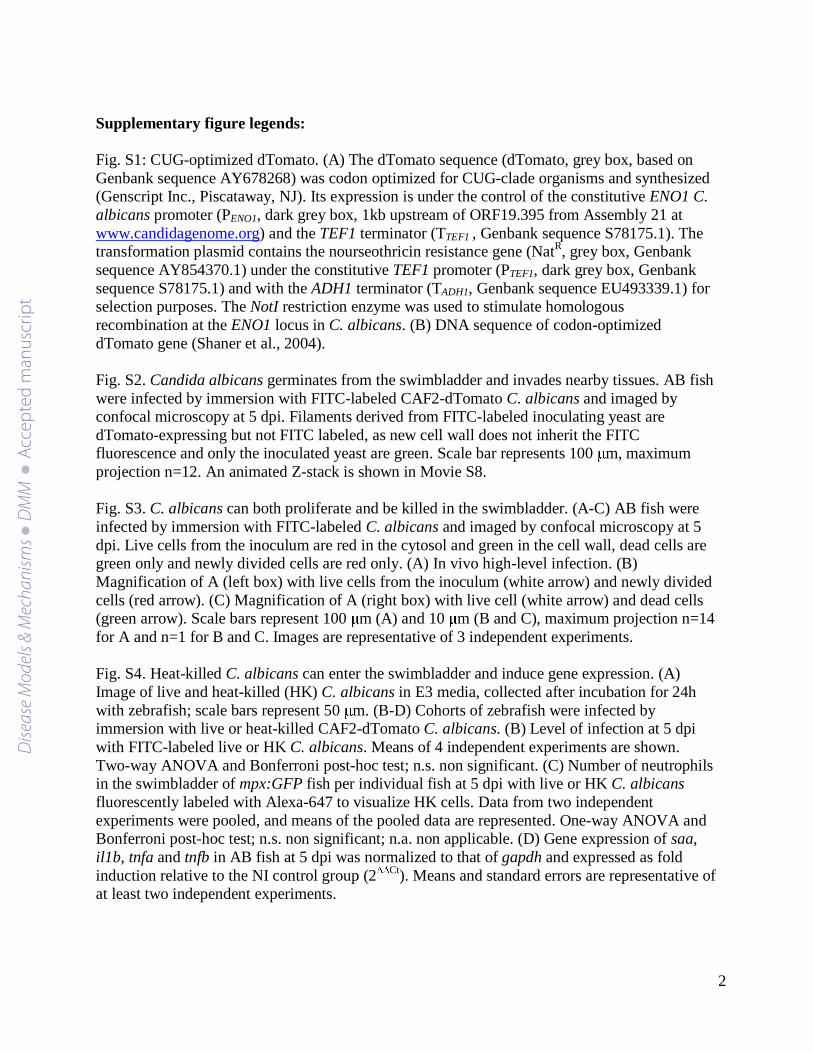
This immersion model results in highly reproducible infection of the swimbladder. At 2 days post-immersion (dpi), the intestinal lumen is entirely filled with fluorescent yeast cells (Fig. 1A). Yeasts are occasionally seen within the pneumatic duct (data not shown), which remains open throughout the zebrafish’s life (Goolish and Okutake, 1999), suggesting they enter the swimbladder through this route. By 5 dpi, approximately 27 % of fish have infected swimbladders (Fig. 1B), 18 % with fewer than 20 yeast cells (low-level infection) and 9 % with over 20 (high-level infection). At low-level infection, the majority of fungi remain as yeast (Fig. 1C) but hyphae occasionally germinate even at low burden. Germination within the swimbladder is more common in high-level infection, where the swimbladder can be observed filled with yeast and hyphae (Fig. 1D,E,F and Movie S1). Although in >99% of cases fungal cells are only observed luminal to the apical surface of the epithelium, in rare cases hyphae can pierce through the swimbladder and reach nearby tissue (Fig. S2). Yeasts are infrequently observed in organs outside the swimbladder and intestinal tract. C. albicans infection of the swimbladder is thus largely limited to the epithelium.
Because active infection is reflected in proliferation of fungi at the infection site, we performed infections with dTomato-expressing fungi that were also labeled with fluorescein isothiocyanate (FITC). This permits the distinction of the inoculum (dTomato-expressing and FITC-positive), new cells that grew within the fish (dTomato-expressing but FITC-negative), and cells from the inoculum that were killed (dTomato negative but FITC positive). These experiments show that both fungal cell division and killing of fungi occur during infection, suggesting that it is a dynamic process (Fig S3).
To assess whether only live yeasts can reach the swimbladder, we performed mock infections with heat-killed (HK) fungi. We find that HK yeast cells can enter the swimbladder at a similar level as live yeasts (Fig. S4); however, high-level exposure of the swimbladder to HK fungi is likely a result of the increased clumping of these cells compared to live C. albicans (Fig. S4A).
Although there are few gross phenotypic signs of infection through 5 dpi, fewer fish with high-level infection have inflated swimbladders (Fig. 1G), suggesting that infection can disturb the normal inflation and/or maintenance of the air bubble. Although we followed survival of larvae beyond 5 dpi (8 dpf), the impact of infection on mortality could not be reliably quantified due to variability in mortality during late larval stages.
Dise
ase
Mod
els &
Mec
hani
sms
D
MM
Acce
pted
man
uscr
ipt

6
In this new model of mucosal candidiasis, we show that when C. albicans yeasts enter the swimbladder they can cause both low and high-level infections, germinating and forming hyphae. The limited germination and proliferation of C. albicans on the epithelial surface is similar to colonization of immunocompetent mammals. The reproducible nature of these infections of the swimbladder in the transparent zebrafish enables non-invasive imaging of host-pathogen dynamics. NF-κB activity is enhanced in vivo during mucosal infection
C. albicans is actively recognized by epithelial cells, which results in activation of several signaling pathways both in vitro and ex vivo (Moyes et al., 2010). The NF-κB transcriptional pathway is an essential component in immune response to infection (Baeuerle and Henkel, 1994). Here, we exploited the swimbladder candidiasis model to study NF-κB activity in vivo using the Tg(NFκB:EGFP) transgenic fish line, in which NF-κB activity drives expression of EGFP (Kanther et al., 2011).
We found that infection of the swimbladder by C. albicans leads to NF-κB activation in epithelial cells. In high-level infection the epithelium of the swimbladder expresses strong NF-κB-driven EGFP fluorescence (Fig. 2A,B). The fluorescence is widespread throughout the epithelial layer of the swimbladder, but restricted to this layer (Fig. 2A,B,C). The magnitude of activation is more obvious ex vivo, where it is clear that the epithelial cell layer in infected swimbladders fluoresce more strongly than those in uninfected swimbladders (Fig. 2D,E). In swimbladders with high-level infection, phagocytes are often seen within the lumen, particularly in swimbladders where the air bubble is not present. These phagocytes can harbor engulfed yeasts or pseudohyphae, and often express low-level EGFP fluorescence (Fig. 2C). Although mammalian phagocytes have been shown to activate NF-kB in response to pathogenic stimuli, expression of EGFP in this reporter line has not yet been explicitly defined (Kanther et al., 2011). Therefore, the lack of robust reporter gene expression may be due to limitations of the reporter rather than a lack of NF-kB activation. In uninfected swimbladders, there is weak fluorescence in the mesothelium and scattered EGFP-positive cells are present in the gut, as previously reported (Kanther et al., 2011). These results show for the first time that epithelial cells respond to mucosal C. albicans infection through NF-κB activation in vivo. Moreover, they demonstrate that the vast majority of the swimbladder's epithelial cells are activated during high-level infection.
In low-level infection, we find that NF-κB activation correlates with direct interaction of yeasts with epithelial cells. In infected fish with a low number of yeasts in the swimbladder, no overall differences in NF-κB activity are seen, as compared with uninfected fish (Fig. 3A,B). In most cases, the yeast cells are engulfed by phagocytic cells and, in contrast with high-level infection, epithelial cells do not express increased levels of EGFP (Fig. 3C,D and Movies S2 and S3). However, if the yeast cells are not contained within phagocytes, the epithelial cell in contact with the pathogen and the neighboring cells have increased fluorescence (Fig. 3F and Movies S4 and S5). When infection foci are categorized into situations in which extracellular fungi are present (Fig. 3G, left) or absent (Fig. 3G, right), we see a much greater likelihood for detectable epithelial EGFP expression when extracellular fungi are present (p<0.05). The activation of NF-κB in neighboring epithelial cells is also observed more often in cases where the swimbladder is only partially inflated and fluid is present in between the air bubble and the epithelium (Fig. 3E). These data suggest that at low pathogen density, an effective phagocyte response limits the activation of NF-κB in epithelial cells. However, at high-level infection, C. albicans is poorly
Dise
ase
Mod
els &
Mec
hani
sms
D
MM
Acce
pted
man
uscr
ipt
7
contained and elicits a widespread activation of NF-κB in the epithelial layer lining the swimbladder. serum amyloid A and tumor necrosis factor a are induced in high-level infection A large number of immune related genes are induced by C. albicans infection at the epithelial level, including several cytokines, chemokines and antimicrobial peptides (Conti et al., 2009; Tomalka et al., 2011). Several of these inflammatory molecules are under NF-κB control (Moyes et al., 2010). To measure downstream effects of increased NF-κB activity due to mucosal infection in the zebrafish, we measured expression of three inflammatory genes shown to have some dependence on NF-κB in zebrafish and/or in mouse (Baeuerle and Henkel, 1994; Kanther et al., 2011): serum amyloid A (saa), interleukin-1β (il1b) and tumor necrosis factor, isoforms a and b (tnfa and tnfb).
Quantitative PCR with whole fish revealed that only expression of saa and tnfa are changed upon infection (Fig. 4). Comparisons with uninfected fish show that fish with high-level infection have increased expression of both saa and tnfa. This is not the case for fish with low-level infection, which have unchanged expression of all cytokines measured. Surprisingly, neither il1b nor tnfb expression is induced at any level of infection. However, the use of total RNA from whole animals could mask local tissue-specific expression differences. Notably, exposure of the swimbladder to heat killed yeast cells induces expression of the same genes as live C. albicans (Fig. S4D).
In sum, the gene expression data shows that some NF-κB-driven inflammatory genes are upregulated upon C. albicans infection in the zebrafish model presented here, similar to mucosal candidiasis in mammals (Dongari-Bagtzoglou and Fidel, 2005). Infection induces localized neutrophilia in proportion to fungal burden
Rapid recruitment of neutrophils to the site of infection is one of the hallmarks of the acute inflammatory response (Sohnle et al., 1976). NF-κB is an important activator of the mucosal immune response, which directs recruitment of neutrophils to the epithelium (Everhart et al., 2006; Mizgerd, 2002; Pantano et al., 2008). The recruitment and activation of these myeloid cells is mediated by chemokines and cytokines, among which TNF and SAA are important in response to C. albicans infection (Badolato et al., 2000; Netea et al., 1999; Weindl et al., 2007). To investigate the neutrophil immune response in swimbladder infection we utilized mpx:GFP transgenic zebrafish (Renshaw et al., 2006), with GFP-expressing neutrophils. We observed strong neutrophilia in the swimbladder in all levels of infection. More neutrophils are present in the swimbladder both in high- and low-level infection (Fig. 5A,B), as compared with the uninfected swimbladder (Fig. 5C,D). In addition, morphometric analysis of confocal Z-stacks reveals that swimbladders with high-level infection have significantly more neutrophils than swimbladders with low-level infection and that both these are significantly different from non-infected groups (Fig. 5E). In low-level infection, neutrophils are found at infection foci and can be seen in direct contact with yeast cells (Fig. 5B and Movie S6). In high-level infection, neutrophils are spread throughout the tissue surrounding the swimbladder and they can also be in direct contact with fungi (Fig. 5A and Movie S7). Neutrophils are present at similar levels when the swimbladder is exposed to HK C. albicans (Fig. S4C). These data show that C. albicans swimbladder infection is associated with an increased neutrophil response at the site of infection, even more so at high-level infection.
Dise
ase
Mod
els &
Mec
hani
sms
D
MM
Acce
pted
man
uscr
ipt
8
DISCUSSION
We describe here a powerful new zebrafish model for the study of mucosal candidiasis. This extends previous models of invertebrate and rodent mucosal fungal infection (Naglik et al., 2008) into a transparent vertebrate reliant on the innate immune system. This zebrafish model shares key aspects of mucosal infection of the human and mouse, including dimorphic fungal growth, epithelial NF-κB activation, induction of NF-κB-dependent proinflammatory genes, and localized neutrophilia. Exploiting unique advantages of the system, we non-invasively document a novel relationship between fungal burden and epithelial NF-κB activity as a function of phagocyte activity. We provide the first in vivo support for a proposed density-dependent model of host response based on in vitro results (Moyes et al., 2010). The amenability of the zebrafish platform for chemical and genetic screening provides a unique opportunity for the discovery of mechanisms underlying host response and fungal virulence.
This is the first reported zebrafish model of innate immune responses to epithelial infection with a human pathogen, and the first description of experimental swimbladder infection in fish larvae. It builds on previous work that has used the zebrafish to elucidate bacterial-intestinal interactions and host mechanisms responsible for gut development and immunity to intestinal pathogens (Flores et al., 2010; Hall et al., 2007; Kanther et al., 2011; O'Toole et al., 2004; Oehlers et al., 2011b; Pressley et al., 2005; Rendueles et al., 2012). In contrast to these studies, we model mucosal immunity to fungal infection in the swimbladder and focus on the cellular and molecular components of a successful and protective innate immune response to a human pathogen. The development of a mucosal model of candidiasis in a transparent vertebrate is especially important because it enables the study of intravital interactions of the pathogen with epithelial and innate immune cells. Striking similarities between the swimbladder and the mammalian lung reinforce the potential for this model to shed light on immune mechanisms involved in pulmonary fungal infection (Bals and Hiemstra, 2004; Cardoso and Lü, 2006; Daniels and Skinner, 1994; Field et al., 2003; Perrin et al., 1999; Prem et al., 2000; Rock and Hogan, 2011; Winata et al., 2009; Zheng et al., 2011). Although not typically regarded as a lung pathogen, recent work suggests that C. albicans may cause and/or exacerbate pulmonary infections (Leclair and Hogan, 2010; Ogba et al., 2013).
We exploited the advantages of this new model of mucosal candidiasis to demonstrate localized NF-κB activation by C. albicans in vivo, finding that it can occur at all infection levels. This is consistent with in vitro evidence that NF-κB is activated in epithelial cells by both high and low levels of C. albicans infection (Moyes et al., 2010; Steubesand et al., 2009). It is also consistent with evidence that NF-κB plays a protective role in candidiasis (Li and Dongari-Bagtzoglou, 2009; Moyes et al., 2010; Pivarcsi et al., 2003; Steubesand et al., 2009). In high-level swimbladder infection, epithelial cells exhibit widespread and strong NF-κB activity, as seen in lung epithelium after prolonged LPS infusion (Everhart et al., 2006). This suggests that high-level infections represent a state where the epithelial cells are being continuously stimulated. In contrast, NF-κB signaling during low-level infection is apparently only activated by close-range signals when C. albicans is not contained within phagocytes. In vitro C. albicans-epithelial infections suggest that NF-κB could be activated either through triggering of receptors by fungal products (Zhu et al., 2012; Zipfel et al., 2011) or through host-derived molecules activating neighboring bystander cells (Dongari-Bagtzoglou et al., 2004; Steubesand et al., 2009). This activation may also be accomplished by direct cell-cell activation through gap-junctions, as has recently been shown for bacterial infection (Kasper et al., 2010). Our finding
Dise
ase
Mod
els &
Mec
hani
sms
D
MM
Acce
pted
man
uscr
ipt
9
that only non-engulfed yeasts efficiently activate NF-κB suggests that fungal products may be required for mediating the activation of close-range bystander epithelial cells, but this does not rule out the participation of host molecules. The combination of C. albicans mutants and the versatile zebrafish toolbox offer unique access to further probe the mechanistic details of NF-κB activation during in vivo infection.
Using non-invasive phenotypic screening to correlate infection level with inflammation, we have identified conserved gene expression responses to mucosal candidiasis. The dependence of inflammatory gene expression on high-level infection is consistent with in vitro data that shows a density dependence of some inflammatory responses (Moyes et al., 2010; Steubesand et al., 2009). Low-level swimbladder infection elicits weak NF-κB activation and no significant overall activation of saa or tnf, whereas high-level infection strongly activates NF-κB and stimulates both saa and tnf transcription. Consistent with these results, colonization with commensal microbiota has been shown to upregulate saa in the intestine, swimbladder and liver in an NF-κB-dependent fashion (Kanther et al., 2011). The upregulation of saa is also seen in an immunocompromised mouse model of oral candidiasis, and has been suggested to play a role in Th17 activation (Ather et al., 2011; Conti et al., 2009; Ivanov et al., 2009; Migita et al., 2010). TNF is highly upregulated by C. albicans and mediates protection against mucosal candidiasis (Farah et al., 2006; Weindl et al., 2007), as well as maintenance of the epithelial barrier (Eyerich et al., 2011). However, recent work suggests TNF might play somewhat different roles in mammals and fish (Roca et al., 2008). Existing morpholino tools in the zebrafish system can be used to elucidate the precise function(s) of SAA and TNF in protection against mucosal candidiasis in vivo.
The strong recruitment and/or retention of neutrophils in the infected swimbladder is consistent with what is seen in mammalian mucosal candidiasis (Challacombe, 1994; Sohnle et al., 1976). The strongest recruitment is seen during high-level swimbladder infection, when both saa and tnf are highly expressed. Interestingly, TNF upregulates chemotaxis through multiple mechanisms (Amulic et al., 2012) and SAA is a potent chemoattractant that may mediate chemotaxis towards C. albicans through the formylated peptide receptor (Edens et al., 1999; Su et al., 1999). However, the finding that neutrophils are recruited to the swimbladder even during low-level infection, in which there is weak NF-κB activity and no overall upregulation of saa or tnf, suggests that multiple pathways are responsible for the recruitment and retention of neutrophils at the infection site. Neutrophils are important in protection against mucosal candidiasis in humans and in animal models, but can also exacerbate symptoms (Akova et al., 1994; Anaissie and Bodey, 1990; Fidel et al., 2004). Their protective role has been established both through neutrophil ablation (Farah et al., 2001) and by IL-17 pathway disturbance (Conti et al., 2009). However, how neutrophils collaborate with epithelial cells is still unclear. Recently, several studies have highlighted a complementary role of neutrophils in enhancement of the expression of receptors and antimicrobial defenses on epithelial cells in mucosal candidiasis (Schaller et al., 2004; Steubesand et al., 2009; Weindl et al., 2007; Weindl et al., 2011). The zebrafish provides a unique model to investigate neutrophil recruitment, using a combination of gene knock-down or chemical inhibitors and time-lapse imaging. The functional relevance of neutrophils in immune-epithelial cross-talk as well as immunopathology (Lionakis et al., 2012; Wheeler et al., 2008) is now readily testable using a recently described conditional ablation transgenic fish line (Gray et al., 2011).
Our findings that heat-killed yeast can elicit both increased pro-inflammatory gene expression and swimbladder neutrophilia suggest that at least some immune responses are due to
Dise
ase
Mod
els &
Mec
hani
sms
D
MM
Acce
pted
man
uscr
ipt
10
direct effects of fungal recognition. This is consistent with numerous in vitro studies that have shown that heat-killed C. albicans elicits strong immune responses from phagocytes (Jeremias Witkin 1991). This also suggests that some immune responses at the epithelium can result from direct recognition of fungi through pattern recognition receptors, independent of the ability of C. albicans to grow invasively. Work in reconstituted human epithelial models has shown that heat-killed hyphae can induce MKP1 expression (Moyes et al., 2010), although heat-killed yeast fail to induce MKP1 or elicit immune responses (Moyes et al., 2010; Schaller et al., 2002; Schaller et al., 2005). Identification of the drivers of both shared and divergent responses in the swimbladder and RHE models may shed light on conserved mechanisms of immune responses to mucosal candidiasis.
The development of the first model of fungal epithelial infection in the tractable larval zebrafish system opens up new possibilities in modeling other human mucosal pathogens. It also enables testing of a new set of hypotheses using real time imaging of both C. albicans pathogenesis and host immune responses during mucosal infection in an intact vertebrate host.
Dise
ase
Mod
els &
Mec
hani
sms
D
MM
Acce
pted
man
uscr
ipt
11
MATERIALS AND METHODS Zebrafish care and maintenance All zebrafish were kept in recirculating systems (Aquatic Habitats) at the University of Maine Zebrafish Facility, under a 14/10 light/dark cycle. Water temperature was kept at 28°C. All zebrafish care protocols and experiments were performed in accordance with NIH guidelines under Institutional Animal Care and Use Committee (IACUC) protocol A2009-11-01. Larvae were rinsed in 0.15% Perosan solution (v/v in E3 media) for 1 min after collection (Phennicie et al., 2010) and kept in a 28°C incubator at 80 fish per 50 mL in E3 media plus 0.00003% methylene blue for 24 hours and E3 media plus PTU (1-phenyl-2-thiourea, Sigma) thereafter (Brand et al., 2002). A concentration of 15 μg/mL PTU is sufficient to inhibit melanization and allows confocal imaging without impacting mortality or development, as reported by others (Karlsson et al., 2001) and routinely confirmed in our experiments by noting survival and gross anatomical defects up to 8 dpf. Fish were fed to 0.01% w/v with dry food (ZM-000), daily from 6 dpf. The fish lines used were AB from ZIRC, Tg(BACmpo:gfp)114 as described (Renshaw et al., 2006) and referred to as mpx:GFP hereafter and Tg(NFκB:EGFP)nc1 as described (Kanther et al., 2011) . When using Tg(NFκB:EGFP), transgenic males were crossed with AB females. All zebrafish care and husbandry procedures were performed as described previously (Westerfield, 2000).
Engineering of C. albicans fluorescent strains The CAF2.1-dTom-NATr strain (Caf2-dTomato) was constructed by transforming CAF2.1 strain (Δura3::imm434/URA3, (Fonzi and Irwin, 1993)) with the pENO1-dTom-NATr plasmid (Fig. S1). This plasmid contains a codon-optimized version of the dTomato gene under the control of the constitutive ENO1 promoter, with the nourseothricin resistance (NATr) selection marker (pUC57 backbone, Genscript, Germany). The transformation was carried out with lithium acetate as previously published (Gietz et al., 1995), using nourseothricin resistance as an integration marker (100 μg/mL NAT, Werner Bioagents). Twenty colonies were selected and screened for fluorescence by flow cytometry (488/585 nm, FACScalibur, Becton Dickinson). PCR check for integration was performed using the following primers to verify for correct plasmid integration (1185 bp): pENO1 FW: 5’-TCCTTGGCTGGCACTGAACTCG-3’ and dTom REV: 5’-AAGGTCTACCTTCACCTTCACC-3’.
Fungal strains and growth conditions Caf2-dTomato was grown on yeast-peptone-dextrose (YPD) agar. For infections, liquid cultures of C. albicans were grown overnight in YPD at 30°C on a roller-drum (New Brunswick Scientific). Overnight cultures were washed twice in phosphate-buffered saline (PBS) and the concentration was adjusted to 4 x 10⁸ colony forming units per ml (cfu/mL). For preparation of heat-killed fungi, Caf2-dTomato was grown in YPD overnight as previously described and the concentration adjusted to 3.2 x 10⁸ cfu/mL in PBS. Heat-killed yeasts (HK yeast) were prepared by incubating in a boiling water bath for 15 minutes. HK and live yeast were centrifuged and resuspended in 100 μL of PBS with 11 μL of Na2CO3 (1M, pH 10) and 1 μL of Alexa Fluor-647 (Invitrogen, succinimidyl ester, 10 mg/mL in DMSO) or 1 μL of FITC (Invitrogen, 100 mg/mL in DMF). HK and live yeasts were incubated in the dark for 1 h and vortexed every 15 minutes. The cells were then washed 4 times in PBS, resuspended in 1 mL of PBS at a concentration of 3.2 x 10⁸ cfu/mL and added to the E3 media as described.
Dise
ase
Mod
els &
Mec
hani
sms
D
MM
Acce
pted
man
uscr
ipt

12
Bath infection Three days post fertilization (dpf) embryos were divided in groups of 20 into 15 mL conical tubes (Falcon, Becton Dickinson) containing 8 mL of E3 media plus PTU. Caf2-dTomato was added to each tube to the appropriate concentration. The tubes were placed in a roller-drum (40 rpm) in a 28°C incubator for the duration of the experiment in order to keep C. albicans in suspension. Media was changed daily (100% of the volume), the fish counted and reinfected immediately at the appropriate concentration.
Fluorescence microscopy For live imaging, fish were anesthetized in Tris-buffered Tricaine (200 μg/mL, Western chemicals) and further immobilized in a solution of 0.4% low-melting-point agarose (LMA, Lonza) in E3 + Tricaine in a 24 well plate glass bottom imaging dish (MatTek Corporation). For dissected swimbladders, fish were euthanized by an overdose of Tricaine and the swimbladder removed with dissection tweezers (#5, Electron Microscopy Sciences). Each dissected swimbladder was immediately placed in an imaging dish with 0.4% LMA and imaged within 10 minutes. Confocal imaging was carried out using an Olympus IX-81 inverted microscope with an FV-1000 laser scanning confocal system (Olympus). Objective lenses with powers of 4x/0.16 numerical aperture (NA), 10x/0.4 NA, and 20x/0.7 NA (Olympus) were used. The enhanced green fluorescent protein (EGFP) and FITC, dTomato fluorescent protein and Alexa Fluor-647 were detected by laser/optical filters for excitation/emission at 488/510 nm, 543/618 nm and 635-668 nm, respectively. Images were collected and processed using Fluoview (Olympus) and Photoshop (Adobe Systems Inc.). Panels are either a single slice for the differential interference contrast channel (DIC) with maximum projection overlays of fluorescence image channels (red-green), or maximum projection overlays of fluorescence channels. The number of slices for each maximum projection is specified as n in the legends of individual figures.
RNA isolation and qPCR Zebrafish infected with Caf2-dTomato were screened at 5 days post immersion (dpi) by confocal microscopy and grouped as Control (no C. albicans immersion), NI (Caf2-dTomato immersion, no infection), Lo (Caf2-dTomato immersion, 1-20 fungal cells in the swimbladder) and Hi (Caf2-dTomato immersion, over 20 fungal cells in the swimbladder). The division between Lo and Hi was chosen to be 20 fungi because this provides a relatively high upper limit of fungal burden in the Lo class, in which there is a more limited immune response. Total RNA was isolated from 20 whole larvae using a combination of Trizol (Invitrogen) and RNeasy column (Qiagen). Briefly, the Trizol isolation protocol was followed and the aqueous phase containing RNA was transferred to an RNeasy column following the manufacturer’s protocol for clean-up of RNA samples. Total RNA was eluted in 20 μL of nuclease free water and stored at -80°C. cDNA was synthesized from 400 ng of tRNA with Improm-II kit (Promega), and a no-RT reaction was carried out for each samples. qPCR primers used in this study are shown in Table 1. A CFX96 thermocycler (Bio-Rad) was used with the following conditions: 95°C for 3 min, followed by 40 cycles of 95°C for 10 sec, 57°C for 10 sec and 72°C for 30 seconds; the final step included a dissociation curve. Threshold cycles (Ct) and dissociation curve were analyzed with Bio-Rad CFX Manager software. Gene expression levels were normalized to zebrafish gapdh (ΔCt) and compared to the NI group (ΔΔCt). Fold induction (2ΔΔCt) is represented. Statistics
Dise
ase
Mod
els &
Mec
hani
sms
D
MM
Acce
pted
man
uscr
ipt
13
Students t-test (two tailed, equal variance) and one/two-way ANOVA (plus Bonferroni post-hoc test for multiple comparison) was carried out using Prism5 (Graphpad Software Inc) and p values were considered significant for p<0.05 (*) and p<0.001 (***).
Dise
ase
Mod
els &
Mec
hani
sms
D
MM
Acce
pted
man
uscr
ipt
14
Acknowledgements We would like to acknowledge Kim Brothers, Alex Hopke, Kim lab members, Henry lab members and Mark Nilan for technical assistance throughout the project. We thank C. Henry and C. Kim for fruitful discussions. We thank Steve Renshaw for the mpx:GFP fish line. Competing interests statement The authors declare that they do not have any competing or financial interests. Author contributions R.L.G. and R.T.W. developed the concept, analyzed data and prepared and edited the manuscript. R.L.G. performed the experiments. J.F.R. contributed essential and unique reagents and edited the manuscript. Funding This work was supported by USDA Hatch Project ME0-H-1-00517-13 (R.T.W.), NIH grants 5P20RR016463, 8P20GM103423, R15AI094406 (R.T.W.) and P30DK034987, and the Pew Scholars Program in the Biomedical Sciences (J.F.R.).
Dise
ase
Mod
els &
Mec
hani
sms
D
MM
Acce
pted
man
uscr
ipt
15
REFERENCES Akova, M., Akalin, H. E., Uzun, O., Hayran, M., Tekuzman, G., Kansu, E., Aslan, S. and
Telatar, H. (1994). Efficacy of fluconazole in the treatment of upper gastrointestinal candidiasis in neutropenic patients with cancer: factors influencing the outcome. Clin Infect Dis 18, 298–304.
Amulic, B., Cazalet, C., Hayes, G. L., Metzler, K. D. and Zychlinsky, A. (2012). Neutrophil function: from mechanisms to disease. Annu Rev Immunol 30, 459–489.
Anaissie, E. J. and Bodey, G. P. (1990). Fungal infections in patients with cancer. Pharmacotherapy 10, 164S–169S.
Ather, J. L., Ckless, K., Martin, R., Foley, K. L., Suratt, B. T., Boyson, J. E., Fitzgerald, K. A., Flavell, R. A., Eisenbarth, S. C. and Poynter, M. E. (2011). Serum amyloid A activates the NLRP3 inflammasome and promotes Th17 allergic asthma in mice. J Immunol 187, 64–73.
Badolato, R., Wang, J. M., Stornello, S. L., Ponzi, A. N., Duse, M. and Musso, T. (2000). Serum amyloid A is an activator of PMN antimicrobial functions: induction of degranulation, phagocytosis, and enhancement of anti-Candida activity. J Leukoc Biol 67, 381–386.
Baeuerle, P. A. and Henkel, T. (1994). Function and activation of NF-kappa B in the immune system. Annu Rev Immunol 12, 141–179.
Bals, R. and Hiemstra, P. S. (2004). Innate immunity in the lung: how epithelial cells fight against respiratory pathogens. Eur Respir J 23, 327–333.
Brand, M., Granato, M. and Nüsslein-Volhard, C. (2002). Keeping and raising zebrafish. In Zebrafish (eds. Nüsslein-Volhard, C. and Dahm, R., pp. 3–37. Oxford: Oxford University Press.
Brothers, K. M., Newman, Z. R. and Wheeler, R. T. (2011). Live imaging of disseminated candidiasis in zebrafish reveals role of phagocyte oxidase in limiting filamentous growth. Eukaryot Cell 10, 932–944.
Cardoso, W. V. and Lü, J. (2006). Regulation of early lung morphogenesis: questions, facts and controversies. Development 133, 1611–1624.
Carvalho, A. and Araújo, L. (2006). Rearing zebrafish (Danio rerio) larvae without live food: evaluation of a commercial, a practical and a purified starter diet on larval performance. Aquaculture Research 37, 1107–1111.
Challacombe, S. J. (1994). Immunologic aspects of oral candidiasis. Oral Surg Oral Med Oral Pathol 78, 202–210.
Dise
ase
Mod
els &
Mec
hani
sms
D
MM
Acce
pted
man
uscr
ipt
16
Chao, C., Hsu, P., Jen, C. and Chen, I. (2010). Zebrafish as a Model Host for Candida albicans Infection. Infect Immun 78, 2512–2521.
Cheng, S.-C., Joosten, L. A. B., Kullberg, B. J. and Netea, M. G. (2012). The interplay between Candida albicans and the mammalian innate host defense. Infect Immun 80, 1304–1313.
Conti, H. R., Shen, F., Nayyar, N., Stocum, E., Sun, J. N., Lindemann, M. J., Ho, A. W., Hai, J. H., Yu, J. J., Jung, J. W., et al. (2009). Th17 cells and IL-17 receptor signaling are essential for mucosal host defense against oral candidiasis. J Exp Med 206, 299–311.
Daniels, C. and Skinner, C. (1994). The composition and function of the surface-active lipids in the Goldfish swimbadder. Physiological Zoology 67, 1230–1256.
Dongari-Bagtzoglou, A. and Fidel, P. L. (2005). The host cytokine responses and protective immunity in oropharyngeal candidiasis. J Dent Res 84, 966–977.
Dongari-Bagtzoglou, A., Kashleva, H. and Villar, C. C. (2004). Bioactive interleukin-1alpha is cytolytically released from Candida albicans-infected oral epithelial cells. Med Mycol 42, 531–541.
Edens, H. A., Parkos, C. A., Liang, T. W., Jesaitis, A. J., Cutler, J. E. and Miettinen, H. M. (1999). Non-serum-dependent chemotactic factors produced by Candida albicans stimulate chemotaxis by binding to the formyl peptide receptor on neutrophils and to an unknown receptor on macrophages. Infect Immun 67, 1063–1071.
Everhart, M. B., Han, W., Sherrill, T. P., Arutiunov, M., Polosukhin, V. V., Burke, J. R., Sadikot, R. T., Christman, J. W., Yull, F. E. and Blackwell, T. S. (2006). Duration and intensity of NF-kappaB activity determine the severity of endotoxin-induced acute lung injury. J Immunol 176, 4995–5005.
Eyerich, S., Wagener, J., Wenzel, V., Scarponi, C., Pennino, D., Albanesi, C., Schaller, M., Behrendt, H., Ring, J., Schmidt-Weber, C. B., et al. (2011). IL-22 and TNF-α represent a key cytokine combination for epidermal integrity during infection with Candida albicans. Eur. J. Immunol. 41, 1894–1901.
Farah, C. S., Elahi, S., Pang, G., Gotjamanos, T., Seymour, G. J., Clancy, R. L. and Ashman, R. B. (2001). T cells augment monocyte and neutrophil function in host resistance against oropharyngeal candidiasis. Infect Immun 69, 6110–6118.
Farah, C. S., Hu, Y., Riminton, S. and Ashman, R. B. (2006). Distinct roles for interleukin-12p40 and tumour necrosis factor in resistance to oral candidiasis defined by gene-targeting. Oral Microbiol Immunol 21, 252–255.
Fidel, P. L., Barousse, M., Espinosa, T., Ficarra, M., Sturtevant, J., Martin, D. H., Quayle, A. J. and Dunlap, K. (2004). An intravaginal live Candida challenge in humans leads to new hypotheses for the immunopathogenesis of vulvovaginal candidiasis. Infect Immun 72, 2939–2946.
Dise
ase
Mod
els &
Mec
hani
sms
D
MM
Acce
pted
man
uscr
ipt
17
Field, H., Ober, E., Roeser, T. and Stainier, D. (2003). Formation of the digestive system in zebrafish. I. Liver morphogenesis. Dev Biol 253, 279–290.
Flores, M. V., Crawford, K. C., Pullin, L. M., Hall, C. J., Crosier, K. E. and Crosier, P. S. (2010). Dual oxidase in the intestinal epithelium of zebrafish larvae has anti-bacterial properties. Biochem Biophys Res Commun 400, 164–168.
Fonzi, W. A. and Irwin, M. (1993). Isogenic strain construction and gene mapping in Candida albicans. Genetics 134, 717–728.
Galuppi, R., Fioravanti, M., Delgado, M., Quaglio, F., Caffara, M. and Tampieri, M. (2001). Segnalazione di due casi do micosi della vescica natatoria in Sparus aurata e Carrassius auratus. Boll Soc It Patol Ittica 32, 26–34.
Gietz, R. D., Schiestl, R. H., Willems, A. R. and Woods, R. A. (1995). Studies on the transformation of intact yeast cells by the LiAc/SS-DNA/PEG procedure. Yeast 11, 355–360.
Goolish, E. and Okutake, K. (1999). Lack of gas bladder inflation by the larvae of zebrafish in the absence of an air�water interface. Journal of Fish Biology 55, 1054–1063.
Gray, C., Loynes, C. A., Whyte, M. K. B., Crossman, D. C., Renshaw, S. A. and Chico, T. J. A. (2011). Simultaneous intravital imaging of macrophage and neutrophil behaviour during inflammation using a novel transgenic zebrafish. Thromb Haemost 105, 811–819.
Hall, C., Flores, M. V., Storm, T., Crosier, K. and Crosier, P. (2007). The zebrafish lysozyme C promoter drives myeloid-specific expression in transgenic fish. BMC Developmental Biology 7, 42.
Hatai, K. (1992). Fungal pathogens of salmonid fish. In (ed. Kimura, T., pp. 283–289. Sapporo, Japan.
Ivanov, I. I., Atarashi, K., Manel, N., Brodie, E. L., Shima, T., Karaoz, U., Wei, D., Goldfarb, K. C., Santee, C. A., Lynch, S. V., et al. (2009). Induction of intestinal Th17 cells by segmented filamentous bacteria. Cell 139, 485–498.
Kanther, M., Sun, X., Mühlbauer, M., Mackey, L. C., Flynn, E. J., Bagnat, M., Jobin, C. and Rawls, J. F. (2011). Microbial Colonization Induces Dynamic Temporal and Spatial Patterns of NF-κB Activation in the Zebrafish Digestive Tract. Gastroenterology 141, 197–207.
Karlsson, J., Hofsten, von, J. and Olsson, P. E. (2001). Generating transparent zebrafish: a refined method to improve detection of gene expression during embryonic development. Mar Biotechnol 3, 522–527.
Kasper, C. A., Sorg, I., Schmutz, C., Tschon, T., Wischnewski, H., Kim, M. L. and Arrieumerlou, C. (2010). Cell-cell propagation of NF-κB transcription factor and MAP kinase activation amplifies innate immunity against bacterial infection. Immunity 33, 804–816.
Dise
ase
Mod
els &
Mec
hani
sms
D
MM
Acce
pted
man
uscr
ipt
18
Kimmel, C. B., Ballard, W. W., Kimmel, S. R., Ullmann, B. and Schilling, T. F. (1995). Stages of embryonic development of the zebrafish. Dev. Dyn. 203, 253–310.
Lapennas, G. and Schmidt-Nielsen, K. (1977). Swimbladder permeability to oxygen. J Exp Biol 67, 175–196.
Leclair, L. W. and Hogan, D. A. (2010). Mixed bacterial-fungal infections in the CF respiratory tract. Med Mycol 48 Suppl 1, S125–32.
Li, L. and Dongari-Bagtzoglou, A. (2009). Epithelial GM-CSF induction by Candida glabrata. J Dent Res 88, 746–751.
Lin, B., Chen, S., Cao, Z., Lin, Y., Mo, D., Zhang, H., Gu, J., Dong, M., Liu, Z. and Xu, A. (2007). Acute phase response in zebrafish upon Aeromonas salmonicida and Staphylococcus aureus infection: striking similarities and obvious differences with mammals. Molecular Immunology 44, 295–301.
Lionakis, M. S., Fischer, B. G., Lim, J. K., Swamydas, M., Wan, W., Richard Lee, C.-C., Cohen, J. I., Scheinberg, P., Gao, J.-L. and Murphy, P. M. (2012). Chemokine Receptor Ccr1 Drives Neutrophil-Mediated Kidney Immunopathology and Mortality in Invasive Candidiasis. PLoS Pathogens 8, e1002865.
Mattingly, C. J., Hampton, T. H., Brothers, K. M., Griffin, N. E. and Planchart, A. (2009). Perturbation of defense pathways by low-dose arsenic exposure in zebrafish embryos. Environmental Health Perspectives 117, 981–987.
Meijer, A. H. and Spaink, H. P. (2011). Host-pathogen interactions made transparent with the zebrafish model. Curr Drug Targets 12, 1000–1017.
Migita, K., Koga, T., Torigoshi, T., Motokawa, S., Maeda, Y., Jiuchi, Y., Izumi, Y., Miyashita, T., Nakamura, M., Komori, A., et al. (2010). Induction of interleukin-23 p19 by serum amyloid A (SAA) in rheumatoid synoviocytes. Clin Exp Immunol 162, 244–250.
Mizgerd, J. P. (2002). Molecular mechanisms of neutrophil recruitment elicited by bacteria in the lungs. Sem Immunol 14, 123–132.
Moran, G., Coleman, D. and Sullivan, D. (2012). An introduction to medically important Candida species. In Candida and Candidiasis (eds. Claderone, R. and Clancy, C., pp. 11–25. Washington, DC: ASM Press.
Moyes, D. L. and Naglik, J. R. (2011). Mucosal Immunity and Candida albicans Infection. Clin Dev Immunol 2011, 346307.
Moyes, D. L., Runglall, M., Murciano, C., Shen, C., Nayar, D., Thavaraj, S., Kohli, A., Islam, A., Mora-Montes, H., Challacombe, S. J., et al. (2010). A biphasic innate immune MAPK response discriminates between the yeast and hyphal forms of Candida albicans in epithelial cells. Cell Host Microbe 8, 225–235.
Dise
ase
Mod
els &
Mec
hani
sms
D
MM
Acce
pted
man
uscr
ipt
19
Naglik, J. R., Fidel, P. L. and Odds, F. C. (2008). Animal models of mucosal Candida infection. FEMS Microbiol Lett 283, 129–139.
Netea, M. G., van Tits, L. J., Curfs, J. H., Amiot, F., Meis, J. F., van der Meer, J. W. and Kullberg, B. J. (1999). Increased susceptibility of TNF-alpha lymphotoxin-alpha double knockout mice to systemic candidiasis through impaired recruitment of neutrophils and phagocytosis of Candida albicans. J Immunol 163, 1498–1505.
O'Toole, R., Hofsten, von, J., Rosqvist, R., Olsson, P.-E. and Wolf-Watz, H. (2004). Visualisation of zebrafish infection by GFP-labelled Vibrio anguillarum. Microb pathog 37, 41–46.
Oehlers, S. H., Flores, M. V., Chen, T., Hall, C. J., Crosier, K. E. and Crosier, P. S. (2011a). Topographical distribution of antimicrobial genes in the zebrafish intestine. Dev Comp Immunol 35, 385–391.
Oehlers, S. H., Flores, M. V., Okuda, K. S., Hall, C. J., Crosier, K. E. and Crosier, P. S. (2011b). A chemical enterocolitis model in zebrafish larvae that is dependent on microbiota and responsive to pharmacological agents. Dev. Dyn. 240, 288–298.
Ogba, O. M., Abia-Bassey, L. N. and Epoke, J. (2013). The relationship between opportunistic pulmonary fungal infections and CD4 count levels among HIV-seropositive patients in Calabar, Nigeria. Transactions of The Royal Society of Tropical Medicine and Hygiene In Press.
Pack, M., Solnica-Krezel, L., Malicki, J., Neuhauss, S. C., Schier, A. F., Stemple, D. L., Driever, W. and Fishman, M. C. (1996). Mutations affecting development of zebrafish digestive organs. Development 123, 321–328.
Pantano, C., Ather, J. L., Alcorn, J. F., Poynter, M. E., Brown, A. L., Guala, A. S., Beuschel, S. L., Whittaker, L. A., Bevelander, M., Irvin, C. G., et al. (2008). Nuclear factor-kappaB activation in airway epithelium induces inflammation and hyperresponsiveness. Am J Respir Crit Care Med 177, 959–969.
Perrin, S., Rich, C., Morris, S., Stone, P. and Foster, J. (1999). The zebrafish swimbladder: a simple model for lung elastin injury and repair. Conn Tissue Res 40, 105–112.
Phennicie, R. T., Sullivan, M. J., Singer, J. T., Yoder, J. A. and Kim, C. H. (2010). Specific resistance to Pseudomonas aeruginosa infection in zebrafish is mediated by the cystic fibrosis transmembrane conductance regulator. Infect Immun 78, 4542–4550.
Pivarcsi, A., Bodai, L., Réthi, B., Kenderessy-Szabó, A., Koreck, A., Széll, M., Beer, Z., Bata-Csörgoo, Z., Magócsi, M., Rajnavölgyi, E., et al. (2003). Expression and function of Toll-like receptors 2 and 4 in human keratinocytes. Int immunol 15, 721–730.
Prem, C., Salvenmoser, W., Wurtz, J. and Pelster, B. (2000). Swim bladder gas gland cells produce surfactant: in vivo and in culture. Am J Physiol Regul Integr Comp Physiol 279, R2336–R2343.
Dise
ase
Mod
els &
Mec
hani
sms
D
MM
Acce
pted
man
uscr
ipt
20
Pressley, M., Phelan, P., III, Eckhard Witten, P., Mellon, M. and Kim, C. (2005). Pathogenesis and inflammatory response to Edwardsiella tarda infection in the zebrafish. Dev Comp Immunol 29, 501–513.
Reef, S. E., Lasker, B. A., Butcher, D. S., McNeil, M. M., Pruitt, R., Keyserling, H. and Jarvis, W. R. (1998). Nonperinatal nosocomial transmission of Candida albicans in a neonatal intensive care unit: prospective study. J Clin Microbiol 36, 1255–1259.
Rendueles, O., Ferrières, L., Frétaud, M., Bégaud, E., Herbomel, P., Levraud, J.-P. and Ghigo, J.-M. (2012). A new zebrafish model of Oro-intestinal pathogen colonization reveals a key role for adhesion in protection by probiotic bacteria. PLoS Pathogens 8, e1002815.
Renshaw, S. A., Loynes, C. A., Trushell, D. M. I., Elworthy, S., Ingham, P. W. and Whyte, M. K. B. (2006). A transgenic zebrafish model of neutrophilic inflammation. Blood 108, 3976–3978.
Rindum, J. L., Stenderup, A. and Holmstrup, P. (1994). Identification of Candida albicans types related to healthy and pathological oral mucosa. J Oral Pathol Med 23, 406–412.
Robertson, G., McGee, C., Dumbarton, T., Croll, R. and Smith, F. (2007). Development of the swimbladder and its innervation in the zebrafish, Danio rerio. J Morphol 268, 967–985.
Roca, F. J., Mulero, I., López-Muñoz, A., Sepulcre, M. P., Renshaw, S. A., Meseguer, J. and Mulero, V. (2008). Evolution of the inflammatory response in vertebrates: fish TNF-alpha is a powerful activator of endothelial cells but hardly activates phagocytes. J Immunol 181, 5071–5081.
Rock, J. R. and Hogan, B. L. M. (2011). Epithelial progenitor cells in lung development, maintenance, repair, and disease. Annu Rev Cell Dev Biol 27, 493–512.
Ross, A., Yasutake, W. and Leek, S. (1975). Phoma herbarum, a fungal plant saprophyte, as a fish pathogen. J Fish Res Board Can 32, 1648–1652.
Schaller, M., Boeld, U., Oberbauer, S., Hamm, G., Hube, B. and Korting, H. C. (2004). Polymorphonuclear leukocytes (PMNs) induce protective Th1-type cytokine epithelial responses in an in vitro model of oral candidosis. Microbiology 150, 2807–2813.
Schaller, M., Mailhammer, R., Grassl, G., Sander, C. A., Hube, B., and Korting, H. C. (2002) Infection of human oral epithelia with Candida species induces cytokine expression correlated to the degree of virulence. J Invest Dermatol 118:652–657.
Schaller, M., Korting, H. C., Borelli, C., Hamm, G., and Hube, B. (2005) Candida albicans-secreted aspartic proteinases modify the epithelial cytokine response in an in vitro model of vaginal candidiasis. Infect Immun 73:2758–2765.
Scully, C., el-Kabir, M. and Samaranayake, L. P. (1994). Candida and oral candidosis: a review. Crit Rev Oral Biol Med 5, 125–157.
Dise
ase
Mod
els &
Mec
hani
sms
D
MM
Acce
pted
man
uscr
ipt
21
Semova, I., Carten, J. D., Stombaugh, J., Mackey, L. C., Knight, R., Farber, S. A. and Rawls, J. F. (2012). Microbiota regulate intestinal absorption and metabolism of fatty acids in the zebrafish. Cell Host Microbe 12, 277–288.
Shaner, N. C., Campbell, R. E., Steinbach, P. A., Giepmans, B. N. G., Palmer, A. E. and Tsien, R. Y. (2004). Improved monomeric red, orange and yellow fluorescent proteins derived from Discosoma sp. red fluorescent protein. Nature biotechnology 22, 1567–1572.
Sohnle, P. G., Frank, M. M. and Kirkpatrick, C. H. (1976). Mechanisms involved in elimination of organisms from experimental cutaneous Candida albicans infections in guinea pigs. J Immunol 117, 523–530.
Soll, D. R., Galask, R., Schmid, J., Hanna, C., Mac, K. and Morrow, B. (1991). Genetic dissimilarity of commensal strains of Candida spp. carried in different anatomical locations of the same healthy women. J Clin Microbiol 29, 1702–1710.
Steubesand, N., Kiehne, K., Brunke, G., Pahl, R., Reiss, K., Herzig, K.-H., Schubert, S., Schreiber, S., Folsch, U. R., Rosenstiel, P., et al. (2009). The expression of the beta-defensins hBD-2 and hBD-3 is differentially regulated by NF-kappaB and MAPK/AP-1 pathways in an in vitro model of Candida esophagitis. BMC Immunol 10, 36.
Su, S. B., Gong, W., Gao, J. L., Shen, W., Murphy, P. M., Oppenheim, J. J. and Wang, J. M. (1999). A seven-transmembrane, G protein-coupled receptor, FPRL1, mediates the chemotactic activity of serum amyloid A for human phagocytic cells. The Journal of experimental medicine 189, 395–402.
Sullivan, L., Daniels, C., Phillips, I., Orgeig, S. and Whitsett, J. (1998). Conservation of surfactant protein A: evidence for a single origin for vertebrate pulmonary surfactant. J Mol Evol 46, 131–138.
Tobin, D. M., May, R. C. and Wheeler, R. T. (2012). Zebrafish: A See-Through Host and a Fluorescent Toolbox to Probe Host–Pathogen Interaction. PLoS Pathogens 8, e1002349.
Tobin, D. M., Vary, J. C., Ray, J. P., Walsh, G. S., Dunstan, S. J., Bang, N. D., Hagge, D. A., Khadge, S., King, M.-C., Hawn, T. R., et al. (2010). The lta4h locus modulates susceptibility to mycobacterial infection in zebrafish and humans. Cell 140, 717–730.
Tomalka, J., Ganesan, S., Azodi, E., Patel, K., Majmudar, P., Hall, B. A., Fitzgerald, K. A. and Hise, A. G. (2011). A Novel Role for the NLRC4 Inflammasome in Mucosal Defenses against the Fungal Pathogen Candida albicans. PLoS Pathogens 7, e1002379.
Weindl, G., Naglik, J. R., Kaesler, S., Biedermann, T., Hube, B., Korting, H. C. and Schaller, M. (2007). Human epithelial cells establish direct antifungal defense through TLR4-mediated signaling. J Clin Invest 117, 3664–3672.
Weindl, G., Wagener, J. and Schaller, M. (2010). Epithelial Cells and Innate Antifungal Defense. J Dent Res 89, 666–675.
Dise
ase
Mod
els &
Mec
hani
sms
D
MM
Acce
pted
man
uscr
ipt
22
Weindl, G., Wagener, J. and Schaller, M. (2011). Interaction of the mucosal barrier with accessory immune cells during fungal infection. Int J Med Microbiol 301, 431–435.
Westerfield, M. (2000). The zebrafish book. Eugene, OR: University of Oregon Press.
Wheeler, R., Kombe, D., Agarwala, S. and Fink, G. R. (2008). Dynamic, morphotype-specific Candida albicans beta-glucan exposure during infection and drug treatment. PLoS Pathog 4, e1000227.
Winata, C., Korzh, S., Kondrychyn, I., Zheng, W., Korzh, V. and Gong, Z. (2009). Development of zebrafish swimbladder: The requirement of Hedgehog signaling in specification and organization of the three tissue layers. Dev Biol 331, 222–236.
Zheng, W., Wang, Z., Collins, J. E., Andrews, R. M., Stemple, D. and Gong, Z. (2011). Comparative transcriptome analyses indicate molecular homology of zebrafish swimbladder and mammalian lung. PLoS ONE 6, e24019.
Zhu, W., Phan, Q. T., Boontheung, P., Solis, N. V., Loo, J. A. and Filler, S. G. (2012). EGFR and HER2 receptor kinase signaling mediate epithelial cell invasion by Candida albicans during oropharyngeal infection. Proc Nat Acad Sci USA 109, 14194–14199.
Zipfel, P. F., Skerka, C., Kupka, D. and Luo, S. (2011). Immune escape of the human facultative pathogenic yeast Candida albicans: the many faces of the Candida Pra1 protein. Int J Med Microbiol 301, 423–430.
Dise
ase
Mod
els &
Mec
hani
sms
D
MM
Acce
pted
man
uscr
ipt
23
Translational impact Clinical issue: Candida albicans is a ubiquitous human commensal that causes non-lethal mucosal infections at numerous sites, most frequently the vaginal and oropharyngeal tract but in rarer cases the lung and skin. The cost of care for Candida vaginitis in the US exceeds $1.8 billion annually, with more than half of women estimated to experience at least one episode during their lifetimes. The innate and adaptive arms of the immune system contribute to both protection and exacerbation of mucosal candidiasis, but our understanding of mucosal candidiasis has significant gaps. Current experimental models of mucosal candidiasis focus on using immunocompromised murine and in vitro reconstituted epithelial systems to identify key mediators of immune response and fungal virulence. However, the complexity of dynamic interactions during infection demands a non-invasive model in which C. albicans, epithelial cells, and immune cells can all be imaged in the context of normal three-dimensional tissue architecture in a fully immunocompetent host. Despite strong conservation of basic immune pathways from fish to human, there is currently no zebrafish model for host-pathogen interaction at the epithelium. Therefore, a transparent zebrafish model with facile genetic manipulation and intravital imaging offers an excellent platform for gaining insights in this disease. Results: This research shows that the zebrafish swimbladder infection with C. albicans resembles mammalian candidiasis and offers a unique window for observing host-pathogen interaction. In this mucosal infection model, C. albicans grows on the swimbladder epithelium as both yeast and hyphae, as found in mammalian infections in vitro and in vivo. C. albicans infection of the mucosa drives NF-κB activation, locally or globally in swimbladder epithelial tissue, depending on the fungal burden. These in vivo observations of differential transcription factor activation and gene expression as a function of fungal numbers confirm recent ground-breaking in vitro findings. Global activation of NF-κB during high-level infection is accompanied by induction of two key pro-inflammatory genes, saa and tnf, that are also induced by C. albicans in mammalian epithelia. Similar to both oral and vulvovaginal candidiasis, neutrophils are present at high numbers at the site of infection. Exploiting the ease of intravital imaging, phagocyte engulfment was found to correlate with limited NF-κB activation and a lack of cytokine induction. This research defines a new transparent model of mucosal candidiasis and exploits its unique attributes to identify novel relationships among fungal location, immune response, and epithelial response. Implications and future directions: The model developed here has important mechanistic resemblances to mucosal candidiasis in mammals. On the pathogen side, the model holds potential for elucidating the genetic requirements for virulence of C. albicans. On the host side, the mechanistic basis for signaling and phagocyte responses can be addressed using non-invasive imaging. This immunocompetent and transparent model also offers a unique tool for the study of cross-talk among epithelial cells and innate immune components in protection against mucosal infection. In the future, this model may be extended to the study of more traditional respiratory pathogens such as mycobacteria and dimorphic fungi. Equally, this model can be extended in genetic or chemical screens to identify novel mediators of epithelial immunity and virulence.
Dise
ase
Mod
els &
Mec
hani
sms
D
MM
Acce
pted
man
uscr
ipt
24
Figure legends Fig. 1. Candida albicans infects the swimbladder of juvenile zebrafish. (A-F) Cohorts of twenty AB fish were infected by immersion with C. albicans CAF2-dTomato and imaged by confocal microscopy at 5 dpi (8 dpf). (A) C. albicans immersion, non-infected (NI) with pseudo-coloring; black outline of the swimbladder; blue: swimbladder air bubble; red: fluid filled regions, anterior with pneumatic duct and posterior; yellow: intestinal tract with red-fluorescent C. albicans. (B) Level of infection at 5 dpi, Lo (1 to 20 yeasts), Hi (over 20 yeast cells) and combination of both. Left is a stacked chart depicting the overall percentage of infected fish, divided by intensity of infection. Right shows the mean and standard errors for 9 independent experiments. (C-F) Representative images of different levels of infection after C. albicans immersion: (C) low-level infection; (D-E) high-level infection, with (D) inflated and (E) non-inflated swimbladder. Animated Z-stack of panel D is shown in Movie S1. (F) Magnification of panel D (white box). Scale bars represent 100 μm (A and C-E) and 20 μm (F), maximum projection slices n=16 for all images. (G) Level of inflation of the swimbladder in different groups. Average and standard error of ten independent experiments are shown. One-way ANOVA and Bonferroni post-hoc test; * p<0.05. Data are representative of at least 3 independent experiments. Fig. 2. NF-κB is highly activated in epithelial cells in vivo in high-level infection. (A-E) Cohorts of twenty NFκB:EGFP fish were infected by immersion with C. albicans CAF2-dTomato and imaged by confocal microscopy at 5 dpi. (A-C) In vivo expression of GFP in high-level infected (A) and uninfected swimbladder (B), yellow arrows indicate the epithelial layer. (C) Magnification of image A (white box), with C. albicans inside a phagocyte (white arrows). (D and E) Ex vivo dissected swimbladder expression of EGFP in high-level infected (D) with highlighted epithelial (green arrow towards green box) and mesothelial (black arrow towards grey box) layers and uninfected swimbladder (E). Scale bars represent 100 μm (A, B, D and E) and 20 μm (C). Maximum projections of n slices: n= 8 for A and B, n=1 for C-E. Images are representative of four independent experiments. Fig. 3. NF-κB is activated in vivo in the swimbladder upon infection and activation is enhanced by interaction of C. albicans with the epithelium. (A-F) Cohorts of twenty NFκB:EGFP fish were infected by immersion with C. albicans CAF2-dTomato and imaged by confocal microscopy at 5 dpi. (A and B) In vivo NF-κB activity in low-level infected (A) and uninfected swimbladder (B). (C-F) Ex vivo dissected swimbladder with low-level infection. (C) Fully inflated dissected swimbladder with yeast cell inside a phagocyte (yellow arrow in D) and (E) partially inflated dissected swimbladder with yeast cells in direct contact with epithelium (white arrow in F) and inside a phagocyte (yellow arrow in F). Animated Z-stack of panel C is shown in movie S2, and zoomed Z-stack is shown in Movie S3. Animated Z-stack of panel E is shown in movie S4, and zoomed Z-stack is shown in Movie S5. (D and F) Magnifications of C and E (white boxes). (G) Schematic representation of the epithelial response to the presence of extracellular fungi (E and F) or phagocytosed fungi only (C and D). Presence of extracellular C. albicans and epithelial cell EGFP expression at 37 infection foci was quantified in images from 13 different fish in 4 independent experiments. There is a significantly higher proportion of cases of detectable EGFP in epithelial cells when there are extracellular fungi present. Fisher's Exact test; * p<0.05. Scale bars represent 100 μm (A-C and E) and 20 μm (D and F). Maximum
Dise
ase
Mod
els &
Mec
hani
sms
D
MM
Acce
pted
man
uscr
ipt
25
projections of n slices: n=11 (A) and n=8 (B-C) and n=1 D and F. Images are representative of four independent experiments. Fig. 4. saa and tnfa are upregulated at high-level infection only. Cohorts of twenty AB fish were infected by immersion with C. albicans CAF2-dTomato. At 5 dpi, fish were divided into groups according to infection level and homogenized for purification of total RNA. cDNA was synthesized and used for qPCR. Gene expression of saa, il1b, tnfa and tnfb in AB fish at 5 dpi was normalized to that of gapdh and NI used as the reference group (∆∆Ct) and expressed as fold induction (2∆∆Ct). Two-way ANOVA and Bonferroni post-hoc test; *** p<0.001. Means and standard errors shown are representative of five independent experiments. Fig. 5. Neutrophils are present at increased levels in swimbladder infection. (A-D) Cohorts of twenty mpx:GFP fish were infected by immersion with C. albicans CAF2-dTomato and imaged by confocal microscopy at 5 dpi. (A and B) Neutrophil accumulation at site of infection. (A) high-level infection in non inflated swimbladder and (B) low-level infection in fully inflated swimbladder. Panel on the right is a magnification of red and green channels (white box) showing direct contact between neutrophil and C. albicans (white arrows). Animated Z-stack of panel A, right, is movie S7. Animated Z-stack of panel B, right, is movie S6. (C and D) Uninfected control fish. (C) C. albicans was not added to the media and (D) C. albicans immersion, no infection. (E) Number of neutrophils in the swimbladder per individual fish. Average and standard error of three independent experiments are shown (pooled data). One-way ANOVA and Bonferroni post-hoc test; *** p<0.001; * p<0.05; n.s. non significant. Scale bars represent 100 μm (left panel) and 20 μm (right panel) for magnification. Maximum projections of n slices: n=7 (A-D) and n=5 for magnification of A and B. Images are representative of four independent experiments.
Dise
ase
Mod
els &
Mec
hani
sms
D
MM
Acce
pted
man
uscr
ipt
26
Supplementary figure legends: Fig. S1: CUG-optimized dTomato. (A) The dTomato sequence (dTomato, grey box, based on Genbank sequence AY678268) was codon optimized for CUG-clade organisms and synthesized (Genscript Inc., Piscataway, NJ). Its expression is under the control of the constitutive ENO1 C. albicans promoter (PENO1, dark grey box, 1kb upstream of ORF19.395 from Assembly 21 at www.candidagenome.org) and the TEF1 terminator (TTEF1 , Genbank sequence S78175.1). The transformation plasmid contains the nourseothricin resistance gene (NatR, grey box, Genbank sequence AY854370.1) under the constitutive TEF1 promoter (PTEF1, dark grey box, Genbank sequence S78175.1) and with the ADH1 terminator (TADH1, Genbank sequence EU493339.1) for selection purposes. The NotI restriction enzyme was used to stimulate homologous recombination at the ENO1 locus in C. albicans. (B) DNA sequence of codon-optimized dTomato gene (Shaner et al., 2004). Fig. S2. Candida albicans germinates from the swimbladder and invades nearby tissues. AB fish were infected by immersion with FITC-labeled CAF2-dTomato C. albicans and imaged by confocal microscopy at 5 dpi. Filaments derived from FITC-labeled inoculating yeast are dTomato-expressing but not FITC labeled, as new cell wall does not inherit the FITC fluorescence and only the inoculated yeast are green. Scale bar represents 100 μm, maximum projection n=12. An animated Z-stack is shown in Movie S8. Fig. S3. C. albicans can both proliferate and be killed in the swimbladder. (A-C) AB fish were infected by immersion with FITC-labeled C. albicans and imaged by confocal microscopy at 5 dpi. Live cells from the inoculum are red in the cytosol and green in the cell wall, dead cells are green only and newly divided cells are red only. (A) In vivo high-level infection. (B) Magnification of A (left box) with live cells from the inoculum (white arrow) and newly divided cells (red arrow). (C) Magnification of A (right box) with live cell (white arrow) and dead cells (green arrow). Scale bars represent 100 μm (A) and 10 μm (B and C), maximum projection n=14 for A and n=1 for B and C. Images are representative of 3 independent experiments. Fig. S4. Heat-killed C. albicans can enter the swimbladder and induce gene expression. (A) Image of live and heat-killed (HK) C. albicans in E3 media, collected after incubation for 24h with zebrafish; scale bars represent 50 μm. (B-D) Cohorts of zebrafish were infected by immersion with live or heat-killed CAF2-dTomato C. albicans. (B) Level of infection at 5 dpi with FITC-labeled live or HK C. albicans. Means of 4 independent experiments are shown. Two-way ANOVA and Bonferroni post-hoc test; n.s. non significant. (C) Number of neutrophils in the swimbladder of mpx:GFP fish per individual fish at 5 dpi with live or HK C. albicans fluorescently labeled with Alexa-647 to visualize HK cells. Data from two independent experiments were pooled, and means of the pooled data are represented. One-way ANOVA and Bonferroni post-hoc test; n.s. non significant; n.a. non applicable. (D) Gene expression of saa, il1b, tnfa and tnfb in AB fish at 5 dpi was normalized to that of gapdh and expressed as fold induction relative to the NI control group (2∆∆Ct). Means and standard errors are representative of at least two independent experiments.
Dise
ase
Mod
els &
Mec
hani
sms
D
MM
Acce
pted
man
uscr
ipt

27
Movie S1. C. albicans swimbladder infection is limited to the epithelium. Animated Z-stack of image shown in Fig. 1D. Yeast and filaments are observed at the interface with the epithelium of the swimbladder. Movie S2. C. albicans contained within a phagocyte do not elicit EGFP fluorescence in surrounding epithelial cells (black arrows). Animated Z-stack of image shown in Fig. 3C. Movie S3. Magnification of Movie S2. Animated, zoomed Z-stack of image shown in Fig. 3C. Black arrow indicates focus of infection with C. albicans within a phagocyte and the surrounding epithelial cells expressing minimal green fluorescence. Movie S4. C. albicans not contained by phagocytes elicit EGFP expression at the foci of infection in adjacent epithelial cells (black arrow). Animated Z-stack of image shown in Fig. 3E. Movie S5. Magnification of Movie S4. Animated Z-stack of image shown in Fig. 3E. Black arrow indicates EGFP fluorescence in cells surrounding C. albicans. Movie S6. Neutrophil (green fluorescence) in direct contact with C. albicans (red fluorescence). Animated Z-stack of image shown in Fig. 5B, right panel. Movie S7. Neutrophil (green fluorescence) in direct contact with C. albicans (red fluorescence). Animated Z-stack of image shown in Fig. 5A, left panel. Movie S8. Invasion of surrounding tissue by C. albicans filaments emanating from the swimbladder. FITC-labeled CAF2-dTomato yeast were used for infection. Filaments derived from these inoculating yeast are dTomato-expressing but not FITC labeled, as new cell wall does not inherit the FITC fluorescence. Animated Z-stack of image shown in Fig. S2.
Dise
ase
Mod
els &
Mec
hani
sms
D
MM
Acce
pted
man
uscr
ipt
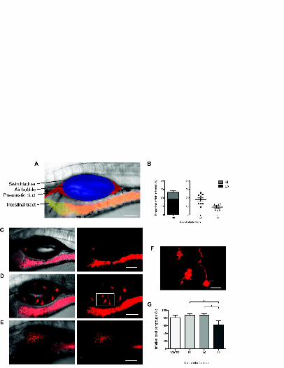
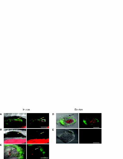

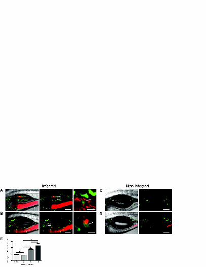
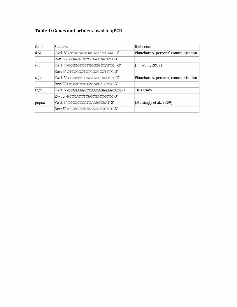
Table 1: Genes and primers used in qPCR Gene Sequence Reference
il1b Fwd: 5’-GTCACACTGAGAGCCGGAAG-3’ Planchart A. personal communication
Rev: 5’-TGGAGATTCCCAAACACACA-3’ saa Fwd: 5’-CGGGGTCCTGGGGGCTATTG -3’ (Lin et al., 2007)
Rev: 5’-GTTGGGGTCTCCGCCGTTTC-3’ tnfa Fwd: 5’-CGCATTTCACAAGGCAATTT-3’ Planchart A. personal communication
Rev: 5’-CTGGTCCTGGTCATCTCTCC-3’ tnfb Fwd: 5’-TCAGAAACCCAACAGAGAACATC-3’ This study
Rev: 5’-ACCCATTTCAGCGATTGTCC-3’ gapdh Fwd: 5’-TGGGCCCATGAAAGGAAT-3’ (Mattingly et al., 2009)
Rev: 5’-ACCAGCGTCAAAGATGGATG-3’
Top Related