


γλώσσες
Σελίδες
Νομικός

77
3 Micro-HPLC
Heather Kalish and Terry M. Phillips
3.1 IntroduCtIon
High-performance liquid chromatography (HPLC) has become a standard separation technique used in both academic and commercial analytical laboratories. However, there are several draw-backs to standard HPLC, including high solvent consumption, large sample quantity, and decreased detection sensitivity. Micro-HPLC (μHPLC) is a term that encompasses a broad range of sample volumes and column sizes (as shown in Table 3.1), but Saito and coworkers provided narrower defi-nitions in their review based on the size of the columns.1
Micro-columns range in size from 0.5 to 1.0 mm internal diameter (i.d.); capillary columns range in size from 0.1 to 0.5 mm i.d., and nano-columns range in size from 0.01 to 0.1 mm i.d., as seen in Figure 3.1. Additionally, the size of the column tends to dictate the materials the col-umns are manufactured from.1 Micro-columns are made from stainless steel tubing, while cap-illary and nano-columns are manufactured from mainly fused silica, glass-lined stainless steel, pressure-resistant plastic (polyetheretherketone—PEEK), or fused silica–lined PEEK (PEEKSil). Fused silica is advantageous to glass-lined stainless steel because it is flexible and inert, but it can allow for chemical bonding to the inner column wall.1 Commercially available capillary and
Contents
3.1 Introduction ............................................................................................................................773.2 μHPLC Systems ..................................................................................................................... 783.3 Advantages of μHPLC Systems .............................................................................................803.4 Columns .................................................................................................................................. 81
3.4.1 Open Tubular .............................................................................................................. 813.4.2 Semi-Packed ............................................................................................................... 813.4.3 Packed ......................................................................................................................... 81
3.5 Stationary Phases .................................................................................................................... 823.5.1 Micro-Particulate ........................................................................................................ 823.5.2 Monolithic ................................................................................................................... 823.5.3 Monoliths Prepared by Porogen Alteration ................................................................ 833.5.4 Monoliths Prepared by Carbon Nanotube Incorporation ........................................... 833.5.5 Monoliths Prepared by Porogen Alteration and Surface Alkylation ..........................843.5.6 Monoliths Prepared by Photo-Initiated Polymerization .............................................843.5.7 Monoliths Prepared by the Sol–Gel Method ..............................................................84
3.6 Gradient Elution Systems .......................................................................................................843.7 Detectors .................................................................................................................................873.8 Applications of μHPLC ..........................................................................................................88
3.8.1 Pre-Concentration .......................................................................................................883.8.2 Multidimensional Liquid Chromatography ................................................................ 913.8.3 Other Applications ......................................................................................................92
References ........................................................................................................................................92

78 Handbook of HPLC
nano-columns are available from a number of companies including the following columns used in the author’s laboratory: MicroTech Scientific (Vista, CA), Dionex/LC Packings (Sunnyvale, CA), Eksigent Technologies (Dublin, CA), Waters Corporation (Milford, MA), Agilent Technologies (Santa Clara, CA), and Shimadzu Scientific Instruments (Kyoto, Japan).
Horvath and coworkers first investigated miniaturization of the HPLC column in the late 1960s in a series of articles examining the separation of nucleotides.2–4 The comparison of open tubular and stainless steel columns with i.d. of 0.5–1.0 mm packed with novel pellicular column materials indicated that the packed columns were superior for LC. Over the next decade, numerous research groups made significant advancements in the reduction of the column size, column construction materials, and packing supports. Ishii and coworkers continued to work with 0.5 mm i.d. columns, but investigated columns made of Teflon that were slurry packed with 30 μm particle diameter pel-licular particles.5–10 High speed, efficient separations were demonstrated by Scott and coworkers on 1.0 mm i.d. columns.11–15 Novotny and coworkers further reduced the column i.d. to 50–200 μm, packed with 10–100 μm particles.16,17 They concluded that with a 70 μm i.d. column packed with 30 μm particles, good efficiency could be obtained without excessive inlet pressure.17 Over the next three decades, significant advancements have been made in the areas of column composition, detector interface, and hardware design, which are the subject of numerous review articles.1,18–23
3.2 µhPlC systeMs
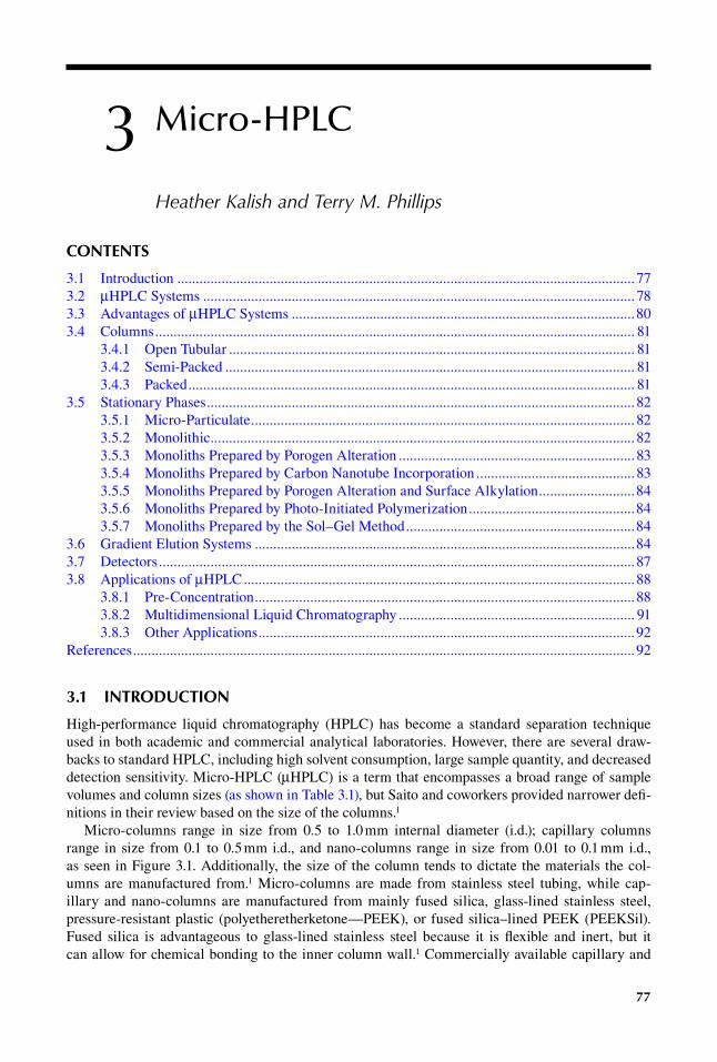
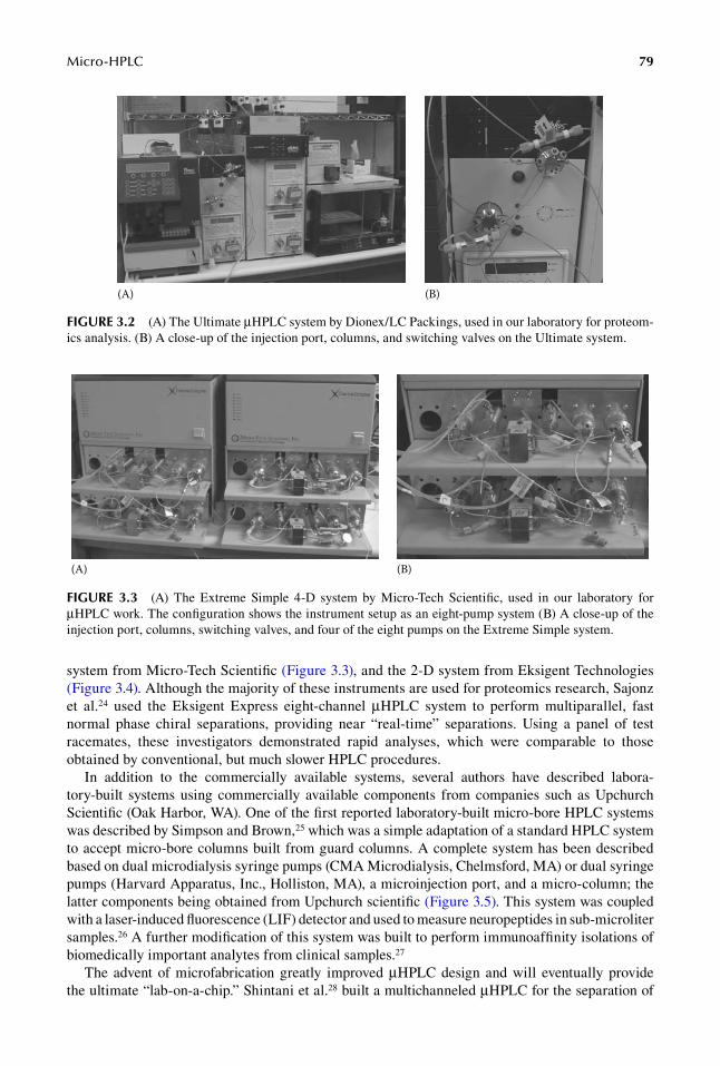
Today nearly all of the major HPLC companies offer a μHPLC system or at least the possibility to modify a standard instrument to accept micro-bore columns. In our laboratory, we routinely use the μHPLC systems Ultimate from Dionex/LC Packing (Figure 3.2), the Extreme Simple 4-D
(A)(B)
(C)
(D)
FIGure 3.1 A comparison of columns used in routine and μHPLC. (A) A laboratory built PEEK nano-flow μHPLC column measuring 100 μm i.d. × 5 cm long, (B) A commercial C-18 PEEK capillary μHPLC column measuring 75 μm i.d. × 25 cm long, (C) A commercial C-8 stainless steel μHPLC column measuring 1 mm i.d. by 10 cm long, (D) a routine commercial reversed-phase stainless steel column measuring 4.6 mm i.d. × 25 cm long, and (E) A commercial HPLC size exclusion preparative column measuring 7.5 mm i.d. by 30 cm.
taBle 3.1units of length and Volume used in μhPlC
symbol Prefix use in Volume (l) use in length (m)
c Centi 10−2 10−2
m Milli 10−3 10−3
μ Micro 10−6 10−6
n Nano 10−9 10−9
p Pico 10−12 10−12
f Femto 10−15 10−15
Micro-HPLC 79
system from Micro-Tech Scientific (Figure 3.3), and the 2-D system from Eksigent Technologies (Figure 3.4). Although the majority of these instruments are used for proteomics research, Sajonz et al.24 used the Eksigent Express eight-channel μHPLC system to perform multiparallel, fast normal phase chiral separations, providing near “real-time” separations. Using a panel of test racemates, these investigators demonstrated rapid analyses, which were comparable to those obtained by conventional, but much slower HPLC procedures.
In addition to the commercially available systems, several authors have described labora-tory-built systems using commercially available components from companies such as Upchurch Scientific (Oak Harbor, WA). One of the first reported laboratory-built micro-bore HPLC systems was described by Simpson and Brown,25 which was a simple adaptation of a standard HPLC system to accept micro-bore columns built from guard columns. A complete system has been described based on dual microdialysis syringe pumps (CMA Microdialysis, Chelmsford, MA) or dual syringe pumps (Harvard Apparatus, Inc., Holliston, MA), a microinjection port, and a micro-column; the latter components being obtained from Upchurch scientific (Figure 3.5). This system was coupled with a laser-induced fluorescence (LIF) detector and used to measure neuropeptides in sub- microliter samples.26 A further modification of this system was built to perform immunoaffinity isolations of biomedically important analytes from clinical samples.27
The advent of microfabrication greatly improved μHPLC design and will eventually provide the ultimate “lab-on-a-chip.” Shintani et al.28 built a multichanneled μHPLC for the separation of
(A) (B)
FIGure 3.2 (A) The Ultimate μHPLC system by Dionex/LC Packings, used in our laboratory for proteom-ics analysis. (B) A close-up of the injection port, columns, and switching valves on the Ultimate system.
(A) (B)
FIGure 3.3 (A) The Extreme Simple 4-D system by Micro-Tech Scientific, used in our laboratory for μHPLC work. The configuration shows the instrument setup as an eight-pump system (B) A close-up of the injection port, columns, switching valves, and four of the eight pumps on the Extreme Simple system.

80 Handbook of HPLC
multiple analytes within the same sample. This system employed an array of monolithic columns driven by a single HPLC pump and a chip-based microinjection device. Detection was achieved with a multichannel ultraviolet (UV) detector based on fiber optics. Further, Yin et al.29 developed an entire μHPLC system on a microfabricated chip made from laminated polyimide layers. Following chromatographic separation on reversed-phase particles, the separated analytes were detected using an ion-trap mass spectrometer, a custom-built interface, and an integrated nanospray tip. A similar chip-based system has been described by Lazar and colleagues.30 The μHPLC system was reported to compare well with a conventional HPLC in the fractionation of a protein tryptic digest.
3.3 adVantaGes oF µhPlC systeMs
μHPLC has some significant advantages over traditional HPLC. The delivery of reliably small samples in μHPLC is often obtained using pre-columns, which concentrate large sample volumes. These pre-columns can accommodate large loading volumes that can dramatically reduce the over-all analysis time and provide protection of small-bore analytical columns from contaminants in the original sample.1
Biological samples, which are often available in limited amounts, can be separated and detected with a drastically improved signal-to-noise ratio, since the volumetric band-broadening (dilution) is much smaller on a capillary column.1 Smaller columns also require less solvent which is an
(A) (B)
FIGure 3.5 (A) A laboratory-built μHPLC system with dual syringe pumps, an electronic injector port, and an Upchurch nano-flow gradient mixer. (B) A picture of the two syringe pumps that comprise the gradient system. The injection valve, gradient system, and detector were controlled via a LabView interface.
(A) (B)
FIGure 3.4 (A) The NanoLC 2-D system by Eksigent Technologies, used in our laboratory for μHPLC protein analysis. (B) A close-up of the injection port, columns, and switching valves on the NanoLC system.
Micro-HPLC 81
economical benefit, from the cost of both the solvent and the solvent disposal.1 Finally, smaller col-umns promote the development of novel analytical columns. The reduced size makes investigating the correlation between the chemical structure of the packing material and its effects on the selec-tivity of the column more cost effective and allows researchers to use materials that are available in limited quantities.1
As the column diameter to particle diameter ratio decreases below 6, the core support region disappears and the support structure becomes dominated by the loosely packed wall region. The packing structure becomes more homogeneous, eliminating one cause of band broadening in HPLC.31 Additionally, an analyte can diffuse easily across a column’s cross section, which can average out any remaining cross-column differences in flow and retention.32
μHPLC systems have also enabled researchers to exploit temperature in liquid chromatographic separations with improved results and fewer problems. In narrow bore columns, thermal mismatches are reduced and can even be neglected due to fast heat transfer across the reduced column size.33
3.4 ColuMns
In μHPLC, there are numerous types of columns used. The comparison and characterization of these columns are often discussed in terms of thermodynamic properties and kinetic character-istics. The retention factor, k, selectivity, α, and the peak asymmetry As are believed to be repre-sentative parameters for the thermodynamic properties, while the kinetic characteristics are often expressed in dimensionless magnitudes of reduced plate height, h, separation impedance, E, and flow resistance factor, ϕ.23
Three broad categories define the analytical columns used in μHPLC: Open tubular, semi-packed, and packed. Each of these three columns types is discussed briefly while making reference to the extensive review articles that cover each area.
3.4.1 open tuBular
Open tubular columns are typically 10–20 μm in diameter and characterized by a stationary phase that is bound to the inner wall of the capillary rather than to the particles packed inside.34 Kennedy and Jorgenson34 effectively used open tubular columns to analyze single cells. The method was advantageous for the analysis of single cells because the columns require small samples, their resolving power equal or exceed conventional HPLC columns, and the detectors typically used with this method have good mass sensitivity. Several other research groups used open tubular columns in their research, many of which are referenced in a review by Saito et al.1 However, for open tubular columns to be effective, Knox and Gilbert calculated that open tubular columns needed to have a diameter of 10 μm or less35 and operating columns of 10 μm or less proved to be quite difficult, thus few reports exist on the use of this column in LC after the early 1990s.
3.4.2 seMi-packed
Semi-packed columns, originally referred to as packed micro-capillaries, were developed by Tsuda and coworkers.17,36 These columns are prepared by packing particles with diameters of 10–100 μm into glass tubes. The packed tubes are then drawn on a glass drawing machine so that the inner diameter is 50–200 μm and roughly two to three times the diameter of the particles.37 Tsuda and coworkers establish that semi-packed columns are effective in significantly reducing the plate heights, but at a cost of decreased sample capacities37 and significantly long analysis times.38
3.4.3 packed
Packed columns, being easier to prepare than semi-packed columns, offer a more feasible way to avoid many of the problems of open tubular columns while still retaining the advantages of
82 Handbook of HPLC
micro-columns.37 Packed columns used in μHPLC are either conventional fused silica capillar-ies packed with alkylated silica particles as the stationary phase or polymeric monolithic columns developed by in-situ polymerization. Numerous reviews on both packed and monolithic columns are found in the literature including the different methods used to pack capillary columns,19 evalu-ation, and comparison of packed capillary columns with conventional-size columns,20 the different organic polymers used to prepare monolithic columns,22 silica gel-based monoliths prepared by the sol–gel method,21 and a comparison of the efficiency of micro-particulate and monolithic capillary columns.18
Packed capillary columns have been used successfully in μHPLC for a number of reasons. It is easy to pack long columns, lengths of 1 m or greater, with 5 μm particles and still maintain low h values, therefore achieving over 100,000 theoretical plates.37 Additionally, they are made from fused silica, which is superior to stainless steel used in conventional columns for several reasons, including increased wall smoothness, and good optical characteristics.37 Development in packed capillary columns has focused on the reduction of the i.d., which leads to high separation efficiency. Karlsson and Novotny were successful in obtaining extremely high efficiencies with a column i.d. of 44 μm.39 Kennedy and Jorgenson successfully reduced the column i.d. even further to 20–50 μm,37 while Hsieh and Jorgenson further reduced the column i.d. to 12–33 μm.32 An additional challenge in the preparation of capillary columns is the packing technique used to prepare the columns. Several methods, including gases, supercritical carbon dioxide, or liquids, have been used to help in transferring the packing material from an external reservoir to the column tubing. Lancas et al. have written an extensive review of these techniques and related work.19
3.5 statIonary Phases
3.5.1 Micro-particulate
The outcome of a chromatographic separation is also influenced by the stationary phase used to pack the column and is highly specific to the type of chromatography being carried out, whether it be normal or reversed phase, ion exchange, or affinity. The most common type of stationary phase for reversed-phase LC is nonpolar, hydrophobic organic species attached by siloxane bonds to the surface of a silica support according to Doyle and Dorsey, who have extensively reviewed the preparation and characterization of reversed-phase stationary phases.40 According to Caude and Jardy, while materials such as alumina, zirconia, titania, and Florisil have been explored, the most common support for normal phase LC is bare silica.41 For ion-exchange chromatography, the most popular supports are those that are based on poly(styrene) cross-linked with divinylbenzene,42 while in high-performance affinity chromatography, the supports commonly used are modified silica or glass, azalacetone beads, and hydroxylated polystyrene media.43
3.5.2 MonolitHic
Monolithic stationary phases are increasingly considered as a viable alternative for micro- particulate columns in HPLC.18 The monolithic column bed has a uniformly porous integral structure thus eliminating the need for retaining frits and enhancing the column’s mechanical stability during pressure changes.44 Furthermore, a large number of readily available chemistries can be applied to functionalize surfaces, and column permeability can be easily adjusted by selecting the appropriate monomer, cross-linker, and porogen.44 Four approaches have been utilized to prepare continuous beds21: (1) polymerization of an organic monomer with additives, (2) formation of a silica-based network using a sol–gel process, (3) fusing porous particulate packing material in a capillary by a sintering process, and (4) organic hybrid materials. Of the four techniques mentioned above, the first two have been the most widely utilized in μHPLC monolithic column preparation.
Micro-HPLC 83
The first monolithic columns reported by Hjerten et al. were based on polyacrylamides.45 This report was soon followed by Svec and Frechet, who reported the preparation of a novel, continu-ous bed column that incorporates both macroporosity and capacity.46 They noted that these rod-shaped columns could be prepared using almost any monomer and offered a tempting alternative to particle-packed columns. Since then, thermally initiated free radical polymerization of acrylates, methacrylates, and dimethacrylates46 and ring-opening metathesis polymerization of norbornane,47 poly(styrene-co-divinylbenzene) prepared in nanospray needle,48 and other monomers have been used to prepare monolithic column beds, and an extensive review article has been written detailing the numerous organic polymers used to prepare monolithic columns.22 Polymer-based monolithic columns have clearly demonstrated their ability to afford excellent separations of peptides, proteins, oligonucleotides, and nucleic acids. Their ease of preparation, tolerance of high flow rates, and the rapid speed of separations that can be achieved at acceptable back pressures make this col-umn format superior in many applications to particle-packed columns.22 However, the efficiency of polymer-based monolithic columns remains low compared with modified silica-based monolithic columns, so further improvements are required.22
3.5.3 MonolitHs prepared By porogen alteration
Porogenic solvent plays a key role in determination of column morphology.44 Altering the porogens in the polymerization of organic polymers to form monolithic column beds has been researched by Premstaller and Huber.49,50 Premstaller et al. first experimented with using monolithic chromato-graphic beds for ion-pair, reversed-phase HPLC separation of single-stranded oligodeoxynucle-otides and double-stranded DNA fragments.49 In order to accomplish successful separations, the synthesis of the monolith had to be tuned such that its morphology resembled that of a chromato-graphic bed formed by nonporous particles. The use of decanol and tetrahydrofuran as porogens in the polymerization process resulted in monolithic column beds whose performance surpassed that of micro-particulate packed columns. Premstaller et al. further altered the morphology of mono-lithic column beds by using tetrahydrofuran/decanol in the polymerization process and produced reversed-phase monolithic columns whose separation efficiency of peptides and proteins rang-ing in molecular mass from a few hundred to more than 55,000 surpassed that of conventional particle-packed columns.50
3.5.4 MonolitHs prepared By carBon nanotuBe incorporation
The alteration of monoliths by carbon nanotube (CNT) incorporation51 or surface alkylation52 has been investigated as another alternative to fine tune the separation efficiencies of organic polymer-based monolithic columns. Because of their curved surface, CNT are expected to show stronger binding affinity for hydrophobic molecules as compared with planar surfaces. Single wall carbon nanotubes (SWNT) consist of a graphene sheet rolled into a cylinder, with a typical diameter of 1 nm.51 The challenge is maintaining their unique structure while obtaining a solubility that allows for their incorporation into the polymer. Since analyte retention after incorporation of SWNT was significantly enhanced without corresponding changes in column porosity, it is proposed that the specific structure, size, and charge of the characteristics of CNT all may play a role.51 Analytes may be drawn onto the nanotube surface or channels between nanotubes due to surface tension and capillary effects and therefore exhibit longer retention times.51 Huang and coworkers sought to increase the chromatographic resolution of peptides by octadecylating the surface of a poly(styrene-divinylbenzene) monolith.52 Then by treating it with a solution containing a Friedel-Crafts catalyst, an alkyl halide, and an organic solvent, Huang et al. were able to alter the surface with C-18 alkyl groups. This surface alteration ultimately improved the reversed-phase LC separation of peptides over unmodified poly(styrene-divinylbenzene) monolithic columns.

84 Handbook of HPLC
3.5.5 MonolitHs prepared By porogen alteration and surface alkylation
A combination of surface alkylation and alteration of the porogens in the polymerization reaction was used by Li and coworkers to manufacture a surface alkylated poly(glycidyl methacrylate ethyl-ene glycol dimethacrylate) monolithic column44 that was copolymerized in situ with dodecanol and toluene as porogenic solvents and further functionalized by alkylation since alkyl groups are the most widely used retentive functions. The porosity of the polymeric monolith could be altered by numerous factors including using the appropriate monomer, cross-linker, porogens, the reaction tem-perature, or the concentration of initiator. Alkylation with linear octadecyl groups showed an appre-ciable improvement in the separation resolution for proteins over nonfunctionalized poly(glycidyl methacrylate ethylene glycol dimethacrylate) monolithic columns.
3.5.6 MonolitHs prepared By pHoto-initiated polyMerization
Lee and coworkers report on a photo-initiated polymerization for the preparation of poly(butyl methylacrylate-co-ethylene dimethylacrylate) within fused silica capillaries.53 UV light-initiated polymerization is well suited to monolith formation in restricted spaces since polymer forms only in those areas that are exposed to irradiation. This leads to far greater control over the length and size of the monolithic column formed. The resulting columns are robust since their separation ability does not deteriorate with time or number of injections and their reproducibility is excellent.53
3.5.7 MonolitHs prepared By tHe sol–gel MetHod
The invention of monolithic silica-based columns can be regarded as a major technological change in column technology.21 The preparation of a porous silica rod by the sol–gel method was reported by Nakanishi and Soga.54,55 The porous silica rods were prepared by hydrolytic polymerization of tetramethoxysilane accompanied by phase separation in the presence of water-soluble organic poly-mers.56 The morphology, determined by phase separation, is solidified by gel formation, resulting in a silica rod with a biporous structure that consists of micrometer-size through-pores and meso- or microporous silica skeletons.56 Siouffi has published an extensive review that describes the sol–gel method of preparation in great detail and provides an extensive list of references.21 Since the preparation of silica-based monoliths, research has been done on modifying them, in similar ways to organic polymer-based monoliths, to improve their separation performance. Minakuchi and cowork-ers derivatized silica-based monoliths to incorporate C18 alkyl groups on the surface.56 The C18 silica rods showed much better performance at high flow rates than conventional columns packed with C18 particles; however, the separation efficiencies obtained were not impressive.57 Luo and cowork-ers sought to improve the separation efficiencies of silica-based monolithic columns by preparing a 20 μm i.d. monolithic column.57 These columns provided a high separation efficiency and sensitivity and show potential in analyzing small amounts of proteomic samples. Continuing to modify the preparation of these columns along similar lines may provide smaller i.d. columns as well.
3.6 GradIent elutIon systeMs
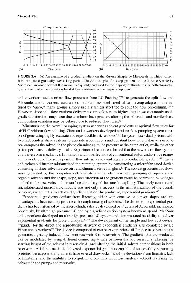
The delivery of accurate and reproducible gradients in μHPLC systems is one of the problems that many researchers are trying to solve. Simple gradients differ from each other in three respects, as seen in Figure 3.6: the shape of the gradient, the slope and curvature of the gradient, and the initial and final concentrations of the more efficient component B.58 Several methods have been developed to deliver accurate and reproducible gradients including flow splitting, miniaturization of high-pressure gradient pumps, exponential gradient formation, preformed gradient loops and multiport switching valves, and high-temperature programming.
Splitting the solvent flow, delivered by the pumps, down to the required flow rate for nano-HPLC is achieved by inserting a variety of devices between the pumps and the injector. While Chervet
Micro-HPLC 85
and coworkers used a micro-flow processor from LC Packings59,60 to generate the split flow and Alexander and coworkers used a modified stainless steel fused silica makeup adapter manufac-tured by Valco,61 many groups simply use a stainless steel tee to split the flow pre-column.62–64 However, since split flow gradient delivery requires flow rates higher than those commonly used, gradient distortions may occur due to column back pressure altering the split ratio, and mobile phase composition variation may be delayed due to reduced flow rates.65
Miniaturizing the overall pumping system generates solvent gradients at optimal flow rates for μHPLC without flow splitting. Zhou and coworkers developed a micro-flow pumping system capa-ble of generating highly accurate and reproducible micro-flows.66 The system uses dual pistons, with two independent drive systems to generate a continuous and constant flow. One piston was used to pre-compress the solvent in the piston chamber up to the pressure at the pump outlet, while the other piston performs its delivery stroke. Experimental results confirmed that the new micro-flow system could overcome mechanical limitations and imperfections of conventional piston and syringe pumps and provide conditions-independent flow rate accuracy and highly reproducible gradient.66 Figeys and Aebersold further miniaturized the pumping system by constructing a microfabricated device consisting of three solvent reservoirs and channels etched in glass.67 The solvent gradients and flows were generated by the computer-controlled differential electroosmotic pumping of aqueous and organic solvents and the shape, slope, and direction of the gradient could be controlled by voltages applied to the reservoirs and the surface chemistry of the transfer capillary. The newly constructed microfabricated microfluidic module was not only a success in the miniaturization of the overall pumping system but also achieved gradient elutions by producing exponential gradients.67
Exponential gradients deviate from linearity, either with concave or convex slopes and are advantageous because they provide a thorough mixing of solvents. The delivery of exponential gra-dients has been attained by the micro-fluidics device developed by Figeys and Aebersold, mentioned previously, by ultrahigh pressure LC and by a gradient elution system known as ηgrad. MacNair and coworkers developed an ultrahigh-pressure LC system and demonstrated its ability to deliver exponential gradients for protein analysis.68,69 The development of the simple and low-cost device, “ηgrad,” for the direct and reproducible delivery of exponential gradients was completed by Le Bihan and coworkers.70 The device is composed of two reservoirs whose difference in solvent height generates a gravity-induced flow from reservoir B to reservoir A. The gradients that are produced can be modulated by using different connecting tubing between the two reservoirs, altering the starting height of the solvent in reservoir A, and altering the initial solvent compositions in both reservoirs. All three methods delivered exponential gradients capable of successfully separating proteins, but exponential gradients have several drawbacks including deviations from linearity, lack of flexibility, and the inability to reequilibrate columns for future analysis without reversing the solvents in the pumps and reservoirs.
0 3 6 9 12 15 18 21 24Time (min)(A) (B)
27 30 33 36 39 42 45 48 51 54 57 60
100908070605040302010
Perc
ent (
%)
Composite percent
0 3 6 9 12 15 18 21 24 27Time (min)
Composite percent
Perc
ent (
%)
30 33 36 39 42 45 4851 54 57 60
100908070605040302010
FIGure 3.6 (A) An example of a gradual gradient on the Xtreme Simple by Microtech, in which solvent B is introduced gradually over a long period. (B) An example of a steep gradient on the Xtreme Simple by Microtech, in which solvent B is introduced quickly and used for the majority of the elution. In both chromato-grams, the gradient ends with solvent A being restored as the major component.
86 Handbook of HPLC
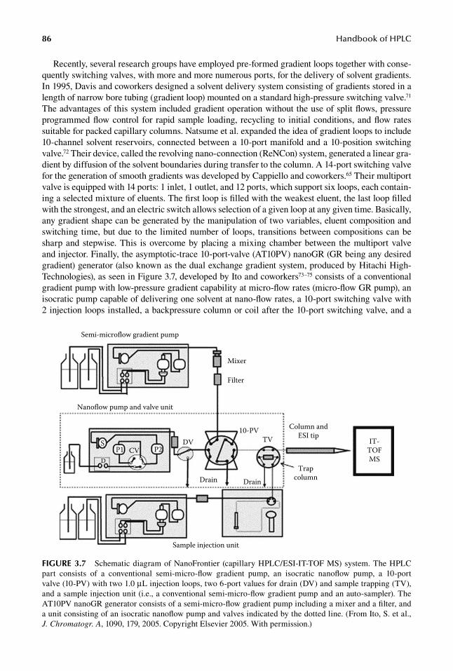
Recently, several research groups have employed pre-formed gradient loops together with conse-quently switching valves, with more and more numerous ports, for the delivery of solvent gradients. In 1995, Davis and coworkers designed a solvent delivery system consisting of gradients stored in a length of narrow bore tubing (gradient loop) mounted on a standard high-pressure switching valve.71 The advantages of this system included gradient operation without the use of split flows, pressure programmed flow control for rapid sample loading, recycling to initial conditions, and flow rates suitable for packed capillary columns. Natsume et al. expanded the idea of gradient loops to include 10-channel solvent reservoirs, connected between a 10-port manifold and a 10-position switching valve.72 Their device, called the revolving nano-connection (ReNCon) system, generated a linear gra-dient by diffusion of the solvent boundaries during transfer to the column. A 14-port switching valve for the generation of smooth gradients was developed by Cappiello and coworkers.65 Their multiport valve is equipped with 14 ports: 1 inlet, 1 outlet, and 12 ports, which support six loops, each contain-ing a selected mixture of eluents. The first loop is filled with the weakest eluent, the last loop filled with the strongest, and an electric switch allows selection of a given loop at any given time. Basically, any gradient shape can be generated by the manipulation of two variables, eluent composition and switching time, but due to the limited number of loops, transitions between compositions can be sharp and stepwise. This is overcome by placing a mixing chamber between the multiport valve and injector. Finally, the asymptotic-trace 10-port-valve (AT10PV) nanoGR (GR being any desired gradient) generator (also known as the dual exchange gradient system, produced by Hitachi High-Technologies), as seen in Figure 3.7, developed by Ito and coworkers73–75 consists of a conventional gradient pump with low-pressure gradient capability at micro-flow rates (micro-flow GR pump), an isocratic pump capable of delivering one solvent at nano-flow rates, a 10-port switching valve with 2 injection loops installed, a backpressure column or coil after the 10-port switching valve, and a
Sample injection unit
Nanoflow pump and valve unit
Semi-microflow gradient pump
Mixer
Filter
DrainDrain
P1 CVDV
D
TVP2
10-PV Column andESI tip
Trapcolumn
IT-TOFMS
S
FIGure 3.7 Schematic diagram of NanoFrontier (capillary HPLC/ESI-IT-TOF MS) system. The HPLC part consists of a conventional semi-micro-flow gradient pump, an isocratic nanoflow pump, a 10-port valve (10-PV) with two 1.0 μL injection loops, two 6-port values for drain (DV) and sample trapping (TV), and a sample injection unit (i.e., a conventional semi-micro-flow gradient pump and an auto-sampler). The AT10PV nanoGR generator consists of a semi-micro-flow gradient pump including a mixer and a filter, and a unit consisting of an isocratic nanoflow pump and valves indicated by the dotted line. (From Ito, S. et al., J. Chromatogr. A, 1090, 179, 2005. Copyright Elsevier 2005. With permission.)
Micro-HPLC 87
controller to control the pumps and switching valve. Micro-flow GR pumps create an original gradi-ent profile by mixing reservoir solvents A and B. The well-mixed solvents are delivered at a flow rate (e.g., 100 μL/min) into injection loop A or B. While A is being loaded, B is delivering solvent loaded in its loop to the capillary column at a nano-flow rate. The roles of A and B switch throughout the sample run and any nano-flow-gradient profile can be generated simply by shortening the switching period of the 10-port switching valve. All the systems mentioned above were able to effectively con-trol the shapes of the gradients, producing accurate and reproducible gradients.
High-temperature generated gradients have been explored in μHPLC by Trones et al.33,76,77 The dimensions of packed capillary columns enable a faster response to temperature changes and exhibit reduced temperature gradients, which contribute to band broadening, within the columns. However, the accuracy and reproducibility of the gradients are not impressive and the research is limited in this area.
3.7 deteCtors
μHPLC systems have been designed with numerous detectors, which are ideally directly connected to the separation column. These detectors include UV, Raman, and infrared (IR) absorbance, fluo-rescence, electrochemical, nuclear magnetic resonance spectroscopy, evaporative light-scattering and electrospray ionization (ESI), and inductively coupled plasma mass spectrometry (ICP-MS). All of these detectors offer advantages and disadvantages, and some are more suitable to the small sample sizes that are collected at the end of a separation. While some of these detectors will be discussed briefly, an extensive review of their use has been published by Vissers et al.23
Raman and mid-IR detectors are of special interest due to the molecular specific fingerprint they provide.78 In addition, their nondestructive character allows their use in sequence as well as coupling with more sensitive detection schemes.78 However, direct application of Raman and IR spectroscopy is made difficult by the low concentration sensitivity of this technique.78 Surowiec and co work-ers have worked to develop a flow-through micro-dispenser used as a solvent elimination interface between micro-bore HPLC and Raman/Fourier transform infrared (FTIR) spectroscopic detectors.78 A dispensing frequency of 10 Hz was chosen to assure deposits with closer size and diameter to the diameter of the IR beam, which assured higher reproducibility and sensitivity. Improved signal reproducibility was obtained with IR than Raman spectroscopy due to a larger IR beam size mea-suring more of an average of the droplet than the Raman beam. While FTIR spectroscopy provides more reproducible measurements, Raman allows for the detection of minute amounts of sample.78
μHPLC has been successfully coupled to ICP-MS79–81; however, the use of gradient elution and high flow rates makes the technique unstable and therefore unusable in most applications. Trones and coworkers have coupled capillary HPLC with ICP-MS and replaced a liquid gradient elution with a temperature gradient elution to study organo-tin and organo-lead detection limits.77 In con-trast to other detection systems such as UV, temperature ramping had no effect on the ICP-MS, the limits of detection were much lower than those of conventional HPLC, and the repeatability of peak height and area were good.77 Additional examples of analytes investigated by μHPLC-ICP-MS are found in Table 3.2.
taBle 3.2examples of analytes Investigated by μhPlC-ICP-Ms
technology used analyte(s)
HPLC-ICP-MS Selenomethionine, carboxymethylated selenocysteine82; Se-methylselenocysteine, γ-gluyamyl-Se-methylselenocysteine, selenosugar, trimethylselenonium, selenomethionine83; Asp-Tyr-SeMet-Gly-Ala-Ala-Lys peptide84; selenomethionyl calmodulin85; and tryptic digest of selenomethionyl calmodulin86
88 Handbook of HPLC
Adapting the evaporative light scattering device (ELSD) to μHPLC was investigated by Gaudin et al.87 Quantitative analysis by ELSD is often hindered by nonlinearity; however, reduction of the flow rate, resulting in better homogeneity of droplet size distribution, has increased the linearity of the response with ELSD. Despite the predictable effect on droplet size in relation to the reduction of the inner diameter of the capillary inside the nebulizer, ELSD is relatively simple to adapt to micro/capillary LC.87
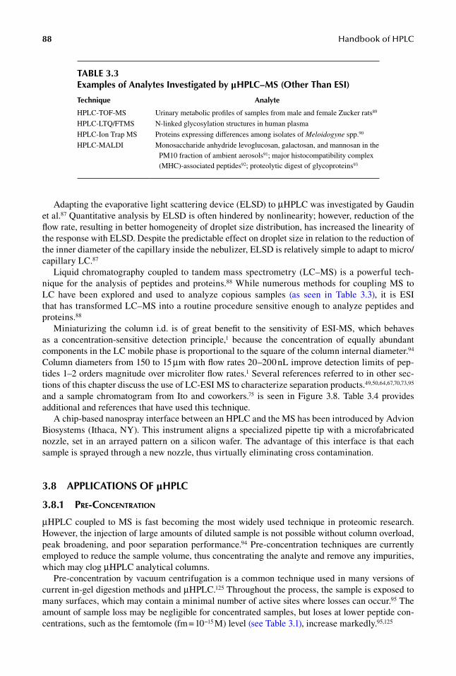
Liquid chromatography coupled to tandem mass spectrometry (LC–MS) is a powerful tech-nique for the analysis of peptides and proteins.88 While numerous methods for coupling MS to LC have been explored and used to analyze copious samples (as seen in Table 3.3), it is ESI that has transformed LC–MS into a routine procedure sensitive enough to analyze peptides and proteins.88
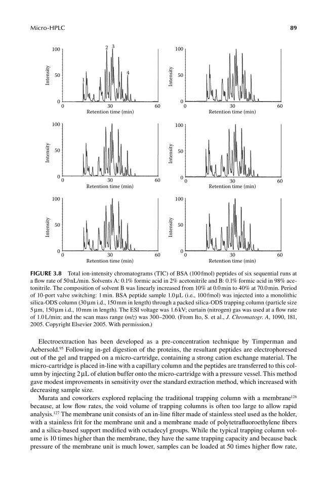
Miniaturizing the column i.d. is of great benefit to the sensitivity of ESI-MS, which behaves as a concentration-sensitive detection principle,1 because the concentration of equally abundant components in the LC mobile phase is proportional to the square of the column internal diameter.94 Column diameters from 150 to 15 μm with flow rates 20–200 nL improve detection limits of pep-tides 1–2 orders magnitude over microliter flow rates.1 Several references referred to in other sec-tions of this chapter discuss the use of LC-ESI MS to characterize separation products.49,50,64,67,70,73,95 and a sample chromatogram from Ito and coworkers.75 is seen in Figure 3.8. Table 3.4 provides additional and references that have used this technique.
A chip-based nanospray interface between an HPLC and the MS has been introduced by Advion Biosystems (Ithaca, NY). This instrument aligns a specialized pipette tip with a microfabricated nozzle, set in an arrayed pattern on a silicon wafer. The advantage of this interface is that each sample is sprayed through a new nozzle, thus virtually eliminating cross contamination.
3.8 aPPlICatIons oF μhPlC
3.8.1 pre-concentration
μHPLC coupled to MS is fast becoming the most widely used technique in proteomic research. However, the injection of large amounts of diluted sample is not possible without column overload, peak broadening, and poor separation performance.94 Pre-concentration techniques are currently employed to reduce the sample volume, thus concentrating the analyte and remove any impurities, which may clog μHPLC analytical columns.
Pre-concentration by vacuum centrifugation is a common technique used in many versions of current in-gel digestion methods and μHPLC.125 Throughout the process, the sample is exposed to many surfaces, which may contain a minimal number of active sites where losses can occur.95 The amount of sample loss may be negligible for concentrated samples, but loses at lower peptide con-centrations, such as the femtomole (fm = 10−15 M) level (see Table 3.1), increase markedly.95,125
taBle 3.3examples of analytes Investigated by μhPlC–Ms (other than esI)
technique analyte
HPLC-TOF-MS Urinary metabolic profiles of samples from male and female Zucker rats89
HPLC-LTQ/FTMS N-linked glycosylation structures in human plasma
HPLC-Ion Trap MS Proteins expressing differences among isolates of Meloidogyne spp.90
HPLC-MALDI Monosaccharide anhydride levoglucosan, galactosan, and mannosan in the PM10 fraction of ambient aerosols91; major histocompatibility complex (MHC)-associated peptides92; proteolytic digest of glycoproteins93
Micro-HPLC 89
Electroextraction has been developed as a pre-concentration technique by Timperman and Aebersold.95 Following in-gel digestion of the proteins, the resultant peptides are electrophoresed out of the gel and trapped on a micro-cartridge, containing a strong cation exchange material. The micro-cartridge is placed in-line with a capillary column and the peptides are transferred to this col-umn by injecting 2 μL of elution buffer onto the micro-cartridge with a pressure vessel. This method gave modest improvements in sensitivity over the standard extraction method, which increased with decreasing sample size.
Murata and coworkers explored replacing the traditional trapping column with a membrane126 because, at low flow rates, the void volume of trapping columns is often too large to allow rapid analysis.127 The membrane unit consists of an in-line filter made of stainless steel used as the holder, with a stainless frit for the membrane unit and a membrane made of polytetrafluoroethylene fibers and a silica-based support modified with octadecyl groups. While the typical trapping column vol-ume is 10 times higher than the membrane, they have the same trapping capacity and because back pressure of the membrane unit is much lower, samples can be loaded at 50 times higher flow rate,
00
50
100
30Retention time (min)
Inte
nsity
60 00
50
100
30Retention time (min)
Inte
nsity
60
00
50
100
30Retention time (min)
Inte
nsity
60
00
50
100
30Retention time (min)
Inte
nsity
60
00
50
100
30Retention time (min)
Inte
nsity
60
0 30
1
2 3
4
Retention time (min)
Inte
nsity
600
50
100
FIGure 3.8 Total ion-intensity chromatograms (TIC) of BSA (100 fmol) peptides of six sequential runs at a flow rate of 50 nL/min. Solvents A: 0.1% formic acid in 2% acetonitrile and B: 0.1% formic acid in 98% ace-tonitrile. The composition of solvent B was linearly increased from 10% at 0.0 min to 40% at 70.0 min. Period of 10-port valve switching: 1 min. BSA peptide sample 1.0 μL (i.e., 100 fmol) was injected into a monolithic silica-ODS column (30 μm i.d., 150 mm in length) through a packed silica-ODS trapping column (particle size 5 μm, 150 μm i.d., 10 mm in length). The ESI voltage was 1.6 kV; curtain (nitrogen) gas was used at a flow rate of 1.0 L/min; and the scan mass range (m/z) was 300–2000. (From Ito, S. et al., J. Chromatogr. A, 1090, 181, 2005. Copyright Elsevier 2005. With permission.)

90 Handbook of HPLC
with as much as 50 μg sample, in spite of the small volume. One drawback observed by researchers was membrane durability that was impaired after 40 injections, but this could be improved by minor changes to the unit structure.
Licklider and co workers experimented with automating the sample introduction step in nano-scale LC–MS.62 In order to achieve pre-concentration and desalting prior to sample analysis, they created a 2 cm vent after the head of the analytical column. Experimental results demonstrated 50 nanoliter (nL) elution peak volumes while retaining low-to subfemtomole detection levels. Additionally, implementing this pre-concentration technique requires minimal changes in current methods and equipment.
The most common form of pre-concentration involves the use of pre-columns.94 Pre-columns combined with column switching techniques help avoid the problems mentioned in the introduc-tion to this section. The type of pre-column used is dictated by experimental conditions. Large sample volumes can be eluted onto pre-columns using mobile phases with low elution powers, allowing for sample concentration and clean-up, prior to injection on the analytical separation col-umn. However, the use of pre-columns for sample concentration can be plagued by void volumes, whose volumes can be several times greater than that of the pre-column, negating any initial sample pre- concentration and leading to poor separation performance.
One type of pre-analytical concentration that is particularly well suited to μHPLC is the incor-poration of an affinity ligand pre-column. The addition of affinity and immunoaffinity ligands for pre-analytical concentration of samples has become popular in the analytical sciences.128,129 The use of affinity pre-columns ranges from making sol–protein gel-derived monolithic columns containing the affinity ligand of interest130 to the employment of true bioaffinity ligands and antibodies. Madera et al.131 employed immobilized lectin micro-columns prior to reversed-phase analysis of glycopro-teins in small samples of human serum. Likewise, Starkey et al.132 used a combination of phenyl
taBle 3.4examples of analytes Investigated by μhPlC-esI
technique analyte(s)
HPLC-ESI Flavonoids of lemon, grapefruit, bergamot, orange, and mandarin96; Bcl-2 antisense phosphothioate oligonucleotides G3139 and metabolites in plasma97; anthocyanins and derived components of red wines98; α-bisabolol in human blood99; anthocyanins in Sicilian wines100; ochratoxin A in grapes101; light-induced lateral migration of photosystem II antennae between appressed and nonappressed thylakoid membranes102; intact yeast proteins103; antiphosphotyrosine antibodies104; N-acylhomoserine lactones105
HPLC–MS-MS (ESI) Integral plasma membrane proteins from a human lung cancer cell line (62 prenylated proteins and 45 Ras family proteins)106; urinary proteins107; AGP-derived glycoproteins108; major and minor populated isoforms of antithrombin109; regulatory lipids in breath condensate; Ser(150), Ser(418), and Ser(476) of human Grb10 zeta110; dissolved proteins in seawater samples111; human milk oligosaccharides derivatized with various esters of aminobenzoic acid112; sulphametoxazole, bezafibrate, metoprolol, carbamazepine, and bisoprolol in water samples113; 16 mycotoxins possibly related to “Sick Building Syndrome”114; 7-fluoro-4-nitrobenzoxadiazole (NBD-F) and 1-fluoro-2,4, dinitrobenzene (DNB-F) tagged amino acids115; six quinic acid derivatives isolated from Baccharis usterii Heering116; protease inhibitors and nonnucleoside reverse transcriptase inhibitors in dried blood spots from HIV/AIDS patients117; urinary 8-hydroxy-2′-deoxyguanosine118; site-specific horseradish peroxidase glycosylation119; “trypsinosome” from specific peptide characteristics120; soluble proteins in wine121
HPLC-ESI-Q-TOF-MS Cereulide from Bacillus cereus122; human plasma proteome103; abiotic stress-tolerant (Mandolina) and an abiotic stress-susceptible (Jubilant) barley cultivar123; neuropeptides from isolated locust corpora cardiaca124

Micro-HPLC 91
borate affinity pre-concentration followed by micro-column chromatography and atmospheric pressure photoionization MS to measure salsolinal and the major catecholamines in experimental animal brain tissue.
Antibody-based immunoaffinity pre-analytical concentrators have been widely used in routine chromatography (see Chapter 13) but are only just becoming introduced into μHPLC, although immunoaffinity pre-analytical concentrators have been described in capillary electrophoresis.133,134 Most applications have employed the micro-columns as immunoaffinity chromatography with direct measurement of the captured analyte. Hodgson and colleagues135 used an antibody-entrapped monolithic column to perform nanoflow immunoaffinity chromatography. Here the column was used as an immunoextraction concentrator with the released analyte being measured by LIF detec-tion. However, as the authors rightly point out, such columns could easily be used as a pre-analytical concentrator.
3.8.2 MultidiMensional liquid cHroMatograpHy
Holland and Jorgenson136 reported separating amines using anion exchange and reversed-phase col-umns in 1995 and since then, there have been numerous reports of combining two LC columns (2D-LC) to achieve efficient sample separation.137–143 In addition to the few references mentioned in this section, see Chapter 4 in this handbook on two-dimensional comprehensive liquid chromatography.
taBle 3.5examples of analytes Investigated by additional detection Methods than those Mentioned in section 3.7
technique analyte(s)
HPLC Clenbuterol in pork, beef, and hog liver145; Co(II) ion as 4-(2-thiazolylazo)resorcinol (TAR) or 5-methyl-4-(2-thiazolylazo)resorcinol (5MTAR)146; total phenols after nitrosation of USEPA classified 11 priority pollutant phenols147; polyprenol and dolichol148
HPLC-UV Mebeverine hydrochloride in raw materials, bulk drugs and formulation149; meloxicam150; platinum in blood and urine samples after administration of cisplatin drug151; budesonide, a novel glucocorticoid prescribed for inflammatory bowel disease152; amikacin153; aromatic amines in water154; cetirizine in human plasma155; Fmoc and z-derivatives of natural and unnatural sulfur containing amino acids156; glycyl- and diastereomeric dipeptides and tripeptides157; gossypol in cotton158; West Nile and Sindbis virus PCR products159; resveratrol and resveratrol-glucosides in Sicilian wines100; glycyrrhizin and glycyrrhetic acid in licorice roots and candies160
HPLC-UV-electrochemical detection
Disodium-2,2′-dithio-bis-ethane sulfonate (BNP7787) intracellular conversion products161; honokiol and magnolol in fresh Magnolia obovata162
HPLC-UV-MS Bioactive compounds from Blumea gariepina163; salicin, salicylic acid, tenoxicam, ketorolac, piroxicam, tolmetin, naproxen, flurbiprofin, diclofenac, and ibuprofen in pharmaceutical formulations and biological samples164
HPLC-UV-NMR Bioactive compounds from Blumea gariepina163
HPLC-electrochemical detection
Baicalin and baicalein in rat plasma165; honokiol and magnolol in branches and leaves of Magnolia obovata162; quercetin in human plasma166
HPLC-chemiluminescence Changes in catecholamines and 3-O-methyl metabolite concentrations in human plasma167
HPLC-fluorescence Erythropoietin in pharmaceutical products168; 3,4-methylenedioxymethamphetamine, 3,4-methylenedioxyamphetamine, amphetamine, and methamphetamine in rat urine169
HPLC-diode array Azoxystrobin, kresoxim-methyl, and trifloxystrobin fungicides170
HPLC-NMR Shape constrained natural compounds (tocopherol homologues, vitamin E)171; protein kinase ZAP-70 tryptic fragment containing amino acids 485–496172; cartenoids from a spinach sample173; isoflavines in Radix astragali174

92 Handbook of HPLC
While 2D-LC can be accomplished using conventional LC columns, the field of proteomics has directed the 2D-LC field toward capillary columns.1 Multidimensional chromatography appears to overcome problems associated with 2D gels, and the peak capacity of the LC system dramatically increases by combining two orthogonal separation techniques.144 The rapid analysis of complex pro-tein digests using a 2D-LC system was investigated by Mitulovic and coworkers.144 A strong cation exchange column was used first to separate peptides based on their electric charge state and charge distribution, whereas a reverse-phase column was used second to separate proteins according to hydrophobicity. The system they used is fully automated, sample loss is low, and they demonstrated that the success of 2-D HPLC is determined by the nature of the loading solvent, the flow rate of the loading pump, and the sample loading time.144
3.8.3 otHer applications
Many of the references cited throughout this chapter use specific analytes to validate the tech-niques they are researching. In addition to these references, there are numerous references that use additional techniques for specific analysis of reagents, proteins, environmental contaminants, etc. Table 3.5 is a summary of research cited from 2004 until early 2006, using μHPLC in conjunction with these methods of detection to study the specific analytes listed.
reFerenCes
1. Saito, Y., Jinno, K., and Greibrokk, T., Capillary columns in liquid chromatography: Between conven-tional columns and microchips, Journal of Separation Science 27(17–18), 1379–1390, 2004.
2. Horvath, C. and Lipsky, S. R., Rapid analysis of ribonucleosides and bases at picomole level using pel-licular cation exchange resin in narrow bore columns, Analytical Chemistry 41(10), 1227–1234, 1969.
3. Horvath, C., Melander, W., Molnar, I., and Molnar, P., Enhancement of retention by ion-pair formation in liquid-chromatography with nonpolar stationary phases, Analytical Chemistry 49(14), 2295–2305, 1977.
4. Horvath, C. G., Preiss, B. A., and Lipsky, S. R., Fast liquid chromatography—An investigation of operat-ing parameters and separation of nucleotides on pellicular ion exchangers, Analytical Chemistry 39(12), 1422–1428, 1967.
5. Ishii, D., Asai, K., Hibi, K., Jonokuchi, T., and Nagaya, M., Study of micro-high-performance liquid-chromatography. 1. Development of technique for miniaturization of high-performance liquid- chromatography, Journal of Chromatography 144(2), 157–168, 1977.
6. Ishii, D., Hibi, K., Asai, K., and Jonokuchi, T., Studies of micro high-performance liquid-chromatography. 2. Application to gel-permeation chromatography of techniques developed for micro high- performance liquid-chromatography, Journal of Chromatography 151(2), 147–154, 1978.
7. Ishii, D., Hibi, K., Asai, K., and Nagaya, M., Studies of micro high-performance liquid-chromatography. 3. Development of a micro-pre-column method for pretreatment of samples, Journal of Chromatography 152(2), 341–348, 1978.
8. Ishii, D., Hibi, K., Asai, K., Nagaya, M., Mochizuki, K., and Mochida, Y., Studies of micro high- performance liquid-chromatography. 4. Application of micro pre-column method to analysis of corticos-teroids in serum, Journal of Chromatography 156(1), 173–180, 1978.
9. Ishii, D., Hirose, A., Hibi, K., and Iwasaki, Y., Studies on micro high-performance liquid chromatogra-phy. 5. Design of a microscale liquid chromatograph and its application to cation-exchange separation of alkali-metals, Journal of Chromatography 157(Sep), 43–50, 1978.
10. Ishii, D., Hirose, A., and Horiuchi, I., Studies on micro-high-performance liquid-chromatography. 6. Application of microscale liquid-chromatographic technique to anion-exchange separation of halide ions, Journal of Radioanalytical Chemistry 45(1), 7–14, 1978.
11. Kucera, P., Design and use of short microbore columns in liquid-chromatography, Journal of Chromatography 198(2), 93–109, 1980.
12. Reese, C. E. and Scott, R. P. W., Microbore columns—Design, construction, and operation, Journal of Chromatographic Science 18(9), 479–486, 1980.
13. Scott, R. P. W., Microbore columns in liquid-chromatography, Journal of Chromatographic Science 18(2), 49–54, 1980.

Micro-HPLC 93
14. Scott, R. P. W. and Kucera, P., Mode of operation and performance-characteristics of microbore columns for use in liquid-chromatography, Journal of Chromatography 169(Feb), 51–72, 1979.
15. Scott, R. P. W., Kucera, P., and Munroe, M., Use of microbore columns for rapid liquid-chromatographic separations, Journal of Chromatography 186(Dec), 475–487, 1979.
16. Hirata, Y. and Novotny, M., Techniques of capillary liquid-chromatography, Journal of Chromatography 186(Dec), 521–528, 1979.
17. Tsuda, T. and Novotny, M., Packed microcapillary columns in high-performance liquid-chromatography, Analytical Chemistry 50(2), 271–275, 1978.
18. Eeltink, S., Decrop, W. M. C., Rozing, G. P., Schoenmakers, P. J., and Kok, W. T., Comparison of the effi-ciency of microparticulate and monolithic capillary columns, Journal of Separation Science 27(17–18), 1431–1440, 2004.
19. Lancas, F. M., Rodrigues, J. C., and Freitas, S. D., Preparation and use of packed capillary columns in chromatographic and related techniques, Journal of Separation Science 27(17–18), 1475–1482, 2004.
20. Pruss, A., Kempter, C., Gysler, J., and Jira, T., Evaluation of packed capillary liquid chromatography columns and comparison with conventional-size columns, Journal of Chromatography A 1030(1–2), 167–176, 2004.
21. Siouffi, A. M., Silica gel-based monoliths prepared by the sol-gel method: Facts and figures, Journal of Chromatography A 1000(1–2), 801–818, 2003.
22. Svec, F., Organic polymer monoliths as stationary phases for capillary HPLC, Journal of Separation Science 27(17–18), 1419–1430, 2004.
23. Vissers, J. P. C., Claessens, H. A., and Cramers, C. A., Microcolumn liquid chromatography: Instrumentation, detection and applications, Journal of Chromatography A 779(1–2), 1–28, 1997.
24. Sajonz, P., Gong, X., Leonard, W. R., Jr., Biba, M., and Welch, C. J., Multiparallel chiral method develop-ment screening using an 8-channel microfluidic HPLC system, Chirality 18(10), 803–813, 2006.
25. Simpson, R. C. and Brown, P. R., Development of a microbore high-performance liquid chromatographic system for biological applications, Journal of Chromatography 385, 41–54, 1987.
26. Phillips, T. M. S. P. D., Immunoaffinity analysis of substance P in complex biological fluids: Analysis of sub-microliter samples, Journal of Liquid Chromatography and Related Technologies 25, 2889–2900, 2002.
27. Peoples, M. C., Phillips, T. M., and Karnes, H. T., A capillary-based microfluidic instrument suitable for immunoaffinity chromatography, Journal of Chromatography. B, Analytical Technologies in the Biomedical and Life Sciences 843(2), 240–246, 2006.
28. Shintani, Y., Hirako, K., Motokawa, M., Iwano, T., Zhou, X., Takano, Y., Furuno, M., Minakuchi, H., and Ueda, M., Development of miniaturized multi-channel high-performance liquid chromatography for high-throughput analysis, Journal of Chromatography A 1073(1–2), 17–23, 2005.
29. Yin, H., Killeen, K., Brennen, R., Sobek, D., Werlich, M., and van de Goor, T., Microfluidic chip for peptide analysis with an integrated HPLC column, sample enrichment column, and nanoelectrospray tip, Analytical Chemistry 77(2), 527–533, 2005.
30. Lazar, I. M., Trisiripisal, P., and Sarvaiya, H. A., Microfluidic liquid chromatography system for proteomic applications and biomarker screening, Analytical Chemistry 78(15), 5513–5524, 2006.
31. Knox, J. H. and Parcher, J. F., Effect of column to particle diameter ratio on dispersion of unsorbed solutes in chromatography, Analytical Chemistry 41(12), 1599–1606, 1969.
32. Hsieh, S. C. and Jorgenson, J. W., Preparation and evaluation of slurry-packed liquid chromatography microcolumns with inner diameters from 12 to 33 μm, Analytical Chemistry 68(7), 1212–1217, 1996.
33. Greibrokk, T. and Andersen, T., High-temperature liquid chromatography, Journal of Chromatography A 1000(1–2), 743–755, 2003.
34. Kennedy, R. T. and Jorgenson, J. W., Quantitative-analysis of individual neurons by open tubular liquid-chromatography with voltammetric detection, Analytical Chemistry 61(5), 436–441, 1989.
35. Knox, J. H. and Gilbert, M. T., Kinetic optimization of straight open-tubular liquid-chromatography, Journal of Chromatography 186(Dec), 405–418, 1979.
36. Tsuda, T., Tanaka, I., and Nakagawa, G., Packed microcapillary liquid-chromatography with reduced Id columns, Journal of Chromatography 239(Apr), 507–513, 1982.
37. Kennedy, R. T. and Jorgenson, J. W., Preparation and evaluation of packed capillary liquid- chromatography columns with inner diameters from 20-Mu-M to 50-Mu-M, Analytical Chemistry 61(10), 1128–1135, 1989.
38. McGuffin, V. L. and Novotny, M., Optimization and evaluation of packed capillary columns for high-performance liquid-chromatography, Journal of Chromatography 255(Jan), 381–393, 1983.
39. Karlsson, K. E. and Novotny, M., Separation efficiency of slurry-packed liquid-chromatography microcolumns with very small inner diameters, Analytical Chemistry 60(17), 1662–1665, 1988.
94 Handbook of HPLC
40. Doyle, C. A. and Dorsey, J. G., Reversed-phase HPLC: Preparation and characterization of reversed phase stationary phases, in Handbook of HPLC, Katz, E. E., Eksteen, R., Schoenmakers, P., and Miller, N. (Eds.), Marcel Dekker, New York, 1998, pp. 293–323.
41. Caude, M. J. and Jardy, A., Normal-phase liquid chromatography, in Handbook of HPLC, Katz, E., Eksteen, R., Schoenmakers, P., and Miller, N. (Eds.), Marcel Dekker, New York, 1998, pp. 325–363.
42. Smith, R. E., HPLC of ions: Ion-exchange chromatography, in Handbook of HPLC, Katz, E., Eksteen, R., Schoenmakers, P., and Miller, N. (Eds.), Marcel Dekker, New York, 1998, pp. 365–411.
43. Hage, D. S., Affinity chromatography, in Handbook of HPLC, Katz, E., Eksteen, R., Schoenmakers, P., and Miller, N., Marcel Dekker, New York, 1998, pp. 483–498.
44. Li, Y., Zhang, J., Xiang, R., Yang, Y. H., and Horvath, C., Preparation and characterization of alky-lated polymethacrylate monolithic columns for microHPLC of proteins, Journal of Separation Science 27(17–18), 1467–1474, 2004.
45. Hjerten, S., Liao, J. L., and Zhang, R., High-performance liquid-chromatography on continuous polymer beds, Journal of Chromatography 473(1), 273–275, 1989.
46. Svec, F. and Frechet, J. M. J., Continuous rods of macroporous polymer as high-performance liquid-chromatography separation media, Analytical Chemistry 64(7), 820–822, 1992.
47. Mayr, B., Holzl, G., Eder, K., and Buchmeiser, C. G., Hydrophobic, pellicular, monolithic capillary columns based on cross-linked polynorbornene for biopolymer separations, Analytical Chemistry 74(23), 6080–6087, 2002.
48. Moore, R. E., Licklider, L., Schumann, D., and Lee, T. D., A microscale electrospray interface incor-porating a monolithic, poly(styrene-divinylbenzene) support for on-line liquid chromatography tandem mass spectrometry analysis of peptides and proteins, Analytical Chemistry 70(23), 4879–4884, 1998.
49. Premstaller, A., Oberacher, H., and Huber, C. G., High-performance liquid chromatography-electrospray ionization mass spectrometry of single- and double-stranded nucleic acids using monolithic capillary columns, Analytical Chemistry 72(18), 4386–4393, 2000.
50. Premstaller, A., Oberacher, H., Walcher, W., Timperio, A. M., Zolla, L., Chervet, J. P., Cavusoglu, N., van Dorsselaer, A., and Huber, C. G., High-performance liquid chromatography-electrospray ionization mass spectrometry using monolithic capillary columns for proteomic studies, Analytical Chemistry 73(11), 2390–2396, 2001.
51. Li, Y., Chen, Y., Xiang, R., Ciuparu, D., Pfefferle, L. D., Horwath, C., and Wilkins, J. A., Incorporation of single-wall carbon nanotubes into an organic polymer monolithic stationary phase for mu-HPLC and capillary electrochromatography, Analytical Chemistry 77(5), 1398–1406, 2005.
52. Huang, X. A., Zhang, S., Schultz, G. A., and Henion, J., Surface-alkylated polystyrene monolithic col-umns for peptide analysis in capillary liquid chromatography-electrospray ionization mass spectrometry, Analytical Chemistry 74(10), 2336–2344, 2002.
53. Lee, D., Svec, F., and Frechet, J. M. J., Photopolymerized monolithic capillary columns for rapid micro high-performance liquid chromatographic separation of proteins, Journal of Chromatography A 1051(1–2), 53–60, 2004.
54. Nakanishi, K. and Soga, N., Phase-separation in gelling silica organic polymer-solution—Systems contain-ing poly(sodium styrenesulfonate), Journal of the American Ceramic Society 74(10), 2518–2530, 1991.
55. Nakanishi, K. and Soga, N., Phase-separation in silica sol-gel system containing polyacrylic-acid. 1. Gel formation behavior and effect of solvent composition, Journal of Non-Crystalline Solids 139(1), 1–13, 1992.
56. Minakuchi, H., Nakanishi, K., Soga, N., Ishizuka, N., and Tanaka, N., Octadecylsilylated porous silica rods as separation media for reversed-phase liquid chromatography, Analytical Chemistry 68(19), 3498–3501, 1996.
57. Luo, Q. Z., Shen, Y. F., Hixson, K. K., Zhao, R., Yang, F., Moore, R. J., Mottaz, H. M., and Smith, R. D., Preparation of 20-mu m-i.d. silica-based monolithic columns and their performance for proteomics analyses, Analytical Chemistry 77(15), 5028–5035, 2005.
58. Yan, C., Dadoo, R., Zare, R. N., Rakestraw, D. J., and Anex, D. S., Gradient elution in capillary electro-chromatography, Analytical Chemistry 68(17), 2726–2730, 1996.
59. Chervet, J. P., Meijvogel, C. J., Ursem, M., and Salzmann, J. P., Recent advances in capillary liquid- chromatography—Delivery of highly reproducible microflows, LC GC—Magazine of Separation Science 10(2), 140–148, 1992.
60. Chervet, J. P., Ursem, M., and Salzmann, J. B., Instrumental requirements for nanoscale liquid chromatography, Analytical Chemistry 68(9), 1507–1512, 1996.
61. Alexander, J. N., Poli, J. B., and Markides, K. E., Evaluation of automated isocratic and gradient nano-liquid chromatography and capillary electrochromatography, Analytical Chemistry 71(13), 2398–2409, 1999.

Micro-HPLC 95
62. Licklider, L. J., Thoreen, C. C., Peng, J. M., and Gygi, S. P., Automation of nanoscale microcapillary liquid chromatography-tandem mass spectrometry with a vented column, Analytical Chemistry 74(13), 3076–3083, 2002.
63. Martin, S. E., Shabanowitz, J., Hunt, D. F., and Marto, J. A., Subfemtomole MS and MS/MS peptide sequence analysis using nano-HPLC micro-ESI Fourier transform ion cyclotron resonance mass spec-trometry, Analytical Chemistry 72(18), 4266–4274, 2000.
64. McCormack, A. L., Schieltz, D. M., Goode, B., Yang, S., Barnes, G., Drubin, D., and Yates, J. R., Direct analysis and identification of proteins in mixtures by LC/MS/MS and database searching at the low-femtomole level, Analytical Chemistry 69(4), 767–776, 1997.
65. Cappiello, A., Famiglini, G., Fiorucci, C., Mangani, F., Palma, P., and Siviero, A., Variable-gradient generator for micro- and nano-HPLC, Analytical Chemistry 75(5), 1173–1179, 2003.
66. Zhou, X., Furushima, N., Terashima, C., Tanaka, H., and Kurano, M., New micro-flow pumping system for liquid chromatography, Journal of Chromatography A 913(1–2), 165–171, 2001.
67. Figeys, D. and Aebersold, R., Nanoflow solvent gradient delivery from a microfabricated device for protein identifications by electrospray ionization mass spectrometry, Analytical Chemistry 70(18), 3721–3727, 1998.
68. MacNair, J. E., Lewis, K. C., and Jorgenson, J. W., Ultrahigh pressure reversed-phase liquid chromato-graphy in packed capillary columns, Analytical Chemistry 69(6), 983–989, 1997.
69. MacNair, J. E., Patel, K. D., and Jorgenson, J. W., Ultrahigh pressure reversed-phase capillary liquid chromatography: Isocratic and gradient elution using columns packed with 1.0-mu m particles, Analytical Chemistry 71(3), 700–708, 1999.
70. Le Bihan, T., Pinto, D., and Figeys, D., Nanoflow gradient generator coupled with mu-LC-ESI-MS/MS for protein identification, Analytical Chemistry 73(6), 1307–1315, 2001.
71. Davis, M. T., Stahl, D. C., and Lee, T. D., Low-flow high-performance liquid-chromatography solvent delivery system designed for tandem capillary liquid-chromatography mass-spectrometry, Journal of the American Society for Mass Spectrometry 6(7), 571–577, 1995.
72. Natsume, T., Yamauchi, Y., Nakayama, H., Shinkawa, T., Yanagida, M., Takahashi, N., and Isobe, T., A direct nanoflow liquid chromatography—Tandem system for interaction proteomics, Analytical Chemistry 74(18), 4725–4733, 2002.
73. Ito, S., Yoshioka, S., Ogata, I., Takeda, A., Yamashita, E., and Deguchi, K., Nanoflow gradient generator for capillary high-performance liquid chromatography-nanoelectrospray mass spectrometry, Journal of Chromatography A 1051(1–2), 19–23, 2004.
74. Deguchi, K., Ito, S., Yoshioka, S., Ogata, I., and Takeda, A., Nanoflow gradient generator for capillary high-performance liquid chromatography, Analytical Chemistry 76(5), 1524–1528, 2004.
75. Ito, S., Yoshioka, S., Ogata, I., Yamashita, E., Nagai, S., Okumoto, T., Ishii, K., Ito, M., Kaji, H., Takao, K., and Deguchi, K., Capillary high-performance liquid chromatography/electrospray ion trap time-of-flight mass spectrometry using a novel nanoflow gradient generator, Journal of Chromatography A 1090(1–2), 178–183, 2005.
76. Trones, R., Andersen, T., Greibrokk, T., and Hegna, D. R., Hindered amine stabilizers investi-gated by the use of packed capillary temperature-programmed liquid chromatography. I. Poly((6-((1,1,3,3-tetramethylbutyl)-amino)-1,3,5-triazine-2,4-diyl)(2,2,6,6-tetramethyl-4-piperidyl)imino)-1,6-hexanediyl ((2,2,6,6-tetramethyl-4-piperidyl)imino), Journal of Chromatography A 874(1), 65–71, 2000.
77. Trones, R., Tangen, A., Lund, W., and Greibrokk, T., Packed capillary high-temperature liquid chro-matography coupled to inductively coupled plasma mass spectrometry, Journal of Chromatography A 835(1–2), 105–112, 1999.
78. Surowiec, I., Baena, J. R., Frank, J., Laurell, T., Nilsson, J., Trojanowicz, M., and Lendl, B., Flow-through microdispenser for interfacing mu-HPLC to Raman and mid-IR spectroscopic detection, Journal of Chromatography A 1080(2), 132–139, 2005.
79. Branch, S., Ebdon, L., and Oneill, P., Determination of arsenic species in fish by directly coupled high-performance liquid chromatography-inductively coupled plasma-mass spectrometry, Journal of Analytical Atomic Spectrometry 9(1), 33–37, 1994.
80. Ebdon, L., Evans, E. H., Pretorius, W. G., and Rowland, S. J., Analysis of geoporphyrins by high- temperature gas-chromatography inductively-coupled plasma-mass spectrometry and high-performance liquid-chromatography inductively-coupled plasma-mass spectrometry, Journal of Analytical Atomic Spectrometry 9(9), 939–943, 1994.
81. Harrington, C. F., Eigendorf, G. K., and Cullen, W. R., The use of high-performance liquid chromatography for the speciation of organotin compounds, Applied Organometallic Chemistry 10(5), 339–362, 1996.
96 Handbook of HPLC
82. Encinar, J. R., Schaumloffel, D., Ogra, Y., and Lobinski, R., Determination of selenomethionine and selenocysteine in human serum using speciated isotope dilution-capillary HPLC-inductively coupled plasma collision cell mass spectrometry, Analytical Chemistry 76(22), 6635–6642, 2004.
83. Ogra, Y. and Suzuki, K. T., Speciation of selenocompounds by capillary HPLC coupled with ICP-MS using multi-mode gel filtration columns, Journal of Analytical Atomic Spectrometry 20(1), 35–39, 2005.
84. Polatajko, A., Encinar, J. R., Schaumloffel, D., and Szpunar, J., Quantification of a selenium-containing protein in yeast extract via an accurate determination of a tryptic peptide by species-specific isotope dilu-tion capillary HPLC-ICP MS, Chemia Analityczna 50(1), 265–278, 2005.
85. Ballihaut, G., Tastet, L., Pecheyran, C., Bouyssiere, B., Donard, O., Grimaud, R., and Lobinski, R., Biosynthesis, purification and analysis of selenomethionyl calmodulin by gel electrophoresis-laser ablation-ICP-MS and capillary HPLC-ICP-MS peptide mapping following in-gel tryptic digestion, Journal of Analytical Atomic Spectrometry 20(6), 493–499, 2005.
86. Giusti, P., Schaumloffel, D., Encinar, J. R., and Szpunar, J., Interfacing reversed-phase nanoHPLC with ICP-MS and on-line isotope dilution analysis for the accurate quantification of selenium-containing pep-tides in protein tryptic digests, Journal of Analytical Atomic Spectrometry 20(10), 1101–1107, 2005.
87. Gaudin, K., Baillet, A., and Chaminade, P., Adaptation of an evaporative light-scattering detector to micro and capillary liquid chromatography and response assessment, Journal of Chromatography A 1051(1–2), 43–51, 2004.
88. Mann, M., Hendrickson, R. C., and Pandey, A., Analysis of proteins and proteomes by mass spectrom-etry, Annual Review of Biochemistry 70, 437–473, 2001.
89. Granger, J., Plumb, R., Castro-Perez, J., and Wilson, I. D., Metabonomic studies comparing capillary and conventional HPLC-oa-TOF MS for the analysis of urine from Zucker obese rats, Chromatographia 61(7–8), 375–380, 2005.
90. Calvo, E., Flores-Romero, P., Lopez, J. A., and Navas, A., Identification of proteins expressing differ-ences among isolates of Meloidogyne spp. (Nematoda: Meloidogynidae) by nano-liquid chromatography coupled to ion-trap mass spectrometry, Journal of Proteome Research 4(3), 1017–1021, 2005.
91. Yttri, K. E., Dye, C., Slordal, L. H., and Braathen, O. A., Quantification of monosaccharide anhydrides by liquid chromatography combined with mass spectrometry: Application to aerosol samples from an urban and a suburban site influenced by small-scale wood burning, Journal of the Air and Waste Management Association 55(8), 1169–1177, 2005.
92. Hofmann, S., Gluckmann, M., Kausche, S., Schmidt, A., Corvey, C., Lichtenfels, R., Huber, C., Albrecht, C., Karas, M., and Herr, W., Rapid and sensitive identification of major histocompatibil-ity complex class I-associated tumor peptides by nano-LC MALDI MS/MS, Molecular and Cellular Proteomics 4(12), 1888–1897, 2005.
93. Lochnit, G. and Geyer, R., An optimized protocol for nano-LC-MALDI-TOF-MS coupling for the analy-sis of proteolytic digests of glycoproteins, Biomedical Chromatography 18(10), 841–848, 2004.
94. Mitulovic, G., Smoluch, M., Chervet, J. P., Steinmacher, I., Kungl, A., and Mechtler, K., An improved method for tracking and reducing the void volume in nano HPLC-MS with micro trapping columns, Analytical and Bioanalytical Chemistry 376(7), 946–951, 2003.
95. Timperman, A. T. and Aebersold, R., Peptide electroextraction for direct coupling of in-gel digests with capillary LC-MS/MS for protein identification and sequencing, Analytical Chemistry 72(17), 4115–4121, 2000.
96. Dugo, P., Presti, M. L., Ohman, M., Fazio, A., Dugo, G., and Mondello, L., Determination of flavonoids in citrus juices by micro-HPLC-ESI/MS, Journal of Separation Science 28(11), 1149–1156, 2005.
97. Dai, G., Wei, X., Liu, Z., Liu, S., Marcucci, G., and Chan, K. K., Characterization and quantification of Bcl-2 antisense G3139 and metabolites in plasma and urine by ion-pair reversed phase HPLC coupled with electrospray ion-trap mass spectrometry, Journal of Chromatography. B, Analytical Technologies in the Biomedical and Life Sciences 825(2), 201–213, 2005.
98. Dugo, P., Favoino, O., Presti, M. L., Luppino, R., Dugo, G., and Mondello, L., Determination of antho-cyanins and related components in red wines by micro- and capillary HPLC, Journal of Separation Science 27(17–18), 1458–1466, 2004.
99. Perbellini, L., Gottardo, R., Caprini, A., Bortolotti, F., Mariotto, S., and Tagliaro, F., Determination of alpha-bisabolol in human blood by micro-HPLC-ion trap MS and head space-GC-MS methods, Journal of Chromatography. B, Analytical Technologies in the Biomedical and Life Sciences 812(1–2), 373–377, 2004.
100. Dugo, G., Salvo, F., Dugo, P., La Torre, G. L., and Mondello, L., Antioxidants in Sicilian wines: Analytic and compositive aspects, Drugs under Experimental and Clinical Research 29(5–6), 189–202, 2003.

Micro-HPLC 97
101. Timperio, A. M., Magro, P., Chilosi, G., and Zolla, L., Assay of ochratoxin A in grape by high-pressure liquid chromatography coupled on line with an ESI-mass spectrometry, Journal of Chromatography. B, Analytical Technologies in the Biomedical and Life Sciences 832(1), 127–133, 2006.
102. Timperio, A. M. and Zolla, L., Investigation of the lateral light-induced migration of photosystem II light-harvesting proteins by nano-high performance liquid chromatography electrospray ionization mass spectrometry, Journal of Biological Chemistry 280(32), 28858–28866, 2005.
103. Wang, H., Clouthier, S. G., Galchev, V., Misek, D. E., Duffner, U., Min, C. K., Zhao, R., Tra, J., Omenn, G. S., Ferrara, J. L. M., and Hanash, S. M., Intact-protein-based high-resolution three-dimensional quan-titative analysis system for proteome profiling of biological fluids, Molecular and Cellular Proteomics 4(5), 618–625, 2005.
104. Ficarro, S. B., Salomon, A. R., Brill, L. M., Mason, D. E., Stettler-Gill, M., Brock, A., and Peters, E. C., Automated immobilized metal affinity chromatography/nano-liquid chromatography/electrospray ion-ization mass spectrometry platform for profiling protein phosphorylation sites, Rapid Communications in Mass Spectrometry 19(1), 57–71, 2005.
105. Frommberger, M., Schmitt-Kopplin, P., Ping, G., Frisch, H., Schmid, M., Zhang, Y., Hartmann, A., and Kettrup, A., A simple and robust set-up for on-column sample preconcentration–nano-liquid chromatography–electrospray ionization mass spectrometry for the analysis of N-acylhomoserine lac-tones, Analytical and Bioanalytical Chemistry 378(4), 1014–1020, 2004.
106. Zhao, Y. X., Zhang, W., Kho, Y. J., and Zhao, Y. M., Proteomic analysis of integral plasma membrane proteins, Analytical Chemistry 76(7), 1817–1823, 2004.
107. Hong, S. S. and Kwon, S. W., Profiling of urinary proteins by nano-high performance liquid chromatography/tandem mass spectrometry, Journal of Liquid Chromatography and Related Technolo-gies 28(6), 805–822, 2005.
108. Imre, T., Schlosser, G., Pocsfalvi, G., Siciliano, R., Molnar-Szollosi, E., Kremmer, T., Malorni, A., and Vekey, K., Glycosylation site analysis of human alpha-1-acid glycoprotein (AGP) by capillary liquid chromatography-electrospray mass spectrometry, Journal of Mass Spectrometry 40(11), 1472–1483, 2005.
109. Plematl, A., Demelbauer, U. M., Josic, D., and Rizzi, A., Determination of the site-specific and isoform-specific glycosylation in human plasma-derived antithrombin by IEF and capillary HPLC-ESI-MS/MS, Proteomics 5(15), 4025–4033, 2005.
110. White, D. C., Geyer, R., Cantu, J., Jo, S. C., Peacock, A. D., Saxton, A. M., Mani, S., Jett, M., and Moss, O. R., Feasibility of assessment of regulatory lipids in breath condensate as potential presymptomatic harbingers of pulmonary pathobiology, Journal of Microbiological Methods 62(3), 293–302, 2005.
111. Langlais, P., Wang, C. H., Dong, L. Q., Carroll, C. A., Weintraub, S. T., and Liu, F., Phosphorylation of Grb10 by mitogen-activated protein kinase: Identification of Ser(150) and Ser(476) of human Grb10 zeta as major phosphorylation site, Biochemistry 44(24), 8890–8897, 2005.
112. Powell, M. J., Sutton, J. N., Del Castillo, C. E., and Timperman, A. I., Marine proteomics: Generation of sequence tags for dissolved proteins in seawater using tandem mass spectrometry, Marine Chemistry 95(3–4), 183–198, 2005.
113. Schmid, D., Behnke, B., Metzger, J., and Kuhn, R., Nano-HPLC-mass spectrometry and MEKC for the analysis of oligosaccharides from human milk, Biomedical Chromatography 16(2), 151–156, 2002.
114. Delmulle, B., De Saeger, S., Adams, A., De Kimpe, N., and Van Peteghem, C., Development of a liquid chromatography/tandem mass spectrometry method for the simultaneous determination of 16 myco-toxins on cellulose filters and in fungal cultures, Rapid Communications in Mass Spectrometry 20(5), 771–776, 2006.
115. Song, Y., Shenwu, M., Zhao, S., Hou, D., and Liu, Y. M., Enantiomeric separation of amino acids derivatized with 7-fluoro-4-nitrobenzoxadiazole by capillary liquid chromatography/tandem mass spec-trometry, Journal of Chromatography A 1091(1–2), 102–109, 2005.
116. Simoes-Pires, C. A., Queiroz, E. F., Henriques, A. T., and Hostettmann, K., Isolation and on-line identifi-cation of antioxidant compounds from three Baccharis species by HPLC-UV-MS/MS with post-column derivatisation, Phytochemical Analysis 16(5), 307–314, 2005.
117. Koal, T., Burhenne, H., Romling, R., Svoboda, M., Resch, K., and Kaever, V., Quantification of antiret-roviral drugs in dried blood spot samples by means of liquid chromatography/tandem mass spectrometry, Rapid Communications in Mass Spectrometry 19(21), 2995–3001, 2005.
118. Sabatini, L., Barbieri, A., Tosi, M., Roda, A., and Violante, F. S., A method for routine quantitation of urinary 8-hydroxy-2′-deoxyguanosine based on solid-phase extraction and micro-high-performance liq-uid chromatography/electrospray ionization tandem mass spectrometry, Rapid Communications in Mass Spectrometry 19(2), 147–152, 2005.

98 Handbook of HPLC
119. Wuhrer, M., Balog, C. I., Koeleman, C. A., Deelder, A. M., and Hokke, C. H., New features of site-specific horseradish peroxidase (HRP) glycosylation uncovered by nano-LC-MS with repeated ion-isolation/fragmentation cycles, Biochimica et Biophysica Acta 1723(1–3), 229–239, 2005.
120. Le Bihan, T., Robinson, M. D., Stewart, I. I., and Figeys, D., Definition and characterization of a “trypsinosome” from specific peptide characteristics by nano-HPLC-MS/MS and in silico analysis of complex protein mixtures, Journal of Proteome Research 3(6), 1138–1148, 2004.
121. Kwon, S. W., Profiling of soluble proteins in wine by nano-high-performance liquid chromatography/tandem mass spectrometry, Journal of Agricultural and Food Chemistry 52(24), 7258–7263, 2004.
122. Pitchayawasin, S., Isobe, M., Kuse, M., Franz, T., Agata, N., and Ohta, M., Molecular diversity of cereu-lide detected by means of nano-HPLC-ESI-Q-TOF-MS, International Journal of Mass Spectrometry 235(2), 123–129, 2004.
123. Sule, A., Vanrobaeys, F., Hajos, G., Van Beeumen, J., and Devreese, B., Proteomic analysis of small heat shock protein isoforms in barley shoots, Phytochemistry 65(12), 1853–1863, 2004.
124. Huybrechts, J., De Loof, A., and Schoofs, L., Melatonin-induced neuropeptide release from isolated locust corpora cardiaca, Peptides 26(1), 73–80, 2005.
125. Speicher, K. D., Kolbas, O., Harper, S., and Speicher, D. W., Systematic analysis of peptide recoveries from in-gel digestions for protein identification in proteome studies., Journal of Biomolecular Techniques 11(2), 74–86, 2000.
126. Murata, K., Mano, N., and Asakawa, N., Development of a large-volume, membrane-based injection system for gradient elution micro-liquid chromatography, Journal of Chromatography A 1106(1–2), 146–151, 2006.
127. Ishihama, Y., Proteomic LC-MS systems using nanoscale liquid chromatography with tandem mass spectrometry, Journal of Chromatography A 1067(1–2), 73–83, 2005.
128. Delaunay, N., Pichon, V., and Hennion, M. C., Immunoaffinity solid-phase extraction for the trace-analysis of low-molecular-mass analytes in complex sample matrices, Journal of Chromatography. B, Biomedical Sciences and Applications 745(1), 15–37, 2000.
129. Hage, D. S., Survey of recent advances in analytical applications of immunoaffinity chromatography, Journal of Chromatography. B, Biomedical Sciences and Applications 715(1), 3–28, 1998.
130. Hodgson, R. J., Chen, Y., Zhang, Z., Tleugabulova, D., Long, H., Zhao, X., Organ, M., Brook, M. A., and Brennan, J. D., Protein-doped monolithic silica columns for capillary liquid chromatography prepared by the sol-gel method: Applications to frontal affinity chromatography, Analytical Chemistry 76(10), 2780–2790, 2004.
131. Madera, M., Mechref, Y., Klouckova, I., and Novotny, M. V., Semiautomated high-sensitivity profiling of human blood serum glycoproteins through lectin preconcentration and multidimensional chromatography/tandem mass spectrometry, Journal of Proteome Research 5(9), 2348–2363, 2006.
132. Starkey, J. A., Mechref, Y., Muzikar, J., McBride, W. J., and Novotny, M. V., Determination of salsolinol and related catecholamines through on-line preconcentration and liquid chromatography/atmospheric pressure photoionization mass spectrometry, Analytical Chemistry 78(10), 3342–3347, 2006.
133. Guzman, N. A., Improved solid-phase microextraction device for use in on-line immunoaffinity capillary electrophoresis, Electrophoresis 24(21), 3718–3727, 2003.
134. Benavente, F., Vescina, M. C., Hernandez, E., Sanz-Nebot, V., Barbosa, J., and Guzman, N. A., Lowering the concentration limits of detection by on-line solid-phase extraction-capillary electrophoresis- electrospray mass spectrometry, Journal of Chromatography A 1140(1–2), 205–212, 2007.
135. Hodgson, R. J., Brook, M. A., and Brennan, J. D., Capillary-scale monolithic immunoaffinity columns for immunoextraction with in-line laser-induced fluorescence detection, Analytical Chemistry 77(14), 4404–4412, 2005.
136. Holland, H. A. and Jorgenson, J. W., Separation of nanoliter samples of biological amines by a comprehensive 2-dimensional microcolumn liquid-chromatography system, Analytical Chemistry 67(18), 3275–3283, 1995.
137. Masuda, J., Maynard, D. A., Nishimura, M., Uedac, T., Kowalak, J. A., and Markey, S. P., Fully automated micro- and nanoscale one- or two-dimensional high-performance liquid chromatography system for liquid chromatography-mass spectrometry compatible with non-volatile salts for ion exchange chroma-tography, Journal of Chromatography A 1063(1–2), 57–69, 2005.
138. Nagele, E., Vollmer, M., and Horth, P., Two-dimensional nano-liquid chromatography-mass spectrom-etry system for applications in proteomics, Journal of Chromatography A 1009(1–2), 197–205, 2003.
139. Shen, Y., Tolic, N., Masselon, C., Pasa-Tolic, L., Camp, D. G., Hixson, K. K., Zhao, R., Anderson, G. A., and Smith, R. D., Ultrasensitive proteomics using high-efficiency on-line micro-SPE-NanoLC-NanoESI MS and MS/MS, Analytical Chemistry 76(1), 144–154, 2004.
Micro-HPLC 99
140. Shen, Y. F., Tolic, N., Zhao, R., Pasa-Tolic, L., Li, L. J., Berger, S. J., Harkewicz, R., Anderson, G. A., Belov, M. E., and Smith, R. D., High-throughput proteomics using high efficiency multiple-capillary liq-uid chromatography with on-line high-performance ESI FTICR mass spectrometry, Analytical Chemistry 73(13), 3011–3021, 2001.
141. Shen, Y. F., Zhao, R., Belov, M. E., Conrads, T. P., Anderson, G. A., Tang, K. Q., Pasa-Tolic, L., Veenstra, T. D., Lipton, M. S., Udseth, H. R., and Smith, R. D., Packed capillary reversed-phase liquid chroma-tography with high-performance electrospray ionization Fourier transform ion cyclotron resonance mass spectrometry for proteomics, Analytical Chemistry 73(8), 1766–1775, 2001.
142. Shen, Y. F., Zhao, R., Berger, S. J., Anderson, G. A., Rodriguez, N., and Smith, R. D., High-efficiency nanoscale liquid chromatography coupled on-line with mass spectrometry using nanoelectrospray ion-ization for proteomics, Analytical Chemistry 74(16), 4235–4249, 2002.
143. Wagner, Y., Sickmann, A., Meyer, H. E., and Daum, G., Multidimensional nano-HPLC for analysis of protein complexes, Journal of the American Society for Mass Spectrometry 14(9), 1003–1011, 2003.
144. Mitulovic, G., Stingl, C., Smoluch, M., Swart, R., Chervet, J. P., Steinmacher, I., Gerner, C., and Mechtler, K., Automated, on-line two-dimensional nano liquid chromatography tandem mass spectrometry for rapid analysis of complex protein digests, Proteomics 4(9), 2545–2557, 2004.
145. Chang, L. Y., Chou, S. S., and Hwang, D. F., High performance liquid chromatography to determine animal drug Clenbuterol in pork, beef and hog liver, Journal of Food and Drug Analysis 13(2), 163–167, 2005.
146. Chung, Y. and Chung, W., Determination of Co(II) ion as a 4-(2-thiazolylazo)resorcinol or 5-methyl- 4-(2-thiazolylazo)resorcinol chelate by reversed-phase capillary high-performance liquid chromatography, Bulletin of the Korean Chemical Society 24(12), 1781–1784, 2003.
147. Chung, Y. S., Determination of total phenols in environmental waters by capillary-HPLC with USEPA classified eleven priority pollutant phenols after nitrosation and their visible spectrophotometric detec-tion, Bulletin of the Korean Chemical Society 26(2), 297–302, 2005.
148. Bamba, T., Fukusaki, E., Minakuchi, H., Nakazawa, Y., and Kobayashi, A., Separation of polyprenol and dolichol by monolithic silica capillary column chromatography, Journal of Lipid Research 46(10), 2295–2298, 2005.
149. Arayne, M. S., Sultana, N., and Siddiqui, F. A., A new RP-HPLC method for analysis of mebever-ine hydrochloride in raw materials and tablets, Pakistan Journal of Pharmaceutical Sciences 18(2), 11–14, 2005.
150. Arayne, M. S., Sultana, N., and Siddiqui, F. A., A new RP-HPLC method for analysis of meloxicam in tablets, Pakistan Journal of Pharmaceutical Sciences 18(1), 58–62, 2005.
151. Lanjwani, S. N., Zhu, R., Khuhawar, M. Y., and Ding, Z., High performance liquid chromatographic determination of platinum in blood and urine samples of cancer patients after administration of cisplatin drug using solvent extraction and N,N’-bis(salicylidene)-1,2-propanediamine as complexation reagent, Journal of Pharmaceutical and Biomedical Analysis 40(4), 833–839, 2006.
152. Gupta, M. and Bhargava, H. N., Development and validation of a high-performance liquid chromato-graphic method for the analysis of budesonide, Journal of Pharmaceutical and Biomedical Analysis 40(2), 423–428, 2006.
153. Ovalles, J. F., Brunetto Mdel, R., and Gallignani, M., A new method for the analysis of amikacin using 6-aminoquinolyl-N-hydroxysuccinimidyl carbamate (AQC) derivatization and high-performance liquid chromatography with UV-detection, Journal of Pharmaceutical and Biomedical Analysis 39(1–2), 294–298, 2005.
154. Yazdi, A. S. and Es’haghi, Z., Two-step hollow fiber-based, liquid-phase microextraction combined with high-performance liquid chromatography: A new approach to determination of aromatic amines in water, Journal of Chromatography A 1082(2), 136–142, 2005.
155. Kim, C. K., Yeon, K. J., Ban, E., Hyun, M. J., Kim, J. K., Kim, M. K., Jin, S. E., and Park, J. S., Narrow-bore high performance liquid chromatographic method for the determination of cetirizine in human plasma using column switching, Journal of Pharmaceutical and Biomedical Analysis 37(3), 603–609, 2005.
156. Piccinini, A. M., Schmid, M. G., Pajpanova, T., Pancheva, S., Grueva, E., and Gubitz, G., Chiral separa-tion of natural and unnatural amino acid derivatives by micro-HPLC on a Ristocetin A stationary phase, Journal of Biochemical and Biophysical Methods 61(1–2), 11–21, 2004.
157. Schmid, M. G., Holbling, M., Schnedlitz, N., and Gubitz, G., Enantioseparation of dipeptides and trip-eptides by micro-HPLC comparing teicoplanin and teicoplanin aglycone as chiral selectors, Journal of Biochemical and Biophysical Methods 61(1–2), 1–10, 2004.
158. Meyer, R., Vorster, S., and Dubery, I. A., Identification and quantification of gossypol in cotton by using packed micro-tips columns in combination with HPLC, Analytical and Bioanalytical Chemistry 380(4), 719–724, 2004.

100 Handbook of HPLC
159. Hernandez, R., Nelson, S., Salm, J. R., Brown, D. T., and Alpert, A. J., Rapid preparative purification of West Nile and Sindbis virus PCR products utilizing a microbore anion-exchange column, Journal of Virological Methods 120(2), 141–149, 2004.
160. Fanali, S., Aturkil, Z., D’Orazio, G., Raggi, M. A., Quaglia, M. G., Sabbioni, C., and Rocco, A., Use of nano-liquid chromatography for the analysis of glycyrrhizin and glycyrrhetic acid in licorice roots and candies, Journal of Separation Science 28(9–10), 982–986, 2005.
161. Verschraagen, M., Boven, E., Torun, E., Hausheer, F. H., Bast, A., and van der Vijgh, W. J. F., Possible (enzymatic) routes and biological sites for metabolic reduction of BNP7787, a new protector against cisplatin-induced side-effects, Biochemical Pharmacology 68(3), 493–502, 2004.
162. Kotani, A., Kojima, S., Hakamata, H., Jin, D., and Kusu, F., Determination of honokiol and magnolol by micro HPLC with electrochemical detection and its application to the distribution analysis in branches and leaves of Magnolia obovata, Chemical and Pharmaceutical Bulletin (Tokyo) 53(3), 319–322, 2005.
163. Queiroz, E. F., Ioset, J. R., Ndjoko, K., Guntern, A., Foggin, C. M., and Hostettmann, K., On-line iden-tification of the bioactive compounds from Blumea gariepina by HPLC-UV-MS and HPLC-UV-NMR, combined with HPLC-micro-fractionation, Phytochemical Analysis 16(3), 166–174, 2005.
164. Sultan, M., Stecher, G., Stoggl, W. M., Bakry, R., Zaborski, P., Huck, C. W., El Kousy, N. M., and Bonn, G. K., Sample pretreatment and determination of non steroidal anti-inflammatory drugs (NSAIDs) in pharmaceutical formulations and biological samples (blood, plasma, erythrocytes) by HPLC-UV-MS and micro-HPLC, Current Medicinal Chemistry 12(5), 573–588, 2005.
165. Kotani, A., Kojima, S., Hakamata, H., and Kusu, F., HPLC with electrochemical detection to examine the pharmacokinetics of baicalin and baicalein in rat plasma after oral administration of a Kampo medicine, Analytical Biochemistry 350(1), 99–104, 2006.
166. Jin, D., Hakamata, H., Takahashi, K., Kotani, A., and Kusu, F., Determination of quercetin in human plasma after ingestion of commercial canned green tea by semi-micro HPLC with electrochemical detec-tion, Biomedical Chromatography 18(9), 662–666, 2004.
167. Tsunoda, M., Nagayama, M., Funatsu, T., Hosoda, S., and Imai, K., Catecholamine analysis with micro-column LC-peroxyoxalate chemiluminescence reaction detection, Clinica Chimica Acta 366(1–2), 168–173, 2006.
168. Luykx, D. M., Dingemanse, P. J., Goerdayal, S. S., and Jongen, P. M., High-performance anion-exchange chromatography combined with intrinsic fluorescence detection to determine erythropoietin in pharma-ceutical products, Journal of Chromatography A 1078(1–2), 113–119, 2005.
169. Wada, M., Nakamura, S., Tomita, M., Nakashima, M. N., and Nakashima, K., Determination of MDMA and MDA in rat urine by semi-micro column HPLC-fluorescence detection with DBD-F and their moni-toring after MDMA administration to rat, Luminescence 20(3), 210–215, 2005.
170. De Melo Abreu, S., Correia, M., Herbert, P., Santos, L., and Alves, A., Screening of grapes and wine for azoxystrobin, kresoxim-methyl and trifloxystrobin fungicides by HPLC with diode array detection, Food Additives and Contaminants 22(6), 549–556, 2005.
171. Krucker, M., Lienau, A., Putzbach, K., Grynbaum, M. D., Schuger, P., and Albert, K., Hyphenation of capillary HPLC to microcoil H-1 NMR spectroscopy for the determination of tocopherol homologues, Analytical Chemistry 76(9), 2623–2628, 2004.
172. Hentschel, P., Krucker, M., Grynbaum, M. D., Putzbach, K., Bischoff, R., and Albert, K., Determination of regulatory phosphorylation sites in nanogram amounts of a synthetic fragment of ZAP-70 using microprobe NMR and on-line coupled capillary HPLC-NMR, Magnetic Resonance in Chemistry 43(9), 747–754, 2005.
173. Putzbach, K., Krucker, M., Grynbaum, M. D., Hentschel, P., Webb, A. G., and Albert, K., Hyphenation of capillary high-performance liquid chromatography to microcoil magnetic resonance spectroscopy—Determination of various carotenoids in a small-sized spinach sample, Journal of Pharmaceutical and Biomedical Analysis 38(5), 910–917, 2005.
174. Xiao, H. B., Krucker, M., Putzbach, K., and Albert, K., Capillary liquid chromatography-microcoil 1H nuclear magnetic resonance spectroscopy and liquid chromatography-ion trap mass spectrometry for on-line structure elucidation of isoflavones in Radix astragali, Journal of Chromatography A 1067(1–2), 135–143, 2005.
Top Related