


γλώσσες
Σελίδες
Νομικός

Advanced Review
Ionic 𝛼-helical polypeptidestoward nonviral gene deliveryRujing Zhang,1 Ziyuan Song,1 Lichen Yin,1 Nan Zheng,1 Haoyu Tang,1
Hua Lu,1 Nathan P. Gabrielson,1 Yao Lin,2 Kyung Kim1 and JianjunCheng1∗
The advent of polymeric materials has significantly promoted the developmentand rapid growth of various technologies in biomedical applications, such as tissueengineering and controlled drug and gene delivery. Water-soluble polypeptidesbearing functional side chains and adopting stable secondary structures are anew class of functional polymeric materials of potentially broad applications inmedicine and biotechnology. In this article, we summarize our recent effort on thedesign and synthesis of the water-soluble 𝛼-helical ionic polypeptides originallydeveloped in our laboratory and highlight their applications in cell membranepenetration and nonviral gene/small interfering RNA (siRNA) delivery. © 2014 WileyPeriodicals, Inc.
How to cite this article:WIREs Nanomed Nanobiotechnol 2015, 7:98–110. doi: 10.1002/wnan.1307
INTRODUCTION
Polypeptides are widely used as biocompati-ble materials,1,2 and a unique characteristic of
polypeptides is their innate ability to adopt orderedconformations such as 𝛼-helices and 𝛽-sheets throughcooperative hydrogen-bonding. These conformationsimpart polypeptides with desirable properties andfunctionalities in various biomedical applications,such as tissue engineering and drug delivery.3,4
Cationic polypeptides were also the first class ofbiomaterials used as nonviral gene delivery vectors.5
However, conventional cationic polypeptides suchas poly-l-lysine (PLL) and its modified deriva-tives often suffer from low transfection efficienciesdespite their abilities to condense anionic plas-mid DNA.6–8 Polymers with better transfectionefficiency, such as polyethyleneimine (PEI), haveprogressively replaced polypeptides as polymerictransfection reagents,9 while PLL has assumeddiminished roles in other functions relevant to
∗Correspondence to: [email protected] of Materials Science and Engineering, University ofIllinois at Urbana—Champaign, Urbana, IL, USA2Department of Chemistry, University of Connecticut, Storrs, CT,USA
Conflict of interest: The authors have declared no conflicts of interestfor this article.
transfection and sometimes is even used as negativecontrol in gene transfection. However, specialtypeptides, such as cell-penetrating peptides (CPPs)exemplified by penetratin,10 transportan,11 andTAT (HIV Tat-derived peptide with the sequence ofRKKRRQRRR),12 have been explored as componentmaterials incorporated into existing delivery vectorsto promote cell internalization, endosomal escape, andoverall transfection efficiency.13 Helical conformationis often observed in CPPs or formed in CPPs duringmembrane transduction, and has been closely con-nected to their membrane activity.14 However, due totheir short length and lack of adequate cationic chargedensity, CPPs are often incompetent as gene deliveryvectors on their own. Therefore, it is of great interestin the design and synthesis of polypeptide-based vec-tors that possess the structural characteristics of CPPs(i.e., helical secondary structure) yet can also functionas stand-alone gene delivery vectors.
Synthesis of Polypeptides usingRing-Opening Polymerizationof N-Carboxyanhydrides (NCAs)Polypeptides are usually prepared throughamine-initiated ring-opening polymerization (ROP)of 𝛼-amino acid N-carboxyanhydrides (NCAs) andthe resulting polypeptides typically have uncontrolled
98 © 2014 Wiley Per iodica ls, Inc. Volume 7, January/February 2015
WIREs Nanomedicine and Nanobiotechnology Ionic 𝛼-helical polypeptides
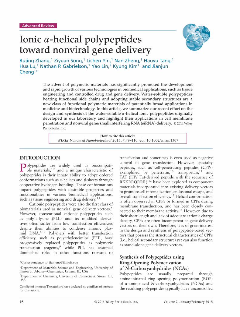
FIGURE 1 | (a) Proposed mechanism of hexamethyldisilazane (HMDS)-initiated controlled ring-opening polymerization (ROP) ofN-Carboxyanhydride (NCA). (Reprinted with permission from Ref 22 Copyright 2007 American Chemical Society) (b) Synthesis of C-terminifunctionalized polypeptides initiated by N-TMS amines. (Reprinted with permission from Ref 23 Copyright 2008 American Chemical Society) (c)Synthesis of water-soluble 𝛼-helical polypeptides PVBLG-X. (Reprinted with permission from Ref 48 Copyright 2011 Nature Publishing Group)
molecular weights (MWs) and broad molecularweight distributions (MWDs).15 In the past decade,a few controlled NCA polymerizations were reportedusing ammonium,16 transition metal complexes asinitiators,17,18 or conventional amine initiators underhigh vacuum.19 Mechanistic studies were also con-ducted on how temperature, pressure, and impuritiesaffect the polymerization.20,21 These achievements col-lectively contributed to the improvement of traditionalamine-initiated NCA polymerization. We recentlyreported hexamethyldisilazane (HMDS)-mediated,controlled NCA polymerization and identifiedtrimethylsilyl carbamate (TMS-CBM) moiety asan unusual chain-propagation group22 (Figure 1(a)).This methodology allows better control over MWand MWD, and thus enables the preparation ofwell-defined block copolypeptides. In addition, thismetal-free living polymerization can be expanded tovarious N-trimethylsilyl (N-TMS) amines and allowsfacile functionalization of C-termini of polypeptides23
(Figure 1(b)) by a variety of functional groups includ-ing alkene, alkyne, and norbornene. For instance, thissystem can be integrated with ring-opening metathesispolymerization (ROMP) by attaching TMS group onROMP monomers or chain transfer agents (CTAs),such that well-defined polypeptides containingbrush-like24 or block polymers25 can be synthesized.
In addition, new NCA monomers derived fromnoncanonical amino acids have emerged as attractivemotifs, and substantial efforts have thus been made in
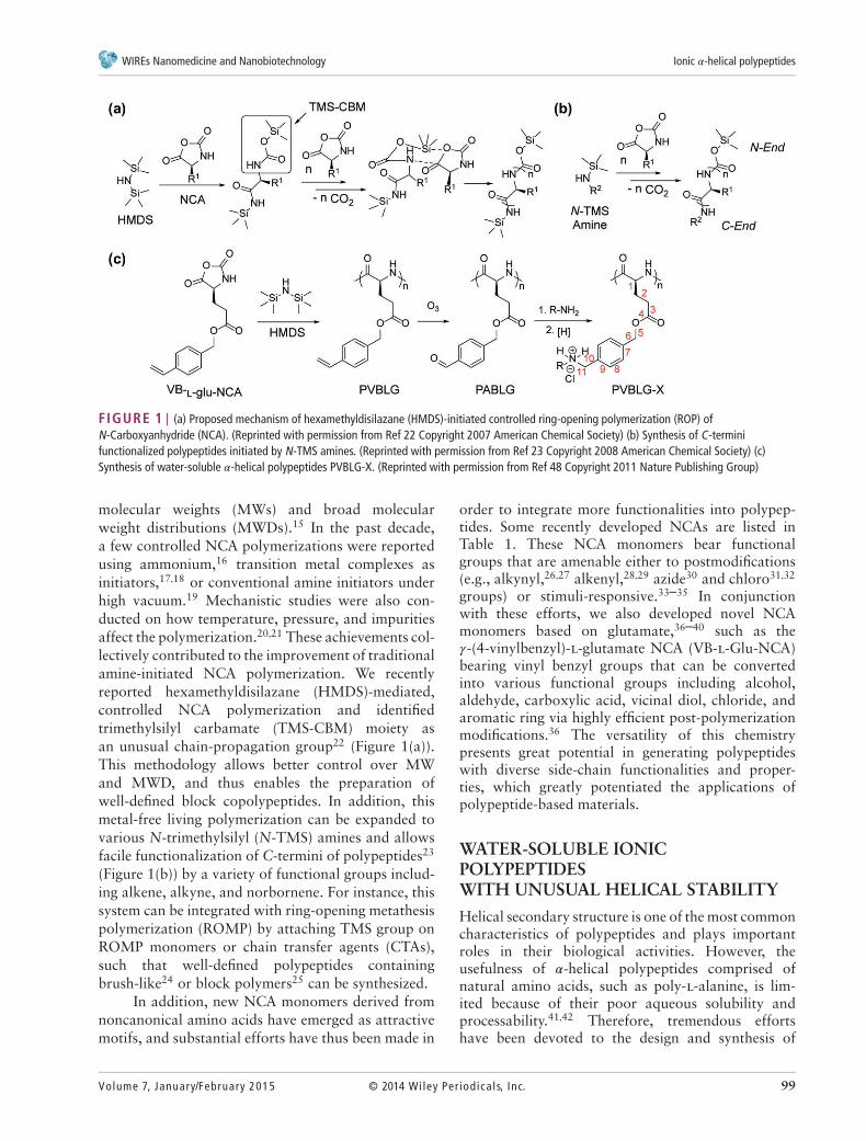
order to integrate more functionalities into polypep-tides. Some recently developed NCAs are listed inTable 1. These NCA monomers bear functionalgroups that are amenable either to postmodifications(e.g., alkynyl,26,27 alkenyl,28,29 azide30 and chloro31,32
groups) or stimuli-responsive.33–35 In conjunctionwith these efforts, we also developed novel NCAmonomers based on glutamate,36–40 such as the𝛾-(4-vinylbenzyl)-l-glutamate NCA (VB-l-Glu-NCA)bearing vinyl benzyl groups that can be convertedinto various functional groups including alcohol,aldehyde, carboxylic acid, vicinal diol, chloride, andaromatic ring via highly efficient post-polymerizationmodifications.36 The versatility of this chemistrypresents great potential in generating polypeptideswith diverse side-chain functionalities and proper-ties, which greatly potentiated the applications ofpolypeptide-based materials.
WATER-SOLUBLE IONICPOLYPEPTIDESWITH UNUSUAL HELICAL STABILITY
Helical secondary structure is one of the most commoncharacteristics of polypeptides and plays importantroles in their biological activities. However, theusefulness of 𝛼-helical polypeptides comprised ofnatural amino acids, such as poly-l-alanine, is lim-ited because of their poor aqueous solubility andprocessability.41,42 Therefore, tremendous effortshave been devoted to the design and synthesis of
Volume 7, January/February 2015 © 2014 Wiley Per iodica ls, Inc. 99
Advanced Review wires.wiley.com/nanomed
TABLE 1 The Structures of Recently DevelopedN-Carboxyanhydrides (NCAs)
NCA Structure Functionalities Ref
Alkyne-azidecycloaddition clickreaction; Thiol-ynereaction
26,27
Thiol-ene reaction 29
Alkyne-azidecycloaddition clickreaction
30
Nucleophilicsubstitution
31,32
Ozonolysis; Olefinmetathesis
36
Thermo-responsive 33
Photo-responsive 35
water-soluble polypeptides with stable helical con-formation. Incorporation of charged groups intopolypeptides significantly improves water solubil-ity, which however, also leads to reduced helicalstability because of increased side-chain chargerepulsion.43,44 Comparatively, introducing neutralhydrophilic groups (e.g., hydroxyl group, sugar moi-eties, or oligo ethylene glycol) onto the side chains canyield water-soluble polypeptides with stable helicalstructures.45–47 However, these neutral polypeptidesmay not be easily used for gene delivery due to theirlack of cationic charges to condense nucleic acids.Charged, water-soluble polypeptides that adopt sta-ble 𝛼-helical conformations would be an interestingalternative to these water-soluble, neutral, helicalpolypeptides in gene delivery.
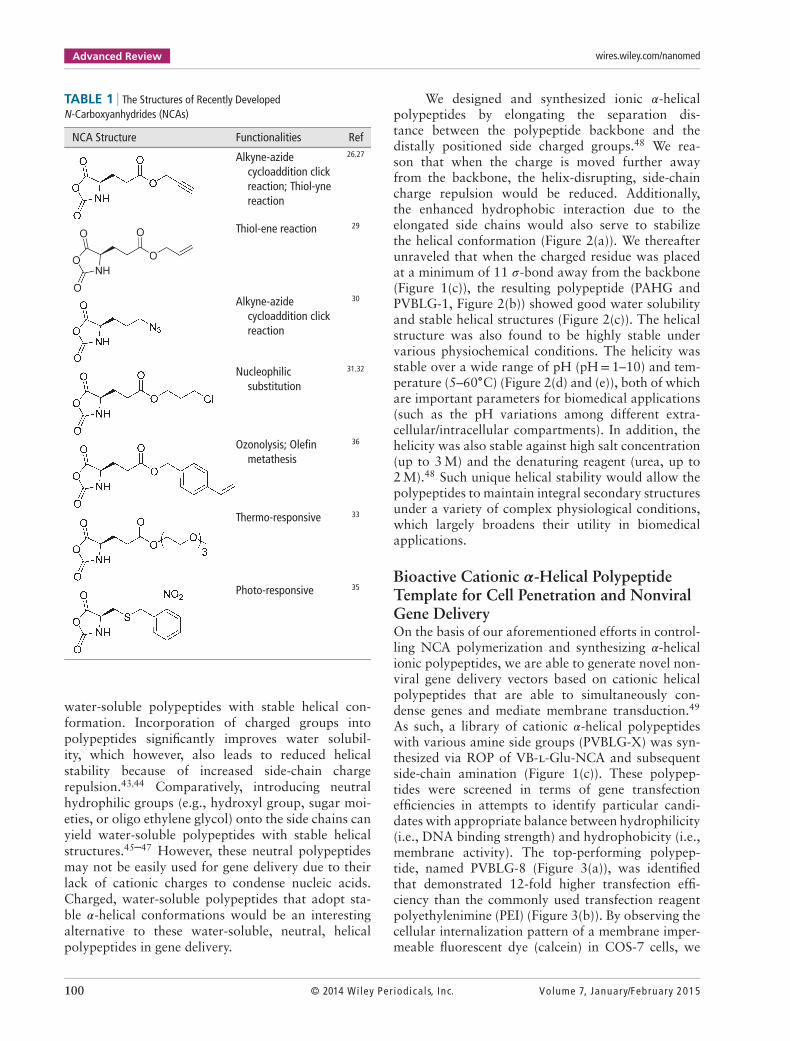
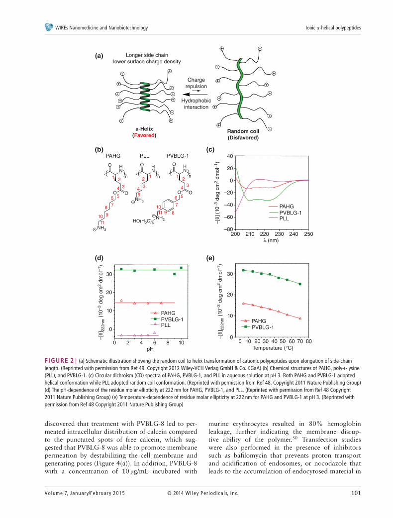
We designed and synthesized ionic 𝛼-helicalpolypeptides by elongating the separation dis-tance between the polypeptide backbone and thedistally positioned side charged groups.48 We rea-son that when the charge is moved further awayfrom the backbone, the helix-disrupting, side-chaincharge repulsion would be reduced. Additionally,the enhanced hydrophobic interaction due to theelongated side chains would also serve to stabilizethe helical conformation (Figure 2(a)). We thereafterunraveled that when the charged residue was placedat a minimum of 11 𝜎-bond away from the backbone(Figure 1(c)), the resulting polypeptide (PAHG andPVBLG-1, Figure 2(b)) showed good water solubilityand stable helical structures (Figure 2(c)). The helicalstructure was also found to be highly stable undervarious physiochemical conditions. The helicity wasstable over a wide range of pH (pH=1–10) and tem-perature (5–60∘C) (Figure 2(d) and (e)), both of whichare important parameters for biomedical applications(such as the pH variations among different extra-cellular/intracellular compartments). In addition, thehelicity was also stable against high salt concentration(up to 3 M) and the denaturing reagent (urea, up to2 M).48 Such unique helical stability would allow thepolypeptides to maintain integral secondary structuresunder a variety of complex physiological conditions,which largely broadens their utility in biomedicalapplications.
Bioactive Cationic 𝜶-Helical PolypeptideTemplate for Cell Penetration and NonviralGene DeliveryOn the basis of our aforementioned efforts in control-ling NCA polymerization and synthesizing 𝛼-helicalionic polypeptides, we are able to generate novel non-viral gene delivery vectors based on cationic helicalpolypeptides that are able to simultaneously con-dense genes and mediate membrane transduction.49
As such, a library of cationic 𝛼-helical polypeptideswith various amine side groups (PVBLG-X) was syn-thesized via ROP of VB-l-Glu-NCA and subsequentside-chain amination (Figure 1(c)). These polypep-tides were screened in terms of gene transfectionefficiencies in attempts to identify particular candi-dates with appropriate balance between hydrophilicity(i.e., DNA binding strength) and hydrophobicity (i.e.,membrane activity). The top-performing polypep-tide, named PVBLG-8 (Figure 3(a)), was identifiedthat demonstrated 12-fold higher transfection effi-ciency than the commonly used transfection reagentpolyethylenimine (PEI) (Figure 3(b)). By observing thecellular internalization pattern of a membrane imper-meable fluorescent dye (calcein) in COS-7 cells, we
100 © 2014 Wiley Per iodica ls, Inc. Volume 7, January/February 2015
WIREs Nanomedicine and Nanobiotechnology Ionic 𝛼-helical polypeptides
00 10 20 30
PAHG
PVBLG-1
PAHG
PVBLG-1PLL
PAHG
PVBLG-1PLL
40
Temperature (°C)
–[θ
] 22
2n
m (
10
–3 d
eg
cm
2 d
mo
l–1)
–[θ
] 22
2n
m (
10
–3 d
eg
cm
2 d
mo
l–1)
50 60 70 80
10
20
30
0
0 2 4 6
pH
8 10
–[θ
] (10
–3 d
eg
cm
2 d
mo
l–1)
200
HO(H2C)6
NH2
NH3
NH3
HOOO
OO
nn
NHN
HN
O O
n
11
11
10
10
7
6
7
6 5
43
5
434
5
3
222111
99 88
–80
–60
–40
–20
20
40
Hydrophobic
interaction
Charge
repulsion
Longer side chain
lower surface charge density(a)
(b) (c)
(d) (e)
Random coil(Disfavored)
a-Helix(Favored)
PVBLG-1PLLPAHG
0
210 220 230
λ (nm)
240 250
10
20
30
FIGURE 2 | (a) Schematic illustration showing the random coil to helix transformation of cationic polypeptides upon elongation of side-chainlength. (Reprinted with permission from Ref 49. Copyright 2012 Wiley-VCH Verlag GmbH & Co. KGaA) (b) Chemical structures of PAHG, poly-L-lysine(PLL), and PVBLG-1. (c) Circular dichroism (CD) spectra of PAHG, PVBLG-1, and PLL in aqueous solution at pH 3. Both PAHG and PVBLG-1 adoptedhelical conformation while PLL adopted random coil conformation. (Reprinted with permission from Ref 48. Copyright 2011 Nature Publishing Group)(d) The pH-dependence of the residue molar ellipticity at 222 nm for PAHG, PVBLG-1, and PLL. (Reprinted with permission from Ref 48 Copyright2011 Nature Publishing Group) (e) Temperature-dependence of residue molar ellipticity at 222 nm for PAHG and PVBLG-1 at pH 3. (Reprinted withpermission from Ref 48 Copyright 2011 Nature Publishing Group)
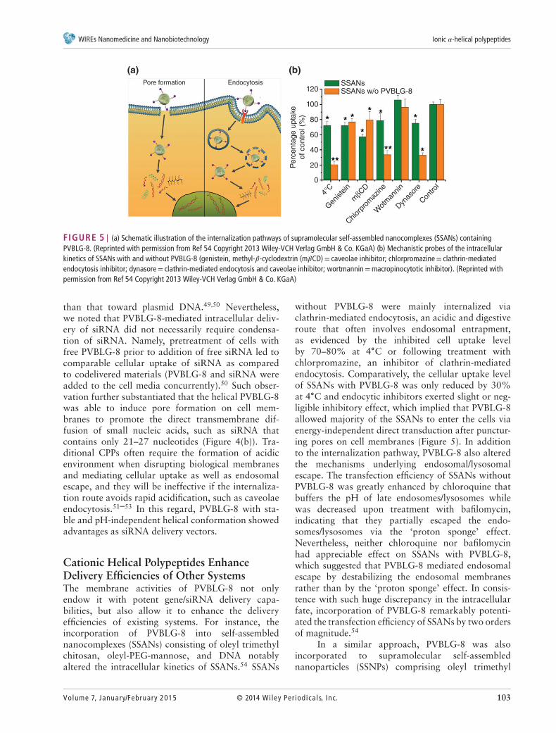
discovered that treatment with PVBLG-8 led to per-meated intracellular distribution of calcein comparedto the punctated spots of free calcein, which sug-gested that PVBLG-8 was able to promote membranepermeation by destabilizing the cell membrane andgenerating pores (Figure 4(a)). In addition, PVBLG-8with a concentration of 10 μg/mL incubated with
murine erythrocytes resulted in 80% hemoglobinleakage, further indicating the membrane disrup-tive ability of the polymer.50 Transfection studieswere also performed in the presence of inhibitorssuch as bafilomycin that prevents proton transportand acidification of endosomes, or nocodazole thatleads to the accumulation of endocytosed material in
Volume 7, January/February 2015 © 2014 Wiley Per iodica ls, Inc. 101
Advanced Review wires.wiley.com/nanomed
PVBLG-8
PVBDLG-8PEI
107(b)(a)
106
105
RLU
/mg p
rote
in
104
O
NH
OClCl
HH
H
N
PVBDLG-8PVBLG-8
N
ClCl
HH
H
NN
O
n
O
NH
O
O
n
103
1:1
5:1
10:1
15:1
20:1
30:1
35:1
PEI25
:1
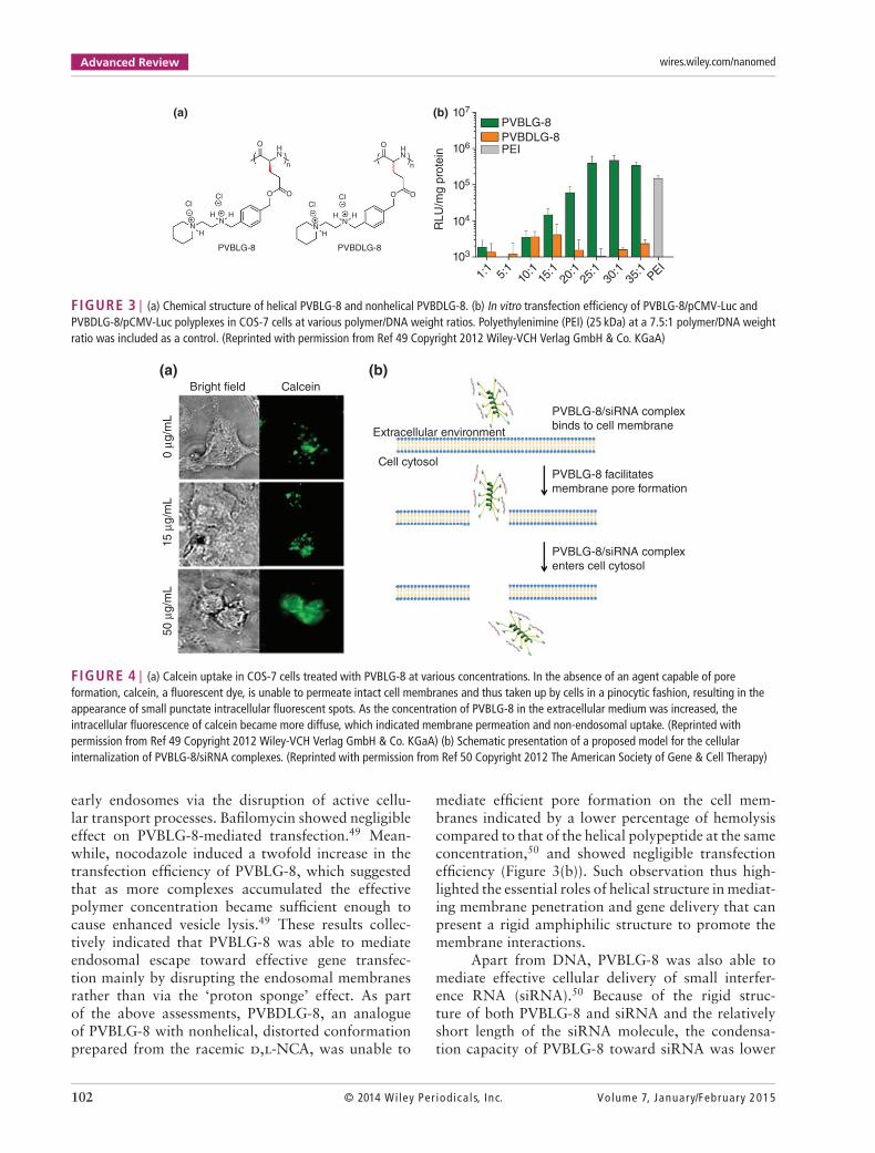
FIGURE 3 | (a) Chemical structure of helical PVBLG-8 and nonhelical PVBDLG-8. (b) In vitro transfection efficiency of PVBLG-8/pCMV-Luc andPVBDLG-8/pCMV-Luc polyplexes in COS-7 cells at various polymer/DNA weight ratios. Polyethylenimine (PEI) (25 kDa) at a 7.5:1 polymer/DNA weightratio was included as a control. (Reprinted with permission from Ref 49 Copyright 2012 Wiley-VCH Verlag GmbH & Co. KGaA)
Cell cytosol
Extracellular environment
Bright field
(a) (b)Calcein
PVBLG-8/siRNA complex
binds to cell membrane
PVBLG-8/siRNA complex
enters cell cytosol
PVBLG-8 facilitates
membrane pore formation
50 μ
g/m
L15 μ
g/m
L0 μ
g/m
L
FIGURE 4 | (a) Calcein uptake in COS-7 cells treated with PVBLG-8 at various concentrations. In the absence of an agent capable of poreformation, calcein, a fluorescent dye, is unable to permeate intact cell membranes and thus taken up by cells in a pinocytic fashion, resulting in theappearance of small punctate intracellular fluorescent spots. As the concentration of PVBLG-8 in the extracellular medium was increased, theintracellular fluorescence of calcein became more diffuse, which indicated membrane permeation and non-endosomal uptake. (Reprinted withpermission from Ref 49 Copyright 2012 Wiley-VCH Verlag GmbH & Co. KGaA) (b) Schematic presentation of a proposed model for the cellularinternalization of PVBLG-8/siRNA complexes. (Reprinted with permission from Ref 50 Copyright 2012 The American Society of Gene & Cell Therapy)
early endosomes via the disruption of active cellu-lar transport processes. Bafilomycin showed negligibleeffect on PVBLG-8-mediated transfection.49 Mean-while, nocodazole induced a twofold increase in thetransfection efficiency of PVBLG-8, which suggestedthat as more complexes accumulated the effectivepolymer concentration became sufficient enough tocause enhanced vesicle lysis.49 These results collec-tively indicated that PVBLG-8 was able to mediateendosomal escape toward effective gene transfec-tion mainly by disrupting the endosomal membranesrather than via the ‘proton sponge’ effect. As partof the above assessments, PVBDLG-8, an analogueof PVBLG-8 with nonhelical, distorted conformationprepared from the racemic d,l-NCA, was unable to
mediate efficient pore formation on the cell mem-branes indicated by a lower percentage of hemolysiscompared to that of the helical polypeptide at the sameconcentration,50 and showed negligible transfectionefficiency (Figure 3(b)). Such observation thus high-lighted the essential roles of helical structure in mediat-ing membrane penetration and gene delivery that canpresent a rigid amphiphilic structure to promote themembrane interactions.
Apart from DNA, PVBLG-8 was also able tomediate effective cellular delivery of small interfer-ence RNA (siRNA).50 Because of the rigid struc-ture of both PVBLG-8 and siRNA and the relativelyshort length of the siRNA molecule, the condensa-tion capacity of PVBLG-8 toward siRNA was lower
102 © 2014 Wiley Per iodica ls, Inc. Volume 7, January/February 2015
WIREs Nanomedicine and Nanobiotechnology Ionic 𝛼-helical polypeptides
0
4°C
Gen
iste
in
Chlor
prom
azine
Wot
man
nin
Dyn
asor
e
Con
trol
mβC
D
20
40
60
Perc
en
tag
e u
pta
ke
of
co
ntr
ol (%
)
80
100
120EndocytosisPore formation
(a) (b)SSANsSSANs w/o PVBLG-8
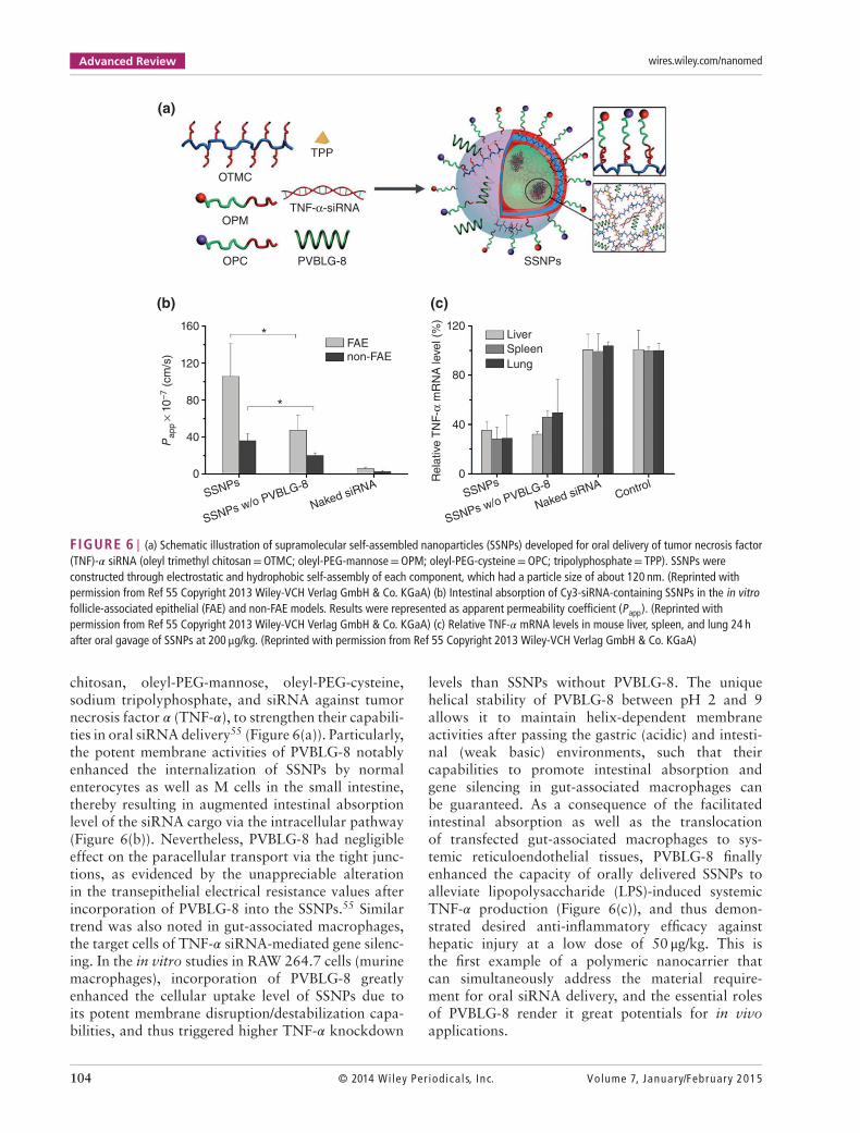
FIGURE 5 | (a) Schematic illustration of the internalization pathways of supramolecular self-assembled nanocomplexes (SSANs) containingPVBLG-8. (Reprinted with permission from Ref 54 Copyright 2013 Wiley-VCH Verlag GmbH & Co. KGaA) (b) Mechanistic probes of the intracellularkinetics of SSANs with and without PVBLG-8 (genistein, methyl-𝛽-cyclodextrin (m𝛽CD)= caveolae inhibitor; chlorpromazine= clathrin-mediatedendocytosis inhibitor; dynasore= clathrin-mediated endocytosis and caveolae inhibitor; wortmannin=macropinocytotic inhibitor). (Reprinted withpermission from Ref 54 Copyright 2013 Wiley-VCH Verlag GmbH & Co. KGaA)
than that toward plasmid DNA.49,50 Nevertheless,we noted that PVBLG-8-mediated intracellular deliv-ery of siRNA did not necessarily require condensa-tion of siRNA. Namely, pretreatment of cells withfree PVBLG-8 prior to addition of free siRNA led tocomparable cellular uptake of siRNA as comparedto codelivered materials (PVBLG-8 and siRNA wereadded to the cell media concurrently).50 Such obser-vation further substantiated that the helical PVBLG-8was able to induce pore formation on cell mem-branes to promote the direct transmembrane dif-fusion of small nucleic acids, such as siRNA thatcontains only 21–27 nucleotides (Figure 4(b)). Tra-ditional CPPs often require the formation of acidicenvironment when disrupting biological membranesand mediating cellular uptake as well as endosomalescape, and they will be ineffective if the internaliza-tion route avoids rapid acidification, such as caveolaeendocytosis.51–53 In this regard, PVBLG-8 with sta-ble and pH-independent helical conformation showedadvantages as siRNA delivery vectors.
Cationic Helical Polypeptides EnhanceDelivery Efficiencies of Other SystemsThe membrane activities of PVBLG-8 not onlyendow it with potent gene/siRNA delivery capa-bilities, but also allow it to enhance the deliveryefficiencies of existing systems. For instance, theincorporation of PVBLG-8 into self-assemblednanocomplexes (SSANs) consisting of oleyl trimethylchitosan, oleyl-PEG-mannose, and DNA notablyaltered the intracellular kinetics of SSANs.54 SSANs
without PVBLG-8 were mainly internalized viaclathrin-mediated endocytosis, an acidic and digestiveroute that often involves endosomal entrapment,as evidenced by the inhibited cell uptake levelby 70–80% at 4∘C or following treatment withchlorpromazine, an inhibitor of clathrin-mediatedendocytosis. Comparatively, the cellular uptake levelof SSANs with PVBLG-8 was only reduced by 30%at 4∘C and endocytic inhibitors exerted slight or neg-ligible inhibitory effect, which implied that PVBLG-8allowed majority of the SSANs to enter the cells viaenergy-independent direct transduction after punctur-ing pores on cell membranes (Figure 5). In additionto the internalization pathway, PVBLG-8 also alteredthe mechanisms underlying endosomal/lysosomalescape. The transfection efficiency of SSANs withoutPVBLG-8 was greatly enhanced by chloroquine thatbuffers the pH of late endosomes/lysosomes whilewas decreased upon treatment with bafilomycin,indicating that they partially escaped the endo-somes/lysosomes via the ‘proton sponge’ effect.Nevertheless, neither chloroquine nor bafilomycinhad appreciable effect on SSANs with PVBLG-8,which suggested that PVBLG-8 mediated endosomalescape by destabilizing the endosomal membranesrather than by the ‘proton sponge’ effect. In consis-tence with such huge discrepancy in the intracellularfate, incorporation of PVBLG-8 remarkably potenti-ated the transfection efficiency of SSANs by two ordersof magnitude.54
In a similar approach, PVBLG-8 was alsoincorporated to supramolecular self-assemblednanoparticles (SSNPs) comprising oleyl trimethyl
Volume 7, January/February 2015 © 2014 Wiley Per iodica ls, Inc. 103
Advanced Review wires.wiley.com/nanomed
120
80
Liver
Spleen
Lung
Re
lative
TN
F-α
mR
NA
leve
l (%
)40
0
120
160
(b)
(a)
(c)
80
Pa
pp
× 1
0–
7 (
cm
/s)
40
FAE
OPC
OPM
OTMC
PVBLG-8
TPP
SSNPs
TNF-α-siRNA
non-FAE
*
*
0
SSNPs
SSNPs w/o PVBLG-8
Naked siRNASSNPs
SSNPs w/o PVBLG-8
Naked siRNAControl
FIGURE 6 | (a) Schematic illustration of supramolecular self-assembled nanoparticles (SSNPs) developed for oral delivery of tumor necrosis factor(TNF)-𝛼 siRNA (oleyl trimethyl chitosan=OTMC; oleyl-PEG-mannose=OPM; oleyl-PEG-cysteine=OPC; tripolyphosphate= TPP). SSNPs wereconstructed through electrostatic and hydrophobic self-assembly of each component, which had a particle size of about 120 nm. (Reprinted withpermission from Ref 55 Copyright 2013 Wiley-VCH Verlag GmbH & Co. KGaA) (b) Intestinal absorption of Cy3-siRNA-containing SSNPs in the in vitrofollicle-associated epithelial (FAE) and non-FAE models. Results were represented as apparent permeability coefficient (Papp). (Reprinted withpermission from Ref 55 Copyright 2013 Wiley-VCH Verlag GmbH & Co. KGaA) (c) Relative TNF-𝛼 mRNA levels in mouse liver, spleen, and lung 24 hafter oral gavage of SSNPs at 200 μg/kg. (Reprinted with permission from Ref 55 Copyright 2013 Wiley-VCH Verlag GmbH & Co. KGaA)
chitosan, oleyl-PEG-mannose, oleyl-PEG-cysteine,sodium tripolyphosphate, and siRNA against tumornecrosis factor 𝛼 (TNF-𝛼), to strengthen their capabili-ties in oral siRNA delivery55 (Figure 6(a)). Particularly,the potent membrane activities of PVBLG-8 notablyenhanced the internalization of SSNPs by normalenterocytes as well as M cells in the small intestine,thereby resulting in augmented intestinal absorptionlevel of the siRNA cargo via the intracellular pathway(Figure 6(b)). Nevertheless, PVBLG-8 had negligibleeffect on the paracellular transport via the tight junc-tions, as evidenced by the unappreciable alterationin the transepithelial electrical resistance values afterincorporation of PVBLG-8 into the SSNPs.55 Similartrend was also noted in gut-associated macrophages,the target cells of TNF-𝛼 siRNA-mediated gene silenc-ing. In the in vitro studies in RAW 264.7 cells (murinemacrophages), incorporation of PVBLG-8 greatlyenhanced the cellular uptake level of SSNPs due toits potent membrane disruption/destabilization capa-bilities, and thus triggered higher TNF-𝛼 knockdown
levels than SSNPs without PVBLG-8. The uniquehelical stability of PVBLG-8 between pH 2 and 9allows it to maintain helix-dependent membraneactivities after passing the gastric (acidic) and intesti-nal (weak basic) environments, such that theircapabilities to promote intestinal absorption andgene silencing in gut-associated macrophages canbe guaranteed. As a consequence of the facilitatedintestinal absorption as well as the translocationof transfected gut-associated macrophages to sys-temic reticuloendothelial tissues, PVBLG-8 finallyenhanced the capacity of orally delivered SSNPs toalleviate lipopolysaccharide (LPS)-induced systemicTNF-𝛼 production (Figure 6(c)), and thus demon-strated desired anti-inflammatory efficacy againsthepatic injury at a low dose of 50 μg/kg. This isthe first example of a polymeric nanocarrier thatcan simultaneously address the material require-ment for oral siRNA delivery, and the essential rolesof PVBLG-8 render it great potentials for in vivoapplications.
104 © 2014 Wiley Per iodica ls, Inc. Volume 7, January/February 2015
WIREs Nanomedicine and Nanobiotechnology Ionic 𝛼-helical polypeptides
107
106
105
421
DNA amount (μg/well)
Homopolymer
Homocopolymer
PVBLG-8
PEG
Graft
Graft
Star
Star
Triblock
Triblock
Diblock
Diblock
30252015
10
0.40.20.10
20
40
60
Ce
ll via
bili
ty (
%)
80
100
HeLa HeLa120
Diblock
Triblock
Sta
r
Gra
ft
Hom
opolym
erLP
F2000
RL
U/m
g p
rote
in
(b)
(a)
(c)
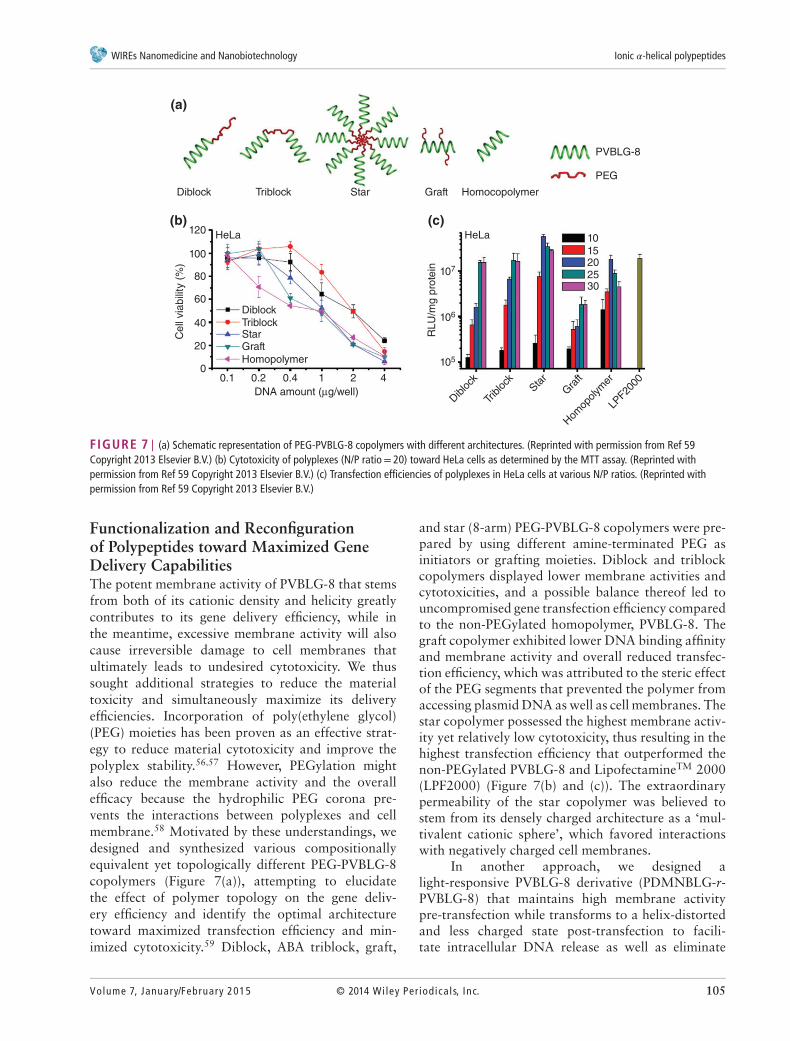
FIGURE 7 | (a) Schematic representation of PEG-PVBLG-8 copolymers with different architectures. (Reprinted with permission from Ref 59Copyright 2013 Elsevier B.V.) (b) Cytotoxicity of polyplexes (N/P ratio= 20) toward HeLa cells as determined by the MTT assay. (Reprinted withpermission from Ref 59 Copyright 2013 Elsevier B.V.) (c) Transfection efficiencies of polyplexes in HeLa cells at various N/P ratios. (Reprinted withpermission from Ref 59 Copyright 2013 Elsevier B.V.)
Functionalization and Reconfigurationof Polypeptides toward Maximized GeneDelivery CapabilitiesThe potent membrane activity of PVBLG-8 that stemsfrom both of its cationic density and helicity greatlycontributes to its gene delivery efficiency, while inthe meantime, excessive membrane activity will alsocause irreversible damage to cell membranes thatultimately leads to undesired cytotoxicity. We thussought additional strategies to reduce the materialtoxicity and simultaneously maximize its deliveryefficiencies. Incorporation of poly(ethylene glycol)(PEG) moieties has been proven as an effective strat-egy to reduce material cytotoxicity and improve thepolyplex stability.56,57 However, PEGylation mightalso reduce the membrane activity and the overallefficacy because the hydrophilic PEG corona pre-vents the interactions between polyplexes and cellmembrane.58 Motivated by these understandings, wedesigned and synthesized various compositionallyequivalent yet topologically different PEG-PVBLG-8copolymers (Figure 7(a)), attempting to elucidatethe effect of polymer topology on the gene deliv-ery efficiency and identify the optimal architecturetoward maximized transfection efficiency and min-imized cytotoxicity.59 Diblock, ABA triblock, graft,
and star (8-arm) PEG-PVBLG-8 copolymers were pre-pared by using different amine-terminated PEG asinitiators or grafting moieties. Diblock and triblockcopolymers displayed lower membrane activities andcytotoxicities, and a possible balance thereof led touncompromised gene transfection efficiency comparedto the non-PEGylated homopolymer, PVBLG-8. Thegraft copolymer exhibited lower DNA binding affinityand membrane activity and overall reduced transfec-tion efficiency, which was attributed to the steric effectof the PEG segments that prevented the polymer fromaccessing plasmid DNA as well as cell membranes. Thestar copolymer possessed the highest membrane activ-ity yet relatively low cytotoxicity, thus resulting in thehighest transfection efficiency that outperformed thenon-PEGylated PVBLG-8 and LipofectamineTM 2000(LPF2000) (Figure 7(b) and (c)). The extraordinarypermeability of the star copolymer was believed tostem from its densely charged architecture as a ‘mul-tivalent cationic sphere’, which favored interactionswith negatively charged cell membranes.
In another approach, we designed alight-responsive PVBLG-8 derivative (PDMNBLG-r-PVBLG-8) that maintains high membrane activitypre-transfection while transforms to a helix-distortedand less charged state post-transfection to facili-tate intracellular DNA release as well as eliminate
Volume 7, January/February 2015 © 2014 Wiley Per iodica ls, Inc. 105
Advanced Review wires.wiley.com/nanomed
Non-UV
UV/NIR
UV
(b)
(a)
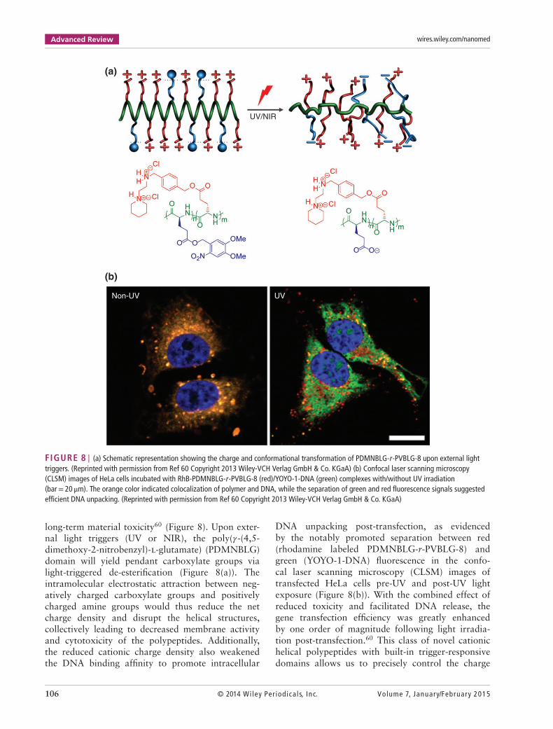
FIGURE 8 | (a) Schematic representation showing the charge and conformational transformation of PDMNBLG-r-PVBLG-8 upon external lighttriggers. (Reprinted with permission from Ref 60 Copyright 2013 Wiley-VCH Verlag GmbH & Co. KGaA) (b) Confocal laser scanning microscopy(CLSM) images of HeLa cells incubated with RhB-PDMNBLG-r-PVBLG-8 (red)/YOYO-1-DNA (green) complexes with/without UV irradiation(bar= 20 μm). The orange color indicated colocalization of polymer and DNA, while the separation of green and red fluorescence signals suggestedefficient DNA unpacking. (Reprinted with permission from Ref 60 Copyright 2013 Wiley-VCH Verlag GmbH & Co. KGaA)
long-term material toxicity60 (Figure 8). Upon exter-nal light triggers (UV or NIR), the poly(𝛾-(4,5-dimethoxy-2-nitrobenzyl)-l-glutamate) (PDMNBLG)domain will yield pendant carboxylate groups vialight-triggered de-esterification (Figure 8(a)). Theintramolecular electrostatic attraction between neg-atively charged carboxylate groups and positivelycharged amine groups would thus reduce the netcharge density and disrupt the helical structures,collectively leading to decreased membrane activityand cytotoxicity of the polypeptides. Additionally,the reduced cationic charge density also weakenedthe DNA binding affinity to promote intracellular
DNA unpacking post-transfection, as evidencedby the notably promoted separation between red(rhodamine labeled PDMNBLG-r-PVBLG-8) andgreen (YOYO-1-DNA) fluorescence in the confo-cal laser scanning microscopy (CLSM) images oftransfected HeLa cells pre-UV and post-UV lightexposure (Figure 8(b)). With the combined effect ofreduced toxicity and facilitated DNA release, thegene transfection efficiency was greatly enhancedby one order of magnitude following light irradia-tion post-transfection.60 This class of novel cationichelical polypeptides with built-in trigger-responsivedomains allows us to precisely control the charge
106 © 2014 Wiley Per iodica ls, Inc. Volume 7, January/February 2015
WIREs Nanomedicine and Nanobiotechnology Ionic 𝛼-helical polypeptides
and conformational change of the polypeptidesusing external stimuli, and ultimately manipulate theintracellular responses to overcome multiple barriersagainst nonviral gene transfer.
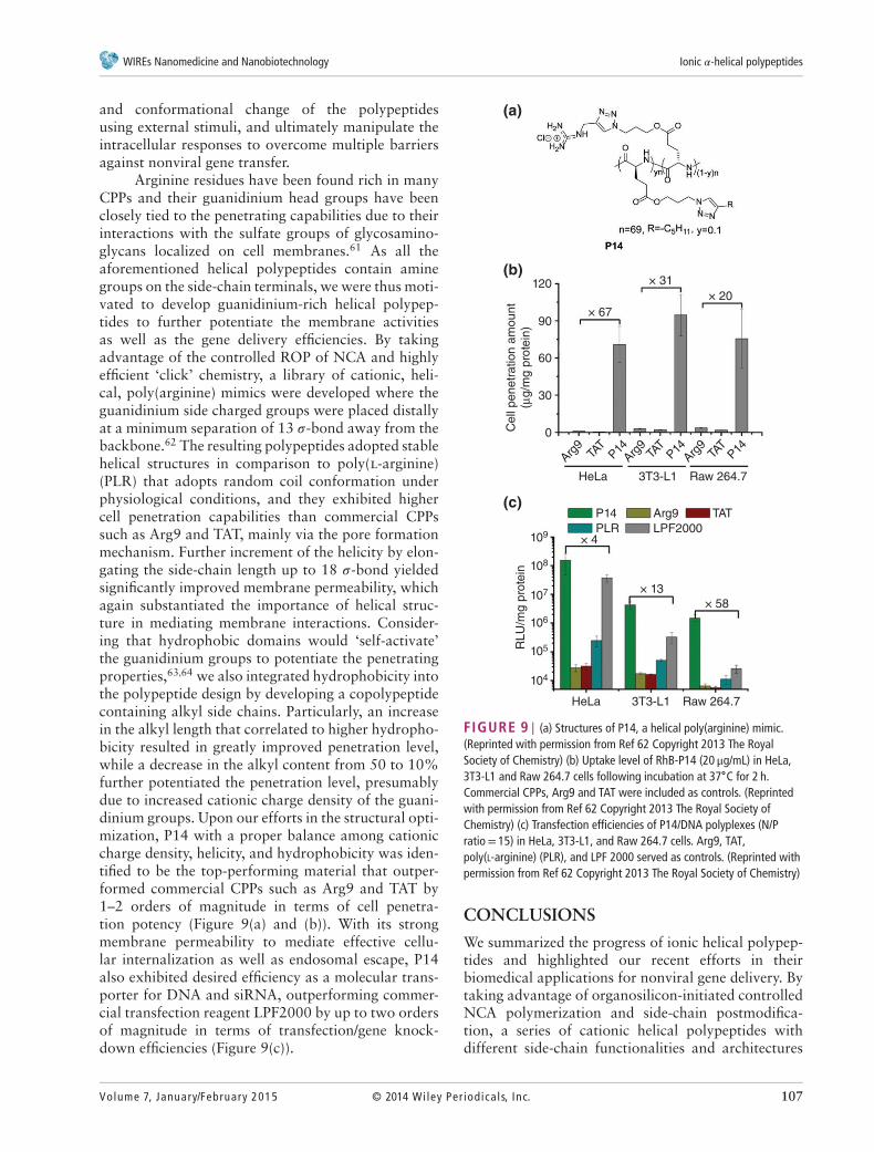
Arginine residues have been found rich in manyCPPs and their guanidinium head groups have beenclosely tied to the penetrating capabilities due to theirinteractions with the sulfate groups of glycosamino-glycans localized on cell membranes.61 As all theaforementioned helical polypeptides contain aminegroups on the side-chain terminals, we were thus moti-vated to develop guanidinium-rich helical polypep-tides to further potentiate the membrane activitiesas well as the gene delivery efficiencies. By takingadvantage of the controlled ROP of NCA and highlyefficient ‘click’ chemistry, a library of cationic, heli-cal, poly(arginine) mimics were developed where theguanidinium side charged groups were placed distallyat a minimum separation of 13 𝜎-bond away from thebackbone.62 The resulting polypeptides adopted stablehelical structures in comparison to poly(l-arginine)(PLR) that adopts random coil conformation underphysiological conditions, and they exhibited highercell penetration capabilities than commercial CPPssuch as Arg9 and TAT, mainly via the pore formationmechanism. Further increment of the helicity by elon-gating the side-chain length up to 18 𝜎-bond yieldedsignificantly improved membrane permeability, whichagain substantiated the importance of helical struc-ture in mediating membrane interactions. Consider-ing that hydrophobic domains would ‘self-activate’the guanidinium groups to potentiate the penetratingproperties,63,64 we also integrated hydrophobicity intothe polypeptide design by developing a copolypeptidecontaining alkyl side chains. Particularly, an increasein the alkyl length that correlated to higher hydropho-bicity resulted in greatly improved penetration level,while a decrease in the alkyl content from 50 to 10%further potentiated the penetration level, presumablydue to increased cationic charge density of the guani-dinium groups. Upon our efforts in the structural opti-mization, P14 with a proper balance among cationiccharge density, helicity, and hydrophobicity was iden-tified to be the top-performing material that outper-formed commercial CPPs such as Arg9 and TAT by1–2 orders of magnitude in terms of cell penetra-tion potency (Figure 9(a) and (b)). With its strongmembrane permeability to mediate effective cellu-lar internalization as well as endosomal escape, P14also exhibited desired efficiency as a molecular trans-porter for DNA and siRNA, outperforming commer-cial transfection reagent LPF2000 by up to two ordersof magnitude in terms of transfection/gene knock-down efficiencies (Figure 9(c)).
120
90
60
30
× 67
× 31
× 13
P14
PLR
Arg9 TAT
LPF2000
× 58
× 4
× 20
0
109
108
107
106
RL
U/m
g p
rote
in
105
104
HeLa 3T3-L1 Raw 264.7
HeLa 3T3-L1 Raw 264.7
Cell
pe
ne
tra
tio
n a
mo
un
t
(μg
/mg
pro
tein
)
Arg9
TAT
TAT
TAT
P14 P14P14Arg
9Arg
9
(c)
(b)
(a)
FIGURE 9 | (a) Structures of P14, a helical poly(arginine) mimic.(Reprinted with permission from Ref 62 Copyright 2013 The RoyalSociety of Chemistry) (b) Uptake level of RhB-P14 (20 μg/mL) in HeLa,3T3-L1 and Raw 264.7 cells following incubation at 37∘C for 2 h.Commercial CPPs, Arg9 and TAT were included as controls. (Reprintedwith permission from Ref 62 Copyright 2013 The Royal Society ofChemistry) (c) Transfection efficiencies of P14/DNA polyplexes (N/Pratio= 15) in HeLa, 3T3-L1, and Raw 264.7 cells. Arg9, TAT,poly(L-arginine) (PLR), and LPF 2000 served as controls. (Reprinted withpermission from Ref 62 Copyright 2013 The Royal Society of Chemistry)
CONCLUSIONS
We summarized the progress of ionic helical polypep-tides and highlighted our recent efforts in theirbiomedical applications for nonviral gene delivery. Bytaking advantage of organosilicon-initiated controlledNCA polymerization and side-chain postmodifica-tion, a series of cationic helical polypeptides withdifferent side-chain functionalities and architectures
Volume 7, January/February 2015 © 2014 Wiley Per iodica ls, Inc. 107
Advanced Review wires.wiley.com/nanomed
were developed. These materials possessed desiredmembrane activities mainly via the pore formationmechanism, and helical secondary structure wasdemonstrated to be essential toward their membranepermeabilities. As such, by facilitating direct trans-duction of the gene cargo across cellular as wellas endosomal membranes, the helical polypeptidesmediated effective gene/siRNA delivery by themselvesor greatly strengthened the delivery efficiencies ofexisting systems. By incorporating multifunctionali-ties or reconfiguring the polypeptide architectures, wewere able to modulate the membrane activities and
material toxicities of the helical polypeptides, thusmaximizing their gene delivery capabilities. Despite allthese success, the detailed mechanism underlying thepolypeptide-mediated membrane penetration remainsto be unraveled using techniques such as small-angleX-ray scattering. A proper structural reconfigurationas well as formulation is also necessitated to maximizethe in vivo gene delivery efficiency toward therapeuticattempts. Therefore, developing new ionic helicalpolypeptides with trigger-responsive, degradable, andtargeting moieties will be a promising subject in thefield.
ACKNOWLEDGMENTS
We acknowledge the financial support from the NSF (CHE-1153122) and the NIH (NIH Director’s NewInnovator Award 1DP2OD007246 and 1R21EB013379).
REFERENCES1. Obeid R, Scholz C. Synthesis and self-assembly of
well-defined poly(amino acid) end-capped poly(ethyleneglycol) and poly(2-methyl-2-oxazoline). Biomacro-molecules 2011, 12:3797.
2. Huang S, Bai M, Wang L. General and facile sur-face functionalization of hydrophobic nanocrystals withpoly(amino acid) for cell luminescence imaging. Sci Rep2013, 3:2023.
3. Deming TJ. Methodologies for preparation of syntheticblock copolypeptides: materials with future promise indrug delivery. Adv Drug Deliv Rev 2002, 54:1145.
4. Deming TJ. Synthetic polypeptides for biomedical appli-cations. Prog Polym Sci 2007, 32:858.
5. Monsigny M, Roche AC, Midoux P, Mayer R. Glyco-conjugates as carriers for specific delivery of therapeuticdrugs and genes. Adv Drug Deliv Rev 1994, 14:1.
6. Ferkol T, Perales JC, Mularo F, Hanson RW.Receptor-mediated gene transfer into macrophages.Proc Natl Acad Sci U S A 1996, 93:101.
7. Putnam D, Gentry CA, Pack DW, Langer R.Polymer-based gene delivery with low cytotoxicityby a unique balance of side-chain termini. Proc NatlAcad Sci U S A 2001, 98:1200.
8. Okuda T, Sugiyama A, Niidome T, Aoyagi H. Charac-ters of dendritic poly(l-lysine) analogues with the ter-minal lysines replaced with arginines and histidines asgene carriers in vitro. Biomaterials 2004, 25:537.
9. Hsu CYM, Uludag H. A simple and rapid nonviralapproach to efficiently transfect primary tissue-derivedcells using polyethylenimine. Nat Protoc 2012, 7:935.
10. Terrone D, Sang SLW, Roudaia L, Silvius JR. Pene-tratin and related cell-penetrating cationic peptides can
translocate across lipid bilayers in the presence of atransbilayer potential. Biochemistry 2003, 42:13787.
11. Pooga M, Hallbrink M, Zorko M, Langel U. Cellpenetration by transportan. FASEB J 1998, 12:67.
12. Brooks H, Lebleu B, Vives E. Tat peptide-mediatedcellular delivery: back to basics. Adv Drug Deliv Rev2005, 57:559.
13. Martin ME, Rice KG. Peptide-guided gene delivery.AAPS J 2007, 9:E18.
14. Derossi D, Calvet S, Trembleau A, Brunissen A,Chassaing G, Prochiantz A. Cell internalization ofthe third helix of the antennapedia homeodomain isreceptor-independent. J Biol Chem 1996, 271:18188.
15. Deming TJ. Polypeptide and polypeptide hybrid copoly-mer synthesis via NCA polymerization. Adv Polym Sci2006, 202:1.
16. Dimitrov I, Schlaad H. Synthesis of nearly monodis-perse polystyrene-polypeptide block copolymers viapolymerisation of N-carboxyanhydrides. Chem Com-mun 2003:2944.
17. Deming TJ. Facile synthesis of block copolypeptides ofdefined architecture. Nature 1997, 390:386.
18. Deming TJ. Amino acid derived nickelacycles: interme-diates in nickel-mediated polypeptide synthesis. J AmChem Soc 1998, 120:4240.
19. Aliferis T, Iatrou H, Hadjichristidis N. Living polypep-tides. Biomacromolecules 2004, 5:1653.
20. Habraken GJM, Peeters M, Dietz CHJT, Kon-ing CE, Heise A. How controlled and versatile isN-carboxy anhydride (NCA) polymerization at 0 oC?Effect of temperature on homo-, block- and graft(co)polymerization. Polym Chem 2010, 1:514.
108 © 2014 Wiley Per iodica ls, Inc. Volume 7, January/February 2015
WIREs Nanomedicine and Nanobiotechnology Ionic 𝛼-helical polypeptides
21. Pickel DL, Politakos N, Avgeropoulos A, Mess-man JM. A mechanistic study of alpha-(aminoacid)-N-carboxyanhydride polymerization: comparinginitiation and termination events in high-vacuum andtraditional polymerization techniques. Macromolecules2009, 42:7781.
22. Lu H, Cheng J. Hexamethyldisilazane-mediatedcontrolled polymerization of alpha-amino acidN-carboxyanhydrides. J Am Chem Soc 2007, 129:14114.
23. Lu H, Cheng J. N-trimethylsilyl amines for con-trolled ring-opening polymerization of amino acidN-carboxyanhydrides and facile end group func-tionalization of polypeptides. J Am Chem Soc 2008,130:12562.
24. Lu H, Wang J, Lin Y, Cheng J. One-pot synthe-sis of brush-like polymers via integrated ring-openingmetathesis polymerization and polymerization of aminoacid N-carboxyanhydrides. J Am Chem Soc 2009,131:13582.
25. Bai Y, Lu H, Ponnusamy E, Cheng J. Synthesis ofhybrid block copolymers via integrated ring-openingmetathesis polymerization and polymerization of NCA.Chem Commun 2011, 47:10830.
26. Engler C, Lee HI, Hammond PT. Highly efficient"grafting onto" a polypeptide backbone using clickchemistry. Angew Chem Int Ed 2009, 48:9334.
27. Huang YG, Zeng YH, Yang JW, Zeng ZH, Zhu FM,Chen XD. Facile functionalization of polypeptides bythiol-yne photochemistry for biomimetic materials syn-thesis. Chem Commun 2011, 47:7509.
28. Sun J, Schlaad H. Thiol-ene clickable polypeptides.Macromolecules 2010, 43:4445.
29. Tang H, Zhang D. Multi-functionalization of helicalblock copoly(alpha-peptide)s by orthogonal chemistry.Polym Chem 2011, 2:1542.
30. Rhodes J, Deming TJ. Soluble, clickable polypeptidesfrom azide-containing N-carboxyanhydride monomers.ACS Macro Lett 2013, 2:351.
31. Tang H, Zhang D. General route toward side-chain-functionalized alpha-helical polypeptides. Bio-macromolecules 2010, 11:1585.
32. Song Z, Zheng N, Ba X, Yin L, Zhang R, Ma L, Cheng J.Polypeptides with quaternary phosphonium side chains:synthesis, characterization, and cell-penetrating proper-ties. Biomacromolecules 2014, 15:1491.
33. Chen C, Wang Z, Li Z. Thermoresponsive polypeptidesfrom pegylated poly-l-glutamates. Biomacromolecules2011, 12:2859.
34. Fu XH, Shen Y, Fu WX, Li ZB. Thermoresponsiveoligo(ethylene glycol) functionalized poly-l-cysteine.Macromolecules 2013, 46:3753.
35. Liu G, Dong CM. Photoresponsive poly(S-(o-nitrobenzyl)-l-cysteine)-b-PEO from a l-cysteine N-car-boxyanhydride monomer: synthesis, self-assembly, and
phototriggered drug release. Biomacromolecules 2012,13:1573.
36. Lu H, Bai Y, Wang J, Gabrielson NP, Wang F, LinY, Cheng J. Ring-opening polymerization of gamma-(4-vinylbenzyl)-l-glutamate N-carboxyanhydride forthe synthesis of functional polypeptides. Macromole-cules 2011, 44:6237.
37. Zhang Y, Lu H, Lin Y, Cheng J. Water-soluble polypep-tides with elongated, charged side chains adopt ultra-stable helical conformations. Macromolecules 2011,44:6641.
38. Tang H, Yin L, Lu H, Cheng J. Water-solublepoly(l-serine)s with elongated and charged side-chains:synthesis, conformations, and cell-penetrating proper-ties. Biomacromolecules 2012, 13:2609.
39. Zheng N, Yin L, Song Z, Ma L, Tang H, Gabrielson NP,Lu H, Cheng J. Maximizing gene delivery efficienciesof cationic helical polypeptides via balanced membranepenetration and cellular targeting. Biomaterials 2014,35:1302.
40. Zhang R, Zheng N, Song Z, Yin L, Cheng J. Theeffect of side-chain functionality and hydrophobicityon the gene delivery capabilities of cationic helicalpolypeptides. Biomaterials 2014, 35:3443.
41. Levy Y, Jortner J, Becker OM. Solvent effects on theenergy landscapes and folding kinetics of polyalanine.Proc Natl Acad Sci U S A 2001, 98:2188.
42. Dobson CM, Sali A, Karplus M. Protein folding: aperspective from theory and experiment. Angew ChemInt Ed 1998, 37:868.
43. Dobson CM. Protein folding and misfolding. Nature2003, 426:884.
44. Dill KA. Dominant forces in protein folding. Biochem-istry 1990, 29:7133.
45. Lotan N, Yaron A, Berger A. Stabilization of alpha-helixin aqueous solution by hydrophobic side-chain interac-tion. Biopolymers 1966, 4:365.
46. Yu M, Nowak AP, Deming TJ, Pochan DJ. Methylatedmono- and diethyleneglycol functionalized polylysines:nonionic, alpha-helical, water-soluble polypeptides. JAm Chem Soc 1999, 121:12210.
47. Kramer JR, Deming TJ. Glycopolypeptides with aredox-triggered helix-to-coil transition. J Am Chem Soc2012, 134:4112.
48. Lu H, Wang J, Bai Y, Lang JW, Liu SY, Lin Y, ChengJ. Ionic polypeptides with unusual helical stability. NatCommun 2011:206.
49. Gabrielson NP, Lu H, Yin L, Li D, Wang F, Cheng J.Reactive and bioactive cationic alpha-helical polypep-tide template for nonviral gene delivery. Angew ChemInt Ed 2012, 51:1143.
50. Gabrielson NP, Lu H, Yin L, Kim KH, Cheng J. Acell-penetrating helical polymer for siRNA delivery tomammalian cells. Mol Ther 2012, 20:1599.
Volume 7, January/February 2015 © 2014 Wiley Per iodica ls, Inc. 109

Advanced Review wires.wiley.com/nanomed
51. Davidson TJ, Harel S, Arboleda VA, Prunell GF, She-lanski ML, Greene LA, Troy CM. Highly efficientsmall interfering RNA delivery to primary mammalianneurons induces microRNA-like effects before mRNAdegradation. J Neurosci 2004, 24:10040.
52. Bjorklund J, Biverstahl H, Graslund A, Maler L,Brzezinski P. Real-time transmembrane translocation ofpenetratin driven by light-generated proton pumping.Biophys J 2006, 91:L29.
53. Rejman J, Bragonzi A, Conese M. Role of clathrin-and caveolae-mediated endocytosis in gene transfermediated by lipo- and polyplexes. Mol Ther 2005, 12:468.
54. Yin L, Song Z, Kim KH, Zheng N, Gabrielson NP,Cheng J. Non-viral gene delivery via membrane-penetrating, mannose-targeting supramolecular self-assembled nanocomplexes. Adv Mater 2013, 25:3063.
55. Yin L, Song Z, Qu Q, Kim KH, Zheng N, Yao C,Chaudhury I, Tang H, Gabrielson NP, Uckun FM, et al.Supramolecular self-assembled nanoparticles mediateoral delivery of therapeutic TNF-alpha siRNA againstsystemic inflammation. Angew Chem Int Ed 2013,52:5757.
56. Deshpande MC, Davies MC, Garnett MC, WilliamsPM, Armitage D, Bailey L, Vamvakaki M, Armes SP,Stolnik S. The effect of poly(ethylene glycol) moleculararchitecture on cellular interaction and uptake of DNAcomplexes. J Control Release 2004, 97:143.
57. Venkataraman S, Ong WL, Ong ZY, Loo SCJ, EePLR, Yang YY. The role of PEG architecture and
molecular weight in the gene transfection performanceof PEGylated poly(dimethylaminoethyl methacrylate)based cationic polymers. Biomaterials 2011, 32:2369.
58. Liu TQ, Thierry B. A solution to the PEG dilemma: effi-cient bioconjugation of large gold nanoparticles for bio-diagnostic applications using mixed layers. Langmuir2012, 28:15634.
59. Yin L, Song Z, Kim KH, Zheng N, Tang H, Lu H,Gabrielson N, Cheng J. Reconfiguring the architecturesof cationic helical polypeptides to control non-viral genedelivery. Biomaterials 2013, 34:2340.
60. Yin L, Tang H, Kim KH, Zheng N, Song Z, GabrielsonNP, Lu H, Cheng J. Light-responsive helical polypep-tides capable of reducing toxicity and unpacking DNA:toward nonviral gene delivery. Angew Chem Int Ed2013, 52:9182.
61. Wender PA, Galliher WC, Goun EA, Jones LR, PillowTH. The design of guanidinium-rich transporters andtheir internalization mechanisms. Adv Drug Deliv Rev2008, 60:452.
62. Tang H, Yin L, Kim KH, Cheng J. Helical poly(arginine)mimics with superior cell-penetrating and moleculartransporting properties. Chem Sci 2013, 4:3839.
63. Som A, Reuter A, Tew GN. Protein transductiondomain mimics: the role of aromatic functionality.Angew Chem Int Ed 2012, 51:980.
64. Som A, Tezgel AO, Gabriel GJ, Tew GN. Self-activationin de novo designed mimics of cell-penetrating peptides.Angew Chem Int Ed 2011, 50:6147.
110 © 2014 Wiley Per iodica ls, Inc. Volume 7, January/February 2015
Top Related