


γλώσσες
Σελίδες
Νομικός

MyD88 Structure and IL1β Signaling
INTERACTIVE SITES IN THE MYD88 TIR DOMAIN RESPONSIBLE
FOR COUPLING TO THE IL1β SIGNALING PATHWAY* Chunsheng Li, Jozef Zienkiewicz and Jacek Hawiger
From the Department of Microbiology and Immunology, Vanderbilt University School of Medicine, Vanderbilt University Medical Center, Nashville, TN 37232
Running Title: MyD88 Structure and IL1β Signaling Address correspondence to: Jacek Hawiger, Department of Microbiology and Immunology, Vanderbilt University School of Medicine, 1161 21st Avenue South, A-5321 MCN, Nashville, TN 37232-2363, Tel: 615 343-8280; Fax: 615 343-8278 ; Email: [email protected]
Myeloid differentiation factor 88
(MyD88) is the essential adaptor protein that integrates and transduces intracellular signals generated by multiple Toll-like receptors (TLRs) including receptor complex for interleukin(IL)1β, a key inflammatory cytokine. IL1β receptor complex interacts with MyD88 via the Toll/IL1 receptor (TIR) domain. Here we report structure-function studies that help define the MyD88 TIR domain binding sites involved in IL1β-induced protein-protein interactions. MyD88 TIR domain, employed as dominant negative inhibitor of IL1β signaling to screen MyD88 TIR mutants, lost its suppressing activity upon truncation of its Box 3. Accordingly, mutations of Box 3 residues 285-286 reversed the dominant negative effect of MyD88 TIR domain on IL1β-induced and NFκB-dependent reporter gene activity and IL6 production. Moreover, mutations of residues 171 in helixαA, 195-197 in Box 2, and 275 in βE strand had similar functional effects. Strikingly, only mutations of residues 195-197 eliminated the TIR-TIR interaction of MyD88 and IL1RAcP while substitution of neighboring canonical Proline 200 by Histidine was without effect. Mutations in Box 2 and 3 prevented homotypic MyD88 oligomerization via TIR domain. Based on this structure-function analysis, a 3-D docking model of TIR-TIR interaction between MyD88 and IL1RAcP was developed.
The importance of Toll-like receptor (TLR1) family in innate immune response to microbial surfaces and nucleic acids is well established. Surprisingly, two members of this
family recognize key inflammatory cytokines Interleukin (IL)1 and IL18. These cytokines induce genes that encode other mediators of inflammation such as pleiotropic inflammatory cytokine IL6 and interferon gamma, respectively (1-5). Consistent with these studies, ILβ is one of the most potent inflammatory cytokines responsible for fever, leukocytosis, thrombocytosis, and production of IL6 and other cytokines (1-3). The signals generated by IL1β binding to its cognate receptor complex, formed by two type 1 transmembrane proteins, IL1 receptor I (IL1RI) and IL1 receptor accessory protein (IL1RAcP), are transduced by their cytoplasmic segments denoted Toll/interleukin 1 receptor (TIR) domain. TIR domain is shared with Drosophila Toll, mammalian TLRs, and cytoplasmic adaptors exemplified by a Myeloid Differentiation 88 (MyD88) (6). MyD88 adaptor integrates signals flowing from IL1R/IL1RAcP and from an array of other TLRs (6,7). This initial IL1 receptor–MyD88 adaptor interaction evoked by IL1β is a critical step in its signaling to the nucleus and, therefore, represents a potential target for new anti-inflammatory agents. MyD88 has a bi-partite structure comprised of amino terminal Death domain and carboxyl terminal TIR domain with a short intervening linker segment (6). Upon IL1β stimulation, IL1RI/IL1RAcP complex recruits MyD88 via its TIR domain (8). In addition, IL1RI-associated kinases are recruited to an IL1RI/IL1RAcP complex including IRAK (9,10), IRAK-2 (11), IRAK-4 (12) and IRAK-M (13). Our current understanding of IL1 receptor complex–MyD88 adaptor interaction is limited. Here we report studies that help to establish the
JBC Papers in Press. Published on April 22, 2005 as Manuscript M503262200
Copyright 2005 by The American Society for Biochemistry and Molecular Biology, Inc.
by guest on March 17, 2018
http://ww
w.jbc.org/
Dow
nloaded from
MyD88 Structure and IL1β Signaling
2
molecular determinants of MyD88 TIR domain interactions in IL1β signaling pathway.
In terms of its structural features, MyD88 TIR domain contains three highly conserved motifs denoted Box 1, 2, and 3 (Figs. 1 and 2). Box 2 forms a loop denoted the BB-loop that contains invariant proline residue at position 200 which in other receptors and adaptors, namely TLR2, TLR4, IL1RAcP, and MAL/TIRAP, is essential for their signaling function (6,7). For example, mutating this residue to histidine in TLR4 renders C3H/HeJ mice hyporesponsive to lipopolysaccharide (LPS) (14). This canonical example indicates that conserved structural motifs in TIR domain of TLRs and their adaptors play highly significant role in proinflammatory ligands- initiated intracellular interactions between TLRs and their adaptors. Depending on the recognition of distinct ligands by TLRs, the preferential usage of its adaptors may require different interacting sites in TIR domain of the same adaptor or an alternative adaptor. The latter applies to TLR3 which requires its adaptor TRIF rather than MyD88 for signaling by viral dsRNA. Conversely, TRIF mediates signaling induced by interaction of LPS with TLR4 in the absence of MyD88 (7). We hypothesized that signaling evoked by IL1β through its cognate receptor complex may depend on different interactive sites on TIR domain of MyD88 than recently reported sites involved in the MyD88 interaction with TIR domains of TLR2 and TLR4 (15).
To test this hypothesis, we undertook our studies focused on the potential role of Box 1, 2 and 3 of MyD88 TIR domain in IL1β signaling. Within these three boxes we focused on residues representing AA-loop, BB-loop, and EE-loop that were reported to participate in interactions of MyD88 with TLR2 and TLR4 (15). The functional consequences of this mutational analysis were monitored by NFκB-dependent reporter gene activity and by IL1β-induced expression of the endogenous gene that encodes inflammatory cytokine IL6. Our structure-function studies of MyD88 TIR domain led to the development of a 3-D docking model of MyD88 and IL1RAcP interaction mediated by their respective TIR domains.
EXPERIMENTAL PROCEDURES Maintenance and Treatment of Cell Lines - Human embryonic kidney (HEK) 293T cells and human fibroblast MRC-5 cells were obtained from the American Type Culture Collection (Manassas, VA). HEK 293T cells were cultured in Dulbecco’s modified Eagle’s medium (DMEM; Cellgro, VA) supplemented with 10% heat-inactivated fetal bovine serum (HI-FBS) containing no detectable LPS (<6 pg/mL) as determined by the manufacturer, Atlanta Biological, Norcross, GA), L-glutamine (2mM), penicillin (100 U/mL) and streptomycin (100 µg/mL). MRC-5 cells were maintained in Minimum essential medium (Eagle) supplemented with 10% HI-FBS, L-glutamine (2mM), penicillin (100 U/mL) and streptomycin (100 µg/mL). All cells were maintained at 37° C in a humidified atmosphere of 5% CO2. Plasmids and Reagents - The NFκB-luciferase reporter construct (NFκB-luc) containing five κB elements was provided by Dean Ballard (Vanderbilt Univ, USA). The Renilla-tk-luciferase reporter construct (RL-TK luc) was purchased from Promega. The AU1-tagged MyD88 expressing plasmid was a gift from Marta Muzio (Mario Negri Institute, Milan, Italy), (16). All MyD88-TIR constructs were cloned into pcDNA3.1. An AU1-tag or Myc-tag was introduced at N-terminus of MyD88 or MyD88-TIR by PCR. IL1RAcP with a myc-tag at N-terminus was cloned by RT-PCR into pcDNA3.1. All constructs were verified by sequencing. Mutagenesis - The mutated MyD88 and MyD88 TIR domain sequences were generated using an in vitro site-directed PCR mutagenesis method and subcloned into plasmid pcDNA3.1 (Invitrogen, Carlsbad, CA) as described previously (17). Briefly, PCR was utilized with a supercoiled dsDNA template and two synthetic complementary oligonucleotides containing the desired mutation and followed by removing methylated parental DNA template with Dpn I. The nicked DNA containing the desired mutations was transformed into DH5a strain of competent E.coli. All the mutants were confirmed by DNA sequencing and subsequently tested in transiently transfected HEK 293T cells. Transient Transfection of HEK 293T cells and NFκB Reporter Gene Activity - The cDNAs for all MyD88 and MyD88 TIR domain mutants were
by guest on March 17, 2018
http://ww
w.jbc.org/
Dow
nloaded from
MyD88 Structure and IL1β Signaling
3
inserted into the pcDNA3.1 vector that drives transcription from a CMV promoter-enhancer and contains an AU1 epitope tag for immunodetection of transiently expressed mutants in HEK 293T cells. Transfection of HEK 293T cells was performed with the indicated cDNAs by a conventional calcium phosphate method. One day before transfection, cells were seeded at the density of 2.5X10⁵ mL-1 per 100 mm plate. After 18 h, the culture medium was replaced with fresh medium, and following 24 h the cells were treated as indicated, harvested, and submitted to subsequent analysis. When the cells were stimulated with IL1β, the culture medium was replaced by a fresh medium containing IL1β (10 ng/mL, CellSciences, Canton, MA) or as indicated otherwise and either further cultured for 6 h or left untreated for the indicated period of time. For NFκB reporter gene activity assay, HEK 293T cells co-transfected with NFκB-luc and RL-TK luc plasmids were harvested and submitted to subsequent Dual-luciferase assay according to manufacture’s protocol (Promega). Western Blotting and Indirect Immunofluorescence - To check the protein expression after transient transfection, HEK 293T cells were seeded (2.5X10⁵ mL-1) onto 100-mm dishes 24 h prior to transfection with combinations of plasmids (20 µg total) or as indicated, using calcium phosphate method. Thirty-six hours post-transfection cells were washed by the addition of 5 mL of ice-cold phosphate-buffered saline. Cells were lysed on ice for 10 min in lysis buffer containing 150 mM NaCl, 2 mM EDTA, 10% glycerol, 0.1% NP-40, 0.2 mM
phenylmethylsulfonyl fluoride, 0.2 mM Na3VO4, and 1 µg of leupeptin mL–1. Cell lysate proteins (50 µg) were separated by SDS-PAGE, and then analyzed by Western blotting. Monoclonal antibodies against the epitope tags c-myc and AU1 were obtained from Covance Company, Princeton, NJ. For indirect immunofluorescence, 105
transfected HEK 293T cells were cytocentrifuged onto glass slide and fixed with 3.5% paraformaldehyde. After washing with PBS, cells were permeabilized with 0.25% Triton X-100 for 10 min and then probed with anti-AU1 antibody followed by Rhodamine Red-X labeled goat anti mouse IgG antibody (Jackson Immuno Research Lab, West Grove, PA) as described (18). Slides with stained cells were mounted in Poly/Mount
(Polysciences, Warrington, PA) and analyzed in an Olympus fluorescence microscope using an X 100 oil immersion lens. Measurement of IL6 Expression Using Cytometric Bead Array (CBA) Assay - One million MRC-5 cells were transfected with Cell Line Nucleofector Kit R (Amaxa, Gaithersburg, MD) program U23 following manufacturer’s protocol. After 8 h cells were washed with HBSS and fresh media added. Cells were stimulated with IL1β for 6 h, and culture medium was collected and analyzed for production of cytokine IL6. Analysis of IL1β-induced expression of cytokine IL6 in human fibroblast MRC-5 cells was performed using the Human Inflammation Kit (BD Biosciences, San Diego, CA) according to manufacture’s protocol. Immunoprecipitation - HEK 293T cells were plated 5X10⁶ per 100 mm plate. Twenty-four hours later, cells were transfected with either (a) 10 µg of myc-IL1RAcP and 10 µg of AU1-MyD88 or MyD88 mutants, or (b) 10 µg of myc-MyD88 and 10 µg of AU1-TIR or TIR mutants using calcium phosphate method. Fresh medium was added 18 h later and incubated for 24 h. In co-immunoprecipitation of IL1RAcP and MyD88, the cells were treated with 100 ng/mL IL1β for 5 min. Then the cells were washed with PBS and resuspended in 400 µL hypotonic gentle lysis buffer (10 mM Tris-HCl pH 7.5, 10 mM NaCl, 10 mM EDTA, 0.5% Triton-X100, 1 mM PMSF, 1 µM aprotinin, 1 µM leupeptin). Then 200 µg of lysate was incubated with 10 µg anti-myc at 4° C overnight. Ten µl Protein-G Sepharose 4 fast flow slurry (Amersham) were added and incubated for 3 h at 4° C. Beads were washed 8 times with 0.5 mL of wash buffer (10 mM Tris-HCl pH 7.5, 150 mM NaCl, 10 mM EDTA, 0.5% Triton-X100, 1 mM PMSF, 1 µM aprotinin, 1 µM leupeptin). Beads were resuspended in 20 µL of 2X Laemmli buffer. The samples were fractionated on 15% SDS-PAGE, transferred to a PVDF membrane and subjected to immunoblotting analysis with anti-AU1 antibody. As a control, 50 µg of total cell lysate was applied to Western blot with both anti-c-myc and anti-AU1 antibodies. Modeling Studies - The 3-D models of MyD88 TIR and IL1RAcP TIR domains were established by comparative (homology) computation modeling method using “Swiss-Model: An Automated Comparative Protein Modeling Server” (http://swissmodel.expasy.org//SWISS-
by guest on March 17, 2018
http://ww
w.jbc.org/
Dow
nloaded from
MyD88 Structure and IL1β Signaling
4
MODEL.html) (19,20). At least six PDB records are available within known 3-D structure of TIR domain from highly homologous members of human TLR family. TIR domain of TLR2 with an alignment identity 31% and >50% of similarity (21) was used for comparative modeling of MyD88 TIR domain (PDB file of 1O77). TIR domain of TLR1 with 33% identity and 66% similarity (22), was used to generate the 3-D model of IL1RAcP (PDB file of 1FYV). All modeling processes were done in four steps: template selection (BLASTP2), target-template alignment (SIM), model building (ProModII), and energy minimization (GROMOS96). Templates, selected by sequence identity with the target sequence were then prepared to create the core of the model by averaging of the backbone atom positions. Atoms with significantly deviating position were excluded from modeling job. The Constraint Space Programming (CSP) protocol was used for generation of insertions coordinates. The best loop was selected using a score scheme including force field energy, steric hindrance, and specific or nonspecific interaction i.e. hydrogen bond formation or dipole-dipole interaction. Side chains were reconstructed by weighting positions of corresponding residues in the template structure and their iso-steric replacement. The final model was optimized by steepest descent energy minimization with GROMOS96. The visualizations of 3-D model were performed in DeepView (Swiss-PdbViewer) freeware program available for download from the Swiss-Model webpage. The 3-D Docking Model of MD88 TIR and IL1RAcP TIR Interaction - Many aspects, such as molecular surface, geometry, surface topology, charge distribution, electrostatic field, and residues localization, were considered before an arrangement was constructed. First step, possible position of two interacting TIR domains, was prepared manually in stereo viewed mode with PSSHOW program (SYBYL-Tripos package) When proper position was chosen, computation was conducted by merging receptor (IL1RAcP-TIR) into adaptor (MyD88-TIR), coordinates of the backbone atoms were then frozen, and the heavy atom aggregate was created and optimized. In the next step, hydrogen atoms were added and final model was optimized by energy minimization followed by molecular dynamics, both performed
with SANDER program (AMBER software package) using integral Newtonian equation of motion. Once the computation process was completed, the coordinates were transformed into a pdb file. The 3-D docking models of dot-surface and contact surface were performed in PSSHOW software. The ribbon structure of docking model was prepared in DeepView (Swiss-PdbViewer). Statistical Analysis - Statistical differences between mean values were analyzed using the two-sided Student’s t test.
RESULTS AND DISCUSSION
The IL1β signaling pathway depends on an orchestrated interplay of intracellular protein-protein interactions (1-3). MyD88 plays a pivotal role in these interactions by directing the flow of signals from IL1β-occupied cognate receptor complex to downstream signal transducers (2,3). Within MyD88, its TIR domain provides an interacting surface for heterotypic interaction with TIR domain of IL1RAcP (3). Therefore, we embarked on structure-function analysis of MyD88 TIR domain that is essential for the transduction of IL1β signaling to downstream effector(s). Our stepwise strategy consisted of analysis of secondary and tertiary structure of MyD88 TIR domain. These data were obtained on the basis of the available crystal structure of TLR2 TIR domain (21). Drawing from these modeling studies, two series of mutagenesis experiments were carried out. In the first set of experiments, construct containing only TIR domain of MyD88, as its dominant negative inhibitor, was mutated and the expressed mutants were screened for their inhibitory effect on IL1β-induced signaling to the nucleus. In the second set of experiments, selected TIR residues were mutated in a full-length MyD88 to assess the impact of specific replacements on heterotypic interaction of MyD88 with IL1RAcP and on homotypic MyD88 oligomerization mediated by its TIR domain. The results of this structure-function analysis led us to the development of the 3-D docking model of MyD88 interaction with IL1RAcP mediated by their respective TIR domains. Structural Characterization of MyD88 TIR domain-MyD88 TIR domain is modeled on the basis of the crystal structure solved for TLR 2 (21).
by guest on March 17, 2018
http://ww
w.jbc.org/
Dow
nloaded from
MyD88 Structure and IL1β Signaling
5
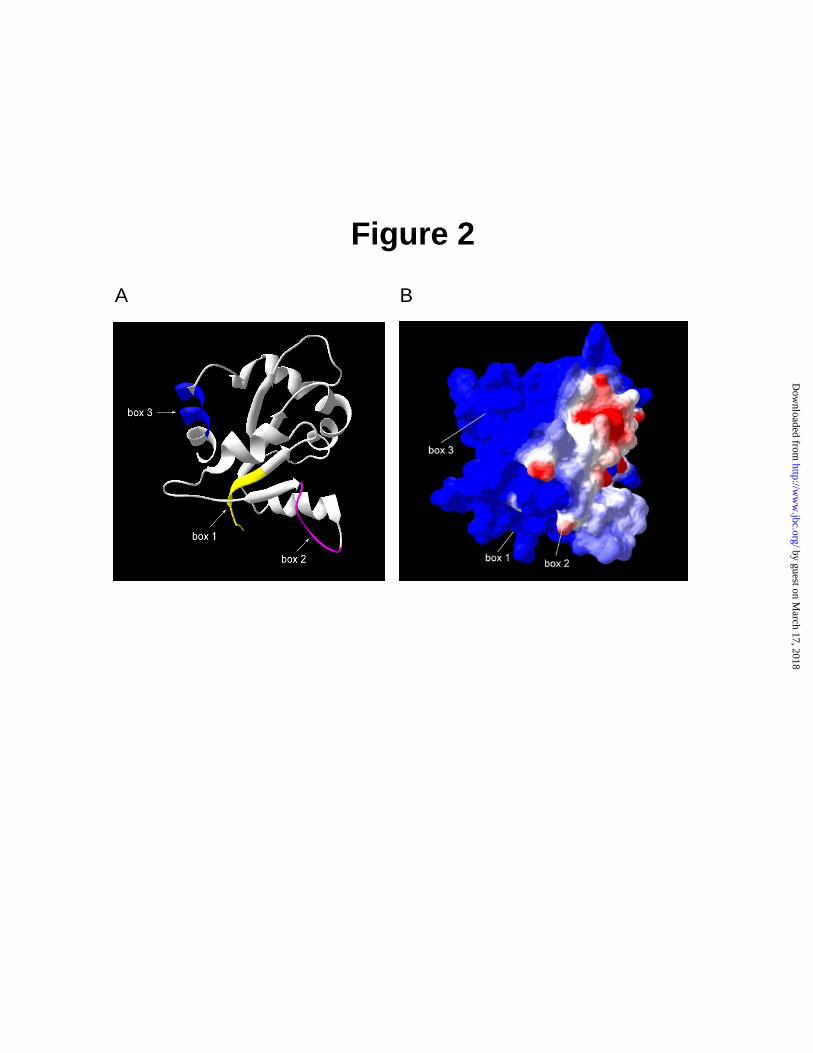
It has an α-β-fold similar to that of the bacterial chemotaxis protein CheY and contains three highly conserved motifs termed Boxes 1, 2, and 3 (3). On the basis of primary structure alignment obtained with program T-COFFEE, presented in Fig. 1, the crystal structure of human TLR2 TIR domain (Protein Data Bank pdb files of 1O77) was selected as the best template for MyD88 TIR domain with 31% identity and >50% similarity. Results obtained from server were visualized in DeepView program (Swiss Pdb viewer). As shown in Fig. 2, the secondary structure of TIR domain consists of five β strands: βA, βB, βC, βD and βE forming the core of molecule which is surrounded by five α-helices: αA, αB, αC, αD and αE. The five loops AA, BB, CC, DD and FF form specific links between β-strand and corresponding α-helix.
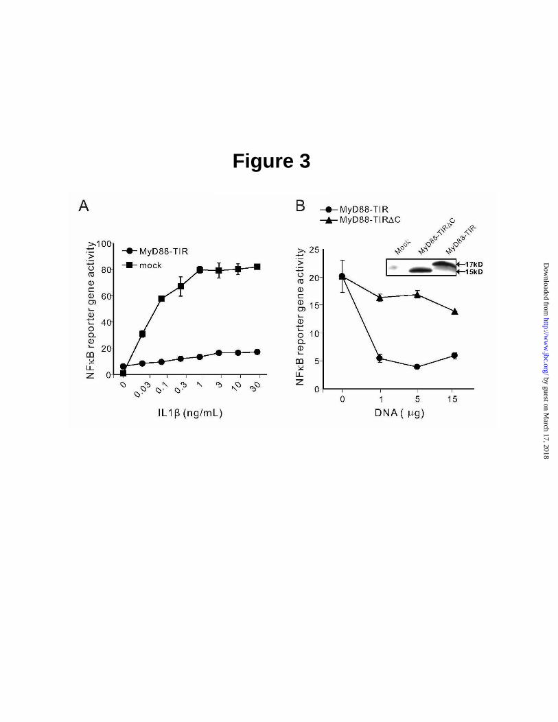
In terms of its tertiary structure, as depicted in Fig. 2, MyD88 TIR domain has a globular shape. Among three highly conserved motifs, Box 1, located at N-terminus of TIR domain, forms partially a βA-strand. Box 2 makes second part of BB loop, while Box 3 creates first part of αE-helix, which is located at the C-terminus of MyD88 TIR domain. As shown in Fig. 2B, distribution of charged residues indicates that molecular surface of MyD88 TIR domain is mostly positively charged (blue) with a few distinct negatively charged knobs (red). They surround a larger swatch of negatively charged surface (red), formed by three loops, AA, BB, and partially DD, and αC-helix. Moreover, the BB loop, projects from the globular TIR domain, forming a quasi- plane on its surface. NFκB Reporter Gene Activity Assay indicates that Box 3 of MyD88 TIR Domain is involved in IL1β –Induced Signaling - NFκB reporter gene activity assay was used to test MyD88 TIR domain as dominant negative inhibitor of IL1β-induced signaling to the nucleus. As documented in Fig. 3, HEK 293T cells transfected with plasmid containing NFκB-dependent luciferase gene responded to sub-nanogram doses of IL1β. This activation reached the maximum at 1 ng/mL of IL1β attesting to the high sensitivity of transfected HEK 293T cells to this inflammatory cytokine. Consistent with prior studies (23,24), MyD88 TIR domain, used as dominant negative inhibitor of IL1β signaling, almost completely suppressed its activating effect on of NFκB reporter gene over
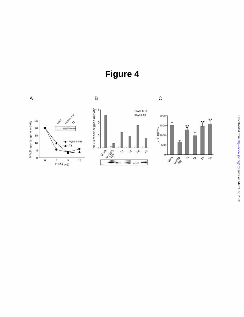
the wide range of IL1β concentrations (Fig. 3). We engineered a deletion mutant of MyD88 TIR domain to establish the utility of NFκB reporter gene activity assay for screening MyD88 TIR domain mutants for their inhibitory effect on IL1β-induced signaling. The deleted segment encompassed Box 3 (282KSWFWTRLAK291) located at the COOH-terminus of MyD88 TIR domain. This deletion caused the loss of the dominant negative inhibitory function of MyD88 TIR (Fig. 3B) suggesting that Box 3 was essential for IL1β-induced signaling. The COOH-terminal deletion mutant was expressed in transfected cells at the level comparable to the intact MyD88 TIR domain (see inset). Mutagenesis of MyD88 TIR Domain: Loss of its Dominant Negative Inhibitory Activity toward IL1β–Induced Signaling - The involvement of the Box 3, as compared to Boxes 1 and 2, in signaling induced by IL1β and mediated by MyD88 TIR domain, was analyzed in the first series of mutagenesis experiments. Mutations of TIR domain (residues 152-296) included two bulky hydrophobic residues (F285 and W286) in highly conserved short motif in Box 3 (SWFWTRL) and proline at the position 200. The canonical Pro712His mutation in TLR4 renders C3H/HeJ mice hyporesponsive to LPS (14). This highly conserved proline residue is located at position 200 in the MyD88 TIR domain (15). It was mutated to histidine in MyD88-TIR domain [152-296 (P200H)]. Furthermore, alignment of members of TLR family reveals two short motifs in Box 1 (PERFDAF) and Box 2 (DRDVLPG) that are most conserved along with Box 3 motif in MyD88 TIR domain. Alanine substitutions were primarily based on selection of charged residues either in the conserved region or predicted to be on the surface. In addition, two mutations, V204 and Q229 were in the region predicted to interact with MAL (15). The expression of mutants varied as compared to that of wild type MyD88 TIR domain (data not shown). Five mutants listed in Table 1 were expressed at the level comparable to that of the wild type MyD88 TIR domain (see insert in Fig. 4). The expression of other mutants was reduced or undetectable presumably due to misfolding and/or degradation. The well expressed mutants listed in Table 1 were screened for potential inhibitory effect on the NFκB reporter gene activation following stimulation with IL1β.
by guest on March 17, 2018
http://ww
w.jbc.org/
Dow
nloaded from
MyD88 Structure and IL1β Signaling
6
Of particular significance is the result with the P200H mutant (T3), analogous to the Pro/His mutation in TLR4, which is responsible for the LPS hyporesponsiveness of C3H/HeJ mice (14). Similar loss of signaling in other TIR-containing molecules like MAL/TIRAP (25) and IL1RAcP (26,27) was reported, indicating that the invariant proline in the BB loop of box 2 in these molecules is one of the interactive sites for other TIR domains-containing proteins. In striking contrast, a similar mutation (P200H) in MyD88 TIR domain, tested within a range of input concentrations, did not change the dominant negative effect of MyD88 TIR on IL1β-induced NFκB reporter gene activation in 293T cells (Fig. 4A). These cells showed a similar level of expression of the wild-type and mutant proteins (insert in Fig. 4A). This result is consistent with recent modeling studies of the interaction of MyD88 with TLR 2 and 4 (15), which suggested that the highly conserved proline residue may not participate in the protein-protein interactions of MyD88 with TLRs.
In contrast to the canonical P200H mutation, the following mutants displayed >2.5 fold loss of the dominant negative effect on IL1β- stimulated NFκB reporter gene activation as compared to the wild type MyD88 TIR domain (Fig. 4B): D171A in helix αA (T1), triple mutant D195A/R196A/D197A (T2) in box 2, D275A in βE strand (T4), and double mutant F285A/W286A (T5) in Box 3. The result with the F285A/W286A mutant is consistent with the loss of inhibition displayed by Box 3-deleted MyD88 TIR domain (Fig. 3B). All these mutants and wild type MyD88 TIR domain were expressed at comparable level in HEK 293T cells (insert in Fig. 4B).
These mutants were chosen for further validation of the functional significance of mutated MyD88 TIR domain residues. We selected IL1β-induced expression of an endogenous gene that encodes inflammatory cytokine, IL6, in human fibroblast MRC5 cells for testing MyD88 TIR domain mutants. The expression of IL6 gene is regulated by NFκB and its mobilization by IL1β is dependent on MyD88 (1-3). Upon stimulation of MRC5 cells with IL1β, an inflammatory cytokine IL6 was expressed. This expression of endogenous IL6 gene was suppressed 2.5 fold by dominant negative TIR domain of MyD88 (Fig. 4C). The observed degree of inhibition of endogenous IL6 gene expression is
smaller than suppression of NFκB reporter gene activity by MyD88 TIR domain in HEK 293T cells, most likely due to lower transfection efficiency of MRC5 cells that minimizes the impact of potential inhibitors on IL6 expression. Nevertheless, inhibition of IL1β-induced IL6 production was depended on residues D171, D275, D195/R196/D197 and F285/W286 because alanine substitutions reduced dominant negative effect of wild-type MyD88 TIR domain. These functional studies of ILβ-induced IL6 production are consistent with NFκB-dependent reporter gene activation (Fig. 4B). Thus, our first series of mutagenesis experiments identified interactive sites within MyD88 TIR domain responsible for coupling IL1β signaling to NFκB translocation to the nucleus and induction of endogenous IL6 gene. Mutagenesis of a Full-Length MyD88 Reveals an Interactive Site for a Direct Contact with IL1RAcP - It is still unknown which of the interactive sites identified in MyD88 TIR domain are responsible for direct contact of MyD88 with IL1 receptor complex subunit, IL1RAcP, which is indispensable for IL1β signaling (26). Alternatively, these interactive sites within MyD88 could participate in IL1β-induced oligomerization of MyD88 through homotypic interactions mediated by its TIR domain.
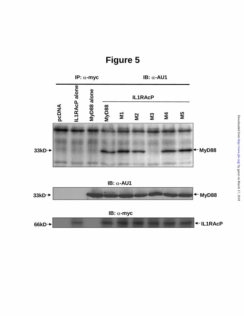
To sort out these possibilities, a second series of mutagenesis experiments was conducted. Five selected mutations were engineered in a full-length MyD88 to allow a comparative analysis of its direct interaction with IL1RAcP (Table 2). HEK 293T cells were cotransfected with myc-IL1RAcP and AU1-MyD88 or its mutant constructs. We used immunoprecipitation followed by Western blotting to assess the effect of mutated residues on the receptor TIR- adaptor TIR interaction. As demonstrated in Fig. 5, only MyD88 D195A/R196A/D197A mutant showed loss of binding to IL1RAcP while other mutants including MyD88 P200H retained their ability to bind IL1RAcP. This selective loss of receptor binding function by MyD88 D195A/R196A/D197A mutant led us to develop the 3-D docking model of MyD88 and IL1RAcP and verify the strategic position of these three residues as a main interactive site on the surface of MyD88 TIR domain for its binding to TIR domain of IL1RAcP.
by guest on March 17, 2018
http://ww
w.jbc.org/
Dow
nloaded from
MyD88 Structure and IL1β Signaling
7
The Development of the 3-D Docking Model of MyD88-IL1RAcP Interaction-The 3-D docking model was developed by optimized superposition of two mutually interacting TIR domains of Myd88 and IL1RAcP. Negatively charged side of the MyD88-TIR (see Fig.2B) was selected as a possible interface of the molecule. This side contains D195/R196/D197 residues that are essential, on the basis of mutagenesis studies (Fig. 5), for MyD88-TIR heterotypic interaction with IL1RAcP-TIR. Then, suitable positively charged site of the IL1RAcP was selected to conduct the modeling computation process. This modeling was based on geometry optimization by energy minimization followed by molecular dynamic computation using program SANDER (AMBER software package). Ribbon structure was developed with Deep View program while the molecular surface of associated proteins was determined by PSSHOW (SYBIL-Tripos software package). Separation surface indicates that there is no crossing of molecular surfaces and distance between them is within the range of 0.4 - 4.7 Å while their topology is diverse and contains several deep pockets. Development of this 3-D model allowed us to verify contribution of the triplet of functionally important residues D195/R196/D197 to the binding reaction with IL1RAcP TIR domain. An analysis of the tertiary structure of two TIR domains that participate in the docking model indicates that three mutated residues (D195/R196/D197), responsible for a loss of MyD88 binding to IL1RAcP, are involved in interaction with residues 527-534 of IL1RAcP previously identified to play a key role in IL1β signaling pathway (26,27). Thus, our study identified a complementary site on MyD88 TIR domain that contributes to its interaction with IL1RAcP TIR domain. We therefore postulate that negatively charged “knob”, partially comprised of the BB loop on the surface of MyD88 TIR domain (Fig. 2B) fits into the positively charged lysine patch formed by residues 527, 530, and 532 of IL1RAcP TIR domain previously identified by Radons et al. as essential for IL1β signaling (26,27). Interactive Sites Involved in Oligomerization of MyD88 through Homotypic Interaction of its TIR Domain-Following IL1β-induced interaction of IL1RAcP with MyD88, this adaptor oligomerizes
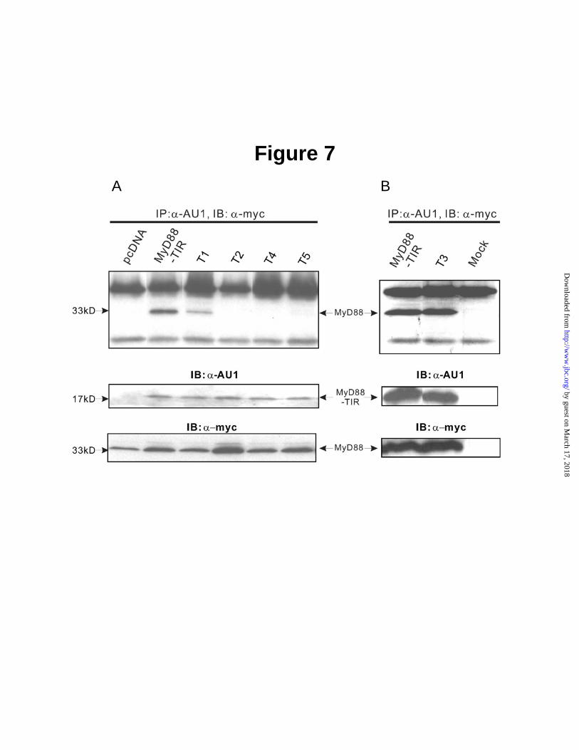
and interacts with downstream signal transducers (2,3,7). Homotypic oligomerization of MyD88 due to its forced expression resulted in robust activation of NFκB reporter gene activity observed in the absence of IL1β stimulation. This receptor-independent effect of ectopically expressed MyD88 oligomers was abolished by cotransfected MyD88-TIR domain (data not shown). Therefore, we examined direct interaction of a full-length MyD88 coexpressed with MyD88 TIR domain or its mutants. We co-transfected HEK 293T cells with full length MyD88 that contained c-myc epitope tag along with the wild type or mutated TIR domain that contained AU1 epitope tag. As demonstrated in Fig. 7, P200H mutant bound to a full-length MyD88 to similar extent as wild-type MyD88 TIR domain. However, mutations in Box 2 (D195A/R196A/D197A) and Box 3 (βE strand D275A and F285A/W286A) caused a loss of binding to MyD88, suggesting that these mutated residues constitute the interactive sites for homotypic oligomerization of the MyD88 TIR domain. Thus, interacting site in Box 2 comprised of residues D195/R196/D197 has potentially an additional function that may encroach on the ability of MyD88 to interact with IL1RAcP. However, in a cascade of signaling steps induced by IL1β, heterotypic interaction of IL1RAcP TIR domain with MyD88 TIR domain antecedes oligomerization of MyD88. The latter depends on homotypic binding mediated by its TIR domain. Therefore, the interactive site in Box 3 is not likely involved in binding of MyD88 to IL1RAcP. Rather, this site participates in homotypic MyD88 oligomerization and possibly other transactions involving downstream signal transducers. This interpretation is consistent with the loss of inhibition of IL1β-induced signaling by MyD88 TIR domain upon truncation of its COOH-terminal segment that contains Box 3. Taken together, our results identify key residues in MyD88 TIR domain that are responsible for its heterotypic interaction with IL1RAcP. In addition, we identified interactive sites for homotypic oligomerization of MyD88. These protein-protein interactions evoked by IL1β are essential for its signaling to the nucleus mediated by NFκB and other proinflammatory stress responsive transcription factors. Mutations identified on the interacting surface of the MyD88
by guest on March 17, 2018
http://ww
w.jbc.org/
Dow
nloaded from
MyD88 Structure and IL1β Signaling
8
TIR domain are functionally important because they interfere with the induction of endogenous gene that encodes IL6. This inflammatory cytokine, along with IL1β, is responsible for cardinal signs of systemic inflammation: fever, leukocytosis, thrombocytosis, acute phase protein response, and tissue injury (1,5). The development of the docking 3-D model of MyD88-IL1RAcP binding, in which a cluster of highly charged residues in Box 2 plays a key role, reaffirms their strategic role in contacting complementary site on IL1RAcP TIR domain. This site is comprised of several positively charged residues identified previously in EE loop residues 527-534 (26,27). Thus, it is not surprising that the invariant Proline 200 is inconsequential for MyD88-ILRAcP interaction. However, canonical mutation of the invariant proline to histidine in TLR4, which abolishes its signaling by LPS (14), indicates that different ligands and their cognate TLRs utilize MyD88 in structurally distinct way. In addition to TLR4, Proline to Histidine mutation attenuated signaling mediated by TLR2, MAL and IL1RAcP (14,25,26). We interpret the later result as indicative of invariant proline playing a significant role in reshaping EE loop in IL1RAcP or in
interactions with TIR domain of IL1RI or with other than MyD88 signaling molecules. In summary, our data indicate that following stimulation with IL1β, MyD88 TIR domain binds to IL1RAcP TIR domain via a highly charged interactive site comprised of residues 195-197 within BB loop of Box 2. We postulate on the basis of the 3-D docking model developed herein that this interactive site is complementary to the previously identified site comprised of residues 527-534 within EE loop of IL1RAcP TIR domain (26,27). Despite its proximity to the interactive site, invariant Proline 200 of MyD88 TIR domain does not play a role in IL1β-induced signaling. However, residues located in Box 3 are essential for subsequent homotypic interaction of MyD88 TIR domain. In a broader context, the results suggest that IL1β signaling pathway differs from other ligands-initiated TLRs intracellular signaling by usage of distinct structural motifs within MyD88 TIR domain. Further mapping of MyD88 surface will expand our understanding of its role in integrating signals derived from a variety of Toll-like receptors.
REFERENCES
1. Dinarello, C. A. (2002) Clin Exp Rheumatol 20(5 Suppl 27), S1-13 2. Yamamoto, M., Takeda, K., and Akira, S. (2004) Mol Immunol 40(12), 861-868 3. Akira, S., and Sato, S. (2003) Scand J Infect Dis 35(9), 555-562 4. Vannier, E., and Dinarello, C. A. (1994) J Biol Chem 269(13), 9952-9956 5. Dinarello, C. A. (1996) Blood 87(6), 2095-2147 6. McGettrick, A. F., and O'Neill, L. A. (2004) Mol Immunol 41(6-7), 577-582 7. Akira, S. (2003) J Biol Chem 278(40), 38105-38108 8. Wesche, H., Henzel, W. J., Shillinglaw, W., Li, S., and Cao, Z. (1997) Immunity 7(6), 837-847 9. Wesche, H., Korherr, C., Kracht, M., Falk, W., Resch, K., and Martin, M. U. (1997) J Biol Chem
272(12), 7727-7731 10. Huang, J., Gao, X., Li, S., and Cao, Z. (1997) Proc Natl Acad Sci U S A 94(24), 12829-12832 11. Volpe, F., Clatworthy, J., Kaptein, A., Maschera, B., Griffin, A. M., and Ray, K. (1997) FEBS
Lett 419(1), 41-44 12. Li, S., Strelow, A., Fontana, E. J., and Wesche, H. (2002) Proc Natl Acad Sci U S A 99(8), 5567-
5572 13. Kobayashi, K., Hernandez, L. D., Galan, J. E., Janeway, C. A., Jr., Medzhitov, R., and Flavell, R.
A. (2002) Cell 110(2), 191-202 14. Poltorak, A., He, X., Smirnova, I., Liu, M. Y., Van Huffel, C., Du, X., Birdwell, D., Alejos, E.,
Silva, M., Galanos, C., Freudenberg, M., Ricciardi-Castagnoli, P., Layton, B., and Beutler, B. (1998) Science 282(5396), 2085-2088
by guest on March 17, 2018
http://ww
w.jbc.org/
Dow
nloaded from
MyD88 Structure and IL1β Signaling
9
15. Dunne, A., Ejdeback, M., Ludidi, P. L., O'Neill, L. A., and Gay, N. J. (2003) J Biol Chem 278(42), 41443-41451
16. Muzio, M., Ni, J., Feng, P., and Dixit, V. M. (1997) Science 278(5343), 1612-1615 17. Kunkel, T. A. (1985) Proc Natl Acad Sci U S A 82(2), 488-492 18. Lin, Y. Z., Yao, S. Y., Veach, R. A., Torgerson, T. R., and Hawiger, J. (1995) J Biol Chem
270(24), 14255-14258 19. Schwede, T., Kopp, J., Guex, N., and Peitsch, M. C. (2003) Nucleic Acids Res 31(13), 3381-3385 20. Guex, N., and Peitsch, M. C. (1997) Electrophoresis 18(15), 2714-2723 21. Xu, Y., Tao, X., Shen, B., Horng, T., Medzhitov, R., Manley, J. L., and Tong, L. (2000) Nature
408(6808), 111-115 22. Tao, X., Xu, Y., Zheng, Y., Beg, A. A., and Tong, L. (2002) Biochem Biophys Res Commun
299(2), 216-221 23. Burns, K., Martinon, F., Esslinger, C., Pahl, H., Schneider, P., Bodmer, J. L., Di Marco, F.,
French, L., and Tschopp, J. (1998) J Biol Chem 273(20), 12203-12209 24. Dupraz, P., Cottet, S., Hamburger, F., Dolci, W., Felley-Bosco, E., and Thorens, B. (2000) J Biol
Chem 275(48), 37672-37678 25. Horng, T., Barton, G. M., and Medzhitov, R. (2001) Nat Immunol 2(9), 835-841 26. Radons, J., Gabler, S., Wesche, H., Korherr, C., Hofmeister, R., and Falk, W. (2002) J Biol Chem
277(19), 16456-16463 27. Radons, J., Dove, S., Neumann, D., Altmann, R., Botzki, A., Martin, M. U., and Falk, W. (2003)
J Biol Chem 278(49), 49145-49153
ACKNOWLEDGMENTS
We thank Dean Ballard for critical reading of the manuscript, Jarrod Smith for experimental advice, and Ana Maria Hernandez for assistance in the preparation of the manuscript.
FOOTNOTES
* This work was supported in part by USPHS National Institutes of Health Grants HL69542, HL62356, and HL68744. The use of core facilities in this study was supported by National Institutes of Health 2P30 CA 68485 to the Vanderbilt Ingram Cancer Center and by 5P30DK058404-03 to the Vanderbilt Digestive Disease Research Center. The costs of publication of this article were defrayed in part by the payment of page charges. This article must therefore be hereby marked “advertisement” in accordance with 18 U.S.C. Section 1734 solely to indicate this fact. 1The abbreviations used are: MyD88, myeloid differentiation factor 88; TIR, Toll/interleukin-1 receptor domain; TLR, toll-like receptor; IL, interleukin; MAL , MyD88-adapter-like protein, IL1RAcP, IL1 receptor accessory protein.
FIGURE LEGENDS
Fig. 1. Sequence alignment of the TIR domains of human MyD88 and of human Toll-like receptor 2 (TLR2) as the basis of homology modeling performed with T-COFFEE. Boxes 1–3 are underlined. Residues substituted with alanine (D171, D195, R196, D197, D275, F285, and W286) or histidine (P200) are printed in bold face.
Fig. 2. Structural model of TIR domain with conserved boxes 1-3. Comparative modeling of MyD88-TIR was performed with SWISS-MODEL (19,20) by aligning to crystal structure of hTLR2 (21,22) optimized by GROMOS 96 and visualized by DeepView/Swiss-pdb viewer. A, Structural features
by guest on March 17, 2018
http://ww
w.jbc.org/
Dow
nloaded from
MyD88 Structure and IL1β Signaling
10
representing the conserved boxes of MyD88 TIR domain are shown in yellow (Box 1), purple (Box 2) and blue (Box 3). B, electrostatic potential of MyD88 TIR with the indicated position of structural features (boxes 1-3) corresponding to same in panel A. The color scale is as follows: red (most negative) and blue (most positive).
Fig. 3. A, Inhibition of IL1β-induced NFκB reporter gene activity by MyD88-TIR domain is reversed upon truncation of its Box 3. A, IL1β-induced NFκB reporter gene activation was inhibited in HEK 293T cells transfected with MyD88-TIR (152-296). The 293T cells were transfected with MyD88-TIR together with NFκB-luc (5µg) and RL-TK (2.5µg). After 24 h, IL1β was added at indicated concentrations for 6 h. NFκB-reporter gene activity was determined and normalized on the basis of RL-TK activity. B, MyD88-TIR∆C (152-283) construct was cloned into pcDNA3.1 using PCR at BamH I and EcoR I sites. HEK 293T cells were transfected with MyD88-TIR or MyD88-TIR∆C (152-283) constructs together with NFκB-luc (5 µg) and RL-TK (2.5 µg). After 24 h, IL1β was added at concentration of 10 ng/mL for 6 h. NFκB reporter gene activity was measured and normalized on the basis of RL-TK activity. Insets, Expression levels of MyD88-TIR and MyD88-TIR∆C (152-283) in transfected 293T cells determined by Western blotting with anti-AU1 antibody. Data show a representative experiment from the series of three performed in triplicates. Error bars indicate the ± S.E. of the mean value of triplicate. Fig. 4. Functional analysis of MyD88-TIR domain mutants tested in NFκB reporter gene activity (panels A and B) and cytokine IL6 production (panel C) assays. A, Concentration-dependent inhibition of NFκB reporter gene activity by wild- type and T3 mutant (Proline200Histidine); B, Inhibition of NFκB reporter gene activity by mutants T1, T2, T4, and T5. HEK 293T cells were transfected with MyD88-TIR or its mutant constructs (1-15 µg in panel A and 12.5 µg in panel B) together with NFκB-luc and RL-TK. After 24 h, IL1β was added at concentration of 10 ng/mL for 6 h. Cells were harvested and split for both reporter assay and Western blotting. NFκB reporter gene activity was analyzed and normalized on the basis of RL-TK activity. The figure shown was a representative of three experiments. Insets, expression levels of TIR and TIR mutants as determined by Western blotting. C, Inhibition of IL6 production by T1, T2, T4 and T5 mutants. MRC-5 cells were transiently transfected with TIR mutants and stimulated with 10 ng/mL IL1β for 6h. IL6 production was analyzed with CBA as described in Experimental Procedures. Data represent combined results from three independent experiments done in triplicate. Error bars indicate the ± S.E. of the mean. ** P<0.01, * P<0.05, by two-sided Student’s t-test. Fig. 5. Interaction of MyD88 mutants with IL1RAcP. HEK 293T cells were transiently transfected with myc-IL1RAcP (10 µg) and full length MyD88 or its mutants (10 µg). After 24 h, the cells were stimulated with 100 ng/mL of IL1β for 5 min, harvested and immunoprecipitated as described in Experimental Procedures. Protein samples bound to anti-myc antibody protein G beads were subjected to SDS-PAGE and the immunoprecipitated MyD88 or its mutants were monitored by immunoblotting with anti-AU1 antibody (top panel). Expression of MyD88 and its mutants (middle panel), IL1RAcP (lower panel) were analyzed by immunoblotting.
Fig. 6. The 3-D models of MyD88 TIR domain, IL-1RAcP TIR domain and docking model of TIR-TIR interaction of MyD88 and IL1RAcP. Panels A and B, ribbon structure of IL1RAcP and MyD88, respectively, obtained from homology computation performed using Swiss-Model: An Automated Comparative Protein Modeling Server, and visualized using Deep View (Swiss Pdb Viewer). Structural features representing the conserved boxes of MyD88 TIR domain are shown in panel B in yellow (Box 1), purple (Box 2) and blue (Box 3). The selected side chains of mutated residues in panel B and C are marked in green. Panel C, ribbon representation of TIR-TIR interaction optimized with SANDER–energy minimization software (AMBER package). The side chains of identified residues (D195, R196, and D197) required for direct binding of MyD88 TIR to IL1RAcP TIR are shown in green. The structural motifs in panel A and C are colored as follow: blue – α-helices, red – β-strands, white – loops.
by guest on March 17, 2018
http://ww
w.jbc.org/
Dow
nloaded from
MyD88 Structure and IL1β Signaling
11
Panel D, top view of docking superposition presented in molecular surface mode. Fig. 7. Homotypic Interaction of TIR domain of MyD88 with a full-length MyD88. The 293T cells were transiently transfected with c-myc tagged- full length MyD88 (5 µg) and AU1 tagged-MyD88-TIR (5 µg) or AU1 tagged-MyD88-TIR mutants (5 µg). After 24 h, the cells were harvested and immunoprecipitated as described in Experimental Procedures. Protein samples bound to anti-AU1 antibody protein G beads were subjected to SDS-PAGE and immunoprecipitated MyD88-TIR or MyD88-TIR mutants were detected with anti-c-myc antibody (top panel). Expression of MyD88-TIR and MyD88-TIR mutants (middle panel) or MyD88 (lower panel) was analyzed by immunoblotting.
by guest on March 17, 2018
http://ww
w.jbc.org/
Dow
nloaded from
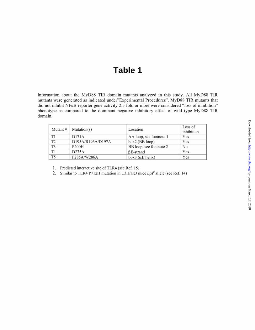
Table 1 Information about the MyD88 TIR domain mutants analyzed in this study. All MyD88 TIR mutants were generated as indicated under”Experimental Procedures”. MyD88 TIR mutants that did not inhibit NFκB reporter gene activity 2.5 fold or more were considered “loss of inhibition” phenotype as compared to the dominant negative inhibitory effect of wild type MyD88 TIR domain.
Mutant # Mutation(s) Location Loss of inhibition
T1 D171A AA loop, see footnote 1 Yes T2 D195A/R196A/D197A box2 (BB loop) Yes T3 P200H BB loop, see footnote 2 No T4 D275A βE-strand Yes T5 F285A/W286A box3 (αE helix) Yes 1. Predicted interactive site of TLR4 (see Ref. 15) 2. Similar to TLR4 P712H mutation in C3H/HeJ mice Lpsd allele (see Ref. 14) by guest on M
arch 17, 2018http://w
ww
.jbc.org/D
ownloaded from
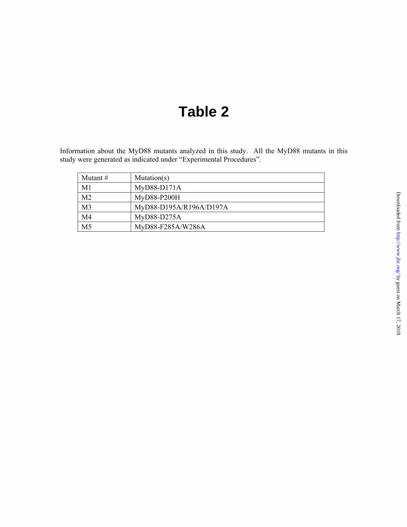
Table 2
Information about the MyD88 mutants analyzed in this study. All the MyD88 mutants in this study were generated as indicated under “Experimental Procedures”.
Mutant # Mutation(s) M1 MyD88-D171A M2 MyD88-P200H M3 MyD88-D195A/R196A/D197A M4 MyD88-D275A M5 MyD88-F285A/W286A
by guest on March 17, 2018
http://ww
w.jbc.org/
Dow
nloaded from

151 Box1 βA αA βB Box2 MyD88-TIR DDPLGHMPERFDAFICYCPSDIQFVQE-MIRQLEQTNYRLKLCVSDRDVLPGAA Tlr2-TIR --------ICYDAFVSYSERDAYWVENLMVQELENFNPPFKLCLHKRDFIPG Cons :***:.*. * :*:: *:::**: * :***: .**.:**
202 αB βC αC MyD88-TIR TCVWSIASELIEKRCRRMVVVVSDDYLQSKECDFQTKFALSLSPGAHQKR Tlr2-TIR KWIIDNIIDSIEK-SHKTVFVLSENFVKSEWSKYELDFSHFRLFAAILIL Cons . : . : *** .:: *.*:*:::::*: ..:: .*: .*
252 βD αD βE Box3 αE MyD88-TIR LIPIKYKAMKKEFPSILRFITVCDYTN----PCTKSWFWTRLAKALSLP Tlr2-TIR LEPIEKKAIPQRFCKLRKIMNTKTYLEWPMDEAQREGFWVNLRAAIKS- Cons * **: **: :.* .: :::.. * : . :. **..* *:.
Figure 1
by guest on March 17, 2018
http://ww
w.jbc.org/
Dow
nloaded from
pcD
NA
IL1R
AcP
alon
e
M3M1
M4
M5
IP: α-myc IB: α-AU1
MyD
88 a
lone
MyD
88
M2
MyD88
IL1RAcP
33kD
IB: α-AU1
IB: α-myc
33kD
66kD
MyD88
IL1RAcP
Figure 5
by guest on March 17, 2018
http://ww
w.jbc.org/
Dow
nloaded from
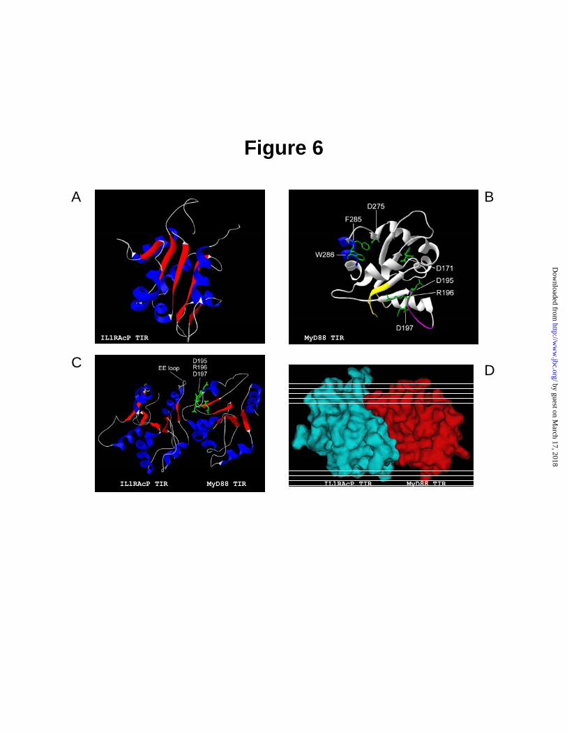
Figure 6
IL1RAcP TIR
IL1RAcP TIR MyD88 TIR
A
C
B
D
IL1RAcP TIR MyD88 TIR
MyD88 TIR
by guest on March 17, 2018
http://ww
w.jbc.org/
Dow
nloaded from
Chunsheng Li, Jozef Zienkiewicz and Jacek Hawigersignaling pathway
βInteractive sites in the MYD88 TIR domain responsible for coupling to the IL1
published online April 22, 2005J. Biol. Chem.
10.1074/jbc.M503262200Access the most updated version of this article at doi:
Alerts:
When a correction for this article is posted•
When this article is cited•
to choose from all of JBC's e-mail alertsClick here
by guest on March 17, 2018
http://ww
w.jbc.org/
Dow
nloaded from
Top Related