

γλώσσες
Σελίδες
Νομικός

Research ArticleGrape Seed Proanthocyanidin Extract Inhibits Human EsophagealSquamous Cancerous Cell Line ECA109 via the NF-κBSignaling Pathway
Fangming Guo ,1,2 Yunhua Hu,1,2 Qiang Niu,1,2 Yu Li,1,2 Yusong Ding,1,2 Rulin Ma,1,2
Xianhua Wang,3 Shugang Li ,1,2 and Jianxin Xie 2,4
1Department of Public Health, Shihezi University School of Medicine, 832000, China2Key Laboratory for Xinjiang Endemic and Ethnic Diseases, 832000, China3Department of Quality Control of Changji Autonomous Prefecture Center for Disease Control and Prevention, 831100, China4Department of Biochemistry, Shihezi University School of Medicine, 832000, China
Correspondence should be addressed to Shugang Li; [email protected] and Jianxin Xie; [email protected]
Fangming Guo and Yunhua Hu contributed equally to this work.
Received 6 April 2018; Revised 17 July 2018; Accepted 5 August 2018; Published 17 December 2018
Academic Editor: Luis I. Terrazas
Copyright © 2018 Fangming Guo et al. This is an open access article distributed under the Creative Commons Attribution License,which permits unrestricted use, distribution, and reproduction in any medium, provided the original work is properly cited.
Esophageal squamous cell carcinoma is the most common type of squamous cell carcinoma. Grape seed proanthocyanidin extract(GSPE) is considered to exhibit anticancer activity against several different types of cancer. We aimed to determine whether GSPEinhibited esophageal squamous cancerous cells and the possible involvement of NF-κB in this process. The human esophagealsquamous cancer cell line ECA109 was treated with GSPE (0–80μg/mL) and BAY11-7082 (10 μmol/L) for 12, 24, and 48 h. TheMTT assay was used to determine cell proliferation; alterations in cell apoptosis were detected by flow cytometry; levels ofinflammatory factors interleukin-6 and cyclooxygenase-2 and apoptotic proteins Bax/Bcl-2 were measured by ELISA; qRT-PCRand western blots were used to examine the activation of caspase-3 and NF-κB signaling. GSPE inhibited the proliferation ofECA109 cells and induced cellular apoptosis in a time- and dose-dependent manner. ELISA results showed that GSPE andBAY11-7082 reduced the secretion of inflammatory cytokines interleukin-6 and cyclooxygenase-2. The results of PCR andwestern blotting indicated that GSPE and BAY11-7082 activated caspase-3 and attenuated the activation of the NF-κB signalingpathway. GSPE induced apoptosis in ECA109 cells and inhibited ECA109 cell proliferation via a reduction in the secretion ofinflammatory cytokines. This mechanism may be related to the attenuation of NF-κB activity and the sensitization of caspase-3.
1. Introduction
Esophageal carcinoma (EC), one of the most common can-cers, is caused by malignant transformation of the esophagus.It is the sixth leading cause of death among malignantcancers, and the most common pathological type is esoph-ageal squamous cell carcinoma (ESCC). The Kazakh areain Xinjiang, China, is a high-risk region for EC. Despiteadvances in understanding the mechanisms of cancerprogression and the development of different therapeuticstrategies, EC is still the leading cause of mortality in
malignant tumor death among the Kazakh population inXinjiang, particularly as a result of metastasis [1].
Chronic esophagitis is one of the most important factorsfor the occurrence of esophageal cancer. Murphy et al. foundthat non-Barrett’s esophagitis increased the risk of ESCC [2].Zhang et al. reported that local infiltration of inflammatorycells led to the interruption and deletion of the local base-ment membrane in esophageal squamous cells [3], whichpromoted cell proliferation and induced EC. Nuclear factorkappa B (NF-κB), a transcription factor that plays an impor-tant role in inflammation, is involved in the progress of
HindawiMediators of InflammationVolume 2018, Article ID 3403972, 12 pageshttps://doi.org/10.1155/2018/3403972
chronic esophagitis [4]. NF-κB participates in cell prolifera-tion [5], cytoskeletal remodelling [6], cell invasion [7], andapoptosis [8]. Studies have found that NF-κB is a key factorin the development of a variety of malignant carcinomas,such as liver cancer [9], colon cancer [10], and breastcancer [11]. However, a direct connection between NF-κBsignaling and EC is less certain.
Proanthocyanidins (PCs), a class of polyphenoliccompounds, are widespread in plants, mostly in the epider-mis and seeds. Our previous studies determined that PCsreduced oxidative damage and inflammation [12, 13]. Recentresearch demonstrated the anticarcinogenic activity of PCs[14], with cytotoxic effects reported in various cancerous celllines (liver [15], colon [16], breast [17], and esophageal [18])that were largely mediated through apoptosis and showed noadverse biological effects on normal cells. Although it wasfound that PCs could induce apoptosis in cancer cells, therole of NF-κB in the reversal of EC, as well as the mechanism,remains unclear. Therefore, we conducted this study todetermine whether GSPE induced apoptosis in esophagealcancer cells and examined any possible involvement ofNF-κB in the process.
2. Materials and Methods
2.1. Reagents. GSPE (≥95.0%) was obtained from JF-NaturalCompany (Tianjin, China). BAY 11-7082 and antibodiesagainst IKK, caspase-3, and NF-κB (p65) were supplied byAbcam (Cambridge, England), and antibodies against IκB,phospho-IκB (p-IκB), and NF-κB (p100/p50) were procuredfrom Cell Signaling Technology Inc. (Danvers, MA). Anti-bodies against GAPDH were purchased from Goodhere Bio-technology (Hangzhou, China). Dulbecco’s modified Eagle’smedium (DMEM), penicillin, streptomycin, fetal bovineserum (FBS), and trypsin/EDTA were purchased fromHyClone (Logan, Utah). 3-(4,5-Dimethylthiazol-2-yl)-3, 5-diphenyltetrazolium bromide (MTT) was obtained fromJiancheng Biotechnology Co. (Nanjing, China). The annexinV-FITC/PI apoptosis kit was procured from Multisciences(Hangzhou, China). ELISA kits for IL-6 and COX-2 werepurchased from Elabscience (Wuhan, China).
2.2. Cell Culture.Human esophageal squamous ECA109 cellswere kindly provided by the Department of Pathology, KeyLaboratory for Xinjiang Endemic and Ethnic Diseases,Shihezi University School of Medicine (Xinjiang, China).All cells were cultured in monolayers with 90% DMEM sup-plemented with 10% FBS and 1% penicillin/streptomycin at37°C in a humidified atmosphere of 5% CO2. The mediumwas changed every second day.
2.3. Cell Viability Assay. The MTT assay was used to measurethe viability of ECA109 cells. The cells were plated into 96-well plates at a density of 2000 cells/well in 200μL DMEM.After incubation at 37°C overnight, GSPE (0–400μg/mL)was added to the cells for 12, 24, and 48h. Each treatmentand time point were assayed in triplicate. After the stipulatedtreatment time with GSPE, MTT was added to the cells for4 h. Subsequently, the supernatant was discarded and the
formazan precipitates were dissolved in 150μL dimethylsulfoxide (DMSO). An automatic microplate spectropho-tometer was used to measure the optical density (OD) foreach well. The detected wavelength was 490nm, and thereference wavelength was 620nm.
2.4. Annexin V-FITC/PI Staining. Apoptosis was determinedin ECA109 cells by using an annexin V-FITC/PI apoptosiskit. After treatment with GSPE (0, 25, 50, and 80μg/mL)for 24 h, the cells were collected and washed twice with coldPBS. Subsequently, 1× 10⁶ cells were suspended in bindingbuffer, stained with annexin V-FITC and PI, and analyzedby using flow cytometry.
2.5. Cell Migration Assay. The effect of GSPE on ECA109migration was analyzed by using a cell scratch test. Cells wereplated into 6-well plates at a density of 5× 10⁶ cells/well in2mL DMEM supplemented with 10% FBS. The cells wereallowed to adhere, scratched by pipette tips, and treated withGSPE (0, 25, 50, and 80μg/mL) for 24h. Each treatment wasassayed in triplicate. After incubation at 37°C overnight, thecells were observed by using an inverted microscope.
2.6. Cell Invasion Assay. A Transwell cell invasion assay wasperformed. Briefly, the upper chamber of Millicell cell cultureinserts was coated with 50μL Matrigel diluted 1 : 8 with PBS.Subsequently, 4× 10⁵ ECA109 cells in 0.4mL serum-freeDMEM, with or without GSPE, were added to the upperchamber. The lower chamber was filled with 0.6mL DMEMsupplemented with 20% FBS as a chemoattractant to induceinvasion. After incubation at 37°C for 24h, the cultureinserts were removed and the noninvasive cells on theupper surface of the culture inserts were removed by usinga cotton swab. The cells that invaded through the Matrigelwere fixed with methanol for 30min and stained with 0.1%crystal violet for 10min at 20°C. Images were captured byusing light microscopy.
2.7. ELISA. Briefly, the cells were cultured with GSPE (0, 25,50, and 80μg/mL) and GSPE (0, 25, 50, and 80μg/mL)+BAY11-7082 (10μmol/L) for 12, 24, and 48h. Superna-tants from experimental cultures were collected and stored at−80°C until use. The levels of IL-6 and COX-2 in the superna-tants were determined by using cytokine detection ELISA kitsin accordance with the manufacturer’s instructions; detectionat 450nm was conducted by using a microplate reader. Theconcentration of Bax and Bcl-2 in the cell culture supernatantwas determined by using a Bax and Bcl-2 detection ELISA kit.
RT-PCR was performed to evaluate the mRNA expres-sion of caspase-3, IKK, NF-κB (p50), and NF-κB (p65) aftertreatment with GSPE (0, 25, 50, and 80μg/mL) and GSPE(0, 25, 50, and 80μg/mL)+BAY11-7082 (10μmol/L) for24 h, as previously described [19]. The designed primers areshown in Table 1.
2.8. Western Blot Analysis. ECA109 cells were treated withGSPE (0, 25, 50, and 80μg/mL) and GSPE (0, 25, 50, and80μg/mL)+BAY11-7082 (10μmol/L) for 24 h. After treat-ment, the cells were collected and washed three times withPBS. The harvested cells were lysed on ice for 30min in
2 Mediators of Inflammation
100mL of lysis buffer. The total protein was collected andquantified by using the Bradford assay. The separated proteinswere transferred onto nitrocellulose membranes, which werefirst incubated with antibodies against caspase-3, IKK, phos-pho-IκB, IκB, NF-κB (p50), NF-κB (p65), and GAPDH, andthen incubated with secondary anti-mouse or anti-rabbit anti-bodies. All western blotting studies were repeated three times.
2.9. Statistical Analysis. All values are expressed as the mean± standard deviation (SD), and analyses were computed byusing SPSS 20.0. Western blotting analysis was calculatedby using Image-Pro Plus software. The comparison of themean among multiple groups was performed with analysisof variance. Pairwise comparison among groups was per-formed with the least significant difference (LSD) tests. Forall preplanned or a priori contrasts stipulated in the mainhypotheses, a significance level of 0.05 or 0.01 was consideredto indicate statistical significance.
3. Results
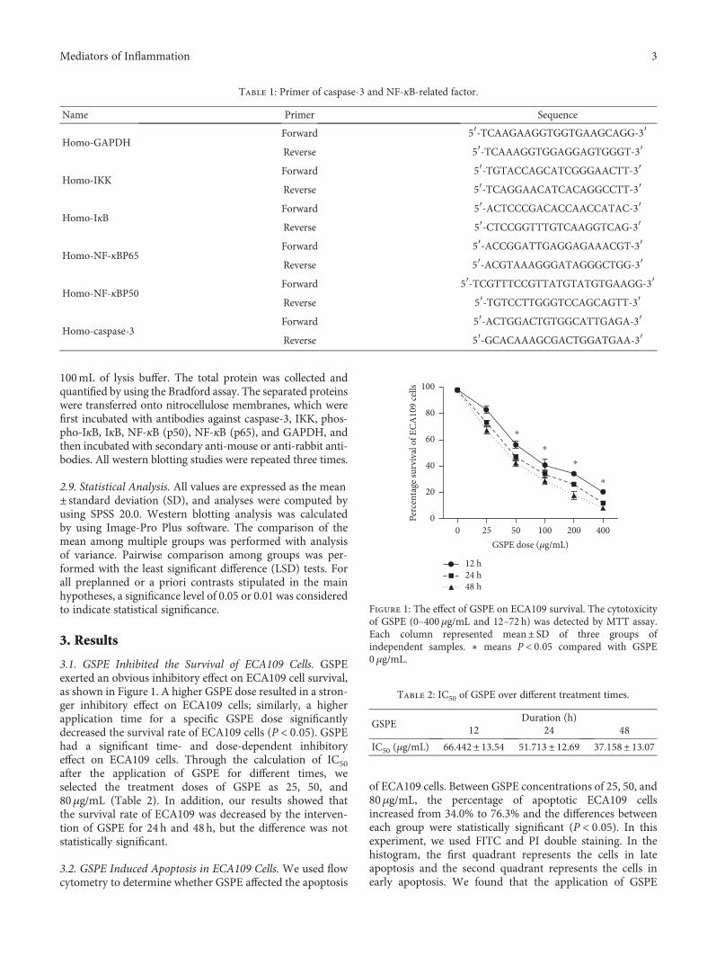
3.1. GSPE Inhibited the Survival of ECA109 Cells. GSPEexerted an obvious inhibitory effect on ECA109 cell survival,as shown in Figure 1. A higher GSPE dose resulted in a stron-ger inhibitory effect on ECA109 cells; similarly, a higherapplication time for a specific GSPE dose significantlydecreased the survival rate of ECA109 cells (P < 0 05). GSPEhad a significant time- and dose-dependent inhibitoryeffect on ECA109 cells. Through the calculation of IC50after the application of GSPE for different times, weselected the treatment doses of GSPE as 25, 50, and80μg/mL (Table 2). In addition, our results showed thatthe survival rate of ECA109 was decreased by the interven-tion of GSPE for 24 h and 48h, but the difference was notstatistically significant.
3.2. GSPE Induced Apoptosis in ECA109 Cells. We used flowcytometry to determine whether GSPE affected the apoptosis
of ECA109 cells. Between GSPE concentrations of 25, 50, and80μg/mL, the percentage of apoptotic ECA109 cellsincreased from 34.0% to 76.3% and the differences betweeneach group were statistically significant (P < 0 05). In thisexperiment, we used FITC and PI double staining. In thehistogram, the first quadrant represents the cells in lateapoptosis and the second quadrant represents the cells inearly apoptosis. We found that the application of GSPE
Table 1: Primer of caspase-3 and NF-κB-related factor.
Name Primer Sequence
Homo-GAPDHForward 5′-TCAAGAAGGTGGTGAAGCAGG-3′Reverse 5′-TCAAAGGTGGAGGAGTGGGT-3′
Homo-IKKForward 5′-TGTACCAGCATCGGGAACTT-3′Reverse 5′-TCAGGAACATCACAGGCCTT-3′
Homo-IκBForward 5′-ACTCCCGACACCAACCATAC-3′Reverse 5′-CTCCGGTTTGTCAAGGTCAG-3′
Homo-NF-κBP65Forward 5′-ACCGGATTGAGGAGAAACGT-3′Reverse 5′-ACGTAAAGGGATAGGGCTGG-3′
Homo-NF-κBP50Forward 5′-TCGTTTCCGTTATGTATGTGAAGG-3′Reverse 5′-TGTCCTTGGGTCCAGCAGTT-3′
Homo-caspase-3Forward 5′-ACTGGACTGTGGCATTGAGA-3′Reverse 5′-GCACAAAGCGACTGGATGAA-3′
12 h24 h48 h
⁎
⁎
⁎
⁎
0
20
40
60
80
100
Perc
enta
ge su
rviv
al o
f ECA
109
cells
25 50 100 200 4000GSPE dose (�휇g/mL)
Figure 1: The effect of GSPE on ECA109 survival. The cytotoxicityof GSPE (0–400μg/mL and 12–72 h) was detected by MTT assay.Each column represented mean± SD of three groups ofindependent samples. ∗ means P < 0 05 compared with GSPE0 μg/mL.
Table 2: IC50 of GSPE over different treatment times.
GSPEDuration (h)
12 24 48
IC50 (μg/mL) 66.442± 13.54 51.713± 12.69 37.158± 13.07
3Mediators of Inflammation
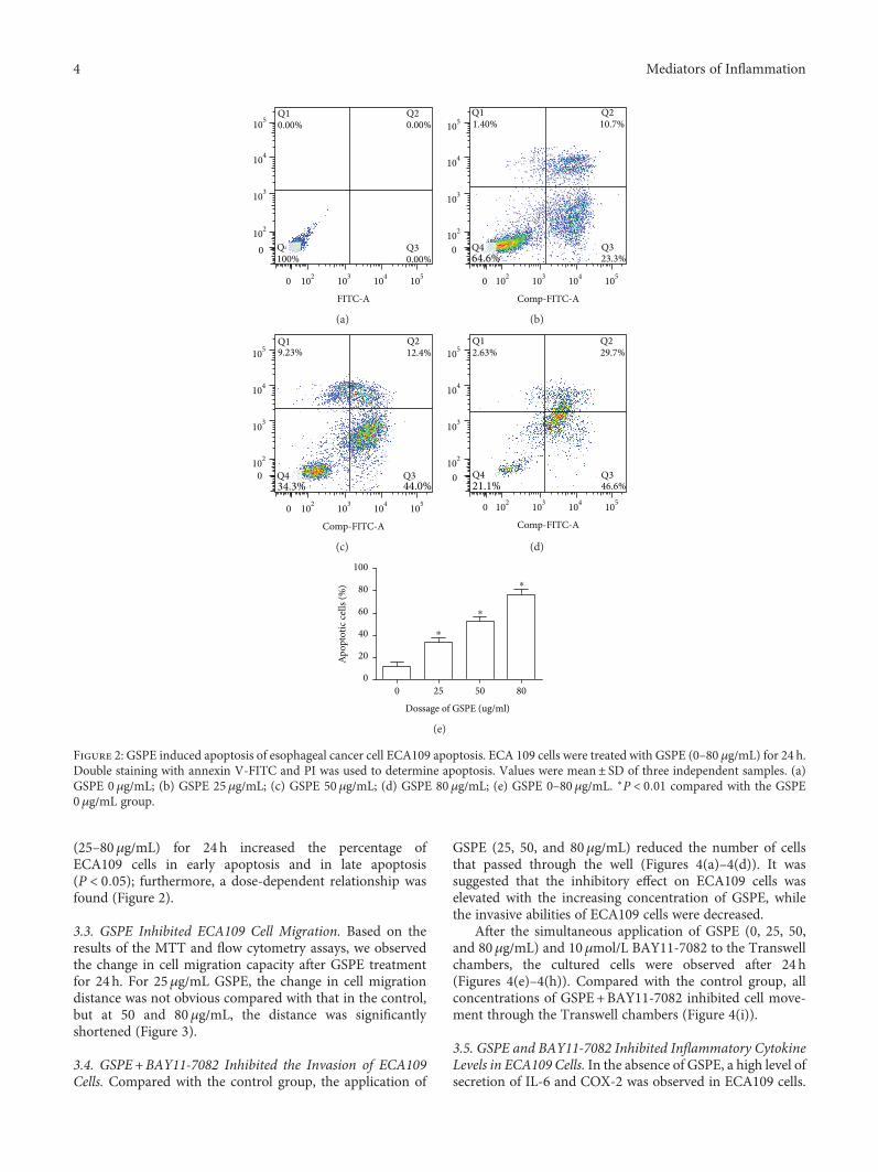
(25–80μg/mL) for 24 h increased the percentage ofECA109 cells in early apoptosis and in late apoptosis(P < 0 05); furthermore, a dose-dependent relationship wasfound (Figure 2).
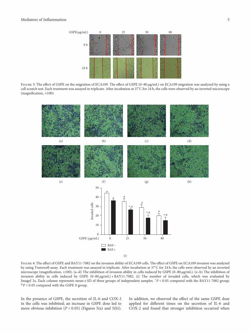
3.3. GSPE Inhibited ECA109 Cell Migration. Based on theresults of the MTT and flow cytometry assays, we observedthe change in cell migration capacity after GSPE treatmentfor 24h. For 25μg/mL GSPE, the change in cell migrationdistance was not obvious compared with that in the control,but at 50 and 80μg/mL, the distance was significantlyshortened (Figure 3).
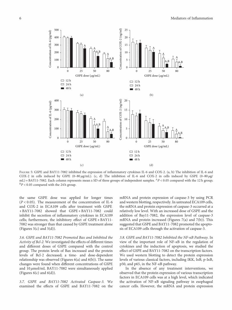
3.4. GSPE+BAY11-7082 Inhibited the Invasion of ECA109Cells. Compared with the control group, the application of
GSPE (25, 50, and 80μg/mL) reduced the number of cellsthat passed through the well (Figures 4(a)–4(d)). It wassuggested that the inhibitory effect on ECA109 cells waselevated with the increasing concentration of GSPE, whilethe invasive abilities of ECA109 cells were decreased.
After the simultaneous application of GSPE (0, 25, 50,and 80μg/mL) and 10μmol/L BAY11-7082 to the Transwellchambers, the cultured cells were observed after 24 h(Figures 4(e)–4(h)). Compared with the control group, allconcentrations of GSPE+BAY11-7082 inhibited cell move-ment through the Transwell chambers (Figure 4(i)).
3.5. GSPE and BAY11-7082 Inhibited Inflammatory CytokineLevels in ECA109 Cells. In the absence of GSPE, a high level ofsecretion of IL-6 and COX-2 was observed in ECA109 cells.
Q1
102
103
104
105
0
0
0.00%
Q4100%
Q20.00%
Q30.00%
103 104 105102
FITC-A
(a)
0
0
Q1 Q2
Q4 Q3
1.40% 10.7%
64.6% 23.3%
102
103
104
105
104103 105102
Comp-FITC-A
(b)
0
0
Q19.23%
Q434.3%
Q212.4%
Q344.0%
102
103
104
105
105104103102
Comp-FITC-A
(c)
Q1
Q4 Q3
0
2.63%
0
Q229.7%
21.1% 46.6%
102
103
104
105
103 104 105102
Comp-FITC-A
(d)
⁎
⁎
⁎
0
20
40
60
80
100
Apop
totic
cells
(%)
25 50 800
Dossage of GSPE (ug/ml)
(e)
Figure 2: GSPE induced apoptosis of esophageal cancer cell ECA109 apoptosis. ECA 109 cells were treated with GSPE (0–80 μg/mL) for 24 h.Double staining with annexin V-FITC and PI was used to determine apoptosis. Values were mean± SD of three independent samples. (a)GSPE 0 μg/mL; (b) GSPE 25μg/mL; (c) GSPE 50 μg/mL; (d) GSPE 80μg/mL; (e) GSPE 0–80μg/mL. ∗P < 0 01 compared with the GSPE0μg/mL group.
4 Mediators of Inflammation
In the presence of GSPE, the secretion of IL-6 and COX-2in the cells was inhibited; an increase in GSPE dose led tomore obvious inhibition (P < 0 05) (Figures 5(a) and 5(b)).
In addition, we observed the effect of the same GSPE doseapplied for different times on the secretion of IL-6 andCOX-2 and found that stronger inhibition occurred when
(a) (b) (c) (d)
(e) (f) (g) (h)
#
#
#
BAY−BAY+
GSPE (�휇g/mL)
⁎
⁎,# ⁎,#
⁎,#
0
10
20
30
40
50
Inva
ded
cells
25 50 800
(i)
Figure 4: The effect of GSPE and BAY11-7082 on the invasion ability of ECA109 cells. The effect of GSPE on ECA109 invasion was analyzedby using Transwell assay. Each treatment was assayed in triplicate. After incubation at 37°C for 24 h, the cells were observed by an invertedmicroscope (magnification, ×100). (a–d) The inhibition of invasion ability in cells induced by GSPE (0–80μg/mL). (e–h) The inhibition ofinvasion ability in cells induced by GSPE (0–80 μg/mL) + BAY11-7082. (i) The number of invaded cells, which was evaluated byImageJ 2x. Each column represents mean± SD of three groups of independent samples. ∗P < 0 05 compared with the BAY11-7082 group;#P < 0 05 compared with the GSPE 0 group.
GSPE(�휇g/mL)
0 h
24 h
0 25 50 80
Figure 3: The effect of GSPE on the migration of ECA109. The effect of GSPE (0–80μg/mL) on ECA109 migration was analyzed by using acell scratch test. Each treatment was assayed in triplicate. After incubation at 37°C for 24 h, the cells were observed by an inverted microscope(magnification, ×100).
5Mediators of Inflammation
the same GSPE dose was applied for longer times(P < 0 05). The measurement of the concentration of IL-6and COX-2 in ECA109 cells after treatment with GSPE+BAY11-7082 showed that GSPE+BAY11-7082 couldinhibit the secretion of inflammatory cytokines in ECA109cells; furthermore, the inhibitory effect of GSPE+BAY11-7082 was stronger than that caused by GSPE treatment alone(Figures 5(c) and 5(d)).
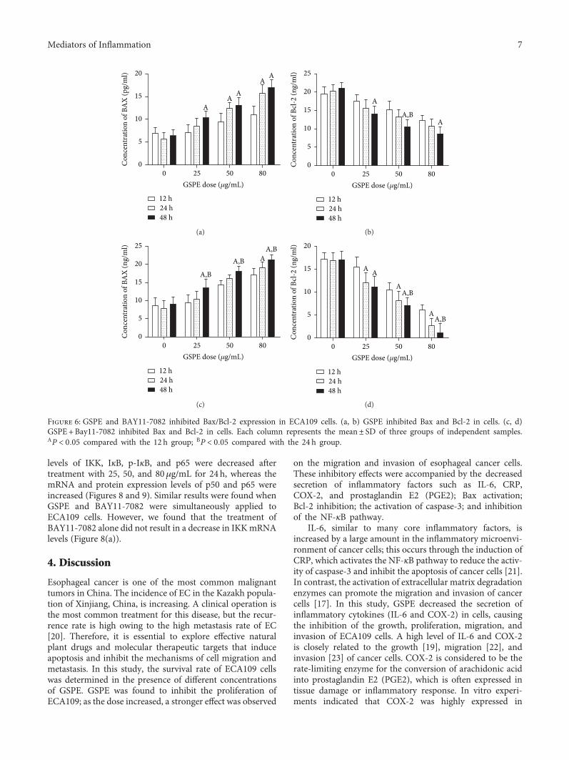
3.6. GSPE and BAY11-7082 Promoted Bax and Inhibited theActivity of Bcl-2.We investigated the effects of different timesand different doses of GSPE compared with the controlgroup. The protein levels of Bax increased and the proteinlevels of Bcl-2 decreased; a time- and dose-dependentrelationship was observed (Figures 6(a) and 6(b)). The samechanges were found when different concentrations of GSPEand 10μmol/mL BAY11-7082 were simultaneously applied(Figures 6(c) and 6(d)).
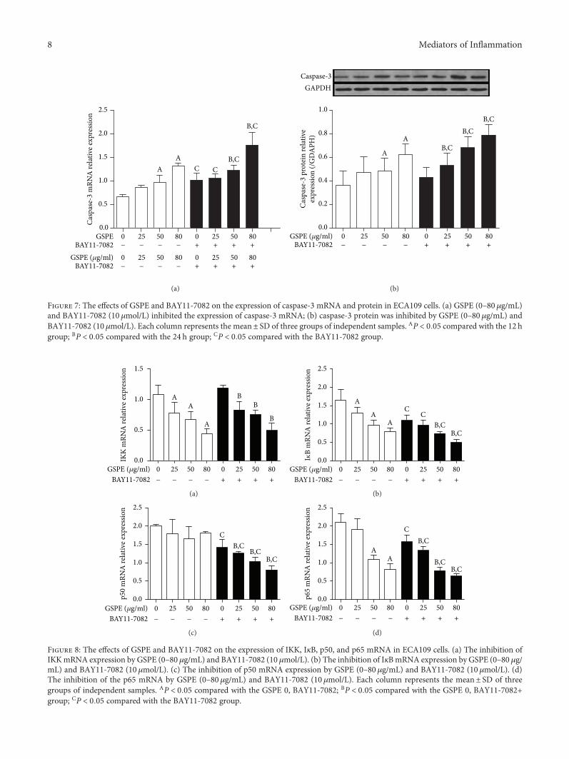
3.7. GSPE and BAY11-7082 Activated Caspase-3. Weexamined the effects of GSPE and BAY11-7082 on the
mRNA and protein expression of caspase-3 by using PCRand western blotting, respectively. In untreated ECA109 cells,the mRNA and protein expression of caspase-3 occurred at arelatively low level. With an increased dose of GSPE and theaddition of Bay11-7082, the expression level of caspase-3mRNA and protein increased (Figures 7(a) and 7(b)). Thissuggested that GSPE and BAY11-7082 promoted the apopto-sis of ECA109 cells through the activation of caspase-3.
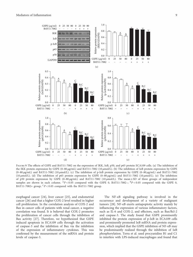
3.8. GSPE and BAY11-7082 Inhibited the NF-κB Pathway. Inview of the important role of NF-κB in the regulation ofcytokines and the induction of apoptosis, we studied theeffect of GSPE and BAY11-7082 on the transcription factors.We used western blotting to detect the protein expressionlevels of various classical factors, including IKK, IκB, p-IκB,p50, and p65, in the NF-κB pathway.
In the absence of any treatment interventions, weobserved that the protein expression of various transcriptionfactors in ECA109 cells was at a high level, which indicatedthe activation of NF-κB signaling pathway in esophagealcancer cells. However, the mRNA and protein expression
AA
AA,B
A,B
12 h24 h48 h
25 50 800GSPE dose (�휇g/mL)
0
100
200
300
400
500
Con
cent
ratio
n of
IL-6
(pg/
ml)
(a)
A
AA
A,B A,B0
5
10
15
20
25
Con
cent
ratio
n of
CO
X-2
(ng/
ml)
25 50 800GSPE dose (�휇g/mL)
12 h24 h48 h
(b)
A
AA,B
A,B
25 50 800GSPE dose (�휇g/mL)
0
100
200
300
400
Con
cent
ratio
n of
IL-6
(pg/
ml)
12 h24 h48 h
(c)
AA
A,B
A,B
25 50 800GSPE dose (�휇g/mL)
0
5
10
15
20
Con
cent
ratio
n of
CO
X-2
(ng/
ml)
12 h24 h48 h
(d)
Figure 5: GSPE and BAY11-7082 inhibited the expression of inflammatory cytokines IL-6 and COX-2. (a, b) The inhibition of IL-6 andCOX-2 in cells induced by GSPE (0–80 μg/mL). (c, d) The inhibition of IL-6 and COX-2 in cells induced by GSPE (0–80μg/mL) + BAY11-7082. Each column represents mean± SD of three groups of independent samples. AP < 0 05 compared with the 12 h group;BP < 0 05 compared with the 24 h group.
6 Mediators of Inflammation
levels of IKK, IκB, p-IκB, and p65 were decreased aftertreatment with 25, 50, and 80μg/mL for 24 h, whereas themRNA and protein expression levels of p50 and p65 wereincreased (Figures 8 and 9). Similar results were found whenGSPE and BAY11-7082 were simultaneously applied toECA109 cells. However, we found that the treatment ofBAY11-7082 alone did not result in a decrease in IKKmRNAlevels (Figure 8(a)).
4. Discussion
Esophageal cancer is one of the most common malignanttumors in China. The incidence of EC in the Kazakh popula-tion of Xinjiang, China, is increasing. A clinical operation isthe most common treatment for this disease, but the recur-rence rate is high owing to the high metastasis rate of EC[20]. Therefore, it is essential to explore effective naturalplant drugs and molecular therapeutic targets that induceapoptosis and inhibit the mechanisms of cell migration andmetastasis. In this study, the survival rate of ECA109 cellswas determined in the presence of different concentrationsof GSPE. GSPE was found to inhibit the proliferation ofECA109; as the dose increased, a stronger effect was observed
on the migration and invasion of esophageal cancer cells.These inhibitory effects were accompanied by the decreasedsecretion of inflammatory factors such as IL-6, CRP,COX-2, and prostaglandin E2 (PGE2); Bax activation;Bcl-2 inhibition; the activation of caspase-3; and inhibitionof the NF-κB pathway.
IL-6, similar to many core inflammatory factors, isincreased by a large amount in the inflammatory microenvi-ronment of cancer cells; this occurs through the induction ofCRP, which activates the NF-κB pathway to reduce the activ-ity of caspase-3 and inhibit the apoptosis of cancer cells [21].In contrast, the activation of extracellular matrix degradationenzymes can promote the migration and invasion of cancercells [17]. In this study, GSPE decreased the secretion ofinflammatory cytokines (IL-6 and COX-2) in cells, causingthe inhibition of the growth, proliferation, migration, andinvasion of ECA109 cells. A high level of IL-6 and COX-2is closely related to the growth [19], migration [22], andinvasion [23] of cancer cells. COX-2 is considered to be therate-limiting enzyme for the conversion of arachidonic acidinto prostaglandin E2 (PGE2), which is often expressed intissue damage or inflammatory response. In vitro experi-ments indicated that COX-2 was highly expressed in
A
AA
AA
0
5
10
15
20
Con
cent
ratio
n of
BA
X (p
g/m
l)
50 800 25GSPE dose (�휇g/mL)
12 h24 h48 h
(a)
A
AA,B
0
5
10
15
20
25
Con
cent
ratio
n of
Bcl-
2 (n
g/m
l)
25 50 800GSPE dose (�휇g/mL)
12 h24 h48 h
(b)
A,B
A,BA,B
A
0
5
10
15
20
25
Con
cent
ratio
n of
BA
X (n
g/m
l)
25 50 800GSPE dose (�휇g/mL)
12 h24 h48 h
(c)
A A
A
A
A,B
A,B
0
5
10
15
20
Con
cent
ratio
n of
Bcl-
2 (n
g/m
l)
50 800 25GSPE dose (�휇g/mL)
12 h24 h48 h
(d)
Figure 6: GSPE and BAY11-7082 inhibited Bax/Bcl-2 expression in ECA109 cells. (a, b) GSPE inhibited Bax and Bcl-2 in cells. (c, d)GSPE +Bay11-7082 inhibited Bax and Bcl-2 in cells. Each column represents the mean± SD of three groups of independent samples.AP < 0 05 compared with the 12 h group; BP < 0 05 compared with the 24 h group.
7Mediators of Inflammation
AA
C C
B,C
B,C
GSPE
GSPE (�휇g/ml)BAY11-7082
BAY11-7082
0.0
0.5
1.0
1.5
2.0
2.5
Casp
ase-
3 m
RNA
relat
ive e
xpre
ssio
n
25 50 80 0 25 50 800−− −− ++ ++
25 50 80 0 25 50 800−− −− ++ ++
(a)
GSPE (�휇g/ml)BAY11-7082
Caspase-3GAPDH
0.0
0.2
0.4
0.6
0.8
1.0
Casp
ase-
3 pr
otei
n re
lativ
eex
pres
sion
(/G
DA
PH)
25 50 80 0 25 50 800− − − + + + +−
A
AB,C
B,CB,C
(b)
Figure 7: The effects of GSPE and BAY11-7082 on the expression of caspase-3 mRNA and protein in ECA109 cells. (a) GSPE (0–80μg/mL)and BAY11-7082 (10 μmol/L) inhibited the expression of caspase-3 mRNA; (b) caspase-3 protein was inhibited by GSPE (0–80μg/mL) andBAY11-7082 (10 μmol/L). Each column represents the mean± SD of three groups of independent samples. AP < 0 05 compared with the 12 hgroup; BP < 0 05 compared with the 24 h group; CP < 0 05 compared with the BAY11-7082 group.
AA
A
BB
B
GSPE (�휇g/ml)BAY11-7082
0.0
0.5
1.0
1.5
IKK
mRN
A re
lativ
e exp
ress
ion
2550 80 025 50 800− −−− + ++ +
(a)
GSPE (�휇g/ml)BAY11-7082
2550 80 025 50 800− −−− + ++ +
A
AA
CC
B,CB,C
0.0
0.5
1.0
1.5
2.0
2.5
I�휅B
mRN
A re
lativ
e exp
ress
ion
(b)
GSPE (�휇g/ml)BAY11-7082
2550 80 025 50 800− −−− + ++ +
CB,C
B,CB,C
0.0
0.5
1.0
1.5
2.0
2.5
p50
mRN
A re
lativ
e exp
ress
ion
(c)
GSPE (�휇g/ml)BAY11-7082
2550 80 025 50 800− −−− + ++ +
AA
CB,C
B,CB,C
0.0
0.5
1.0
1.5
2.0
2.5
p65
mRN
A re
lativ
e exp
ress
ion
(d)
Figure 8: The effects of GSPE and BAY11-7082 on the expression of IKK, IκB, p50, and p65 mRNA in ECA109 cells. (a) The inhibition ofIKKmRNA expression by GSPE (0–80 μg/mL) and BAY11-7082 (10 μmol/L). (b) The inhibition of IκBmRNA expression by GSPE (0–80μg/mL) and BAY11-7082 (10 μmol/L). (c) The inhibition of p50 mRNA expression by GSPE (0–80μg/mL) and BAY11-7082 (10 μmol/L). (d)The inhibition of the p65 mRNA by GSPE (0–80μg/mL) and BAY11-7082 (10 μmol/L). Each column represents the mean± SD of threegroups of independent samples. AP < 0 05 compared with the GSPE 0, BAY11-7082; BP < 0 05 compared with the GSPE 0, BAY11-7082+group; CP < 0 05 compared with the BAY11-7082 group.
8 Mediators of Inflammation
esophageal cancer [24], liver cancer [25], and endometrialcancer [26] and that a higher COX-2 level resulted in highercell proliferation. In the correlation analysis of COX-2 andBax in cancer cells of patients with renal cancer, a negativecorrelation was found. It is believed that COX-2 promotesthe proliferation of cancer cells through the inhibition ofBax activity [27]. Therefore, we hypothesized that GSPEinduced apoptosis in ECA109 cells through the activationof caspase-3 and the inhibition of Bax via the inhibitionof the expression of inflammatory cytokines. This wasconfirmed by the measurement of the mRNA and proteinlevels of caspase-3.
The NF-κB signaling pathway is involved in theoccurrence and development of a variety of malignanttumors [28]. NF-κB exerts antiapoptotic activity mainly byinfluencing the expression of various inflammatory factors,such as IL-6 and COX-2, and effectors, such as Bax/Bcl-2and caspase-3. The study found that GSPE prominentlyinhibited the protein expression of p-IκB in ECA109 cellsand prominently promoted IκB mRNA and protein expres-sion, which implied that the GSPE inhibition of NF-κB maybe predominantly realized through the inhibition of IκBphosphorylation. Terra et al. used procyanidins B1 and C1to interfere with LPS-induced macrophages and found that
GSPE (�휇g/ml)BAY11-7082
0 25 50 80 0 25 50 80− − − − + + + +
IKK
I�휅B
p-I�휅B
p65
p50
GAPDH GSPE (�휇g/ml)BAY11-7082
AA
0.0
0.2
0.4
0.6
0.8
1.0
Rela
ive p
rote
in ex
pres
sion
(IKK
/GD
APH
)
8050 80 0 25 50250− − − + + + +−
(a)
GSPE (�휇g/ml)BAY11-7082
8050 80 0 25 50250− − − + + + +−
A AA
C
B,CB,C
B,C
0.0
0.1
0.2
0.3
0.4
0.5
Rela
ive p
rote
in ex
pres
sion
(I�휅
B/G
DA
PH)
(b)
GSPE (�휇g/ml)BAY11-7082
8050 80 0 25 50250− − − + + + +−
AA
CB,C
B,CB,C
0.0
0.2
0.4
0.6
0.8
1.0
Rela
ive p
rote
in ex
pres
sion
(p-I�휅
B/G
DA
PH)
(c)
GSPE (�휇g/ml)BAY11-7082
8050 80 0 25 50250− − − + + + +−
AA
C
B,C
B,CB,C
0.0
0.2
0.4
0.6
0.8
1.0
Rela
ive p
rote
in ex
pres
sion
(p65
/GD
APH
)
(d)
GSPE (�휇g/ml)BAY11-7082
8050 80 0 25 50250− − − + + + +−
AC
B,C B,C B,C
0.0
0.2
0.4
0.6
0.8
Rela
ive p
rote
in ex
pres
sion
(p50
/GD
APH
)
(e)
Figure 9: The effects of GSPE and BAY11-7082 on the expression of IKK, IκB, p50, and p65 protein ECA109 cells. (a) The inhibition ofthe IKK protein expression by GSPE (0–80 μg/mL) and BAY11-7082 (10 μmol/L). (b) The inhibition of IκB protein expression by GSPE(0–80 μg/mL) and BAY11-7082 (10 μmol/L). (c) The inhibition of p-IκB protein expression by GSPE (0–80μg/mL) and BAY11-7082(10 μmol/L). (d) The inhibition of p65 protein expression by GSPE (0–80μg/mL) and BAY11-7082 (10 μmol/L). (e) The inhibitionof p50 protein expression by GSPE (0–80 μg/mL) and BAY11-7082 (10 μmol/L). The mean± SD of three groups of independentsamples are shown in each column. AP < 0 05 compared with the GSPE 0, BAY11-7082–; BP < 0 05 compared with the GSPE 0,BAY11-7082+ group; CP < 0 05 compared with the BAY11-7082 group.
9Mediators of Inflammation
the proanthocyanidins inhibited the activation of the NF-κBpathway by inhibiting the phosphorylation of IκB [29]. How-ever, Zhao et al. found that GSPE inhibited IκB in humanovarian cancer A2780 cells, which inhibited the NF-κBpathway and subsequently promoted apoptosis [15]. Basedon the effects of GSPE, we also investigated the treatmentof the NF-κB-specific inhibitor BAY11-7082 and found thatGSPE+BAY11-7082 was a more effective inhibitor of thephosphorylation level of IκB compared with GSPE alone.This suggested that the inhibition of NF-κB by GSPE wasachieved by the inhibition of IκB phosphorylation; a similareffect occurred with BAY11-7082, showing that GSPE andBAY11-7082 may have a synergistic inhibitory effect on theNF-κB in ECA109 cells.
In addition, we found that GSPE inhibited the expressionof NF-κB p50/p65 mRNA and protein in cells. NF-κB p50/p65, the most common heterogeneous dimer in the NF-κBsignaling pathway, is also an important protein for the func-tion of NF-κB. In resting cells, NF-κB p50/p65 and IκB formcomplexes, which exist in the cytoplasm in an inactive form.When the cell is stimulated by an extracellular signal, the IκBkinase complex (IKK) activates the phosphorylation of IκB,and the NF-κB is exposed to the nuclear localization site.The dissociated NF-κB is rapidly shifted to the nucleus, bind-ing to a specific κB sequence and inducing the transcriptionof related genes. The GSPE inhibition of NF-κB p50/p65resulted from the inhibition of IκB phosphorylation by
GSPE, which was consistent with the research of Mackenzieet al. [30]. Some studies have suggested that the ability ofprocyanidins to inhibit NF-κB p50/p65 expression inhibitionmay result from the appearance of the procyanidin dimersthat may mimic the arginine residues of the NF-κB p50/p65sequence, with respect to hydrogen bonding, to inhibit theexpression of p50/p65 [31]. However, our study did notindicate whether the chemical structure of GSPE was relatedto the expression of NF-κB p50/p65.
In addition, we found that the mRNA and proteinexpressions of IKK were both inhibited by GSPE. How-ever, there was no significant difference between the GSPEgroup and the GSPE+BAY11-7082 group. BAY11-7082, aspecific inhibitor of NF-κB, inhibits the phosphorylation ofIκB. Therefore, our findings also suggest that GSPE maydirectly affect IKK, inhibit the activation of IKK, andinhibit the phosphorylation of IκB; together, this inhibitsthe NF-κB pathway.
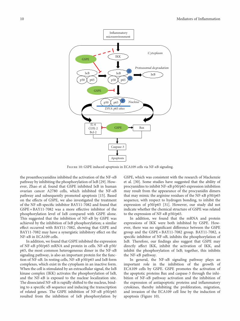
In general, the NF-κB signaling pathway plays animportant role in the inhibition of the growth ofECA109 cells by GSPE. GSPE promotes the activation ofthe apoptotic proteins Bax and caspase-3 through the inhi-bition of NF-κB pathway activation and the inhibition ofthe expression of antiapoptotic proteins and inflammatorycytokines, thereby inhibiting the proliferation, migration,and invasion of the ECA109 cell line by the induction ofapoptosis (Figure 10).
p50p65p50
Proteasomal degradation
Cytoplasm
p65p50
DNA p65 sites
Nucleus
p65
IKK
GSPE
GSPE
IL-6COX-2Bcl-2
Bax
Caspase-3
Apoptosis
GSPE
Inflammatorymicroenvironment
P
P
PI�휅BI�휅BI�휅B
Figure 10: GSPE induced apoptosis in ECA109 cells via NF-κB signaling.
10 Mediators of Inflammation
5. Conclusions
Our study has illustrated a possible molecular mechanism forthe action of GSPE against cancer; however, the occurrenceand development of cancer and the migration and inva-sion of cancer cells are complex and involve multiplefactors. Therefore, the specific mechanism requires extensiveresearch to explore the anticancer effect of procyanidins andprovide a basis for their effective use. The results and discus-sion may be presented separately, or in one combined sec-tion, and may optionally be divided into headed subsections.
Data Availability
The datasets used or analyzed during the current studyare available from the corresponding author on reason-able request.
Conflicts of Interest
The authors declare no conflict of interests regarding thepublication of this paper.
Acknowledgments
This study was supported by the National Natural ScienceFoundation of China (nos. 81760584 and 81560517), theKey Areas of Science and Technology Research Projectof Xinjiang Production and Construction Corps (no.2014BA039 and no. 2015AG014), and the InternationalCooperative Project of Shihezi University (no. GJHZ201602).
References
[1] A. Qukuerhan and X. Xaymardan, “The epidemiologicalanalysis of 904 cases of malignant tumor of Kazak in Xinjiang,”Tumor, vol. 23, no. 1, 2003.
[2] S. J. Murphy, A. E. Hughes, C. C. Patterson et al., “Apopulation-based association study of SNPs of GSTP 1,MNSOD, GPX 2 and Barrett’s esophagus and esophagealadenocarcinoma,” Carcinogenesis, vol. 28, no. 6, pp. 1323–1328, 2007.
[3] G. H. Zhang, M. Su, and D. P. Tian, “Effect of chronicinflammation-induced basement membrane changes onesophageal carcinogenesis,” Ai Zheng, vol. 24, no. 9,pp. 1071–1075, 2005.
[4] Y. R. Liu, X. Yan, H. X. Yu et al., “NLRC 5 promotes cellproliferation via regulating the NF-κB signaling pathwayin rheumatoid arthritis,” Molecular Immunology, vol. 91,pp. 24–34, 2017.
[5] M. Zhang and F. Jin, “1α,25-Dihydroxyvitamin D3 amelioratesseawater aspiration-induced lung injury by inhibiting thetranslocation of NF-κB and RhoA,” Inflammation, vol. 40,no. 3, pp. 832–839, 2017.
[6] L. Yu, Y. Mu, N. Sa, H. Wang, and W. Xu, “Tumor necrosisfactor α induces epithelial-mesenchymal transition andpromotes metastasis via NF-κB signaling pathway-mediatedTWIST expression in hypopharyngeal cancer,” OncologyReports, vol. 31, no. 1, pp. 321–327, 2014.
[7] M. Sha, J. Ye, L. X. Zhang, Z. Y. Luan, and Y. B. Chen,“Celastrol induces apoptosis of gastric cancer cells by miR-
146a inhibition of NF-κB activity,” Cancer Cell International,vol. 13, no. 1, p. 50, 2013.
[8] T. Lawrence, “The nuclear factor NF-κB pathway in inflamma-tion,” Cold Spring Harbor Perspectives in Biology, vol. 1, no. 6,2009.
[9] Q. Li and I. M. Verma, “Erratum: NF-κB regulation in theimmune system,” Nature Reviews Immunology, vol. 2, no. 12,pp. 975–975, 2002.
[10] D. Joyce, C. Albanese, J. Steer, M. Fu, B. Bouzahzah, andR. Pestell, “NF-κB and cell-cycle regulation: the cyclin connec-tion,” Cytokine & Growth Factor Reviews, vol. 12, no. 1,pp. 73–90, 2001.
[11] A. E. Harvey, “Energy balance, inflammation, and tumorprogression: the role of NF-κB,” Journal of the AmericanVeterinary Medical Association, vol. 228, no. 4, pp. 522–527,2011.
[12] S. Li, M. Xu, Q. Niu et al., “Efficacy of procyanidins againstin vivo cellular oxidative damage: a systematic review andmeta-analysis,” PLoS One, vol. 10, no. 10, article e0139455,2015.
[13] M. Wei, F. Guo, D. Rui et al., “Alleviation of arsenic-inducedpulmonary oxidative damage by GSPE as shown duringin vivo and in vitro experiments,” Biological Trace ElementResearch, vol. 183, no. 1, pp. 80–91, 2018.
[14] K. C. Choi, S. Park, B. J. Lim et al., “Procyanidin B3, aninhibitor of histone acetyltransferase, enhances the action ofantagonist for prostate cancer cells via inhibition of p300-dependent acetylation of androgen receptor,” BiochemicalJournal, vol. 433, no. 1, pp. 235–244, 2011.
[15] B. X. Zhao, Y. B. Sun, S. Q. Wang et al., “Grape seed procyani-din reversal of p-glycoprotein associated multi-drug resistancevia down-regulation of NF-κB and MAPK/ERK mediatedYB-1 activity in a2780/T cells,” PLoS One, vol. 8, no. 8, arti-cle e71071, 2013.
[16] C. Minker, L. Duban, D. Karas, P. Järvinen, A. Lobstein, andC. D. Muller, “Impact of procyanidins from different berrieson caspase 8 activation in colon cancer,” Oxidative Medicineand Cellular Longevity, vol. 2015, Article ID 154164, 13 pages,2015.
[17] S. Dinicola, A. Pasqualato, A. Cucina et al., “Grape seed extractsuppresses MDA-MB231 breast cancer cell migration andinvasion,” European Journal of Nutrition, vol. 53, no. 2,pp. 421–431, 2014.
[18] M. I. Wei, L. I. Wu, Y. I. Shuying, Y. I. Weijie, S. H. Tala, andH. A. Wenting, “Hawthorn procyanidins regulate theexpression of COX-2 and induce the apoptosis of ECA109esophageal cancer cells,” Food Science, vol. 37, no. 3,pp. 176–181, 2016.
[19] M. Thill, A. Woeste, K. Reichert et al., “Vitamin D inhibitsovarian cancer cell line proliferation in combination withcelecoxib and suppresses cyclooxygenase-2 expression,” Anti-cancer Research, vol. 35, no. 2, pp. 1197–1203, 2015.
[20] C.-L. Li, F.-L. Zhang, Y.-D. Wang et al., “Characteristics ofrecurrence after radical esophagectomy with two-field lymphnode dissection for thoracic esophageal cancer,” OncologyLetters, vol. 5, no. 1, pp. 355–359, 2013.
[21] B. Dobrzycka, B. Mackowiak-Matejczyk, K. M. Terlikowska,B. Kulesza-Bronczyk, M. Kinalski, and S. J. Terlikowski,“Serum levels of IL-6, IL-8 and CRP as prognostic factorsin epithelial ovarian cancer,” European Cytokine Network,vol. 24, no. 3, pp. 106–113, 2013.
11Mediators of Inflammation
[22] J. Karavitis, L. M. Hix, Y. H. Shi, R. F. Schultz, K. Khazaie,and M. Zhang, “Regulation of COX-2 expression in mousemammary tumor cells controls bone metastasis and PGE2-induction of regulatory t cell migration,” PLoS One, vol. 7,no. 9, article e46342, 2012.
[23] T. Li, J. Zhong, X. Dong et al., “Meloxicam suppresseshepatocellular carcinoma cell proliferation and migration bytargeting COX-2/PGE2-regulated activation of the β-cateninsignaling pathway,” Oncology Reports, vol. 35, no. 6,pp. 3614–3622, 2016.
[24] M. Bardou, “Chronic intake of NSAIDs and COX-2 inhibitorsreduce risk of esophageal cancer,” European Journal of Cancer,vol. 46, no. 18, pp. 3417–3424, 2010.
[25] H. Xiong, B. Li, J. He, Y. Zeng, Y. Zhang, and F. He, “lncRNAHULC promotes the growth of hepatocellular carcinoma cellsvia stabilizing COX-2 protein,” Biochemical and BiophysicalResearch Communications, vol. 490, no. 3, pp. 693–699, 2017.
[26] K. Hasegawa, K. Ishikawa, S. Kawai et al., “Overcomingpaclitaxel resistance in uterine endometrial cancer using aCOX-2 inhibitor,” Oncology Reports, vol. 30, no. 6, pp. 2937–2944, 2013.
[27] F. S. Pehlivan, A. Sarı, S. N. Görgel et al., “Relationship ofCOX-2, Bax, Bcl-2, Ki 67, p 53 expression to clinicopathologicparameters and their impact on prognosis in renal cellcarcinoma,” Journal of Turgut Ozal Medical Center, vol. 21,no. 3, 2014.
[28] F. Li, J. Zhang, F. Arfuso et al., “Nf-κB in cancer therapy,”Archives of Toxicology, vol. 89, no. 5, pp. 711–731, 2015.
[29] X. Terra, P. Palozza, J. Fernandez-Larrea et al., “Procyanidindimer b1 and trimer c1 impair inflammatory responsesignalling in human monocytes,” Free Radical Research,vol. 45, no. 5, pp. 611–619, 2011.
[30] G. G. Mackenzie, A. M. Adamo, N. P. Decker, and P. I. Oteiza,“Dimeric procyanidin B2 inhibits constitutively active NF-κBin Hodgkin’s lymphoma cells independently of the presenceof IκB mutations,” Biochemical Pharmacology, vol. 75, no. 7,pp. 1461–1471, 2008.
[31] C. G. Fraga and P. I. Oteiza, “Dietary flavonoids: role of(−)-epicatechin and related procyanidins in cell signaling,”Free Radical Biology & Medicine, vol. 51, no. 4, pp. 813–823,2011.
12 Mediators of Inflammation

Stem Cells International
Hindawiwww.hindawi.com Volume 2018
Hindawiwww.hindawi.com Volume 2018
MEDIATORSINFLAMMATION
of
EndocrinologyInternational Journal of
Hindawiwww.hindawi.com Volume 2018
Hindawiwww.hindawi.com Volume 2018
Disease Markers
Hindawiwww.hindawi.com Volume 2018
BioMed Research International
OncologyJournal of
Hindawiwww.hindawi.com Volume 2013
Hindawiwww.hindawi.com Volume 2018
Oxidative Medicine and Cellular Longevity
Hindawiwww.hindawi.com Volume 2018
PPAR Research
Hindawi Publishing Corporation http://www.hindawi.com Volume 2013Hindawiwww.hindawi.com
The Scientific World Journal
Volume 2018
Immunology ResearchHindawiwww.hindawi.com Volume 2018
Journal of
ObesityJournal of
Hindawiwww.hindawi.com Volume 2018
Hindawiwww.hindawi.com Volume 2018
Computational and Mathematical Methods in Medicine
Hindawiwww.hindawi.com Volume 2018
Behavioural Neurology
OphthalmologyJournal of
Hindawiwww.hindawi.com Volume 2018
Diabetes ResearchJournal of
Hindawiwww.hindawi.com Volume 2018
Hindawiwww.hindawi.com Volume 2018
Research and TreatmentAIDS
Hindawiwww.hindawi.com Volume 2018
Gastroenterology Research and Practice
Hindawiwww.hindawi.com Volume 2018
Parkinson’s Disease
Evidence-Based Complementary andAlternative Medicine
Volume 2018Hindawiwww.hindawi.com
Submit your manuscripts atwww.hindawi.com
Top Related