


![Mechanische Beanspruchung und subchondrale · PDF fileDie 4-Fragment-Fraktur des proximalen Oberarms 422 The four-fragment fracture of the proximal humerus W. Knopp, Β. ... [14],](https://static.fdocument.org/doc/165x107/5a7b54f17f8b9a2e6e8bd25f/mechanische-beanspruchung-und-subchondrale-4-fragment-fraktur-des-proximalen.jpg)
γλώσσες
Σελίδες
Νομικός

Fragment-guided design of subnanomolarβ-lactamase inhibitors active in vivoOliv Eidama,1, Chiara Romagnolib,1, Guillaume Dalmassoc, Sarah Bareliera, Emilia Casellib, Richard Bonnetc,d,2,Brian K. Shoicheta,2, and Fabio Pratib,2
aDepartment of Pharmaceutical Chemistry, University of California, 1700 Fourth Street, Byers Hall, San Francisco, CA 94158; bDipartimento di Chimica,Università degli Studi di Modena e Reggio Emilia, Via Campi 183, 41125 Modena, Italy; cMicrobes, Intestin, Inflammation et Susceptibilité de l'Hôte,UMR1071 Inserm, Université d’Auvergne, Unité Sous Contrat Institut National de la Recherche Agronomique 2018, Clermont-Ferrand F-63001, France;and dService de Bactériologie, Centre Hospitalier Universitaire, Clermont-Ferrand F-63001, France
Edited by Gregory A. Petsko, Brandeis University, Waltham, MA, and approved September 11, 2012 (received for review May 16, 2012)
Fragment-based design was used to guide derivatization of alead series of β-lactamase inhibitors that had heretofore resistedoptimization for in vivo activity. X-ray structures of fragmentsoverlaid with the lead suggested new, unanticipated functionalityand points of attachment. Synthesis of three derivatives improvedaffinity over 20-fold and improved efficacy in cell culture. Crystalstructures were consistent with the fragment-based design, en-abling further optimization to a K i of 50 pM, a 500-fold improve-ment that required the synthesis of only six derivatives. One ofthese, compound 5, was tested in mice. Whereas cefotaximealone failed to cure mice infected with β-lactamase-expressingEscherichia coli, 65% were cleared of infection when treated witha cefotaxime:5 combination. Fragment complexes offer a patharound design hurdles, even for advanced molecules; the seriesdescribed here may provide leads to overcome β-lactamase-basedresistance, a key clinical challenge.
antibiotic resistance ∣ antimicrobial ∣ drug discovery ∣ structure-based ∣boronic acid
Lead optimization in chemical biology and drug discovery is amultifactorial problem and frequently stalls on the way to
tool molecules or clinical candidates. Confronted with an other-wise attractive compound series for which affinity or efficacy hasleveled off, for instance, one seeks an efficient derivatizationstrategy in the face of many possible chemotypes and points ofderivatization. Multiple paths may be considered, from combina-torial derivatization among accessible side chains, to structure-based placement of specific groups. Neither of these strategiescan fully promise to overcome the challenge of knowing exactlywhere and how to derivatize a lead.
We faced such a challenge with a series of boronic acid β-lac-tamase inhibitors. β-lactamases are the most common cause ofresistance to β-lactam antibiotics, such as penicillins and cepha-losporins; they threaten what remains the most widely used classof antibiotics and have attracted much recent attention (1–5).Third- and fourth-generation cephalosporins were partly intro-duced to overcome these enzymes, as were inhibitors like clavu-lanic acid, but resistance arose rapidly to these agents, which areall themselves β-lactams and thus potential substrates of β-lacta-mases. Boronic acids, as non-lactam transition-state analogs, areimpervious to such mutants and are often transparent to otherβ-lactam resistance mechanisms, such as β-lactamase upregula-tion. Using β-lactam functional groups on a boronic acid scaffold,we rapidly optimized an initial class of boronic acids to mid-nanomolar affinity (6, 7), and with further derivatization to 1 nMaffinity against the class C β-lactamase AmpC (8). Notwithstand-ing their high affinity, these compounds had relatively modestactivity in bacterial cell culture. Though they certainly reducedminimum inhibitory concentrations (MICs) of third-generationcephalosporins, they were unable to break through the empiricalresistance threshold—typically 2 μg∕mL—for most pathogens(6–9). Subsequent efforts failed to further improve affinity (9).
Also, these molecules used functionality well explored amongβ-lactams themselves to achieve their affinity—utilizing, for in-stance, the R1 side chains of cephalothin and ceftazidime, and theC3(4)’ carboxylate of penicillins and cephalosporins—and sothey were subject to pre-evolved resistant mutant enzymes, suchas the inhibitor-resistant TEM30 (10).
To escape this cul-de-sac, we thought to replace the boronicacid R1 carboxamide, which mimics the analogous group ubiqui-tous among β-lactam drugs, with a sulfonamide, as this wouldnot only change the electronic character of a key interactionbut also the geometry of the inhibitor (11). This switch scrambledthe structure-activity relationship previously observed with the car-boxamides: Boronic acids with a carboxylate analogous to the C3(4)’ group of penicillins and β-lactams lost 10- to 100-fold activity,relative to the carboxamide series; conversely, small derivatives onthe R1 side, such as compounds 1 and 2 (Table 1), achieved K ivalues as low as 70 and 25 nM, 8- to 20-fold better than observedfor analogous carboxamides. Unfortunately, further derivatizationfailed to improve affinity; indeed, even the addition of a carbox-ylate to 1, leading to 2, improved activity only threefold, whereasa similar derivative in an earlier series had improved affinity byover 20-fold (12). We were stuck again.
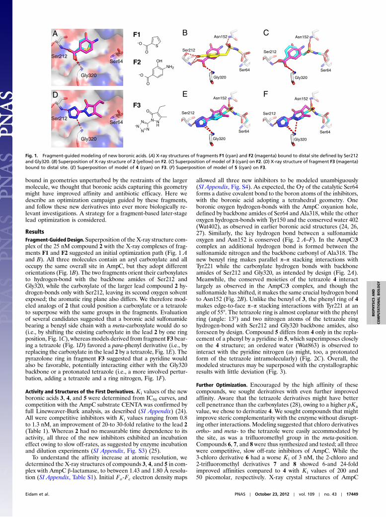
Fragment-based discovery has anchored early lead discoveryfor targets that have resisted traditional methods. Fragmentscan optimally explore receptor pockets (13–18) and better coverchemical space (19–22), and we wondered if they could guidea late-stage optimization that had thus far floundered. In a pre-vious fragment-based docking screen against AmpC (22) and ina defragmentation study of a known AmpC inhibitor (23), crystalstructures of three anionic fragments were determined: athiophene carboxylic acid (F1), a benzoic acid (F2), and an aryltetrazole (F3), all of which bind in the same distal region of theactive site where the aryl carboxylate of compound 2 was placed(Fig. 1 A, B, and D). The poses adopted by these fragments over-lapped one another, and though in roughly the same spot as thecarboxylate of compound 2, they differed in their angle of attackfrom the larger inhibitor (Fig. 1B). Consequently, in their crystal-lographic complexes with AmpC they hydrogen-bonded with thebackbone amides of Ser212 and Gly320, rather than with Ser212alone as did the 2 carboxylate. Reasoning that the fragments
Author contributions: O.E., C.R., G.D., R.B., B.K.S., and F.P. designed research; O.E., C.R.,G.D., S.B., and R.B. performed research; C.R. contributed new reagents/analytic tools; O.E.,C.R., G.D., S.B., E.C., R.B., B.K.S., and F.P. analyzed data; and O.E. and B.K.S. wrote the paper.
The authors declare no conflict of interest.
This article is a PNAS Direct Submission.
Data deposition: The atomic coordinates and structure factors have been depositedin the Protein Data Bank, www.pdb.org (PDB ID codes 4E3I, 4E3J, 4E3K, 4E3L, 4E3M, 4E3N,and 4E3O).1O.E. and C.R. contributed equally to this work.2To whom correspondence may be addressed. E-mail: [email protected] [email protected] or [email protected].
This article contains supporting information online at www.pnas.org/lookup/suppl/doi:10.1073/pnas.1208337109/-/DCSupplemental.
17448–17453 ∣ PNAS ∣ October 23, 2012 ∣ vol. 109 ∣ no. 43 www.pnas.org/cgi/doi/10.1073/pnas.1208337109
bound in geometries unperturbed by the restraints of the largermolecule, we thought that boronic acids capturing this geometrymight have improved affinity and antibiotic efficacy. Here wedescribe an optimization campaign guided by these fragments,and follow these new derivatives into ever more biologically re-levant investigations. A strategy for a fragment-based later-stagelead optimization is considered.
ResultsFragment-Guided Design. Superposition of the X-ray structure com-plex of the 25 nM compound 2 with the X-ray complexes of frag-ments F1 and F2 suggested an initial optimization path (Fig. 1 Aand B). All three molecules contain an aryl carboxylate and alloccupy the same overall site in AmpC, but they adopt differentorientations (Fig. 1B). The two fragments orient their carboxylatesto hydrogen-bond with the backbone amides of Ser212 andGly320, while the carboxylate of the larger lead compound 2 hy-drogen-bonds only with Ser212, leaving its second oxygen solventexposed; the aromatic ring plane also differs. We therefore mod-eled analogs of 2 that could position a carboxylate or a tetrazoleto superpose with the same groups in the fragments. Evaluationof several candidates suggested that a boronic acid sulfonamidebearing a benzyl side chain with a meta-carboxylate would do so(i.e., by shifting the existing carboxylate in the lead 2 by one ringposition, Fig. 1C), whereasmodels derived from fragment F3 bear-ing a tetrazole (Fig. 1D) favored a para-phenyl derivative (i.e., byreplacing the carboxylate in the lead 2 by a tetrazole, Fig. 1E). Thepyrazolone ring in fragment F3 suggested that a pyridine wouldalso be favorable, potentially interacting either with the Gly320backbone or a protonated tetrazole (i.e., a more involved pertur-bation, adding a tetrazole and a ring nitrogen, Fig. 1F).
Activity and Structures of the First Derivatives.K i values of the newboronic acids 3, 4, and 5 were determined from IC50 curves, andcompetition with the AmpC substrate CENTA was confirmed byfull Lineweaver-Burk analysis, as described (SI Appendix) (24).All were competitive inhibitors with K i values ranging from 0.8to 1.3 nM, an improvement of 20-to 30-fold relative to the lead 2(Table 1). Whereas 2 had no measurable time dependence to itsactivity, all three of the new inhibitors exhibited an incubationeffect owing to slow off-rates, as suggested by enzyme incubationand dilution experiments (SI Appendix, Fig. S3) (25).
To understand the affinity increase at atomic resolution, wedetermined the X-ray structures of compounds 3, 4, and 5 in com-plex with AmpC β-lactamase, to between 1.43 and 1.80 Å resolu-tion (SI Appendix, Table S1). Initial Fo-Fc electron density maps
allowed all three new inhibitors to be modeled unambiguously(SI Appendix, Fig. S4). As expected, the Oγ of the catalytic Ser64forms a dative covalent bond to the boron atoms of the inhibitors,with the boronic acid adopting a tetrahedral geometry. Oneboronic oxygen hydrogen-bonds with the AmpC oxyanion hole,defined by backbone amides of Ser64 and Ala318, while the otheroxygen hydrogen-bonds with Tyr150 and the conserved water 402(Wat402), as observed in earlier boronic acid structures (24, 26,27). Similarly, the key hydrogen bond between a sulfonamideoxygen and Asn152 is conserved (Fig. 2 A–F). In the AmpC/3complex an additional hydrogen bond is formed between thesulfonamide nitrogen and the backbone carbonyl of Ala318. Thenew benzyl ring makes parallel π–π stacking interactions withTyr221 while the carboxylate hydrogen bonds with backboneamides of Ser212 and Gly320, as intended by design (Fig. 2A).Meanwhile, the conserved moieties of the tetrazole 4 interactlargely as observed in the AmpC/3 complex, and though thesulfonamide has shifted, it makes the same crucial hydrogen bondto Asn152 (Fig. 2B). Unlike the benzyl of 3, the phenyl ring of 4makes edge-to-face π–π stacking interactions with Tyr221 at anangle of 55°. The tetrazole ring is almost coplanar with the phenylring (angle: 13°) and two nitrogen atoms of the tetrazole ringhydrogen-bond with Ser212 and Gly320 backbone amides, alsoforeseen by design. Compound 5 differs from 4 only in the repla-cement of a phenyl by a pyridine in 5, which superimposes closelyon the 4 structure; an ordered water (Wat863) is observed tointeract with the pyridine nitrogen (as might, too, a protonatedform of the tetrazole intramolecularly) (Fig. 2C). Overall, themodeled structures may be superposed with the crystallographicresults with little deviation (Fig. 3).
Further Optimization. Encouraged by the high affinity of thesecompounds, we sought derivatives with even further improvedaffinity. Aware that the tetrazole derivatives might have bettercell penetrance than the carboxylates (28), owing to a higher pKavalue, we chose to derivatize 4. We sought compounds that mightimprove steric complementarity with the enzyme without disrupt-ing other interactions. Modeling suggested that chloro derivativesortho- and meta- to the tetrazole were easily accommodated bythe site, as was a trifluoromethyl group in the meta-position.Compounds 6, 7, and 8 were thus synthesized and tested; all threewere competitive, slow off-rate inhibitors of AmpC. While the3-chloro derivative 6 had a worse K i of 3 nM, the 2-chloro and2-trifluoromethyl derivatives 7 and 8 showed 6-and 24-foldimproved affinities compared to 4 with K i values of 200 and50 picomolar, respectively. X-ray crystal structures of AmpC
N
O
O
B C
E F
Gly320
Ser212Ser64
A
Gly320
Ser212Ser64
D
-
O
NH2
OH
-
O
S
F2
F1
N-
N N
N NH
OF3
Ser64
Asn152
Ser212
Gly320
Ser64
Asn152
Ser212
Gly320
Ser64
Asn152
Ser212
Gly320
Ser64
Asn152
Ser212
Gly320
Fig. 1. Fragment-guided modeling of new boronic acids. (A) X-ray structures of fragments F1 (cyan) and F2 (magenta) bound to distal site defined by Ser212and Gly320. (B) Superposition of X-ray structure of 2 (yellow) on F2. (C) Superposition of model of 3 (cyan) on F2. (D) X-ray structure of fragment F3 (magenta)bound to distal site. (E) Superposition of model of 4 (cyan) on F3. (F) Superposition of model of 5 (cyan) on F3.
Eidam et al. PNAS ∣ October 23, 2012 ∣ vol. 109 ∣ no. 43 ∣ 17449
BIOPH
YSICSAND
COMPU
TATIONALBIOLO
GY
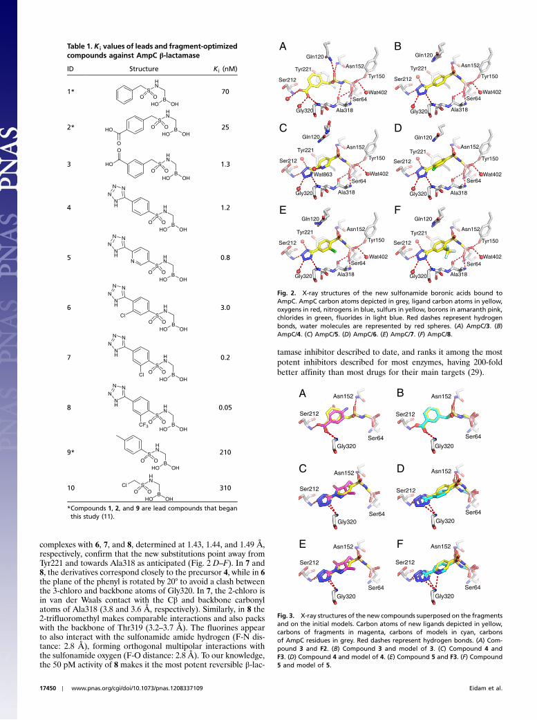
complexes with 6, 7, and 8, determined at 1.43, 1.44, and 1.49 Å,respectively, confirm that the new substitutions point away fromTyr221 and towards Ala318 as anticipated (Fig. 2 D–F). In 7 and8, the derivatives correspond closely to the precursor 4, while in 6the plane of the phenyl is rotated by 20° to avoid a clash betweenthe 3-chloro and backbone atoms of Gly320. In 7, the 2-chloro isin van der Waals contact with the Cβ and backbone carbonylatoms of Ala318 (3.8 and 3.6 Å, respectively). Similarly, in 8 the2-trifluoromethyl makes comparable interactions and also packswith the backbone of Thr319 (3.2–3.7 Å). The fluorines appearto also interact with the sulfonamide amide hydrogen (F-N dis-tance: 2.8 Å), forming orthogonal multipolar interactions withthe sulfonamide oxygen (F-O distance: 2.8 Å). To our knowledge,the 50 pM activity of 8 makes it the most potent reversible β-lac-
tamase inhibitor described to date, and ranks it among the mostpotent inhibitors described for most enzymes, having 200-foldbetter affinity than most drugs for their main targets (29).
BGln120
Tyr150
Ala318
Tyr221
Ser64
Asn152
Wat402
Ser212
Gly320
AGln120
Tyr150
Ala318
Tyr221
Ser64
Asn152
Wat402
Ser212
Gly320
CGln120
Tyr150
Ala318
Tyr221
Ser64
Asn152
Wat402
Ser212
Gly320
DGln120
Tyr150
Ala318
Tyr221
Ser64
Asn152
Wat402
Ser212
Gly320
EGln120
Tyr150
Ala318
Tyr221
Ser64
Asn152
Wat402
Ser212
Gly320
FGln120
Tyr150
Ala318
Tyr221
Ser64
Asn152
Wat402
Ser212
Gly320
Wat863
Fig. 2. X-ray structures of the new sulfonamide boronic acids bound toAmpC. AmpC carbon atoms depicted in grey, ligand carbon atoms in yellow,oxygens in red, nitrogens in blue, sulfurs in yellow, borons in amaranth pink,chlorides in green, fluorides in light blue. Red dashes represent hydrogenbonds, water molecules are represented by red spheres. (A) AmpC/3. (B)AmpC/4. (C) AmpC/5. (D) AmpC/6. (E) AmpC/7. (F) AmpC/8.
Table 1. K i values of leads and fragment-optimizedcompounds against AmpC β-lactamase
ID Structure Ki (nM)
1* S
HN
O O BHO OH
70
2*S
HN
O O BHO OH
O
HO 25
3 S
HN
O O BHO OH
HO
O
1.3
4S
HN
O O BHO OH
NN
NNH 1.2
5
S
HN
O O BHO OH
N
NN
NNH 0.8
6S
HN
O O BHO OH
NN
NNH
Cl
3.0
7S
HN
O O BHO OH
NN
NNH
Cl
0.2
8S
HN
O O BHO OH
NN
NNH
CF3
0.05
9* S
HN
O O BHO OH
210
10 S
HN
O O BHO OH
Cl 310
*Compounds 1, 2, and 9 are lead compounds that beganthis study (11).
C
E
Ser64
Asn152
Gly320
D
F
Ser64
Asn152
Ser212
Gly320
B
Ser64
Asn152
Ser212
Gly320
A
Ser64
Asn152
Ser212
Gly320
Ser212
Ser64
Asn152
Ser212
Gly320
Ser64
Asn152
Ser212
Gly320
Fig. 3. X-ray structures of the new compounds superposed on the fragmentsand on the initial models. Carbon atoms of new ligands depicted in yellow,carbons of fragments in magenta, carbons of models in cyan, carbonsof AmpC residues in grey. Red dashes represent hydrogen bonds. (A) Com-pound 3 and F2. (B) Compound 3 and model of 3. (C) Compound 4 andF3. (D) Compound 4 and model of 4. (E) Compound 5 and F3. (F) Compound5 and model of 5.
17450 ∣ www.pnas.org/cgi/doi/10.1073/pnas.1208337109 Eidam et al.
Selectivity. To assess the selectivity of the new molecules wedetermined K i values against three common serine proteases—Trypsin, Elastase, and α-Chymotrypsin—as well as that of a classA β-lactamase, CTX-M-9 (SI Appendix, Table S2) (30–33). Affi-nity for AmpC was typically 105-to 106-fold better than for theserine proteases (SI Appendix, Fig. S5). Notwithstanding theboronic acid warhead shared by these inhibitors, the compoundsshow clear specificity for their target over protease off-targets.Affinity was also substantially better for AmpC than CTX-M-9,which, though speaking to specificity, may portend difficulties forclinical relevance, as one would ideally prefer a compound activeagainst both class C and class A enzymes. Still, several of theanalogs retained substantial affinity for CTX-M-9, especially 3,which was a 45 nM inhibitor of CTX-M-9.
Microbiology. The anti-resistance activity of inhibitors was inves-tigated by the determination of the minimum inhibitory concen-trations (MICs) of the β-lactam/inhibitor combination necessaryto inhibit the growth of clinically isolated bacteria resistant tothird-generation cephalosporins via expression of class A or classC β-lactamases (Table 2). Used by themselves, the antibioticscefotaxime and ceftazidime had high MIC values, often greaterthan 64 μg∕mL, certainly much higher than the break point forempirical resistance levels ≥2 μg∕mL (34). Conversely, in com-bination with the new inhibitors, the MIC values of these third-generation cephalosporins improved substantially, typically by64-fold or more. For 75% of the clinical isolates measured, MICsdropped into the susceptible range (MICs ≤1 μg∕mL) with com-pounds 3 and 5, for 50% with compounds 7 and 8, and for 25% ofthose treated with the cephalosporins and 4 or 6 (SI Appendix,Table S3). For many clinical isolates, MIC values for ceftazidimeand cefotaxime combined with 5 and 7 dropped to 0.5 μg∕mLand below (SI Appendix, Fig. S6), which represents an 8-to 16-foldimprovement of MIC values compared to previously tested boro-nic acids (6, 8, 11).
Intriguingly, substantial decreases in MIC values were ob-served for a strain producing the plasmid-mediated class A β-lac-tamase CTX-M-14 (8-to 64-fold), especially for compounds 3and 7, which had the broadest spectrums of activity. This offerspreliminary evidence that the sulfonamide boronic acids may in-hibit both class C and class A β-lactamases, consistent with theirin vitro activity against this class of enzymes.
Efficacy in a Mouse Model of Infection. We had not observed suchsubstantial reversal of bacterial resistance to β-lactams, acrosssuch a broad spectrum of clinical isolates, for previous series ofboronic acid inhibitors of β-lactamase; indeed, this lack of effi-
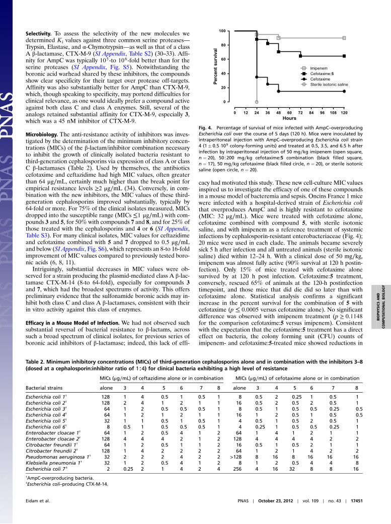
cacy had motivated this study. These new cell-culture MIC valuesinspired us to investigate the efficacy of one of these compoundsin a mouse model of bacteremia and sepsis. Oncins France 1 micewere infected with a hospital-derived strain of Escherichia colithat overproduces AmpC and is highly resistant to cefotaxime(MIC: 32 μg∕mL). Mice were treated with cefotaxime alone,cefotaxime combined with compound 5, with sterile isotonicsaline, and with imipenem as a reference treatment of systemicinfections by cephalosporin-resistant enterobacteriaceae (Fig. 4);20 mice were used in each clade. The animals became severelysick 5 h after infection and all untreated animals (sterile isotonicsaline) died within 12–24 h. With a clinical dose of 50 mg∕kg,imipenem was almost fully active (90% survival at 120 h postin-fection). Only 15% of mice treated with cefotaxime alonesurvived by at 120 h post infection. Cefotaxime:5 treatment,conversely, rescued 65% of animals at the 120-h postinfectiontimepoint, and those mice that did die did so later than withcefotaxime alone. Statistical analysis confirms a significantincrease in the percent survival for the combination of 5 withcefotaxime (p ≤ 0.0005 versus cefotaxime alone). No significantdifference was observed with imipenem treatment (p ≥ 0.1148for the comparison cefotaxime:5 versus imipenem). Consistentwith the expectation that the cefotaxime:5 treatment has a directeffect on bacteria, the colony forming unit (CFU) counts ofimipenem- and cefotaxime:5-treated mice showed reductions in
Fig. 4. Percentage of survival of mice infected with AmpC–overproducingEscherichia coli over the course of 5 days (120 h). Mice were inoculated byintraperitoneal injection with AmpC–overproducing Escherichia coli strain4 (1� 0.5 109 colony-forming units) and treated at 0.5, 3.5, and 6.5 h afterinfection by intraperitoneal injection of 50 mg∕kg imipenem (open square,n ¼ 20), 50∶200 mg∕kg cefotaxime:5 combination (black filled square,n ¼ 17), 50 mg∕kg cefotaxime (black filled circle, n ¼ 20), or sterile isotonicsaline (open circle, n ¼ 20).
Table 2. Minimum inhibitory concentrations (MICs) of third-generation cephalosporins alone and in combination with the inhibitors 3–8(dosed at a cephalosporin:inhibitor ratio of 1∶4) for clinical bacteria exhibiting a high level of resistance
Bacterial strains
MICs (μg∕mL) of ceftazidime alone or in combination MICs (μg∕mL) of cefotaxime alone or in combination
alone 3 4 5 6 7 8 alone 3 4 5 6 7 8
Escherichia coli 1† 128 1 4 0.5 1 0.5 1 8 0.5 2 0.25 1 0.5 1Escherichia coli 2† 128 2 4 1 2 1 1 16 0.5 2 0.5 2 0.5 1Escherichia coli 3† 64 1 2 0.5 0.5 0.5 1 8 0.5 1 0.5 0.5 0.25 0.5Escherichia coli 4† 64 1 2 1 2 1 1 16 1 2 0.5 1 0.5 0.5Escherichia coli 5† 32 1 1 0.5 1 0.5 1 4 0.5 1 0.5 2 0.5 1Escherichia coli 6† 8 0.5 1 0.5 0.5 0.5 1 4 0.25 1 0.5 0.5 0.25 1Enterobacter cloacae 1† 64 1 2 0.5 4 1 2 64 1 4 1 2 1 1Enterobacter cloacae 2† 128 4 4 4 2 1 2 128 4 4 4 4 2 2Citrobacter freundii 1† 64 1 2 0.5 1 1 2 16 0.5 1 0.5 2 1 1Citrobacter freundii 2† 128 1 4 2 2 2 2 64 1 2 1 4 2 2Pseudomonas aeruginosa 1† 32 2 2 2 4 2 2 >128 8 16 8 16 16 16Klebsiella pneumonia 1† 32 1 2 0.5 4 1 2 8 1 2 0.5 4 4 8Escherichia coli 7 ‡ 2 0.25 2 1 4 2 4 256 4 16 32 8 8 16†AmpC-overproducing bacteria.‡Escherichia coli–producing CTX-M-14.
Eidam et al. PNAS ∣ October 23, 2012 ∣ vol. 109 ∣ no. 43 ∣ 17451
BIOPH
YSICSAND
COMPU
TATIONALBIOLO
GY
all organs and blood compared to treatment with cefotaximealone and to untreated controls (SI Appendix, Fig. S7 andTable S4).
DiscussionThe use of fragments in hit-to-lead development has becomepopular in drug discovery, especially for difficult drug targets.Fragments benefit from binding to pockets and surfaces unper-turbed by restraints found in larger molecules, and often do sowith high ligand efficiency. By merging, linking, or growing frag-ments, high-affinity leads may be obtained. A second advantageof fragments is that they cover much more chemical space thanlead-like molecules (13–15, 19–21).
Here we used both virtues to optimize a series already exhibit-ing decent affinity but insufficient biological activity. First, weexploited the geometric information contained in aryl-carboxy-late fragments. The lead compound 2 had a K i of 25 nM againstAmpC β-lactamase and lowered MIC values eightfold on aver-age. Modeling suggested that 3 could pick up the interactionsobserved in fragments F1 and F2. In fact, only a different orienta-tion for the benzoic acid substructure contained in the lead 2seemed necessary, which could be obtained by moving the carbox-ylate from para- to meta-. This improved affinity of compound 3almost 20-fold over 2, while the affinity 3 is 54-fold better than 1(ΔΔG ¼ 1.9 kcal∕mol), which can be attributed to the carboxylatein a preferred environment. Indeed, the placement of the distalcarboxylate of 3 between Ser212 and Gly320 superposes well withthat observed in F2 (Fig. 3A), recapitulating the designed structurewith a RMSD of 0.3 Å (Fig. 3B). The improved affinity alsoimproved antimicrobial activity: MIC values dropped 64-fold onaverage and the median MIC for 3 was 1 μg∕mL against 12 highlyresistant strains (SI Appendix, Table S3), below the empiricalbreak point for hospital infections. Fortuitously, compound 3 alsoinhibits class A β-lactamases efficiently, with a K i of 45 nM againstCTX-M-9. Correspondingly, it lowers MIC values for bacteriaexpressing this enzyme, in combination with ceftazidime and cefo-taxime, by 8-and 64-fold respectively, making it the compound withbest broad-spectrum activity within this series.
In the tetrazole series, we were guided by the geometric infor-mation contained in fragment F3. The tetrazoles of the designedmolecules 4 and 5 superpose well with that of F3, and the initialmodels agree well with the subsequent crystal structures, withRMSD values of 0.9 Å and 0.7 Å, respectively (Fig. 3 C–F). Com-parison with the K i of molecule 9 (K i 210 nM) suggests that thetetrazole added about 3 kcal∕mol of affinity, improving the K i170-fold in compound 4 and 250-fold in the pyridine derivative5. Tetrazoles are common bioisosteres of carboxylates and oftenhave better bioavailability (28, 35). Although they are not unpre-cedented in β-lactam antibiotics (e.g., cefazolin), boronic acidinhibitors of β-lactamases have not yet exploited this chemotypein this region of the active site. While previous generations ofboronic acids focused on mimicking β-lactam substrates, thesefragment-derived boronic acids exhibit greater novelty and maybe more robust against pre-evolved mutant enzymes that over-come boronic acids more closely resembling β-lactams (10).
Whereas fragments have been used previously for new chemo-type discovery (13–22) and merging, their use in late stage opti-mization has remained largely unexplored. Of course nothingprevented this, and indeed such an idea is implicit in the fragmentapproach and anticipated by computational design methods likeLUDI, HOOK, GrowMol, and MCSS (36–39). Still, late-stageoptimization with fragments seems underdeveloped; it can revealderivatization strategies, both in geometry and in chemotype,that may otherwise remain unknown without an industrial-scalehit-to-lead campaign.
Certain caveats deserve attention. This approach to optimizingleads with fragments is restricted to targets where proximal bind-ing sites can be detected and for which fragment orientations
can be accurately determined. It also requires a decomposablelead series where substantial inhibition remains with only a corechemotype, which is not always the case (23, 40). Whereas thesecompounds did turn out to be additive in affinity gained—com-pared to the naked sulfonamide they added over 2-logs, while theK i values of the fragments were between 3 and 40 mM—thistoo will not always hold. Indeed, in another series of analogs thatalso tried to exploit the fragment placement, no improvement inaffinity was achieved. There are also cautions to the mouse ex-periments—in the cephalosporin/inhibitor combination clade,we preserved the cefotaxime/inhibitor ratio of 1∶4 of the MICexperiments. This resulted in a final concentration of 200 mg∕kgfor inhibitor 5, which is very high. Future studies may focus onthe analysis and improvement of pharmacokinetic propertiesand proper evaluation of toxicity and activity against a largerpanel of bacteria. Fortunately, because the molecules remainsmall (molecular weights range from 270 to 350 Da), there isroom for further optimization.
These caveats should not obscure the central observation ofthis study—two series of fragments, bound in a particular pocketrevealed an opportunity to derivatize a relatively advanced seriesin a direction, and with chemotypes, that had not been previouslyexplored or imagined. This overcame what had been an unsur-mounted barrier in efficacy. Not only was the resulting seriespotent, with sub-nanomolar to mid-picomolar affinities, but alsoit had clinically relevant MIC values and activity in a mouse mod-el of bacterial infection. In short, beginning with a structural studyand guided synthesis, we ended up with molecules active in vivoin a mammalian system. In themselves, these inhibitors holdpromise as leads to overcome a pervasive and growing threat topublic health. More generally, whereas fragments are widely usedto nucleate early discovery (13–22), this study suggests that theyalso may be used to guide late-stage optimization into chemo-types and geometries that would be hard to systematically sampleby other methods.
MethodsModeling of Distal Site Binders. Boronic acids were modeled manually andsubsequently minimized using PLOP (41) (SI Appendix, Supporting Methods).
Synthesis. Sulfonamidomethaneboronic acids (1–10) were obtained fromfunctionalized sulfonyl chlorides. Microwave assisted cycloaddition yieldedtetrazoles 4–8 (SI Appendix, Supporting Methods).
Enzymology. Enzyme inhibition was measured by the method of initial rates(SI Appendix, Supporting Methods).
Crystallography. All AmpC/inhibitor X-ray structures were obtained byco-crystallization and determined by molecular replacement (SI Appendix,Supporting Methods). The atomic coordinates and structure factors for AmpCwith compounds 3–8 and 10 have been deposited in the Protein Data Bank(PDB), www.rcsb.org (PDB ID codes 4E3I, 4E3J, 4E3K, 4E3L, 4E3M, 4E3N, 4E3O).
Microbiology. Susceptibility testing followed the guidelines of CLSI (34). EachMIC value reported reflects the average of three independent experiments(SI Appendix, Supporting Methods).
In Vivo Efficacy Studies. The experiments were approved by the Animal CareCommittee of Auvergne University, Clermont-Ferrand, France (SI Appendix,Supporting Methods).
ACKNOWLEDGMENTS. We thank Prof. Y. Chen and A. Doak for CTX-M-9 andAmpC and Dr. A. O’Donoghue for serine proteases and substrates. We thankDrs. K. Ziebart and M. Merski for assisting with the slow binding kinetics andI. Fish and S. Pierre for structure refinements. We thank Dr. H.T.T. Nguyenfor technical assistance Microbes, Intestin, Inflammation et Susceptibilitéde l'Hôte and Dr. A. Alloui for animal care. We thank Dr. M. Fischer andA. Doak for reading this manuscript, and the Centro InterdipartimentaleGrandi Strumenti of Modena for NMR and MS spectra. This study wassupported by National Institutes of Health Grant GM63815 and by InstitutNational de la Santé et de la Recherche Médicale and Institut National dela Recherche Agronomique.
17452 ∣ www.pnas.org/cgi/doi/10.1073/pnas.1208337109 Eidam et al.
1. Frere JM (1995) Beta-lactamases and bacterial resistance to antibiotics. Mol Microbiol16:385–395.
2. Bush K (1999) Beta-lactamases of increasing clinical importance. Curr Pharm Des5:839–845.
3. NukagaM, Kumar S, Nukaga K, Pratt RF, Knox JR (2004) Hydrolysis of third-generationcephalosporins by class C beta-lactamases: Structures of a transition state analog ofcefotaxime in wild-type and extended spectrum enzymes. J Biol Chem 279:9344–9352.
4. Fisher JF, Meroueh SO, Mobashery S (2005) Bacterial resistance to beta-lactam antibio-tics: Compelling opportunism, compelling opportunity. Chem Rev 105:395–424.
5. Drawz SM, Bonomo RA (2010) Three decades of beta-lactamase inhibitors. Clin Micro-biol Rev 23:160–201.
6. Caselli E, et al. (2001) Energetic, structural, and antimicrobial analyses of beta-lactamside chain recognition by beta-lactamases. Chem Biol 8:17–31.
7. Powers RA, Caselli E, Focia PJ, Prati F, Shoichet BK (2001) Structures of ceftazidime andits transition-state analogue in complex with AmpC beta-lactamase: Implications forresistance mutations and inhibitor design. Biochemistry 40:9207–9214.
8. Morandi F, et al. (2003) Nanomolar inhibitors of AmpC beta-lactamase. J Am Chem Soc125:685–695.
9. Morandi S, Morandi F, Caselli E, Shoichet BK, Prati F (2008) Structure-based optimiza-tion of cephalothin-analogue boronic acids as beta-lactamase inhibitors. Bioorg MedChem 16:1195–1205.
10. Wang X, et al. (2003) Recognition and resistance in TEM beta-lactamase. Biochemistry42:8434–8444.
11. Eidam O, et al. (2010) Design, synthesis, crystal structures, and antimicrobial activity ofsulfonamide boronic acids as beta-lactamase inhibitors. J Med Chem 53:7852–7863.
12. Tondi D, Morandi F, Bonnet R, Costi MP, Shoichet BK (2005) Structure-based optimiza-tion of a non-beta-lactam lead results in inhibitors that do not up-regulate beta-lactamase expression in cell culture. J Am Chem Soc 127:4632–4639.
13. Rees DC, Congreve M, Murray CW, Carr R (2004) Fragment-based lead discovery. NatRev Drug Discovery 3:660–672.
14. Murray CW, Blundell TL (2010) Structural biology in fragment-based drug design. CurrOpin Struct Biol 20:497–507.
15. Fischer M, Hubbard RE (2009) Fragment-based ligand discovery. Mol Interventions9:22–30.
16. Ciulli A, Williams G, Smith AG, Blundell TL, Abell C (2006) Probing hot spots at protein-ligand binding sites: A fragment-based approach using biophysical methods. J MedChem 49:4992–5000.
17. Allen KN, et al. (1996) An experimental approach to mapping the binding surfaces ofcrystalline proteins. J Phys Chem 100:2605–2611.
18. Landon MR, et al. (2009) Detection of ligand binding hot spots on protein surfaces viafragment-based methods: Application to DJ-1 and glucocerebrosidase. J ComputAided Mol Des 23:491–500.
19. Hann MM, Leach AR, Harper G (2001) Molecular complexity and its impact on theprobability of finding leads for drug discovery. J Chem Inf Comput Sci 41:856–864.
20. Fink T, Bruggesser H, Reymond JL (2005) Virtual exploration of the small-molecule che-mical universe below 160 daltons. Angew Chem Int Ed Engl 44:1504–1508.
21. Fink T, Reymond JL (2007) Virtual exploration of the chemical universe up to 11 atomsof C, N, O, F: Assembly of 26.4 million structures (110.9 million stereoisomers) and ana-
lysis for new ring systems, stereochemistry, physicochemical properties, compoundclasses, and drug discovery. J Chem Inf Model 47:342–353.
22. Teotico DG, et al. (2009) Docking for fragment inhibitors of AmpC beta-lactamase.Proc Natl Acad Sci USA 106:7455–7460.
23. Babaoglu K, Shoichet BK (2006) Deconstructing fragment-based inhibitor discovery.Nat Chem Biol 2:720–723.
24. Weston GS, Blazquez J, Baquero F, Shoichet BK (1998) Structure-based enhancementof boronic acid-based inhibitors of AmpC beta-lactamase. J Med Chem 41:4577–4586.
25. Morrison JF, Walsh CT (1988) The behavior and significance of slow-binding enzymeinhibitors. Adv Enzymol Relat Areas Mol Biol 61:201–301.
26. Strynadka NC,Martin R, Jensen SE, GoldM, Jones JB (1996) Structure-based design of apotent transition state analogue for TEM-1 beta-lactamase. Nat Struct Biol 3:688–695.
27. Chen Y, Minasov G, Roth TA, Prati F, Shoichet BK (2006) The deacylation mechanism ofAmpC beta-lactamase at ultrahigh resolution. J Am Chem Soc 128:2970–2976.
28. Lemke TL, Williams DA (2007) Foye’s Principles of Medicinal Chemistry (LippincottWilliams & Wilkins, Philadelphia), 6th Ed.
29. Overington JP, Al-Lazikani B, Hopkins AL (2006) Howmany drug targets are there?NatRev Drug Discovery 5:993–996.
30. Pouvreau L, et al. (1998) Effect of pea and bovine trypsin inhibitors on wild-type andmodified trypsins. FEBS Lett 423:167–172.
31. Del Mar EG, Largman C, Brodrick JW, Fassett M, Geokas MC (1980) Substrate specificityof human pancreatic elastase 2. Biochemistry 19:468–472.
32. Rodriguez-Martinez JA, Rivera-Rivera I, Sola RJ, Griebenow K (2009) Enzymatic activityand thermal stability of PEG-alpha-chymotrypsin conjugates. Biotechnol Lett31:883–887.
33. Chen Y, Delmas J, Sirot J, Shoichet B, Bonnet R (2005) Atomic resolution structuresof CTX-M beta-lactamases: Extended spectrum activities from increased mobilityand decreased stability. J Mol Biol 348:349–362.
34. Clinical and Laboratory Standards Institute (2010) Performance standards for antimi-crobial susceptibility testing. 20th Informational Supplement pp M100–S20.
35. Meanwell NA (2011) Synopsis of some recent tactical application of bioisosteres indrug design. J Med Chem 54:2529–2591.
36. BohmHJ (1992) The computer program Ludi: A newmethod for the de novo design ofenzyme inhibitors. J Comput Aided Mol Des 6:61–78.
37. EisenMB,Wiley DC, KarplusM, Hubbard RE (1994) HOOK: A program for finding novelmolecular architectures that satisfy the chemical and steric requirements of a macro-molecule binding site. Proteins 19:199–221.
38. Bohacek RS, McMartin C (1994) Multiple highly diverse structures complementary toenzyme binding sites: Results of extensive application of a de novo design methodincorporating combinatorial growth. J Am Chem Soc 116:5560–5571.
39. Joseph-McCarthy D, Hogle JM, KarplusM (1997) Use of themultiple copy simultaneoussearch (MCSS) method to design a new class of picornavirus capsid binding drugs.Proteins 29:32–58.
40. Barelier S, Pons J, Marcillat O, Lancelin JM, Krimm I (2010) Fragment-based deconstruc-tion of Bcl-x(L) inhibitors. J Med Chem 53:2577–2588.
41. Kalyanaraman C, Bernacki K, Jacobson MP (2005) Virtual screening against highlycharged active sites: Identifying substrates of alpha-beta barrel enzymes. Biochemistry44:2059–2071.
Eidam et al. PNAS ∣ October 23, 2012 ∣ vol. 109 ∣ no. 43 ∣ 17453
BIOPH
YSICSAND
COMPU
TATIONALBIOLO
GY
Top Related