


γλώσσες
Σελίδες
Νομικός

European Respiratory Society statement:diagnosis and treatment of pulmonarydisease in α1-antitrypsin deficiency
Marc Miravitlles (co-chair)1, Asger Dirksen2, Ilaria Ferrarotti3,Vladimir Koblizek4, Peter Lange5, Ravi Mahadeva6, Noel G. McElvaney7,David Parr8, Eeva Piitulainen9, Nicolas Roche10, Jan Stolk11, Gabriel Thabut12,13,Alice Turner14, Claus Vogelmeier15 and Robert A. Stockley (co-chair)16
Affiliations: 1Pneumology Dept, Hospital Universitari Vall d’Hebron, CIBER de Enfermedades Respiratorias(CIBERES), Barcelona, Spain. 2Dept of Respiratory Medicine, Gentofte Hospital, University of Copenhagen,Hellerup, Denmark. 3Dept of Internal Medicine and Therapeutics, Pneumology Unit, IRCCS San MatteoHospital Foundation, University of Pavia, Pavia, Italy. 4Pulmonary Dept, Czech Multicentre Research Databaseof COPD, Charles University, Faculty of Medicine in Hradec Kralove, Hradec Kralove, Czech Republic. 5Sectionof Respiratory Medicine, Hvidovre Hospital, Copenhagen University, Copenhagen, Denmark. 6Dept of Medicine,Cambridge NIHR BRC, University of Cambridge, Cambridge, UK. 7Irish Centre for Rare Lung Diseases, RoyalCollege of Surgeons in Ireland, Beaumont Hospital, Dublin, Ireland. 8Dept of Respiratory Medicine, UniversityHospitals Coventry and Warwickshire NHS Trust, Coventry, UK. 9Dept of Respiratory Medicine and Allergology,Skåne University Hospital, Lund University, Malmö, Sweden. 10Respiratory and Intensive Care Medicine,Cochin Hospital (AP-HP), University Paris Descartes, Paris, France. 11Dept of Pulmonology, Leiden UniversityMedical Center, Leiden, The Netherlands. 12Service de Pneumologie et Transplantation Pulmonaire, HôpitalBichat, Paris, France. 13INSERM U1152, Université Paris Diderot, Paris, France. 14 Centre for TranslationalInflammation Research, University of Birmingham, Birmingham, UK. 15Dept of Medicine, Pulmonary andCritical Care Medicine, University Medical Center Giessen and Marburg, Philipps-Universität Marburg,Member of the German Center for Lung Research (DZL), Marburg, Germany. 16Lung Investigation UnitMedicine – University Hospitals Birmingham NHS Foundation Trust, Queen Elizabeth Hospital, Birmingham, UK.
Correspondence: Robert A. Stockley, Lung Investigation Unit Medicine – University Hospitals BirminghamNHS Foundation Trust, Queen Elizabeth Hospital Birmingham, Mindelsohn Way, Edgbaston, Birmingham, B152GW. UK. E-mail: [email protected]
@ERSpublicationsDescription of the best practice in diagnosis and treatment of pulmonary disease in alpha-1antitrypsin deficiency http://ow.ly/fD3S30fuy4H
Cite this article as: Miravitlles M, Dirksen A, Ferrarotti I, et al. European Respiratory Society statement:diagnosis and treatment of pulmonary disease in α1-antitrypsin deficiency. Eur Respir J 2017; 50: 1700610[https://doi.org/10.1183/13993003.00610-2017].
ABSTRACT α1-antitrypsin deficiency (AATD) is the most common hereditary disorder in adults. It isassociated with an increased risk of developing pulmonary emphysema and liver disease. The pulmonaryemphysema in AATD is strongly linked to smoking, but even a proportion of never-smokers developprogressive lung disease. A large proportion of individuals affected remain undiagnosed and thereforewithout access to appropriate care and treatment.
The most recent international statement on AATD was published by the American Thoracic Society andthe European Respiratory Society in 2003. Since then there has been a continuous development of novel,more accurate and less expensive genetic diagnostic methods. Furthermore, new outcome parameters havebeen developed and validated for use in clinical trials and a new series of observational and randomisedclinical trials have provided more evidence concerning the efficacy and safety of augmentation therapy, theonly specific treatment available for the pulmonary disease associated with AATD.
As AATD is a rare disease, it is crucial to organise national and international registries and collect informationprospectively about the natural history of the disease. Management of AATD patients must be supervised bynational or regional expert centres and inequalities in access to therapies across Europe should be addressed.
Copyright ©ERS 2017
https://doi.org/10.1183/13993003.00610-2017 Eur Respir J 2017; 50: 1700610
ERS OFFICIAL DOCUMENTERS STATEMENT
IntroductionIt has been over 50 years since the first cases of α1-antitrypsin deficiency (AATD) were described [1] andmuch has been learnt about the condition since then, especially in recent years. More than 100 geneticvariants have been described and those associated with severe plasma deficiency (<11 μM or 0.5 g·L−1) arerecognised as increasing susceptibility to the development of emphysema even in never-smokers. The PiZZhomozygous genotype is by far the most prevalent severe deficiency state and its additionalextrapulmonary associations with liver cirrhosis, hepatocellular cancer, vasculitis and panniculitis are wellrecognised. Knowledge has progressed through the development of local, national and internationalregistries and the mechanisms leading to emphysema and cirrhosis and the natural history of the diseaseare now documented in greater detail. The prevalence of AATD in Europe varies from 1 in 1368 inDenmark to 1 in 58319 in Poland following migration patterns [2].
The complexity of interpreting the genetic variants, their importance and the role of patient and familyscreening as well as disease management requires expertise only gained by seeing patients on a regularbasis. The role and instigation of augmentation therapy and transplantation need careful andmultidisciplinary approaches. The development of new therapies such as gene silencing strategies, smallmolecule drugs and other anti-inflammatory and anti-proteinase therapies requires the application of novelapproaches to the design and implementation of clinical trials to overcome some of the inherentchallenges of conducting clinical research in rare diseases. The establishment of patient advocacyorganisations and close collaboration with clinicians and other healthcare workers have played, and willcontinue to play, a key role in the acquisition of new knowledge and the design and delivery of newclinical trials. The views and concerns of patients were sought through the European Lung Foundationtogether with national AATD patient organisations. These have both added to the development of thecurrent document and are include in the text and summarised verbatim in the online supplementarymaterial.
This European Respiratory Society (ERS) statement provides a broad update on the “state of the art”knowledge in the study and management of pulmonary disease associated with AATD.
MethodThe task force co-chairs (R.A. Stockley and M. Miravitlles) led all aspects of project management andselected the panellists, which included 13 specialists with experience in AATD and/or non-deficientchronic obstructive pulmonary disease (COPD) management, basic and clinical research. The co-chairsand panellists discussed the evidence and formulated the statements. All panel members were required todisclose any potential conflicts of interest. At least one third of the panel was free from any such conflicts.
Task force members compiled a list of issues that they considered important and relevant to the diagnosisand management of pulmonary disease in AATD. Our literature search used the previous AmericanThoracic Society (ATS)/ ERS statement published in 2003 as a starting point [3]. The following databaseswere searched up until June 2016 with no language restrictions (online supplementary material):MEDLINE, MEDLINE In Process and EMBASE (via Ovid), and Cochrane Library (Wiley) CENTRAL,CDSR, HTA, EED and DARE databases. In addition, Conference Proceedings Citation Index via Web ofScience and British Library’s ZETOC were searched for conference proceedings and abstracts. Thereferences of included studies and reviews were checked. The search was confined to α1-antitrypsindeficiency of homozygous Z genotype, Null genotype or Null/Z genotype, all abbreviated for thisdocument as AATD. The searches and the formulation of statements were supervised by one of the ERSmethodologists (D. Rigau).
AATD and lung diseaseThe initial five cases of AATD reported by LAURELL and ERIKSSON [1] included three patients withemphysematous lung disease and one young and one old person without obvious signs of COPD.Subsequently ERIKSSON [4] collected a further 33 patients via hospital records and hence represented apartially biased cohort. Again variability in the presence and severity of lung disease was noted, although,in general, the patients had early onset of COPD and basal panlobular emphysema. This pattern of diseasebecame recognised as the classical clinical phenotype, leading to predominant testing of younger patients
This article has supplementary material available from erj.ersjournals.com
Received: March 23 2017 | Accepted after revision: Aug 16 2017
This document was endorsed by the ERS Science Council and Executive Committee in September 2017.
Conflict of interest: Disclosures can be found alongside this article at erj.ersjournals.com
https://doi.org/10.1183/13993003.00610-2017 2
ERS STATEMENT | M. MIRAVITLLES ET AL.

presenting with severe lung disease. However, existing guidelines recommend testing all COPD patientsirrespective of age and severity [3, 5–7]. The pathophysiology has been thought to reflect the lack ofα1-antitrypsin (AAT) function (a primary serum and lung inhibitor of serine proteinases) protecting thelung tissues from proteolytic destruction, which is largely neutrophil dependent. However, althoughsmoking amplifies neutrophil recruitment to the lung and is recognised as an important risk factor, it failsto explain the diversity of clinical and structural impact in both smokers and never-smokers with AATD.
More recently, the establishment of AATD registries [8, 9] and follow-up data from the 1972 Swedish birthcohort [10] have highlighted the diversity of impact and type of lung disease, including its effect on healthstatus and lung physiology. One of the most comprehensive databases is the UK national registry, whichnow includes in-depth data and longitudinal follow-up data from over a thousand patients [11]. Theclinical features of these patients have highlighted many facets of the disease that are not entirelyconsistent with a simple AAT–neutrophil proteinase balance concept.
First, there is lack of concordance found between siblings raised with the same environmental background.There is no relationship between the forced expiratory volume in 1 s (FEV1) of such siblings even thoughgas transfer is related [12]. This raises two issues, namely, that FEV1 is a poor surrogate measure foremphysema in these patients, and that other unknown environmental or genetic influences may play a rolein determining the outcome.
Secondly, the FEV1 decline reflects a late physiological change in the disease process only starting todeviate from normal in the fourth decade, whereas gas transfer is reduced much earlier indicating that itmay be a more sensitive and specific test of emphysema development [13, 14]. This is supported by theobservational monitoring of the Swedish birth cohort, where abnormal gas transfer was present in somepatients in their fourth decade while FEV1 remained within normal ranges [15].
Thirdly, a proportion of never-smokers develop emphysema with reduction in FEV1 in middle age,whereas some retain normal spirometry into old age [16], and the life expectancy of never-smokers isclose to normal [17].
Fourthly, physiological assessment confirms a discordance of lung function with some patients retainingnormal gas transfer even with reduced FEV1 and vice versa [18], which reflects (at least in part) thedistribution of emphysema [14, 19]. Indeed, the classical basal distribution of emphysema is not alwayspresent and a proportion of patients display a pattern of emphysema distribution more typical ofnon-AATD deficient COPD [19]. These issues have a significant bearing on monitoring patients forstability/progression. As indicated above, the earliest change is a decline in gas transfer, which may beindependent of and faster than FEV1 decline [20]. This is particularly evident in very severe disease whengas transfer impairment and progression is greatest and average FEV1 decline is least [21, 22].
Finally, patients can demonstrate the same clinical features as non-deficient COPD, including increasedevidence of bronchiectasis, chronic bronchitis, bacterial colonisation, frequent exacerbations (but with agreater degree of pulmonary inflammation and associated progression), impaired health status and adegree of reversibility of airflow obstruction [20, 23, 24]. Therefore, the World Health Organization(WHO) guidance for testing every patient with a diagnosis of COPD or adult-onset asthma for AATDshould be standard [25].
Statement• The clinical impact of AATD is highly variable. Heterogeneity in lung disease is only partly explained byexposure to known risk factors, such as cigarette smoke.
• Lung disease in AATD generally presents at a younger age than “usual” COPD and may bemisdiagnosed as asthma.
• Although the patients’ clinical phenotype may vary they are more likely to have basal emphysema thanpatients with usual COPD.
• The WHO recommends all patients with a diagnosis of COPD or adult-onset asthma should be testedfor AATD.
Laboratory diagnosis and hierarchy of testingThe laboratory diagnosis of AATD has evolved over the past 50 years since the first cases of the disorderwere reported, based on a low or absent AAT band seen on paper electrophoresis [1]. We have reviewedthe testing hierarchy for the diagnosis of AATD.
The quantitative determination of AAT levels in blood is a crucial first test to identify AATD. Originalmethods, such as radial immunodiffusion and rocket electrophoresis, are no longer used in laboratories.
https://doi.org/10.1183/13993003.00610-2017 3
ERS STATEMENT | M. MIRAVITLLES ET AL.
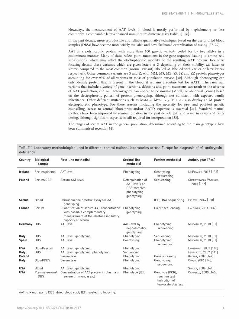
Nowadays, the measurement of AAT levels in blood is mostly performed by nephelometry or, lesscommonly, a comparable latex-enhanced immunoturbidimetric assay (table 1) [26].
In the past decade, more reproducible and reliable quantitative techniques based on the use of dried bloodsamples (DBSs) have become more widely available and have facilitated centralisation of testing [27–29].
AAT is a polymorphic protein with more than 100 genetic variants coded for by two alleles in acodominant manner. Many of these reflect point mutations in the gene sequence leading to amino acidsubstitutions, which may affect the electrophoretic mobility of the resulting AAT protein. Isoelectricfocusing detects these variants, which are given letters A–Z depending on their mobility, i.e. faster orslower, compared to the most common (normal variant) labelled M labelled with earlier or later letters,respectively. Other common variants are S and Z, with MM, MS, MZ, SS, SZ and ZZ protein phenotypesaccounting for over 99% of all variants in most of population surveys [30]. Although phenotyping canonly identify protein that is present in the blood, it remains a routine test for AATD. The rarer nullvariants that include a variety of gene insertions, deletions and point mutations can result in the absenceof AAT production, and null heterozygotes can appear to be normal (Mnull) or abnormal (Znull) basedon the electrophoretic pattern of protein phenotyping, although not consistent with expected familyinheritance. Other deficient mutations such as MMalton, MWurzburg, MHeerlen also display an M proteinelectrophoretic phenotype. For these reasons, including the necessity for pre- and post-test geneticcounselling, access to central laboratories and/or AATD expertise is essential [31]. Standard manualmethods have been improved by semi-automation in the past decade [32] and result in easier and fastertesting, although significant expertise is still required for interpretation [33].
The ranges of serum AAT in the general population, determined according to the main genotypes, havebeen summarised recently [34].
TABLE 1 Laboratory methodologies used in different central national laboratories across Europe for diagnosis of α1-antitrypsindeficiency
Country Biologicalsample
First-line method(s) Second-linemethod(s)
Further method(s) Author, year [Ref.]
Ireland Serum/plasma AAT level Phenotyping Genotyping,sequencing
MCELVANEY, 2015 [136]
Poland Serum/DBS Serum AAT level Determination ofAAT levels onDBS samples,phenotyping,genotyping
Sequencing CHOROSTOWSKA-WYNIMKO,2015 [137]
Serbia Blood Immunonephelometric assay for AAT;genotyping
IEF; DNA sequencing BELETIC, 2014 [138]
France Serum Quantification of serum AAT concentrationwith possible complementarymeasurement of the elastase inhibitorycapacity of serum
Phenotyping,genotyping
Direct sequencing BALDUYCK, 2014 [139]
Germany DBS AAT level AAT level bynephelometry,genotyping
Phenotyping,sequencing
MIRAVITLLES, 2010 [31]
Italy DBS AAT level, genotyping Phenotyping Sequencing MIRAVITLLES, 2010 [31]Spain DBS AAT level Genotyping Phenotyping;
sequencingMIRAVITLLES, 2010 [31]
USA Blood/serum AAT level, genotyping Phenotyping BORNHORST, 2007 [140]Italy DBS AAT level, genotyping, phenotyping Sequencing FERRAROTTI, 2007 [141]Poland Serum level Phenotyping Gene screening KACZOR, 2007 [142]Italy Blood/DBS Serum level Phenotyping Genotyping,
sequencingCORDA, 2006 [143]
USA Blood AAT level, genotyping Phenotyping SNYDER, 2006 [144]USA Plasma-serum/
DBSConcentration of AAT protein in plasma orserum (immunoassay)
Phenotype (IEF) Genotype (PCR),function test(inhibition ofleukocyte elastase)
CAMPBELL, 2000 [145]
AAT: α1-antitrypsin; DBS: dried blood spot; IEF: isoelectric focusing.
https://doi.org/10.1183/13993003.00610-2017 4
ERS STATEMENT | M. MIRAVITLLES ET AL.
The optimal threshold level for AAT to discriminate normal PI*MM from other genotypes carrying atleast one deficient S or Z allele was 24.4 μM (1.1 g·L−1) with 73.4% sensitivity and 88.5% specificity [34].AAT is an acute phase protein and, despite average concentrations varying with protein phenotype, thereis a high degree of overlap, such that plasma level alone is an insufficient parameter to diagnoseintermediate deficiency due to M heterozygosity with certainty. The acute phase response known toinfluence AAT can be partly recognised by the simultaneous quantification of C-reactive protein (CRP).However, these issues are overcome by protein phenotyping or specifically by genotyping, both of whichare independent of AAT level.
Genotyping describes the detection of specific AAT gene mutations, mainly S and Z. This approachutilises the principles of PCR and can also detect rare and null variants such as the MMalton [35–37].However, it can only detect known sequence defects and requires specific primers for each of these, someof which (e.g. F and I) are routinely used by some laboratories. The absence of specific primers can lead tofalse results.
Whole gene sequencing (which is becoming cheaper) will detect stop mutations and help elucidate thenature of rarer variants such as E, F, G, I and P without the need for specific primers, as well as identifycurrently unrecognised variants. The optimal results of genotyping are obtained when performed in expertlaboratories and interpreted in conjunction with the protein level and familial relationships by experiencedAATD clinicians/geneticists.
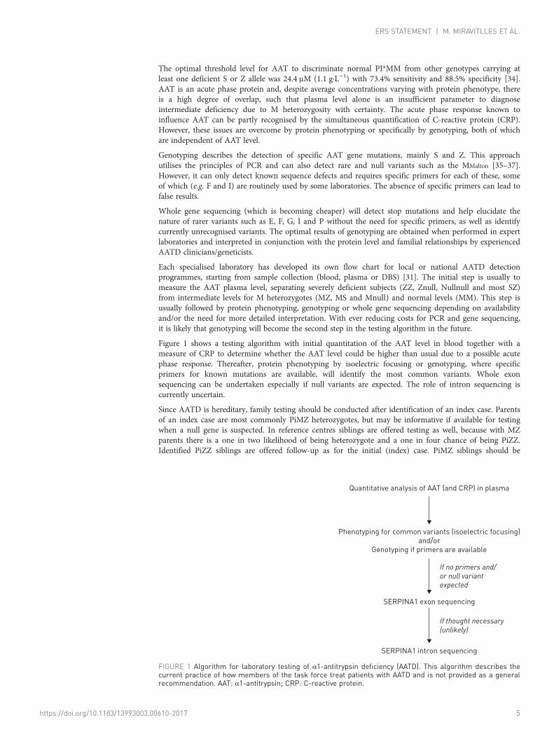
Each specialised laboratory has developed its own flow chart for local or national AATD detectionprogrammes, starting from sample collection (blood, plasma or DBS) [31]. The initial step is usually tomeasure the AAT plasma level, separating severely deficient subjects (ZZ, Znull, Nullnull and most SZ)from intermediate levels for M heterozygotes (MZ, MS and Mnull) and normal levels (MM). This step isusually followed by protein phenotyping, genotyping or whole gene sequencing depending on availabilityand/or the need for more detailed interpretation. With ever reducing costs for PCR and gene sequencing,it is likely that genotyping will become the second step in the testing algorithm in the future.
Figure 1 shows a testing algorithm with initial quantitation of the AAT level in blood together with ameasure of CRP to determine whether the AAT level could be higher than usual due to a possible acutephase response. Thereafter, protein phenotyping by isoelectric focusing or genotyping, where specificprimers for known mutations are available, will identify the most common variants. Whole exonsequencing can be undertaken especially if null variants are expected. The role of intron sequencing iscurrently uncertain.
Since AATD is hereditary, family testing should be conducted after identification of an index case. Parentsof an index case are most commonly PiMZ heterozygotes, but may be informative if available for testingwhen a null gene is suspected. In reference centres siblings are offered testing as well, because with MZparents there is a one in two likelihood of being heterozygote and a one in four chance of being PiZZ.Identified PiZZ siblings are offered follow-up as for the initial (index) case. PiMZ siblings should be
Quantitative analysis of AAT (and CRP) in plasma
Phenotyping for common variants (isoelectric focusing)
and/or
Genotyping if primers are available
SERPINA1 exon sequencing
SERPINA1 intron sequencing
If no primers and/or null variantexpected
If thought necessary(unlikely)
FIGURE 1 Algorithm for laboratory testing of α1-antitrypsin deficiency (AATD). This algorithm describes thecurrent practice of how members of the task force treat patients with AATD and is not provided as a generalrecommendation. AAT: α1-antitrypsin; CRP: C-reactive protein.
https://doi.org/10.1183/13993003.00610-2017 5
ERS STATEMENT | M. MIRAVITLLES ET AL.
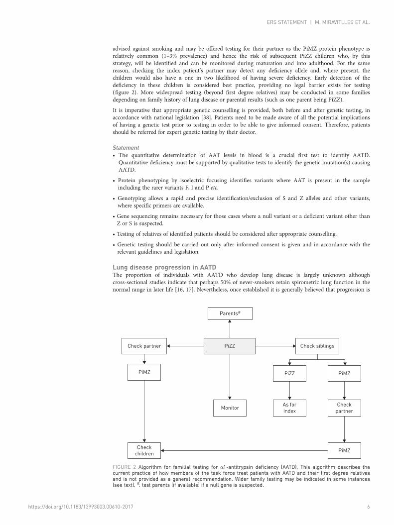
advised against smoking and may be offered testing for their partner as the PiMZ protein phenotype isrelatively common (1–3% prevalence) and hence the risk of subsequent PiZZ children who, by thisstrategy, will be identified and can be monitored during maturation and into adulthood. For the samereason, checking the index patient’s partner may detect any deficiency allele and, where present, thechildren would also have a one in two likelihood of having severe deficiency. Early detection of thedeficiency in these children is considered best practice, providing no legal barrier exists for testing(figure 2). More widespread testing (beyond first degree relatives) may be conducted in some familiesdepending on family history of lung disease or parental results (such as one parent being PiZZ).
It is imperative that appropriate genetic counselling is provided, both before and after genetic testing, inaccordance with national legislation [38]. Patients need to be made aware of all the potential implicationsof having a genetic test prior to testing in order to be able to give informed consent. Therefore, patientsshould be referred for expert genetic testing by their doctor.
Statement• The quantitative determination of AAT levels in blood is a crucial first test to identify AATD.Quantitative deficiency must be supported by qualitative tests to identify the genetic mutation(s) causingAATD.
• Protein phenotyping by isoelectric focusing identifies variants where AAT is present in the sampleincluding the rarer variants F, I and P etc.
• Genotyping allows a rapid and precise identification/exclusion of S and Z alleles and other variants,where specific primers are available.
• Gene sequencing remains necessary for those cases where a null variant or a deficient variant other thanZ or S is suspected.
• Testing of relatives of identified patients should be considered after appropriate counselling.
• Genetic testing should be carried out only after informed consent is given and in accordance with therelevant guidelines and legislation.
Lung disease progression in AATDThe proportion of individuals with AATD who develop lung disease is largely unknown althoughcross-sectional studies indicate that perhaps 50% of never-smokers retain spirometric lung function in thenormal range in later life [16, 17]. Nevertheless, once established it is generally believed that progression is
Parents#
PiZZ
PiZZ PiMZPiMZ
PiMZ
MonitorAs for
index
Check
partner
Check partner
Check
children
Check siblings
FIGURE 2 Algorithm for familial testing for α1-antitrypsin deficiency (AATD). This algorithm describes thecurrent practice of how members of the task force treat patients with AATD and their first degree relativesand is not provided as a general recommendation. Wider family testing may be indicated in some instances(see text). #: test parents (if available) if a null gene is suspected.
https://doi.org/10.1183/13993003.00610-2017 6
ERS STATEMENT | M. MIRAVITLLES ET AL.
more rapid than in non-AATD patients with COPD, especially in smokers. However, smoking cessationcan “normalise” this progression to that of AATD in never-smokers [20].
Conventionally, FEV1 has been used as the major indicator for the presence, progression and severity oflung disease. However, FEV1 is a poor surrogate of emphysema, while gas transfer is more specific.Although these two measures correlate in cohort studies, both have to be measured to a high degree ofspecification and do not provide the same information on the clinical phenotype or the rate of progressionor stability. Similarly, the progression of emphysema measured by lung density in computed tomography(CT) continues even when FEV1 is stable [14, 18, 20, 22].
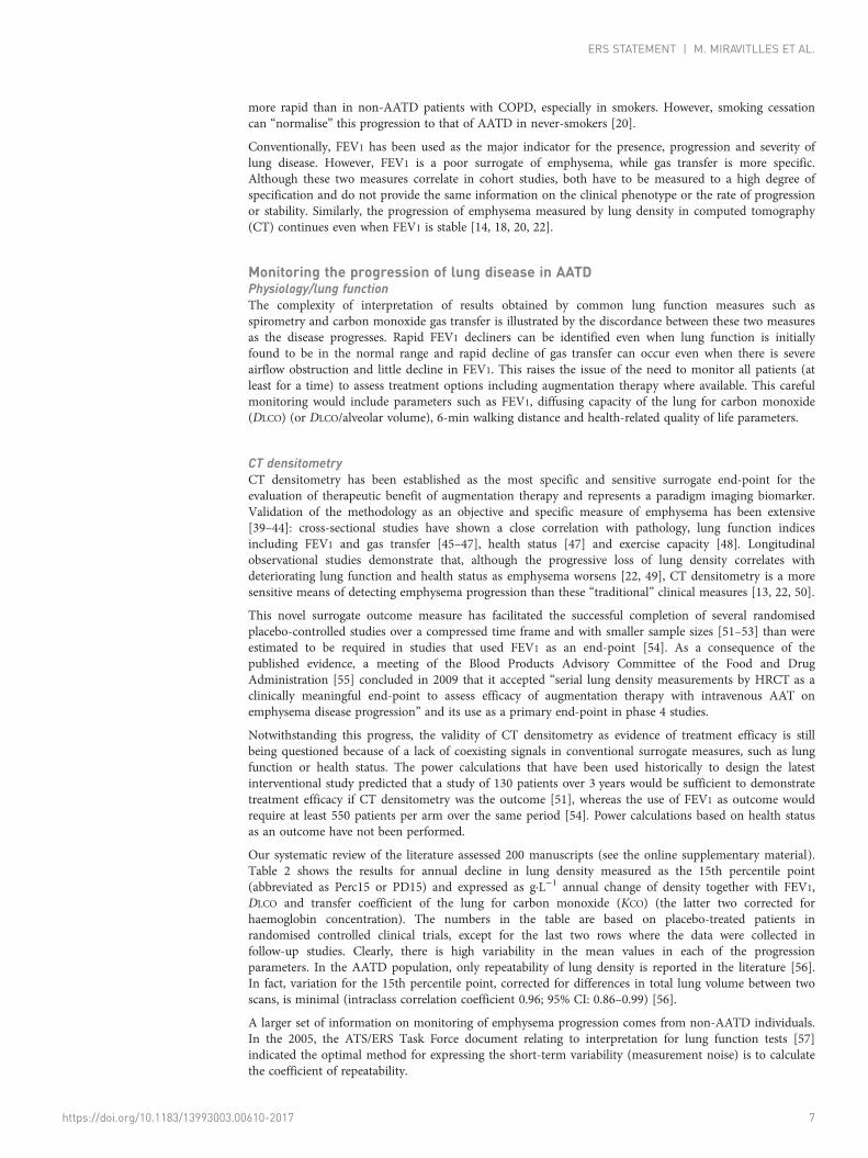
Monitoring the progression of lung disease in AATDPhysiology/lung functionThe complexity of interpretation of results obtained by common lung function measures such asspirometry and carbon monoxide gas transfer is illustrated by the discordance between these two measuresas the disease progresses. Rapid FEV1 decliners can be identified even when lung function is initiallyfound to be in the normal range and rapid decline of gas transfer can occur even when there is severeairflow obstruction and little decline in FEV1. This raises the issue of the need to monitor all patients (atleast for a time) to assess treatment options including augmentation therapy where available. This carefulmonitoring would include parameters such as FEV1, diffusing capacity of the lung for carbon monoxide(DLCO) (or DLCO/alveolar volume), 6-min walking distance and health-related quality of life parameters.
CT densitometryCT densitometry has been established as the most specific and sensitive surrogate end-point for theevaluation of therapeutic benefit of augmentation therapy and represents a paradigm imaging biomarker.Validation of the methodology as an objective and specific measure of emphysema has been extensive[39–44]: cross-sectional studies have shown a close correlation with pathology, lung function indicesincluding FEV1 and gas transfer [45–47], health status [47] and exercise capacity [48]. Longitudinalobservational studies demonstrate that, although the progressive loss of lung density correlates withdeteriorating lung function and health status as emphysema worsens [22, 49], CT densitometry is a moresensitive means of detecting emphysema progression than these “traditional” clinical measures [13, 22, 50].
This novel surrogate outcome measure has facilitated the successful completion of several randomisedplacebo-controlled studies over a compressed time frame and with smaller sample sizes [51–53] than wereestimated to be required in studies that used FEV1 as an end-point [54]. As a consequence of thepublished evidence, a meeting of the Blood Products Advisory Committee of the Food and DrugAdministration [55] concluded in 2009 that it accepted “serial lung density measurements by HRCT as aclinically meaningful end-point to assess efficacy of augmentation therapy with intravenous AAT onemphysema disease progression” and its use as a primary end-point in phase 4 studies.
Notwithstanding this progress, the validity of CT densitometry as evidence of treatment efficacy is stillbeing questioned because of a lack of coexisting signals in conventional surrogate measures, such as lungfunction or health status. The power calculations that have been used historically to design the latestinterventional study predicted that a study of 130 patients over 3 years would be sufficient to demonstratetreatment efficacy if CT densitometry was the outcome [51], whereas the use of FEV1 as outcome wouldrequire at least 550 patients per arm over the same period [54]. Power calculations based on health statusas an outcome have not been performed.
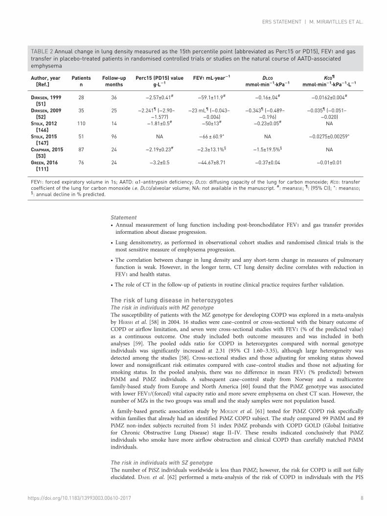
Our systematic review of the literature assessed 200 manuscripts (see the online supplementary material).Table 2 shows the results for annual decline in lung density measured as the 15th percentile point(abbreviated as Perc15 or PD15) and expressed as g·L−1 annual change of density together with FEV1,DLCO and transfer coefficient of the lung for carbon monoxide (KCO) (the latter two corrected forhaemoglobin concentration). The numbers in the table are based on placebo-treated patients inrandomised controlled clinical trials, except for the last two rows where the data were collected infollow-up studies. Clearly, there is high variability in the mean values in each of the progressionparameters. In the AATD population, only repeatability of lung density is reported in the literature [56].In fact, variation for the 15th percentile point, corrected for differences in total lung volume between twoscans, is minimal (intraclass correlation coefficient 0.96; 95% CI: 0.86–0.99) [56].
A larger set of information on monitoring of emphysema progression comes from non-AATD individuals.In the 2005, the ATS/ERS Task Force document relating to interpretation for lung function tests [57]indicated the optimal method for expressing the short-term variability (measurement noise) is to calculatethe coefficient of repeatability.
https://doi.org/10.1183/13993003.00610-2017 7
ERS STATEMENT | M. MIRAVITLLES ET AL.
Statement• Annual measurement of lung function including post-bronchodilator FEV1 and gas transfer providesinformation about disease progression.
• Lung densitometry, as performed in observational cohort studies and randomised clinical trials is themost sensitive measure of emphysema progression.
• The correlation between change in lung density and any short-term change in measures of pulmonaryfunction is weak. However, in the longer term, CT lung density decline correlates with reduction inFEV1 and health status.
• The role of CT in the follow-up of patients in routine clinical practice requires further validation.
The risk of lung disease in heterozygotesThe risk in individuals with MZ genotypeThe susceptibility of patients with the MZ genotype for developing COPD was explored in a meta-analysisby HERSH et al. [58] in 2004. 16 studies were case–control or cross-sectional with the binary outcome ofCOPD or airflow limitation, and seven were cross-sectional studies with FEV1 (% of the predicted value)as a continuous outcome. One study included both outcome measures and was included in bothanalyses [59]. The pooled odds ratio for COPD in heterozygotes compared with normal genotypeindividuals was significantly increased at 2.31 (95% CI 1.60–3.35), although large heterogeneity wasdetected among the studies [58]. Cross-sectional studies and those adjusting for smoking status showedlower and nonsignificant risk estimates compared with case–control studies and those not adjusting forsmoking status. In the pooled analysis, there was no difference in mean FEV1 (% predicted) betweenPiMM and PiMZ individuals. A subsequent case–control study from Norway and a multicentrefamily-based study from Europe and North America [60] found that the PiMZ genotype was associatedwith lower FEV1/(forced) vital capacity ratio and more severe emphysema on chest CT scan. However, thenumber of MZs in the two groups was small and the study samples were not population based.
A family-based genetic association study by MOLLOY et al. [61] tested for PiMZ COPD risk specificallywithin families that already had an identified PiMZ COPD subject. The study compared 99 PiMM and 89PiMZ non-index subjects recruited from 51 index PiMZ probands with COPD GOLD (Global Initiativefor Chronic Obstructive Lung Disease) stage II–IV. These results indicated conclusively that PiMZindividuals who smoke have more airflow obstruction and clinical COPD than carefully matched PiMMindividuals.
The risk in individuals with SZ genotypeThe number of PiSZ individuals worldwide is less than PiMZ; however, the risk for COPD is still not fullyelucidated. DAHL et al. [62] performed a meta-analysis of the risk of COPD in individuals with the PIS
TABLE 2 Annual change in lung density measured as the 15th percentile point (abbreviated as Perc15 or PD15), FEV1 and gastransfer in placebo-treated patients in randomised controlled trials or studies on the natural course of AATD-associatedemphysema
Author, year[Ref.]
Patientsn
Follow-upmonths
Perc15 (PD15) valueg·L−1
FEV1 mL·year−1 DLCO
mmol·min−1·kPa−1KCO¶
mmol·min−1·kPa−1·L−1
DIRKSEN, 1999[51]
28 36 −2.57±0.41# −59.1±11.9# −0.16±.04# −0.0162±0.004#
DIRKSEN, 2009[52]
35 25 −2.241¶ (−2.90–−1.577)
−23 mL¶ (−0.043–−0.004)
−0.343¶ (−0.489–−0.196)
−0.035¶ (−0.051–−0.020)
STOLK, 2012[146]
110 14 −1.81±0.5# −50±13# −0.23±0.05# NA
STOLK, 2015[147]
51 96 NA −66 ± 60.9+ NA −0.0275±0.00259+
CHAPMAN, 2015[53]
87 24 −2.19±0.23# −2.3±13.1%§ −1.5±19.5%§ NA
GREEN, 2016[111]
76 24 −3.2±0.5 −44.67±8.71 −0.37±0.04 −0.01±0.01
FEV1: forced expiratory volume in 1s; AATD: α1-antitrypsin deficiency; DLCO: diffusing capacity of the lung for carbon monoxide; KCO: transfercoefficient of the lung for carbon monoxide i.e. DLCO/alveolar volume; NA: not available in the manuscript. #: mean±SE; ¶: (95% CI); +: mean±SD;§: annual decline in % predicted.
https://doi.org/10.1183/13993003.00610-2017 8
ERS STATEMENT | M. MIRAVITLLES ET AL.
allele. 21 studies were included and there were six case–control and cross-sectional studies. In the pooledanalysis there were 42 PiSZ individuals of whom 27 had COPD. The summary odds ratio for COPD inPiSZ individuals was significantly elevated at 3.26 (95% CI 1.24–8.57) compared with PiMM; however,when a significant outlier was removed from the analysis the odds ratio was no longer significantlyincreased [63]. SEERSHOLM et al. [64], in a study of 94 PiSZ individuals of whom 66 were non-index cases,observed that index PiSZ cases had reduced survival. Data from the Italian and Spanish registries [24, 65]found that PiSZ subjects were older at diagnosis and had more preserved lung function despite highersmoking exposure than PiZZ patients. Similarly, a more recent study [66] suggested that PiSZ subjectswere less susceptible to cigarette smoke than PiZZ and that the pattern of emphysema found on the CTscan at diagnosis was similar to that seen in patients with usual COPD rather than the predominantlybasal distribution of panlobular emphysema of PiZZ individuals.
Taken together, these data suggest increased susceptibility of the SZ phenotype for the development ofCOPD in smokers, but more research is needed, including the effect of environmental factors in a similarway to that undertaken for MZ subjects [61].
Rarer mutation heterozygotes (such as FZ, IZ, MMalton and Mnull mutations)There is a paucity of data for rarer AATD mutations, but some studies are emerging indicating that the F,I and MMalton mutations confer increased susceptibility to COPD when inherited with a Z allele. Althoughthe Null mutations associated with AATD are rare, studies have shown that Null homozygotes have moresevere lung disease than PiZZ or PiSZ individuals [67–70] and Mnull individuals have increased lungsymptomatology and obstructive lung disease [71].
Statement• Never-smoking PiMZ subjects do not have an increased risk for COPD.
• Smoking PiMZ and PiSZ subjects have an increased risk of COPD compared to smoking PiMM subjects.
• The role of other heterozygotes remains unknown due to their rarity and potential ascertainment biasfrom measuring AAT in unusual cases of lung or liver disease.
Role and benefits of screeningThere are different approaches to identify individuals with AATD. The first is population-based, in whichunselected groups have been tested (screening studies). The second is targeted-detection studies in whichpatients with an enhanced suspicion of having AATD are tested including those with early-onset COPD(<40 years age), basal panlobular emphysema, family history of COPD or AATD, and those with perinataljaundice, cirrhosis, vasculitis or panniculitis.
No randomised controlled study determining the efficacy of screening programmes for AATD has beenperformed. Most screening studies have been selective and did not involve random population samples,but included individuals that are healthier (blood donors) or sicker (hospital outpatients) than the generalpopulation. A few population-based studies that randomly screened the general population [72, 73] orlarge numbers of newborns [10, 74] provided a less biased and more accurate prevalence estimate ofspecific AATD phenotypes. The newborn studies have also provided valuable insight into the naturalhistory of AATD, with unbiased assessments in the risks of liver and lung diseases. More specifically, thesestudies have found that individuals tested often had lung function in the normal range at mean ages of15 years [75] and 30 years [76]. Data collected at age 35 years suggest that at least some have developed areduction in gas transfer and lung density [15]. This is consistent with the retrograde analysis thathighlighted the higher sensitivity of these measures to detect early changes compared to spirometry [14].The most recent birth cohort information at ages 37 to 40 years showed that two of the four currentsmokers already had COPD [77].
Potential benefits of systematic screening include genetic counselling, lifestyle recommendations (smokingprevention or cessation, avoidance of high-risk occupations, alcohol intake limitation), and considerationfor earlier augmentation therapy. The potential harms include psychological effects, social discriminatoryeffects and costs. These effects can be addressed, at least partially, by reassurance as never-smokers whoare non-index cases with AATD have a normal life expectancy [17, 78].
The largest population-based study published for AATD was carried out in 200000 newborn children inSweden between 1972 and 1974 leading to the identification of 127 PIZ individuals and 48 PISZindividuals [10]. The major purpose was to reduce exposure of the child to parental smoking duringchildhood and adolescence and to prevent active smoking. Neonatal screening reduced the smoking ratesin18–20-year-olds compared to age-matched subjects [79], with 6% being current smokers versus 17%(p<0.05) and 88% being never-smokers versus 65% (p<0.05), although this failed to affect smoking among
https://doi.org/10.1183/13993003.00610-2017 9
ERS STATEMENT | M. MIRAVITLLES ET AL.
the parents. Similar results were also found in another neonatal screening study in Oregon, withsignificantly lower smoking initiation rates in subjects who had been diagnosed with AATD than incontrol subjects (27.3% versus 56.9%; p=0.02) [75].
Neonatal screening produced no adverse psychological effects in adolescents identified at birth withAATD, although parental distress and adverse effects were identified in the mother–child relationship [80]and 20 years later the mothers had significantly more anxiety than control mothers [81]. These concernshave inhibited the reintroduction of neonatal screening in Sweden although a clearer understanding of therisks/benefits and natural history of the disease is both helpful and reassuring.
Statement• Most screening studies have been biased as they did not involve random population samples.
• Population-based screening studies provide less biased prevalence estimates of specific AATD proteinand clinical phenotypes as well as valuable insights into the natural history of AATD.
• Neonatal screening has been shown to be effective in reducing the smoking rates for 18–20-year-oldscompared to age-matched individuals.
• Screening may have negative psychological effects on parents and on mother–child bonding. However,these negative effects can be addressed by comprehensive genetic counselling and care provision atcentres of excellence for AATD.
Augmentation therapy for AATDSince augmentation is currently the only specific therapy for AATD, it has been a topic of intense debatein the literature and the subject of numerous review and opinion articles. In rare diseases such as AATD,the difficulty of recruitment to clinical trials, coupled with the lack of sensitivity to change of typicaloutcome measures has challenged the development and delivery of clinical trials. Furthermore, there is theunusual situation where augmentation has been advocated (and given) for some time on the basis ofbiochemical effect (namely raising AAT level) [82] and hence has become established as treatment inmany areas of the world, without the level of evidence now expected for respiratory outcomes such asFEV1, quality of life and mortality. There have been two previous systematic reviews, one focused onrandomised controlled trials (RCTs) of augmentation [83] and the other which considered all controlledstudy designs of augmentation (including nonrandomised studies) and presented analyses of FEV1 decline [84].In addition, an individual patient data analysis of the CT densitometry data from two of therandomised controlled trials has been reported [85]. The latter two studies were supportive ofaugmentation as a treatment capable of reducing, albeit not eliminating, emphysema progression [84, 85].The meta-analysis conducted by GØTZSCHE et al. [83] also indicated that CT density decline was lower onaugmentation therapy than placebo, but concluded that this did not equate to efficacy. A similar view wasalso expressed in the more recent update by the same authors [86], following publication of the RAPIDtrial [53].
In order to obtain all the evidence about augmentation and minimise bias we used standard systematicreview methods as described in the online supplementary material. There have been eight RCTs ofintravenous augmentation, three against placebo [51–53, 87] and five against another active comparator[88–92], generally a newer brand of augmentation therapy. In addition there have been six observationalstudies reporting a control group [8, 93–97], largely assessing data from registries, and 11 uncontrolledobservational studies [82, 98–108], focused on pharmacokinetics, safety or novel outcomes. There are alsotwo ongoing trials (ClinicalTrials.gov identifiers NCT00242385 and NCT01213043). In the interests ofbrevity, the published placebo-controlled RCTs are discussed here in detail, whereas other publishedstudies are shown in table 3, which contains a brief summary of study characteristics and results.
The RCTs included a total of 315 patients. The earliest RCT included 58 ex-smoking PiZZ patients withmoderate-to-severe emphysema, treated for a minimum of 3 years and randomised to an infusion of AATat 250 mg·kg−1 or human albumin every 4 weeks [51]. FEV1 was the primary outcome; secondaryoutcomes included KCO, DLCO and change in lung density measured by CT. There was no difference inphysiological decline, but a strong trend towards reduced decline in CT-measured lung density.
The EXACTLE trial included 77 participants with severe AATD treated using weekly infusions of AAT at60 mg·kg−1 or placebo for 2 years, with an optional 6-month extension [52]. Primary outcome wasprogression rate of emphysema determined by annual CT lung density at total lung capacity (TLC), butthis was in part an exploratory study, as the optimum method of image analysis was uncertain at the time.A strong trend toward reduced density deterioration was seen, consistently across the four differentanalytical methods used. In one of these, conventional statistical significance was reached (p=0.049).
https://doi.org/10.1183/13993003.00610-2017 10
ERS STATEMENT | M. MIRAVITLLES ET AL.
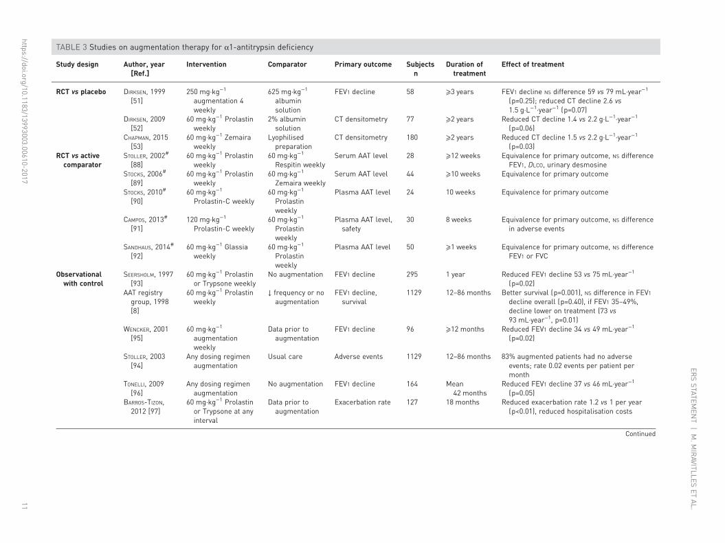
TABLE 3 Studies on augmentation therapy for α1-antitrypsin deficiency
Study design Author, year[Ref.]
Intervention Comparator Primary outcome Subjectsn
Duration oftreatment
Effect of treatment
RCT vs placebo DIRKSEN, 1999[51]
250 mg·kg−1
augmentation 4weekly
625 mg·kg−1
albuminsolution
FEV1 decline 58 ⩾3 years FEV1 decline NS difference 59 vs 79 mL·year−1
(p=0.25); reduced CT decline 2.6 vs1.5 g·L−1·year−1 (p=0.07)
DIRKSEN, 2009[52]
60 mg·kg−1 Prolastinweekly
2% albuminsolution
CT densitometry 77 ⩾2 years Reduced CT decline 1.4 vs 2.2 g·L−1·year−1
(p=0.06)CHAPMAN, 2015[53]
60 mg·kg−1 Zemairaweekly
Lyophilisedpreparation
CT densitometry 180 ⩾2 years Reduced CT decline 1.5 vs 2.2 g·L−1·year−1
(p=0.03)RCT vs activecomparator
STOLLER, 2002#
[88]60 mg·kg−1 Prolastin
weekly60 mg·kg−1
Respitin weeklySerum AAT level 28 ⩾12 weeks Equivalence for primary outcome, NS difference
FEV1, DLCO, urinary desmosineSTOCKS, 2006#
[89]60 mg·kg−1 Prolastin
weekly60 mg·kg−1
Zemaira weeklySerum AAT level 44 ⩾10 weeks Equivalence for primary outcome
STOCKS, 2010#
[90]60 mg·kg−1
Prolastin-C weekly60 mg·kg−1
Prolastinweekly
Plasma AAT level 24 10 weeks Equivalence for primary outcome
CAMPOS, 2013#
[91]120 mg·kg−1
Prolastin-C weekly60 mg·kg−1
Prolastinweekly
Plasma AAT level,safety
30 8 weeks Equivalence for primary outcome, NS differencein adverse events
SANDHAUS, 2014#
[92]60 mg·kg−1 Glassia
weekly60 mg·kg−1
Prolastinweekly
Plasma AAT level 50 ⩾1 weeks Equivalence for primary outcome, NS differenceFEV1 or FVC
Observationalwith control
SEERSHOLM, 1997[93]
60 mg·kg−1 Prolastinor Trypsone weekly
No augmentation FEV1 decline 295 1 year Reduced FEV1 decline 53 vs 75 mL·year−1
(p=0.02)AAT registrygroup, 1998[8]
60 mg·kg−1 Prolastinweekly
↓ frequency or noaugmentation
FEV1 decline,survival
1129 12–86 months Better survival (p=0.001), NS difference in FEV1decline overall (p=0.40), if FEV1 35–49%,decline lower on treatment (73 vs93 mL·year−1, p=0.01)
WENCKER, 2001[95]
60 mg·kg−1
augmentationweekly
Data prior toaugmentation
FEV1 decline 96 ⩾12 months Reduced FEV1 decline 34 vs 49 mL·year−1
(p=0.02)
STOLLER, 2003[94]
Any dosing regimenaugmentation
Usual care Adverse events 1129 12–86 months 83% augmented patients had no adverseevents; rate 0.02 events per patient permonth
TONELLI, 2009[96]
Any dosing regimenaugmentation
No augmentation FEV1 decline 164 Mean42 months
Reduced FEV1 decline 37 vs 46 mL·year−1
(p=0.05)BARROS-TIZON,2012 [97]
60 mg·kg−1 Prolastinor Trypsone at anyinterval
Data prior toaugmentation
Exacerbation rate 127 18 months Reduced exacerbation rate 1.2 vs 1 per year(p<0.01), reduced hospitalisation costs
Continued
https://doi.org/10.1183/13993003.00610-201711
ERSSTATEM
ENT
|M.M
IRAVITLLES
ETAL.
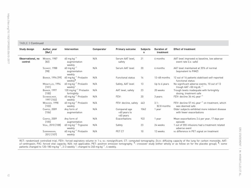
TABLE 3 Continued
Study design Author, year[Ref.]
Intervention Comparator Primary outcome Subjectsn
Duration oftreatment
Effect of treatment
Observational, nocontrol
WEWERS, 1987[82]
60 mg·kg−1
augmentationweekly
N/A Serum AAT level,safety
21 6 months AAT level improved vs baseline, low adverseevent rate (i.e. safe)
SCHMIDT, 1988[98]
60 mg·kg−1
augmentationweekly
N/A Serum AAT level 20 6 months AAT level maintained at 35% of normal(equivalent to PiMZ)
BARKER, 1994 [99] 60 mg·kg−1 Prolastinweekly¶
N/A Functional status 14 12–48 months 12 out of 14 patients stabilised self-reportedfunctional status
MIRAVITLLES, 1994[101]
60 mg·kg−1 Prolastinweekly+
N/A Safety, AAT level 13 Up to 6 years No significant adverse events; 10 out of 13trough AAT >50 mg·dL−1
BARKER, 1997[100]
120 mg·kg−1 Prolastinfortnightly
N/A AAT level, safety 23 20 weeks Trough levels inadequate with fortnightlydosing, treatment safe
SCHWAIBLMAIR,1997 [102]
60 mg·kg−1 Prolastinweekly
N/A FEV1 20 3 years FEV1 decline 36 mL·year−1
WENCKER, 1998[103]
60 mg·kg−1 Prolastinweekly
N/A FEV1 decline, safety 443 3.1–82.8 months
FEV1 decline 57 mL·year−1 on treatment, whichwas deemed safe
CAMPOS, 2009[106]
Any form ofaugmentation
N/A Compared age>60 years to<60 years
1062 1 year Older subjects exhibited more indolent diseasewith fewer exacerbations
CAMPOS, 2009[105]
Any form ofaugmentation
N/A Exacerbations 922 1 year Mean exacerbations 2.4 per year, 17 days perepisode
VIDAL, 2010 [108] 60 mg·kg−1 Trypsoneweekly
N/A Safety 23 24 weeks 1 out of 555 infusions had a treatment relatedadverse event
SUBRAMANIAN,2012 [107]
60 mg·kg−1 Prolastinweekly
N/A PET CT 10 12 weeks NS difference in PET signal on treatment
RCT: randomised controlled trial; FEV1: forced expiratory volume in 1 s; NS: nonsignificant; CT: computed tomography; DLCO: diffusing capacity of the lung for carbon monoxide; AAT:α1-antitrypsin; FVC: forced vital capacity; N/A: not applicable; PET: positron emission tomography. #: crossover study (either wholly or as follow on for the placebo group); ¶: somepatients changed to 120–180 mg·kg−1, 2–3 weekly +: changed to 240 mg·kg−1, 4 weekly.
https://doi.org/10.1183/13993003.00610-201712
ERSSTATEM
ENT
|M.M
IRAVITLLES
ETAL.
Secondary outcomes included patient-reported exacerbation frequency, DLCO and quality of life. No trendsin these measures were seen between active treatment and placebo, although there was a reduction inhospital admissions for exacerbations in the active treatment arm.
The most recently performed study (RAPID) included 180 patients with emphysema secondary to AATDand FEV1 of 35–70% predicted [53]. Patients received either weekly infusions of AAT at 60 mg·kg−1 orplacebo for 2 years, with a 2-year open label extension for some participants [87]. This study was the firstto be powered to detect a treatment effect on the annual rate of decrease in lung density measured by CTscan; secondary outcomes included exacerbation rate, change in FEV1 % predicted, quality of life using theSt George’s Respiratory Questionnaire (SGRQ) and change in DLCO. The CT imaging protocol obtainedscans at full inspiration (TLC) and at relaxed expiration (functional residual capacity (FRC)). While thechosen primary end-point was a combination of CT lung density (PD15) measured at TLC and FRC(which failed to achieve statistical significance), the separate imaging series at TLC and FRC were includedas secondary outcomes. The main finding was a reduced rate of lung density decline, as measured by CTscanning, in the treated patients. This treatment effect was statistically significant when quantified usingCT imaging obtained at full inspiration (TLC), as in previous studies (discussed earlier). During the openlabel extension, the patients previously on placebo exhibited a change in CT density decline, becomingsimilar to that seen in patients treated in the randomised phase. However, as in previous RCTs in thisarea, no significant effect was seen on other outcome measures, such as lung function and quality of life [87].A supplementary report to the trial has also detailed reduced circulating desmosine, indicating an effect ofaugmentation on body elastin breakdown [109].
Augmentation is considered safe across the larger number of studies where this is reported. Adverse eventrates were similar between treated and placebo groups in both EXACTLE [52] and RAPID [53], but werenot reported in the earlier RCT [51].
The consistency of the trial data with respect to CT density decline, and the fact that CT density has beenshown in cross-sectional and longitudinal studies to relate well to other clinical outcomes, such asmortality and quality of life [47, 110], indicates that it is a clinically relevant measure. Moreover decline inCT density has also been shown to relate to mortality [111], indicating that the RCT results with respectto CT density decline are consistent with the longer observational work suggesting a mortality benefit [8].Survival was also reported in the most recent RCT (one death on augmentation, three on placebo), but thelow mortality rate prevented any conclusion.
While many of the observational studies imply a benefit of treatment on the rate of FEV1 decline, thepotential for bias is greater than in a RCT and the data should be interpreted with caution. The effect ofaugmentation on exacerbations of AATD lung disease remains uncertain, with inconsistent effects in thoseRCTs which reported them [52, 53], and reduced rates in one retrospective observational study [97].Longer duration trials, with use of symptom diaries, and/or selection for frequent exacerbations might helpto confirm any treatment effect on clinical symptoms, but again such studies would require large samplesizes and the inclusion of a placebo control group would probably be considered unethical given theevident benefit on CT density decline.
Statement• Several randomised clinical trials in severe AATD have shown intravenous augmentation therapy toreduce the progression of emphysema as assessed by CT densitometry.
• There is no evidence to support efficacy of AAT augmentation therapy in PiSZ, PiMZ or currentsmokers of any protein phenotype.
• Clinical trials have used fixed doses of AAT determined by body weight. Whether individualising dosagebased on trough levels for each patient has any benefit requires confirmation.
Patient assessment and management stepsThis stepwise approach describes the current practice of how members of the task force assess and treatpatients with AATD and is not intended as a general recommendation.
1) Identify patient with severe AAT deficiency.
2) Ensure smoking is stopped if the patient was a smoker.
3) Identify and modify any other potential risk factor(s).
4) Optimise current COPD therapy.
5) Assess patient in an expert reference centre.
https://doi.org/10.1183/13993003.00610-2017 13
ERS STATEMENT | M. MIRAVITLLES ET AL.
6) Instigate augmentation therapy if indicated.
7) Continue monitoring.
Lung volume reduction surgery in AATDPatients with severe emphysema suffer breathlessness in part due to emphysematous hyperinflation. It iswell established that targeted resection of these areas, in selected patients with COPD, can result insignificant improvements in quality of life and mortality. The previous ATS/ERS statement concluded thatbilateral lung volume reduction surgery (LVRS) offered short-term benefit only and was not recommendedfor AATD-related emphysema until more data was available [3]. STOLLER et al. [112] reported outcomesfrom a National Emphysema Treatment Trial study in 10 AATD patients having bilateral LVRS (five withupper lobe predominant emphysema). The authors identified a higher mortality than medical treatmentand a trend towards reduced magnitude and duration of beneficial effect compared with usual COPD.Specifically with respect to unilateral LVRS in AATD, DAURIAT et al. [113] compared outcomes in 17patients with AATD versus 35 individuals with non-AATD-related COPD, finding improvements in bothgroups in terms of FEV1, dyspnoea score and arterial oxygen tension at 3–6 months. There was a loss ofeffect on walking distance, but preserved FEV1 and dyspnoea score at 12 months in the AATD group.
These studies were performed a decade or more ago, since when there have been significant advances inpatient selection, surgery and the advent of RCTs utilising devices to achieve medical lung volumereduction without the need for surgery (i.e. endobronchial valves (EBV), endobronchial coils, lung sealant,thermal vapour). Patient selection is currently advocated through a multidisciplinary team approachincluding a physician, surgeon, radiologist and interventional bronchoscopist with a special interest in lungvolume reduction (LVR). It is recognised that patients being considered are, by definition, those withadvanced disease and thus at higher risk, hence risk/benefit analysis is central to the multidisciplinaryteam assessment. Early outcomes from surgical LVR are now improved, probably a reflection of improvedpatient selection, multidisciplinary team approach and the fact that most cases involve minimally invasivevideo assisted thoracoscopic surgery (VATS) and a unilateral rather than a bilateral procedure. Whetherthis has an influence on long-term outcomes is unknown. However, mortality rate is 3% over 20 years postLVRS [114], with benefit in both lower and upper lobe disease and very low mortality from unilateralVATS [115].
The range of treatments in development may allow specific patterns of emphysema to be treated. The twobest evaluated devices are endobronchial coils and EBV. Coils have been evaluated in patients withemphysema and significant improvements have been shown in 6-min walking distance, FEV1 and qualityof life. Some patients with emphysema associated with AATD have been treated with coils, but no resultshave been provided for this specific subgroup of patients. Studies have shown that morbidity is increasedand therefore personalised risk/benefit analysis is critical [116, 117]. EBVs are unidirectional valves placedbronchoscopically in the airways supplying the target lobe. In common with LVRS, the success of thisapproach remains optimal patient selection; in the case of an EBV the absence of collateral ventilationbetween target and non-target lobe is vital to a successful outcome. The most recent RCT demonstratedsignificant improvements in 6-min walking distance, FEV1 and quality of life at 6 months, and alsodemonstrated that centres need to be aware of potential morbidity of pneumothorax related to non-targetlobe expansion and be proactive in performing valve maintenance to a high standard [118]. The study alsoincluded some AATD individuals treated with an EBV. However, owing the rarity of AATD large studiesare not currently available to provide detailed assessment of coils, EBV or LVRS in AATD alone. UnlikeEBV, coils can be used in patients with homogeneous emphysema, irrespective of collateral ventilation, butthere is no specific data in AATD. The promising results from EBV therapy have meant that specialistLVR units no longer exclude appropriate AATD individuals from these therapies, although more researchis required.
Statement• Surgical volume reduction and EBV placement may be considered in selected patients with AATD, butfurther studies are needed to confirm the role of such therapies.
• The optimal results of these techniques are obtained when a careful appraisal of risks and benefits areperformed by a multidisciplinary team experienced in LVR and AATD.
Lung transplantation for emphysema associated with AATDSevere AATD-related emphysema accounted for 5.4% of all lung transplants performed between 1995 and2014 [119]. Since the last ATS/ERS statement [3], there have been several publications reporting outcomesin many established transplant centres in different countries, but all studies have been retrospective.DE PERROT et al. [120] reported a higher early mortality from sepsis in AATD and lower 10-year survival
https://doi.org/10.1183/13993003.00610-2017 14
ERS STATEMENT | M. MIRAVITLLES ET AL.
in AATD post-transplantation compared with usual non-deficient COPD, and, similarly, THABUT et al.[121] have shown that patients with AATD had less survival benefit than patients with non-deficientCOPD. This may relate to associated excess inflammation at times of infection in post-transplant AATDindividuals [122, 123], due to the lack of the normal anti-inflammatory role of AAT [124]. However, thishigher, early mortality for AATD post-transplant has not been confirmed, for example BURTON et al. [125]reported no differences in early or late mortality for AATD compared with non-deficient COPD.
In terms of survival compared with non-transplanted AATD patients, TANASH et al. [126] observed thattransplantation increased survival considerably from 5 to 11 years compared with non-transplanted AATDpatients matched for FEV1, age, sex and smoking history. The most common cause of death was pulmonaryinfection among the transplant patients and respiratory failure among the controls. In contrast, a UK study [127]also matched transplanted and non-transplanted AATD individuals for FEV1, age and sex, but found thatAATD patients who underwent lung transplant had lower gas transfer and quality of life pre-transplantcompared with non-transplant patients. Further matching adjusted for quality of life (SGRQ), gas transferfactor and pre-transplant rate of lung function decline showed that transplantation did not increasepost-surgical survival although quality of life was much improved. These controversial results underscorethat the survival benefit of lung transplant is complex to assess and studies that compare matched patientswith and without transplant are (by nature) biased [128]. Consequently, survival benefit remains unclearand thus the main indication for transplantation relates to improvement in quality of life.
The evaluation of comorbidities is crucial in the assessment of candidates for lung transplantation, andhepatic evaluation is particularly critical in AATD [129]. Some centres perform systematic liver biopsy incandidates, although the detection of liver disease per se does not preclude lung transplant in thesepatients. There are experiences of combined liver and lung transplantation with satisfactory results.
Statement• The survival benefit of lung transplant in AATD patients is not clear.
• In general, patients with AATD have improved quality of life following lung transplantation.
• Referral timing, rate of decline in lung function, health status and social support differ from patient topatient, and will have an influence on the evaluation for transplant.
• The role of post-transplant augmentation therapy in particular needs to be explored.
New lines of research in AATDThere are several aspects of lung disease in AATD that require further research. The main topics identifiedby the group are: 1) biomarkers of emphysema progression in AATD; 2) biomarkers of response toaugmentation therapy; 3) research on the minimum clinically important difference in rate of decline inlung density; 4) personalised augmentation therapy, with individualised selection of therapeutic regimenaccording to the patient needs; 5) development of genetic and regenerative therapies; 6) other types oftreatment, such as biochemical inhibitors of neutrophil proteinases; 7) development of specificpatient-reported outcomes for patients with emphysema associated with AATD; and 8) efficacy ofaugmentation therapy after lung transplant in AATD patients.
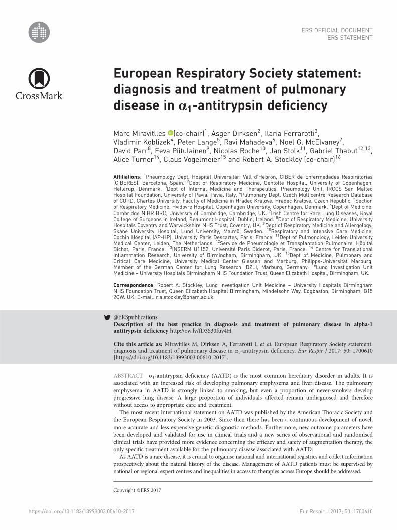
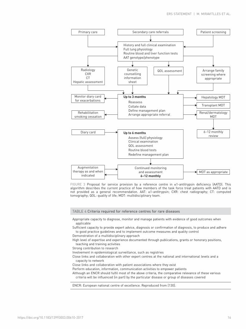
Organisation of care: reference centres and registriesDue to the low prevalence and underdiagnosis, AATD is considered a rare disease. It is almost impossiblefor an individual clinician or a single centre to accumulate enough expertise in diagnosis and managementof the disease. Therefore, the care for patients with AATD is best organised in reference centres that canprovide the highest standard of care and advice to the individuals affected and their families while alsocontributing to knowledge accumulation. An optimised format of service provision by a reference centre inAATD as outlined in figure 3, although other models may be applicable.
The European Commission also recommends the development of reference centres for rare diseases. Theestablishment of European reference networks (ERNs) for rare diseases should therefore serve as researchand knowledge centres, updating and contributing to the latest scientific findings, treating patients fromother member states and ensuring the availability of subsequent treatment facilities where necessary. Thedefinition of ERNs should also reflect the need for services and expertise to be distributed across theEuropean Union (EU). In a document released by the European Commission in 2006, the criteria forreference centres of rare diseases are clearly specified (table 4) [130].
Reference centres must establish a registry of their activity and collect information prospectively about thenatural history of the patients being monitored. These data can be shared at national and internationallevels and be the foundation of the registries of AATD. The development of registries is crucial as the only
https://doi.org/10.1183/13993003.00610-2017 15
ERS STATEMENT | M. MIRAVITLLES ET AL.
Primary care Secondary care referrals Patient screening
History and full clinical examination
Full lung physiology
Routine blood and liver function tests
AAT genotype/phenotype
Radiology
CXR
CT
Hepatic assessment
Monitor diary card
for exacerbations
Rehabilitation
smoking cessation
Diary card
Augmentation
therapy as and when
indicated
Continued monitoring
and assessment
6–12 monthly
Up to 3 months
Up to 6 months
Reassess
Hepatology MDT
Transplant MDT
Renal/dermatology
MDT
6–12 monthly
review
MDT as appropriate
Collate data
Define management plan
Arrange appropriate referral
Assess (full) physiologyClinical examination
QOL assessment
Routine blood tests
Redefine management plan
Genetic
counselling
information
sheet
QOL assessment Arrange family
screening where
appropriate
FIGURE 3 Proposal for service provision by a reference centre in α1-antitrypsin deficiency (AATD). Thisalgorithm describes the current practice of how members of the task force treat patients with AATD and isnot provided as a general recommendation. AAT: α1-antitrypsin; CXR: chest radiography; CT: computedtomography; QOL: quality of life; MDT: multidisciplinary team.
TABLE 4 Criteria required for reference centres for rare diseases
Appropriate capacity to diagnose, monitor and manage patients with evidence of good outcomes whenapplicable
Sufficient capacity to provide expert advice, diagnosis or confirmation of diagnosis, to produce and adhereto good practice guidelines and to implement outcome measures and quality control
Demonstration of a multidisciplinary approachHigh level of expertise and experience documented through publications, grants or honorary positions,teaching and training activities
Strong contribution to researchInvolvement in epidemiological surveillance, such as registriesClose links and collaboration with other expert centres at the national and international levels and acapacity to network
Close links and collaboration with patient associations where they existPerform education, information, communication activities to empower patientsAlthough an ENCR should fulfil most of the above criteria, the comparative relevance of these variouscriteria will be influenced (in part) by the particular disease or group of diseases covered
ENCR: European national centre of excellence. Reproduced from [130].
https://doi.org/10.1183/13993003.00610-2017 16
ERS STATEMENT | M. MIRAVITLLES ET AL.
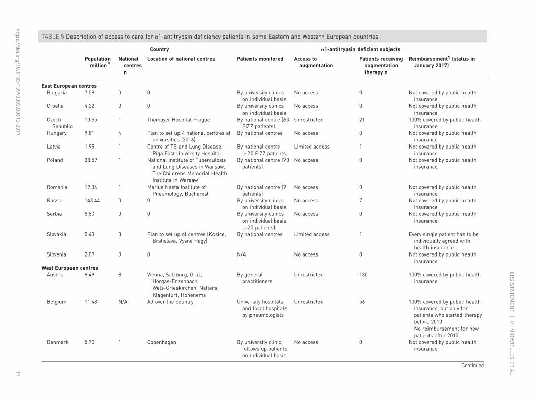
TABLE 5 Description of access to care for α1-antitrypsin deficiency patients in some Eastern and Western European countries
Country α1-antitrypsin deficient subjects
Populationmillion#
Nationalcentresn
Location of national centres Patients monitored Access toaugmentation
Patients receivingaugmentationtherapy n
Reimbursement¶ (status inJanuary 2017)
East European centresBulgaria 7.09 0 0 By university clinics
on individual basisNo access 0 Not covered by public health
insuranceCroatia 4.22 0 0 By university clinics
on individual basisNo access 0 Not covered by public health
insuranceCzechRepublic
10.55 1 Thomayer Hospital Prague By national centre (63PiZZ patients)
Unrestricted 21 100% covered by public healthinsurance
Hungary 9.81 4 Plan to set up 4 national centres atuniversities (2016)
By national centres No access 0 Not covered by public healthinsurance
Latvia 1.95 1 Centre of TB and Lung Disease,Riga East University Hospital
By national centre(∼20 PiZZ patients)
Limited access 1 Not covered by public healthinsurance
Poland 38.59 1 National Institute of Tuberculosisand Lung Diseases in Warsaw,The Childrens Memorial HealthInstitute in Warsaw
By national centre (70patients)
No access 0 Not covered by public healthinsurance
Romania 19.34 1 Marius Nasta Institute ofPneumology, Bucharest
By national centre (7patients)
No access 0 Not covered by public healthinsurance
Russia 143.44 0 0 By university clinicson individual basis
No access 7 Not covered by public healthinsurance
Serbia 8.80 0 0 By university clinicson individual basis(∼20 patients)
No access 0 Not covered by public healthinsurance
Slovakia 5.43 3 Plan to set up of centres (Kosice,Bratislava, Vysne Hagy)
By national centres Limited access 1 Every single patient has to beindividually agreed withhealth insurance
Slovenia 2.09 0 0 N/A No access 0 Not covered by public healthinsurance
West European centresAustria 8.49 8 Vienna, Salzburg, Graz,
Hörgas-Enzenbach,Wels-Grieskirchen, Natters,Klagenfurt, Hohenems
By generalpractitioners
Unrestricted 130 100% covered by public healthinsurance
Belgium 11.48 N/A All over the country University hospitalsand local hospitalsby pneumologists
Unrestricted 56 100% covered by public healthinsurance, but only forpatients who started therapybefore 2010No reimbursement for newpatients after 2010
Denmark 5.70 1 Copenhagen By university clinic,follows up patientson individual basis
No access 0 Not covered by public healthinsurance
Continued
https://doi.org/10.1183/13993003.00610-201717
ERSSTATEM
ENT
|M.M
IRAVITLLES
ETAL.
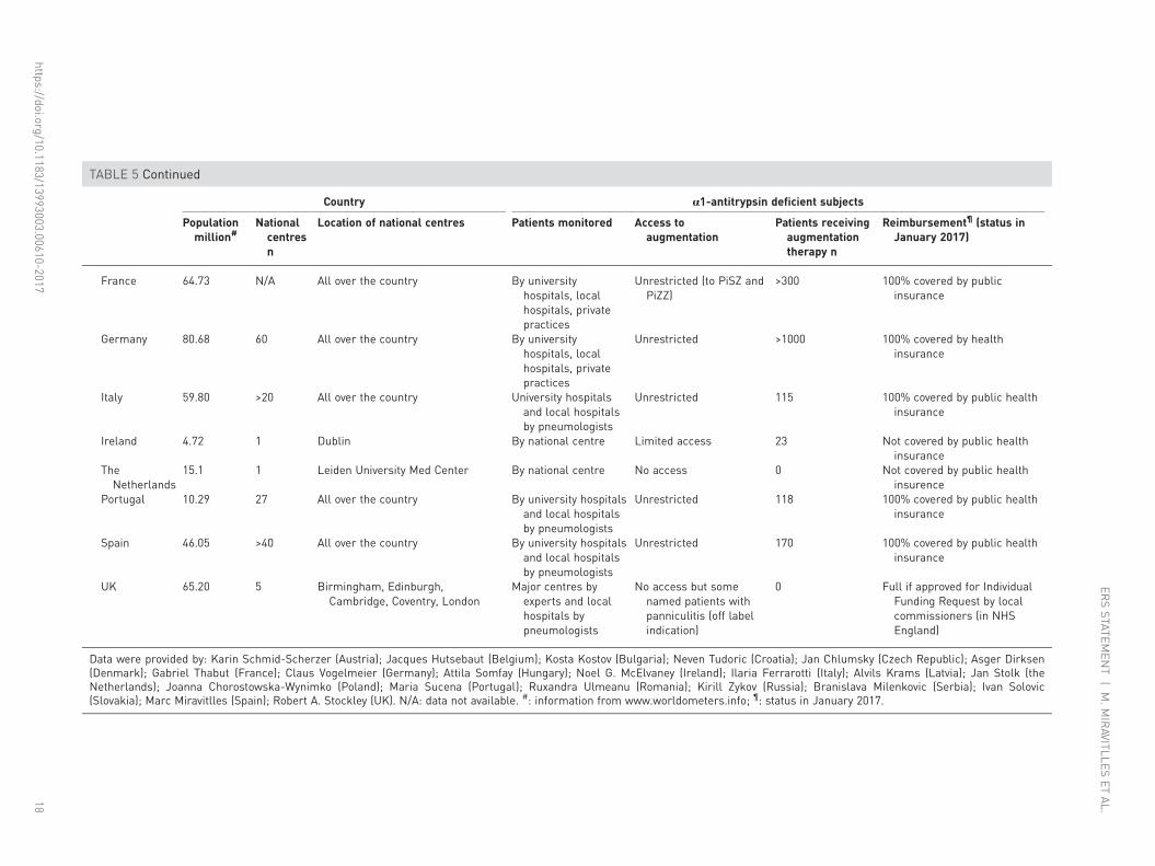
TABLE 5 Continued
Country α1-antitrypsin deficient subjects
Populationmillion#
Nationalcentresn
Location of national centres Patients monitored Access toaugmentation
Patients receivingaugmentationtherapy n
Reimbursement¶ (status inJanuary 2017)
France 64.73 N/A All over the country By universityhospitals, localhospitals, privatepractices
Unrestricted (to PiSZ andPiZZ)
>300 100% covered by publicinsurance
Germany 80.68 60 All over the country By universityhospitals, localhospitals, privatepractices
Unrestricted >1000 100% covered by healthinsurance
Italy 59.80 >20 All over the country University hospitalsand local hospitalsby pneumologists
Unrestricted 115 100% covered by public healthinsurance
Ireland 4.72 1 Dublin By national centre Limited access 23 Not covered by public healthinsurance
TheNetherlands
15.1 1 Leiden University Med Center By national centre No access 0 Not covered by public healthinsurence
Portugal 10.29 27 All over the country By university hospitalsand local hospitalsby pneumologists
Unrestricted 118 100% covered by public healthinsurance
Spain 46.05 >40 All over the country By university hospitalsand local hospitalsby pneumologists
Unrestricted 170 100% covered by public healthinsurance
UK 65.20 5 Birmingham, Edinburgh,Cambridge, Coventry, London
Major centres byexperts and localhospitals bypneumologists
No access but somenamed patients withpanniculitis (off labelindication)
0 Full if approved for IndividualFunding Request by localcommissioners (in NHSEngland)
Data were provided by: Karin Schmid-Scherzer (Austria); Jacques Hutsebaut (Belgium); Kosta Kostov (Bulgaria); Neven Tudoric (Croatia); Jan Chlumsky (Czech Republic); Asger Dirksen(Denmark); Gabriel Thabut (France); Claus Vogelmeier (Germany); Attila Somfay (Hungary); Noel G. McElvaney (Ireland); Ilaria Ferrarotti (Italy); Alvils Krams (Latvia); Jan Stolk (theNetherlands); Joanna Chorostowska-Wynimko (Poland); Maria Sucena (Portugal); Ruxandra Ulmeanu (Romania); Kirill Zykov (Russia); Branislava Milenkovic (Serbia); Ivan Solovic(Slovakia); Marc Miravitlles (Spain); Robert A. Stockley (UK). N/A: data not available. #: information from www.worldometers.info; ¶: status in January 2017.
https://doi.org/10.1183/13993003.00610-201718
ERSSTATEM
ENT
|M.M
IRAVITLLES
ETAL.
way to achieve the successful accumulation of knowledge about the clinical characteristics, evolution,natural history and response to treatment of patients with rare diseases, such as AATD.
Europe was pioneer in the development of national registries for AATD. As early as the 1970s Sweden [15]and Denmark [78] initiated their registries, followed by other countries such as the Netherlands, Spain,Italy, Germany, Ireland, the UK and more recently Switzerland, Latvia, Estonia, Czech Republic, Poland,Austria, Belgium and France, among others. However, the low prevalence of the disease stimulated theorganisation of an international registry, not restricted to Europe, but with predominance of Europeancountries, the Alpha One International Registry (AIR), which was founded in 1997 [9] following therecommendation from the WHO to establish such a registry of AATD [25]. AIR has been a successfulplatform for the development of clinical trials with new and existing therapies for the disease and hascontributed to increase the awareness of the disease among healthcare professionals across Europe [131].
Statement• According to the European Council, management of patients with AATD should be supervised byreference centres of excellence at a national or regional level.
• The systematic collection of data concerning clinical characteristics and natural history of patients withAATD in national and international registries will enhance knowledge about the evolution of this diseaseand its optimal management.
• For many AATD individuals a respiratory service is the first point of diagnosis. The operational pathwayincludes varying assessments and follow-up depending on personalising the patients’ risk and definingthe respiratory phenotype. Links to multidisciplinary teams will ensure the best quality of care.
Access to optimal care and augmentation therapy for AATD in EuropeIn Europe, healthcare policy is largely a devolved matter and decisions related to provision of healthcareservices, disease management and prescription medicines rest with national, regional or local policymakers, health technology assessment (HTA) agencies and payers. This results in different standards ofcare for AATD and contributes to geographical inequalities in access to optimal healthcare services,clinical expertise and effective therapies. Notably, access to high-cost, rare disease therapies, such asaugmentation therapy for AATD, can vary significantly across jurisdictions [132]. Augmentation therapy isfully reimbursed in some countries such as Germany, Italy, Spain, Portugal, France and others, but notreimbursed in the majority of Eastern European countries and in some Western European countries suchas the UK, Ireland, Denmark or Sweden (table 5).
Increasing efforts are being made by policy makers to ensure that patients with rare diseases such asAATD have timely, better and more equitable access to high quality care. Examples of these initiativesinclude: the European Council recommendation on an action in the field of rare diseases, that required allEU member states to adopt national plans and policies for rare diseases by the end of 2013 [133]; theEuropean Network of HTA agencies (EUnetHTA) that aims to assist in the development of reliable,timely, transparent and transferable information to contribute to HTAs in European countries [134]; andthe European Medicine Agency’s adaptive pathways pilot for medicine development and data generation,which allows for early and progressive patient access to a medicine [135].
In the absence of harmonised legislation that regulates AATD healthcare provision and access toaugmentation therapy and other specific treatments across Europe, only multi-stakeholder collaborationand continuous improvement of the available evidence-base for efficacy and cost-effectiveness of AATDtherapies is likely to achieve greater equity in implementation of best practice.
AcknowledgementsThe authors want to acknowledge Courtney Coleman (European Lung Foundation, Sheffield, UK) for coordinating andproviding the opinions of patients through National AATD patient organisation, David Rigau (European RespiratorySociety methodologist) for supervising the development of this statement, Sandra Nestler-Parr (Trustee, Alpha-1 UKSupport Group) for her comments on the organisation and access of care in Europe and Anita Pye (Lung InvestigationUnit, University Hospitals Birmingham NHS Foundation Trust Queen Elizabeth Hospital Birmingham, Birmingham,UK) for working party organisation and collation of individual contributions.
References1 Laurell CB, Eriksson S. The electrophoretic alpha1-globulin pattern of serum in alpha1-antitrypsin deficiency.
Scan J Clin Lab Invest 1963; 15: 132–140.2 Blanco I, de Serres FJ, Fernández-Bustillo E, et al. Estimates of the prevalence of alpha-1 antitrypsin deficiency
PI*S and PI*Z alleles and the numbers at risk in Europe countries. Eur Respir J 2006; 27: 77–84.3 American Thoracic Society/European Respiratory Society Statement: standards for the diagnosis and
management of individuals with alpha-1 antitrypsin deficiency. Am J Respir Crit Care Med 2003; 168: 820–899.4 Eriksson S. Pulmonary emphysema and alpha1-antitrypsin deficiency. Acta Med Scand 1964; 175: 197–205.
https://doi.org/10.1183/13993003.00610-2017 19
ERS STATEMENT | M. MIRAVITLLES ET AL.

5 Casas F, Blanco I, Martínez MT, et al. Indications for active case searches and intravenous alpha-1 antitrypsintreatment for patients with alpha-1 antitrypsin deficiency chronic obstructive pulmonary disease: an update. ArchBronconeumol 2015; 51: 185–192.
6 Marciniuk DD, Hernandez P, Balter M, et al. Alpha-1 antitrypsin deficiency targeted testing and augmentationtherapy: a Canadian Thoracic Society clinical practice guideline. Can Respir J 2012; 19: 109–116.
7 Sandhaus RA, Turino G, Brantly ML, et al. The diagnosis and management of alpha-1 antitrypsin deficiency inthe adult. Chronic Obstr Pulm Dis (Miami) 2016; 3: 668–682.
8 Survival and FEV1 decline in individuals with severe deficiency of alpha-1 antitrypsin. The Alpha-1 AntitrypsinDeficiency Registry Study Group. Am J Respir Crit Care Med 1998; 158: 49–59.
9 Luisetti M, Miravitlles M, Stockley RA. Alpha-1 antitrypsin deficiency: a report from the 2nd meeting of theAlpha One International Registry, Rapallo (Genoa, Italy), 2001. Eur Respir J 2002; 20: 1050–1056.
10 Sveger T. Liver disease in alpha-1 antitrypsin deficency detected by screening of 200,000 infants. N Engl J Med1976; 294: 1316–1321.
11 Stockley RA. Antitrypsin deficiency assessment and programme for treatment (ADAPT): the United Kingdomregistry. COPD 2015; 12: Suppl. 1, 63–68.
12 Wood AM, Needham M, Simmonds MJ, et al. Phenotypic differences in alpha-1 antitrypsin deficient siblingpairs may relate to genetic variation. COPD 2008; 5: 353–359.
13 Stolk J, Cooper BG, Stoel B, et al. Retinoid treatment of Emphysema in Patients on the Alpha-1 InternationalRegistry. The REPAIR study: study design, methodology and quality control of study assessments. Ther AdvRespir Dis 2010; 4: 319–332.
14 Holme J, Stockley RA. Radiologic and clinical features of COPD patients with discordant pulmonary physiology:lessons from alpha-1 antitrypsin deficiency. Chest 2007; 132: 909–915.
15 Piitulainen E, Montero LC, Nystedt-Düzakin M, et al. Lung function and CT densitometry in subjects withalpha-1 antitrypsin deficiency and healthy controls at 35 years of age. COPD 2015; 12: 162–167.
16 Piitulainen F, Tornling G, Eriksson S. Effect of age and occupational exposure to airway irritants on lungfunction in non-smoking individuals with alpha-1 antitrypsin deficiency (PiZZ). Thorax 1997; 52: 244–248.
17 Tanash HA, Nilsson PM, Nilsson JA, et al. Survival in severe alpha-1 antitrypsin deficiency (Pi ZZ). Respir Res2010; 11: 44.
18 Ward H, Turner AM, Stockley RA. Spirometric and gas transfer discordance in alpha-1 antitrypsin deficiency;patient characteristics and progression. Chest 2014; 145: 1316–1324.
19 Parr DG, Stoel BC, Stolk J, et al. Pattern of emphysema distribution in alpha1-antitrypsin deficiency influenceslung function impairment. Am J Respir Crit Care Med 2004; 170: 1172–1178.
20 Stockley RA, Edgar RG, Pillai A, et al. Individualized lung function trends in alpha-1-antitrypsin deficiency: aneed for patience in order to provide patient centered management? Int J Chron Obst Pulm Dis 2016; 11:1745–1756.
21 Dawkins PA, Dawkins CL, Wood AM, et al. Rate of progression of lung function impairment inalpha-1-antitrypsin deficiency. Eur Respir J 2009; 33: 1338–1344.
22 Parr DG, Stoel BC, Stolk J, et al. Validation of computed tomographic lung densitometry for monitoringemphysema in alpha1-antitrypsin deficiency. Thorax 2006; 61: 485–490.
23 Piitulainen E, Tanash HA. The clinical profile of subjects included in the Swedish National Register onindividuals with severe alpha-1 antitrypsin deficiency. COPD 2015; 12: Suppl. 1, 36–41.
24 Piras B, Ferrarotti I, Lara B, et al. Clinical phenotypes of Italian and Spanish patients with α1-antitrypsindeficiency. Eur Respir J 2013; 42: 54–64.
25 Alpha1-antitrypsin deficiency: memorandum from a WHO meeting. Bull WHO 1997; 75: 397–415.26 Ledue TB, Collins MF. Development and validation of 14 human serum protein assays on the Roche cobas (c)
501. J Clin Lab Anal 2011; 25: 52e60.27 Costa X, Jardi R, Rodriguez F, et al. Simple method for alpha1-antitrypsin deficiency screening by use of dried
blood spot specimens. Eur Respir J 2000; 15: 1111–1115.28 Gorrini M, Ferrarotti I, Lupi A, et al. Validation of a rapid and simple method to measure a1-antitrypsin. Clin
Chem 2006; 52: 899–901.29 Zillmer LR, Russo R, Manzano BM, et al. Validation and development of an immunonephelometric assay for the
determination of alpha-1 antitrypsin levels in dried blood spots from patients with COPD. J Bras Pneumol 2013;39: 547–554.
30 Blanco I, Bueno P, Diego I, et al. Alpha-1 antitrypsin Pi*Z gene frequency and Pi*ZZ genotype numbersworldwide: an update. Int J Chron Obstruct Pulmon Dis 2017; 12: 561–569.
31 Miravitlles M, Herr C, Ferrarotti I, et al. Laboratory testing of individuals with severe alpha1-antitrypsindeficiency in three European centres. Eur Respir J 2010; 35: 960–968.
32 Zerimech F, Hennache G, Bellon F, et al. Evaluation of a new Sebia isoelectrofocusing kit for alpha 1-antitrypsinphenotyping with the Hydrasys System. Clin Chem Lab Med 2008; 46: 260–263.
33 Greene DN, Elliott-Jelf MC, Straseski JA, et al. Facilitating the laboratory diagnosis of α1-antitrypsin deficiency.Am J Clin Pathol 2013; 139: 184–191.
34 Ferrarotti I, Thun GA, Zorzetto M, et al. Serum levels and genotype distribution of α1-antitrypsin in the generalpopulation. Thorax 2012; 67: 669–674.
35 Orrù G, Faa G, Pillai S, et al. Rapid PCR real-time genotyping of M-Malton alpha1-antitrypsin deficiency allelesby molecular beacons. Diagn Mol Pathol 2005; 14: 237–244.
36 Denden S, Lakhdar R, Keskes NB, et al. PCR-based screening for the most prevalent alpha 1 antitrypsindeficiency mutations (PI S, Z, and Mmalton) in COPD patients from Eastern Tunisia. Biochem Genet 2013; 51:677–685.
37 Belmonte I, Montoto L, Miravitlles M, et al. Rapid detection of Mmalton α1-antitrypsin deficiency allele byreal-time PCR and melting curves in whole blood, serum and dried blood spot samples. Clin Chem Lab Med2016; 54: 241–248.
38 Soini, S. Genetic testing legislation in Western Europe—a fluctuating regulatory target. J Community Genet 2012;3: 143–153.
https://doi.org/10.1183/13993003.00610-2017 20
ERS STATEMENT | M. MIRAVITLLES ET AL.
39 Hruban RH, Meziane MA, Zerhouni EA, et al. High resolution computed tomography of inflation-fixed lungs.Pathologic-radiologic correlation of centrilobular emphysema. Am Rev Respir Dis 1987; 136: 935–940.
40 Hayhurst MD, MacNee W, Flenley DC, et al. Diagnosis of pulmonary emphysema by computerised tomography.Lancet 1984; 2: 320–322.
41 Gould GA, MacNee W, McLean A, et al. CT measurements of lung density in life can quantitate distal airspaceenlargement–an essential defining feature of human emphysema. Am Rev Respir Dis 1988; 137: 380–392.
42 Gevenois PA, de Maertelaer V, De Vuyst P, et al. Comparison of computed density and macroscopicmorphometry in pulmonary emphysema. Am J Respir Crit Care Med 1995; 152: 653–657.
43 Gevenois PA, De Vuyst P, de Maertelaer V, et al. Comparison of computed density and microscopicmorphometry in pulmonary emphysema. Am J Respir Crit Care Med 1996; 154: 187–192.
44 Muller NL, Staples CA, Miller RR, et al. “Density mask”. An objective method to quantitate emphysema usingcomputed tomography. Chest 1988; 94: 782–787.
45 Gould GA, Redpath AT, Ryan M, et al. Lung CT density correlates with measurements of airflow limitation andthe diffusing capacity. Eur Respir J 1991; 4: 141–146.
46 Kinsella M, Muller NL, Abboud RT, et al. Quantitation of emphysema by computed tomography using a“density mask” program and correlation with pulmonary function tests. Chest 1990; 97: 315–321.
47 Dowson LJ, Guest PJ, Hill SL, et al. High-resolution computed tomography scanning in alpha1-antitrypsindeficiency: relationship to lung function and health status. Eur Respir J 2001; 17: 1097–1104.
48 Dowson LJ, Newall C, Guest PJ, et al. Exercise capacity predicts health status in alpha1-antitrypsin deficiency.Am J Respir Crit Care Med 2001; 163: 936–941.
49 Stolk J, Ng WH, Bakker ME, et al. Correlation between annual change in health status and computertomography derived lung density in subjects with alpha1-antitrypsin deficiency. Thorax 2003; 58: 1027–1030.
50 Dowson LJ, Guest PJ, Stockley RA. Longitudinal changes in physiological, radiological, and health statusmeasurements in alpha1-antitrypsin deficiency and factors associated with decline. Am J Respir Crit Care Med2001; 164: 1805–1809.
51 Dirksen A, Dijkman JH, Madsen F, et al. A randomized clinical trial of alpha1-antitrypsin augmentation therapy.Am J Respir Crit Care Med 1999; 160: 1468–1472.
52 Dirksen A, Piitulainen E, Parr DG, et al. Exploring the role of CT densitometry: a randomised study ofaugmentation therapy in alpha1-antitrypsin deficiency. Eur Respir J 2009; 33: 1345–1353.
53 Chapman KR, Burdon JG, Piitulainen E, et al. Intravenous augmentation treatment and lung density in severe α1antitrypsin deficiency (RAPID): a randomised, double-blind, placebo-controlled trial. Lancet 2015; 386: 360–368.
54 Schluchter MD, Stoller JK, Barker AF, et al. Feasibility of a clinical trial of augmentation therapy foralpha1-antitrypsin deficiency. The Alpha 1-Antitrypsin Deficiency Registry Study Group. Am J Respir Crit CareMed 2000; 161: 796–801.
55 Plasma Protein Therapeutics Association (PPTA). Clinical and Surrogate Endpoints for Evaluating Efficacyof Alpha1-Proteinase Inhibitor (Human) Augmentation Therapy. www.pptaglobal.org/images/regulatory/FDAA09009_A1-PIStatement_BPAC_final.pdf Date last updated: July 20, 2009.
56 Stolk J, Dirksen A, van der Lugt AA, et al. Repeatability of lung density measurements with low-dose computedtomography in subjects with alpha-1-antitrypsin deficiency-associated emphysema. Invest Radiol 2001; 36:648–651.
57 Miller MR, Crapo R, Hankinson J, et al. General considerations for lung function testing. Eur Respir J 2005; 26:153–161.
58 Hersh CP, Dahl M, Ly NP, et al. Chronic obstructive pulmonary disease in alpha1-antitrypsin PI MZheterozygotes: a meta-analysis. Thorax 2004; 59: 843–849.
59 Dahl M, Tybjaerg-Hansen A, Lange P, et al. Change in lung function and morbidity from chronic obstructivepulmonary disease in alpha-1-antitrypsin MZ heterozygotes: a longitudinal study of the general population. AnnIntern Med 2002; 136: 270–279.
60 Sørheim IC, Bakke P, Gulsvik A, et al. α1-Antitrypsin protease inhibitor MZ heterozygosity is associated withairflow obstruction in two large cohorts. Chest 2010; 138: 1125–1132.
61 Molloy K, Hersh CP, Morris VB, et al. Clarification of the risk of chronic obstructive pulmonary disease inα1-antitrypsin deficiency PiMZ heterozygotes. Am J Respir Crit Care Med 2014; 189: 419–427.
62 Dahl M, Hersh CP, Ly NP, et al. The protease inhibitor PI*S allele and COPD: a meta-analysis. Eur Respir J2005; 26: 67–76.
63 Bartmann K, Fooke-Achterrath M, Koch G, et al. Heterozygosity in the Pi-system as a pathogenetic cofactor inchronic obstructive pulmonary disease (COPD). Eur J Respir Dis 1985; 66: 284–296.
64 Seersholm N, Kok-Jensen A. Intermediate alpha 1-antitrypsin deficiency PiSZ: a risk factor for pulmonaryemphysema? Respir Med 1998; 92: 241–245.
65 Lara B, Miravitlles M. Spanish registry of patients with alpha-1 antitrypsin deficiency; comparison of thecharacteristics of PISZ and PIZZ individuals. COPD 2015; 12: Suppl. 1, 27–31.
66 Green CE, Vayalapra S, Hampson JA, et al. PiSZ alpha-1 antitrypsin deficiency (AATD): pulmonary phenotypeand prognosis relative to PiZZ AATD and PiMM COPD. Thorax 2015; 70: 939–945.
67 Cox DW, Levison H. Emphysema of early onset associated with a complete deficiency of alpha-1-antitrypsin(null homozygotes). Am Rev Respir Dis 1988; 137: 371–375.
68 Cook L, Janus ED, Brenton S, et al. Absence of alpha-1-antitrypsin (Pi Null Bellingham) and the early onset ofemphysema. Aust N Z J Med 1994; 24: 263–269.
69 Fregonese L, Stolk J, Frants RR, et al. Alpha-1 antitrypsin Null mutations and severity of emphysema. RespirMed 2008; 102: 876–884.
70 Rodríguez-Frías F, Miravitlles M, Vidal R, et al. Rare alpha-1-antitrypsin variants: are they really so rare? TherAdv Respir Dis 2012; 6: 79–85.
71 Ferrarotti I, Carroll TP, Ottaviani S, et al. Identification and characterisation of eight novel SERPINA1 Nullmutations. Orphanet J Rare Dis 2014; 9: 172.
72 Carroll TP, O’Connor CA, Floyd O, et al. The prevalence of alpha-1 antitrypsin deficiency in Ireland. Respir Res2011; 12: 91.
https://doi.org/10.1183/13993003.00610-2017 21
ERS STATEMENT | M. MIRAVITLLES ET AL.
73 Kaczor MP, Sanak M, Libura-Twardowska M, et al. The prevalence of alpha1-antitrypsin deficiency in arepresentative population sample from Poland. Respir Med 2007; 101: 2520–2525.
74 O’Brien ML, Buist NR, Murphey WH. Neonatal screening for alpha1-antitrypsin deficiency. J Pediatr 1978; 92:1006–1010.
75 Wall M, Moe E, Eisenberg J, et al. Long-term follow-up of a cohort of children with alpha-1-antitrypsindeficiency. J Pediatr 1990; 116: 248–251.
76 Bernspang E, Sveger T, Piitulainen E. Respiratory symptoms and lung function in 30-year-old individuals withalpha-1-antitrypsin deficiency. Respir Med 2007; 101: 1971–1976.
77 Piitulainen E, Mostafavi B, Tanash HA. Health status and lung function in the Swedish alpha 1-antitrypsindeficient cohort, identified by neonatal screening, at the age of 37–40 years. Int J Chron Obstruct Pulmon Dis2017; 12: 495–500.
78 Seersholm N, Kok-Jensen A, Dirksen A. Survival of patients with severe alpha 1-antitrypsin deficiency withspecial reference to non-index cases. Thorax 1994; 49: 695–698.
79 Thelin T, Sveger T, McNeil TF. Primary prevention in a high-risk group: smoking habits in adolescents withhomozygous alpha-1-antitrypsin deficiency (ATD). Acta Paediatr 1996; 85: 1207–1212.
80 McNeil TF, Sveger T, Thelin T. Psychosocial effects of screening for somatic risk: the Swedish alpha 1 antitrypsinexperience. Thorax 1988; 43: 505–507.
81 Sveger T, Thelin T, McNeil TF. Neonatal alpha1-antitrypsin screening: parents’ views and reactions 20 years afterthe identification of the deficiency state. Acta Paediatr 1999; 88: 315–318.
82 Wewers MD, Casolaro MA, Sellers SE, et al. Replacement therapy for alpha 1-antitrypsin deficiency associatedwith emphysema. N Engl J Med 1987; 316: 1055–1062.
83 Gøtzsche PC, Johansen HK. Intravenous alpha-1 antitrypsin augmentation therapy for treating patients withalpha-1 antitrypsin deficiency and lung disease. Cochrane Database Syst Rev 2010; 7: CD007851.
84 Chapman KR, Stockley RA, Dawkins C, et al. Augmentation therapy for alpha1 antitrypsin deficiency: ameta-analysis. COPD 2009; 6: 177–184.
85 Stockley RA, Parr DG, Piitulainen E, et al. Therapeutic efficacy of alpha-1 antitrypsin augmentation therapy onthe loss of lung tissue: an integrated analysis of 2 randomised clinical trials using computed tomographydensitometry. Respir Res 2010; 11: 136.
86 Gøtzsche PC, Johansen HK. Intravenous alpha-1 antitrypsin augmentation therapy for treating patients withalpha-1 antitrypsin deficiency and lung disease. Cochrane Database Syst Rev 2016; in press [https://doi.org/10.1002/14651858.CD007851.pub3].
87 McElvaney NG, Burdon J, Holmes M, et al. Long-term efficacy and safety of α1 proteinase inhibitor treatmentfor emphysema caused by severe α1 antitrypsin deficiency: an open-label extension trial (RAPID-OLE). LancetRespir Med 2017; 5: 51–60.
88 Stoller JK, Rouhani F, Brantly M, et al. Biochemical efficacy and safety of a new pooled human plasmaalpha1-antitrypsin, Respitin. Chest 2002; 122: 66–74.
89 Stocks JM, Brantly M, Pollock D, et al. Multi-center study: the biochemical efficacy, safety and tolerability of anew alpha1-proteinase inhibitor, Zemaira. COPD 2006; 3: 17–23.
90 Stocks JM, Brantly ML, Wang-Smith L, et al. Pharmacokinetic comparability of Prolastin(R)-C to Prolastin(R) inalpha1-antitrypsin deficiency: a randomized study. BMC Clin Pharmacol 2010; 10: 13.
91 Campos MA, Kueppers F, Stocks JM, et al. Safety and pharmacokinetics of 120 mg/kg versus 60 mg/kg weeklyintravenous infusions of alpha-1 proteinase inhibitor in alpha-1 antitrypsin deficiency: a multicenter,randomized, double-blind, crossover study (SPARK). COPD 2013; 10: 687–695.
92 Sandhaus RA, Stocks J, Rouhani FN, et al. Biochemical efficacy and safety of a new, ready-to-use, liquidalpha-1-proteinase inhibitor, GLASSIA (alpha1-proteinase inhibitor (human), intravenous). COPD 2014; 11:17–25.
93 Seersholm N, Wencker M, Banik N, et al. Does alpha1-antitrypsin augmentation therapy slow the annual declinein FEV1 in patients with severe hereditary alpha1-antitrypsin deficiency? Wissenschaftliche Arbeitsgemeinschaftzur Therapie von Lungenerkrankungen (WATL) alpha1-AT study group. Eur Respir J 1997; 10: 2260–2263.
94 Stoller JK, Fallat R, Schluchter MD, et al. Augmentation therapy with alpha1-antitrypsin: patterns of use andadverse events. Chest 2003; 123: 1425–1434.
95 Wencker M, Fuhrmann B, Banik N, et al. Longitudinal follow-up of patients with alpha1-protease inhibitordeficiency before and during therapy with IV alpha1-protease inhibitor. Chest 2001; 119: 737–744.
96 Tonelli AR, Rouhani F, Li N, et al. Alpha-1-antitrypsin augmentation therapy in deficient individuals enrolled inthe Alpha-1 Foundation DNA and Tissue Bank. Int J Chron Obstruct Pulmon Dis 2009; 4: 443–452.
97 Barros-Tizon JC, Torres ML, Blanco I, et al. Reduction of severe exacerbations and hospitalization-derived costsin alpha-1-antitrypsin-deficient patients treated with alpha-1-antitrypsin augmentation therapy. Ther Adv RespirDis 2012; 6: 67–78.
98 Schmidt EW, Rasche B, Ulmer WT, et al. Replacement therapy for alpha-1-protease inhibitor deficiency in PiZsubjects with chronic obstructive lung disease. Am J Med 1988; 84: 63–69.
99 Barker AF, Siemsen F, Pasley D, et al. Replacement therapy for hereditary alpha1-antitrypsin deficiency. Aprogram for long-term administration. Chest 1994; 105: 1406–1410.
100 Barker AF, Iwata-Morgan I, Oveson L, et al. Pharmacokinetic study of alpha1-antitrypsin infusion inalpha1-antitrypsin deficiency. Chest 1997; 112: 607–613.
101 Miravitlles M, Vidal R, Torrella M, et al. Evaluacion del tratamiento sustitutivo del enfisema por deficit dealfa-1-antitripsina [Evaluation of replacement therapy in emphysema caused by alpha 1-antitrypsin deficiency].Arch Bronconeumol 1994; 30: 479–484.
102 Schwaiblmair M, Vogelmeier C, Fruhmann G. Long-term augmentation therapy in twenty patients with severealpha-1-antitrypsin deficiency–three-year follow-up. Respiration 1997; 64: 10–15.
103 Wencker M, Banik N, Buhl R, et al. Langzeittherapie des alpha 1-Antitrypsin-mangelassoziiertenLungenemphysems mit humanem alpha 1-Antitrypsin [Long-term therapy of alpha 1-antitrypsin-deficiency-associated pulmonary emphysema with human alpha 1-antitrypsin]. Pneumologie 1998; 52:545–552.
https://doi.org/10.1183/13993003.00610-2017 22
ERS STATEMENT | M. MIRAVITLLES ET AL.
104 Wencker M, Banik N, Buhl R, et al. Long-term treatment of alpha1-antitrypsin deficiency-related pulmonaryemphysema with human alpha1-antitrypsin. Wissenschaftliche Arbeitsgemeinschaft zur Therapie vonLungenerkrankungen (WATL)-alpha1-AT-study group. Eur Respir J 1998; 11: 428–433.
105 Campos MA, Alazemi S, Zhang G, et al. Exacerbations in subjects with alpha-1 antitrypsin deficiency receivingaugmentation therapy. Respir Med 2009; 103: 1532–1539.
106 Campos MA, Alazemi S, Zhang G, et al. Clinical characteristics of subjects with symptoms of alpha1-antitrypsindeficiency older than 60 years. Chest 2009; 135: 600–608.
107 Subramanian DR, Jenkins L, Edgar R, et al. Assessment of pulmonary neutrophilic inflammation in emphysemaby quantitative positron emission tomography. Am J Respir Crit Care Med 2012; 186: 1125–1132.
108 Vidal R, Barros-Tizón JC, Gáldiz JB, et al. Tolerance and safety of Trypsone®: prospective follow-up in alpha-1antitrypsin deficient subjects with pulmonary emphysema. Minerva Pneumologica 2010; 49: 83–91.
109 Ma S, Lin YY, Cantor JO, et al. The effect of alpha-1 proteinase inhibitor on biomarkers of elastin degradation inalpha-1 antitrypsin deficiency: an analysis of the RAPID/RAPID extension trials. Chronic Obst Pulm Dis (Miami)2017; 4: 34–44.
110 Dawkins PA, Wood A, Nightingale P, et al. Mortality in alpha-1-antitrypsin deficiency in the United Kingdom.Respir Med 2009; 103: 1540–1547.
111 Green CE, Parr DG, Edgar R, et al. Lung density decline associates with mortality in alpha 1 antitrypsin deficientpatients. Respir Med 2016; 112: 81–87.
112 Stoller JK, Gildea TR, Ries AL, et al. Lung volume reduction surgery in patients with emphysema and alpha-1antitrypsin deficiency. Ann Thorac Surg 2007; 83: 241–251.
113 Dauriat G, Mal H, Jebrak G, et al. Functional results of unilateral lung volume reduction surgery inalpha1-antitrypsin deficient patients. Int J Chron Obstruct Pulmon Dis 2006; 1: 201–206.
114 Rathinam S, Oey I, Steiner M, et al. The role of the emphysema multidisciplinary team in a successful lungvolume reduction surgery programme. Eur J Cardiothorac Surg 2014; 46: 1021–1026.
115 Clark SJ, Zoumot Z, Bamsey O, et al. Surgical approaches for lung volume reduction in emphysema. Clin Med(Lond) 2014; 14: 122–127.
116 Sciurba FC, Criner GJ, Strange C, et al. Effect of endobronchial coils vs usual care on exercise tolerance inpatients with severe emphysema: the RENEW randomized clinical trial. JAMA 2016; 315: 2178–2189.
117 Shah PL, Herth FJ, van Geffen WH, et al. Lung volume reduction for emphysema. Lancet Respir Med 2017; 5:147–156.
118 Klooster K, ten Hacken NH, Hartman JE, et al. Endobronchial valves for emphysema without interlobarcollateral ventilation. N Engl J Med 2015; 373: 2325–2335.
119 International Society for Heart and Lung Transplantation. Registries – Heart/Lung Registries. https://www.ishlt.org/registries/slides.asp?slides=heartLungRegistry Date last accessed: March 15, 2017.
120 de Perrot M, Chaparro C, McRae K, et al. Twenty-year experience of lung transplantation at a single centre:influence of recipient diagnosis on long-term survival. J Thorac Cardiovasc Surg 2004; 127: 1493–1501.
121 Thabut G, Ravaud P, Christie JD, et al. Determinants of the survival benefit of lung transplantation in patientswith chronic obstructive pulmonary disease. Am J Respir Crit Care Med 2008; 177: 1156–1163.
122 Trulock EP. Lung transplantation for alpha 1-antitrypsin deficiency emphysema. Chest 1996; 110: Suppl.,284S–294S.
123 King MB, Campbell EJ, Gray BH, et al. The proteinase-antiproteinase balance in alpha-1-proteinaseinhibitor-deficient lung transplant recipients. Am J Respir Crit Care Med 1994; 149: 966–971.
124 Janciauskiene S, Welte T. Well-known and less well-known functions of alpha-1 antitrypsin. Its role in chronicobstructive pulmonary disease and other disease developments. Ann Am Thorac Soc 2016; 13: Suppl. 4,S280–S288.
125 Burton CM, Milman N, Carlsen J, et al. The Copenhagen National Lung Transplant Group: after single lung,double lung, and heart-lung transplantation. J Heart Lung Transplant 2005; 24: 1834–1843.
126 Tanash HA, Riise GC, Hansson L, et al. Survival benefit of lung transplantation in individuals with severeα1-anti-trypsin deficiency (PiZZ) and emphysema. J Heart Lung Transplant 2011; 30: 1342–1347.
127 Stone HM, Edgar RG, Thompson RD, et al. Lung transplantation in alpha-1-antitrypsin deficiency. COPD 2016;13: 146–152.
128 Thabut G. Estimating the survival benefit of lung transplantation: considering the disease course during the wait.Ann Am Thorac Soc 2017; 14: 163–164.
129 Morer L, Choudat L, Dauriat G, et al. Liver involvement in patients with PiZZ-emphysema, candidates for lungtransplantation. Am J Transplant 2016; 17: 1389–1395.
130 Centres of Reference for rare diseases in Europe: State-of-the-art in 2006 and recommendations of the RareDiseases Task Force. http://ec.europa.eu/health/archive/ph_threats/non_com/docs/contribution_policy.pdf Datelast accessed: March 20, 2017.
131 Stockley RA, Luisetti M, Miravitlles M, et al. Ongoing research in Europe: Alpha One International Registry(AIR) objectives and development. Eur Respir J 2007; 29: 582–586.
132 Luisetti M, Balfour-Lynn IM, Johnson S, et al. Perspectives for improving the evaluation and access of therapiesfor rare lung diseases in Europe. Respir Med 2012; 106: 759–768.
133 Official Journal of the European Union. Council recommendation of 8 June 2009 on an action in the field of rarediseases, 2009/C 151/02. http://eur-lex.europa.eu/LexUriServ/LexUriServ.do?uri=OJ:C:2009:151:0007:0010:EN:PDF Date last accessed: March 13, 2017.
134 EUnetHTA. http://www.eunethta.eu/about-us. Date last accessed: March 13, 2017.135 European Medicines Agency. Adaptive pathways. www.ema.europa.eu/ema/index.jsp?curl=pages/regulation/
general/general_content_000601.jsp Date last accessed: March 13, 2017.136 McElvaney NG. Diagnosing α1-antitrypsin deficiency: how to improve the current algorithm. Eur Respir Rev
2015; 24: 52–57.137 Chorostowska-Wynimko J. Targeted screening programmes in COPD: how to identify individuals with
α1-antitrypsin deficiency. Eur Respir Rev 2015; 24: 40–45.
https://doi.org/10.1183/13993003.00610-2017 23
ERS STATEMENT | M. MIRAVITLLES ET AL.

138 Beletic A, Dudvarski-Ilic A, Milenkovic B, et al. Is an integrative laboratory algorithm more effective in detectingalpha-1-antitrypsin deficiency in patients with premature chronic obstructive pulmonary disease than AATconcentration based screening approach? Biochem Med (Zagreb) 2014; 24: 293–298.
139 Balduyck M, Odou MF, Zerimech F, et al. Diagnosis of alpha-1 antitrypsin deficiency: modalities, indicationsand diagnosis strategy. Rev Mal Respir 2014; 31: 729–745.
140 Bornhorst JA, Procter M, Meadows C, et al. Evaluation of an integrative diagnostic algorithm for theidentification of people at risk for alpha1-antitrypsin deficiency. Am J Clin Pathol 2007; 128: 482–490.
141 Ferrarotti I, Scabini R, Campo I, et al. Laboratory diagnosis of alpha1-antitrypsin deficiency. Transl Res 2007;150: 267–274.
142 Kaczor MP, Sanak M, Szczeklik A. Molecular diagnostics of α1-antitrypsin deficiency. Expert Opin Med Diagn2007; 1: 253–265.
143 Corda L, Bertella E, Pini L, et al. Diagnostic flow chart for targeted detection of alpha1-antitrypsin deficiency.Respir Med 2006; 100: 463–470.
144 Snyder MR, Katzmann JA, Butz ML, et al. Diagnosis of alpha-1-antitrypsin deficiency: an algorithm ofquantification, genotyping, and phenotyping. Clin Chem 2006; 52: 2236–2242.
145 Campbell EJ. Alpha1-antitrypsin deficiency: incidence and detection program. Respir Med 2000; 94: Suppl. C,S18–S21.
146 Stolk J, Stockley RA, Stoel BC, et al. Randomised controlled trial for emphysema with a selective agonist of theγ-type retinoic acid receptor. Eur Respir J 2012; 40: 306–312.
147 Stolk J, Stockley RA, Piitulainen E, et al. Relationship between change in lung density and long-term progressionof lung function. Am J Respir Crit Care Med 2015; 192: 114–116.
https://doi.org/10.1183/13993003.00610-2017 24
ERS STATEMENT | M. MIRAVITLLES ET AL.
Top Related