γλώσσες
Σελίδες
Νομικός

Submit Manuscript | http://medcraveonline.com
IntroductionYeasts have fundamentally the same subcellular structure as
cells of higher animals and plants, and have been routinely used as food, feed, and medicine.1 Yeasts can be submitted to autolysis where intracellular enzymes are activated by appropriate process conditions resulting in a partial degradation of the cell wall structures. After autolysis, yeast cream can be centrifuged in order to remove the yeast extract and isolate the yeast cell wall.2 Finally, yeast cell wall, a typical prebiotic, can be submitted to further purification process in order to remove the mannan- protein outer layer to form a purified β-glucan. In this purified form, the β-1,3;1,6-glucan can be recognized and captured in the gut by specialized cells (M cells and macrophages), with subsequent unrolling of complex biodynamics and biokinetics processes, suggesting more important systemic effects than only microbial immunomodulatory effect.3,4 β-Glucans represent a group of biologically active polysaccharides originating from different sources including the cell wall of filamentous fungus and yeasts, seaweed, oat, and barley.5 These macromolecules present α or β conformation linkages in different degrees of polymerization, where the β-1,3 glucose linear chain core has abundant lateral branches β-1,6. Its functions include protection, energy reserve, and osmotic stability to the cell wall.6 β-Glucans from yeast and fungi consist of a backbone of glycopyranosyl molecules joined by 1,3-β-links; from this backbone, side chains can be joined by 1,6-β-links, producing a branched molecular structure. Cereal cell wall consists of not branched β-glucans with glucopyranose molecules linked by 1,3-β and 1,4-β linkages. On the other hand, bacterial β-glucans are unbranched with only 1,3-β-linkages between the glycopyranosyl molecules; similarly, barley and most cereal grains contain unbranched β-glucan chains.7
The biological effect of β-glucans depends on the route of application, as well as on other characteristics such as origin, solubility, size of
the molecules, conformation, and purity.5,8 Beneficial effects of β-glucans on animal and human health have been widely described in the literature including immunomodulatory, anti-infective, antitumor, antimutagenic, anti-allergic, regenerative, antithrombogenic, anticoagulant, antioxidant, lipid-lowering, and radioprotective activity.5,6,9–14 In addition, they have also been widely used in animal feed to improve the growth and performance of farm animals.15,17
The fungal-like Pythium insidiosum is an aquatic oomycete that affects mammals. Pythiosis, an emerging life-threatening illness with an unfavorable prognosis, is caused by Pythium insidiosum.18,19 Despite increased studies evaluating different therapeutic protocols for the treatment of pythiosis, this disease remains extremely difficult to treat and cure. Pythiosis has been reported in species inhabiting marshy environments in tropical and subtropical areas, with most cases occurring in the Americas, Asia, and Australia.20 Most cases have been reported in dogs, horses, and humans.21 Depending on the site of entry, infection can result in different forms: cutaneous, vascular, ocular, gastrointestinal, and a less common systemic form.18 In dogs, it has a more common incidence in young animals of medium and large breeds. Clinical signs of the gastrointestinal form include vomiting, diarrhea, and abdominal pain, all contributing to a condition of progressive weight loss.22
With the increasing incidence of pythiosis in humans and companion animals and its unfavorable prognosis, it is clear that studies evaluating new therapies and ways to improve the immune responses are required. β-Glucans, with their significant biological and particularly immunostimulant effects, could be an interesting adjuvant to the existing therapies. Thus, the purpose of this study was to evaluate the effects of purified β-glucan using rabbits as experimental model.
Int Clin Pathol J. 2020;8(1):14‒20. 14©2020 Santurio et al. This is an open access article distributed under the terms of the Creative Commons Attribution License, which permits unrestricted use, distribution, and build upon your work non-commercially.
Effect of yeast purified β-glucan in experimental treatment of pythiosis in rabbits
Volume 8 Issue 1 - 2020
Janio Morais Santurio,1 Sydney Hartz Alves,1 Daniela Isabel Brayer Pereira,2 Vaclav Vetvicka,3 Carlos AF de Oliveira4
1Laboratory of Mycological Research, Department of Microbiology and Parasitology/Health Sciences Center, Federal University of Santa Maria, Brazil2Mycology Laboratory, Department of Microbiology and Parasitology/Institute of Biology, Federal University of Pelotas, Brazil3University of Louisville, Department of Pathology, USA4Department of Research and Development, Biorigin Company, Fazenda São José s/n, 17290- 000 Macatuba, São Paulo, Brazil
Correspondence: Vaclav Vetvicka, University of Louisville, School of Medicine, Department of Pathology, 511 S. Floyd, Louisville, KY 40202, USA, Tel (502) 852-1612, Email
Received: April 08, 2020 | Published: April 16, 2020
Abstract
Purified β-1,3-1,6-glucan derived from the cell wall of yeast has important biological properties and is considered to be a potent immunomodulatory compound. Pythium insidiosum is an aquatic oomycete affecting mammals and causing infections of unfavorable prognosis. Treatment is difficult, and therapeutic options commonly include surgery, immunotherapy, and antimicrobial agents. This study evaluated the clinical effects and antioxidant activity of yeast-purified β-glucan in the treatment of experimental pythiosis in rabbits. The disease was induced in 30 rabbits and we compared the effects of food supplementation with β-glucan with oral treatment with a combination of antifungals. Results suggested that the β-glucan showed antioxidant, immunomodulatory, and anti-inflammatory effects. Although there has not been a clinical cure of the treated animals, it appears that the inclusion of the β-glucan is beneficial as an adjuvant to the treatment of pythiosis via strengthening of nonspecific immunity and improvement of antioxidant status of the animal.
Keywords: immunotherapy, glucans, pythiosis, rabbit
International Clinical Pathology Journal
Research Article Open Access
Effect of yeast purified β-glucan in experimental treatment of pythiosis in rabbits 15Copyright:
©2020 Santurio et al.
Citation: Santurio JM, Alves SH, Pereira DIB, et al. Effect of yeast purified β-glucan in experimental treatment of pythiosis in rabbits. Int Clin Pathol J. 2020;8(1):14‒20. DOI: 10.15406/icpjl.2020.08.00199
Material and methodsExperimental design and treatments
All procedures involving the animals have been approved by the Ethics Committee on Animal Experimentation of the Federal University of Santa Maria. Thirty 3-month-old male and female rabbits (New Zealand) were subcutaneously inoculated with 1 mL P. insidiosum zoospores, as previously described by Santurio & Pereira et al.23,24 The animals were kept in individual cages under adequate conditions of hygiene, light and temperature, receiving water ad libitum and feed according to body weight. Forty-five days post inoculation (PI) and after evaluation of the evolution of subcutaneous lesions, the animals were randomly divided into six groups of five as follows:
1. Group 1–untreated animals
2. Group 2–untreated animals fed diet supplemented with β-glucan BG 01 (500 mg/day).
3. Biorigin has developed this sample of purified insoluble particulate glucan from Saccharomyces cerevisiae, 77.30 % pure, using a special biotechnological process, as previously described by de Oliveira et al.10
4. Group 3–animals treated with six doses of the immunotherapeutic (Pitium-Vac, vaccine made by the Laboratory of Mycological Research (LAPEMI) of the Federal University of Santa Maria) subcutaneously at 14-day intervals
5. Group 4–animals treated with six doses of the immunotherapy (Pitium-Vac) subcutaneously at 14-day intervals and diet supplemented with β-glucan (500 mg/day)
6. Group 5–animals treated with the combination of itraconazole antifungals (Sigma- Aldrich) (8 mg/ Kg /day) and terbinafine (Novartis) (125 mg/day) administered orally by gavage
7. Group 6–animals treated with the combination of itraconazole antifungals (8 mg/kg/day) and terbinafine (125 mg/day) by gavage, and diet supplemented with β-glucan (500 mg/day)
Prior to animal inoculation (Day 0) and every 15 days, blood samples were obtained by cardiac puncture for hematological evaluation and biochemical profile. The area of the lesions was measured horizontally and transversely (cm2) with the aid of a pachymeter, on the first day of treatment (45 days PI) and later on days 7, 15, 30, and 45. All animals were individually weighed on the same dates. At the end of the experiment, the animals were submitted to necropsy and fragments representative of the subcutaneous lesions were collected, fixed in 10% formalin, routinely processed for histopathological analysis, and stained with hematoxylin-eosin and Grocott.
Evaluation of hematological and biochemical profile
Biochemical parameters (total protein, albumin/globulin (A/G) ratio, serum urea, creatinine, and alkaline phosphatase) were performed using commercial kits (Labtest, Brazil) according to manufacturer’s instructions. Hematological profile (hematocrit and hemoglobin) was determined by automated hematology analyzer and by preparation of stained hematological slides for microscopic observation of cells, as previously described by Loreto et al.25
Tests of catalase
Catalase activity was evaluated in hydrogen peroxide medium as previously described by Aebi.26
Superoxide dismutase (SOD)
The determination of SOD was performed by a commercial kit (Ransod - Randox, United Kingdom) in an automatic analyzer (Cobas Miras - Roche Rotkreuz, Switzerland) according to manufacturer’s instruction with the result expressed in IU.
Lipid Peroxidation (TBARS)
Thiobarbituric acid reactive substances (TBARS) measures the degree of lipid peroxidation and, consequently, oxidative stress. It was evaluated by the formation of TBARS in hot acid reaction as previously described by Esterbauer & Cheeseman.27
Phagocytic activity of leukocytes
A 3% dextran solution was added to the heparinized blood samples followed by resting for leukocyte separation. The supernatant was then discarded and, in another tube, ammonium chloride (8.26g ammonium chloride in 1 L distilled water) was mixed for lysis of residual red blood cells. After centrifugation and discarding of the supernatant, the pellet was resuspended in Hanks solution when the cell number was adjusted to 2x10⁶ cells/ml. The zymosan particles were suspended in Hanks solution (Sigma-Aldrich) and added to the leukocyte suspension in a ratio of 5:1. After 30 min at 37°C, smears were prepared in cytocentrifuge and stained with May- Greenwald-Giemsa. Under optical microscopy, 100 cells per slide were counted.
Histology
At the end of the experiment (90 days after infection) the animals of all groups were sacrificed and fragments of the tissues with the lesions were studied for histopathological aspects. Tissue reactions were evaluated under Hematoxylin–Eosin (HE) staining and for the primary observation of fungi in lesions, Grocott-Gomori staining was used, based on the precipitation of silver salts.
Statistical Analysis
The performance data were analyzed with Tukey test when appropriate. The analyses were performed using the statistical software SAS (SAS Institute, Cary, USA), with a significance level of 5% (P<.05). The values were reported as the mean±standard error.
ResultsBiochemical and hematological profile
Rabbits with experimental pythiosis showed leukocytosis, whose intensity correlated with the duration of the lesions. However, there was no significant difference between treatments (P>.05) when applying Dunn multiple comparison test (data not shown). Additionally, we found that the other hematological and biochemical parameters evaluated, including hematocrit, hemoglobin, total protein, albumin/globulin (A/G) ratio, serum urea, creatinine, and alkaline phosphatase values, did not differ between the animals of the different groups (P>.05) (data not shown).
Effect of yeast purified β-glucan in experimental treatment of pythiosis in rabbits 16Copyright:
©2020 Santurio et al.
Citation: Santurio JM, Alves SH, Pereira DIB, et al. Effect of yeast purified β-glucan in experimental treatment of pythiosis in rabbits. Int Clin Pathol J. 2020;8(1):14‒20. DOI: 10.15406/icpjl.2020.08.00199
Body weight evaluation, lesion evolution, and histopathological analysis
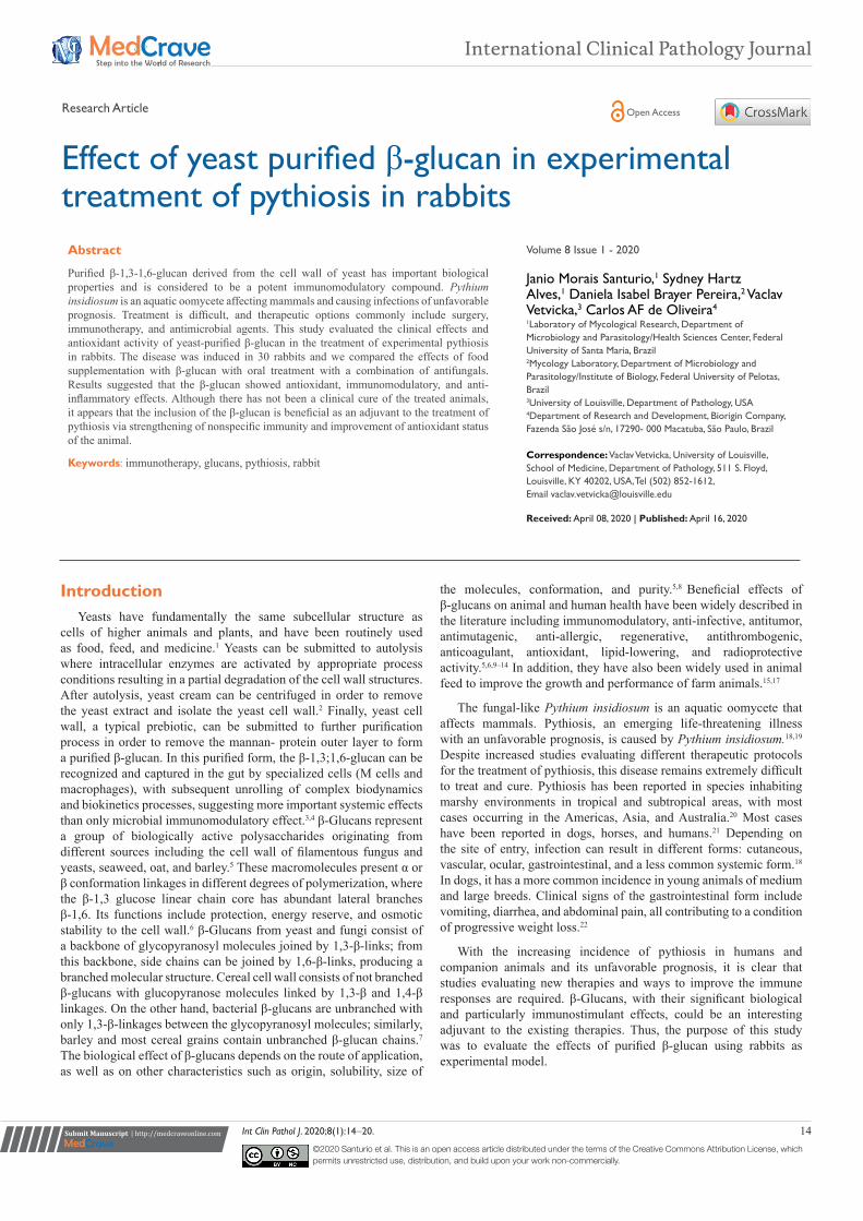
In Group 1, 45 days PI without treatment, the weight of the animals ranged from 2265g to 3130g. After 45 days of treatment, the weight of the animals ranged from 2200g to 3500g (Table 1). Statistical analysis revealed that the weight of animals under various treatments differed when evaluated by the Kruskal-Wallis test (P=.0094). However, when Dunn multiple comparison test was used, it was evident that Group 4 (immunotherapy + β-glucan intake) (P< .01) and Group 5 (itraconazole+terbinafine combination) (P<.05) showed significant weight gain compared to Group 1 (no treatment). Initially, the measured area of the lesions (Day 0 PI) ranged from 2.25cm2 to 22.09cm2, with an average of 10.32cm2. The animals of Group 5 (itraconazole+terbinafine) showed significant lesion growth in relation to the other groups. Significant differences in lesion growth were also observed in Group 3 (immunotherapy-treated) and in Group 5 (itraconazole+terbinafine combination) (P = .0040; P < .01) (Figure
1). The histological observation of the lesions by Grocott staining was similar in all groups, with no variation in the number of P. insidiosum fungal hyphae between the individual groups (Figure 2&3).
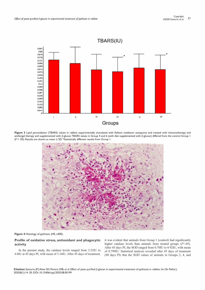
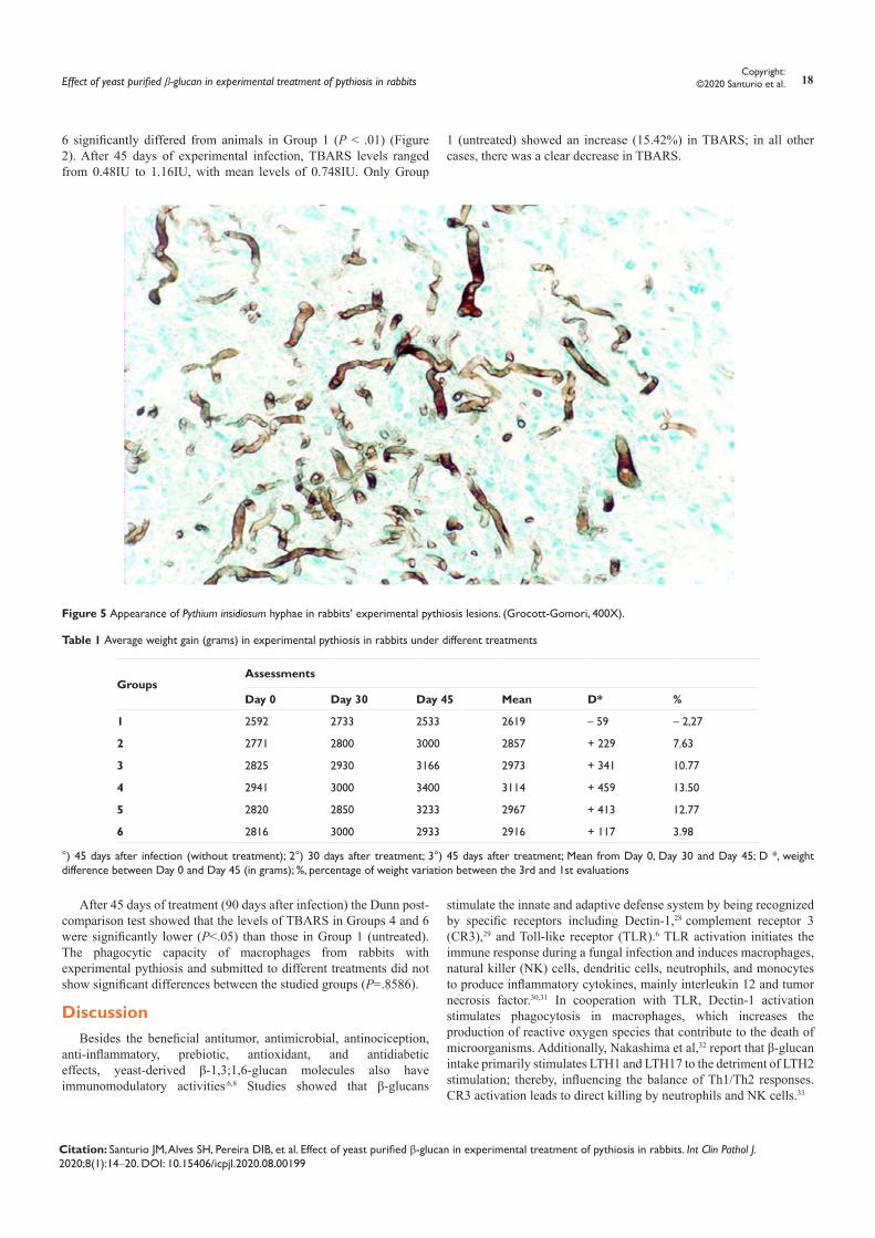
Histopathology in hematoxylin-eosin–stained slides was characterized by the presence of multifocal to coalescent areas of necrosis delimited by inflammatory infiltrates, consisting predominantly of polymorphonucleated cells with predominance of eosinophils (Figure 4). Inside and in the center of the areas of necrosis, we observed the presence of negative tubuliform images referring to P. insidiosum hyphae. These hyphae were surrounded by irregular and eosinophilic areas of necrosis compatible with the Splendori-Hoeppli reaction (Figure 5). Some of these reactions were bound by giant Langerhans and foreign body cells. There was also intense proliferation of fibrous connective tissue with eosinophils, plasmocytes, lymphocytes, macrophages, some giant cells, and epithelioid cells.
Figure 1 Lesion dimensions in rabbits experimentally inoculated with Pythium insidiosum zoospores and treated with immunotherapy and antifungal therapy and supplemented with β- glucan. Injuries of Group 5 animals differed from those of Group 3 animals at P < .01 and from Groups 1, 2, 3, and 6 at P < .05. Results are shown as mean ± SD. *Statistically different results from Group 1.
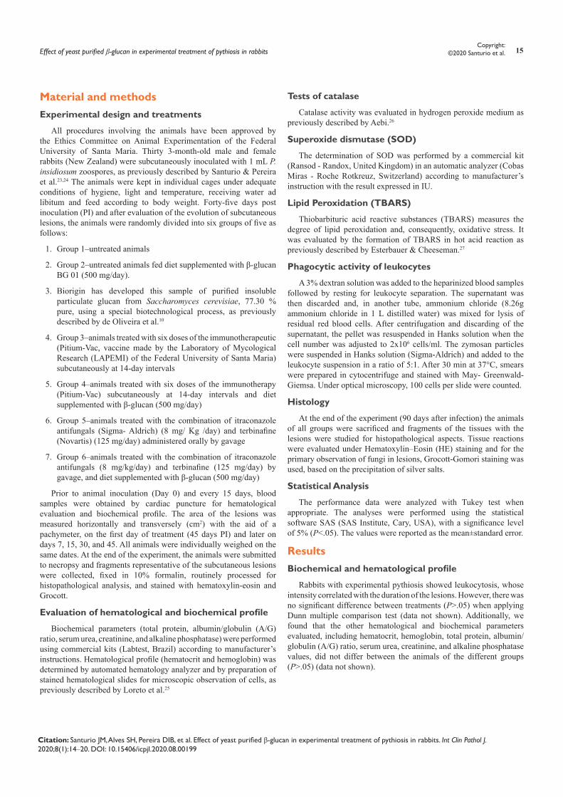
Figure 2 Superoxide dismutase (SOD) values in rabbits experimentally inoculated with Pythium insidiosum zoospores and treated with immunotherapy and antifungal therapy and supplemented with β-glucan. SOD values in the animals of Groups 2, 4, and 6 (diets supplemented with β- glucan) differed significantly (P < .01) from the animals of the control Group 1. Results are shown as mean ± SD. *Statistically different results from Group 1.
Effect of yeast purified β-glucan in experimental treatment of pythiosis in rabbits 17Copyright:
©2020 Santurio et al.
Citation: Santurio JM, Alves SH, Pereira DIB, et al. Effect of yeast purified β-glucan in experimental treatment of pythiosis in rabbits. Int Clin Pathol J. 2020;8(1):14‒20. DOI: 10.15406/icpjl.2020.08.00199
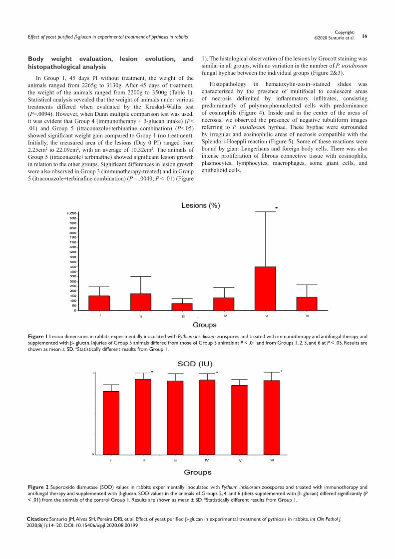
Figure 3 Lipid peroxidation (TBARS) values in rabbits experimentally inoculated with Pythium insidiosum zoospores and treated with immunotherapy and antifungal therapy and supplemented with β-glucan. TBARS values in Group 4 and 6 (with diet supplemented with β-glucan) differed from the control Group 1 (P < .05). Results are shown as mean ± SD. *Statistically different results from Group 1.
Figure 4 Histology of pythiosis. (HE, x400).
Profile of oxidative stress, antioxidant and phagocytic activity
In the present study, the catalase levels ranged from 2.52IU to 4.0IU at 45 days PI, with mean of 3.16IU. After 45 days of treatment,
it was evident that animals from Group 1 (control) had significantly higher catalase levels than animals from treated groups (P<.05). After 45 days PI, the SOD ranged from 0.70IU to 0.92IU, with mean of 0.799IU. Statistical analysis revealed after 45 days of treatment (90 days PI) that the SOD values of animals in Groups 2, 4, and
Effect of yeast purified β-glucan in experimental treatment of pythiosis in rabbits 18Copyright:
©2020 Santurio et al.
Citation: Santurio JM, Alves SH, Pereira DIB, et al. Effect of yeast purified β-glucan in experimental treatment of pythiosis in rabbits. Int Clin Pathol J. 2020;8(1):14‒20. DOI: 10.15406/icpjl.2020.08.00199
6 significantly differed from animals in Group 1 (P < .01) (Figure 2). After 45 days of experimental infection, TBARS levels ranged from 0.48IU to 1.16IU, with mean levels of 0.748IU. Only Group
1 (untreated) showed an increase (15.42%) in TBARS; in all other cases, there was a clear decrease in TBARS.
Figure 5 Appearance of Pythium insidiosum hyphae in rabbits’ experimental pythiosis lesions. (Grocott-Gomori, 400X).
Table 1 Average weight gain (grams) in experimental pythiosis in rabbits under different treatments
Groups Assessments
Day 0 Day 30 Day 45 Mean D* %
1 2592 2733 2533 2619 – 59 – 2,27
2 2771 2800 3000 2857 + 229 7.63
3 2825 2930 3166 2973 + 341 10.77
4 2941 3000 3400 3114 + 459 13.50
5 2820 2850 3233 2967 + 413 12.77
6 2816 3000 2933 2916 + 117 3.98
°) 45 days after infection (without treatment); 2°) 30 days after treatment; 3°) 45 days after treatment; Mean from Day 0, Day 30 and Day 45; D *, weight difference between Day 0 and Day 45 (in grams); %, percentage of weight variation between the 3rd and 1st evaluations
After 45 days of treatment (90 days after infection) the Dunn post-comparison test showed that the levels of TBARS in Groups 4 and 6 were significantly lower (P<.05) than those in Group 1 (untreated). The phagocytic capacity of macrophages from rabbits with experimental pythiosis and submitted to different treatments did not show significant differences between the studied groups (P=.8586).
DiscussionBesides the beneficial antitumor, antimicrobial, antinociception,
anti-inflammatory, prebiotic, antioxidant, and antidiabetic effects, yeast-derived β-1,3;1,6-glucan molecules also have immunomodulatory activities.6,8 Studies showed that β-glucans
stimulate the innate and adaptive defense system by being recognized by specific receptors including Dectin-1,28 complement receptor 3 (CR3),29 and Toll-like receptor (TLR).6 TLR activation initiates the immune response during a fungal infection and induces macrophages, natural killer (NK) cells, dendritic cells, neutrophils, and monocytes to produce inflammatory cytokines, mainly interleukin 12 and tumor necrosis factor.30,31 In cooperation with TLR, Dectin-1 activation stimulates phagocytosis in macrophages, which increases the production of reactive oxygen species that contribute to the death of microorganisms. Additionally, Nakashima et al,32 report that β-glucan intake primarily stimulates LTH1 and LTH17 to the detriment of LTH2 stimulation; thereby, influencing the balance of Th1/Th2 responses. CR3 activation leads to direct killing by neutrophils and NK cells.33
Effect of yeast purified β-glucan in experimental treatment of pythiosis in rabbits 19Copyright:
©2020 Santurio et al.
Citation: Santurio JM, Alves SH, Pereira DIB, et al. Effect of yeast purified β-glucan in experimental treatment of pythiosis in rabbits. Int Clin Pathol J. 2020;8(1):14‒20. DOI: 10.15406/icpjl.2020.08.00199
The animals of Group 5 (itraconazole+terbinafine) showed significant lesion growth compared to the other groups (Figure 1). Possibly due to the immunological effect of itraconazole.34 Thus, treatment strategies should consider the immune status of the host with fungal infections. Considering that Groups 5 and 6 were treated with an antifungal combination and that Group 6 was supplemented with β-glucan, we believe that the lower growth of lesions in this group was probably due to the protective effects of β-glucan to many kinds of chemotherapies, concomitantly with its immunomodulatory properties.
In the present study, we found that glucan supplementation resulted in significant antioxidant activity. These results were demonstrated by higher levels of SOD, only observed in the groups with β-glucan in the diet. Thus, it can be inferred that ingestion of β-glucan had effects on maintaining high levels of the antioxidant reserve, in an oxidative challenge promoted by chronic granulomatous infection caused by P. insidiosum. Additionally, ingestion of β-glucan by animals in Group 4 (immunotherapy-treated) and Group 6 (antifungal combination-treated) induced lower lipid peroxidation (oxidative stress), demonstrating the strong antioxidant action of β- glucan. Previously, Kayali et al,35 reported significant antioxidant effect of β-glucan when administered to rats with spinal cord injury. However, Tang et al.36 stated that the antioxidant activity of glucans may vary depending on the purification methods, and Bacha et al,9 demonstrated different effects of glucans on lipid peroxidation when administered 2% and 4% glucans in rat diets.
Nonsignificant results in phagocytosis assays were not expected since β-glucan, when recognized by CR3, TLRs and Dectin-1, stimulates phagocytosis in macrophages and induces inflammatory cytokine production.31,37 However, it is important to note that our experiments evaluated only the capture capacity of zymosan particle by macrophages in vitro. Other kind of protocols evaluating macrophage stimulation parameters suggested the activity β-glucan on macrophage activation.10 The treatment of monocytes with β-glucan inhibits the spontaneous apoptosis of these cells, as well as being able to stimulate their differentiation into competent macrophages that play an important role in the control of inflammatory processes.38 The evaluation of the hematological and biochemical profile of rabbits with experimental pythiosis showed no alterations, indicating absence of effects of β-glucan on these parameters. This shows that β-glucan intake is safe and can be administered to animals without any contraindications or side effects. On the other hand, the hematological changes observed in animals infected with P. insidiosum were consistent with the cellular changes observed in animals with pythiosis.25 Our study demonstrated that the effect of β-glucan on weight gain in animals treated with immunotherapy (Group 4) was significant when compared to the control group. These results highlighted the beneficial and immunoadjuvant effect of β-glucan on immunotherapy. Similar effects were reported by others15,31,39 using other pathogens. Additionally, when assessing the histological characteristics of the lesions, we found that animals from Group 6 (treated with antifungal combination and supplemented with β-glucan) showed lower lesion growth, particularly when compared to those from Group 5 (treated with combination of itraconazole+terbinafine). We hypothesize that the lower growth of lesions in this group is due to the immunostimulatory and anti-inflammatory effects of the β-glucan, suggesting that the addition of β-glucan to the animals’ diet may help in the treatment of pythiosis.
These findings allow us to infer that the intake of β-glucans associated with treatments for pythiosis significantly reduced the phenomenon of lipid peroxidation (oxidative stress) promoted by experimental infection. The immunoadjuvant effect of β-glucans has been reported by different authors. However, to our knowledge, we are the first to report the beneficial effect in conjunction with the associated antifungal therapy. The ability of β-glucans to reduce the phenomenon of lipid peroxidation is in agreement with findings relating β-glucans to SOD.
Although no clinical cure was observed in the treated groups, it is noteworthy that this result is similar to previous studies that evaluated different therapies in experimental pythiosis in rabbits, with only few successfully cured animals, denoting the difficulty of treating P. insidiosum infection.23–25,40–44 This may be because therapies begin only 45 days after a strong subcutaneous challenge with P. insidiosum zoospores. Thus, the absence of responses to the clinical and histopathological parameters could be interpreted as due to the ineffectiveness of primary therapies in this specific protocol and not because of absent β-glucan activity.
ConclusionThe inclusion of purified β-1,3;1,6-glucan derived from the cell
wall of yeast as an adjuvant in immunotherapy-based and antifungal-based therapies for the treatment of experimental pythiosis has been found to be beneficial in strengthening nonspecific immunity and antioxidant reserves. Based on the importance of the possibility of extrapolating these findings for humans and dogs, further studies are required to evaluate the eventual prophylactic effect of glucans as well as protocols with rabbits naturally infected with P. insidiosum.
AcknowledgmentsThe authors would like to thank Biorigin for their β-glucan
donation and financial support. The funders had no role in the study design, data collection and analysis, or decision to publish.
Conflicts of interestAuthors declare that there are no conflicts of interest.
References1. Jach ME, Serefko A, Sajnaga E, et al. Dietary supplements based on the
yeast biomass. Curr Top Nutraceutical Res. 2015;13:83–88
2. Podpora B, Świderski F, Sadowska A, et al. Spent brewer’s yeast extracts as a new component of functional food. Czech J Food Sci. 2016;34:554–563.
3. Chan GC, Chan WK, Sze DM. The effects of beta-glucan on human immune and cancer cells. J Hematol Oncol. 2009;2:25.
4. Vetvicka V. β-Glucans as Natural Biological Response Modifiers. New York: Nova Science Publishers, Inc. 2013.
5. Jesenak M, Banovcin P, Rennerova Z, et al. β-Glucans in the treatment and prevention of allergic diseases. Allergol Immunopathol (Madr). 2014;42(2):149–156.
6. Dalonso N, Goldman GH, Gern RM. β-(1-->3),(1-->6)-Glucans: medicinal activities, characterization, biosynthesis and new horizons. Appl Microbiol Biotechnol. 2015;99(19):7893–906.
7. Laroche C, Michaud P. New developments and prospective applications for beta (1,3) glucans. Recent Pat Biotechnol. 2007;1(1):59–73.
Effect of yeast purified β-glucan in experimental treatment of pythiosis in rabbits 20Copyright:
©2020 Santurio et al.
Citation: Santurio JM, Alves SH, Pereira DIB, et al. Effect of yeast purified β-glucan in experimental treatment of pythiosis in rabbits. Int Clin Pathol J. 2020;8(1):14‒20. DOI: 10.15406/icpjl.2020.08.00199
8. Novak M, Vetvicka V. Glucans as biological response modifiers. Endocr Metab Immune Disord Drug Targets. 2009;9(1):67–75.
9. Bacha U, Nasir M, Iqbal S, et al. Nutraceutical, anti-inflammatory, and immune modulatory effects of beta-glucan isolated from yeast. Biomed Res Int. 2017;8972678.
10. de Oliveira CAF, Vetvicka V, Zanuzzo FS. β-Glucan successfully stimulated the immune system in different jawed vertebrate species. Comp Immunol Microbiol Infect Dis. 2019;62:1–6.
11. Delatte SJ, Evans J, Hebra A, et al. Effectiveness of beta-glucan collagen for treatment of partial-thickness burns in children. J Pediatr Surg. 2001;36(1):113–118.
12. Krizkova L, Durackova Z, Sandula J, et al. Fungal beta-(1-3)-D-glucan derivatives exhibit high antioxidative and antimutagenic activity in vitro. Anticancer Res. 2003;23(3B):2751–2756
13. Richter J, Svozil V, Kral V, et al. β-Glucan affects mucosal immunity in children with chronic respiratory problems under physical stress: clinical trials. Ann Transl Med. 2015;3(4):52.
14. Toklu HZ, Sener G, Jahovic N, et al. Beta-glucan protects against burn-induced oxidative organ damage in rats. Int Immunopharmacol. 2006;6(2):156–169.
15. Horst G, Levine R, Chick R, et al. Effects of beta-1,3-glucan (AletaTM) on vaccination response in broiler chickens. Poult Sci. 2019;98(4):1643–1647.
16. Petit J, Wiegertjes GF. Long-lived effects of administering beta-glucans: Indications for trained immunity in fish. Dev Comp Immunol. 2016;64:93–102.
17. Wu C, Xu Q, Wang R, et al. Effects of dietary beta-glucan supplementation on growth performance and immunological and metabolic parameters of weaned pigs administered with Escherichia coli lipopolysaccharide. Food Funct. 2018;9(6):3338–3343.
18. Gaastra W, Lipman LJ, De Cock AW, et al. Pythium insidiosum: an overview. Vet Microbiol. 2010;146(1–2):1–16.
19. Santurio JM, Alves SH, Pereira DB, et al. Pythiosis: an emergent mycosis. Acta Sci Vet. 2006;34:1–14.
20. Mendoza L, Vilela R. The mammalian pathogenic oomycetes. Curr Fungal Infect Rep.2013;7:198–208.
21. Permpalung N, Worasilchai N, Chindamporn A. Human pythiosis: emergence of fungal-like organism. Mycopathologia. 2019.
22. Hunning PS, Rigon G, Faraco CS, et al. Intestinal obstruction by Pythium insidiosum in a dog: case report]. Arquivo Brasileiro de Medicina Veterinária e Zootecnia. 2010;62:801–805.
23. Santurio JM, Leal AT, Leal AB, et al. Three types of immunotherapics against pythiosis insidiosi developed and evaluated. Vaccine. 2003;21(19–20):2535–2540.
24. Pereira DI, Santurio JM, Alves SH, et al. Caspofungin in vitro and in vivo activity against Brazilian Pythium insidiosum strains isolated from animals. J Antimicrob Chemother. 2007;60(5):1168–1171.
25. Loreto ES, Alves SH, Santurio JM, et al. Diphenyl diselenide in vitro and in vivo activity against the oomycete Pythium insidiosum. Vet Microbiol. 2012;156(1–2):222–226.
26. Aebi H. Catalase in vitro. Methods Enzymol. 1984;105:121–126.
27. Esterbauer H, Cheeseman KH. Determination of aldehydic lipid peroxidation products: malonaldehyde and 4-hydroxynonenal. Methods Enzymol. 1990;186:407–421.
28. Brown GD, Gordon S. Immune recognition. A new receptor for beta-glucans. Nature. 2001;413(685):36–37.
29. Ross GD, Vetvicka V, Yan J, et al. Therapeutic intervention with complement and beta-glucan in cancer. Immunopharmacology. 1999;42(1–3):61–74.
30. Gantner BN, Simmons RM, Canavera SJ, et al. Collaborative induction of inflammatory responses by dectin-1 and Toll-like receptor 2. J Exp Med. 2003;197(9):1107–1117.
31. Jin Y, Li P, Wang F. β-Glucans as potential immunoadjuvants: A review on the adjuvanticity, structure-activity relationship and receptor recognition properties. Vaccine. 2018;36(35):5235–5244.
32. Nakashima A, Yamada K, Iwata O, et al. Beta-Glucan in foods and its physiological functions. J Nutr Sci Vitaminol (Tokyo). 2018;64(1):8–17.
33. Ross GD, Vetvicka V. CR3 (CD11b, CD18): A phagocyte and NK cell membrane receptor with multiple ligand specificities and functions. Clin Exp Immunol. 1993;92(2):181–184.
34. Muenster S, Bode C, Diedrich B, et al. Antifungal antibiotics modulate the pro-inflammatory cytokine production and phagocytic activity of human monocytes in an in vitro sepsis model. Life Sci. 2015;141:128–136.
35. Kayali H, Ozdag MF, Kahraman S, et al. The antioxidant effect of beta-glucan on oxidative stress status in experimental spinal cord injury in rats. Neurosurg Rev. 2005;28(4):298–302.
36. Tang Q, Huang G, Zhao F, et al. The antioxidant activities of six (1-->3)-beta-d-glucan derivatives prepared from yeast cell wall. Int J Biol Macromol. 2017;98:216–221.
37. Vetvicka V, Vetvickova J. Natural immunomodulators and their stimulation of immune reaction: true or false? Anticancer Res. 2014;34(5):2275–2282.
38. Leonhardt J, Grosse S, Marx C, et al. Candida albicans beta-glucan differentiates human monocytes into a specific subset of macrophages. Front Immunol. 2018;9:2818.
39. Williams DL, Yaeger RG, Pretus HA, et al. Immunization against Trypanosoma cruzi: adjuvant effect of glucan. Int J Immunopharmacol. 1989;11(4):403–410
40. Argenta JS, Alves SH, Silveira F, et al. In vitro and in vivo susceptibility of two-drug and three-drug combinations of terbinafine, itraconazole, caspofungin, ibuprofen and fluvastatin against Pythium insidiosum. Vet Microbiol. 2012;157(1–2):137–142.
41. Fonseca AO, Pereira DI, Botton SA, et al. Treatment of experimental pythiosis with essential oils of Origanum vulgare and Mentha piperita singly, in association and in combination with immunotherapy. Vet Microbiol. 2015;178(3–4):265–269.
42. Jesus FP, Loreto ES, Ferreiro L, et al. In vitro and in vivo antimicrobial activities of minocycline in combination with azithromycin, clarithromycin, or tigecycline against Pythium insidiosum. Antimicrob Agents Chemother. 2016;60(1):87–91.
43. Loreto ES, Tondolo JSM, Oliveira DC, et al. In vitro activities of miltefosine and antibacterial agents from the macrolide, oxazolidinone, and pleuromutilin classes against Pythium insidiosum and Pythium aphanidermatum. Antimicrob Agents Chemother. 2018;62(3):e01678–1717.
44. Zanette RA, Jesus FP, Pilotto MB, et al. Micafungin alone and in combination therapy with deferasirox against Pythium insidiosum. J Mycol Med. 2015;25(1):91–94.
Top Related