


γλώσσες
Σελίδες
Νομικός

NEW DERIVATIVES OF TROVACENE (η7-C7H7)V(η5-C5H5): STUDIES OF REDOX
SPLITTING, EXCHANGE COUPLING AND HYDROGEN BONDING
INAUGURAL-DISSERTATION
zur
Erlangung des Grades eines Doktors
der Naturwissenschaften
(Dr. rer. nat.)
vorgelegt
dem Fachbereich Chemie
der Philipps-Universität Marburg/Lahn
von
Francesca Paganelli
aus
Rimini
Marburg/Lahn 2003

Dedicated to my father
Danksagung
Herrn Prof. Dr. Ch. Elschenbroich danke ich für die Überlassung des Themas und für seine
Unterstützung. Mein ganz besonderer Dank gilt Prof. D. Braga und Prof. F. Grepioni für die
Kooperation mit ihrer Arbeitsgruppe.
Herrn Dr. Olaf Burghaus danke ich für die Simulationen der EPR-Spektren und
immerwährende Hilfsbereitschaft bei EPR-spektroskopischen Problemen.
Herrn C. Pietzonka danke ich für die Durchführung und Auswertung der magnetischen
Messungen, und Herrn Prof. Dr. B. Neumüller für die schnelle Durchführung der
Kristallstrukturanalyse.
Bei Herrn Dipl.-Chem. S. Horst und Herrn Dipl.-Chem. B. Schäfer möchte ich mich für die
Polymeranalytik bedanken.
Den ehemaligen Mitgliedern des Arbeitskreises danke ich für das hervorragende
Arbeitsklima. Mein besonderer Dank gilt Herrn Dr. M. Nowotny für zahlreiche fachliche
Diskussionen sowie Frau T. Schmidt für die von ihr durchgeführten Synthesen und die
cyclovoltammetrischen Messungen.
Weiterhin danke ich dem Personal der zentralen Serviceeinrichtungen für die erbrachten
Leistungen, vor allem den Herren K. Stutz (glastechnische Werkstatt), Prof. Dr. W. Massa
(Röntgenstukturanalyse), Dr. K. Steinbach (Massenspektroskopie), Dr. J. Knecht (CHN-
Analytik).
Sehr herzlich bedanken möchte ich mich bei meinen Mitbewohnern Roman und Sarah sowie
bei meinen AGP-Kollegen Thomas und bei meinen Auslandskorrespondenten Marina, Paolo,
Daniele und Domenico für ihre Freundschaft und ihre moralische Unterstützung.
I
Table of Contents
Numbered Compounds IV
Abbreviations VI
1. Introduction 1
1.1 Crystal Engineering 1
1.2 Research Objectives 3
1.3 Electron Transfer and Exchange Interactions through Hydrogen Bond 4
2. Metallation of (η7-Cycloheptatrienyl)(η5-cyclopentadienyl)vanadium and
(η7-Cycloheptatrienyl)(η5-pentamethylcyclopentadienyl)vanadium 7
2.1 ENDOR Studies of Trovacene Metallation 13
2.1.1 Fundamentals of ENDOR Spectroscopy 13
2.1.2 Results 15
3. Determination of the Structure of (η7-Cycloheptatrienyl)(η5-
cyclopentadienyl)vanadium, 1• , and (η7-cycloheptatrienyl)(η5-
pentamethylcyclopentadienyl)vanadium, 2• 19
3.1 Structure of (η7-Cycloheptatrienyl)(η5-cyclopentadienyl)vanadium, 1• 19
3.2 Structure of (η7-Cycloheptatrienyl)(η5-pentamethylcyclopentadienyl)
vanadium, 2• 21
4. Aldehydes and Carboxylic Acids (η7-cycloheptatrienyl)(η5-
cyclopentadienyl)vanadium, 1• , and (η7-cycloheptatrienyl)(η5-
pentamethylcyclopentadienyl)vanadium, 2• 25
4.1 Synthesis of (Carboxy-η7-cycloheptatrienyl)(carboxy-η5-
cyclopentadienyl)vanadium, 13• , and (Formyl-η7-cycloheptatrienyl)
(formyl-η5-cyclopentadienyl)vanadium, 14• 25
4.2 Synthesis of (Carboxy-η7-cycloheptatrienyl)(η5-
pentamethylcyclopentadienyl)vanadium, 15• , and (Formyl-η7-cycloheptatrienyl)
(η5-pentamethylcyclopentadienyl)vanadium, 16• 26
4.3 Structure of (Carboxy-η7-cycloheptatrienyl)(carboxy-η5-
cyclopentadienyl)vanadium, 13• 27
4.4 Structure of (Formyl-η7-cycloheptatrienyl)
(η5-pentamethylcyclopentadienyl)vanadium,16• 30
II
4.5 Cyclic Voltammetry of 13• , 14•, 15• , 16• 33
4.5.1 Fundamentals of Cyclic Voltammetry 33
4.5.2 Results 34
4.6 Electron Paramagnetic Resonance of 13• , 14• , 15•, 16• 42
4.6.1 Fundamentals of Electron Paramagnetic Resonance 42
4.6.2 Results 44
4.7 ENDOR of 2• , 14•, 16•, 17• 51
5. A Binuclear Complex of (η7-Cycloheptatrienyl)
(η5-pentamethylcyclopentadienyl)vanadium 54
5.1 Synthesis of E-1,2-Bis[(η7-cycloheptatrienyl)(pentamethyl-η5-
cyclopentadienyl)vanadium]ethene, 18•• 55
5.2 Cyclic Voltammetry of 18•• 56
5.2.1 Fundamentals of Cyclic Voltammetry of Binuclear Species 56
5.2.2 Results 57
5.3 Electron Paramagnetic Resonance of 18•• 59
5.3.1 Fundamentals of Electron Paramagnetic Resonance of Biradicals 59
5.3.2 Results 62
5.4 Magnetic Measurement of 18•• 65
5.4.1 Fundamentals of Magnetic Susceptibility 65
5.4.2 Results 67
6. Sila-trovacenophanes 70
6.1 Synthesis of [1,8-(Diphenylsilanediyl)(η7-cycloheptatrienyl)(η5-
cyclopentadienyl)vanadium], 24• , and [1,8-(Tetramethyldisilane-1,2-diyl)(η7-
cycloheptatrienyl)(η5-cyclopentadienyl)vanadium], 25• 71
6.2 Structure of [1,8-(Diphenylsilandiyl)(η7-cycloheptatrienyl)(η5-
cyclopentadienyl)vanadium] 24• 72
6.3 Structure of [1,8-(Tetramethyldisilan-1,2-diyl)(η7-cycloheptatrienyl-η5-
cyclopentadienyl)vanadium], 25• 74
6.4 Comparison of the two Structures 77
6.5 Cyclic Voltammetry of 24• and 25• 79
6.6 EPR of 24• and 25• 84
6.7 Poly(trovacenylsilane) 86
6.7.1 Polymerisation Attempts and Analysis of the Products 88
III
7. Benzoic Acid and Benzoate π-Complexes of Chromium 92
7.1 Synthesis of 1,1’-Bis(carboxy-η6-benzene)chromium, 4 93
7.2 Structure of 1,1’-Bis(carboxy-η6-benzene)chromium, 4 94
7.3 Synthesis of [1,1’-Bis(carboxy-η6-benzene)chromium][hexafluorophosphate],
[4(H)2]+•[PF6]
−, (α and β forms)
and [1,1’-Bis(carboxyl-η6-benzene)chromium]
[ammoniumhexafluorophosphate], [4(H)•]•[NH4PF6] 96
7.4 Polymorphic Modification of the Salt 31+• 97
7.4.1 Fundamentals of Polymorphism 97
7.4.2 Structures of [4(H)2]+
•[PF6]− α and β Form 98
7.5 Structure of the Zwitterion as Co-crystal with [NH4][PF6], [4(H)•]•[NH4PF6] 102
7.6 Hydrogen Bonding Interaction in the three Species 105
8. Summary 107
9. Zusammenfassung 111
10. Experimental Section 115
10.1 Materials 115
10.2 Instrumental Analysis 116
10.3 Preparations 118
11. Literature 125
Appendix A 132
Appendix B 138
Appendix C 148
IV
Numbered Compounds
1• (η7-Cycloheptatrienyl)(η5-cyclopentadienyl)vanadium, [trovacene]
2• (η7-cycloheptatrienyl)(η5-pentamethylcyclopentadienyl)vanadium
3 Bis(benzene)chromium
4 1,1’-Bis(carboxyl-η6-benzene)chromium
[4(H)2]+•[PF6]
− [1,1’-Bis(carboxy-η6-benzene)chromium][hexafluorophosphate],
(α and β forms)
[4(H)•]•[NH4PF6] [1,1’-Bis(carboxyl-η6-benzene)chromium]
[ammoniumhexafluorophosphate]
4•− [1,1’-Bis(carboxyl-η6-benzene)chromium]
5 [1,1’-Bis(carboxyl-η6-benzene)chromium][squarate]
6• (η7-Cycloheptatrienyl)(carboxy-η5-cyclopentadienyl)vanadium
7 Ferrocene
8 (η7-Cycloheptatrienyl)(η5-cyclopentadienyl)titanium
9 (η7-Cycloheptatrienyl)(η5-cyclopentadienyl)chromium
10 Tetracarbonyl(η5-cyclopentadienyl)vanadium
11• (Deuterio-η5-cyclopentadienyl)(η7-cycloheptatrienyl)vanadium
12• (Deuterio-η5-cyclopentadienyl)(deuterio-η7-cycloheptatrienyl)vanadium
13• (Carboxy-η7-cycloheptatrienyl)(carboxy-η5-cyclopentadienyl)vanadium
14• (Formyl-η7-cycloheptatrienyl)(formyl-η5-cyclopentadienyl)vanadium
15• (Carboxy-η7-cycloheptatrienyl)(η5-pentamethylcyclopentadienyl)vanadium
16• (Formyl-η7-cycloheptatrienyl)(η5-pentamethylcyclopentadienyl)vanadium
17• (η7-Cycloheptatrienyl)(formyl-η5-cyclopentadienyl)vanadium
18•• E-1,2-bis[(η7-cycloheptatrienyl)(η5-pentamethylcyclopentadienyl)vanadium]ethene
19•• [5-5]-Bitrovacene
20•• [7-7]-Bitrovacene
21•• Bis([5]-trovacenyl)ethene
22•• 1,4-Bis([5]trovacenyl)benzene)
23 1,1’-Ferrocenyldiphenylsilane
24• [1,8-(Diphenylsilanediyl)(η7-cycloheptatrienyl)(η5-cyclopentadienyl)vanadium]
25• [1,8-(Tetramethyldisilane-1,2-diyl)(η7-cycloheptatrienyl)(η5-
cyclopentadienyl)vanadium]
V
26 µ-(1,1’-Ferrocenyldiphenylsilane)
27 µ-(1,1’,6,6’-Tetraphenylsilanechromium)
28 µ-(1,1’-Ferrocenyltetramethyldisilanylene)
29• [5]-Trovacenol
30 1,1-Dimethyl-1-silaferrocenophane
31• [1,8-(Dimethylsilandiyl)(η7-cycloheptatrienyl)(η5-cyclopentadienyl)vanadium]
32 1,1’-Ferrocenedicarboxylic acid
33+ [1,1’-Cobaltoceniumdicarboxylic acid][hexafluorophosphate]
34• 1,1’-Bis(carboxyl-η6-benzene)vanadium
VI
Abbreviations
a, A isotropic, anisotropic hyperfine coupling constant
a, b, c lattice constants
AO atomic orbitals
β Bohr magneton
BBC bis(benzene)chromium
c concentration
BuLi n-butyllithium
Cp cyclopentadienyl
C Curie constant
D zero field splitting
DME 1,2-dimethoxyethane
DMF dimethylformamide
E energy, zero field splitting
E1/2 half wave potential
Eλ switching potential
Epa, Epc anodic, cathodic peak potential
EPR electron paramagnetic resonance
ENDOR electron-nuclear double-resonance
f frequency
g g-value
G Gauss
h Planck constant
Hab electronic matrix element
HOMO highest occupied molecular orbital
I nuclear spin, current
Ipa, Ipc anodic, cathodic peak current
IR infra red
J exchange coupling interaction
K equilibrium constant
kB Boltzmann constant
λ reorganization term
VII
LUMO lowest occupied molecular orbital
M transition metal
µ magnetic moment
mI nuclear spin quantum number
Ms magnetic electronic quantum number
Me methyl
MS mass spectroscopy
MO molecular orbital
NMR nuclear magnetic resonance
ν sweep velocity
PE petroleum ether
Ph phenyl
r distance, radius
ROP ring opening polymerization
S electronic spin
SCE saturated calomel electrode
T temperature
TBAP tetrabutylammoniumperchlorate
THF tetrahydrofurane
TMEDA N,N,N’,N’-tetramethylethylenediamine
Tr cycloheptatrienyl, tropylium (C7H7+)
TVC trovacene, (η7-tropilium)vanadium(η5-cyclopentadienyl)
TVC* (η7-cycloheptatrienyl)(η5-pentamethylcyclopentadienyl)vanadium
Θ Weiss constant
χ susceptibility

1
1. Introduction
The independent discovery of ferrocene1 in 1951 by Pauson and Miller and the rational
synthesis of bis(benzene)chromium2 by Fischer and Hafner in 1955 heralded a new era in
organometallic chemistry, that of “sandwich-complexes”.
The introduction of substituents in the ring periphery leads to molecules with new structural,
electronic and electrochemical properties. Variation of metal and ligands offers a great
number of combinations obtained by different synthetic methods. In this context should be
mentioned:
a) the metallation reaction employing n-butyllithium,3 which gives mono- and 1,1'-
dimetallated products. Subsequent reactions give a variety of mono- and 1,1'-disubstituted
bis(arene)metal derivatives including 1,1'-heteroatom substitution in the ring periphery;
b) the metal-ligand co-condensation, introduced in 1969 by Timms.4 Thanks to this technique,
complexes containing a large variety and number of substituents as well as complexes
containing different types of transition- and post-transition metals became accessible.
Sandwich complexes play a central role in many areas:
-ligands in catalysts for stereoselective synthesis (i.e. 1,1'-bis(diphenylphosphino)ferrocene
derivatives);5
-building blocks of redox active macrocycles in supramolecular chemistry;6
-application in medical science (biosensors, anti tumor agens);7
-components of molecular magnets;8
-utilization in non linear optics.9
1.1 Crystal Engineering10
Originally, crystal engineering was concerned with the design of more efficient
topochemical11 reactions, but now has greatly increased its scope to the design of molecular
crystals for a wide variety of physical and chemical purposes.
Crystal engineering is the planning and the execution of a crystal structure synthesis from the
constituent molecules.12 Molecules and ions, chosen on the basis of their size, shape, and
extramolecular bonding capacity, are the ultimate constituents. The assembly of these, i.e., the
nucleation and growth of a molecular crystal, is one of the most basic processes in solid-state
2
chemistry and also one of the most impressive examples of molecular recognition. Crystal
Engineering proceeds via the essential steps of analysis, synthesis and application.
Molecular crystal engineering is an area where supramolecular chemistry and material
chemistry meet13: both disciplines are concerned with the utilization of non-covalent
interactions to predetermine chemical and physical properties of supramolecular aggregates.
The interest in organometallic crystal engineering stems from the enormous potential arising
from the possibility of combining, inter alia, the electronic and magnetic characteristics of
metal-bound ligands with those of metal atoms. The properties of solid organometallic
materials depend on the electronic nature of the metals as well as on the characteristic of the
ligands. These are in general organic molecules as fragments that, in most cases, retain their
original extramolecular bonding capacity upon metal coordination because the peripheral
atomic groups are not affected.
The crystals are the most accessible systems for a detailed study of geometry and energy of
non-covalent interactions. X-ray crystallography has always played an important role in the
development of organometallic chemistry. Ferrocene14 represents the earliest example of an
organometallic crystal structure determination. The relevance of X-ray crystallography in this
chemistry arose because the complex structural features of organometallic compounds (many
of which are air- and moinsture-sensitive) could not be determined by any other known
methods.15 This is particularly true for paramagnetic complexes where high-resolution NMR
spectroscopy fails as an analytical tool. Despite this, the almost exclusive interest of
organometallic chemists lay in the molecular structure and stereochemistry of organometallic
compounds rather than in their crystal structures and packing characteristics.16 However,
crystals of organometallic complexes are molecular in nature, and so they must be held
together by interactions that are, at least, similar to the interactions responsible for the
assembly of purely organic molecular crystals.17 The interactions utilized in the construction
of the desired superstructure are
a) the coordinative bonds between multidentate ligands and metal centers, in the case of
coordinative networks, and
b) van der Waals and hydrogen bonding between building blocks that possess a defined
structure in solution, in the case of molecular networks.
The hydrogen bond,18 that may most generally considered to be a three-center four-electron
interaction, is the master-key interaction in crystal engineering because it combines
directionality with strength (nevertheless anion-cation19 and van der Waals20 interactions take
part to the process of molecular recognition and crystal cohesion). Length and directionality
3
permit the planning of molecular aggregates formed by molecules bearing donor and acceptor
groups opportunely chosen.21 This idea is now largely used to produce new crystalline
materials22 with suitable characteristics to confer useful properties in non linear optics,
optoelectronics and photonics, magnetism, conductivity, nanoporosity, as well as applications
in catalysis, molecular traps, reservoirs and sieves, solid state reactivity, mechanics etc.
1.2 Research Objectives
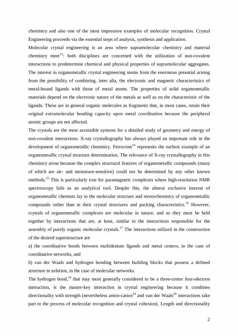
Organometallic complex offers the possibility to combine the large variety of the organic
synthetic methods with the stability of paramagnetic complexes. In particular our research is
focused on the paramagnetic complexes [(η7-C7H7)(η5-C5H5)V] (TVC) [trovacene, (η7-
tropilium)vanadium(η5-cyclopentadienyl)], 1• , and [V(η7-C7H7)(η5-C5Me5)V] (TVC*), 2• , in
their neutral form, and the paramagnetic complex [(η6-C6H6)2Cr] (BBC),
bis(benzene)chromium, 3+• , in its cationic form.
The followed line of research is devoted to preparation and characterization of novel building
blocks and of their precursors for the construction of crystalline materials via cooperative
strong and weak hydrogen bonds.23 The prerequisites of the chosen candidate building blocks
are essentially 3-fold: (a) chemical stability to yield robust materials, (b) specific and
predictable capacity for the formation of a large number of extramolecular interactions, in
particular O−H---O and C−H---O types24, and (c) suitable shape for molding intermolecular
H-bond networks to achieve highly organized superstructures.
Carboxylic acids and aldehydes of 1• , and 2• were chosen as buildings blocks, since, due to
their paramagnetic properties, they permit an investigation of the electron spin-exchange
interactions mediated by O−H---O hydrogen bonds in solution, employing EPR25
spectroscopy, in addition to that in solid state, employing X-ray diffraction.
4
When paramagnetic complexes are linked by a bridging ligand to give a binuclear complex, it
is possible to observe intermetallic interactions between both spin centers, depending on the
nature of the spacers. The study of electron spin exchange coupling aid in understanding
which factors control the rate of electron-transfer, as found in oxidation-reduction, and
electrochemical processes. The bridged complexes of 1• , and 2• provide the opportunity to
study intramolecular electron-transfer between electron-donor and electron-acceptor as a
function of the relative orientation and separation of donor and acceptor. In this context, is of
interest, which factors determine both magnetic exchange and intramolecular electron-
transfer.26
Moreover, varying the oxidation states of the individual metal centers can be used to control
the spin state, hence the magnetic properties of the complex. Magnetic materials, that are
based on organometallic open-shell complexes, have been studied extensively. Hence, in
organometallic systems the neutral or charged nature of the ligands, combined with the
variable oxidation states of metal atoms, allows the study of the same building block in both
neutral and ionic environments.10 Bis(benzoic acid)chromium, [Cr(η6-C6H5COOH)2], 4, is a
good candidate to prepare magnetic materials due to the possibility of combining in a
controlled manner the charge (varied via a redox process) with the hydrogen bonding
capacity.
1.3 Electron Transfer and Exchange Interactions mediated through
Hydrogen Bonding
Electron transfer reactions are known to be transmitted through non-covalent interactions such
as hydrogen bonds, which play an important role in biological electron-transfer systems for
example, non-covalent bonded proteins, model complexes for photosynthesis.27
Long range donor-acceptor electron transfer, mediated by a molecular bridge, represents one
of the fundamental charge transfer processes in chemical and biological systems.
Due to the possible relation between ket and J (exchange interaction coupling), the more facile
experimental access to J could deliver valuable information about electron transfer processes
through hydrogen bonds in biological molecules.
The hydrogen bond is the principal non-covalent interaction in the formation of molecular
crystals, because it combines strength with directionality.28 Etter’s elaboration29 of Linus
Pauling’s definition describes “a hydrogen bond as an interaction that directs the association

5
of a covalently bonded hydrogen atom with one or more others atoms, groups of atoms, or
molecules into an aggregate structure that is sufficiently stable to make it convenient for the
chemist to consider it as independent chemical species”. However, for most purposes,
hydrogen bonding is defined as an interaction between a X-H donor and a Y acceptor, X and
Y being electronegative atoms or electron-rich groups, in the same or in another molecule
(X−H--Y).30 This interaction is generally stronger (often much stronger) than the strongest
intra- and intermolecular van der Waals interactions (X−H--Y). The H-Y and X-Y separations
are shorter than van der Waals contact distances, and X-H-Y angles tend to adopt the
linearity. Hydrogen bridges can be found in the solid state, in solution, and in the gas-phase.31
Charge assistance32 to hydrogen bond is the enhancement of donor and acceptor systems’
polarity by utilizing cationic donors and anionic acceptors instead of neutral systems. The
favourable location of charges increases proton acidity and acceptor basicity.
Spectroscopic characteristics, and detection methods of hydrogen bond are shown in Table 1.
Properties Detection Methods
The distance H--Y is shorter than the sum of
van der Waals radii
X-ray Diffraction
Neutron Diffraction
The distance X−H increases compared to the
free molecules
X-ray Diffraction/Neutron Diffraction
Red-Shift of X---H Stretching in IR
The electron density at the H atom decreases
with the formation of hydrogen bridges Low Field Shift in NMR
Table 1: Properties and detection methods of hydrogen bonds.
The classical O-H···O hydrogen bonds formed by COOH and OH groups are among the
strongest neutral hydrogen bonds. However, the bond can be strengthened if the polarity of
the acceptor is increased via deprotonation. Negatively charged O-H···O− bonds have been
shown to possess dissociation energies in the range 60-120 kJmol-1.33
The utilization of carboxylic acids permits the simultaneous use of neutral O-H···O and
charged O-H···O− bonding interactions. The latter interactions can be grouped in two distinct
categories: the O-H···O− interactions, in which the donors belongs to a neutral molecule and
the acceptor is an anion, and the interaction O-H−···O−, where both donor and acceptor groups
belong to an anion. In all cases the O···O distances are considerably shorter than the sum of
the van der Waals radii and there is a marked preference for linearity. O-H···O− and O-H−···O−
6
interactions, although possessing the same geometrical properties as neutral O-H···O bonds,
are generally associated with O···O distances shorter than in the case of neutral systems
(roughly 2.45 against 2.65 Å).34
A recent example of engineering of a supramolecular arrangement with target magnetic
properties is provided by crystalline [1,1'-bis(carboxyl-η6-benzene)chromium][squarate],
[(η6-C6H6)2CrI]+[HC4O4]−, 5, obtained by reacting squaric acid (3,4-dihydroxy-3-cyclobutene-
1,2-dione) with [(η6-C6H6)2CrI]+, 3+• .35 The anion [HC4O4]− self-assembles into chains linked
by (−)O-H···O(−) interactions and simultaneously intercalates between the benzene ligands
forming π-stacking interactions. The presence of a charge transfer transition was detected by
diffuse reflectance UV-spectroscopy.
An EPR study of exchange interactions mediated by O-H---O bonds for hydrogen bonded
dimmers of the paramagnetic complex (η7-cycloheptatrienyl)(carboxy-η5-
cyclopentadienyl)vanadium, [(η7-C7H7)(η5-C5H4COOH)V0], 6• , has been reported
previously.25
7
2. Metallation of (η7-Cycloheptatrienyl)(η5-cyclopentadienyl)vanadium, 1• and
(η7-Cycloheptatrienyl)(η5-pentamethylcyclopentadienyl)vanadium, 2•
The reaction between an organic (organometallic) compound containing a relatively acidic
hydrogen atom (R-H) and an organolithium reagent (R’-Li) is of considerably synthetic utility
since the resulting new organolithium reagent (R’-Li) can be used in a wide variety of
subsequent reactions:36
R-H + R’-Li → R-Li + R’-H
Ring hydrogen atoms of cyclopentadienyl- as well as arene-metal complexes have weakly
acidic character. Reactions with alkyllithium reagens result in ligand metallation under
formation of highly reactive products, which can be converted into a wide variety of ring-
substituted complexes.
The extent of polylithiation, which may occur in the case of the more acidic complexes, can
be controlled to some degree by an appropriate choice of the reaction conditions. For
example, addition of tertiary amines, such as N,N,N’,N’-tetramethylethylendiamine
(TMEDA), and 1,4-diazabicyclo[2.2.2]octane (DABCO), accelerates lithiation by
coordination to the lithium, which in turn promotes dissociation of aggregated structures.37
The stable coordination complexes formed are considerably more reactive in the metallation
reaction than the organolithium reagents alone.
The enhanced metalating ability of the couple n-butyllithium/TMEDA may possibly arise
from the interation of the diamine with vacant 2p orbitals of the lithium ion. Such a process
may polarize the carbon-lithium bond to such an extent that the butyl group becomes a
considerably more powerful nucleophile than n-butyllithium alone. The presence of the
diamine does not affect subsequent reactions of the newly formed organolithium compounds.
The ability of various (η5-cyclopentadienyl)metal compounds to undergo the hydrogen
lithium exchange (metallation) reaction has played an important role in the development of
the chemistry of metallocenes. Metallation of ferrocene, 7, with n-butyllithium in ether
solution, first reported independently by two research group in 1954,38 leads to lithio-
ferrocene and 1,1'-dilithio-ferrocene, that are very readily converted to numerous derivatives.
Studies of the early transition metals have shown that (η7-cycloheptatrienyl)(η5-
cyclopentadienyl)titanium, 8, is readly monometalated by 1 equiv of n-butyllithium in ether
solution at 0°C, and the metallation take place preferentially at the seven membered ring.39
Treatment of this mixed sandwich complex with 2.4 equiv of n-butyllithium/TMEDA affords
8
the dimetalated product in high yield.40 This is in contrast to the behaviour of the
corresponding vanadium and chromium compounds which are preferentially metalated at the
five-membered ring. The mono-metallation of (η7-cycloheptatrienyl)(η5-cyclopenta-
dienyl)vanadium can be achieved by employing 1.5 equiv of n-butyllithium in ether solution
at room temperature overnight41, whereas the metallation of (η7-cycloheptatrienyl)(η5-
cyclopentadienyl)chromium, 9, is more difficult, but again the products isolated are
substituted mainly in the five-membered ring.42
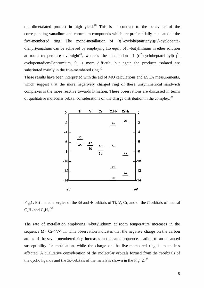
These results have been interpreted with the aid of MO calculations and ESCA measurements,
which suggest that the more negatively charged ring of these unsymmetrical sandwich
complexes is the more reactive towards lithiation. These observations are discussed in terms
of qualitative molecular orbital considerations on the charge distribution in the complex.39
Fig.1: Estimated energies of the 3d and 4s orbitals of Ti, V, Cr, and of the π-orbitals of neutral
C7H7 and C5H5.39
The rate of metallation employing n-butyllithium at room temperature increases in the
sequence M= Cr< V< Ti. This observation indicates that the negative charge on the carbon
atoms of the seven-membered ring increases in the same sequence, leading to an enhanced
susceptibility for metallation, while the charge on the five-membered ring is much less
affected. A qualitative consideration of the molecular orbitals formed from the π-orbitals of
the cyclic ligands and the 3d-orbitals of the metals is shown in the Fig. 2.39
9
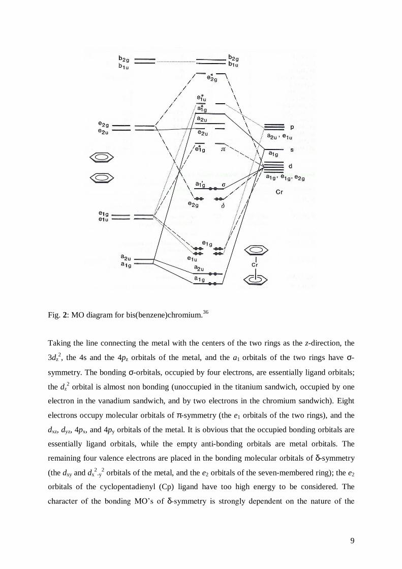
Fig. 2: MO diagram for bis(benzene)chromium.36
Taking the line connecting the metal with the centers of the two rings as the z-direction, the
3dz2, the 4s and the 4pz orbitals of the metal, and the a1 orbitals of the two rings have σ-
symmetry. The bonding σ-orbitals, occupied by four electrons, are essentially ligand orbitals;
the dz2 orbital is almost non bonding (unoccupied in the titanium sandwich, occupied by one
electron in the vanadium sandwich, and by two electrons in the chromium sandwich). Eight
electrons occupy molecular orbitals of π-symmetry (the e1 orbitals of the two rings), and the
dxz, dyz, 4px, and 4py orbitals of the metal. It is obvious that the occupied bonding orbitals are
essentially ligand orbitals, while the empty anti-bonding orbitals are metal orbitals. The
remaining four valence electrons are placed in the bonding molecular orbitals of δ-symmetry
(the dxy and dx2
-y2 orbitals of the metal, and the e2 orbitals of the seven-membered ring); the e2
orbitals of the cyclopentadienyl (Cp) ligand have too high energy to be considered. The
character of the bonding MO’s of δ-symmetry is strongly dependent on the nature of the

10
metal, as the 3d-orbitals of the metals (which increase in the sequence Cr< V< Ti)43, and the
e2 orbitals of the seven-membered ring are close in energy.44
It is argued that the ligand character of these occupied orbitals will increase in the sequence
Cr< V< Ti. This picture predicts that the bonding MO’s of δ-symmetry are mainly metal
orbitals in 9, but mainly C7H7 orbitals in 8. Hence, the Cp ligand is more negative than the
cycloheptatrienyl ligand in the chromium compound, and vice versa in the titanium
compound, whereas in 1• the two rings carry about an equal (negative) charge.45
In this case the charge of the seven-membered ring resides predominantly on the hydrogen
atoms, the carbon atoms being almost neutral, while the charge of the Cp ring is distributed
over the carbon and hydrogen atoms. In any case, it is shown that the negative charge on the
seven-membered ring of (C5H5)M(C7H7) increases in the sequence M= Cr< V< Ti. The
interatomic distances in 939 [Cr-C(C5H5)= 2.18 Å; Cr-C(C7H7)= 2.16 Å] and (C5H5)V(C7H7)
[V-C(C5H5)= 2.210 Å; V-C(C7H7)= 2.208 Å] (see next section for the values) have normal
values; while 846 [Ti-C(C5H5)= 2.32 Å; Ti-C(C7H7)= 2.19 Å] displays abnormally short Ti-
C(C7H7) distances, as a result of the very short distance (1.49 Å) of the metal to the plane of
the seven-membered ring.
This observation demonstrates the importance of δ-bonding in the Ti-(C7H7) moiety; a
decrease of the distance of the metal from the ring plane greatly increases the overlap of the
ligand e2 orbitals with the dx2
-y2 orbitals of the metal.
Mono-substituted complexes of trovacene are known, carrying the functional groups in the
cycloheptatrienyl- or in the Cp-ring. Compounds of the first type, [V0(η5-C5H5)(η7-C7H6R)],
(where R= -CH3, -Ph, -CN, -OCH3…) are obtained by refluxing the appropriate
cycloheptatriene derivative with tetracarbonyl(η5-cyclopentadienyl)vanadium, [V0(η5-
C5H5)(CO)4], 10.47 This method is limited by the accessibility of the cycloheptatrienyl-
derivative (for example, it is very difficult to introduce aldehyde and acid groups to the
cycloheptatrienyl ring).
Because of the only marginally greater negative charge on the Cp, with respect to that on the
cycloheptatrienyl ligand, trovacene reacts slowly with n-buthillithium (1 equivalent of
trovacene and 1.5 equivalent of n-buthillithium) in ether at room temperature (maximum
yield: 50%),39 resulting in metallation of the five-membered ring, as indicated by a colour
change of the solution from violet to dark-red. The mono-lithio derivative can be converted to
compounds of the type [V0(η5-C5H4R)(η7-C7H7)], where R= -CH348, -CHO49, -COOH50, -I51, -
Br52, -PPh253, -B(OH)2
54…
11
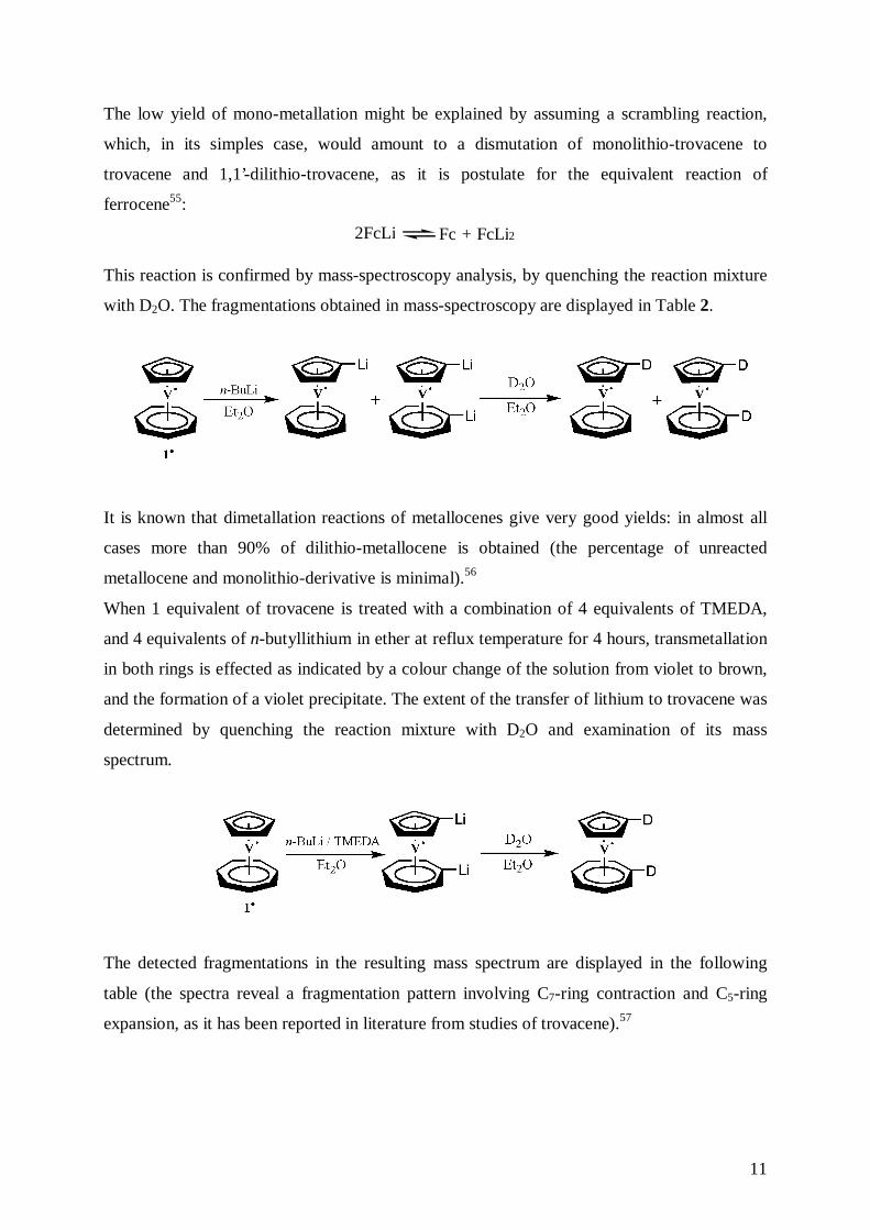
The low yield of mono-metallation might be explained by assuming a scrambling reaction,
which, in its simples case, would amount to a dismutation of monolithio-trovacene to
trovacene and 1,1’-dilithio-trovacene, as it is postulate for the equivalent reaction of
ferrocene55:
2FcLi Fc + FcLi2
This reaction is confirmed by mass-spectroscopy analysis, by quenching the reaction mixture
with D2O. The fragmentations obtained in mass-spectroscopy are displayed in Table 2.
It is known that dimetallation reactions of metallocenes give very good yields: in almost all
cases more than 90% of dilithio-metallocene is obtained (the percentage of unreacted
metallocene and monolithio-derivative is minimal).56
When 1 equivalent of trovacene is treated with a combination of 4 equivalents of TMEDA,
and 4 equivalents of n-butyllithium in ether at reflux temperature for 4 hours, transmetallation
in both rings is effected as indicated by a colour change of the solution from violet to brown,
and the formation of a violet precipitate. The extent of the transfer of lithium to trovacene was
determined by quenching the reaction mixture with D2O and examination of its mass
spectrum.
The detected fragmentations in the resulting mass spectrum are displayed in the following
table (the spectra reveal a fragmentation pattern involving C7-ring contraction and C5-ring
expansion, as it has been reported in literature from studies of trovacene).57
12
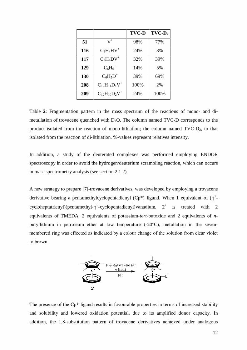
TVC-D TVC-D2
51 V+ 98% 77%
116 C5H4HV+ 24% 3%
117 C5H4DV+ 32% 39%
129 C6H6+ 14% 5%
130 C6H5D+ 39% 69%
208 C12H11D1V+ 100% 2%
209 C12H10D2V+ 24% 100%
Table 2: Fragmentation pattern in the mass spectrum of the reactions of mono- and di-
metallation of trovacene quenched with D2O. The column named TVC-D corresponds to the
product isolated from the reaction of mono-lithiation; the column named TVC-D2, to that
isolated from the reaction of di-lithiation. %-values represent relatives intensity.
In addition, a study of the deuterated complexes was performed employing ENDOR
spectroscopy in order to avoid the hydrogen/deuterium scrambling reaction, which can occurs
in mass spectrometry analysis (see section 2.1.2).
A new strategy to prepare [7]-trovacene derivatives, was developed by employing a trovacene
derivative bearing a pentamethylcyclopentadienyl (Cp*) ligand. When 1 equivalent of (η7-
cycloheptatrienyl)(pentamethyl-η5-cyclopentadienyl)vanadium, 2• is treated with 2
equivalents of TMEDA, 2 equivalents of potassium-tert-butoxide and 2 equivalents of n-
butyllithium in petroleum ether at low temperature (-20°C), metallation in the seven-
membered ring was effected as indicated by a colour change of the solution from clear violet
to brown.
The presence of the Cp* ligand results in favourable properties in terms of increased stability
and solubility and lowered oxidation potential, due to its amplified donor capacity. In
addition, the 1,8-substitution pattern of trovacene derivatives achieved under analogous
13
reaction conditions of metallated intermediates with electrophiles, is of course not possible in
the case of (η7-cycloheptatrienyl)(pentamethyl-η5-cyclopentadienyl)vanadium moiety, which
might be of advantage with regard to a selective 1’-monofunctionalization.
2.1 ENDOR Studies of Trovacene Metallation
2.1.1 Fundamentals of ENDOR Spectroscopy58
ENDOR (Electron-Nuclear Double-Resonance) spectroscopy is a multiple-resonance
technique in which the spin system is simultaneously irradiated by a microwave and a radio
frequency field. This technique was introduced in 195659 to resolve hyperfine and nuclear
quadrupole interactions which were not accessible in the EPR spectra.
One disadvantage of EPR spectroscopy in rigid media is that the EPR lines are additionally
broadened by the g anisotropy and the hyperfine anisotropy of the interacting nuclei. As a
consequence, ligand hyperfine couplings are often not resolved in the EPR display.
In an ordinary EPR experiment the magnetic field is swept through the region of resonance
H= ωe/gβ (where H= magnetic field strength, � � � �π, ωe = microwave frequency, g=
spectroscopic splitting factor, β= Bohr magneton). An ENDOR experiment uses two
frequencies: a microwave frequency to partially saturate electronic Zeeman transitions and
monitor the intensity of the EPR signal, and a strong radio frequency, which is varied in order
to excite nuclear (NMR) Zeeman transition. The induced NMR transitions alter the spin
polarization or the spin alignment, leading to a change of the EPR signal intensity. By plotting
these EPR intensity changes versus the radio frequency, the ENDOR spectrum is obtained.
In Fig. 3 the hyperfine coupling of one unpaired electron with four equivalent nuclei (I= ½) in
a high field is show.
The four nuclear spins can be combined and classified according to their total spins values I=
2, 1, 0 with degeneracies D(I=2) = 1, D(I=1) = 4, D(I=0) = 6. Owing to the transition
frequency degeneracies, five EPR transitions are obtained with binomial intensity distribution
(1:4:6:4:1). The ENDOR spectrum, on the other hand, exhibits only two signals, since all
NMR transitions in the same Ms state are degenerate. To generalize, each group of equivalent
nuclei contributes only two ENDOR lines to the spectrum. Addition of non-equivalent nuclei
to the system causes a multiplicative increases of the number of signals in the EPR spectrum,

14
but only an additive increase in the ENDOR spectrum. The number of lines in an ENDOR
spectrum is considerably less, so the effective resolution is much greater.
Fig. 3: Energy-level diagram of a hyperfine-coupled spin system in a high magnetic field,
consisting of one unpaired electron (S= ½) and four equivalent nuclei (I= ½).58
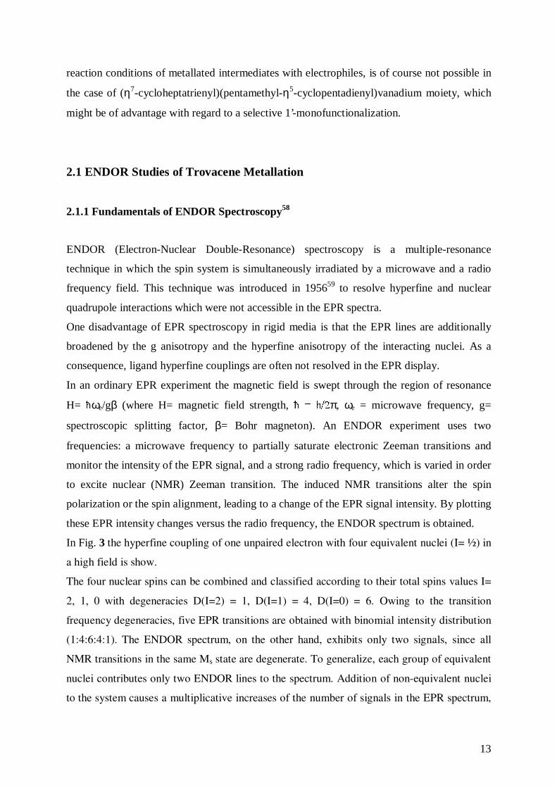
The narrow signal usually achieved in fluid solutions or in single crystals are no longer
obtained in ENDOR experiments of disordered solid, i.e., glasses or powders. Due to
absorption from a range of orientations of the radicals with respect to the direction of the
magnetic field, the ENDOR lines broaden and may become very weak. In this situation the
ENDOR absorptions are spread out over a range of anisotropic hyperfine coupling values
with some buildup of intensity at three values corresponding to those radicals that have their
principal axes along the magnetic field (see Fig. 4). Fairly narrow lines are observed only if
the hyperfine anisotropy pertaining to the nucleus is small.
15
Fig. 4: ENDOR lines shapes (absorption: top, first derivate: bottom) resulting from axially
symmetric hyperfine tensor.58
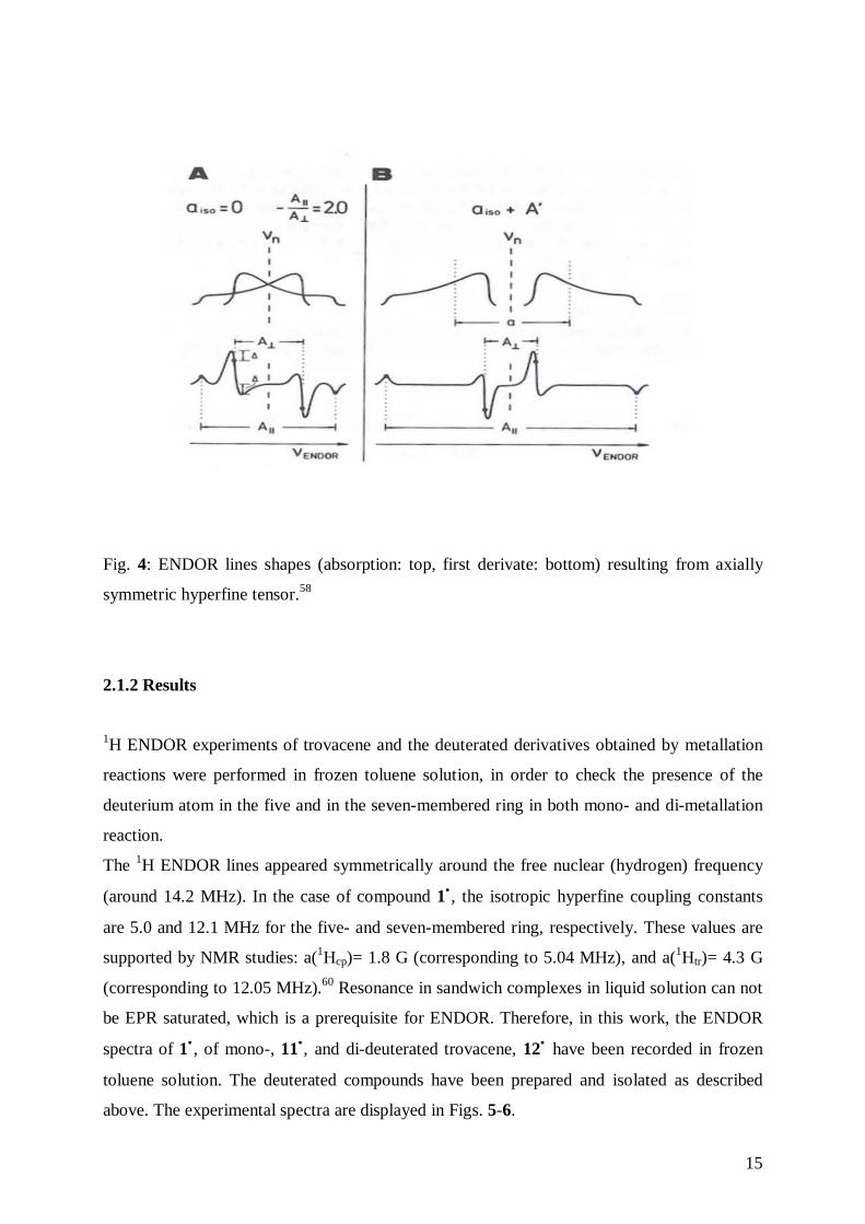
2.1.2 Results
1H ENDOR experiments of trovacene and the deuterated derivatives obtained by metallation
reactions were performed in frozen toluene solution, in order to check the presence of the
deuterium atom in the five and in the seven-membered ring in both mono- and di-metallation
reaction.
The 1H ENDOR lines appeared symmetrically around the free nuclear (hydrogen) frequency
(around 14.2 MHz). In the case of compound 1• , the isotropic hyperfine coupling constants
are 5.0 and 12.1 MHz for the five- and seven-membered ring, respectively. These values are
supported by NMR studies: a(1Hcp)= 1.8 G (corresponding to 5.04 MHz), and a(1Htr)= 4.3 G
(corresponding to 12.05 MHz).60 Resonance in sandwich complexes in liquid solution can not
be EPR saturated, which is a prerequisite for ENDOR. Therefore, in this work, the ENDOR
spectra of 1• , of mono-, 11•, and di-deuterated trovacene, 12• have been recorded in frozen
toluene solution. The deuterated compounds have been prepared and isolated as described
above. The experimental spectra are displayed in Figs. 5-6.
16
0 5 10 15 20 25 30
Frequency [MHz]
TVC
νΗ
a(1H)Cp
a(1H)Tr
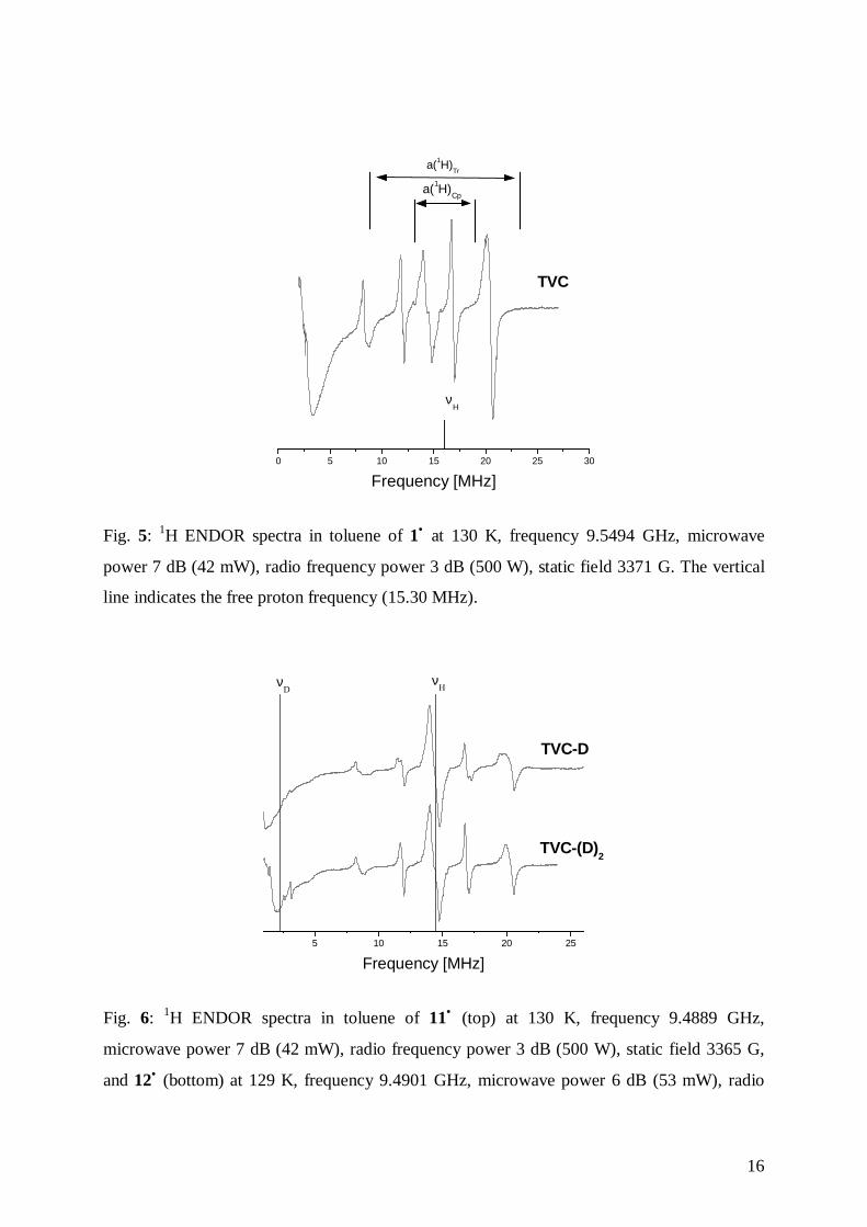
Fig. 5: 1H ENDOR spectra in toluene of 1• at 130 K, frequency 9.5494 GHz, microwave
power 7 dB (42 mW), radio frequency power 3 dB (500 W), static field 3371 G. The vertical
line indicates the free proton frequency (15.30 MHz).
5 10 15 20 25
νD
TVC-(D)2
TVC-D
Frequency [MHz]
νH
Fig. 6: 1H ENDOR spectra in toluene of 11• (top) at 130 K, frequency 9.4889 GHz,
microwave power 7 dB (42 mW), radio frequency power 3 dB (500 W), static field 3365 G,
and 12• (bottom) at 129 K, frequency 9.4901 GHz, microwave power 6 dB (53 mW), radio
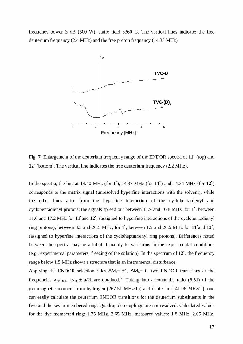
17
frequency power 3 dB (500 W), static field 3360 G. The vertical lines indicate: the free
deuterium frequency (2.4 MHz) and the free proton frequency (14.33 MHz).
1 2 3 4 5
TVC-(D)2
TVC-D
Frequency [MHz]
νD
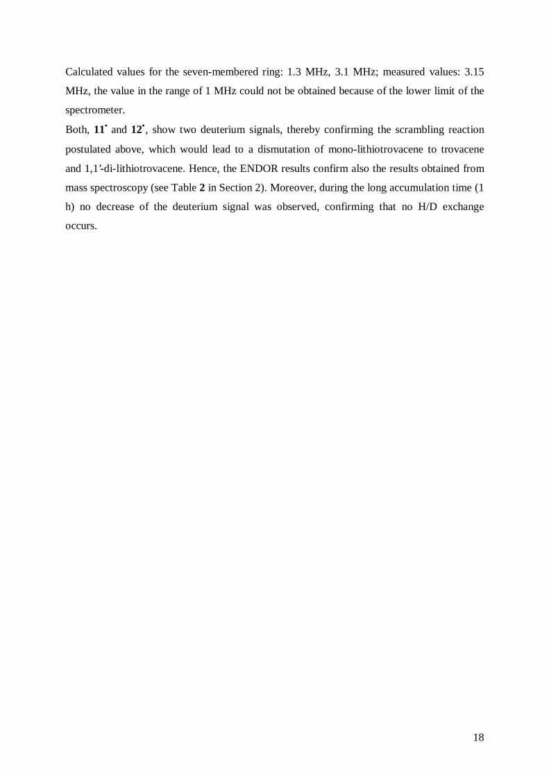
Fig. 7: Enlargement of the deuterium frequency range of the ENDOR spectra of 11• (top) and
12• (bottom). The vertical line indicates the free deuterium frequency (2.2 MHz).
In the spectra, the line at 14.40 MHz (for 1•), 14.37 MHz (for 11•) and 14.34 MHz (for 12•)
corresponds to the matrix signal (unresolved hyperfine interactions with the solvent), while
the other lines arise from the hyperfine interaction of the cycloheptatrienyl and
cyclopentadienyl protons: the signals spread out between 11.9 and 16.8 MHz, for 1• , between
11.6 and 17.2 MHz for 11•and 12• , (assigned to hyperfine interactions of the cyclopentadienyl
ring protons); between 8.3 and 20.5 MHz, for 1•, between 1.9 and 20.5 MHz for 11•and 12• ,
(assigned to hyperfine interactions of the cycloheptatrienyl ring protons). Differences noted
between the spectra may be attributed mainly to variations in the experimental conditions
(e.g., experimental parameters, freezing of the solution). In the spectrum of 12• , the frequency
range below 1.5 MHz shows a structure that is an instrumental disturbance.
Applying the ENDOR selection rules ∆MI= ±1, ∆MS= 0, two ENDOR transitions at the
frequencies νENDOR=ν D ± a/2 are obtained.58 Taking into account the ratio (6.51) of the
gyromagnetic moment from hydrogen (267.51 MHz/T)) and deuterium (41.06 MHz/T), one
can easily calculate the deuterium ENDOR transitions for the deuterium substituents in the
five and the seven-membered ring. Quadrupole couplings are not resolved. Calculated values
for the five-membered ring: 1.75 MHz, 2.65 MHz; measured values: 1.8 MHz, 2.65 MHz.
18
Calculated values for the seven-membered ring: 1.3 MHz, 3.1 MHz; measured values: 3.15
MHz, the value in the range of 1 MHz could not be obtained because of the lower limit of the
spectrometer.
Both, 11• and 12•, show two deuterium signals, thereby confirming the scrambling reaction
postulated above, which would lead to a dismutation of mono-lithiotrovacene to trovacene
and 1,1’-di-lithiotrovacene. Hence, the ENDOR results confirm also the results obtained from
mass spectroscopy (see Table 2 in Section 2). Moreover, during the long accumulation time (1
h) no decrease of the deuterium signal was observed, confirming that no H/D exchange
occurs.
19
3. Determination of the Structure of (η7-Cycloheptatrienyl)(η5-cyclopentadienyl)-
vanadium, 1• and (η7-Cycloheptatrienyl)(η5-pentamethylcyclopentadienyl)-
vanadium, 2•
3.1 Structure of (η7-Cycloheptatrienyl)(η5-cyclopentadienyl)vanadium, 1•
The first low precision determination of the structure of trovacene, 1• (present discrepancy
factor, R= 9.6%) was published in 1963.61 In the present work, a redetermination of the
unsymmetric sandwich complex was undertaken in order to improve the low accuracy of the
original determination, which did not allow for a precise discussion of structural parameters.
Crystals suitable an X-ray crystallographic determination were grown by sublimation as violet
hexagonal plates.
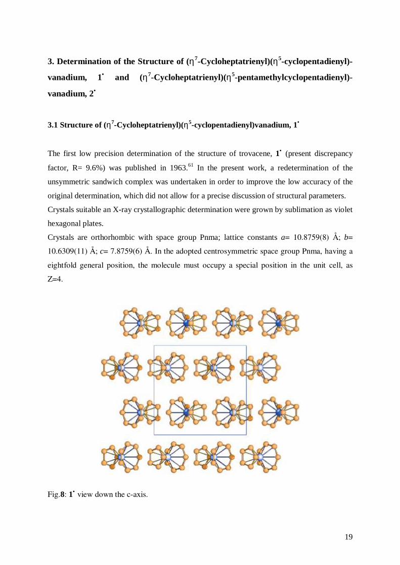
Crystals are orthorhombic with space group Pnma; lattice constants a= 10.8759(8) Å; b=
10.6309(11) Å; c= 7.8759(6) Å. In the adopted centrosymmetric space group Pnma, having a
eightfold general position, the molecule must occupy a special position in the unit cell, as
Z=4.
Fig.8: 1• view down the c-axis.
20
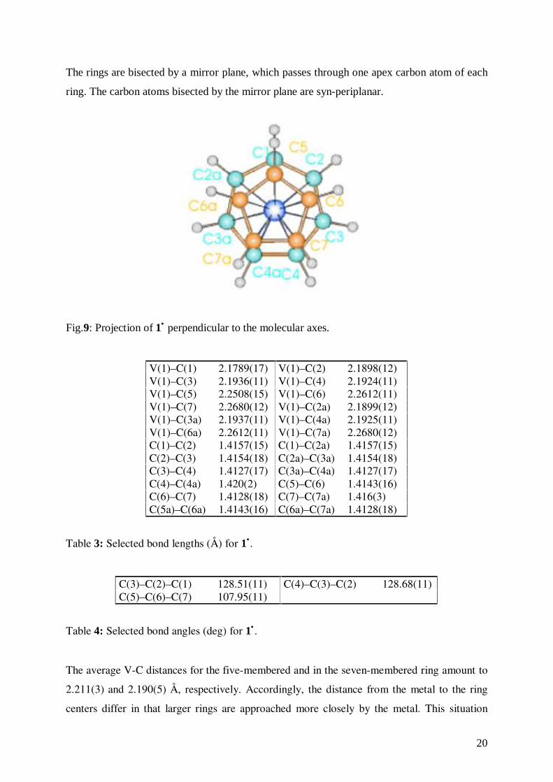
The rings are bisected by a mirror plane, which passes through one apex carbon atom of each
ring. The carbon atoms bisected by the mirror plane are syn-periplanar.
Fig.9: Projection of 1• perpendicular to the molecular axes.
V(1)–C(1) 2.1789(17) V(1)–C(2) 2.1898(12)V(1)–C(3) 2.1936(11) V(1)–C(4) 2.1924(11)V(1)–C(5) 2.2508(15) V(1)–C(6) 2.2612(11)V(1)–C(7) 2.2680(12) V(1)–C(2a) 2.1899(12)V(1)–C(3a) 2.1937(11) V(1)–C(4a) 2.1925(11)V(1)–C(6a) 2.2612(11) V(1)–C(7a) 2.2680(12)C(1)–C(2) 1.4157(15) C(1)–C(2a) 1.4157(15)C(2)–C(3) 1.4154(18) C(2a)–C(3a) 1.4154(18)C(3)–C(4) 1.4127(17) C(3a)–C(4a) 1.4127(17)C(4)–C(4a) 1.420(2) C(5)–C(6) 1.4143(16)C(6)–C(7) 1.4128(18) C(7)–C(7a) 1.416(3)C(5a)–C(6a) 1.4143(16) C(6a)–C(7a) 1.4128(18)
Table 3: Selected bond lengths (Å) for 1•.
C(3)–C(2)–C(1) 128.51(11) C(4)–C(3)–C(2) 128.68(11)C(5)–C(6)–C(7) 107.95(11)
Table 4: Selected bond angles (deg) for 1• .
The average V-C distances for the five-membered and in the seven-membered ring amount to
2.211(3) and 2.190(5) Å, respectively. Accordingly, the distance from the metal to the ring
centers differ in that larger rings are approached more closely by the metal. This situation
21
leads to the relatively poor metal-ligand orbital overlap, so that centrosymmetric bonding of a
first row transition metal to a large ring is less favourable than to a smaller ring. No
significant differences have been observed between the mean C-C bond lengths of both rings
(1.414(2) and 1.415(2) Å for the five-membered and the seven-membered ring, respectively).
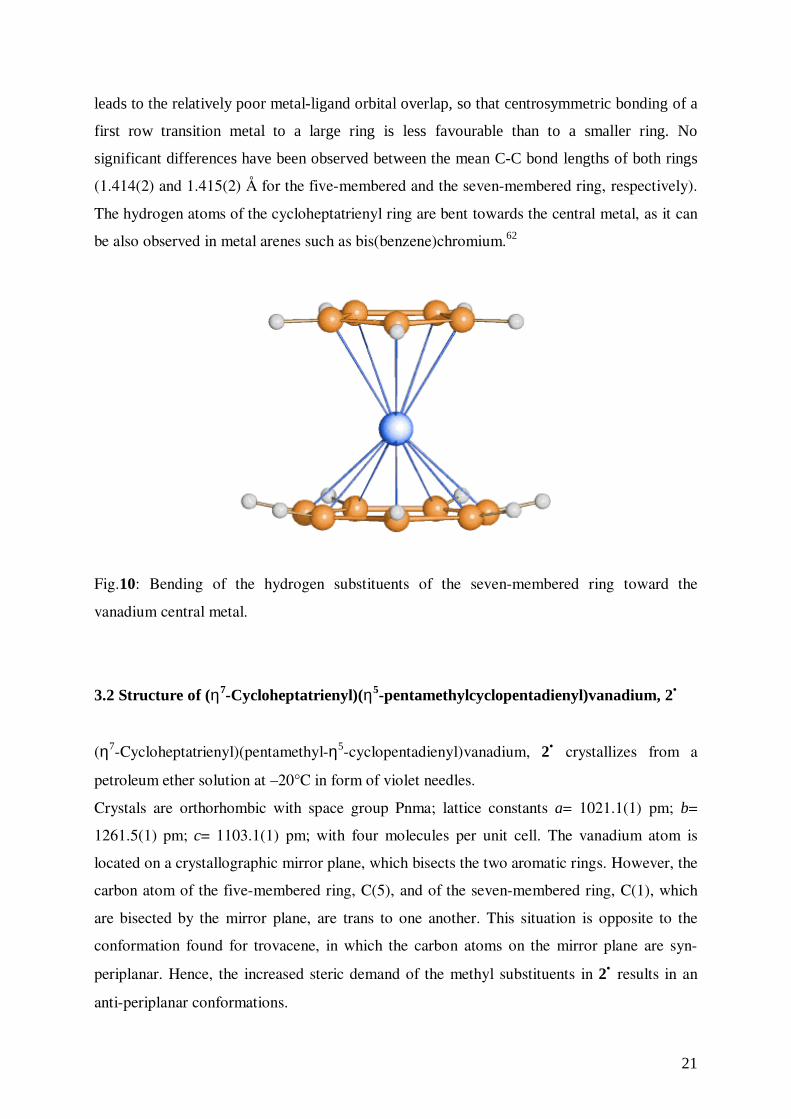
The hydrogen atoms of the cycloheptatrienyl ring are bent towards the central metal, as it can
be also observed in metal arenes such as bis(benzene)chromium.62
Fig.10: Bending of the hydrogen substituents of the seven-membered ring toward the
vanadium central metal.
3.2 Structure of (η7-Cycloheptatrienyl)(η5-pentamethylcyclopentadienyl)vanadium, 2•
(η7-Cycloheptatrienyl)(pentamethyl-η5-cyclopentadienyl)vanadium, 2• crystallizes from a
petroleum ether solution at –20°C in form of violet needles.
Crystals are orthorhombic with space group Pnma; lattice constants a= 1021.1(1) pm; b=
1261.5(1) pm; c= 1103.1(1) pm; with four molecules per unit cell. The vanadium atom is
located on a crystallographic mirror plane, which bisects the two aromatic rings. However, the
carbon atom of the five-membered ring, C(5), and of the seven-membered ring, C(1), which
are bisected by the mirror plane, are trans to one another. This situation is opposite to the
conformation found for trovacene, in which the carbon atoms on the mirror plane are syn-
periplanar. Hence, the increased steric demand of the methyl substituents in 2• results in an
anti-periplanar conformations.

22
Fig.11: Projection of 2• perpendicular to the molecular axes.
V(1)–C(1) 2.179(3) V(1)–C(2) 2.181(2)V(1)–C(3) 2.183(2) V(1)–C(4) 2.195(2)V(1)–C(5) 2.247(2) V(1)–C(6) 2.251(2)V(1)–C(7) 2.261(2) C(1)–C(2) 1.420(3)C(2)–C(3) 1.404(4) C(3)–C(4) 1.393(4)C(5)–C(6) 1.420(2) C(6)–C(7) 1.423(2)C(5)–C(51) 1.507(3) C(6)–C(61) 1.501(2)C(7)–C(71) 1.495(2) C(7)–C(7a) 1.427(3)C(1)–H(1) 0.86(5) C(2)–H(2) 0.97(3)C(3)–H(3) 1.01(3) C(4)–H(4) 0.94(3)C(51)–H(511) 0.95(4) C(51)–H(512) 0.98(3)C(61)–H(611) 0.98 C(61)–H(612) 0.98C(61)–H(613) 0.98 C(71)–H(711) 0.98C(71)–H(712) 0.98 C(71)–H(713) 0.98
Table 5: Selected bond lengths (Å) for 2•.
C(1)–C(2)–C(3) 127.9(2) C(3)–C(4)–C(4a) 128.4(2)C(5)–C(6)–C(7) 107.9(1) C(6)–C(5)–C(51) 125.8(1)C(7)–C(6)–C(61) 125.6(2) C(6)–C(7)–C(71) 126.6(2)
Table 6: Selected bond angles (deg) for 2• .
As expected, the two ligand planes are essentially parallel (angle of Tr plane to Cp* plane is
3°). The average distances of the vanadium atom to the carbon atoms of the cycloheptatrienyl-
and Cp*-ring are 2.185(7) and 2.254(6) Å, respectively. The virtual identity of the average
carbon-vanadium bond distances for the Cp ligand in 1• and 2• despite the higher steric
23
demand of the Cp* ligand may be attributed to a very small structural trans effect, indicating
the tighter bonding of the more electron-rich Cp* ligand. The static structural differences in
M-Cp and M-Cp* bonding should reflect related electronic differences in M-Cp and M-Cp*
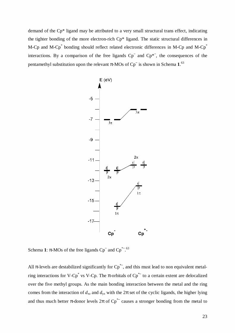
interactions. By a comparison of the free ligands Cp− and Cp*−, the consequences of the
pentamethyl substitution upon the relevant π-MOs of Cp− is shown in Schema 1.63
Schema 1: π-MOs of the free ligands Cp− and Cp*−.63
All π-levels are destabilized significantly for Cp*−, and this must lead to non equivalent metal-
ring interactions for V-Cp* vs V-Cp. The π-orbitals of Cp*− to a certain extent are delocalized
over the five methyl groups. As the main bonding interaction between the metal and the ring
comes from the interaction of dxz and dyz with the 2π set of the cyclic ligands, the higher lying
and thus much better π-donor levels 2π of Cp*− causes a stronger bonding from the metal to
24
the carbon atoms of the pentamethyl substituted ring and, the weaker donating Cp− ligand
keeps more of its electron density in its 2π-level set.
In conclusion, the new electronic and steric environment created by the Cp* moiety does not
greatly effect the overall structure.
25
4. Aldehydes and Carboxylic Acids of (η7-Cycloheptatrienyl)(η5-
cyclopentadienyl)vanadium, 1• and (η7-Cycloheptatrienyl)(η5-
pentamethylcyclopentadienyl)vanadium, 2•
Whereas the study of intramolecular electron transfer requires highly sophisticated techniques
of time resolved spectroscopy, intramolecular communication, namely spin exchange
interaction in biradicals, can be explored by measuring the exchange coupling constant J via
EPR.64
A structural unit that plays an important role in electron transfer pathway in non-covalently
linked proteins and in photosynthetic models is the hydrogen bond. Organometallic molecules
bonded by hydrogen bonds have been widely studied in recent years.65
The synthesis of paramagnetic organometallic aldehydes and carboxylic acids permits to
combine the two seemingly unrelated fields, in order to study electron-electron spin-spin
exchange in the dimers formed by hydrogen bridge interactions.
For this purpose, as a supplement to the previously prepared mono-aldehyde49 and mono-
carboxylic acid50 of 1• , in which the functional groups had been introduced into the Cp ligand,
in the present work the dicarboxylic acid, 13• , and the bis-aldehyde of 1• , 14• , have been
prepared. Furthermore, the mono-carboxylic acid, 15• , and the mono-aldehyde, 16• ,
derivatives of 2• , bearing the respective functional group in the seven-membered ring have
been synthesized. All new compounds have been studied by EPR spectroscopy and cyclic
voltammetry, and 13• and 16• have been further characterized by X-ray diffraction.
4.1 Synthesis of (Carboxy-η7-cycloheptatrienyl)(carboxy-η5-cyclopentadienyl)vanadium,
[(η7-C7H6COOH)(η5-C5H4COOH)V0], 13•, and (Formyl-η7-cycloheptatrienyl)(formyl-
η5-cyclopentadienyl)vanadium, [(η7-C7H6CHO)(η5-C5H4CHO)V0], 14•
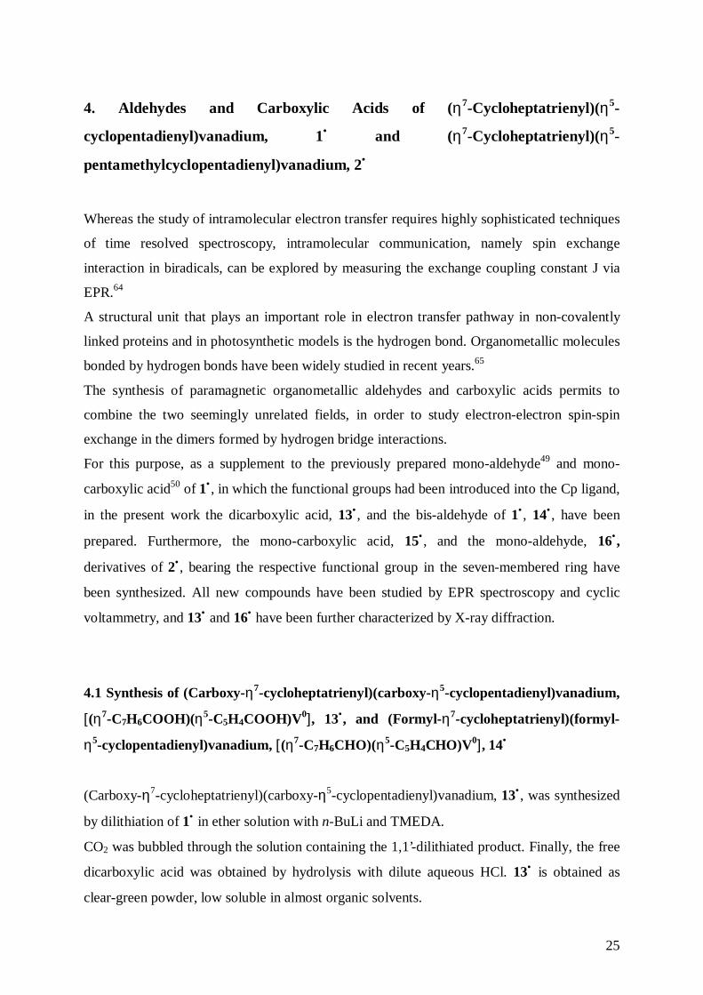
(Carboxy-η7-cycloheptatrienyl)(carboxy-η5-cyclopentadienyl)vanadium, 13• , was synthesized
by dilithiation of 1• in ether solution with n-BuLi and TMEDA.
CO2 was bubbled through the solution containing the 1,1’-dilithiated product. Finally, the free
dicarboxylic acid was obtained by hydrolysis with dilute aqueous HCl. 13• is obtained as
clear-green powder, low soluble in almost organic solvents.
26
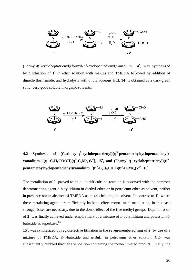
(Formyl-η7-cycloheptatrienyl)(formyl-η5-cyclopentadienyl)vanadium, 14• , was synthesized
by dilithiation of 1• in ether solution with n-BuLi and TMEDA followed by addition of
dimethylformamide, and hydrolysis with dilute aqueous HCl. 14• is obtained as a dark-green
solid, very good soluble in organic solvents.
4.2 Synthesis of (Carboxy-η7-cycloheptatrienyl)(η5-pentamethylcyclopentadienyl)-
vanadium, [(η7-C7H6COOH)(η5-C5Me5)V0], 15•, and (Formyl-η7-cycloheptatrienyl)(η5-
pentamethylcyclopentadienyl)vanadium, [(η7-C7H6CHO)(η5-C5Me5)V0], 16•
The metallation of 2• proved to be quite difficult: no reaction is observed with the common
deprotonanting agent n-butyllithium in diethyl ether or in petroleum ether as solvent, neither
in presence nor in absence of TMEDA as metal chelating co-solvent. In contrast to 1• , where
these metalating agents are sufficiently basic to effect mono- or di-metallation, in this case
stronger bases are necessary, due to the donor effect of the five methyl groups. Deprotonation
of 2• was finally achieved under employment of a mixture of n-butyllithium and potassium-t-
butoxide as superbase.66
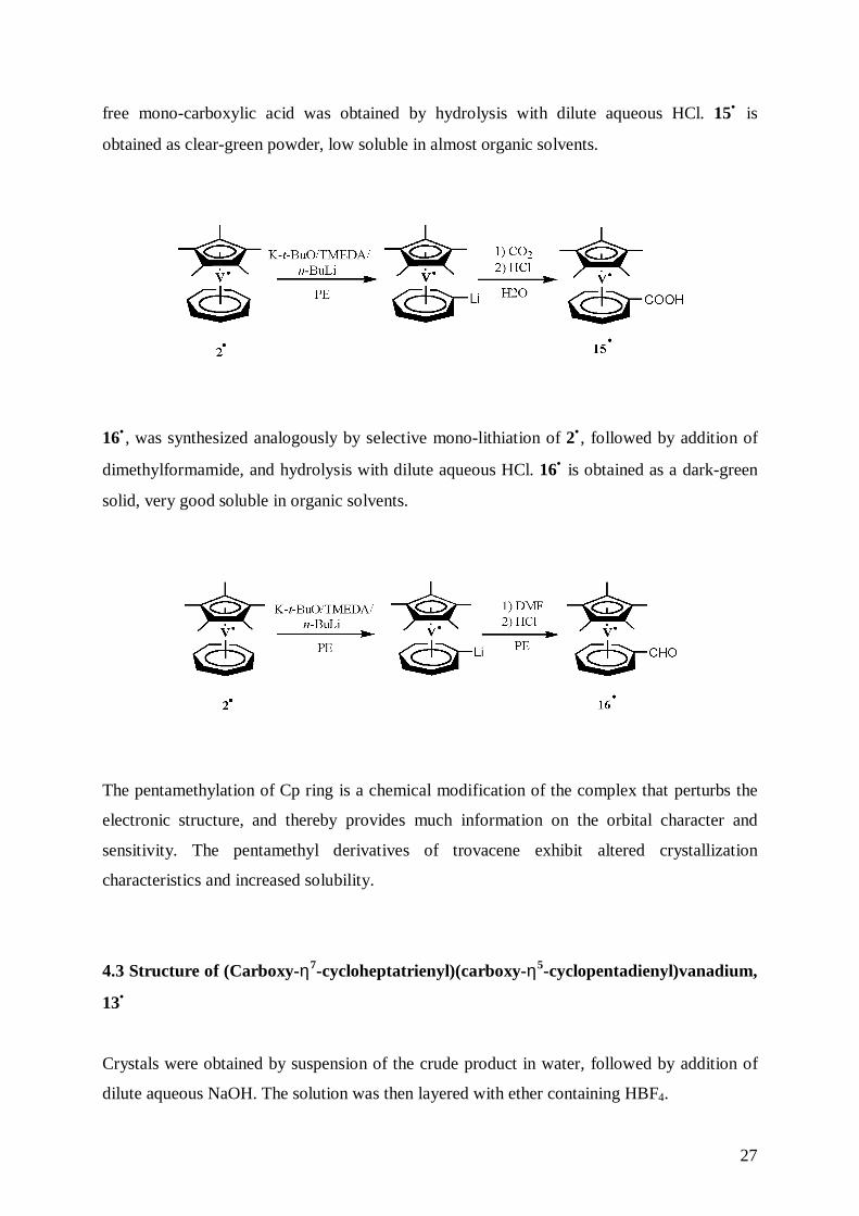
15• , was synthesized by regioselective lithiation in the seven-membered ring of 2• by use of a
mixture of TMEDA, K-t-butoxide and n-BuLi in petroleum ether solution. CO2 was
subsequently bubbled through the solution containing the mono-lithiated product. Finally, the
27
free mono-carboxylic acid was obtained by hydrolysis with dilute aqueous HCl. 15• is
obtained as clear-green powder, low soluble in almost organic solvents.
16• , was synthesized analogously by selective mono-lithiation of 2• , followed by addition of
dimethylformamide, and hydrolysis with dilute aqueous HCl. 16• is obtained as a dark-green
solid, very good soluble in organic solvents.
The pentamethylation of Cp ring is a chemical modification of the complex that perturbs the
electronic structure, and thereby provides much information on the orbital character and
sensitivity. The pentamethyl derivatives of trovacene exhibit altered crystallization
characteristics and increased solubility.
4.3 Structure of (Carboxy-η7-cycloheptatrienyl)(carboxy-η5-cyclopentadienyl)vanadium,
13•
Crystals were obtained by suspension of the crude product in water, followed by addition of
dilute aqueous NaOH. The solution was then layered with ether containing HBF4.
28
Crystals are monoclinic with space group P21/c; lattice constants a= 6.9723(14) Å; b=
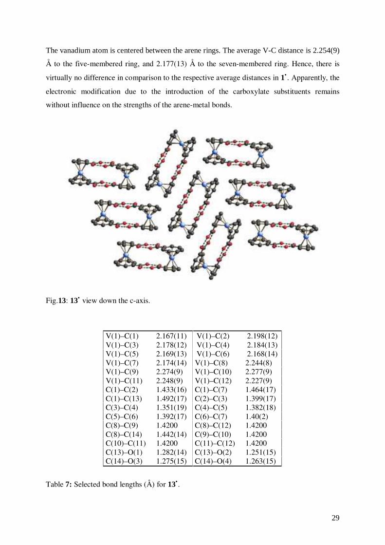
7.8419(16) Å; c= 21.642(4) Å; with four molecules per unit cell. The molecular structure of
one of the dimers in the unit cell is presented in Fig. 12, while the packing of the molecules
inside the unit cell is displayed in Fig. 13.
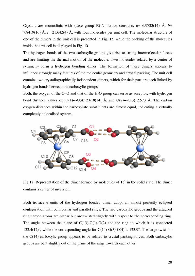
The hydrogen bonds of the two carboxylic groups give rise to strong intermolecular forces
and are limiting the thermal motion of the molecule. Two molecules related by a center of
symmetry form a hydrogen bonding dimer. The formation of these dimers appears to
influence strongly many features of the molecular geometry and crystal packing. The unit cell
contains two crystallographically independent dimers, which for their part are each linked by
hydrogen bonds between the carboxylic groups.
Both, the oxygen of the C=O and that of the H-O group can serve as acceptor, with hydrogen
bond distance values of: O(1)---O(4) 2.618(14) Å, and O(2)---O(3) 2.573 Å. The carbon
oxygen distances within the carboxylate substituents are almost equal, indicating a virtually
completely delocalised system.
Fig.12: Representation of the dimer formed by molecules of 13• in the solid state. The dimer
contains a center of inversion.
Both trovacene units of the hydrogen bonded dimer adopt an almost perfectly eclipsed
configuration with both planar and parallel rings. The two carboxylic groups and the attached
ring carbon atoms are planar but are twisted slightly with respect to the corresponding ring.
The angle between the plane of C(13)-O(1)-O(2) and the ring to which it is connected
122.4(12)°, while the corresponding angle for C(14)-O(3)-O(4) is 123.9°. The large twist for
the C(14) carboxylic group appears to be related to crystal packing forces. Both carboxylic
groups are bent slightly out of the plane of the rings towards each other.
29
The vanadium atom is centered between the arene rings. The average V-C distance is 2.254(9)
Å to the five-membered ring, and 2.177(13) Å to the seven-membered ring. Hence, there is
virtually no difference in comparison to the respective average distances in 1• . Apparently, the
electronic modification due to the introduction of the carboxylate substituents remains
without influence on the strengths of the arene-metal bonds.
Fig.13: 13• view down the c-axis.
V(1)–C(1) 2.167(11) V(1)–C(2) 2.198(12)V(1)–C(3) 2.178(12) V(1)–C(4) 2.184(13)V(1)–C(5) 2.169(13) V(1)–C(6) 2.168(14)V(1)–C(7) 2.174(14) V(1)–C(8) 2.244(8)V(1)–C(9) 2.274(9) V(1)–C(10) 2.277(9)V(1)–C(11) 2.248(9) V(1)–C(12) 2.227(9)C(1)–C(2) 1.433(16) C(1)–C(7) 1.464(17)C(1)–C(13) 1.492(17) C(2)–C(3) 1.399(17)C(3)–C(4) 1.351(19) C(4)–C(5) 1.382(18)C(5)–C(6) 1.392(17) C(6)–C(7) 1.40(2)C(8)–C(9) 1.4200 C(8)–C(12) 1.4200C(8)–C(14) 1.442(14) C(9)–C(10) 1.4200C(10)–C(11) 1.4200 C(11)–C(12) 1.4200C(13)–O(1) 1.282(14) C(13)–O(2) 1.251(15)C(14)–O(3) 1.275(15) C(14)–O(4) 1.263(15)
Table 7: Selected bond lengths (Å) for 13•.
30
C(2)–C(1)–C(7) 126.7(12) C(3)–C(2)–C(1) 126.9(13)C(4)–C(3)–C(2) 131.6(14) C(3)–C(4)–C(5) 128.8(13)C(4)–C(5)–C(6) 129.2(14) C(5)–C(6)–C(7) 129.0(14)C(6)–C(7)–C(1) 127.7(13) C(9)–C(8)–C(12) 108.0C(10)–C(9)–C(8) 108.0 C(10)–C(11)–C(12) 108.0C(11)–C(10)–C(9) 108.0 C(11)–C(12)–C(8) 108.0C(2)–C(1)–C(13) 117.3(12) C(7)–C(1)–C(13) 115.7(12)C(9)–C(8)–C(14) 125.9(8) C(12)–C(8)–C(14) 125.8(8)O(2)–C(13)–O(1) 122.4(12) O(2)–C(13)–C(1) 118.7(12)O(1)–C(13)–C(1) 118.9(13) O(4)–C(14)–O(3) 123.9(12)O(4)–C(14)–C(8) 118.1(11) O(3)–C(14)–C(8) 117.9(12)
Table 8: Selected bond angles (deg) for 13• .
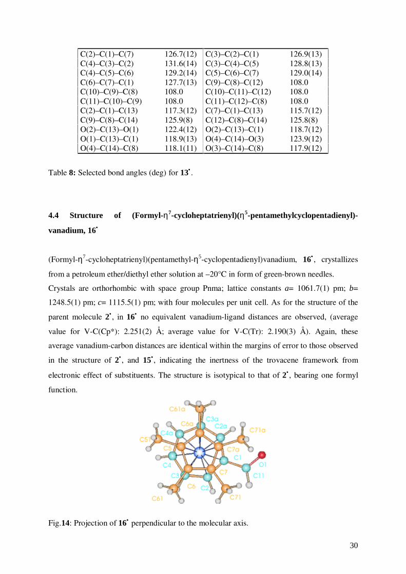
4.4 Structure of (Formyl-η7-cycloheptatrienyl)(η5-pentamethylcyclopentadienyl)-
vanadium, 16•
(Formyl-η7-cycloheptatrienyl)(pentamethyl-η5-cyclopentadienyl)vanadium, 16• , crystallizes
from a petroleum ether/diethyl ether solution at –20°C in form of green-brown needles.
Crystals are orthorhombic with space group Pnma; lattice constants a= 1061.7(1) pm; b=
1248.5(1) pm; c= 1115.5(1) pm; with four molecules per unit cell. As for the structure of the
parent molecule 2• , in 16• no equivalent vanadium-ligand distances are observed, (average
value for V-C(Cp*): 2.251(2) Å; average value for V-C(Tr): 2.190(3) Å). Again, these
average vanadium-carbon distances are identical within the margins of error to those observed
in the structure of 2• , and 15•, indicating the inertness of the trovacene framework from
electronic effect of substituents. The structure is isotypical to that of 2• , bearing one formyl
function.
Fig.14: Projection of 16• perpendicular to the molecular axis.
31
The complex resides on a crystallographic mirror plane containing the vanadium atom and
bisecting both aromatic rings. For this reason the C(O)H-function is disordered equally over
two positions (thermal ellipsoid 20%).
V(1)–C(1) 2.186(3) V(1)–C(2) 2.179(2)V(1)–C(3) 2.204(3) V(1)–C(4) 2.192(3)V(1)–C(5) 2.249(3) V(1)–C(6) 2.250(2)V(1)–C(7) 2.254(2) O(1)–C(11) 1.252(8)C(1)–C(2) 1.428(3) C(1)–C(11) 1.452(6)C(2)–C(3) 1.397(4) C(3)–C(4) 1.408(4)C(4)–C(4a) 1.406(6) C(5)–C(6) 1.426(3)C(6)–C(7) 1.424(3) C(7)–C(7a) 1.429(5)C(6)–C(61) 1.499(3) C(7)–C(71) 1.499(3)C(2)–H(2) 0.92(3) C(3)–H(3) 1.02(4)C(4)–H(4) 0.91(3) C(11)–H(11) 0.96(8)C(51)–H(151) 0.92(5) C(51)–H(152) 0.92(3)C(61)–H(161) 0.98 C(61)–H(162) 0.98C(61)–H(163) 0.98 C(71)–H(171) 0.98C(71)–H(172) 0.98 C(71)–H(173) 0.98
Table 9: Selected bond lengths (Å) for 16•.
C(2)–C(1)–C(2a) 126.3(3) C(2)–C(1)–C(11) 116.4(2)C(1)–C(2)–C(3) 129.6(2) C(3)–C(4)–C(4a) 129.0(3)C(2)–C(3)–C(4) 128.1(3) C(5)–C(6)–C(7) 108.1(2)C(6)–C(5)–C(51) 126.0(1) C(6)–C(5)–C(6a) 107.9(2)C(51)–C(5)–C(6a) 126.0(1) C(5)–C(6)–C(61) 125.8(2)C(7)–C(6)–C(61) 126.1(2) C(6)–C(7)–C(71) 126.6(2)C(6)–C(7)–C(7a) 107.9(2) C(71)–C(7)–C(7a) 125.4(2)O(1)–C(11)–C(1) 122.8(4) O(1)–C(11)–H(11) 120.(6)O(1)–C(11)–H(11a) 10.(6) O(1a)–C(11)–H(11a) 120.(6)
Table 10: Selected bond angles (deg) for 16• .
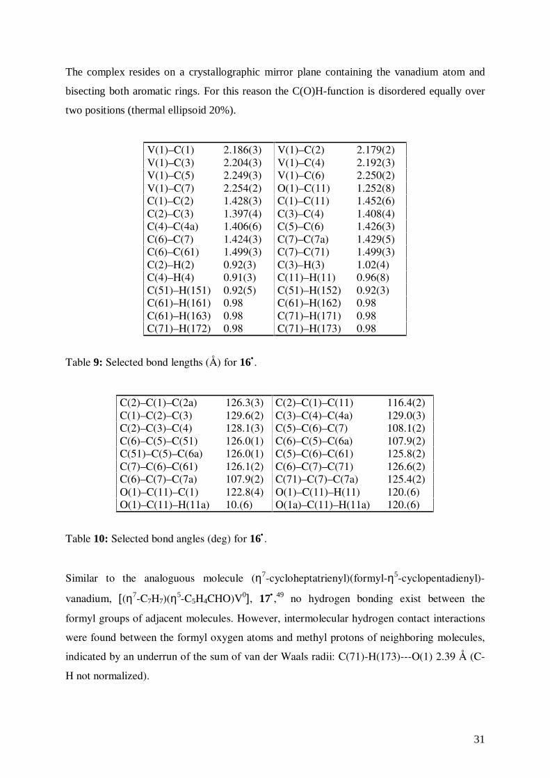
Similar to the analoguous molecule (η7-cycloheptatrienyl)(formyl-η5-cyclopentadienyl)-
vanadium, [(η7-C7H7)(η5-C5H4CHO)V0], 17• ,49 no hydrogen bonding exist between the
formyl groups of adjacent molecules. However, intermolecular hydrogen contact interactions
were found between the formyl oxygen atoms and methyl protons of neighboring molecules,
indicated by an underrun of the sum of van der Waals radii: C(71)-H(173)---O(1) 2.39 Å (C-
H not normalized).
32
Fig.15: Intermolecular hydrogen bonding in 16• (only one of the two disorder images is
shown).
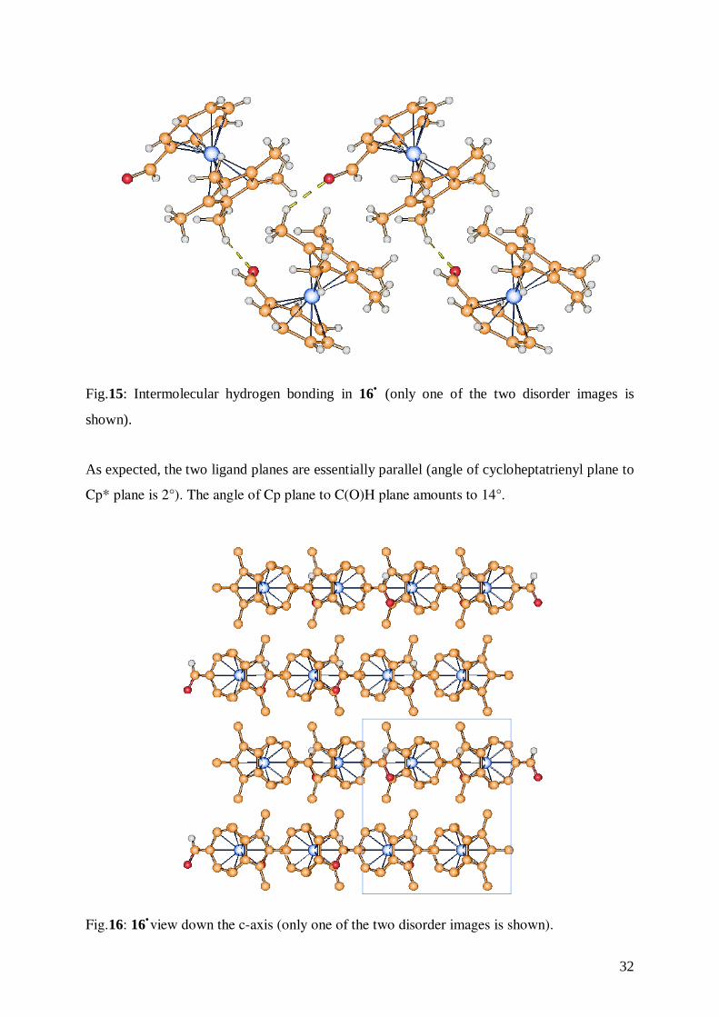
As expected, the two ligand planes are essentially parallel (angle of cycloheptatrienyl plane to
Cp* plane is 2°). The angle of Cp plane to C(O)H plane amounts to 14°.
Fig.16: 16•view down the c-axis (only one of the two disorder images is shown).
33
4.5 Cyclic Voltammetry of 13• , 14•, 15• , 16•
4.5.1 Fundamentals of Cyclic Voltammetry67
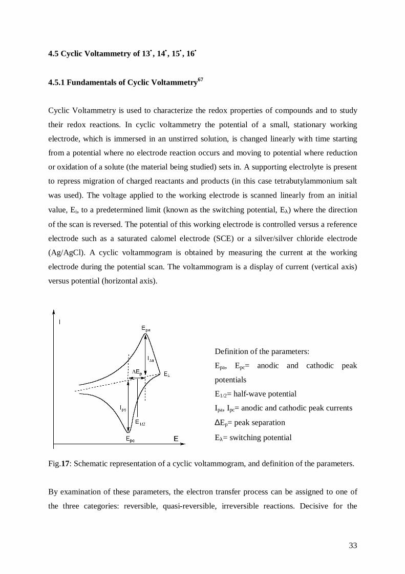
Cyclic Voltammetry is used to characterize the redox properties of compounds and to study
their redox reactions. In cyclic voltammetry the potential of a small, stationary working
electrode, which is immersed in an unstirred solution, is changed linearly with time starting
from a potential where no electrode reaction occurs and moving to potential where reduction
or oxidation of a solute (the material being studied) sets in. A supporting electrolyte is present
to repress migration of charged reactants and products (in this case tetrabutylammonium salt
was used). The voltage applied to the working electrode is scanned linearly from an initial
value, Ei, to a predetermined limit (known as the switching potential, Eλ) where the direction
of the scan is reversed. The potential of this working electrode is controlled versus a reference
electrode such as a saturated calomel electrode (SCE) or a silver/silver chloride electrode
(Ag/AgCl). A cyclic voltammogram is obtained by measuring the current at the working
electrode during the potential scan. The voltammogram is a display of current (vertical axis)
versus potential (horizontal axis).
Definition of the parameters:
Epa, Epc= anodic and cathodic peak
potentials
E1/2= half-wave potential
Ipa, Ipc= anodic and cathodic peak currents
∆Ep= peak separation
Eλ= switching potential
Fig.17: Schematic representation of a cyclic voltammogram, and definition of the parameters.
By examination of these parameters, the electron transfer process can be assigned to one of
the three categories: reversible, quasi-reversible, irreversible reactions. Decisive for the
34
electrochemical reversibility is the rate of heterogeneous electron transfer at the electrode
surface given by ks (standard heterogeneous electron transfer rate constant).
a) Reversible process
ks > 2·10-2 cm/s: the electron transfer process is faster than the diffusion.
The reaction is diffusion-controlled (no other processes limit the current). The criteria for
diffusion control is that Ipc increases with v1/2 (v is the sweep rate, dE/dt) and is directly
proportional to the concentration. The electron transfer reaction at the electrode surface is so
rapid that equilibrium conditions are maintained even with a substantial net current and a
rapidly changing potential. The criteria of reversibility are ∆Ep= Epa-Epc= 57/n mV at 298 K,
where n is number of electrons transferred per ion (equivalent/mol), values which must be
independent of scan rate and concentration. The E1/2 value is situated exactly (within 2/n mV)
midway between Epa and Epc. The values of Ipa and Ipc should be identical for a simple
reversible (fast) couple, that is Ipa/Ipc= 1.
b) Quasi-reversible process
2·10-2 cm/s > ks > 5·10-5 cm/s: the rate of the electron transfer process is equal to that of the
diffusion.
The rate of electron transfer and diffusion are comparable. The peak separation ∆Ep of a
quasi-reversible process increases with v1/2. The values of Ipa and Ipc should be identical, that
is Ipa/Ipc= 1. Ipa and Ipc are not proportional to v1/2.
c) Irreversible process
ks > 5·10-5 cm/s: the electron transfer process is slower than the diffusion.
Electrochemical irreversibility is caused by slow electron exchange of the redox species with
the working electrode. Irreversibility manifest itself through ∆Ep> 57/n mV at 298 K, ∆Ep
increasing with increasing v, and it is characterized by a separation of peak potentials greater
than indicated by the expression ∆Ep= Epa-Epc≅ 0,059/n.
4.5.2 Results
The 17-VE-complex trovacene, 1• , exhibits in its crystalline phase air stability of a few
minutes.68 This property is confirmed by the cyclovoltammetry experiment, indicated by a
redox potential E1/2(+/ 0) of 0.26 V, [vs. 0.54 V of ferrocene, 7, a sandwich complex stable to

35
air] (these values have been measured in DME/0.1 M TBAP; T=−40 °C; ν= 100 mV/s; glassy
carbon electrode vs. SCE). Neutral trovacene possesses the ground state configuration
[(e2)4(a1)
1(e1)0], while the cationic form has the low spin configuration [(e2)
4(a1)0(e1)
0]. The
e2-orbitals are so low in energy, that the configuration [(e2)4] is more favorable also in the
ionized state. The introduction of electron-withdrawing substituents on the rings of trovacene
causes a reduction of the electron density at the central metal. In consequence, the effective
positive charge at the vanadium atom increases, resulting in a contraction of the essentially
non-bonding a1 (V-3dz2) redox orbital and in a stronger binding of the occupying electron.
Therefore, the oxidation becomes more difficult and an anodic shift of the redox potential is
observed. Similar to the parent molecule trovacene, the derivatives (carboxy-η7-
cycloheptatrienyl)(carboxy-η5-cyclopentadienyl)vanadium, 13• , and (formyl-η7-
cycloheptatrienyl)(formyl-η5-cyclopentadienyl)vanadium, 14• show a second oxidation wave
(2+/+), that is irreversible for all complexes (the species formed has not been identified so
far). Trovacene is reversibly reduced to the mono-anion at –2.55 V: the transferred electron
will occupy the metal dominated a1g-orbital. The derivatives 13• and 14• are reduced also, but
more readily. This effect is explained by the increase of stability of the a1g-orbital, due to the
presence of the electron-withdrawing substituents. It has to be noted, that the anodic shifts of
the oxidation steps to the monocations are smaller than the shifts of the corresponding
reduction steps to the monoanions. Hence, a transfer of an electron into a ligand-centered
LUMO would also be conceivable, resulting in the formation of a paramagnetic biradical
species. This explanation may be adopted also for the second reduction of the formyl
complexes. The π-bonded arene ring bearing a carbonyl substituent has the ability to stabilize
an electron transferred into a ligand-centered LUMO by delocalization. Due to the low energy
and the corresponding higher electron affinity of this ligand-centered LUMO, a further
reduction of the previously formed monoanion would be facilitated.
Cyclovoltammetric data for (carboxy-η5-cyclopentadienyl)(η7-cycloheptatrienyl)vanadium,25
6• , (carboxy-η7-cycloheptatrienyl)(carboxy-η5-cyclopentadienyl)vanadium, 13• , (formyl-η5-
cyclopentadienyl)(η7-cycloheptatrienyl)vanadium,49 17• , (formyl-η7-cycloheptatrienyl)-
(formyl-η5-cyclopentadienyl)vanadium, 14• , and for the parent molecule trovacene, 1• , are
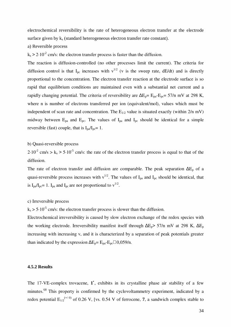
shown in Table 11. Typical cyclovoltammetric traces for 13• and 14• are depicted in Figs. 18-
19.
36
1• 6• 13• 17• 14•
E1/2(+/ 0) [V] 0.260 0.480 0.615 c 0.496 0.688
∆Ep [mV] a 64 74 74 90 60r= ipa/ipc 0.93 0.94 0.52 1 0.34Epa [V] 1.03 1.043 b 1.034 1.100 1.102E1/2
(0/−) [V] -2.55 -2.687 -2.554 c -2.088 -1.843 c
∆Ep [mV] a 66 88 96 66r= ipa/ip 1 0.56 0.41 0.96E1/2
(−/2−) [V] c -2.499 -2.070∆Ep [mV] a 136 152r= ipa/ip 0.30 0.86
a ∆Ep= (Epa – Epc)b irreversible c the r values do not strictly confirm reversibility
Table 11: Cyclovoltammetric data for 1• , 6• , 13•, 17• , 14•measured in DME/0.1 M TBAP, vs.
SCE, ν= 0.1 V/s, T=-40°C.
+ 1 .7+1 .00-1 .0-2 .0-2 .9
[ V ]
Fig.18: Cyclovoltammetric trace for 13•.
37
+1.7+1.00-1.0-2.0-2.9
[ V ]
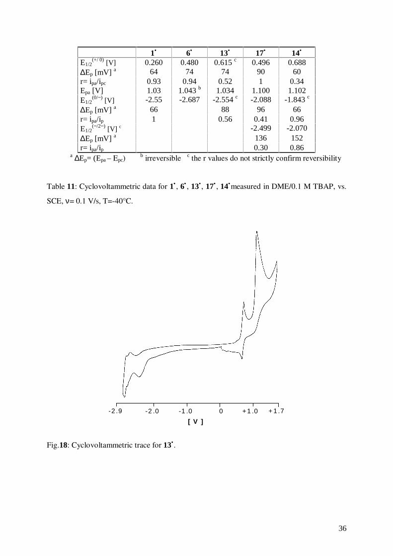
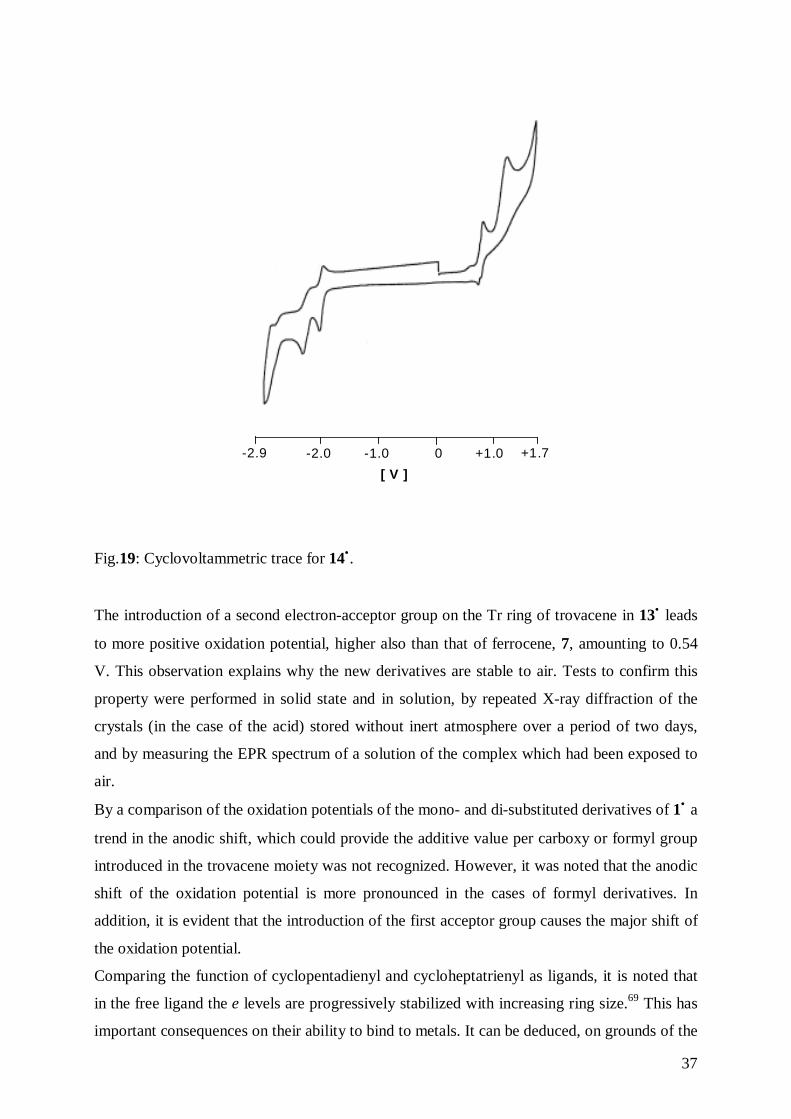
Fig.19: Cyclovoltammetric trace for 14•.
The introduction of a second electron-acceptor group on the Tr ring of trovacene in 13• leads
to more positive oxidation potential, higher also than that of ferrocene, 7, amounting to 0.54
V. This observation explains why the new derivatives are stable to air. Tests to confirm this
property were performed in solid state and in solution, by repeated X-ray diffraction of the
crystals (in the case of the acid) stored without inert atmosphere over a period of two days,
and by measuring the EPR spectrum of a solution of the complex which had been exposed to
air.
By a comparison of the oxidation potentials of the mono- and di-substituted derivatives of 1• a
trend in the anodic shift, which could provide the additive value per carboxy or formyl group
introduced in the trovacene moiety was not recognized. However, it was noted that the anodic
shift of the oxidation potential is more pronounced in the cases of formyl derivatives. In
addition, it is evident that the introduction of the first acceptor group causes the major shift of
the oxidation potential.
Comparing the function of cyclopentadienyl and cycloheptatrienyl as ligands, it is noted that
in the free ligand the e levels are progressively stabilized with increasing ring size.69 This has
important consequences on their ability to bind to metals. It can be deduced, on grounds of the
38
energy separation, that the interaction of the e1 orbitals with a metal is likely to decrease with
increasing ring size, while that of the e2 orbitals is likely to increase. Because of energy of the
metal valence orbitals concerned is approximately intermediate between the e1 and e2 orbitals
for the early transition metals, both the e1 and e2 molecular orbitals, will increase in ligand
character with increasing ring size. When π-Cp is a ligand, interaction of the e1 orbitals is the
most important source of bonding, and as these orbitals are not fully occupied in the free
ligand (the Cp radical), this interaction is able to remove charge from the metal. When π-
cycloheptatrienyl is a ligand, its e1 interaction with the metal is likely to be very small and
chief source of bonding is the e2 interaction. This theoretical consideration is supported by a
systematic analysis of the species [M(ηm-CmHm) (ηn-CnHm)] (M= Ti, Cr, V; m, n= 5, 6, 7, 8)
by photoelectronic-spectroscopy, which confirmed that the contribution of the ring to the e2-
MO’s of sandwich complex increases with increasing ring size, while it decreases with
increasing atomic number of the transition metal.70
The substitution of Cp for Cp* is one of the best known methods for increasing steric
congestion at a metal center. This modification results also in increasing stability of reactive
metal complexes and altering of their catalytic properties. The favorable properties of the Cp*
ligand, such as increased electron donation, steric bulk, and enhanced solubility in
comparison to the ubiquitous Cp ligand, are finding increasing use in organometallic
chemistry.71
The ligand-field strength of the Cp ring is significantly enhanced by the complete replacement
of the hydrogens with electron-donating methyl groups. The increased electron density, and
donor strength of the permethylated ring reflects itself, for instance, in low oxidation
potential.72
The influence of the introduction of Cp* into the trovacene framework has been studied by
cyclic voltammetry measurements, demonstrating a cathodic shift of the redox potential
relative to parent 1•.
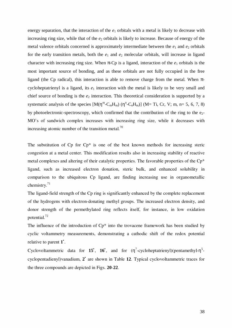
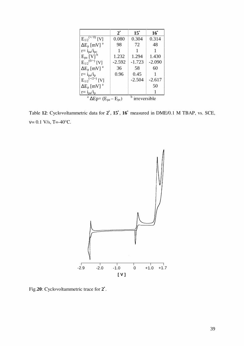
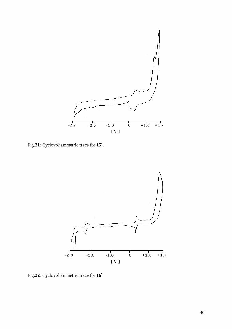
Cyclovoltammetric data for 15• , 16•, and for (η7-cycloheptatrienyl)(pentamethyl-η5-
cyclopentadienyl)vanadium, 2• are shown in Table 12. Typical cyclovoltammetric traces for
the three compounds are depicted in Figs. 20-22.
39
2• 15• 16•
E1/2(+/ 0) [V] 0.080 0.304 0.314
∆Ep [mV] a 98 72 48r= ipa/ipc 1 1 1Epa [V] b 1.232 1.294 1.430E1/2
(0/−) [V] -2.592 -1.723 -2.090∆Ep [mV] a 36 58 60r= ipa/ip 0.96 0.45 1E1/2
(−/2−) [V] -2.504 -2.617∆Ep [mV] a 50r= ipa/ip 1
a ∆Ep= (Epa – Epc)b irreversible
Table 12: Cyclovoltammetric data for 2• , 15• , 16• measured in DME/0.1 M TBAP, vs. SCE,
ν= 0.1 V/s, T=-40°C.
+1.7+1.00-1.0-2.0-2.9
[ V ]
Fig.20: Cyclovoltammetric trace for 2•.
40
+1 .7+1 .00-1 .0-2 .0-2 .9
[ V ]
Fig.21: Cyclovoltammetric trace for 15•.
+1 .7+1.00-1 .0-2 .0-2 .9
[ V ]
Fig.22: Cyclovoltammetric trace for 16•
41
All complexes show a fully reversible oxidation step, and the carboxylic acid and formyl
derivatives show an anodic shift due to the electron withdrawing character of the subtituent
groups, as expected. For 16• , as for all formyl derivatives, a further reduction of the formed
anion is possible due to the increased electron affinity.
The effect of the transition from Cp to Cp* on the redox potential of the Fe(0)/Fe(+1) couple
has been studied on a series of substituted ferrocenes.73 In this study, a shift of the redox
potential by –0.203 V due to the replacement of Cp by Cp* was determined. Moreover, it was
revealed that the contributions of various substituents to the overall potential shift were
additive.
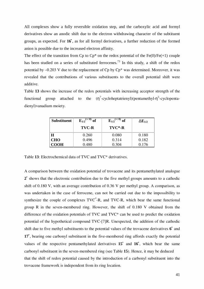
Table 13 shows the increase of the redox potentials with increasing acceptor strength of the
functional group attached to the (η7-cycloheptatrienyl)(pentamethyl-η5-cyclopenta-
dienyl)vanadium moiety.
Substituent E1/2(+/ 0) of
TVC-R
E1/2(+/ 0) of
TVC*-R
∆E1/2
H 0.260 0.080 0.180CHO 0.496 0.314 0.182COOH 0.480 0.304 0.176
Table 13: Electrochemical data of TVC and TVC* derivatives.
A comparison between the oxidation potential of trovacene and its pentamethylated analogue
2• shows that the electronic contribution due to the five methyl groups amounts to a cathodic
shift of 0.180 V, with an average contribution of 0.36 V per methyl group. A comparison, as
was undertaken in the case of ferrocene, can not be carried out due to the impossibility to
synthesize the couple of complexes TVC*-R, and TVC-R, which bear the same functional
group R in the seven-membered ring. However, the shift of 0.180 V obtained from the
difference of the oxidation potentials of TVC and TVC* can be used to predict the oxidation
potential of the hypothetical compound TVC-[7]R. Unexpected, the addition of the cathodic
shift due to five methyl substituents to the potential values of the trovacene derivatives 6• and
17• , bearing one carbonyl substituent in the five-membered ring affords exactly the potential
values of the respective pentamethylated derivatives 15• and 16• , which bear the same
carbonyl substituent in the seven-membered ring (see Table 15). Hence, it may be deduced
that the shift of redox potential caused by the introduction of a carbonyl substituent into the
trovacene framework is independent from its ring location.
42
4.6 Electron Paramagnetic Resonance of 13• , 14• , 15• , 16•
4.6.1 Fundamentals of Electron Paramagnetic Resonance74
Electron paramagnetic resonance is a technique applicable to systems with net electron spin
angular momentum. Free radicals in solid, liquid or gaseous states are systems that fulfil this
condition. The unpaired electron with spin S= 1/2 has two possible values of the quantum
numbers MS, +1/2 and –1/2. A strong and homogeneous magnetic field is employed along the
z direction of laboratory coordinates, to separate the formerly degenerate spin states (Zeeman
effect). The magnetic moments of the electron spin µZ are oriented along the direction of the
applied magnetic field H:
µZ= -MS g βe = ±1/2 g βe
where, g= electronic gyromagnetic ratio (g= 2.0023 for a free electron), βe= Bohr magneton
(= e � � � � � � � � � � � -24 JT-1).
The quantization of spin angular momentum in a specified direction leads to the quantization
of energy levels of a system of magnetic dipoles in a magnetic field. Application of the
expression E= -µZ Ho (E= energy of a magnetic dipole of moment µZ in a field Ho) and
substitution of -MS g βe for µZ, gives a set of energies:
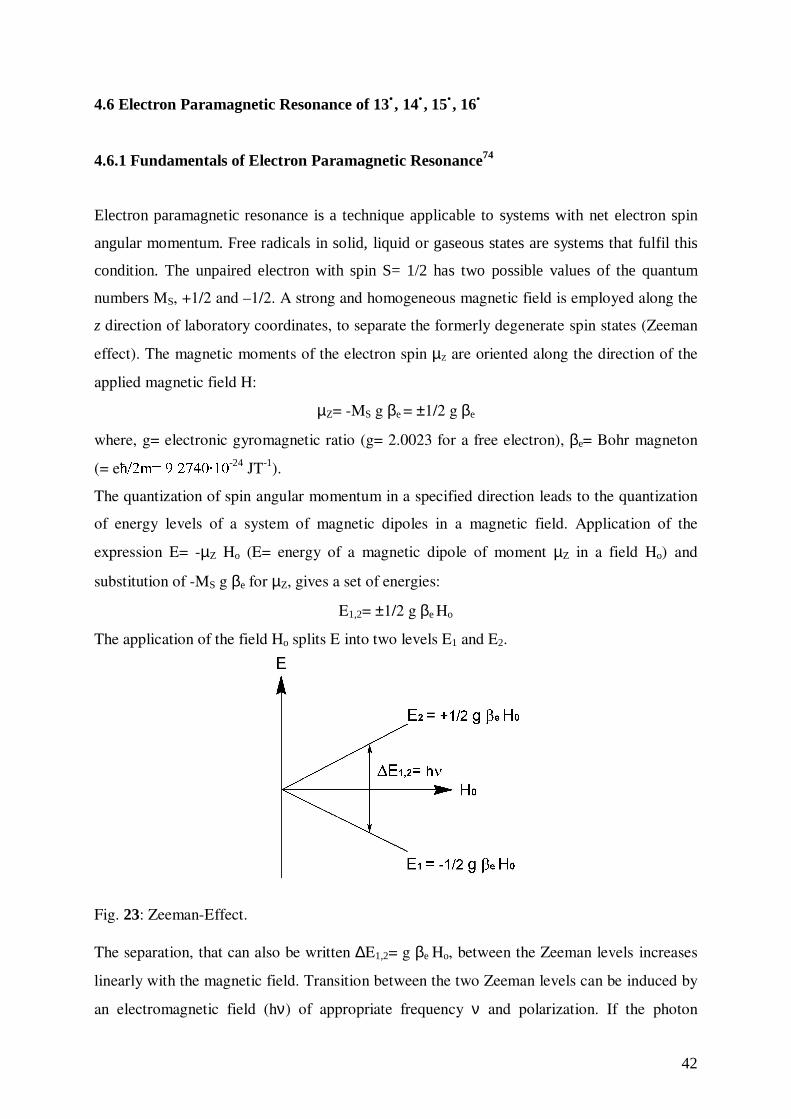
E1,2= ±1/2 g βe Ho
The application of the field Ho splits E into two levels E1 and E2.
Fig. 23: Zeeman-Effect.
The separation, that can also be written ∆E1,2= g βe Ho, between the Zeeman levels increases
linearly with the magnetic field. Transition between the two Zeeman levels can be induced by
an electromagnetic field (hν) of appropriate frequency ν and polarization. If the photon
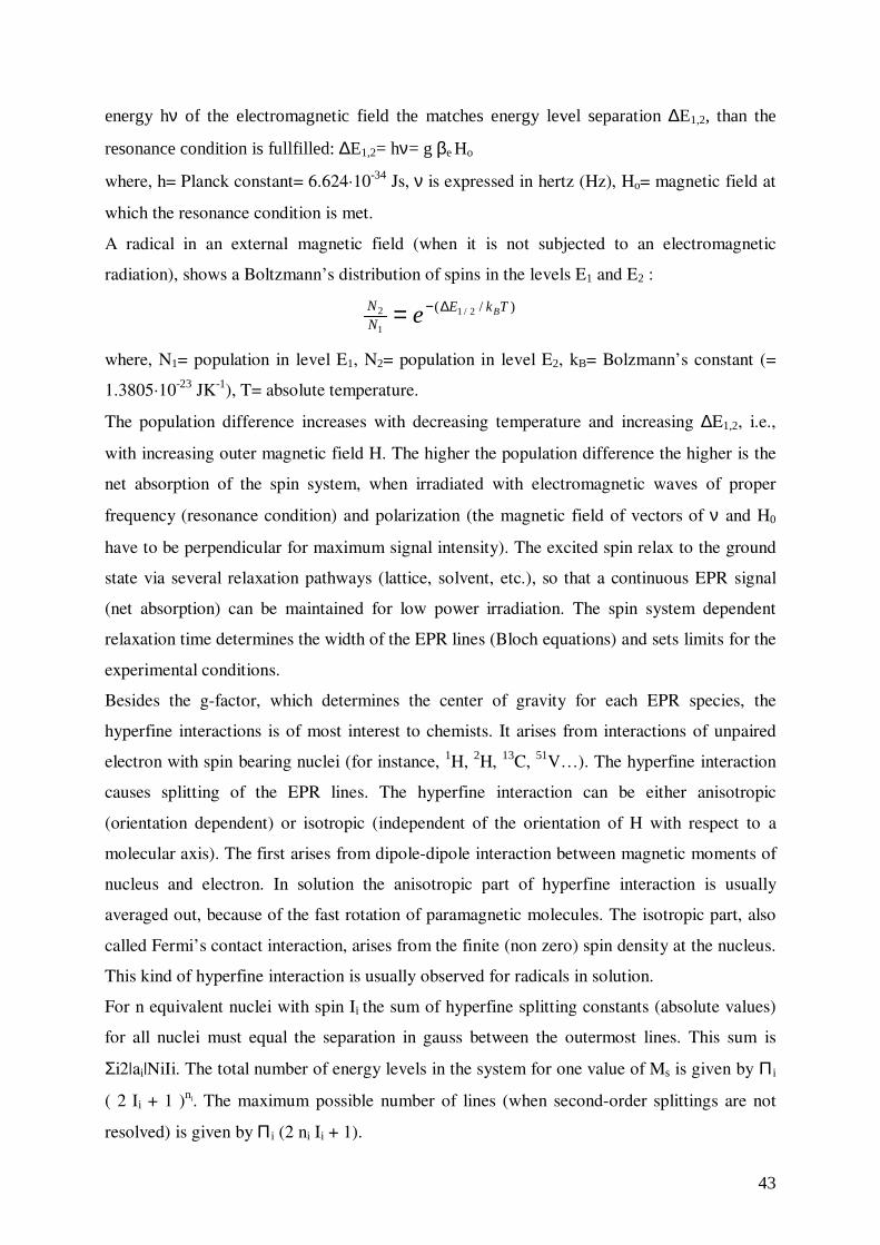
43
energy hν of the electromagnetic field the matches energy level separation ∆E1,2, than the
resonance condition is fullfilled: ∆E1,2= hν= g βe Ho
where, h= Planck constant= 6.624·10-34 Js, ν is expressed in hertz (Hz), Ho= magnetic field at
which the resonance condition is met.
A radical in an external magnetic field (when it is not subjected to an electromagnetic
radiation), shows a Boltzmann’s distribution of spins in the levels E1 and E2 :
)/( 2/1
1
2 TkENN Be ∆−=
where, N1= population in level E1, N2= population in level E2, kB= Bolzmann’s constant (=
1.3805·10-23 JK-1), T= absolute temperature.
The population difference increases with decreasing temperature and increasing ∆E1,2, i.e.,
with increasing outer magnetic field H. The higher the population difference the higher is the
net absorption of the spin system, when irradiated with electromagnetic waves of proper
frequency (resonance condition) and polarization (the magnetic field of vectors of ν and H0
have to be perpendicular for maximum signal intensity). The excited spin relax to the ground
state via several relaxation pathways (lattice, solvent, etc.), so that a continuous EPR signal
(net absorption) can be maintained for low power irradiation. The spin system dependent
relaxation time determines the width of the EPR lines (Bloch equations) and sets limits for the
experimental conditions.
Besides the g-factor, which determines the center of gravity for each EPR species, the
hyperfine interactions is of most interest to chemists. It arises from interactions of unpaired
electron with spin bearing nuclei (for instance, 1H, 2H, 13C, 51V…). The hyperfine interaction
causes splitting of the EPR lines. The hyperfine interaction can be either anisotropic
(orientation dependent) or isotropic (independent of the orientation of H with respect to a
molecular axis). The first arises from dipole-dipole interaction between magnetic moments of
nucleus and electron. In solution the anisotropic part of hyperfine interaction is usually
averaged out, because of the fast rotation of paramagnetic molecules. The isotropic part, also
called Fermi’s contact interaction, arises from the finite (non zero) spin density at the nucleus.
This kind of hyperfine interaction is usually observed for radicals in solution.
For n equivalent nuclei with spin Ii the sum of hyperfine splitting constants (absolute values)
for all nuclei must equal the separation in gauss between the outermost lines. This sum is
Σi2|ai|NiIi. The total number of energy levels in the system for one value of Ms is given by Π i
( 2 Ii + 1 )ni. The maximum possible number of lines (when second-order splittings are not
resolved) is given by Π i (2 ni Ii + 1).
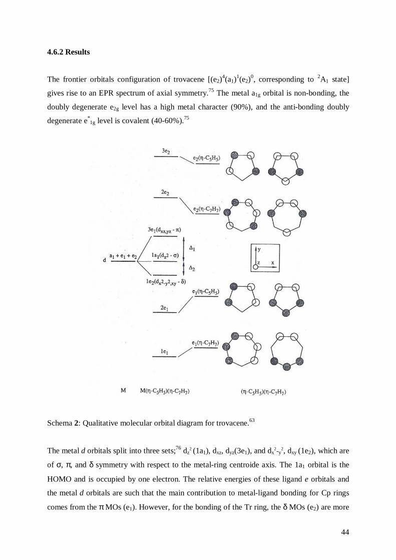
44
4.6.2 Results
The frontier orbitals configuration of trovacene [(e2)4(a1)
1(e2)0, corresponding to 2A1 state]
gives rise to an EPR spectrum of axial symmetry.75 The metal a1g orbital is non-bonding, the
doubly degenerate e2g level has a high metal character (90%), and the anti-bonding doubly
degenerate e*1g level is covalent (40-60%).75
Schema 2: Qualitative molecular orbital diagram for trovacene.63
The metal d orbitals split into three sets;76 dz2 (1a1), dxz, dyz(3e1), and dx
2-y2, dxy
(1e2), which are
of σ, π, and δ symmetry with respect to the metal-ring centroide axis. The 1a1 orbital is the
HOMO and is occupied by one electron. The relative energies of these ligand e orbitals and
the metal d orbitals are such that the main contribution to metal-ligand bonding for Cp rings
comes from the π MOs (e1). However, for the bonding of the Tr ring, the δ MOs (e2) are more
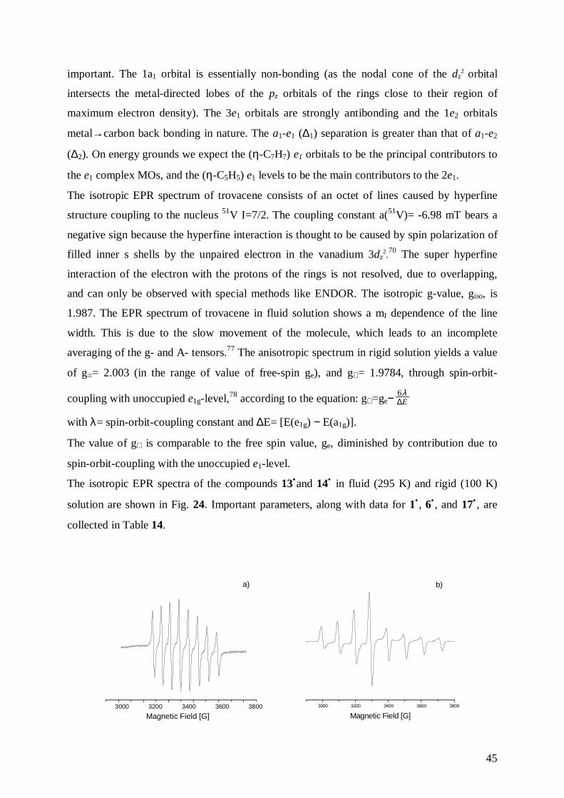
45
important. The 1a1 orbital is essentially non-bonding (as the nodal cone of the dz2 orbital
intersects the metal-directed lobes of the pz orbitals of the rings close to their region of
maximum electron density). The 3e1 orbitals are strongly antibonding and the 1e2 orbitals
metal→carbon back bonding in nature. The a1-e1 (∆1) separation is greater than that of a1-e2
(∆2). On energy grounds we expect the (η-C7H7) e1 orbitals to be the principal contributors to
the e1 complex MOs, and the (η-C5H5) e1 levels to be the main contributors to the 2e1.
The isotropic EPR spectrum of trovacene consists of an octet of lines caused by hyperfine
structure coupling to the nucleus 51V I=7/2. The coupling constant a(51V)= -6.98 mT bears a
negative sign because the hyperfine interaction is thought to be caused by spin polarization of
filled inner s shells by the unpaired electron in the vanadium 3dz2.70 The super hyperfine
interaction of the electron with the protons of the rings is not resolved, due to overlapping,
and can only be observed with special methods like ENDOR. The isotropic g-value, giso, is
1.987. The EPR spectrum of trovacene in fluid solution shows a mI dependence of the line
width. This is due to the slow movement of the molecule, which leads to an incomplete
averaging of the g- and A- tensors.77 The anisotropic spectrum in rigid solution yields a value
of g = 2.003 (in the range of value of free-spin ge), and g⊥ = 1.9784, through spin-orbit-
coupling with unoccupied e1g-level,78 according to the equation: g⊥ =ge− E∆λ6
with λ= spin-orbit-coupling constant and ∆E= [E(e1g) − E(a1g)].
The value of g⊥ is comparable to the free spin value, ge, diminished by contribution due to
spin-orbit-coupling with the unoccupied e1-level.
The isotropic EPR spectra of the compounds 13•and 14• in fluid (295 K) and rigid (100 K)
solution are shown in Fig. 24. Important parameters, along with data for 1• , 6• , and 17• , are
collected in Table 14.
3000 3200 3400 3600 3800
Magnetic Field [G]
a)
3000 3200 3400 3600 3800
Magnetic Field [G]
b)
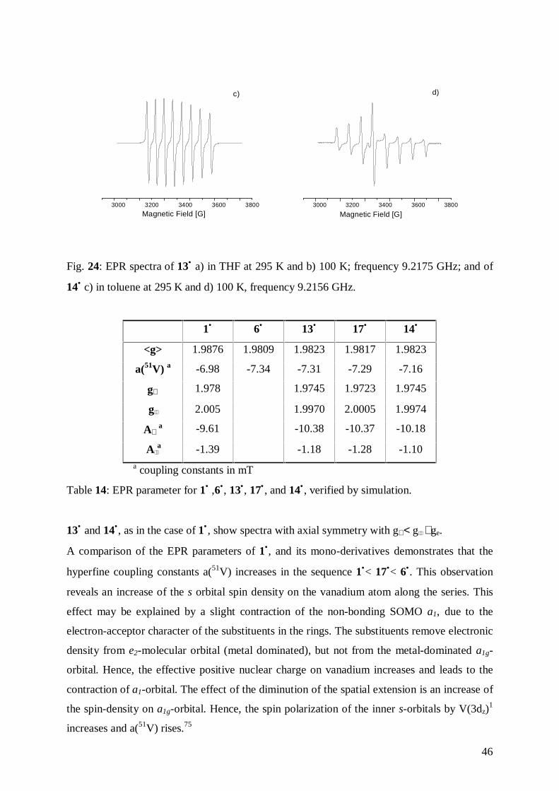
46
3000 3200 3400 3600 3800
Magnetic Field [G]
c)
3000 3200 3400 3600 3800
Magnetic Field [G]
d)
Fig. 24: EPR spectra of 13• a) in THF at 295 K and b) 100 K; frequency 9.2175 GHz; and of
14• c) in toluene at 295 K and d) 100 K, frequency 9.2156 GHz.
1• 6• 13• 17• 14•
<g> 1.9876 1.9809 1.9823 1.9817 1.9823
a(51V) a -6.98 -7.34 -7.31 -7.29 -7.16
g⊥ 1.978 1.9745 1.9723 1.9745
g 2.005 1.9970 2.0005 1.9974
A⊥ a -9.61 -10.38 -10.37 -10.18
A a -1.39 -1.18 -1.28 -1.10
a coupling constants in mT
Table 14: EPR parameter for 1• ,6•, 13•, 17• , and 14• , verified by simulation.
13• and 14•, as in the case of 1• , show spectra with axial symmetry with g⊥ < g ≅ ge.
A comparison of the EPR parameters of 1•, and its mono-derivatives demonstrates that the
hyperfine coupling constants a(51V) increases in the sequence 1•< 17•< 6•. This observation
reveals an increase of the s orbital spin density on the vanadium atom along the series. This
effect may be explained by a slight contraction of the non-bonding SOMO a1, due to the
electron-acceptor character of the substituents in the rings. The substituents remove electronic
density from e2-molecular orbital (metal dominated), but not from the metal-dominated a1g-
orbital. Hence, the effective positive nuclear charge on vanadium increases and leads to the
contraction of a1-orbital. The effect of the diminution of the spatial extension is an increase of
the spin-density on a1g-orbital. Hence, the spin polarization of the inner s-orbitals by V(3dz)1
increases and a(51V) rises.75
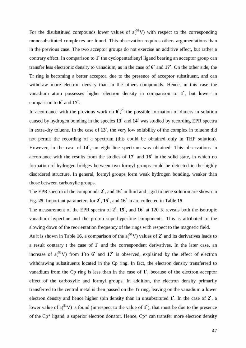
47
For the disubstitued compounds lower values of a(51V) with respect to the corresponding
monosubstituted complexes are found. This observation requires others argumentations than
in the previous case. The two acceptor groups do not exercise an additive effect, but rather a
contrary effect. In comparison to 1• the cyclopentadienyl ligand bearing an acceptor group can
transfer less electronic density to vanadium, as in the case of 6• and 17•. On the other side, the
Tr ring is becoming a better acceptor, due to the presence of acceptor substituent, and can
withdraw more electron density than in the others compounds. Hence, in this case the
vanadium atom possesses higher electron density in comparison to 1•, but lower in
comparison to 6• and 17•.
In accordance with the previous work on 6•,25 the possible formation of dimers in solution
caused by hydrogen bonding in the species 13• and 14• was studied by recording EPR spectra
in extra-dry toluene. In the case of 13•, the very low solubility of the complex in toluene did
not permit the recording of a spectrum (this could be obtained only in THF solution).
However, in the case of 14•, an eight-line spectrum was obtained. This observations in
accordance with the results from the studies of 17• and 16• in the solid state, in which no
formation of hydrogen bridges between two formyl groups could be detected in the highly
disordered structure. In general, formyl groups form weak hydrogen bonding, weaker than
those between carboxylic groups.
The EPR spectra of the compounds 2• , and 16• in fluid and rigid toluene solution are shown in
Fig. 25. Important parameters for 2• , 15•, and 16• in are collected in Table 15.
The measurement of the EPR spectra of 2•, 15•, and 16• at 120 K reveals both the isotropic
vanadium hyperfine and the proton superhyperfine components. This is attributed to the
slowing down of the reorientation frequency of the rings with respect to the magnetic field.
As it is shown in Table 16, a comparison of the a(51V) values of 2• and its derivatives leads to
a result contrary t the case of 1• and the correspondent derivatives. In the later case, an
increase of a(51V) from 1•to 6• and 17• is observed, explained by the effect of electron
withdrawing substituents located in the Cp ring. In fact, the electron density transferred to
vanadium from the Cp ring is less than in the case of 1•, because of the electron acceptor
effect of the carboxylic and formyl groups. In addition, the electron density primarily
transferred to the central metal is then passed on the Tr ring, leaving on the vanadium a lower
electron density and hence higher spin density than in unsubstituted 1•. In the case of 2•, a
lower value of a(51V) is found (in respect to the value of 1•), that must be due to the presence
of the Cp* ligand, a superior electron donator. Hence, Cp* can transfer more electron density
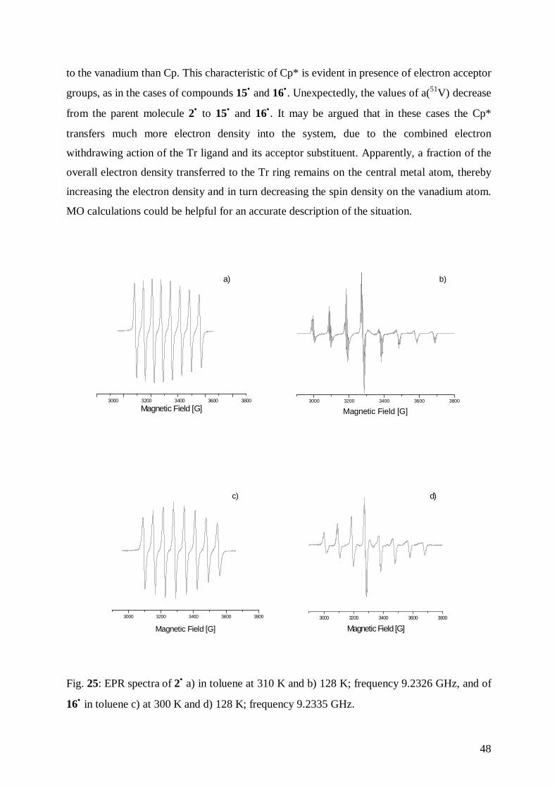
48
to the vanadium than Cp. This characteristic of Cp* is evident in presence of electron acceptor
groups, as in the cases of compounds 15• and 16•. Unexpectedly, the values of a(51V) decrease
from the parent molecule 2• to 15• and 16•. It may be argued that in these cases the Cp*
transfers much more electron density into the system, due to the combined electron
withdrawing action of the Tr ligand and its acceptor substituent. Apparently, a fraction of the
overall electron density transferred to the Tr ring remains on the central metal atom, thereby
increasing the electron density and in turn decreasing the spin density on the vanadium atom.
MO calculations could be helpful for an accurate description of the situation.
3000 3200 3400 3600 3800
Magnetic Field [G]
a)
3000 3200 3400 3600 3800
Magnetic Field [G]
b)
3000 3200 3400 3600 3800
Magnetic Field [G]
c)
3000 3200 3400 3600 3800
Magnetic Field [G]
d)
Fig. 25: EPR spectra of 2• a) in toluene at 310 K and b) 128 K; frequency 9.2326 GHz, and of
16• in toluene c) at 300 K and d) 128 K; frequency 9.2335 GHz.
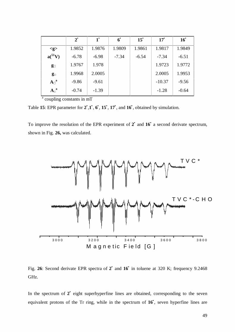
49
2• 1• 6• 15• 17• 16•
<g> 1.9852 1.9876 1.9809 1.9861 1.9817 1.9849
a(51V) -6.78 -6.98 -7.34 -6.54 -7.34 -6.51
g⊥ 1.9767 1.978 1.9723 1.9772
g 1.9968 2.0005 2.0005 1.9953
A⊥a -9.86 -9.61 -10.37 -9.56
A a -0.74 -1.39 -1.28 -0.64a coupling constants in mT
Table 15: EPR parameter for 2•,1• , 6• , 15•, 17• , and 16• , obtained by simulation.
To improve the resolution of the EPR experiment of 2• and 16• a second derivate spectrum,
shown in Fig. 26, was calculated.
3 0 0 0 3 2 0 0 3 4 0 0 3 6 0 0 3 8 0 0
T V C *
T V C * - C H O
M a g n e t ic F ie l d [ G ]
Fig. 26: Second derivate EPR spectra of 2• and 16• in toluene at 320 K; frequency 9.2468
GHz.
In the spectrum of 2• eight superhyperfine lines are obtained, corresponding to the seven
equivalent protons of the Tr ring, while in the spectrum of 16•, seven hyperfine lines are
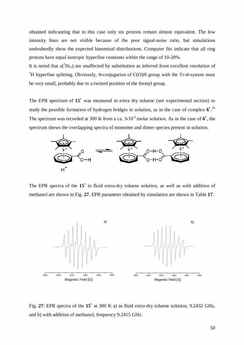
50
obtained indicanting that in this case only six protons remain almost equivalent. The low
intensity lines are not visible because of the poor signal-noise ratio, but simulations
undoubtedly show the expected binominal distributions. Computer fits indicate that all ring
protons have equal isotropic hyperfine constants within the range of 10-20%.
It is noted that a(1HTr) are unaffected by substitution as inferred from excellent resolution of1H hyperfine splitting. Obviously, π-conjugation of C(O)H group with the Tr-σ-system must
be very small, probably due to a twisted position of the formyl group.
The EPR spectrum of 15• was measured in extra dry toluene (see experimental section) to
study the possible formation of hydrogen bridges in solution, as in the case of complex 6• .25
The spectrum was recorded at 300 K from a ca. 3•10-3 molar solution. As in the case of 6• , the
spectrum shows the overlapping spectra of monomer and dimer species present in solution.
The EPR spectra of the 15• in fluid extra-dry toluene solution, as well as with addition of
methanol are shown in Fig. 27. EPR parameter obtained by simulation are shown in Table 17.
2800 3000 3200 3400 3600 3800
Magnetic Field [G]
a)
2800 3000 3200 3400 3600 3800
Magnetic Field [G]
b)
Fig. 27: EPR spectra of the 15• at 300 K a) in fluid extra-dry toluene solution, 9.2432 GHz,
and b) with addition of methanol, frequency 9.2415 GHz.
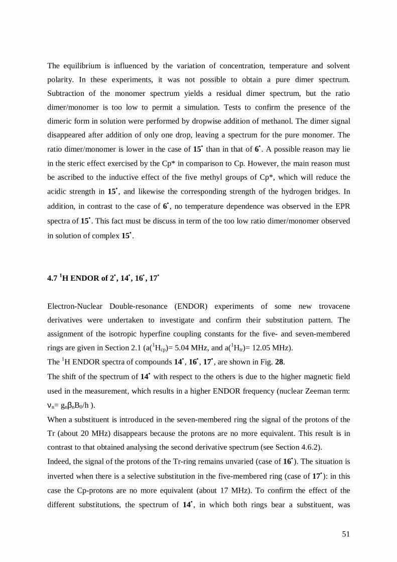
51
The equilibrium is influenced by the variation of concentration, temperature and solvent
polarity. In these experiments, it was not possible to obtain a pure dimer spectrum.
Subtraction of the monomer spectrum yields a residual dimer spectrum, but the ratio
dimer/monomer is too low to permit a simulation. Tests to confirm the presence of the
dimeric form in solution were performed by dropwise addition of methanol. The dimer signal
disappeared after addition of only one drop, leaving a spectrum for the pure monomer. The
ratio dimer/monomer is lower in the case of 15• than in that of 6• . A possible reason may lie
in the steric effect exercised by the Cp* in comparison to Cp. However, the main reason must
be ascribed to the inductive effect of the five methyl groups of Cp*, which will reduce the
acidic strength in 15• , and likewise the corresponding strength of the hydrogen bridges. In
addition, in contrast to the case of 6• , no temperature dependence was observed in the EPR
spectra of 15• . This fact must be discuss in term of the too low ratio dimer/monomer observed
in solution of complex 15• .
4.7 1H ENDOR of 2•, 14•, 16• , 17•
Electron-Nuclear Double-resonance (ENDOR) experiments of some new trovacene
derivatives were undertaken to investigate and confirm their substitution pattern. The
assignment of the isotropic hyperfine coupling constants for the five- and seven-membered
rings are given in Section 2.1 (a(1Hcp)= 5.04 MHz, and a(1Htr)= 12.05 MHz).
The 1H ENDOR spectra of compounds 14• , 16• , 17• , are shown in Fig. 28.
The shift of the spectrum of 14• with respect to the others is due to the higher magnetic field
used in the measurement, which results in a higher ENDOR frequency (nuclear Zeeman term:
νn= gnβnB0/h ).
When a substituent is introduced in the seven-membered ring the signal of the protons of the
Tr (about 20 MHz) disappears because the protons are no more equivalent. This result is in
contrast to that obtained analysing the second derivative spectrum (see Section 4.6.2).
Indeed, the signal of the protons of the Tr-ring remains unvaried (case of 16•). The situation is
inverted when there is a selective substitution in the five-membered ring (case of 17•): in this
case the Cp-protons are no more equivalent (about 17 MHz). To confirm the effect of the
different substitutions, the spectrum of 14• , in which both rings bear a substituent, was
52
recorded. As expected, both signals, that derived from the protons of the Cp, and that from the
protons of the Tr, disappear. (For the parameters assignment see Section 2.1.2).
0 5 10 15 20 25
νΗ
Frequency [MHz]
TVC*
TVC*-CHO
TVC-CHO
TVC-(CHO)2
2.
16.
17.
14.νΗ
Fig. 28: 1H ENDOR spectra in toluene of 2• at 121 K, frequency 9.5178 GHz, microwave
power 13 dB (10 mW), radio frequency power 10 dB (125 W), static field 3373 G; 15• at 121
K, frequency 9.5021 GHz, microwave power 7 dB (42 mW), radio frequency power 3 dB
(500 W), static field 3373 G; 17• at 121 K, frequency 9.5067 GHz, microwave power 7 dB
(42 mW), radio frequency power 3 dB (500 W), static field 3372 G; 14• at 122 K, frequency,
9.4882 GHz, microwave power 3 dB (100 mW), radio frequency power 3 dB (500 W), static
field 3760 G; free proton frequency= 14.38 MHz (spectra of 2• , 15• and 17•); free proton
frequency= 16.03 MHz (spectra of 14•).
The ENDOR spectrum of 2• (shown in Fig. 28) was also recorded to obtain the values of the
proton hyperfine coupling of the methyl groups in the five-membered ring, which are
unaccessible by EPR. The following values of aH(Tr) are obtained: 1A1= 4.7, 1A2= 4.4, 1A3=
3.8 MHz (derived via simulation). From ENDOR the values of aH(Cp) are obtained: 1A1= 2.7
MHz, 1A2= 1.32 MHz, 1A3= 0.12 MHz (directly measured on spectrometer) reflecting an
orthorhombic symmetry. The spectrum shows three lines in the center, one for each methyl
proton. All methyl protons are equivalent, because of their free, fast rotation. Moreover, the
ENDOR spectrum affords the values of A⊥ = 12.79 MHz and A = 11.05 MHz, indicative of
53
axial symmetry. It is important to taking account that the isotropic hyperfine constants in
liquid solution is the time average of all equally possible orientations of the hyperfine tensor
with respect to the quantization axis given by the outer magnetic field (see EPR spectrum
shown in Fig. 26). In frozen solutions, in powder (see ENDOR spectrum shown in Fig. 28),
the complete hyperfine tensor has to be taken. The isotropic part, or better the 1/3 trace of this
tensor is invariant against transformation of coordinates and is comparable with the isotropic
hyperfine constant obtained in liquid solution. Hence, no matter which direction a hyperfine
tensor points to, the isotropic part remains constant. The tensor components of the seven-
membered ring protons in (η7-cycloheptatrienyl)(η5-pentamethylcyclopentadienyl)vanadium,
2• , have equal 1/3 trace <A> value, but different anisotropic parts because of their individual
orientations with respect to the seven-membered ring plane. At 130 K (temperature used in
the measurement) the seven-membered ring is still rotating faster than the interaction time at
the ENDOR experiment. Therefore, time average of all the individual hyperfine tensors are
obtained in the ENDOR experiment. The ENDOR seven-membered ring lines of (η7-
cycloheptatrienyl)(η5-pentamethylcyclopentadienyl)vanadium, 2• , (see also ENDOR of
trovacene, 2•) are axial symmetric and relative sharp. At lower temperatures the lines will be
broaded and finally all anisotropic hyperfine couplings for each proton in the ring will
become visible (T< 4K).79 Substitution of the Cp or Tr ring slows down the ring rotation and
enhances the differences of the anisotropic part as well as the isotropic part of the individual
proton hyperfine constants.
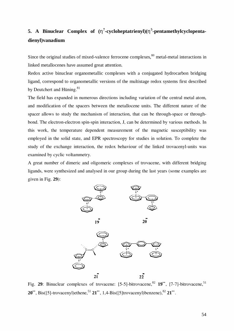
54
5. A Binuclear Complex of (η7-cycloheptatrienyl)(η5-pentamethylcyclopenta-
dienyl)vanadium
Since the original studies of mixed-valence ferrocene complexes,80 metal-metal interactions in
linked metallocenes have assumed great attention.
Redox active binuclear organometallic complexes with a conjugated hydrocarbon bridging
ligand, correspond to organometallic versions of the multistage redox systems first described
by Deutchert and Hüning.81
The field has expanded in numerous directions including variation of the central metal atom,
and modification of the spacers between the metallocene units. The different nature of the
spacer allows to study the mechanism of interaction, that can be through-space or through-
bond. The electron-electron spin-spin interaction, J, can be determined by various methods. In
this work, the temperature dependent measurement of the magnetic susceptibility was
employed in the solid state, and EPR spectroscopy for studies in solution. To complete the
study of the exchange interaction, the redox behaviour of the linked trovacenyl-units was
examined by cyclic voltammetry.
A great number of dimeric and oligomeric complexes of trovacene, with different bridging
ligands, were synthesized and analysed in our group during the last years (some examples are
given in Fig. 29):
Fig. 29: Binuclear complexes of trovacene: [5-5]-bitrovacene,82 19•• , [7-7]-bitrovacene,51
20•• , Bis([5]-trovacenyl)ethene,51 21•• , 1,4-Bis([5]trovacenyl)benzene),82 21•• .
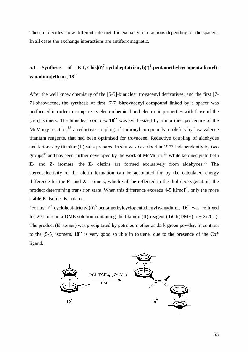
55
These molecules show different intermetallic exchange interactions depending on the spacers.
In all cases the exchange interactions are antiferromagnetic.
5.1 Synthesis of E-1,2-bis[(η7-cycloheptatrienyl)(η5-pentamethylcyclopentadienyl)-
vanadium]ethene, 18••
After the well know chemistry of the [5-5]-binuclear trovacenyl derivatives, and the first [7-
7]-bitrovacene, the synthesis of first [7-7]-bitrovacenyl compound linked by a spacer was
performed in order to compare its electrochemical and electronic properties with those of the
[5-5] isomers. The binuclear complex 18•• was synthesized by a modified procedure of the
McMurry reaction,83 a reductive coupling of carbonyl-compounds to olefins by low-valence
titanium reagents, that had been optimised for trovacene. Reductive coupling of aldehydes
and ketones by titanium(II) salts prepared in situ was described in 1973 independently by two
groups84 and has been further developed by the work of McMurry.85 While ketones yield both
E- and Z- isomers, the E- olefins are formed exclusively from aldehydes.86 The
stereoselectivity of the olefin formation can be accounted for by the calculated energy
difference for the E- and Z- isomers, which will be reflected in the diol deoxygenation, the
product determining transition state. When this difference exceeds 4-5 kJmol-1, only the more
stable E- isomer is isolated.
(Formyl-η7-cycloheptatrienyl)(η5-pentamethylcyclopentadienyl)vanadium, 16• was refluxed
for 20 hours in a DME solution containing the titanium(II)-reagent (TiCl3(DME)1,5 + Zn/Cu).
The product (E isomer) was precipitated by petroleum ether as dark-green powder. In contrast
to the [5-5] isomers, 18•• is very good soluble in toluene, due to the presence of the Cp*
ligand.
56
5.2 Cyclic Voltammetry of 18••
5.2.1 Fundamentals of Cyclic Voltammetry of Binuclear Species
In bridged compounds two types of interaction between the two moieties are possible:
interaction propagated through the bond (arising from an electronic system delocalised over a
diamagnetic spacer or by the mechanism of spin-polarization of the electrons in the double
occupied bridging ligand orbitals by the unpaired electrons) and through space (electric field
effect, that describes the direct overlapping of both singuly occupied atomic orbitals).
Generally, a decreasing interaction with increasing metal-metal distance is observed. The
redox properties of dinuclear complexes of transition metal ions depend on the metal, the
chemical nature of the bridging ligands, and the geometry of the bridge.
Cyclic voltammetry is a method to study the metal-metal interactions via the parameter of the
redox splitting δE1/2 (potential difference of two subsequent redox steps), complementary to
the determination of the exchange interaction, J, between the electronic spins of the metal
centers. The redox splitting results from the repulsion between equal charges. In a complex in
which the two metal centers are separated from each other two cases are possible:
a) when an electron is transferred from one metal center, the second electron is transferred to
the other metal center at the same potential (δE1/2 < 70 mV);
b) when the distance between the two metal centers is small, there is electrostatic repulsion
between the first negative charge and the second added negative charge, hence, a shift to
cathodic potential is observed in the second electron transfer.
The difference of potential between both electron transfers is the redox splitting δE1/2. δE1/2 is
a measure of the extent of the interaction between both metal centers. Presupposition for a
determination of δE1/2 from the cyclic voltammogram is the reversibility of the electron
transfer.
In an electrochemical experiment,87 a separation, ∆E, of (RT/F)ln2n is observed between the
first and last redox events.88
For a molecule with two redox centers, a separation of approximately 36 mV should be
expected at 20 °C (rather smaller separation than that typically resolvable in routine
electrochemical experiments). Larger, measurable separations are indicative of an interaction
between the two sites.
An intramolecular electron transfer can occur between two metal centers of different
oxidation states. It can be distinguished between two types of compounds:
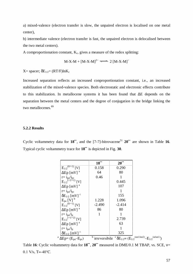
57
a) mixed-valence (electron transfer is slow, the unpaired electron is localised on one metal
center),
b) intermediate valence (electron transfer is fast, the unpaired electron is delocalised between
the two metal centers).
A comproportionation constant, Kc, gives a measure of the redox splitting:
M-X-M + [M-X-M]2− 2 [M-X-M]−
X= spacer; δE1/2= (RT/F)lnKc
Increased separation reflects an increased conproportionation constant, i.e., an increased
stabilization of the mixed-valence species. Both electrostatic and electronic effects contribute
to this stabilization. In metallocene systems it has been found that ∆E depends on the
separation between the metal centers and the degree of conjugation in the bridge linking the
two metallocenes.89
5.2.2 Results
Cyclic voltammetry data for 18•• , and the [7-7]-bitrovacene51 20•• are shown in Table 16.
Typical cyclic voltammetry trace for 18•• is depicted in Fig. 30.
18•• 20••
E1/2(0/+1) [V] 0.158 0.290
∆Ep [mV] a 64 80r= ipa/ipc 0.46 1E1/2
(+2/+1) [V] 0.445∆Ep [mV] a 107r= ipa/ipc 1δE1/2 [mV] c 155Epa [V] b 1.228 1.096E1/2
(0/−1) [V] -2.490 -2.414∆Ep [mV] a 86 80r= ipa/ip 1 1E1/2
(−1/−2) [V] 2.739∆Ep [mV] a 63r= ipa/ip 1δE1/2 [mV] c 325
a ∆Ep= (Epa–Epc)b irreversibile c δE1/2= (E1/2
(n±1/n±2)–E1/2(n/n±1))
Table 16: Cyclic voltammetry data for 18•• , 20•• measured in DME/0.1 M TBAP, vs. SCE, ν=
0.1 V/s, T=-40°C.
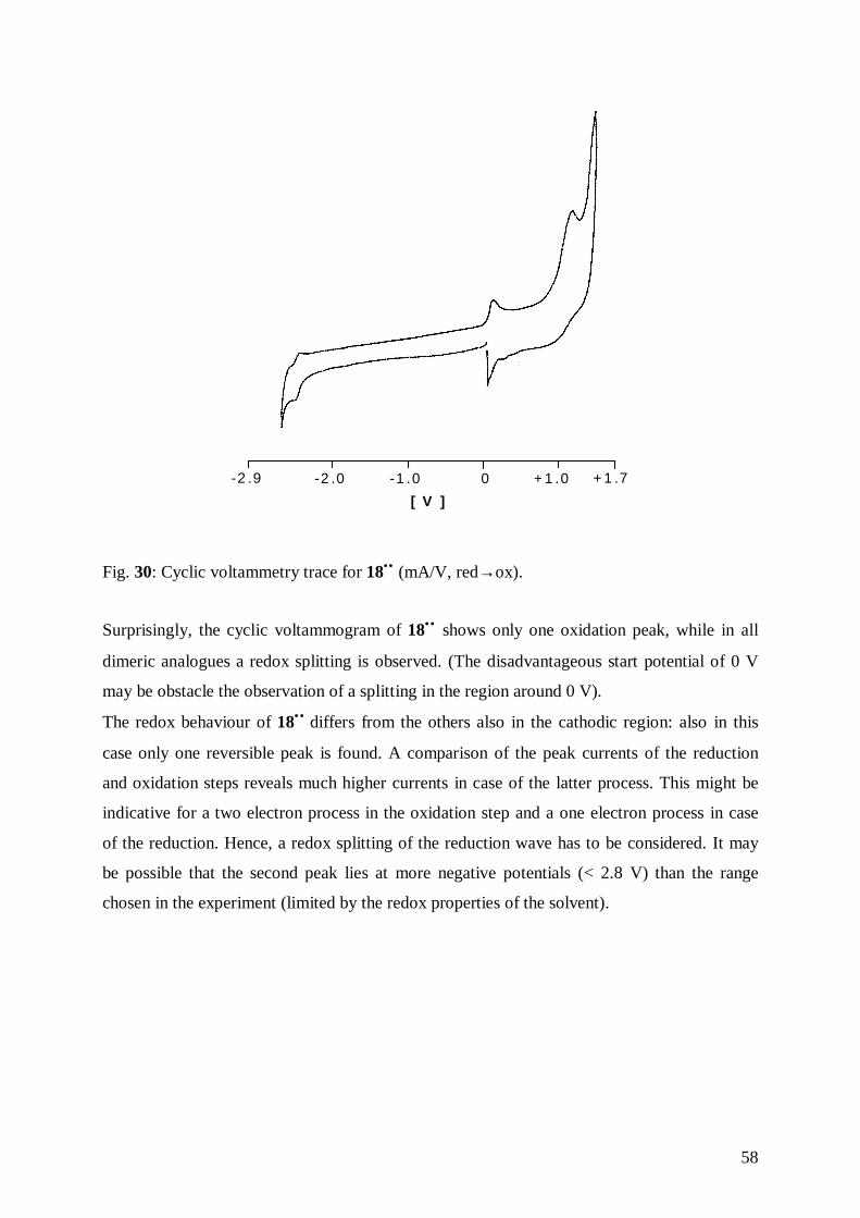
58
+ 1 .7+ 1 .00-1 .0-2 .0-2 .9
[ V ]
Fig. 30: Cyclic voltammetry trace for 18•• (mA/V, red→ox).
Surprisingly, the cyclic voltammogram of 18•• shows only one oxidation peak, while in all
dimeric analogues a redox splitting is observed. (The disadvantageous start potential of 0 V
may be obstacle the observation of a splitting in the region around 0 V).
The redox behaviour of 18•• differs from the others also in the cathodic region: also in this
case only one reversible peak is found. A comparison of the peak currents of the reduction
and oxidation steps reveals much higher currents in case of the latter process. This might be
indicative for a two electron process in the oxidation step and a one electron process in case
of the reduction. Hence, a redox splitting of the reduction wave has to be considered. It may
be possible that the second peak lies at more negative potentials (< 2.8 V) than the range
chosen in the experiment (limited by the redox properties of the solvent).
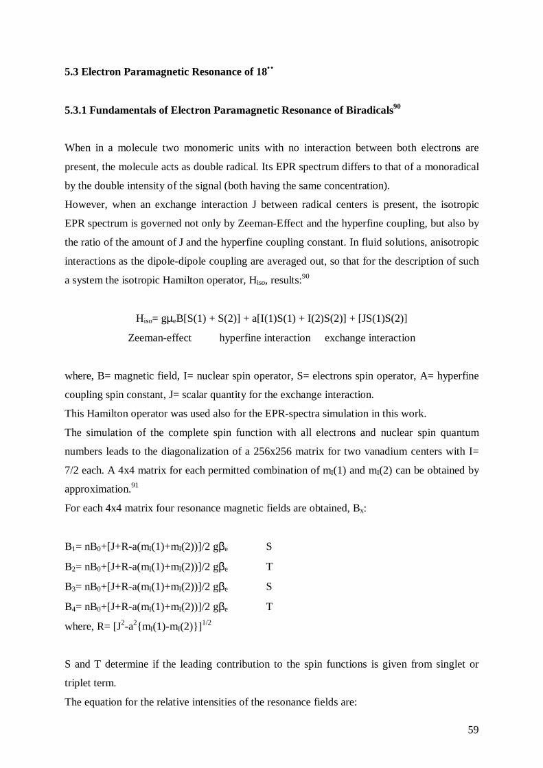
59
5.3 Electron Paramagnetic Resonance of 18••
5.3.1 Fundamentals of Electron Paramagnetic Resonance of Biradicals90
When in a molecule two monomeric units with no interaction between both electrons are
present, the molecule acts as double radical. Its EPR spectrum differs to that of a monoradical
by the double intensity of the signal (both having the same concentration).
However, when an exchange interaction J between radical centers is present, the isotropic
EPR spectrum is governed not only by Zeeman-Effect and the hyperfine coupling, but also by
the ratio of the amount of J and the hyperfine coupling constant. In fluid solutions, anisotropic
interactions as the dipole-dipole coupling are averaged out, so that for the description of such
a system the isotropic Hamilton operator, Hiso, results:90
Hiso= gµeB[S(1) + S(2)] + a[I(1)S(1) + I(2)S(2)] + [JS(1)S(2)]
Zeeman-effect hyperfine interaction exchange interaction
where, B= magnetic field, I= nuclear spin operator, S= electrons spin operator, A= hyperfine
coupling spin constant, J= scalar quantity for the exchange interaction.
This Hamilton operator was used also for the EPR-spectra simulation in this work.
The simulation of the complete spin function with all electrons and nuclear spin quantum
numbers leads to the diagonalization of a 256x256 matrix for two vanadium centers with I=
7/2 each. A 4x4 matrix for each permitted combination of mI(1) and mI(2) can be obtained by
approximation.91
For each 4x4 matrix four resonance magnetic fields are obtained, Bx:
B1= nB0+[J+R-a(mI(1)+mI(2))]/2 gβe S
B2= nB0+[J+R-a(mI(1)+mI(2))]/2 gβe T
B3= nB0+[J+R-a(mI(1)+mI(2))]/2 gβe S
B4= nB0+[J+R-a(mI(1)+mI(2))]/2 gβe T
where, R= [J2-a2{mI(1)-mI(2)}]1/2
S and T determine if the leading contribution to the spin functions is given from singlet or
triplet term.
The equation for the relative intensities of the resonance fields are:
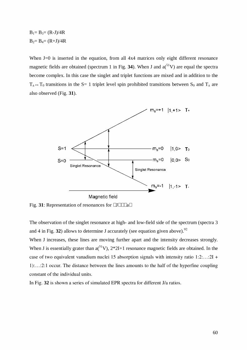
60
B1= B3= (R-J)/4R
B2= B4= (R+J)/4R
When J=0 is inserted in the equation, from all 4x4 matrices only eight different resonance
magnetic fields are obtained (spectrum 1 in Fig. 34). When J and a(51V) are equal the spectra
become complex. In this case the singlet and triplet functions are mixed and in addition to the
T±↔T0 transitions in the S= 1 triplet level spin prohibited transitions between S0 and T± are
also observed (Fig. 31).
Fig. 31: Representation of resonances for J≅ a
The observation of the singlet resonance at high- and low-field side of the spectrum (spectra 3
and 4 in Fig. 32) allows to determine J accurately (see equation given above).92
When J increases, these lines are moving further apart and the intensity decreases strongly.
When J is essentially grater than a(51V), 2*2I+1 resonance magnetic fields are obtained. In the
case of two equivalent vanadium nuclei 15 absorption signals with intensity ratio 1:2:…:2I +
1):…:2:1 occur. The distance between the lines amounts to the half of the hyperfine coupling
constant of the individual units.
In Fig. 32 is shown a series of simulated EPR spectra for different J/a ratios.
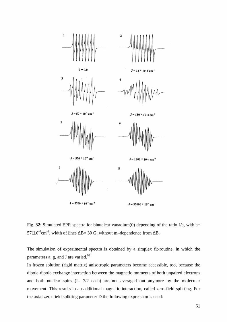
61
Fig. 32: Simulated EPR-spectra for binuclear vanadium(0) depending of the ratio J/a, with a=
57∗ 10-4cm-1, width of lines ∆B= 30 G, without mI-dependence from ∆B.
The simulation of experimental spectra is obtained by a simplex fit-routine, in which the
parameters a, g, and J are varied.93
In frozen solution (rigid matrix) anisotropic parameters become accessible, too, because the
dipole-dipole exchange interaction between the magnetic moments of both unpaired electrons
and both nuclear spins (I= 7/2 each) are not averaged out anymore by the molecular
movement. This results in an additional magnetic interaction, called zero-field splitting. For
the axial zero-field splitting parameter D the following expression is used:
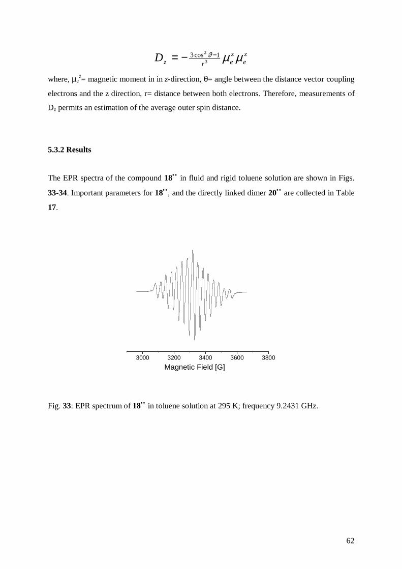
62
ze
zerzD µµϑ
3
2 1cos3 −−=
where, µez= magnetic moment in in z-direction, θ= angle between the distance vector coupling
electrons and the z direction, r= distance between both electrons. Therefore, measurements of
Dz permits an estimation of the average outer spin distance.
5.3.2 Results
The EPR spectra of the compound 18•• in fluid and rigid toluene solution are shown in Figs.
33-34. Important parameters for 18•• , and the directly linked dimer 20•• are collected in Table
17.
3000 3200 3400 3600 3800
Magnetic Field [G]
Fig. 33: EPR spectrum of 18•• in toluene solution at 295 K; frequency 9.2431 GHz.
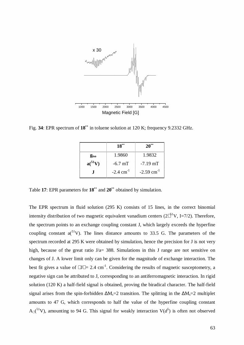
63
1000 1500 2000 2500 3000 3500 4000 4500
Magnetic Field [G]
x 30
Fig. 34: EPR spectrum of 18•• in toluene solution at 120 K; frequency 9.2332 GHz.
18•• 20••
giso 1.9860 1.9832
a(51V) -6.7 mT -7.19 mT
J -2.4 cm-1 -2.59 cm-1
Table 17: EPR parameters for 18•• and 20•• obtained by simulation.
The EPR spectrum in fluid solution (295 K) consists of 15 lines, in the correct binomial
intensity distribution of two magnetic equivalent vanadium centers (2∗ 51V, I=7/2). Therefore,
the spectrum points to an exchange coupling constant J, which largely exceeds the hyperfine
coupling constant a(51V). The lines distance amounts to 33.5 G. The parameters of the
spectrum recorded at 295 K were obtained by simulation, hence the precision for J is not very
high, because of the great ratio J/a= 388. Simulations in this J range are not sensitive on
changes of J. A lower limit only can be given for the magnitude of exchange interaction. The
best fit gives a value of J = 2.4 cm-1. Considering the results of magnetic susceptometry, a
negative sign can be attributed to J, corresponding to an antiferromagnetic interaction. In rigid
solution (120 K) a half-field signal is obtained, proving the biradical character. The half-field
signal arises from the spin-forbidden ∆Ms=2 transition. The splitting in the ∆Ms=2 multiplet
amounts to 47 G, which corresponds to half the value of the hyperfine coupling constant
A⊥ (51V), amounting to 94 G. This signal for weakly interaction V(d5) is often not observed
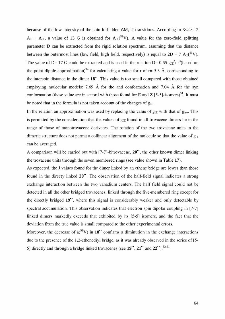
64
because of the low intensity of the spin-forbidden ∆Ms=2 transitions. According to 3<a>= 2
A⊥ + A , a value of 13 G is obtained for A (51V). A value for the zero-field splitting
parameter D can be extracted from the rigid solution spectrum, assuming that the distance
between the outermost lines (low field, high field, respectively) is equal to 2D + 7 A⊥ (51V).
The value of D= 17 G could be extracted and is used in the relation D= 0.65 g2/ r3(based on
the point-dipole approximation)94 for calculating a value for r of r= 5.3 Å, corresponding to
the interspin distance in the dimer 18•• . This value is too small compared with those obtained
employing molecular models: 7.69 Å for the anti conformation and 7.04 Å for the syn
conformation (these value are in accord with those found for E and Z [5-5]-isomers)51. It must
be noted that in the formula is not taken account of the changes of g .
In the relation an approximation was used by replacing the value of g with that of giso. This
is permitted by the consideration that the values of g found in all trovacene dimers lie in the
range of those of monotrovacene derivates. The rotation of the two trovacene units in the
dimeric structure does not permit a collinear alignment of the molecule so that the value of g
can be averaged.
A comparison will be carried out with [7-7]-bitrovacene, 20•• , the other known dimer linking
the trovacene units through the seven membered rings (see value shown in Table 17).
As expected, the J values found for the dimer linked by an ethene bridge are lower than those
found in the directy linked 20•• . The observation of the half-field signal indicates a strong
exchange interaction between the two vanadium centers. The half field signal could not be
detected in all the other bridged trovacenes, linked through the five-membered ring except for
the directly bridged 19•• , where this signal is considerably weaker and only detectable by
spectral accumulation. This observation indicates that electron spin dipolar coupling in [7-7]
linked dimers markedly exceeds that exhibited by its [5-5] isomers, and the fact that the
deviation from the true value is small compared to the other experimental errors.
Moreover, the decrease of a(51V) in 18•• confirms a diminution in the exchange interactions
due to the presence of the 1,2-ethenediyl bridge, as it was already observed in the series of [5-
5] directly and through a bridge linked trovacenes (see 19•• , 21•• and 22•• ).82,51
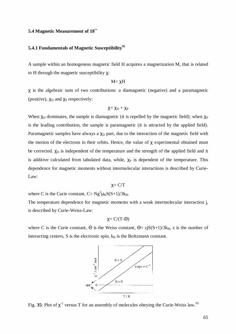
65
5.4 Magnetic Measurement of 18••
5.4.1 Fundamentals of Magnetic Susceptibility95
A sample within an homogeneus magnetic field H acquires a magnetization M, that is related
to H through the magnetic susceptibility χ:
M= χH
χ is the algebraic sum of two contributions: a diamagnetic (negative) and a paramagnetic
(positive), χD and χP respectively:
χ= χD + χP
When χD dominates, the sample is diamagnetic (it is repelled by the magnetic field); when χP
is the leading contribution, the sample is paramagnetic (it is attracted by the applied field).
Paramagnetic samples have always a χD part, due to the interaction of the magnetic field with
the motion of the electrons in their orbits. Hence, the value of χ experimental obtained must
be corrected. χD is independent of the temperature and the strength of the applied field and it
is additive calculated from tabulated data, while, χP is dependent of the temperature. This
dependence for magnetic moments without intermolecular interactions is described by Curie-
Law:
χ= C/T
where C is the Curie constant, C= Ng2µBS(S+1)/3kB.
The temperature dependence for magnetic moments with a weak intermolecular interaction j,
is described by Curie-Weiss-Law:
χ= C/(T-Θ)
where C is the Curie constant, Θ is the Weiss constant, Θ= zjS(S+1)/3kB, z is the number of
interacting centers, S is the electronic spin, kB is the Boltzmann constant.
Fig. 35: Plot of χ-1 versus T for an assembly of molecules obeying the Curie-Weiss law.95
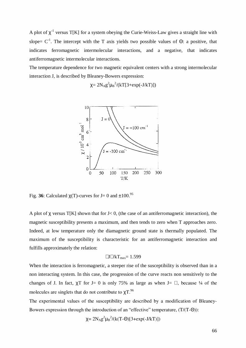
66
A plot of χ-1 versus T[K] for a system obeying the Curie-Weiss-Law gives a straight line with
slope= C-1. The intercept with the T axis yields two possible values of Θ: a positive, that
indicates ferromagnetic intermolecular interactions, and a negative, that indicates
antiferromagnetic intermolecular interactions.
The temperature dependence for two magnetic equivalent centers with a strong intermolecular
interaction J, is described by Bleaney-Bowers expression:
χ= 2NAg2µB2/(kT[3+exp(-J/kT)])
Fig. 36: Calculated χ(T)-curves for J= 0 and ±100.95
A plot of χ versus T[K] shown that for J< 0, (the case of an antiferromagnetic interaction), the
magnetic susceptibility presents a maximum, and then tends to zero when T approaches zero.
Indeed, at low temperature only the diamagnetic ground state is thermally populated. The
maximum of the susceptibility is characteristic for an antiferromagnetic interaction and
fulfills approximately the relation:
J /kTmax= 1.599
When the interaction is ferromagnetic, a steeper rise of the susceptibility is observed than in a
non interacting system. In this case, the progression of the curve reacts non sensitively to the
changes of J. In fact, χT for J= 0 is only 75% as large as when J= ∝ , because ¼ of the
molecules are singlets that do not contribute to χT.96
The experimental values of the susceptibility are described by a modification of Bleaney-
Bowers expression through the introduction of an “effective” temperature, (T/(T-Θ)):
χ= 2NAg2µB2/(k(T-Θ)[3+exp(-J/kT)])
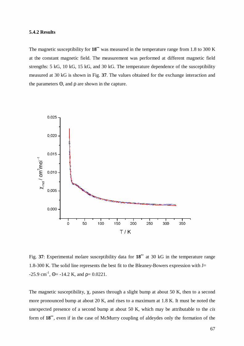
67
5.4.2 Results
The magnetic susceptibility for 18•• was measured in the temperature range from 1.8 to 300 K
at the constant magnetic field. The measurement was performed at different magnetic field
strengths: 5 kG, 10 kG, 15 kG, and 30 kG. The temperature dependence of the susceptibility
measured at 30 kG is shown in Fig. 37. The values obtained for the exchange interaction and
the parameters Θ, and ρ are shown in the capture.
Fig. 37: Experimental molare susceptibility data for 18•• at 30 kG in the temperature range
1.8-300 K. The solid line represents the best fit to the Bleaney-Bowers expression with J=
-25.9 cm-1, Θ= -14.2 K, and ρ= 0.0221.
The magnetic susceptibility, χ, passes through a slight bump at about 50 K, then to a second
more pronounced bump at about 20 K, and rises to a maximum at 1.8 K. It must be noted the
unexpected presence of a second bump at about 50 K, which may be attributable to the cis
form of 18•• , even if in the case of McMurry coupling of aldeydes only the formation of the
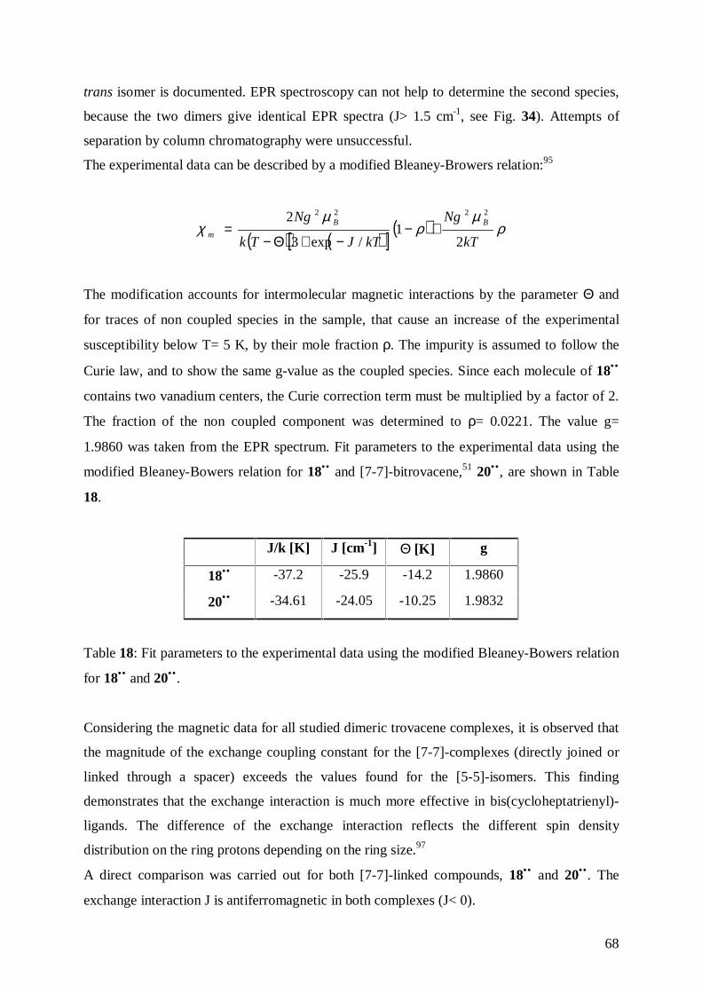
68
trans isomer is documented. EPR spectroscopy can not help to determine the second species,
because the two dimers give identical EPR spectra (J> 1.5 cm-1, see Fig. 34). Attempts of
separation by column chromatography were unsuccessful.
The experimental data can be described by a modified Bleaney-Browers relation:95
( ) ( )[ ]( ) ρ
µρ
µχ
kT
Ng
kTJTk
Ng BB
m2
1/exp3
2 2222
+−−+Θ−
=
The modification accounts for intermolecular magnetic interactions by the parameter Θ and
for traces of non coupled species in the sample, that cause an increase of the experimental
susceptibility below T= 5 K, by their mole fraction ρ. The impurity is assumed to follow the
Curie law, and to show the same g-value as the coupled species. Since each molecule of 18••
contains two vanadium centers, the Curie correction term must be multiplied by a factor of 2.
The fraction of the non coupled component was determined to ρ= 0.0221. The value g=
1.9860 was taken from the EPR spectrum. Fit parameters to the experimental data using the
modified Bleaney-Bowers relation for 18•• and [7-7]-bitrovacene,51 20•• , are shown in Table
18.
J/k [K] J [cm-1] Θ [K] g
18•• -37.2 -25.9 -14.2 1.9860
20•• -34.61 -24.05 -10.25 1.9832
Table 18: Fit parameters to the experimental data using the modified Bleaney-Bowers relation
for 18•• and 20•• .
Considering the magnetic data for all studied dimeric trovacene complexes, it is observed that
the magnitude of the exchange coupling constant for the [7-7]-complexes (directly joined or
linked through a spacer) exceeds the values found for the [5-5]-isomers. This finding
demonstrates that the exchange interaction is much more effective in bis(cycloheptatrienyl)-
ligands. The difference of the exchange interaction reflects the different spin density
distribution on the ring protons depending on the ring size.97
A direct comparison was carried out for both [7-7]-linked compounds, 18•• and 20•• . The
exchange interaction J is antiferromagnetic in both complexes (J< 0).
69
The value of J obtained by magnetic measurement corresponds to the sum of J (symmetric
anisotropic), J (asymmetric anisotropic), and J (isotropic). The value of J (isotropic) can not
be determined alone by magnetic measurements. In the EPR experiments the anisotropic
values of J are cancelled due to the molecular motion. In addition, EPR measurements in
dilute solutions are not disturbed by intermolecular interactions. In magnetic measurements in
the solid state strong intermolecular interactions can be anticipated, because it is reasonable to
believe that the crystal structure must be similar to those of 19•• and 20•• , in which the
vanadium-vanadium intramolecular distance is comparable to the intermoleculare distance
between adjacent molecules.98 Unfortunately, J obtained by EPR spectroscopy are limited to
values below 1.5 cm-1.
In the case of the complex 18•• (in which the two monomer units are linked by a spacer), a
lower value of J than for 20•• (in which the two monomer units directly linked) was expected.
A possible reason for this result is the presence of the Cp* ligand, which posses amplified
donor capacity in comparison to the Cp ligand. In the case of the dimer 18•• spin density is
transfer into the spacer, resulting in a stronger overlap of orbitals, which finally leads to the
unexpected high value of J.
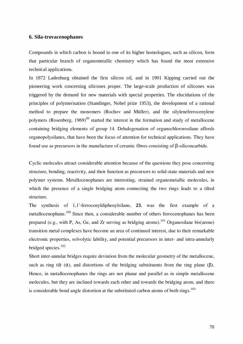
70
6. Sila-trovacenophanes
Compounds in which carbon is bound to one of its higher homologues, such as silicon, form
that particular branch of organometallic chemistry which has found the most extensive
technical applications.
In 1872 Ladenburg obtained the first silicon oil, and in 1901 Kipping carried out the
pioneering work concerning silicones proper. The large-scale production of silicones was
triggered by the demand for new materials with special properties. The elucidations of the
principles of polymerisation (Standinger, Nobel prize 1953), the development of a rational
method to prepare the monomers (Rochov and Müller), and the silyleneferrocenylene
polymers (Rosenberg, 1969)99 started the interest in the formation and study of metallocene
containing bridging elements of group 14. Dehalogenation of organochlororosilane affords
organopolysilanes, that have been the focus of attention for technical applications. They have
found use as precursors in the manufacture of ceramic fibres consisting of β-siliconcarbide.
Cyclic molecules attract considerable attention because of the questions they pose concerning
structure, bonding, reactivity, and their function as precursors to solid-state materials and new
polymer systems. Metallocenophanes are interesting, strained organometallic molecules, in
which the presence of a single bridging atom connecting the two rings leads to a tilted
structure.
The synthesis of 1,1'-ferrocenyldiphenylsilane, 23, was the first example of a
metallocenophane.100 Since then, a considerable number of others ferrocenophanes has been
prepared (e.g., with P, As, Ge, and Zr serving as bridging atoms).101 Organosilane bis(arene)
transition metal complexes have become an area of continued interest, due to their remarkable
electronic properties, solvolytic lability, and potential precursors in inter- and intra-annularly
bridged species.102
Short inter-annular bridges require deviation from the molecular geometry of the metallocene,
such as ring tilt (α), and distortions of the bridging substituents from the ring plane (β).
Hence, in metallocenophanes the rings are not planar and parallel as in simple metallocene
molecules, but they are inclined towards each other and towards the bridging atom, and there
is considerable bond angle distortion at the substituted carbon atoms of both rings.103
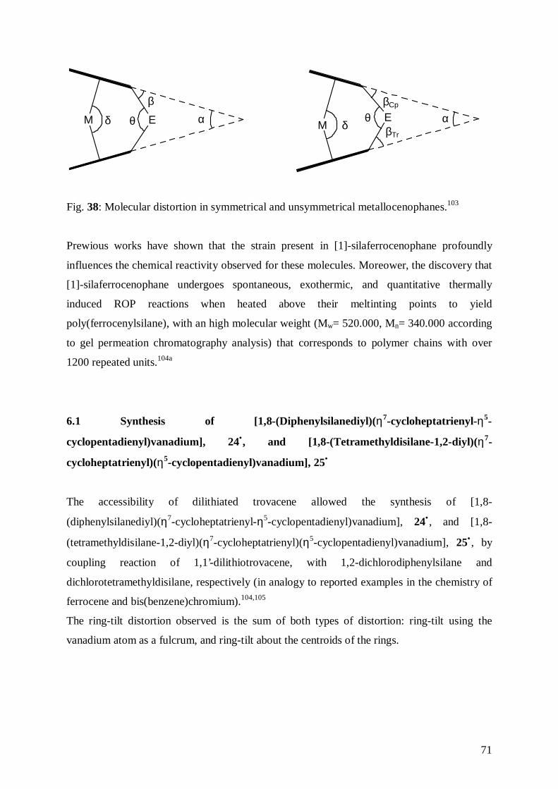
71
E αβ
θδM ME αβCp
βTr
θδ
Fig. 38: Molecular distortion in symmetrical and unsymmetrical metallocenophanes.103
Prewious works have shown that the strain present in [1]-silaferrocenophane profoundly
influences the chemical reactivity observed for these molecules. Moreower, the discovery that
[1]-silaferrocenophane undergoes spontaneous, exothermic, and quantitative thermally
induced ROP reactions when heated above their meltinting points to yield
poly(ferrocenylsilane), with an high molecular weight (Mw= 520.000, Mn= 340.000 according
to gel permeation chromatography analysis) that corresponds to polymer chains with over
1200 repeated units.104a
6.1 Synthesis of [1,8-(Diphenylsilanediyl)(η7-cycloheptatrienyl-η5-
cyclopentadienyl)vanadium], 24•, and [1,8-(Tetramethyldisilane-1,2-diyl)(η7-
cycloheptatrienyl)(η5-cyclopentadienyl)vanadium], 25•
The accessibility of dilithiated trovacene allowed the synthesis of [1,8-
(diphenylsilanediyl)(η7-cycloheptatrienyl-η5-cyclopentadienyl)vanadium], 24• , and [1,8-
(tetramethyldisilane-1,2-diyl)(η7-cycloheptatrienyl)(η5-cyclopentadienyl)vanadium], 25• , by
coupling reaction of 1,1’-dilithiotrovacene, with 1,2-dichlorodiphenylsilane and
dichlorotetramethyldisilane, respectively (in analogy to reported examples in the chemistry of
ferrocene and bis(benzene)chromium).104,105
The ring-tilt distortion observed is the sum of both types of distortion: ring-tilt using the
vanadium atom as a fulcrum, and ring-tilt about the centroids of the rings.
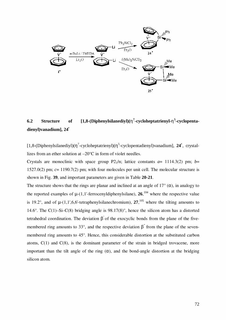
72
6.2 Structure of [1,8-(Diphenylsilanediyl)(η7-cycloheptatrienyl-η5-cyclopenta-
dienyl)vanadium], 24•
[1,8-(Diphenylsilanediyl)(η7-cycloheptatrienyl)(η5-cyclopentadienyl)vanadium], 24• , crystal-
lizes from an ether solution at –20°C in form of violet needles.
Crystals are monoclinic with space group P21/n; lattice constants a= 1114.3(2) pm; b=
1527.0(2) pm; c= 1190.7(2) pm; with four molecules per unit cell. The molecular structure is
shown in Fig. 39, and important parameters are given in Table 20-21.
The structure shows that the rings are planar and inclined at an angle of 17° (α), in analogy to
the reported examples of µ-(1,1'-ferrocenyldiphenylsilane), 26,104 where the respective value
is 19.2°, and of µ-(1,1',6,6'-tetraphenylsilanechromium), 27,105 where the tilting amounts to
14.6°. The C(1)–Si–C(8) bridging angle is 98.17(8)°, hence the silicon atom has a distorted
tetrahedral coordination. The deviation β’ of the exocyclic bonds from the plane of the five-
membered ring amounts to 33°, and the respective deviation β’’ from the plane of the seven-
membered ring amounts to 45°. Hence, this considerable distortion at the substituted carbon
atoms, C(1) and C(8), is the dominant parameter of the strain in bridged trovacene, more
important than the tilt angle of the ring (α), and the bond-angle distortion at the bridging
silicon atom.
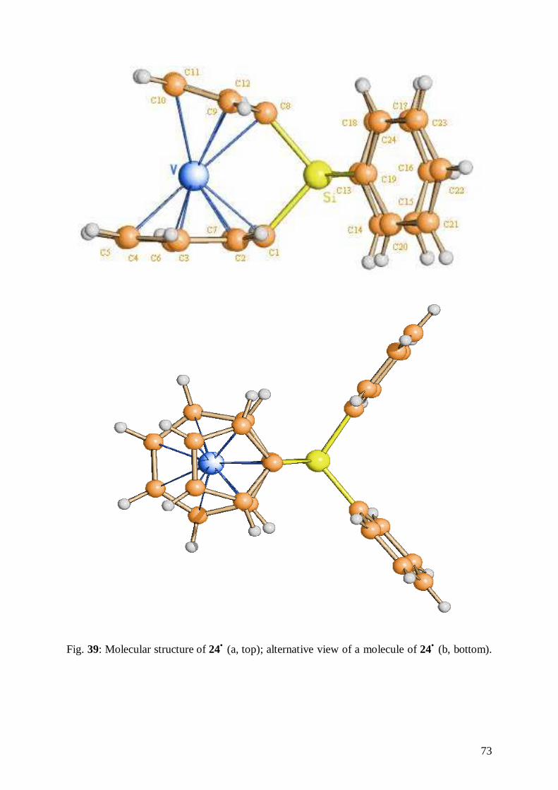
73
Fig. 39: Molecular structure of 24• (a, top); alternative view of a molecule of 24• (b, bottom).
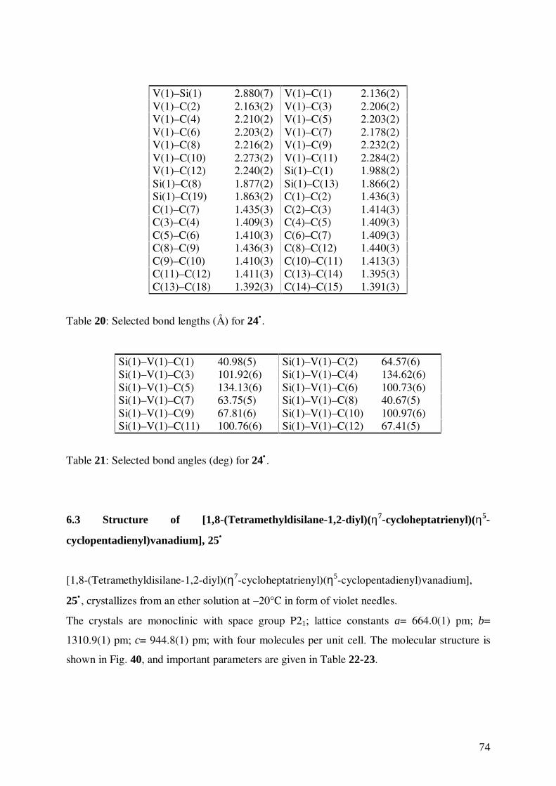
74
V(1)–Si(1) 2.880(7) V(1)–C(1) 2.136(2)V(1)–C(2) 2.163(2) V(1)–C(3) 2.206(2)V(1)–C(4) 2.210(2) V(1)–C(5) 2.203(2)V(1)–C(6) 2.203(2) V(1)–C(7) 2.178(2)V(1)–C(8) 2.216(2) V(1)–C(9) 2.232(2)V(1)–C(10) 2.273(2) V(1)–C(11) 2.284(2)V(1)–C(12) 2.240(2) Si(1)–C(1) 1.988(2)Si(1)–C(8) 1.877(2) Si(1)–C(13) 1.866(2)Si(1)–C(19) 1.863(2) C(1)–C(2) 1.436(3)C(1)–C(7) 1.435(3) C(2)–C(3) 1.414(3)C(3)–C(4) 1.409(3) C(4)–C(5) 1.409(3)C(5)–C(6) 1.410(3) C(6)–C(7) 1.409(3)C(8)–C(9) 1.436(3) C(8)–C(12) 1.440(3)C(9)–C(10) 1.410(3) C(10)–C(11) 1.413(3)C(11)–C(12) 1.411(3) C(13)–C(14) 1.395(3)C(13)–C(18) 1.392(3) C(14)–C(15) 1.391(3)
Table 20: Selected bond lengths (Å) for 24•.
Si(1)–V(1)–C(1) 40.98(5) Si(1)–V(1)–C(2) 64.57(6)Si(1)–V(1)–C(3) 101.92(6) Si(1)–V(1)–C(4) 134.62(6)Si(1)–V(1)–C(5) 134.13(6) Si(1)–V(1)–C(6) 100.73(6)Si(1)–V(1)–C(7) 63.75(5) Si(1)–V(1)–C(8) 40.67(5)Si(1)–V(1)–C(9) 67.81(6) Si(1)–V(1)–C(10) 100.97(6)Si(1)–V(1)–C(11) 100.76(6) Si(1)–V(1)–C(12) 67.41(5)
Table 21: Selected bond angles (deg) for 24• .
6.3 Structure of [1,8-(Tetramethyldisilane-1,2-diyl)(η7-cycloheptatrienyl)(η5-
cyclopentadienyl)vanadium], 25•
[1,8-(Tetramethyldisilane-1,2-diyl)(η7-cycloheptatrienyl)(η5-cyclopentadienyl)vanadium],
25• , crystallizes from an ether solution at –20°C in form of violet needles.
The crystals are monoclinic with space group P21; lattice constants a= 664.0(1) pm; b=
1310.9(1) pm; c= 944.8(1) pm; with four molecules per unit cell. The molecular structure is
shown in Fig. 40, and important parameters are given in Table 22-23.
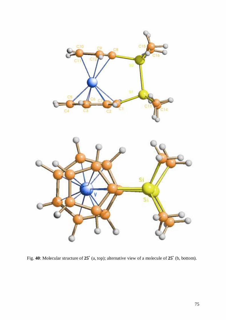
75
Fig. 40: Molecular structure of 25• (a, top); alternative view of a molecule of 25• (b, bottom).
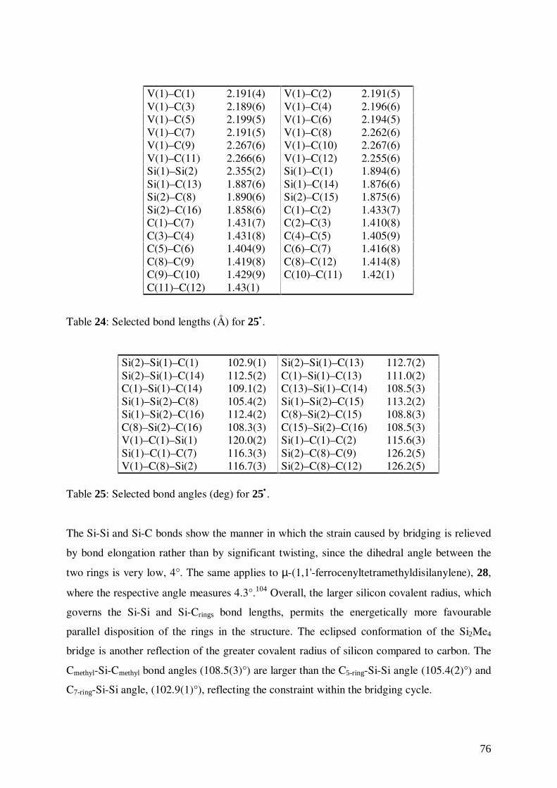
76
V(1)–C(1) 2.191(4) V(1)–C(2) 2.191(5)V(1)–C(3) 2.189(6) V(1)–C(4) 2.196(6)V(1)–C(5) 2.199(5) V(1)–C(6) 2.194(5)V(1)–C(7) 2.191(5) V(1)–C(8) 2.262(6)V(1)–C(9) 2.267(6) V(1)–C(10) 2.267(6)V(1)–C(11) 2.266(6) V(1)–C(12) 2.255(6)Si(1)–Si(2) 2.355(2) Si(1)–C(1) 1.894(6)Si(1)–C(13) 1.887(6) Si(1)–C(14) 1.876(6)Si(2)–C(8) 1.890(6) Si(2)–C(15) 1.875(6)Si(2)–C(16) 1.858(6) C(1)–C(2) 1.433(7)C(1)–C(7) 1.431(7) C(2)–C(3) 1.410(8)C(3)–C(4) 1.431(8) C(4)–C(5) 1.405(9)C(5)–C(6) 1.404(9) C(6)–C(7) 1.416(8)C(8)–C(9) 1.419(8) C(8)–C(12) 1.414(8)C(9)–C(10) 1.429(9) C(10)–C(11) 1.42(1)C(11)–C(12) 1.43(1)
Table 24: Selected bond lengths (Å) for 25•.
Si(2)–Si(1)–C(1) 102.9(1) Si(2)–Si(1)–C(13) 112.7(2)Si(2)–Si(1)–C(14) 112.5(2) C(1)–Si(1)–C(13) 111.0(2)C(1)–Si(1)–C(14) 109.1(2) C(13)–Si(1)–C(14) 108.5(3)Si(1)–Si(2)–C(8) 105.4(2) Si(1)–Si(2)–C(15) 113.2(2)Si(1)–Si(2)–C(16) 112.4(2) C(8)–Si(2)–C(15) 108.8(3)C(8)–Si(2)–C(16) 108.3(3) C(15)–Si(2)–C(16) 108.5(3)V(1)–C(1)–Si(1) 120.0(2) Si(1)–C(1)–C(2) 115.6(3)Si(1)–C(1)–C(7) 116.3(3) Si(2)–C(8)–C(9) 126.2(5)V(1)–C(8)–Si(2) 116.7(3) Si(2)–C(8)–C(12) 126.2(5)
Table 25: Selected bond angles (deg) for 25• .
The Si-Si and Si-C bonds show the manner in which the strain caused by bridging is relieved
by bond elongation rather than by significant twisting, since the dihedral angle between the
two rings is very low, 4°. The same applies to µ-(1,1'-ferrocenyltetramethyldisilanylene), 28,
where the respective angle measures 4.3°.104 Overall, the larger silicon covalent radius, which
governs the Si-Si and Si-Crings bond lengths, permits the energetically more favourable
parallel disposition of the rings in the structure. The eclipsed conformation of the Si2Me4
bridge is another reflection of the greater covalent radius of silicon compared to carbon. The
Cmethyl-Si-Cmethyl bond angles (108.5(3)°) are larger than the C5-ring-Si-Si angle (105.4(2)°) and
C7-ring-Si-Si angle, (102.9(1)°), reflecting the constraint within the bridging cycle.
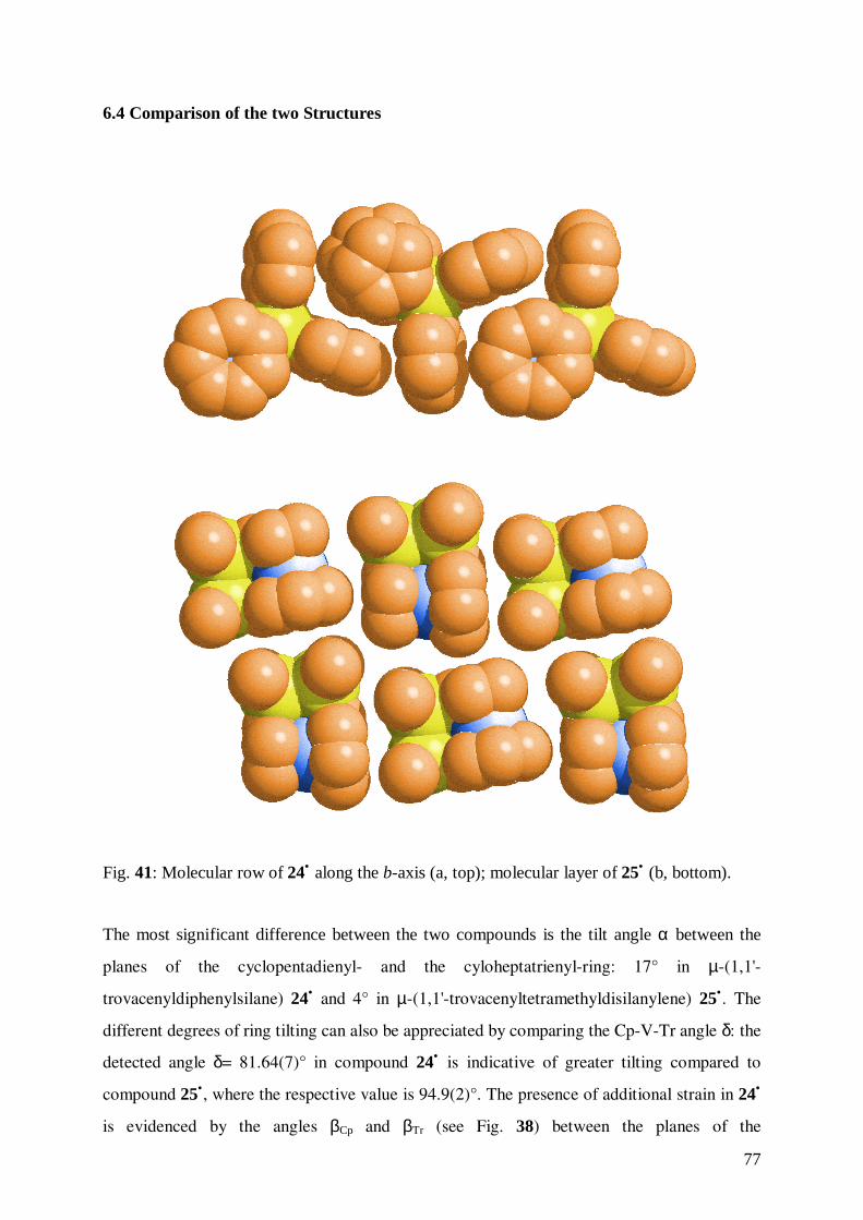
77
6.4 Comparison of the two Structures
Fig. 41: Molecular row of 24• along the b-axis (a, top); molecular layer of 25• (b, bottom).
The most significant difference between the two compounds is the tilt angle α between the
planes of the cyclopentadienyl- and the cyloheptatrienyl-ring: 17° in µ-(1,1'-
trovacenyldiphenylsilane) 24• and 4° in µ-(1,1'-trovacenyltetramethyldisilanylene) 25•. The
different degrees of ring tilting can also be appreciated by comparing the Cp-V-Tr angle δ: the
detected angle δ= 81.64(7)° in compound 24• is indicative of greater tilting compared to
compound 25•, where the respective value is 94.9(2)°. The presence of additional strain in 24•
is evidenced by the angles βCp and βTr (see Fig. 38) between the planes of the
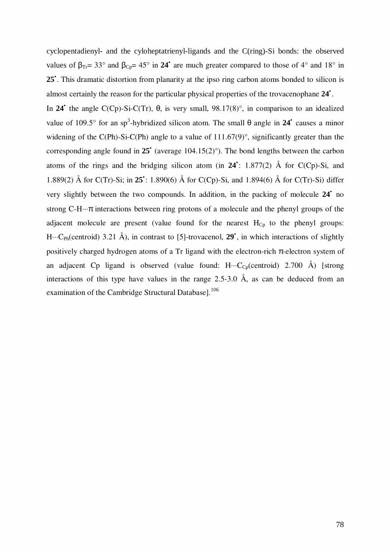
78
cyclopentadienyl- and the cyloheptatrienyl-ligands and the C(ring)-Si bonds: the observed
values of βTr= 33° and βCp= 45° in 24• are much greater compared to those of 4° and 18° in
25• . This dramatic distortion from planarity at the ipso ring carbon atoms bonded to silicon is
almost certainly the reason for the particular physical properties of the trovacenophane 24• .
In 24• the angle C(Cp)-Si-C(Tr), θ, is very small, 98.17(8)°, in comparison to an idealized
value of 109.5° for an sp3-hybridized silicon atom. The small θ angle in 24• causes a minor
widening of the C(Ph)-Si-C(Ph) angle to a value of 111.67(9)°, significantly greater than the
corresponding angle found in 25• (average 104.15(2)°). The bond lengths between the carbon
atoms of the rings and the bridging silicon atom (in 24• : 1.877(2) Å for C(Cp)-Si, and
1.889(2) Å for C(Tr)-Si; in 25• : 1.890(6) Å for C(Cp)-Si, and 1.894(6) Å for C(Tr)-Si) differ
very slightly between the two compounds. In addition, in the packing of molecule 24• no
strong C-H···π interactions between ring protons of a molecule and the phenyl groups of the
adjacent molecule are present (value found for the nearest HCp to the phenyl groups:
H···CPh(centroid) 3.21 Å), in contrast to [5]-trovacenol, 29• , in which interactions of slightly
positively charged hydrogen atoms of a Tr ligand with the electron-rich π-electron system of
an adjacent Cp ligand is observed (value found: H···CCp(centroid) 2.700 Å) [strong
interactions of this type have values in the range 2.5-3.0 Å, as can be deduced from an
examination of the Cambridge Structural Database].106
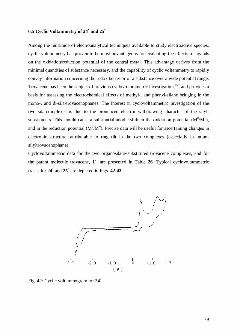
79
6.5 Cyclic Voltammetry of 24• and 25•
Among the multitude of electroanalytical techniques available to study electroactive species,
cyclic voltammetry has proven to be most advantageous for evaluating the effects of ligands
on the oxidation/reduction potential of the central metal. This advantage derives from the
minimal quantities of substance necessary, and the capability of cyclic voltammetry to rapidly
convey information concerning the redox behavior of a substance over a wide potential range.
Trovacene has been the subject of previous cyclovoltammetric investigation,107 and provides a
basis for assessing the electrochemical effects of methyl-, and phenyl-silane bridging in the
mono-, and di-sila-trovacenophanes. The interest in cyclovoltammetric investigation of the
two sila-complexes is due to the pronunced electron-withdrawing character of the silyl-
substituents. This should cause a substantial anodic shift in the oxidation potential (M0/M+),
and in the reduction potential (M0/M−). Precise data will be useful for ascertaining changes in
electronic structure, attributable to ring tilt in the two complexes (especially in mono-
silyltrovacenophane).
Cyclovoltammetric data for the two organosilane-substituted trovacene complexes, and for
the parent molecule trovacene, 1• , are presented in Table 26. Typical cyclovoltammetric
traces for 24• and 25• are depicted in Figs. 42-43.
+ 1 .7+1 .00-1 .0-2 .0-2 .9
[ V ]
Fig. 42: Cyclic voltammogram for 24• .
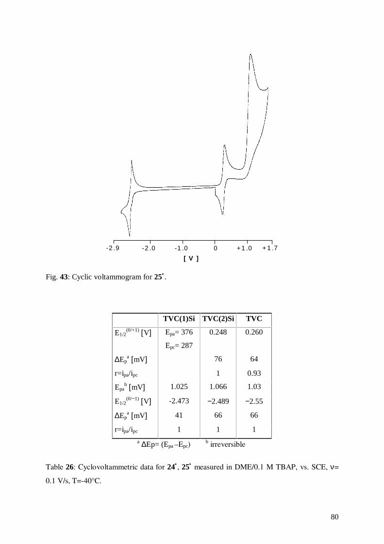
80
+ 1 .7+1 .00-1 .0-2 .0-2 .9
[ V ]
Fig. 43: Cyclic voltammogram for 25• .
TVC(1)Si TVC(2)Si TVC
E1/2(0/+1) [V] Epa= 376
Epc= 287
0.248 0.260
∆Epa [mV] 76 64
r=ipa/ipc 1 0.93
Epab [mV] 1.025 1.066 1.03
E1/2(0/−1) [V] -2.473 −2.489 −2.55
∆Epa [mV] 41 66 66
r=ipa/ipc 1 1 1
a ∆Ep= (Epa –Epc)b irreversible
Table 26: Cyclovoltammetric data for 24• , 25• measured in DME/0.1 M TBAP, vs. SCE, ν=
0.1 V/s, T=-40°C.
81
The silatrovacenophane 25• displays an oxidation potential almost equal to that of the parent
complex trovacene, 1• . This finding may be rationalized in terms of electron donation from
the silicon-silicon σ bond to the rings π orbitals (σ-π conjugation), and a withdrawal effect of
ring electron density through silicon d orbital overlap. The sum of both effects gives no
oxidation potential shift.
Due to the high sensitivity of the strained trovacenophane 24• , bearing a single silicon atom
bridge, the cyclic voltammogram is reproducible at the scan rates used in this study only for a
few cycles. This cycling results in a gradual decomposition that produces a transient
uncharacterized species. This fact reflects the aggregate action of silyl-substitution and
bending of the sandwich structure. It may be postulated a ring opening induced by electron
transfer. The same effect has been reported also for the corresponding ferrocenophane,108 and
for the respective bis(benzene)chromium derivative,109 where a collection of data is prevented
by its high sensitivity towards bridge cleavage. Oxidation of the metal center promotes bridge
cleavage, thereby releasing the considerable structural strain inflicted by the bridging unit. In
contrast, the reversibility noted for 25•, reflects the lower degree of structural strain due to the
-R4Si2- bridge. In this context, the data are in accordance with those collected for the
analoguous bis(benzene)chromium derivative.
The quantum mechanical calculations of Hoffman and Lauher110 for the titanocene molecule,
Cp2Ti, are helpful to explain the effect of deformation of the sandwich structure in
bis(arene)metal-types, especially the tilting of the ring-metal-ring axis, on their redox
potential. The authors studied the dependence of the energies of frontier orbitals on the tilting
angle θ (see Figs. 44 and 45). An important result is the destabilization of a1-orbitals, that
reflects itself in a cathodic shift of the redox potential of the tilted complex with respect to its
undistorted analogue. The change in reactivity induced by tilting is based not only on the
increased steric interaction between the rings and their substituents, but also on the
modification of the nature and energy of the frontier orbitals. Hence, the illustrated results
predict an easier oxidation of a tilted metallocene.
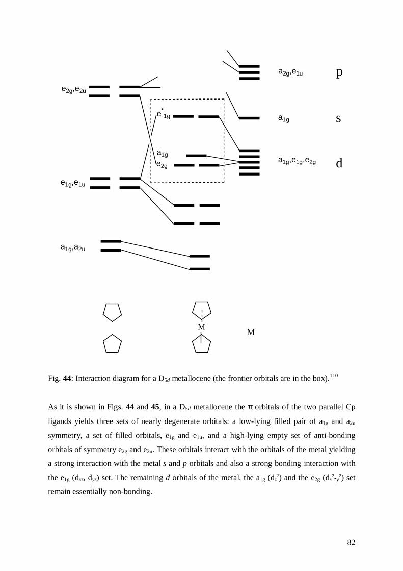
82
a1g,e1g,e2g
a1g
a2g,e1u
e*1g
a1g
e2g
e2g,e2u
e1g,e1u
a1g,a2u
s
p
d
MM
Fig. 44: Interaction diagram for a D5d metallocene (the frontier orbitals are in the box).110
As it is shown in Figs. 44 and 45, in a D5d metallocene the π orbitals of the two parallel Cp
ligands yields three sets of nearly degenerate orbitals: a low-lying filled pair of a1g and a2u
symmetry, a set of filled orbitals, e1g and e1u, and a high-lying empty set of anti-bonding
orbitals of symmetry e2g and e2u. These orbitals interact with the orbitals of the metal yielding
a strong interaction with the metal s and p orbitals and also a strong bonding interaction with
the e1g (dxz, dyz) set. The remaining d orbitals of the metal, the a1g (dz2) and the e2g (dx
2-y2) set
remain essentially non-bonding.
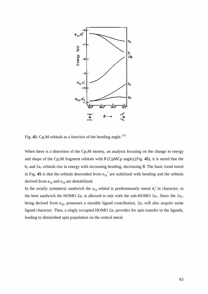
83
Fig. 45: Cp2M orbitals as a function of the bending angle.110
When there is a distortion of the Cp2M moiety, an analysis focusing on the change in energy
and shape of the Cp2M fragment orbitals with θ (CpMCp angle),(Fig. 45), it is noted that the
b2 and 2a1 orbitals rise in energy with increasing bending, decreasing θ. The basic trend noted
in Fig. 45 is that the orbitals descended from e1g* are stabilized with bending and the orbitals
derived from a1g and e2g are destabilized.
In the axially symmetric sandwich the a1g orbital is predominantly metal dz2 in character; in
the bent sandwich the HOMO 2a1 is allowed to mix with the sub-HOMO 1a1. Since the 1a1,
being derived from e2g, possesses a sizeable ligand contribution, 2a1 will also acquire some
ligand character. Thus, a singly occupied HOMO 2a1 provides for spin transfer to the ligands,
leading to diminished spin population on the central metal.
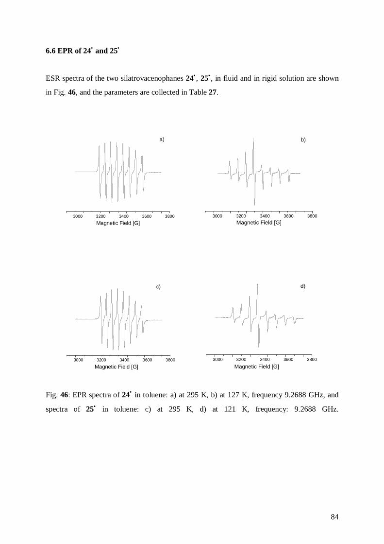
84
6.6 EPR of 24• and 25•
ESR spectra of the two silatrovacenophanes 24• , 25• , in fluid and in rigid solution are shown
in Fig. 46, and the parameters are collected in Table 27.
3000 3200 3400 3600 3800
Magnetic Field [G]
a)
3000 3200 3400 3600 3800
Magnetic Field [G]
b)
3000 3200 3400 3600 3800
Magnetic Field [G]
c)
3000 3200 3400 3600 3800
Magnetic Field [G]
d)
Fig. 46: EPR spectra of 24• in toluene: a) at 295 K, b) at 127 K, frequency 9.2688 GHz, and
spectra of 25• in toluene: c) at 295 K, d) at 121 K, frequency: 9.2688 GHz.
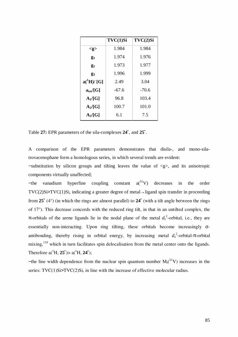
85
TVC(1)Si TVC(2)Si
<g> 1.984 1.984
g1 1.974 1.976
g2 1.973 1.977
g3 1.996 1.999
a(1H)/ [G] 2.49 3.04
aiso/[G] -67.6 -70.6
A1/[G] 96.8 103.4
A2/[G] 100.7 101.0
A3/[G] 6.1 7.5
Table 27: EPR parameters of the sila-complexes 24• , and 25• .
A comparison of the EPR parameters demonstrates that disila-, and mono-sila-
trovacenophane form a homologous series, in which several trends are evident:
−substitution by silicon groups and tilting leaves the value of <g>, and its anisotropic
components virtually unaffected;
−the vanadium hyperfine coupling constant a(51V) decreases in the order
TVC(2)Si>TVC(1)Si, indicating a greater degree of metal→ligand spin transfer in proceeding
from 25• (4°) (in which the rings are almost parallel) to 24• (with a tilt angle between the rings
of 17°). This decrease concords with the reduced ring tilt, in that in an untilted complex, the
π-orbitals of the arene ligands lie in the nodal plane of the metal dz2-orbital, i.e., they are
essentially non-interacting. Upon ring tilting, these orbitals become increasingly σ-
antibonding, thereby rising in orbital energy, by increasing metal dz2-orbital-π-orbital
mixing,110 which in turn facilitates spin delocalisation from the metal center onto the ligands.
Therefore a(1H, 25•)> a(1H, 24•);
−the line width dependence from the nuclear spin quantum number MI(51V) increases in the
series: TVC(1)Si>TVC(2)Si, in line with the increase of effective molecular radius.
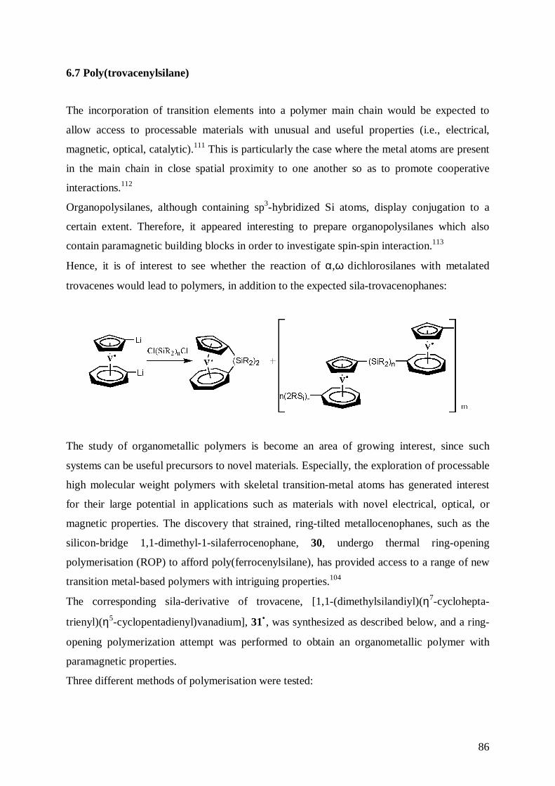
86
6.7 Poly(trovacenylsilane)
The incorporation of transition elements into a polymer main chain would be expected to
allow access to processable materials with unusual and useful properties (i.e., electrical,
magnetic, optical, catalytic).111 This is particularly the case where the metal atoms are present
in the main chain in close spatial proximity to one another so as to promote cooperative
interactions.112
Organopolysilanes, although containing sp3-hybridized Si atoms, display conjugation to a
certain extent. Therefore, it appeared interesting to prepare organopolysilanes which also
contain paramagnetic building blocks in order to investigate spin-spin interaction.113
Hence, it is of interest to see whether the reaction of α,ω dichlorosilanes with metalated
trovacenes would lead to polymers, in addition to the expected sila-trovacenophanes:
The study of organometallic polymers is become an area of growing interest, since such
systems can be useful precursors to novel materials. Especially, the exploration of processable
high molecular weight polymers with skeletal transition-metal atoms has generated interest
for their large potential in applications such as materials with novel electrical, optical, or
magnetic properties. The discovery that strained, ring-tilted metallocenophanes, such as the
silicon-bridge 1,1-dimethyl-1-silaferrocenophane, 30, undergo thermal ring-opening
polymerisation (ROP) to afford poly(ferrocenylsilane), has provided access to a range of new
transition metal-based polymers with intriguing properties.104
The corresponding sila-derivative of trovacene, [1,1-(dimethylsilandiyl)(η7-cyclohepta-
trienyl)(η5-cyclopentadienyl)vanadium], 31• , was synthesized as described below, and a ring-
opening polymerization attempt was performed to obtain an organometallic polymer with
paramagnetic properties.
Three different methods of polymerisation were tested:

87
Since ring strain provides an important driving force for ring-opening polymerisation, 31•
would be an excellent candidate for polymerisation studies. The monomer 31• was
synthesised by slow addition of dimethyldichlorosilane to 1-1’-dilithiotrovacene, affording a
violet oil. Unexpectedly, the elementary analysis of the product showed a nitrogen content of
2.6%, which may be attributed to an incorporation of TMEDA that was used as chelating
agent in the dimetalation of trovacene. In the analysis of the fragmentation pattern of the mass
spectrum, in addition to the molecular peak of the monomer (m/z= 264), an unexpected peak
at m/z= 734, corresponding to a trimer after loss of a dimethylsilyl unit, was observed. This
result shows that a successful polymerisation cannot be excluded.
Fig. 47: Fragmentation pattern in the mass spectrum of the product 31• .
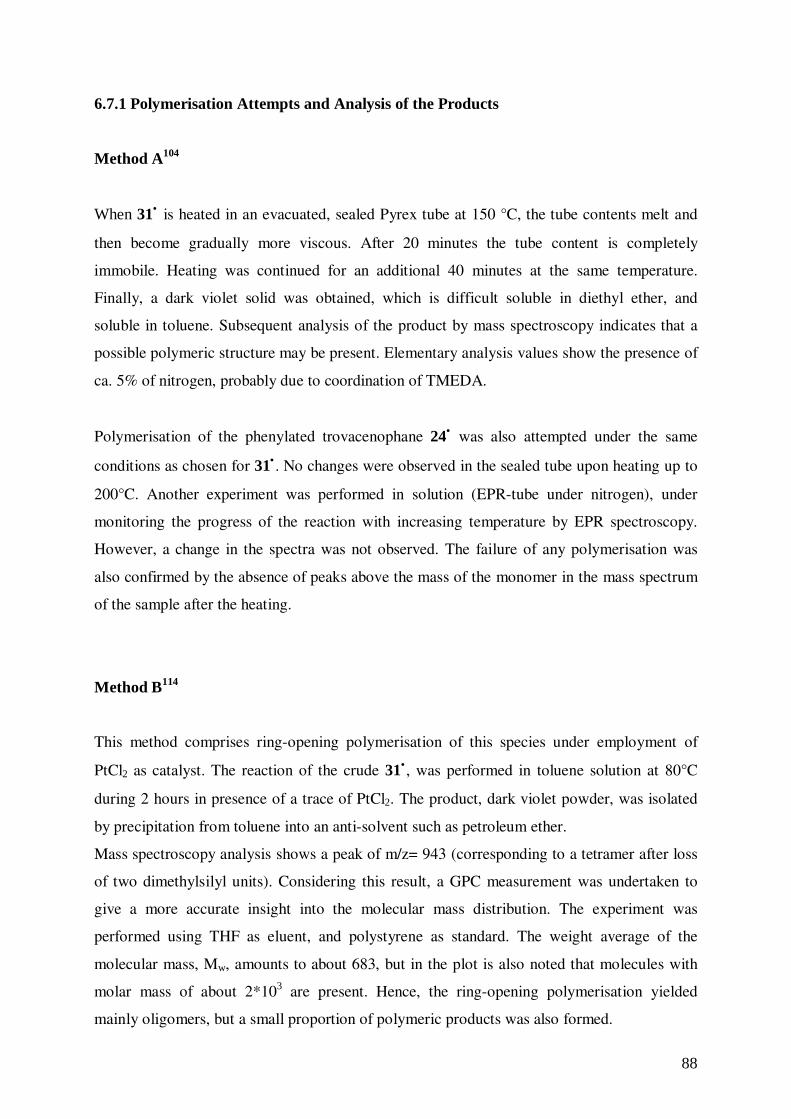
88
6.7.1 Polymerisation Attempts and Analysis of the Products
Method A104
When 31• is heated in an evacuated, sealed Pyrex tube at 150 °C, the tube contents melt and
then become gradually more viscous. After 20 minutes the tube content is completely
immobile. Heating was continued for an additional 40 minutes at the same temperature.
Finally, a dark violet solid was obtained, which is difficult soluble in diethyl ether, and
soluble in toluene. Subsequent analysis of the product by mass spectroscopy indicates that a
possible polymeric structure may be present. Elementary analysis values show the presence of
ca. 5% of nitrogen, probably due to coordination of TMEDA.
Polymerisation of the phenylated trovacenophane 24• was also attempted under the same
conditions as chosen for 31• . No changes were observed in the sealed tube upon heating up to
200°C. Another experiment was performed in solution (EPR-tube under nitrogen), under
monitoring the progress of the reaction with increasing temperature by EPR spectroscopy.
However, a change in the spectra was not observed. The failure of any polymerisation was
also confirmed by the absence of peaks above the mass of the monomer in the mass spectrum
of the sample after the heating.
Method B114
This method comprises ring-opening polymerisation of this species under employment of
PtCl2 as catalyst. The reaction of the crude 31• , was performed in toluene solution at 80°C
during 2 hours in presence of a trace of PtCl2. The product, dark violet powder, was isolated
by precipitation from toluene into an anti-solvent such as petroleum ether.
Mass spectroscopy analysis shows a peak of m/z= 943 (corresponding to a tetramer after loss
of two dimethylsilyl units). Considering this result, a GPC measurement was undertaken to
give a more accurate insight into the molecular mass distribution. The experiment was
performed using THF as eluent, and polystyrene as standard. The weight average of the
molecular mass, Mw, amounts to about 683, but in the plot is also noted that molecules with
molar mass of about 2*103 are present. Hence, the ring-opening polymerisation yielded
mainly oligomers, but a small proportion of polymeric products was also formed.
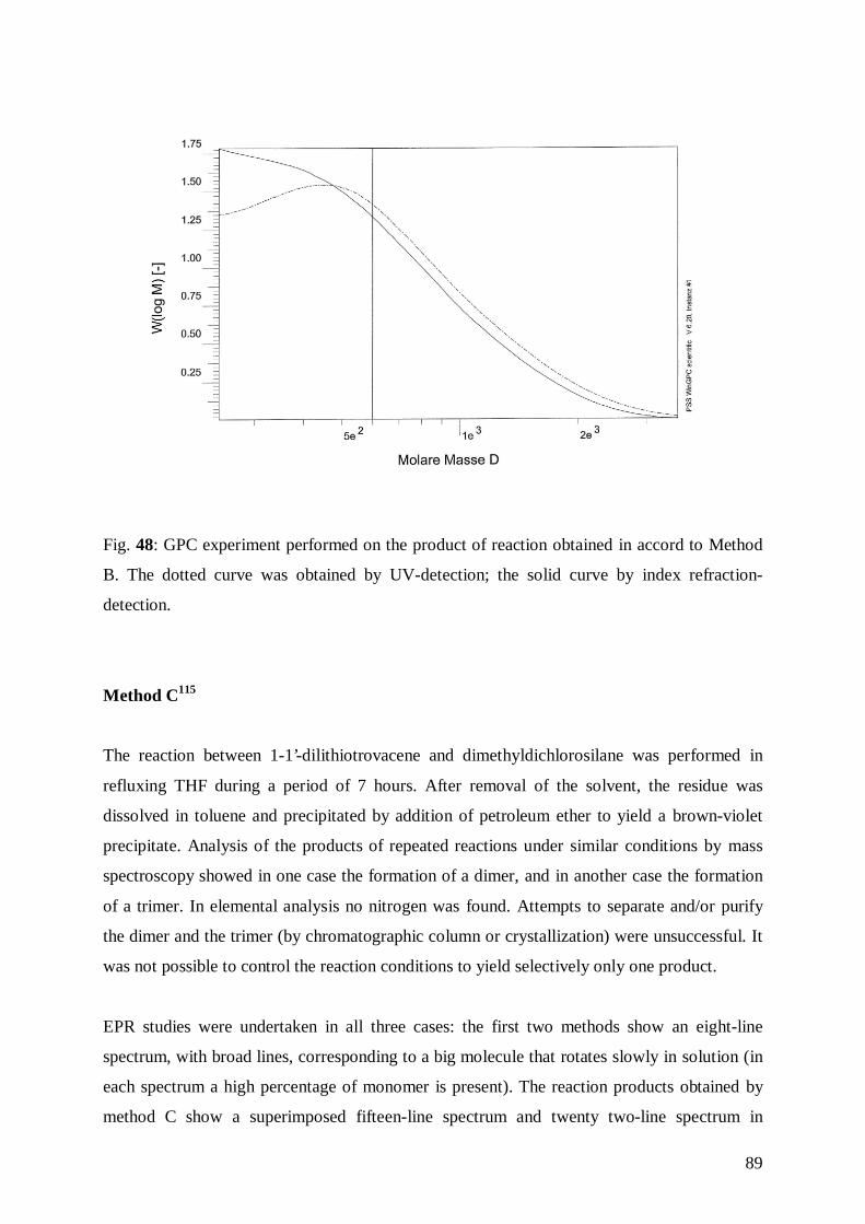
89
Fig. 48: GPC experiment performed on the product of reaction obtained in accord to Method
B. The dotted curve was obtained by UV-detection; the solid curve by index refraction-
detection.
Method C115
The reaction between 1-1’-dilithiotrovacene and dimethyldichlorosilane was performed in
refluxing THF during a period of 7 hours. After removal of the solvent, the residue was
dissolved in toluene and precipitated by addition of petroleum ether to yield a brown-violet
precipitate. Analysis of the products of repeated reactions under similar conditions by mass
spectroscopy showed in one case the formation of a dimer, and in another case the formation
of a trimer. In elemental analysis no nitrogen was found. Attempts to separate and/or purify
the dimer and the trimer (by chromatographic column or crystallization) were unsuccessful. It
was not possible to control the reaction conditions to yield selectively only one product.
EPR studies were undertaken in all three cases: the first two methods show an eight-line
spectrum, with broad lines, corresponding to a big molecule that rotates slowly in solution (in
each spectrum a high percentage of monomer is present). The reaction products obtained by
method C show a superimposed fifteen-line spectrum and twenty two-line spectrum in
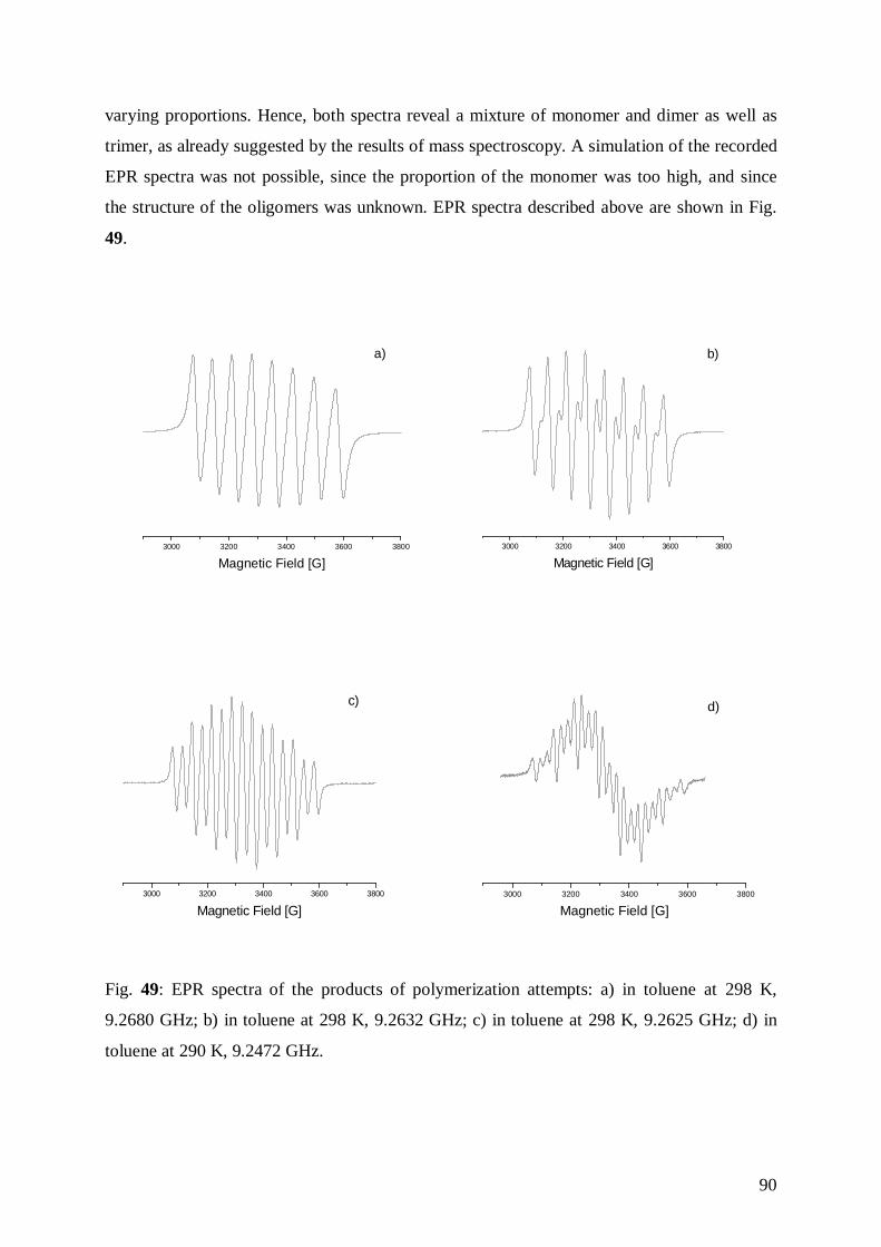
90
varying proportions. Hence, both spectra reveal a mixture of monomer and dimer as well as
trimer, as already suggested by the results of mass spectroscopy. A simulation of the recorded
EPR spectra was not possible, since the proportion of the monomer was too high, and since
the structure of the oligomers was unknown. EPR spectra described above are shown in Fig.
49.
3000 3200 3400 3600 3800
Magnetic Field [G]
a)
3000 3200 3400 3600 3800
Magnetic Field [G]
b)
3000 3200 3400 3600 3800
Magnetic Field [G]
c)
3000 3200 3400 3600 3800
Magnetic Field [G]
d)
Fig. 49: EPR spectra of the products of polymerization attempts: a) in toluene at 298 K,
9.2680 GHz; b) in toluene at 298 K, 9.2632 GHz; c) in toluene at 298 K, 9.2625 GHz; d) in
toluene at 290 K, 9.2472 GHz.
91
In order to interpret the results obtained from EPR and mass spectrometry it is helpful to take
account of the mechanisms proposed for the ROP of [1]ferrocenophanes, in which the
presence of dimeric and trimeric species is postulated.
The mechanism for the polymerisation of sila-metallocenophanes is still under investigation.
However, experiments have suggested that the bond cleavage during thermal ROP of silicon-
bridged [1]ferrocenophanes occurs via heterolytic cleavage of the Si-Cp bond rather than a
homolytic process.116 Such cleavage might be initiated by minute traces of nucleophilic
impurities (e.g. water). Alternatively, thermal heterolytic cleavage might afford a small
population of zwitterionic, resonance stabilized species with positive charge at silicon and
negative charge at the Cp ring, which could then initiate chain propagation by nucleophilic
attack at the silicon atom of the initial monomer. However, a mechanism such as ring fusion
via σ-bond metathesis is also possible.117 In this case, the Si-C(Cp) bonds of the
silaferrocenophane add to those of another molecule to yield a cyclic dimmers via a four-
center transition state. The cyclic dimmer formed could than undergo the same reaction with
monomer to yield a cyclic trimer, and these ring-fusion processes could continue until
polymer is generated.
A mechanism proposed for the bond cleavage by metal halide catalyst (e.g. PtCl2) involves
initial oxidative-addition of a strained Si-C bond to the coordinatively unsatured metal center,
chlorine transfer from the metal to silicon generating a growing chain and repetitive oxidative
addition/reductive elimination steps for chain propagation.118
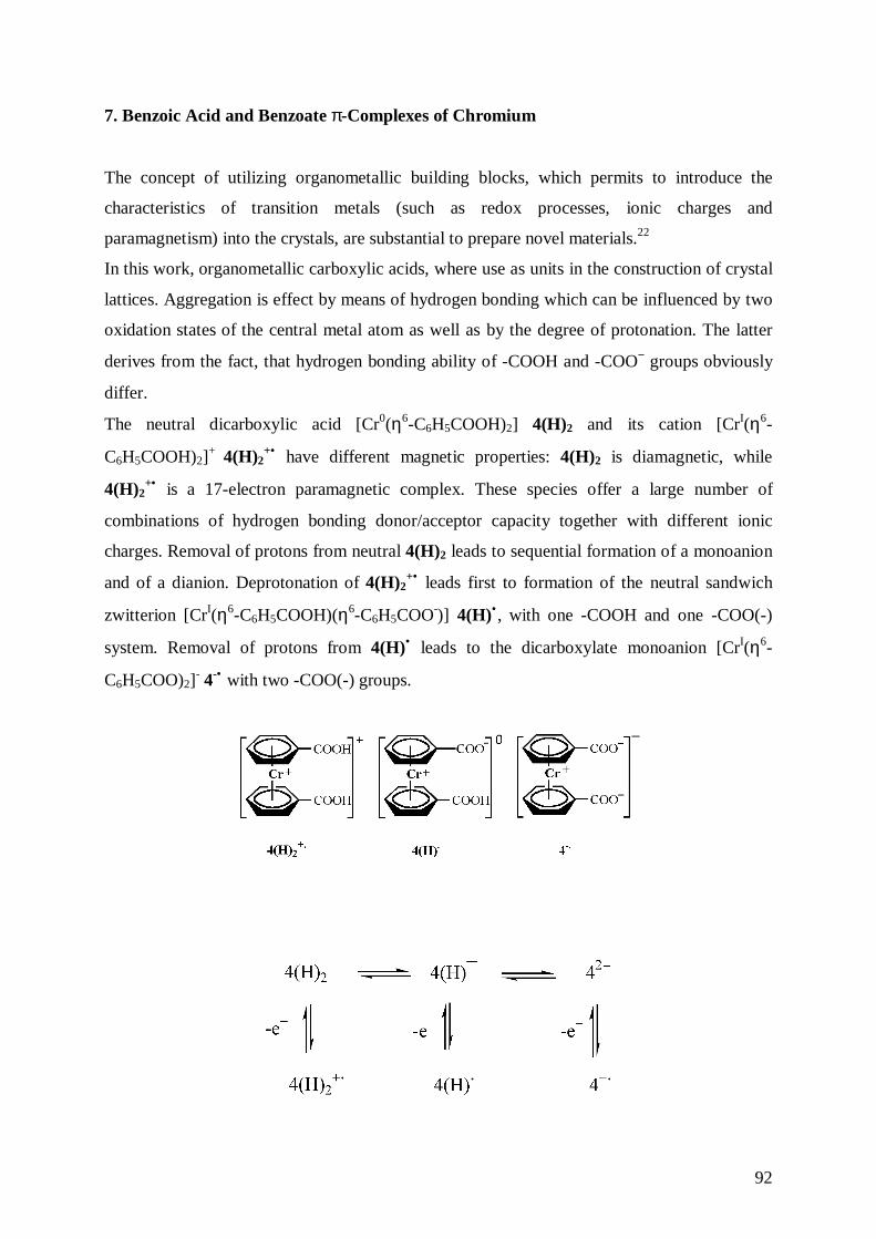
92
7. Benzoic Acid and Benzoate π-Complexes of Chromium
The concept of utilizing organometallic building blocks, which permits to introduce the
characteristics of transition metals (such as redox processes, ionic charges and
paramagnetism) into the crystals, are substantial to prepare novel materials.22
In this work, organometallic carboxylic acids, where use as units in the construction of crystal
lattices. Aggregation is effect by means of hydrogen bonding which can be influenced by two
oxidation states of the central metal atom as well as by the degree of protonation. The latter
derives from the fact, that hydrogen bonding ability of -COOH and -COO− groups obviously
differ.
The neutral dicarboxylic acid [Cr0(η6-C6H5COOH)2] 4(H)2 and its cation [CrI(η6-
C6H5COOH)2]+ 4(H)2
+• have different magnetic properties: 4(H)2 is diamagnetic, while
4(H)2+• is a 17-electron paramagnetic complex. These species offer a large number of
combinations of hydrogen bonding donor/acceptor capacity together with different ionic
charges. Removal of protons from neutral 4(H)2 leads to sequential formation of a monoanion
and of a dianion. Deprotonation of 4(H)2+• leads first to formation of the neutral sandwich
zwitterion [CrI(η6-C6H5COOH)(η6-C6H5COO-)] 4(H)• , with one -COOH and one -COO(-)
system. Removal of protons from 4(H)• leads to the dicarboxylate monoanion [CrI(η6-
C6H5COO)2]- 4-• with two -COO(-) groups.
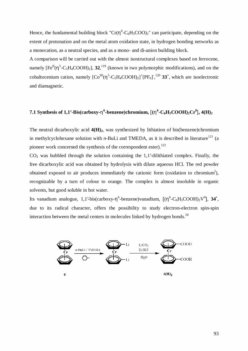
93
Hence, the fundamental building block "Cr(η6-C6H5COO)2" can participate, depending on the
extent of protonation and on the metal atom oxidation state, in hydrogen bonding networks as
a monocation, as a neutral species, and as a mono- and di-anion building block.
A comparison will be carried out with the almost isostructural complexes based on ferrocene,
namely [FeII(η5-C5H4COOH)2], 32,119 (known in two polymorphic modifications), and on the
cobaltocenium cation, namely [CoIII(η5-C5H4COOH)2]+[PF6]
-,120 33+, which are isoelectronic
and diamagnetic.
7.1 Synthesis of 1,1’-Bis(carboxy-η6-benzene)chromium, [(η6-C6H5COOH)2Cr0], 4(H)2
The neutral dicarboxylic acid 4(H)2, was synthesized by lithiation of bis(benzene)chromium
in methylcyclohexane solution with n-BuLi and TMEDA, as it is described in literature121 (a
pioneer work concerned the synthesis of the correspondent ester).122
CO2 was bubbled through the solution containing the 1,1’-dilithiated complex. Finally, the
free dicarboxylic acid was obtained by hydrolysis with dilute aqueous HCl. The red powder
obtained exposed to air produces immediately the cationic form (oxidation to chromiumI),
recognizable by a turn of colour to orange. The complex is almost insoluble in organic
solvents, but good soluble in hot water.
Its vanadium analogue, 1,1’-bis(carboxy-η6-benzene)vanadium, [(η6-C6H5COOH)2V0], 34• ,
due to its radical character, offers the possibility to study electron-electron spin-spin
interaction between the metal centers in molecules linked by hydrogen bonds.50
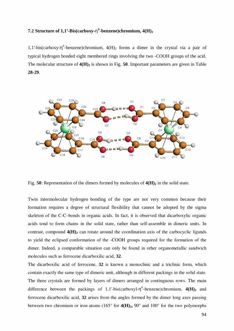
94
7.2 Structure of 1,1’-Bis(carboxy-η6-benzene)chromium, 4(H)2
1,1’-bis(carboxy-η6-benzene)chromium, 4(H)2 forms a dimer in the crystal via a pair of
typical hydrogen bonded eight membered rings involving the two -COOH groups of the acid.
The molecular structure of 4(H)2 is shown in Fig. 50. Important parameters are given in Table
28-29.
Fig. 50: Representation of the dimers formed by molecules of 4(H)2 in the solid state.
Twin intermolecular hydrogen bonding of the type are not very common because their
formation requires a degree of structural flexibility that cannot be adopted by the sigma
skeleton of the C-C–bonds in organic acids. In fact, it is observed that dicarboxylic organic
acids tend to form chains in the solid state, rather than self-assemble in dimeric units. In
contrast, compound 4(H)2 can rotate around the coordination axis of the carbocyclic ligands
to yield the eclipsed conformation of the -COOH groups required for the formation of the
dimer. Indeed, a comparable situation can only be found in other organometallic sandwich
molecules such as ferrocene dicarboxilic acid, 32.
The dicarboxilic acid of ferrocene, 32 is known a monoclinic and a triclinic form, which
contain exactly the same type of dimeric unit, although in different packings in the solid state.
The three crystals are formed by layers of dimers arranged in continguous rows. The main
difference between the packings of 1,1'-bis(carboxyl-η6-benzene)chromium, 4(H)2 and
ferrocene dicarboxilic acid, 32 arises from the angles formed by the dimer long axes passing
between two chromium or iron atoms (165° for 4(H)2, 90° and 100° for the two polymorphs
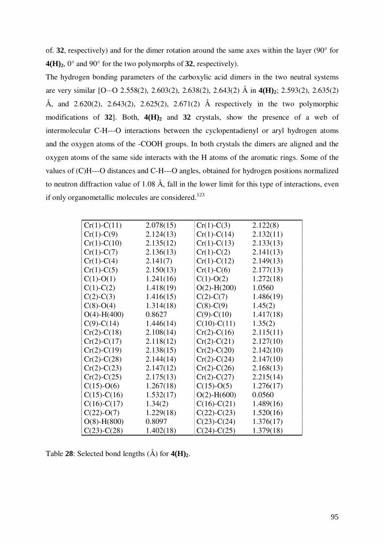
95
of. 32, respectively) and for the dimer rotation around the same axes within the layer (90° for
4(H)2, 0° and 90° for the two polymorphs of 32, respectively).
The hydrogen bonding parameters of the carboxylic acid dimers in the two neutral systems
are very similar [O···O 2.558(2), 2.603(2), 2.638(2), 2.643(2) Å in 4(H)2; 2.593(2), 2.635(2)
Å, and 2.620(2), 2.643(2), 2.625(2), 2.671(2) Å respectively in the two polymorphic
modifications of 32]. Both, 4(H)2 and 32 crystals, show the presence of a web of
intermolecular C-H---O interactions between the cyclopentadienyl or aryl hydrogen atoms
and the oxygen atoms of the -COOH groups. In both crystals the dimers are aligned and the
oxygen atoms of the same side interacts with the H atoms of the aromatic rings. Some of the
values of (C)H---O distances and C-H---O angles, obtained for hydrogen positions normalized
to neutron diffraction value of 1.08 Å, fall in the lower limit for this type of interactions, even
if only organometallic molecules are considered.123
Cr(1)-C(11) 2.078(15) Cr(1)-C(3) 2.122(8)Cr(1)-C(9) 2.124(13) Cr(1)-C(14) 2.132(11)Cr(1)-C(10) 2.135(12) Cr(1)-C(13) 2.133(13)Cr(1)-C(7) 2.136(13) Cr(1)-C(2) 2.141(13)Cr(1)-C(4) 2.141(7) Cr(1)-C(12) 2.149(13)Cr(1)-C(5) 2.150(13) Cr(1)-C(6) 2.177(13)C(1)-O(1) 1.241(16) C(1)-O(2) 1.272(18)C(1)-C(2) 1.418(19) O(2)-H(200) 1.0560C(2)-C(3) 1.416(15) C(2)-C(7) 1.486(19)C(8)-O(4) 1.314(18) C(8)-C(9) 1.45(2)O(4)-H(400) 0.8627 C(9)-C(10) 1.417(18)C(9)-C(14) 1.446(14) C(10)-C(11) 1.35(2)Cr(2)-C(18) 2.108(14) Cr(2)-C(16) 2.115(11)Cr(2)-C(17) 2.118(12) Cr(2)-C(21) 2.127(10)Cr(2)-C(19) 2.138(15) Cr(2)-C(20) 2.142(10)Cr(2)-C(28) 2.144(14) Cr(2)-C(24) 2.147(10)Cr(2)-C(23) 2.147(12) Cr(2)-C(26) 2.168(13)Cr(2)-C(25) 2.175(13) Cr(2)-C(27) 2.215(14)C(15)-O(6) 1.267(18) C(15)-O(5) 1.276(17)C(15)-C(16) 1.532(17) O(2)-H(600) 0.0560C(16)-C(17) 1.34(2) C(16)-C(21) 1.489(16)C(22)-O(7) 1.229(18) C(22)-C(23) 1.520(16)O(8)-H(800) 0.8097 C(23)-C(24) 1.376(17)C(23)-C(28) 1.402(18) C(24)-C(25) 1.379(18)
Table 28: Selected bond lengths (Å) for 4(H)2.
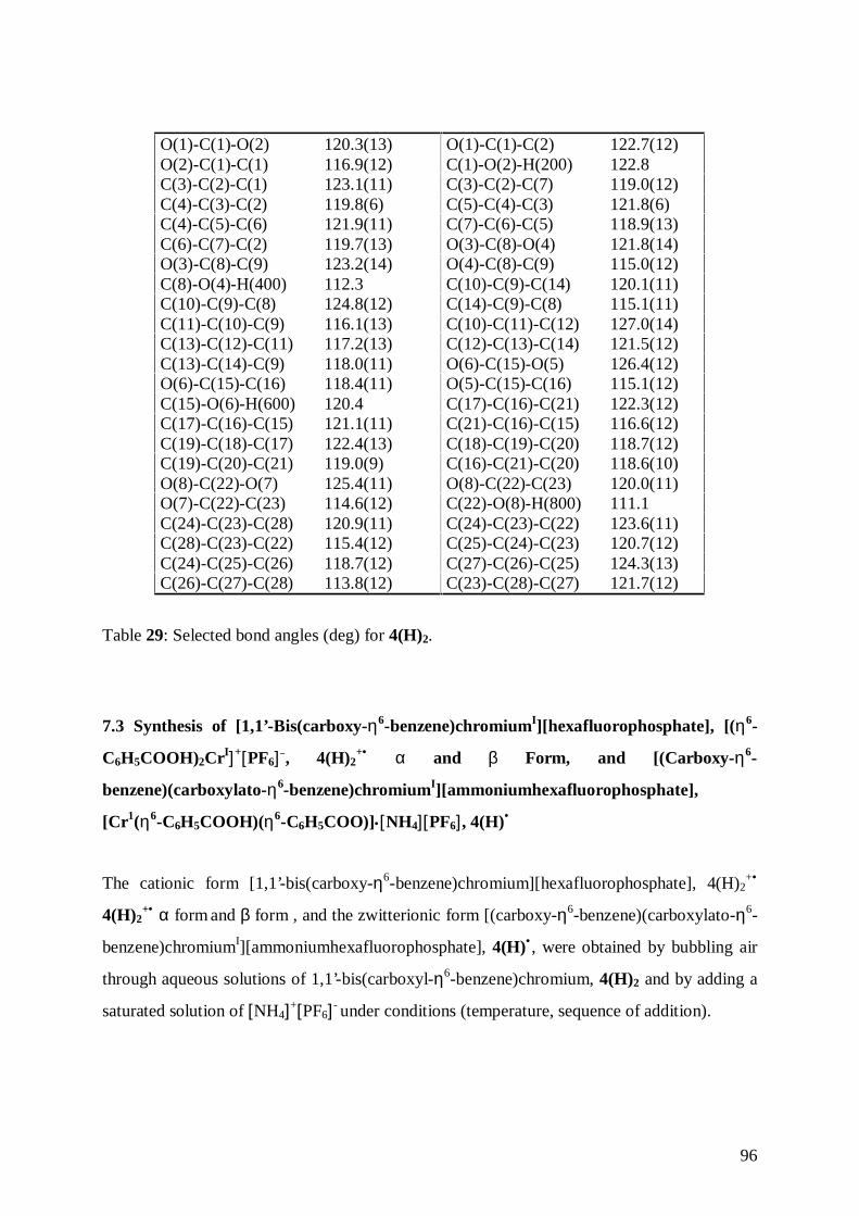
96
O(1)-C(1)-O(2) 120.3(13) O(1)-C(1)-C(2) 122.7(12)O(2)-C(1)-C(1) 116.9(12) C(1)-O(2)-H(200) 122.8C(3)-C(2)-C(1) 123.1(11) C(3)-C(2)-C(7) 119.0(12)C(4)-C(3)-C(2) 119.8(6) C(5)-C(4)-C(3) 121.8(6)C(4)-C(5)-C(6) 121.9(11) C(7)-C(6)-C(5) 118.9(13)C(6)-C(7)-C(2) 119.7(13) O(3)-C(8)-O(4) 121.8(14)O(3)-C(8)-C(9) 123.2(14) O(4)-C(8)-C(9) 115.0(12)C(8)-O(4)-H(400) 112.3 C(10)-C(9)-C(14) 120.1(11)C(10)-C(9)-C(8) 124.8(12) C(14)-C(9)-C(8) 115.1(11)C(11)-C(10)-C(9) 116.1(13) C(10)-C(11)-C(12) 127.0(14)C(13)-C(12)-C(11) 117.2(13) C(12)-C(13)-C(14) 121.5(12)C(13)-C(14)-C(9) 118.0(11) O(6)-C(15)-O(5) 126.4(12)O(6)-C(15)-C(16) 118.4(11) O(5)-C(15)-C(16) 115.1(12)C(15)-O(6)-H(600) 120.4 C(17)-C(16)-C(21) 122.3(12)C(17)-C(16)-C(15) 121.1(11) C(21)-C(16)-C(15) 116.6(12)C(19)-C(18)-C(17) 122.4(13) C(18)-C(19)-C(20) 118.7(12)C(19)-C(20)-C(21) 119.0(9) C(16)-C(21)-C(20) 118.6(10)O(8)-C(22)-O(7) 125.4(11) O(8)-C(22)-C(23) 120.0(11)O(7)-C(22)-C(23) 114.6(12) C(22)-O(8)-H(800) 111.1C(24)-C(23)-C(28) 120.9(11) C(24)-C(23)-C(22) 123.6(11)C(28)-C(23)-C(22) 115.4(12) C(25)-C(24)-C(23) 120.7(12)C(24)-C(25)-C(26) 118.7(12) C(27)-C(26)-C(25) 124.3(13)C(26)-C(27)-C(28) 113.8(12) C(23)-C(28)-C(27) 121.7(12)
Table 29: Selected bond angles (deg) for 4(H)2.
7.3 Synthesis of [1,1’-Bis(carboxy-η6-benzene)chromiumI][hexafluorophosphate], [(η6-
C6H5COOH)2CrI]+[PF6]−, 4(H)2+• α and β Form, and [(Carboxy-η6-
benzene)(carboxylato-η6-benzene)chromiumI][ammoniumhexafluorophosphate],
[Cr1(η6-C6H5COOH)(η6-C6H5COO)]•[NH4][ PF6], 4(H)•
The cationic form [1,1’-bis(carboxy-η6-benzene)chromium][hexafluorophosphate], 4(H)2+•
4(H)2+• α form and β form , and the zwitterionic form [(carboxy-η6-benzene)(carboxylato-η6-
benzene)chromiumI][ammoniumhexafluorophosphate], 4(H)• , were obtained by bubbling air
through aqueous solutions of 1,1’-bis(carboxyl-η6-benzene)chromium, 4(H)2 and by adding a
saturated solution of [NH4]+[PF6]- under conditions (temperature, sequence of addition).
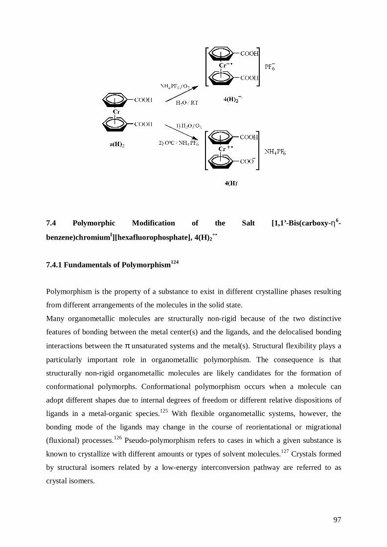
97
7.4 Polymorphic Modification of the Salt [1,1’-Bis(carboxy-η6-
benzene)chromiumI][hexafluorophosphate], 4(H)2+•
7.4.1 Fundamentals of Polymorphism124
Polymorphism is the property of a substance to exist in different crystalline phases resulting
from different arrangements of the molecules in the solid state.
Many organometallic molecules are structurally non-rigid because of the two distinctive
features of bonding between the metal center(s) and the ligands, and the delocalised bonding
interactions between the π unsaturated systems and the metal(s). Structural flexibility plays a
particularly important role in organometallic polymorphism. The consequence is that
structurally non-rigid organometallic molecules are likely candidates for the formation of
conformational polymorphs. Conformational polymorphism occurs when a molecule can
adopt different shapes due to internal degrees of freedom or different relative dispositions of
ligands in a metal-organic species.125 With flexible organometallic systems, however, the
bonding mode of the ligands may change in the course of reorientational or migrational
(fluxional) processes.126 Pseudo-polymorphism refers to cases in which a given substance is
known to crystallize with different amounts or types of solvent molecules.127 Crystals formed
by structural isomers related by a low-energy interconversion pathway are referred to as
crystal isomers.
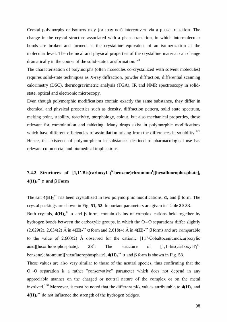
98
Crystal polymorphs or isomers may (or may not) interconvert via a phase transition. The
change in the crystal structure associated with a phase transition, in which intermolecular
bonds are broken and formed, is the crystalline equivalent of an isomerization at the
molecular level. The chemical and physical properties of the crystalline material can change
dramatically in the course of the solid-state transformation.128
The characterization of polymorphs (often molecules co-crystallized with solvent molecules)
requires solid-state techniques as X-ray diffraction, powder diffraction, differential scanning
calorimetry (DSC), thermogravimetric analysis (TGA), IR and NMR spectroscopy in solid-
state, optical and electronic microscopy.
Even though polymorphic modifications contain exactly the same substance, they differ in
chemical and physical properties such as density, diffraction pattern, solid state spectrum,
melting point, stability, reactivity, morphology, colour, but also mechanical properties, those
relevant for comminution and tableting. Many drugs exist in polymorphic modifications
which have different efficiencies of assimilation arising from the differences in solubility.129
Hence, the existence of polymorphism in substances destined to pharmacological use has
relevant commercial and biomedical implications.
7.4.2 Structures of [1,1’-Bis(carboxyl-η6-benzene)chromiumI][hexafluorophosphate],
4(H)2+• α and β Form
The salt 4(H)2+• has been crystallized in two polymorphic modifications, α, and β form. The
crystal packings are shown in Fig. 51, 52. Important parameters are given in Table 30-33.
Both crystals, 4(H)2+• α and β form, contain chains of complex cations held together by
hydrogen bonds between the carboxylic groups, in which the O···O separations differ slightly
(2.629(2), 2.634(2) Å in 4(H)2+• α form and 2.618(4) Å in 4(H)2
+• β form) and are comparable
to the value of 2.600(2) Å observed for the cationic [1,1'-Cobaltoceniumdicarboxylic
acid][hexafluorophosphate], 33+. The structure of [1,1'-bis(carboxyl-η6-
benzene)chromium][hexafluorophosphate], 4(H)2+• α and β form is shown in Fig. 53.
These values are also very similar to those of the neutral species, thus confirming that the
O···O separation is a rather "conservative" parameter which does not depend in any
appreciable manner on the charged or neutral nature of the complex or on the metal
involved.130 Moreover, it must be noted that the different pKS values attributable to 4(H)2 and
4(H)2+• do not influence the strength of the hydrogen bridges.
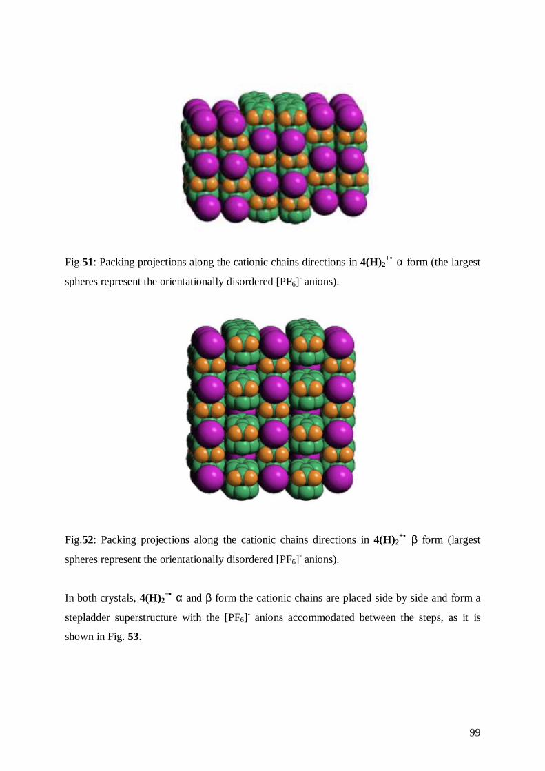
99
Fig.51: Packing projections along the cationic chains directions in 4(H)2+• α form (the largest
spheres represent the orientationally disordered [PF6]- anions).
Fig.52: Packing projections along the cationic chains directions in 4(H)2+• β form (largest
spheres represent the orientationally disordered [PF6]- anions).
In both crystals, 4(H)2+• α and β form the cationic chains are placed side by side and form a
stepladder superstructure with the [PF6]- anions accommodated between the steps, as it is
shown in Fig. 53.

100
Fig.53: Stepladder hydrogen bonding superstructure formed by the cationic chains in
crystalline 4(H)2+• α and β form with [PF6]
- anions accommodated between the steps.
Cr(1)-C(6) 2.1251(16) Cr(1)-C(6a) 2.1251(16)Cr(1)-C(5) 2.1298(17) Cr(1)-C(5a) 2.1298(17)Cr(1)-C(1) 2.1350(17) Cr(1)-C(1a) 2.1350(17)Cr(1)-C(4) 2.1444(19) Cr(1)-C(4a) 2.1444(19)Cr(1)-C(2) 2.1496(18) Cr(1)-C(2a) 2.1496(18)Cr(1)-C(3) 2.154(2) Cr(1)-C(3a) 2.154(2)C(1)-C(2) 1.3900 C(1)-C(6) 1.3900C(2)-C(3) 1.3900 C(3)-C(4) 1.3900C(4)-C(5) 1.3900 C(5)-C(6) 1.3900C(6)-C(7) 1.509(3) C(7)-O(1) 1.260(3)C(7)-O(2) 1.264(3) P(1)-F(1) 1.569(4)P(1)-F(2) 1.580(4) P(1)-F(3) 1.575(4)P(1)-F(4) 1.588(4) P(1)-F(5) 1.547(4)P(1)-F(6) 1.568(4)
Table 30: Selected bond lengths (Å) for 4(H)2+• α form.
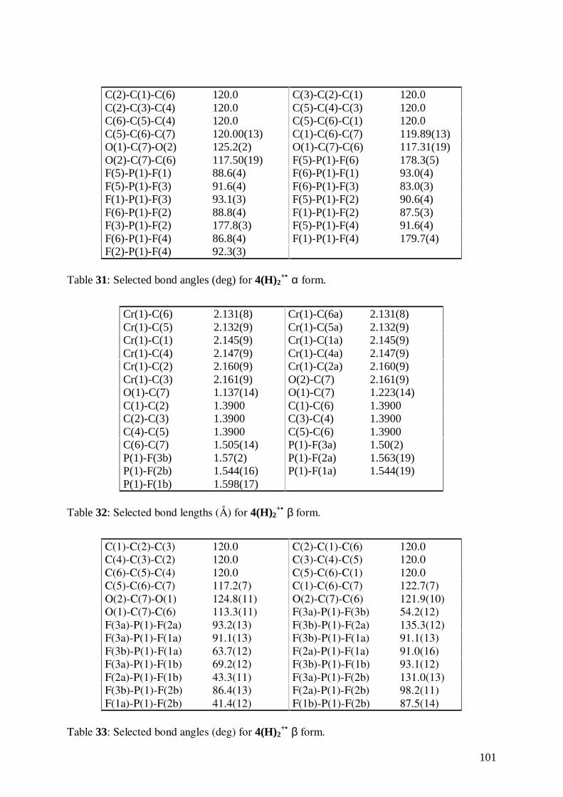
101
C(2)-C(1)-C(6) 120.0 C(3)-C(2)-C(1) 120.0C(2)-C(3)-C(4) 120.0 C(5)-C(4)-C(3) 120.0C(6)-C(5)-C(4) 120.0 C(5)-C(6)-C(1) 120.0C(5)-C(6)-C(7) 120.00(13) C(1)-C(6)-C(7) 119.89(13)O(1)-C(7)-O(2) 125.2(2) O(1)-C(7)-C(6) 117.31(19)O(2)-C(7)-C(6) 117.50(19) F(5)-P(1)-F(6) 178.3(5)F(5)-P(1)-F(1) 88.6(4) F(6)-P(1)-F(1) 93.0(4)F(5)-P(1)-F(3) 91.6(4) F(6)-P(1)-F(3) 83.0(3)F(1)-P(1)-F(3) 93.1(3) F(5)-P(1)-F(2) 90.6(4)F(6)-P(1)-F(2) 88.8(4) F(1)-P(1)-F(2) 87.5(3)F(3)-P(1)-F(2) 177.8(3) F(5)-P(1)-F(4) 91.6(4)F(6)-P(1)-F(4) 86.8(4) F(1)-P(1)-F(4) 179.7(4)F(2)-P(1)-F(4) 92.3(3)
Table 31: Selected bond angles (deg) for 4(H)2+• α form.
Cr(1)-C(6) 2.131(8) Cr(1)-C(6a) 2.131(8)Cr(1)-C(5) 2.132(9) Cr(1)-C(5a) 2.132(9)Cr(1)-C(1) 2.145(9) Cr(1)-C(1a) 2.145(9)Cr(1)-C(4) 2.147(9) Cr(1)-C(4a) 2.147(9)Cr(1)-C(2) 2.160(9) Cr(1)-C(2a) 2.160(9)Cr(1)-C(3) 2.161(9) O(2)-C(7) 2.161(9)O(1)-C(7) 1.137(14) O(1)-C(7) 1.223(14)C(1)-C(2) 1.3900 C(1)-C(6) 1.3900C(2)-C(3) 1.3900 C(3)-C(4) 1.3900C(4)-C(5) 1.3900 C(5)-C(6) 1.3900C(6)-C(7) 1.505(14) P(1)-F(3a) 1.50(2)P(1)-F(3b) 1.57(2) P(1)-F(2a) 1.563(19)P(1)-F(2b) 1.544(16) P(1)-F(1a) 1.544(19)P(1)-F(1b) 1.598(17)
Table 32: Selected bond lengths (Å) for 4(H)2+• β form.
C(1)-C(2)-C(3) 120.0 C(2)-C(1)-C(6) 120.0C(4)-C(3)-C(2) 120.0 C(3)-C(4)-C(5) 120.0C(6)-C(5)-C(4) 120.0 C(5)-C(6)-C(1) 120.0C(5)-C(6)-C(7) 117.2(7) C(1)-C(6)-C(7) 122.7(7)O(2)-C(7)-O(1) 124.8(11) O(2)-C(7)-C(6) 121.9(10)O(1)-C(7)-C(6) 113.3(11) F(3a)-P(1)-F(3b) 54.2(12)F(3a)-P(1)-F(2a) 93.2(13) F(3b)-P(1)-F(2a) 135.3(12)F(3a)-P(1)-F(1a) 91.1(13) F(3b)-P(1)-F(1a) 91.1(13)F(3b)-P(1)-F(1a) 63.7(12) F(2a)-P(1)-F(1a) 91.0(16)F(3a)-P(1)-F(1b) 69.2(12) F(3b)-P(1)-F(1b) 93.1(12)F(2a)-P(1)-F(1b) 43.3(11) F(3a)-P(1)-F(2b) 131.0(13)F(3b)-P(1)-F(2b) 86.4(13) F(2a)-P(1)-F(2b) 98.2(11)F(1a)-P(1)-F(2b) 41.4(12) F(1b)-P(1)-F(2b) 87.5(14)
Table 33: Selected bond angles (deg) for 4(H)2+• β form.
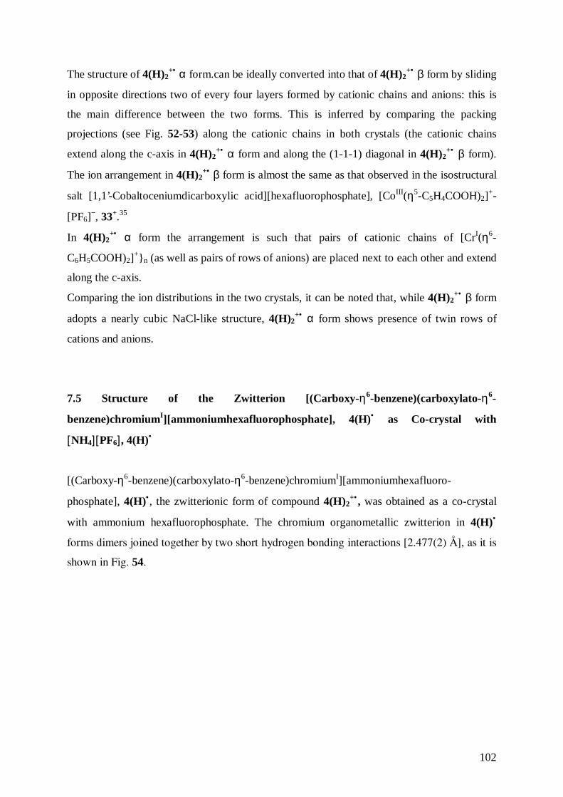
102
The structure of 4(H)2+• α form.can be ideally converted into that of 4(H)2
+• β form by sliding
in opposite directions two of every four layers formed by cationic chains and anions: this is
the main difference between the two forms. This is inferred by comparing the packing
projections (see Fig. 52-53) along the cationic chains in both crystals (the cationic chains
extend along the c-axis in 4(H)2+• α form and along the (1-1-1) diagonal in 4(H)2
+• β form).
The ion arrangement in 4(H)2+• β form is almost the same as that observed in the isostructural
salt [1,1’-Cobaltoceniumdicarboxylic acid][hexafluorophosphate], [CoIII(η5-C5H4COOH)2]+-
[PF6]−, 33+.35
In 4(H)2+• α form the arrangement is such that pairs of cationic chains of [CrI(η6-
C6H5COOH)2]+}n (as well as pairs of rows of anions) are placed next to each other and extend
along the c-axis.
Comparing the ion distributions in the two crystals, it can be noted that, while 4(H)2+• β form
adopts a nearly cubic NaCl-like structure, 4(H)2+• α form shows presence of twin rows of
cations and anions.
7.5 Structure of the Zwitterion [(Carboxy-η6-benzene)(carboxylato-η6-
benzene)chromiumI][ammoniumhexafluorophosphate], 4(H)• as Co-crystal with
[NH4][ PF6], 4(H)•
[(Carboxy-η6-benzene)(carboxylato-η6-benzene)chromiumI][ammoniumhexafluoro-
phosphate], 4(H)•, the zwitterionic form of compound 4(H)2+• , was obtained as a co-crystal
with ammonium hexafluorophosphate. The chromium organometallic zwitterion in 4(H)•
forms dimers joined together by two short hydrogen bonding interactions [2.477(2) Å], as it is
shown in Fig. 54.
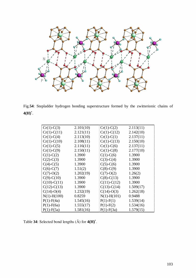
103
Fig.54: Stepladder hydrogen bonding superstructure formed by the zwitterionic chains of
4(H)•.
Cr(1)-C(3) 2.101(10) Cr(1)-C(2) 2.113(11)Cr(1)-C(11) 2.121(11) Cr(1)-C(12) 2.142(10)Cr(1)-C(4) 2.113(10) Cr(1)-C(1) 2.137(11)Cr(1)-C(10) 2.108(11) Cr(1)-C(13) 2.150(10)Cr(1)-C(5) 2.116(11) Cr(1)-C(6) 2.137(11)Cr(1)-C(9) 2.150(11) Cr(1)-C(8) 2.177(10)C(1)-C(2) 1.3900 C(1)-C(6) 1.3900C(2)-C(3) 1.3900 C(3)-C(4) 1.3900C(4)-C(5) 1.3900 C(5)-C(6) 1.3900C(6)-C(7) 1.51(2) C(8)-C(9) 1.3900C(7)-O(2) 1.202(19) C(7)-O(2) 1.26(2)C(9)-C(10) 1.3900 C(8)-C(13) 1.3900C(10)-C(11) 1.3900 C(11)-C(12) 1.3900C(12)-C(13) 1.3900 C(13)-C(14) 1.509(17)C(14)-O(4) 1.232(19) C(14)-O(3) 1.262(18)N(1)-H(100) 0.8259 N(1)-H(101) 0.9488P(1)-F(4a) 1.545(16) P(1)-F(1) 1.539(14)P(1)-F(6a) 1.555(17) P(1)-F(2) 1.534(16)P(1)-F(5a) 1.581(16) P(1)-F(3a) 1.579(15)
Table 34: Selected bond lengths (Å) for 4(H)• .
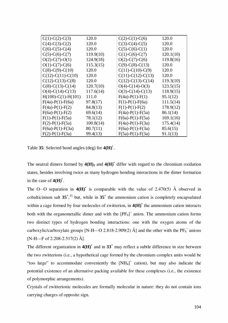
104
C(1)-C(2)-C(3) 120.0 C(2)-C(1)-C(6) 120.0C(4)-C(3)-C(2) 120.0 C(3)-C(4)-C(5) 120.0C(6)-C(5)-C(4) 120.0 C(5)-C(6)-C(1) 120.0C(5)-C(6)-C(7) 119.9(10) C(1)-C(6)-C(7) 120.1(10)O(2)-C(7)-O(1) 124.9(18) O(2)-C(7)-C(6) 119.8(16)O(1)-C(7)-C(6) 115.3(15) C(9)-C(8)-C(13) 120.0C(8)-C(9)-C(10) 120.0 C(11)-C(10)-C(9) 120.0C(12)-C(11)-C(10) 120.0 C(11)-C(12)-C(13) 120.0C(12)-C(13)-C(8) 120.0 C(12)-C(13)-C(14) 119.3(10)C(8)-C(13)-C(14) 120.7(10) O(4)-C(14)-O(3) 123.5(15)O(4)-C(14)-C(13) 117.6(14) O(3)-C(14)-C(13) 118.9(15)H(100)-C(1)-H(101) 111.0 F(4a)-P(1)-F(1) 95.1(12)F(4a)-P(1)-F(6a) 97.8(17) F(1)-P(1)-F(6a) 111.5(14)F(4a)-P(1)-F(2) 84.8(13) F(1)-P(1)-F(2) 178.9(12)F(6a)-P(1)-F(2) 69.6(14) F(4a)-P(1)-F(5a) 86.1(14)F(1)-P(1)-F(5a) 78.1(12) F(6a)-P(1)-F(5a) 169.1(16)F(2)-P(1)-F(5a) 100.8(14) F(4a)-P(1)-F(3a) 175.4(14)F(6a)-P(1)-F(3a) 80.7(11) F(6a)-P(1)-F(3a) 85.6(15)F(2)-P(1)-F(3a) 99.4(13) F(5a)-P(1)-F(3a) 91.1(13)
Table 35: Selected bond angles (deg) for 4(H)• .
The neutral dimers formed by 4(H)2 and 4(H)• differ with regard to the chromium oxidation
states, besides involving twice as many hydrogen bonding interactions in the dimer formation
in the case of 4(H)• .
The O···O separation in 4(H)• is comparable with the value of 2.470(5) Å observed in
cobalticinium salt 35+,35 but, while in 35+ the ammonium cation is completely encapsulated
within a cage formed by four molecules of zwitterion, in 4(H)• the ammonium cation interacts
both with the organometallic dimer and with the [PF6]− anion. The ammonium cation forms
two distinct types of hydrogen bonding interactions: one with the oxygen atoms of the
carboxylic/carboxylate groups [N-H---O 2.818-2.909(2) Å] and the other with the PF6− anions
[N-H---F of 2.208-2.517(2) Å].
The different organization in 4(H)• and in 33+ may reflect a subtle difference in size between
the two zwitterions (i.e., a hypothetical cage formed by the chromium complex units would be
“too large” to accommodate conveniently the [NH4]+ cation), but may also indicate the
potential existence of an alternative packing available for these complexes (i.e., the existence
of polymorphic arrangements).
Crystals of zwitterionic molecules are formally molecular in nature: they do not contain ions
carrying charges of opposite sign.
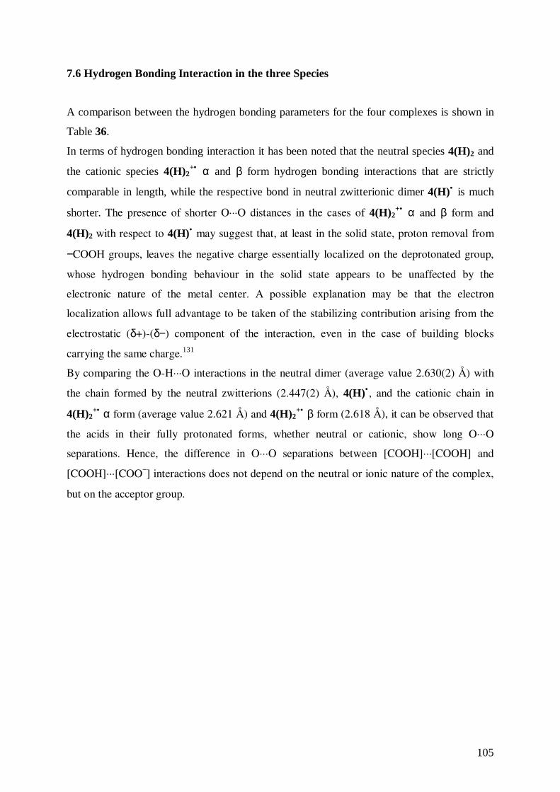
105
7.6 Hydrogen Bonding Interaction in the three Species
A comparison between the hydrogen bonding parameters for the four complexes is shown in
Table 36.
In terms of hydrogen bonding interaction it has been noted that the neutral species 4(H)2 and
the cationic species 4(H)2+• α and β form hydrogen bonding interactions that are strictly
comparable in length, while the respective bond in neutral zwitterionic dimer 4(H)• is much
shorter. The presence of shorter O···O distances in the cases of 4(H)2+• α and β form and
4(H)2 with respect to 4(H)• may suggest that, at least in the solid state, proton removal from
−COOH groups, leaves the negative charge essentially localized on the deprotonated group,
whose hydrogen bonding behaviour in the solid state appears to be unaffected by the
electronic nature of the metal center. A possible explanation may be that the electron
localization allows full advantage to be taken of the stabilizing contribution arising from the
electrostatic (δ+)-(δ−) component of the interaction, even in the case of building blocks
carrying the same charge.131
By comparing the O-H···O interactions in the neutral dimer (average value 2.630(2) Å) with
the chain formed by the neutral zwitterions (2.447(2) Å), 4(H)• , and the cationic chain in
4(H)2+• α form (average value 2.621 Å) and 4(H)2
+• β form (2.618 Å), it can be observed that
the acids in their fully protonated forms, whether neutral or cationic, show long O···O
separations. Hence, the difference in O···O separations between [COOH]···[COOH] and
[COOH]···[COO−] interactions does not depend on the neutral or ionic nature of the complex,
but on the acceptor group.
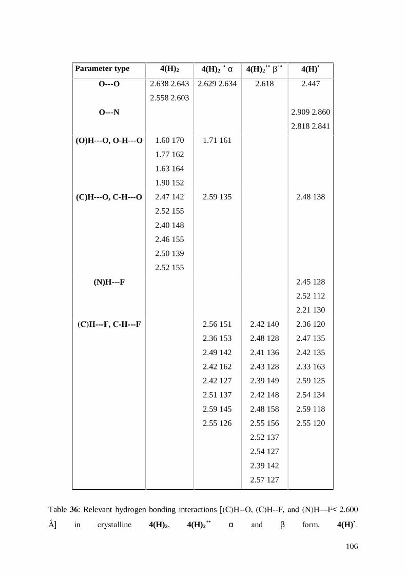
106
Parameter type 4(H)2 4(H)2+• α 4(H)2
+• β+• 4(H)•
O---O 2.638 2.643
2.558 2.603
2.629 2.634 2.618 2.447
O---N 2.909 2.860
2.818 2.841
(O)H---O, O-H---O 1.60 170
1.77 162
1.63 164
1.90 152
1.71 161
(C)H---O, C-H---O 2.47 142
2.52 155
2.40 148
2.46 155
2.50 139
2.52 155
2.59 135 2.48 138
(N)H---F 2.45 128
2.52 112
2.21 130
(C)H---F, CH---F 2.56 151
2.36 153
2.49 142
2.42 162
2.42 127
2.51 137
2.59 145
2.55 126
2.42 140
2.48 128
2.41 136
2.43 128
2.39 149
2.42 148
2.48 158
2.55 156
2.52 137
2.54 127
2.39 142
2.57 127
2.36 120
2.47 135
2.42 135
2.33 163
2.59 125
2.54 134
2.59 118
2.55 120
Table 36: Relevant hydrogen bonding interactions [(C)H--O, (C)H--F, and (N)H—F< 2.600
Å] in crystalline 4(H)2, 4(H)2+• α and β form, 4(H)• .
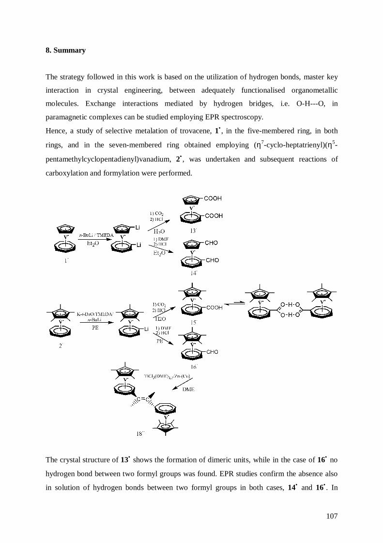
107
8. Summary
The strategy followed in this work is based on the utilization of hydrogen bonds, master key
interaction in crystal engineering, between adequately functionalised organometallic
molecules. Exchange interactions mediated by hydrogen bridges, i.e. O-H---O, in
paramagnetic complexes can be studied employing EPR spectroscopy.
Hence, a study of selective metalation of trovacene, 1• , in the five-membered ring, in both
rings, and in the seven-membered ring obtained employing (η7-cyclo-heptatrienyl)(η5-
pentamethylcyclopentadienyl)vanadium, 2• , was undertaken and subsequent reactions of
carboxylation and formylation were performed.
The crystal structure of 13• shows the formation of dimeric units, while in the case of 16• no
hydrogen bond between two formyl groups was found. EPR studies confirm the absence also
in solution of hydrogen bonds between two formyl groups in both cases, 14• and 16• . In
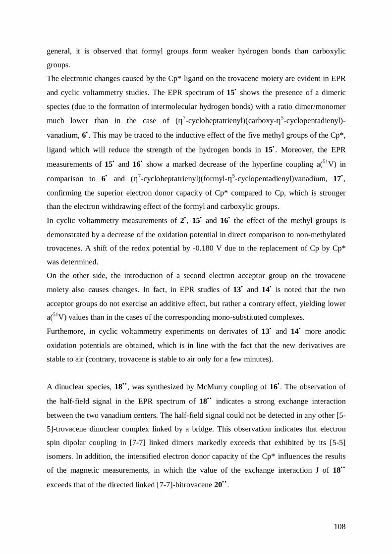
108
general, it is observed that formyl groups form weaker hydrogen bonds than carboxylic
groups.
The electronic changes caused by the Cp* ligand on the trovacene moiety are evident in EPR
and cyclic voltammetry studies. The EPR spectrum of 15• shows the presence of a dimeric
species (due to the formation of intermolecular hydrogen bonds) with a ratio dimer/monomer
much lower than in the case of (η7-cycloheptatrienyl)(carboxy-η5-cyclopentadienyl)-
vanadium, 6• . This may be traced to the inductive effect of the five methyl groups of the Cp*,
ligand which will reduce the strength of the hydrogen bonds in 15• . Moreover, the EPR
measurements of 15• and 16• show a marked decrease of the hyperfine coupling a(51V) in
comparison to 6• and (η7-cycloheptatrienyl)(formyl-η5-cyclopentadienyl)vanadium, 17• ,
confirming the superior electron donor capacity of Cp* compared to Cp, which is stronger
than the electron withdrawing effect of the formyl and carboxylic groups.
In cyclic voltammetry measurements of 2• , 15• and 16• the effect of the methyl groups is
demonstrated by a decrease of the oxidation potential in direct comparison to non-methylated
trovacenes. A shift of the redox potential by -0.180 V due to the replacement of Cp by Cp*
was determined.
On the other side, the introduction of a second electron acceptor group on the trovacene
moiety also causes changes. In fact, in EPR studies of 13• and 14• is noted that the two
acceptor groups do not exercise an additive effect, but rather a contrary effect, yielding lower
a(51V) values than in the cases of the corresponding mono-substituted complexes.
Furthemore, in cyclic voltammetry experiments on derivates of 13• and 14• more anodic
oxidation potentials are obtained, which is in line with the fact that the new derivatives are
stable to air (contrary, trovacene is stable to air only for a few minutes).
A dinuclear species, 18•• , was synthesized by McMurry coupling of 16• . The observation of
the half-field signal in the EPR spectrum of 18•• indicates a strong exchange interaction
between the two vanadium centers. The half-field signal could not be detected in any other [5-
5]-trovacene dinuclear complex linked by a bridge. This observation indicates that electron
spin dipolar coupling in [7-7] linked dimers markedly exceeds that exhibited by its [5-5]
isomers. In addition, the intensified electron donor capacity of the Cp* influences the results
of the magnetic measurements, in which the value of the exchange interaction J of 18••
exceeds that of the directed linked [7-7]-bitrovacene 20•• .
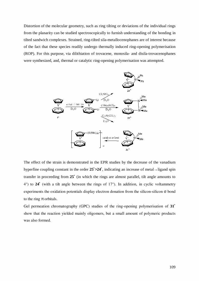
109
Distortion of the molecular geometry, such as ring tilting or deviations of the individual rings
from the planarity can be studied spectroscopically to furnish understanding of the bonding in
tilted sandwich complexes. Strained, ring-tilted sila-metallocenophanes are of interest because
of the fact that these species readily undergo thermally induced ring-opening polymerisation
(ROP). For this purpose, via dilithiation of trovacene, monosila- and disila-trovacenophanes
were synthesized, and, thermal or catalytic ring-opening polymerisation was attempted.
The effect of the strain is demonstrated in the EPR studies by the decrease of the vanadium
hyperfine coupling constant in the order 25•>24•, indicating an increase of metal→ligand spin
transfer in proceeding from 25• (in which the rings are almost parallel, tilt angle amounts to
4°) to 24• (with a tilt angle between the rings of 17°). In addition, in cyclic voltammetry
experiments the oxidation potentials display electron donation from the silicon-silicon σ bond
to the ring π orbitals.
Gel permeation chromatography (GPC) studies of the ring-opening polymerisation of 31•
show that the reaction yielded mainly oligomers, but a small amount of polymeric products
was also formed.
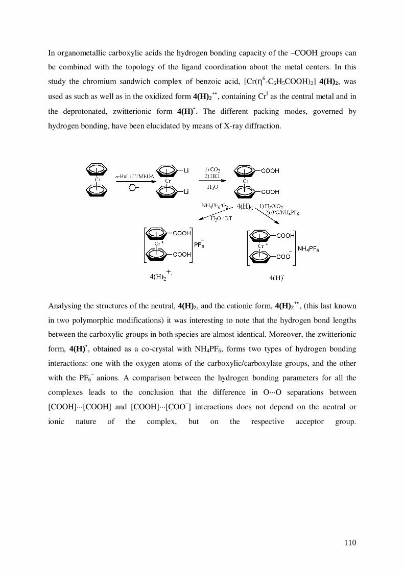
110
In organometallic carboxylic acids the hydrogen bonding capacity of the –COOH groups can
be combined with the topology of the ligand coordination about the metal centers. In this
study the chromium sandwich complex of benzoic acid, [Cr(η6-C6H5COOH)2] 4(H)2, was
used as such as well as in the oxidized form 4(H)2+• , containing CrI as the central metal and in
the deprotonated, zwitterionic form 4(H)•. The different packing modes, governed by
hydrogen bonding, have been elucidated by means of X-ray diffraction.
Analysing the structures of the neutral, 4(H)2, and the cationic form, 4(H)2+• , (this last known
in two polymorphic modifications) it was interesting to note that the hydrogen bond lengths
between the carboxylic groups in both species are almost identical. Moreover, the zwitterionic
form, 4(H)•, obtained as a co-crystal with NH4PF6, forms two types of hydrogen bonding
interactions: one with the oxygen atoms of the carboxylic/carboxylate groups, and the other
with the PF6− anions. A comparison between the hydrogen bonding parameters for all the
complexes leads to the conclusion that the difference in O···O separations between
[COOH]···[COOH] and [COOH]···[COO−] interactions does not depend on the neutral or
ionic nature of the complex, but on the respective acceptor group.
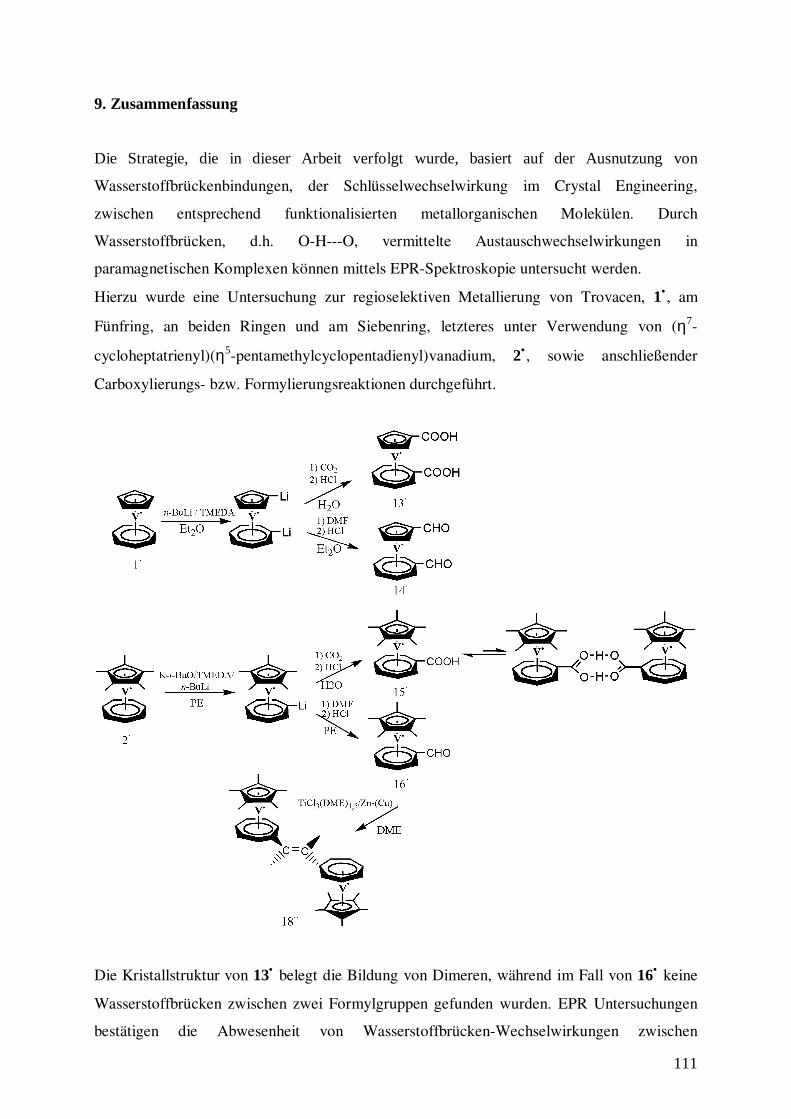
111
9. Zusammenfassung
Die Strategie, die in dieser Arbeit verfolgt wurde, basiert auf der Ausnutzung von
Wasserstoffbrückenbindungen, der Schlüsselwechselwirkung im Crystal Engineering,
zwischen entsprechend funktionalisierten metallorganischen Molekülen. Durch
Wasserstoffbrücken, d.h. O-H---O, vermittelte Austauschwechselwirkungen in
paramagnetischen Komplexen können mittels EPR-Spektroskopie untersucht werden.
Hierzu wurde eine Untersuchung zur regioselektiven Metallierung von Trovacen, 1• , am
Fünfring, an beiden Ringen und am Siebenring, letzteres unter Verwendung von (η7-
cycloheptatrienyl)(η5-pentamethylcyclopentadienyl)vanadium, 2• , sowie anschließender
Carboxylierungs- bzw. Formylierungsreaktionen durchgeführt.
Die Kristallstruktur von 13• belegt die Bildung von Dimeren, während im Fall von 16• keine
Wasserstoffbrücken zwischen zwei Formylgruppen gefunden wurden. EPR Untersuchungen
bestätigen die Abwesenheit von Wasserstoffbrücken-Wechselwirkungen zwischen
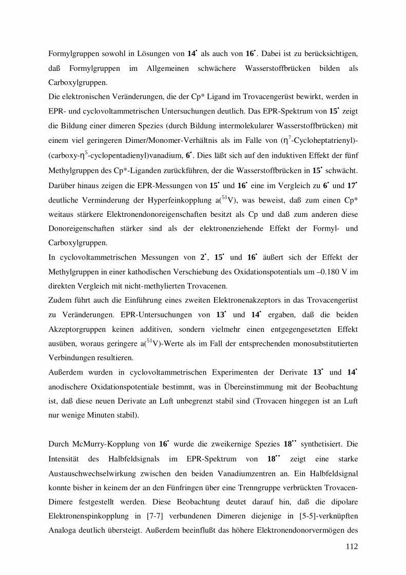
112
Formylgruppen sowohl in Lösungen von 14• als auch von 16• . Dabei ist zu berücksichtigen,
daß Formylgruppen im Allgemeinen schwächere Wasserstoffbrücken bilden als
Carboxylgruppen.
Die elektronischen Veränderungen, die der Cp* Ligand im Trovacengerüst bewirkt, werden in
EPR- und cyclovoltammetrischen Untersuchungen deutlich. Das EPR-Spektrum von 15• zeigt
die Bildung einer dimeren Spezies (durch Bildung intermolekularer Wasserstoffbrücken) mit
einem viel geringeren Dimer/Monomer-Verhältnis als im Falle von (η7-Cycloheptatrienyl)-
(carboxy-η5-cyclopentadienyl)vanadium, 6• . Dies läßt sich auf den induktiven Effekt der fünf
Methylgruppen des Cp*-Liganden zurückführen, der die Wasserstoffbrücken in 15• schwächt.
Darüber hinaus zeigen die EPR-Messungen von 15• und 16• eine im Vergleich zu 6• und 17•
deutliche Verminderung der Hyperfeinkopplung a(51V), was beweist, daß zum einen Cp*
weitaus stärkere Elektronendonoreigenschaften besitzt als Cp und daß zum anderen diese
Donoreigenschaften stärker sind als der elektronenziehende Effekt der Formyl- und
Carboxylgruppen.
In cyclovoltammetrischen Messungen von 2• , 15• und 16• äußert sich der Effekt der
Methylgruppen in einer kathodischen Verschiebung des Oxidationspotentials um –0.180 V im
direkten Vergleich mit nicht-methylierten Trovacenen.
Zudem führt auch die Einführung eines zweiten Elektronenakzeptors in das Trovacengerüst
zu Veränderungen. EPR-Untersuchungen von 13• und 14• ergaben, daß die beiden
Akzeptorgruppen keinen additiven, sondern vielmehr einen entgegengesetzten Effekt
ausüben, woraus geringere a(51V)-Werte als im Fall der entsprechenden monosubstitutierten
Verbindungen resultieren.
Außerdem wurden in cyclovoltammetrischen Experimenten der Derivate 13• und 14•
anodischere Oxidationspotentiale bestimmt, was in Übereinstimmung mit der Beobachtung
ist, daß diese neuen Derivate an Luft unbegrenzt stabil sind (Trovacen hingegen ist an Luft
nur wenige Minuten stabil).
Durch McMurry-Kopplung von 16• wurde die zweikernige Spezies 18•• synthetisiert. Die
Intensität des Halbfeldsignals im EPR-Spektrum von 18•• zeigt eine starke
Austauschwechselwirkung zwischen den beiden Vanadiumzentren an. Ein Halbfeldsignal
konnte bisher in keinem der an den Fünfringen über eine Trenngruppe verbrückten Trovacen-
Dimere festgestellt werden. Diese Beobachtung deutet darauf hin, daß die dipolare
Elektronenspinkopplung in [7-7] verbundenen Dimeren diejenige in [5-5]-verknüpften
Analoga deutlich übersteigt. Außerdem beeinflußt das höhere Elektronendonorvermögen des
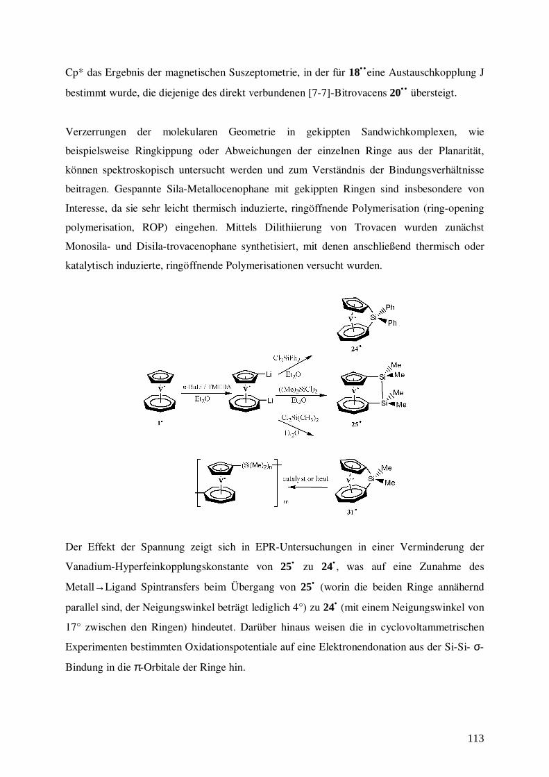
113
Cp* das Ergebnis der magnetischen Suszeptometrie, in der für 18•• eine Austauschkopplung J
bestimmt wurde, die diejenige des direkt verbundenen [7-7]-Bitrovacens 20•• übersteigt.
Verzerrungen der molekularen Geometrie in gekippten Sandwichkomplexen, wie
beispielsweise Ringkippung oder Abweichungen der einzelnen Ringe aus der Planarität,
können spektroskopisch untersucht werden und zum Verständnis der Bindungsverhältnisse
beitragen. Gespannte Sila-Metallocenophane mit gekippten Ringen sind insbesondere von
Interesse, da sie sehr leicht thermisch induzierte, ringöffnende Polymerisation (ring-opening
polymerisation, ROP) eingehen. Mittels Dilithiierung von Trovacen wurden zunächst
Monosila- und Disila-trovacenophane synthetisiert, mit denen anschließend thermisch oder
katalytisch induzierte, ringöffnende Polymerisationen versucht wurden.
Der Effekt der Spannung zeigt sich in EPR-Untersuchungen in einer Verminderung der
Vanadium-Hyperfeinkopplungskonstante von 25• zu 24• , was auf eine Zunahme des
Metall→Ligand Spintransfers beim Übergang von 25• (worin die beiden Ringe annähernd
parallel sind, der Neigungswinkel beträgt lediglich 4°) zu 24• (mit einem Neigungswinkel von
17° zwischen den Ringen) hindeutet. Darüber hinaus weisen die in cyclovoltammetrischen
Experimenten bestimmten Oxidationspotentiale auf eine Elektronendonation aus der Si-Si- σ-
Bindung in die π-Orbitale der Ringe hin.
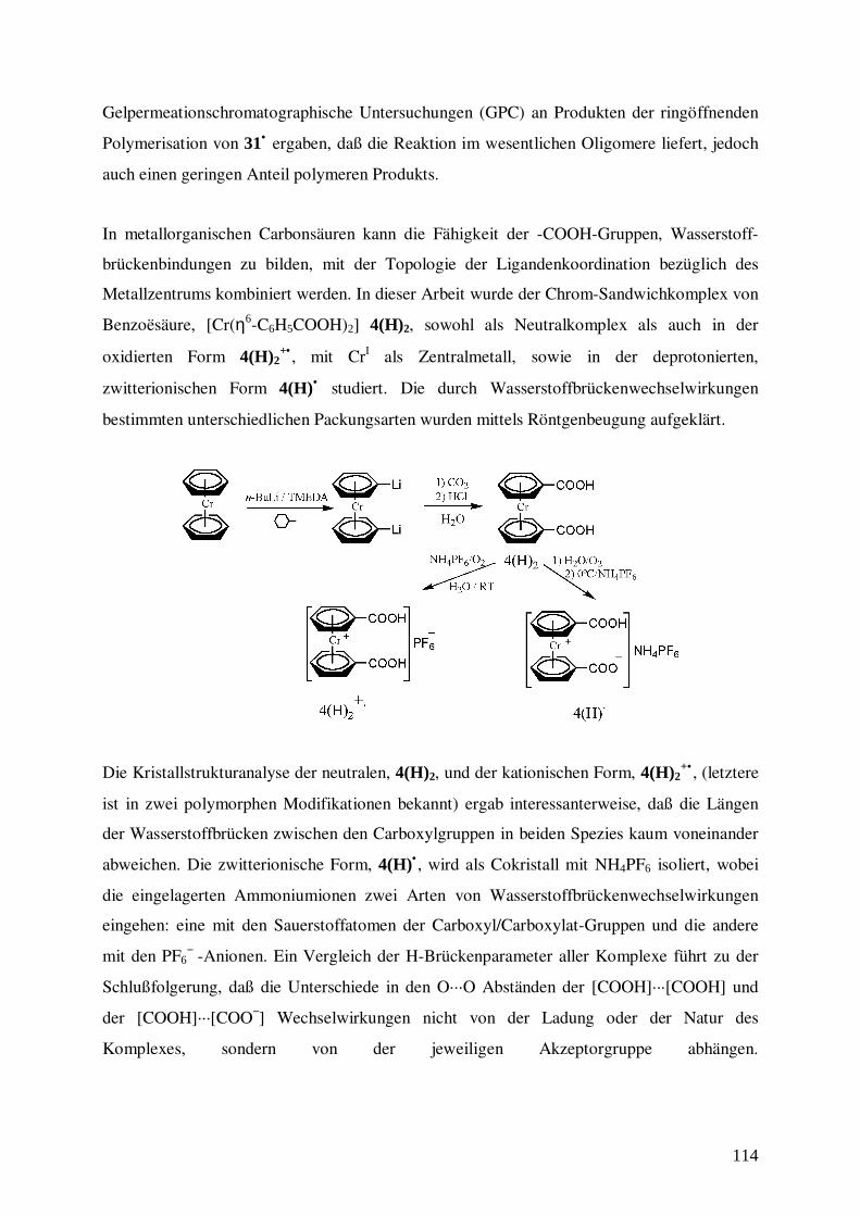
114
Gelpermeationschromatographische Untersuchungen (GPC) an Produkten der ringöffnenden
Polymerisation von 31• ergaben, daß die Reaktion im wesentlichen Oligomere liefert, jedoch
auch einen geringen Anteil polymeren Produkts.
In metallorganischen Carbonsäuren kann die Fähigkeit der -COOH-Gruppen, Wasserstoff-
brückenbindungen zu bilden, mit der Topologie der Ligandenkoordination bezüglich des
Metallzentrums kombiniert werden. In dieser Arbeit wurde der Chrom-Sandwichkomplex von
Benzoësäure, [Cr(η6-C6H5COOH)2] 4(H)2, sowohl als Neutralkomplex als auch in der
oxidierten Form 4(H)2+• , mit CrI als Zentralmetall, sowie in der deprotonierten,
zwitterionischen Form 4(H)• studiert. Die durch Wasserstoffbrückenwechselwirkungen
bestimmten unterschiedlichen Packungsarten wurden mittels Röntgenbeugung aufgeklärt.
Die Kristallstrukturanalyse der neutralen, 4(H)2, und der kationischen Form, 4(H)2+• , (letztere
ist in zwei polymorphen Modifikationen bekannt) ergab interessanterweise, daß die Längen
der Wasserstoffbrücken zwischen den Carboxylgruppen in beiden Spezies kaum voneinander
abweichen. Die zwitterionische Form, 4(H)• , wird als Cokristall mit NH4PF6 isoliert, wobei
die eingelagerten Ammoniumionen zwei Arten von Wasserstoffbrückenwechselwirkungen
eingehen: eine mit den Sauerstoffatomen der Carboxyl/Carboxylat-Gruppen und die andere
mit den PF6− -Anionen. Ein Vergleich der H-Brückenparameter aller Komplexe führt zu der
Schlußfolgerung, daß die Unterschiede in den O···O Abständen der [COOH]···[COOH] und
der [COOH]···[COO−] Wechselwirkungen nicht von der Ladung oder der Natur des
Komplexes, sondern von der jeweiligen Akzeptorgruppe abhängen.
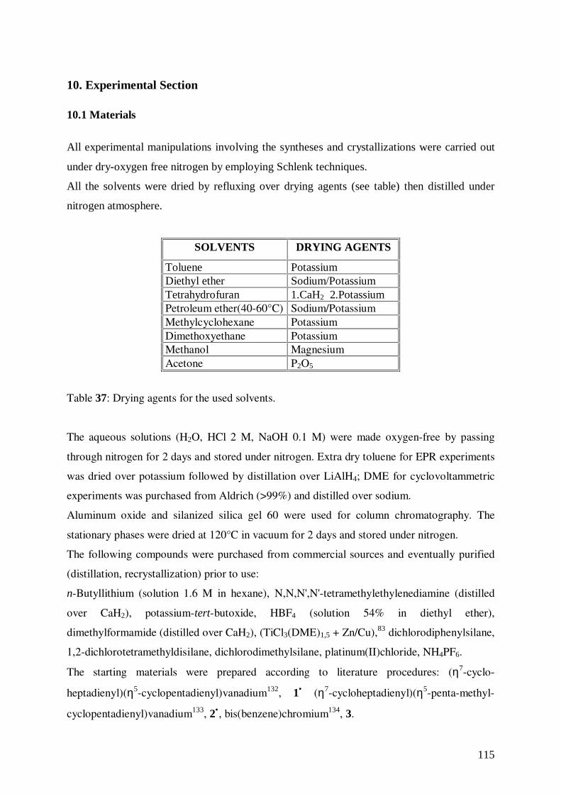
115
10. Experimental Section
10.1 Materials
All experimental manipulations involving the syntheses and crystallizations were carried out
under dry-oxygen free nitrogen by employing Schlenk techniques.
All the solvents were dried by refluxing over drying agents (see table) then distilled under
nitrogen atmosphere.
SOLVENTS DRYING AGENTS
Toluene PotassiumDiethyl ether Sodium/PotassiumTetrahydrofuran 1.CaH2 2.PotassiumPetroleum ether(40-60°C) Sodium/PotassiumMethylcyclohexane PotassiumDimethoxyethane PotassiumMethanol MagnesiumAcetone P2O5
Table 37: Drying agents for the used solvents.
The aqueous solutions (H2O, HCl 2 M, NaOH 0.1 M) were made oxygen-free by passing
through nitrogen for 2 days and stored under nitrogen. Extra dry toluene for EPR experiments
was dried over potassium followed by distillation over LiAlH4; DME for cyclovoltammetric
experiments was purchased from Aldrich (>99%) and distilled over sodium.
Aluminum oxide and silanized silica gel 60 were used for column chromatography. The
stationary phases were dried at 120°C in vacuum for 2 days and stored under nitrogen.
The following compounds were purchased from commercial sources and eventually purified
(distillation, recrystallization) prior to use:
n-Butyllithium (solution 1.6 M in hexane), N,N,N',N'-tetramethylethylenediamine (distilled
over CaH2), potassium-tert-butoxide, HBF4 (solution 54% in diethyl ether),
dimethylformamide (distilled over CaH2), (TiCl3(DME)1,5 + Zn/Cu),83 dichlorodiphenylsilane,
1,2-dichlorotetramethyldisilane, dichlorodimethylsilane, platinum(II)chloride, NH4PF6.
The starting materials were prepared according to literature procedures: (η7-cyclo-
heptadienyl)(η5-cyclopentadienyl)vanadium132, 1• (η7-cycloheptadienyl)(η5-penta-methyl-
cyclopentadienyl)vanadium133, 2•, bis(benzene)chromium134, 3.
116
10.2 Instrumental Analysis
Mass Spectrometry: mass spectra samples were prepared in special glass equipment under
nitrogen and measured on a CH7 (EI-MS) (70 eV) Spectrometer, (Varian MAT).
Elemental Analysis (C, H, N): the tin-crucibles (filled in special devise under nitrogen
atmosphere) containing the substance were burned in a oxygen-stream. The analytical
measurements were made employing Elementar Analyser (Vario EL).
In the elementary analyses of vanadium sandwich complexes is noted that value found for
carbon is often lower than calculated. This is due to the formation of stable carbides of
vanadium. The effect is more pronounced in the case of (η7-cycloheptatrienyl)(η5-
pentamethylcyclopentadienyl)vanadium and its derivates, because of the presence of the
additional five methyl groups. The combustion of the carbon atoms is not complete.
Combustion would be complete at temperatures higher than those accessible with the analyse
available.
Infrared Spectroscopy: the samples were prepared as KBr-pellets and the spectra were
recorded on Interferometer IFS 88, (Bruker), from 4000 to 400 cm-1.
Gel Permeation Chromatography: samples with concentration of ca. 1 mg/mL were measured
in THF relative to polystyrene standards. A column (600 x 8 mm, Type SDV linear, 10 µL,
PSS) were used. Detection was performed employing a refractive index detector K731
(Knauer), and a UV/VIS-detector UV 3071 (Knauer). The evaluation of the measured data
was carried out by the computer program WinGPC Version 4.01 (PSS).
1H NMR Spectroscopy: the spectra were measured on a AM-500 Spectrometer (Bruker), 500
MHz. d8-THF δ= 1.73, 3.58 ppm was used as solvent. Sample solutions were submitted in
sealed tubes; the solutions were tempered (at 60°C for 12 hours) prior to recording the spectra
in order to destroy CrI complex radical cations adventitiously present.
Electron Paramagnetic Resonance: all measurements were performed in solution 10-5-10-4
molar of the complexes on a X-Band Spectrometer ESP 300 (Bruker). The sample were
prepared in inert atmosphere in quartz or glass tubes (∅ 4 mm) and flame-sealed under
117
vacuum. A modified TE 102 Square-Pulse Resonator (Varian) was employed as cavity. The
frequency was determined using the Frequency-Counter Mod. TR 5214 (Advantest).
ENDOR: 1H ENDOR spectra were recorded on a ESP 300 spectrometer (Bruker) with EN 200
S/E/T ENDOR-Triple accessory. The sample were prepared in inert atmosphere in glass tube
(∅ 5 mm) and flame-sealed under vacuum.
Cyclic Voltammetry: the measurements were made in a special cell containing unstirred
millimolar solution of the complex in DME in the presence of (n-Bu)4NClO4 (0.1 M) as
supporting electrolyte at –40°C and with sweep rate (v= dE/dt) 0.1 V/s. Working electrode: 2
mm glassy carbon electrode (EG & G); counter electrode: platinum wire (Amel); reference
electrode: mercury/calomel electrode (Amel). The equipment consisted of the potentiostat
(Amel, Mod. 522), function generator (Amel, Mod. 560), multipurpose unit (Amel, Mod.
563). The voltammogramms were registered by a digital storage oscilloscope (Nicolet, Mod.
3091) and X-Y-recorder (Kipp and Zonen, Mod. BD 90). Cyclovoltammetry was performed
under argon protection. Dimethoxyethane was dried over calcium hydride.
Crystal Structure Analysis: X-ray diffraction data were collected on a NONIUS CAD-4
diffractometer equipped with a liquid nitrogen Oxford-Cryostream device. Crystal date and
details of measurements are summarized in Tables. Common to all compounds: MoKα
radiation, λ= 0.71073 Å, monochromator graphite, psi-scan absorption correction.
SHELXL97135 was used for structure solution and refinement based on F2.
SCHAKAL97136 was used for all graphical representations.
Correspondence between the structures determined by X-ray diffraction and the bulk material
was confirmed by powder diffraction. Powder spectra were measured on an automated PW
1100 Powder Diffractometer with monochromatic Cu-Kα radiation.
Magnetic Susceptibility: magnetic susceptibility was studied with a SQUID Magnetometer
(Quantum Design) in the temperature range 1.8-300 K. The samples for magnetic
measurements were prepared in special sealed teflon-crucibles (filled under nitrogen
atmosphere). The samples contained ca. 10 mg of substance. The data were corrected for
magnetization of the sample holder (KLF), and diamagnetic corrections were applied to the
data based on Pascal’s constant.
118
10.3 Preparations
Synthesis of (Carboxy-η7-cycloheptatrienyl)(carboxy-η5-cyclopentadienyl)vanadium,
[(η7-C7H6COOH)(η5-C5H4COOH)V0], 13•. A 100 mL flask equipped with a magnetic
stirring bar was charged with (η7-cycloheptatrienyl)(η5-cyclopentadienyl)vanadium (0.70 g,
3.38 mmol), diethyl ether (40 mL), and TMEDA (2.04 mL, 13.52 mmol). Then n-BuLi (8.34
mL, 13.52 mmol, solution 1.6 M in hexane) was added dropwise and the mixture was refluxed
for 4 hours. After cooling to room temperature, CO2 was bubbled through the solution
containing the dark-violet suspension for 3 hours. The colour turned immediately to green.
The solvent was removed in vacuum and the residue was dissolved in water. After filtration,
HCl (12 mL, 2 M) was added to the filtrate, yielding a clear green precipitate, which was
collected by filtration and dried under vacuum. Suitable crystal for X-ray diffraction were
obtained by suspending 0.10 g of crude product in water, followed by addition of NaOH (2
mL, 0.1 M). The solution was then layered with 10 mL of diethyl ether containing HBF4 (3
drops, solution 58% in diethyl ether).
Yield: 0.83 g, 83%.
MS, m/e: 295 (75%, M +), 251 (38%, M+-COOH), 175 (25%, VC6H6COOH+), 159 (92%,
VCpCOOH+), 129 (21%,C6H6V+), 91 (98%,C7H7
+), 51 (14%,V+).
Anal. Calculated for C14H12O4V (295.19): C, 56.97; H, 4.11. Found: C, 56.56, H, 4.39.
IR (KBr): νO-H 2653 cm-1, νC=O 1678 cm-1, νC=C 1498, 1476 cm-1, νC-O 1308 cm-1, δC-H,ring 795
cm-1.
Synthesis of (Formyl-η7-cycloheptatrienyl)(formyl-η5-cyclopentadienyl)vanadium, [(η7-
C7H6CHO)(η5-C5H4CHO)V0], 14•. A 100 mL flask equipped with a magnetic stirring bar
was charged with (η7-cycloheptatrienyl)(η5-cyclopentadienyl)vanadium (0.70 g, 3.38 mmol),
diethyl ether (50 mL), and TMEDA (2.04 mL, 13.52 mmol). Then n-BuLi (8.34 mL, 13.52
mmol, solution 1.6 M in hexane) was added dropwise and the mixture was refluxed for 4
hours. After cooling to room temperature, dimethylformamide (2.70 mL, 27.04 mmol) was
added to the dark-violet suspension at –60 °C. The resulting violet mixture was stirred at –60
°C for 1 hour and at room temperature for additional 2 hours. HCl (1.75 mL, 2 M) was added
at –5 °C and the green suspension was stirred at room temperature for 1 hour, then the solvent
was removed in vacuum. The residue was dissolved in toluene and dried over MgSO4. The
product was chromatographed on aluminium oxide (neutral, 0% water). The first fraction,
eluted with toluene, contained unreacted trovacene, the second, eluted with toluene/THF
119
(20:1), consisted of (η7-cycloheptatrienyl)(formyl-η5-cyclopentadienyl)vanadium, the last,
eluted with THF/toluene (3:1), contained the title compound. The solvent was removed under
vacuum to yield a dark green powder.
Yield: 0.52 g, 58%.
MS, m/e: 263 (100%, M +), 235 (8%, M+-CHO), 207 (25%, TVC+), 157 (6%, VC6H6CO+),
144 (8%, VCpCO+), 141 (1%, VC6H6C+), 128 (3%, VCpC+), 129 (16%, C6H6V
+), 116 (40%,
CpV+), 91 (85%,C7H7+), 51 (22%,V+).
Anal. Calculated for C14H12O2V (263.19): C, 63.89; H, 4.60. Found: C, 63.46, H, 4.59.
IR (KBr): νC(O)-H 2961 cm-1, νC=O 1682 cm-1, νC=C 1497, 1463 cm-1, δC-H,ring 799 cm-1.
Synthesis of (Carboxy-η7-cycloheptatrienyl)(η5-pentamethylcyclopentadienyl)vanadium,
[(η7-C7H6COOH)(η5-C5Me5)V0], 15•. A 100 mL flask equipped with a magnetic stirring bar
was charged with (η7-cycloheptatrienyl)(η5-pentamethylcyclopentadienyl)vanadium (0.15 g,
0.50 mmol), and petroleum ether (20 mL). At –50°C, TMEDA (0.2 mL, 1.0 mmol),
potassium-tert-butoxide (0.13 g, 1.0 mmol) was added and the solution was stirred for 10
minutes. Then, at -20°C, n-BuLi (0.9 mL, 1.0 mmol, solution 1.6 M in hexane) was added
dropwise and the mixture was stirred at this temperature for 1 hour. Then, at room
temperature, CO2 was bubbled through the solution containing the brown suspension for 3
hours. The colour turned immediately to clear green. The solvent was removed in vacuum and
the residue was dissolved in water. After filtration of the unreacted (η7-cycloheptatrienyl)(η5-
pentamethylcyclopentadienyl)vanadium, HCl (4 mL, 2 M) was added to the filtrate, yielding a
clear green precipitate, which was collected by filtration and dried under vacuum.
Yield: 0.034 g, 20%.
MS, m/e: 321 (50%, M+), 277 (74%, M+-COOH), 136 (100%, C5(CH3)5+), 91 (31%,C7H7
+),
51 (1%,V+).
Anal. Calculated for C18H22O2V (321.31): C, 67.29; H, 6.90. Found: C, 63.96, H, 6.69.
IR (KBr): νC-CH3 2909 cm-1, νO-H, free 2627 cm-1, νO-H, bonded 2542 cm-1, νC=O 1651 cm-1, νC=C
1496, 1466 cm-1, δas-CH3 1430 cm-1, δs-CH3 1375 cm-1, νC-O 1319 cm-1.
Synthesis of (Formyl-η7-cycloheptatrienyl)(η5-pentamethylcyclopentadienyl)vanadium,
[(η7-C7H6CHO)(η5-C5Me5)V0], 16•. A 100 mL flask equipped with a magnetic stirring bar
was charged with (η7-cycloheptatrienyl)(η5-pentamethylcyclopentadienyl)vanadium (0.20 g,
0.72 mmol), and petroleum ether (20 mL). At –50°C TMEDA (0.3 mL, 1.44 mmol), and
potassium-tert-butoxide (0.18 g, 1.44 mmol) were added and the solution was stirred for 10
120
minutes. Then, at -20°C, n-BuLi (1.2 mL, 1.44 mmol, solution 1.6 M in hexane) was added
dropwise and the mixture was stirred at this temperature for 1 hour. Dimethylformamide (0.2
mL, 2.88 mmol) was added to the brown suspension at –60 °C. The resulting red-violet
mixture was stirred at –60 °C for 1 hour and at room temperature for an additional 2 hours.
HCl 2 M (0.3 mL) was added at –5 °C and the green suspension was stirred at room
temperature for 1 hour. After filtration, the solution was dried over MgSO4. The product was
chromatographed on silica gel. The first fraction, eluted with petroleum ether, contained
unreacted (η7-cycloheptatrienyl)(η5-pentamethylcyclopentadienyl)vanadium, the second,
eluted with petroleum ether/diethyl ether (10:1), contained the product. The solvent was
removed under vacuum to yield green-brown crystals.
Yield: 0.044 g, 20%.
MS, m/e: 305 (100%, M+), 277 (29%, M+-CHO), 91 (85%,C7H7+).
Anal. Calculated for C18H22OV (263.19): C, 70.81; H, 7.26. Found: C, 68.98; H, 7.54.
IR (KBr): νC-CH3 2961 cm-1, νC(O)-H 2771 cm-1, δH-C=O 2717 cm-1, νC=O 1674 cm-1, νC=C 1491,
δas-CH3 1426 cm-1, δs-CH3 1374 cm-1, νC-O 1306 cm-1.
Synthesis of E-1,2-Bis[(η7-cycloheptatrienyl)(η5-pentamethylcyclopentadienyl)-
vanadium]ethene, 18•• . A 100 mL flask equipped with a magnetic stirring bar was charged
with TiCl3(DME)1,5 (1.14 g, 3.94 mmol) and zinc-copper couple (3.80 g, 58.10 mmol) in 50
mL of DME. The mixture was heated at 80°C for 2 hours to form the active titanium reagent.
After cooling to room-temperature, 1-formyl(η7-cycloheptatrienyl)(η5-pentamethyl-
cyclopentadienyl)vanadium, (0.28 g, 0.90 mmol) in 10 mL of DME was added by a syringe,
and the mixture was heated for 20 hours. The reaction slurry was cooled to room temperature
and petroleum ether was added. The mixture was filtered through a small layer of silica gel to
remove inorganic salts. The filtrate was concentrate under vacuum and dissolved in petroleum
ether. From the solution a clear-green powder precipitated, which was collected, washed with
petroleum ether/diethyl ether and dried under vacuum to give 0.3 g (Yield= 58 %) of pure E-
18•• isomer. The solution was concentrated to 5 mL and chromatographed on silicagel. A first
clear-green fraction was eluted with petroleum ether. This contains a few milligrams of E
isomer. A second dark-green fraction eluted with petroleum ether/toluene contained unreacted
(formyl-η7-cycloheptatrienyl)(η5-pentamethylcyclopentadienyl)vanadium.
MS, m/e: 578 (100%, M+), 290 (6%, M++), 277 (39%, TVC*), 185 (1%, Cp*V).
Anal. Calculated for C36H44V2 (578.63): C, 74.73; H, 7.66. Found: C, 70.39, H, 7.39.
IR (KBr): νC-CH3 2961 cm-1, νC=C 1629, 1692, δas-CH3 1449 cm-1, δs-CH3 1375 cm-1.
121
Synthesis of [1,8-(Diphenylsilanediyl)(η7-cycloheptatrienyl)(η5-cyclopentadienyl)
vanadium], [(Ph2Si-η7-C7H6)(η5-C5H4)V0], 24•. A 100 mL flask equipped with a magnetic
stirring bar was charged with (η7-cycloheptatrienyl)(η5-cyclopentadienyl)vanadium (0.20 g,
0.97 mmol), diethyl ether (30 mL), and TMEDA (0.58 mL, 3.83 mmol). Then, n-BuLi (2.41
mL, 3.83 mmol, solution 1.6 M in hexane) was added dropwise and the mixture was refluxed
for 4 hours. After cooling to room temperature, dichlorodiphenylsilane (0.2 mL, 0.97 mmol)
dissolved in 10 mL petroleum ether was added slowly (2 h) to the dark-violet suspension at –
20°C. The colour of the mixture turned from dark-brown to violet and LiCl precipitated. The
mixture was stirred at room temperature overnight. Then, LiCl was filtered off. Violet crystals
suitable for X-ray diffraction were obtained by storing the solution at –20°C.
Yield: 0.24 g, 64%.
MS, m/e: 387 (100%, M +), 309 (14%, M+-Ph), 232 (14%, M+-2Ph), 205 (3%, M+-Si(Ph)2).
Anal. Calculated for C24H20SiV (387.45): C, 74.40; H, 5.20. Found: C, 73.14, H, 5.78.
Synthesis of [1,8-(Tetramethyldisilane-1,2-diyl)(η7-cycloheptatrienyl)(η5-
cyclopentadienyl)-vanadium], [(Me2SiSiMe2-η7-C7H6)(η5-C5H4)V0], 25• . A 100 mL flask
equipped with a magnetic stirring bar was charged with (η7-cycloheptatrienyl)(η5-
cyclopentadienyl)vanadium (0.15 g, 0.72 mmol), diethyl ether (30 mL), and TMEDA (0.43
mL, 2.88 mmol). Then, n-BuLi (1.81 mL, 2.88 mmol, solution 1.6 M in hexane) was added
dropwise and the mixture was refluxed for 4 hours. After cooling to room temperature, 1,2-
dichlorotetramethyldisilane (0.13 mL, 0.72 mmol) dissolved in 10 mL petroleum ether was
added slowly (2 h) to the dark-violet suspension at –20°C. The colour of the mixture turned
from dark-brown to clear violet and LiCl precipitated. The mixture was stirred at room
temperature overnight. Then, LiCl was filtered off. Violet crystals suitable for X-ray
diffraction were obtained by storing the solution at –20°C.
Yield: 0.10 g, 45%.
MS, m/e: 321 (9%, M+), 205 (37%, M+-Si(CH3)2-Si(CH3)2), 116 (54%, CpV+), 91
(20%,C7H7+).
Anal. Calculated for C24H22Si2V (321.46): C, 59.78; H, 6.90. Found: C, 56.83, H, 7.04.
122
Attempted Synthesis of Poly(trovacenylsilane), [-Si(CH3)2(η7-C7H6)V0(η7-C5H4)-]n.
Synthesis of [1,8-(Dimethylsilanediyl)(η7-cycloheptatrienyl)(η5-cyclopentadienyl)
vanadium], [(Me2Si-η7-C7H6)(η5-C5H4)V0], 31•. A 100 mL flask equipped with a magnetic
stirring bar was charged with (η7-cycloheptatrienyl)(η5-cyclopentadienyl)vanadium (0.20 g,
0.97 mmol), diethyl ether (30 mL), and TMEDA (0.58 mL, 3.83 mmol). Then, n-BuLi (2.41
mL, 3.83 mmol, solution 1.6 M in hexane) was added dropwise and the mixture was refluxed
for 4 hours. After cooling to room temperature, dichlorodimethylsilane (0.12 mL, 0.97 mmol)
dissolved in 10 mL petroleum ether was added slowly (2 h) to the dark-violet suspension at –
20°C. The colour of the mixture turned from dark-brown to violet and LiCl precipitated. The
mixture was stirred at room temperature overnight. Then, LiCl was filtered off, and the
solution was evaporated under vacuum to yield 31• as violet oil.
MS, m/e: 734 (50%, (TVC-Si(CH3))3+-Si(CH3)), 264 (15%, M+), 207 (2%, TVC+), 91
(100%,C7H7+).
Anal. Calculated for C14H16SiV (263.31): C, 63.86; H, 6.12. Found: C, 58.55, H, 6.82, N 2.57.
Method A: [1,8-(Dimethylsilanediyl)(η7-cycloheptatrienyl)(η5-cyclopentadienyl)vanadium]
was sealed in an evacuated Pyrex tube and heated at 150 °C for 1 hour. The content was
dissolved in THF, filtered and precipitated by addition of petroleum ether. The dark-violet
solid was collected by filtration, washed with additional petroleum ether and dried under
vacuum.
MS, m/e: 470 (100%, (TVC)2Si2+), 207 (13%, TVC+), 91 (3%,C7H7
+), 51 (1%,V+).
Anal. Calculated for C14H16SiV (263.31): C, 63.86; H, 6.12. Found: C, 58.18, H, 7.73, N 3.28
(calculated on the monomer unit).
Method B: [1,8-(Dimethylsilanediyl)(η7-cycloheptatrienyl)(η5-cyclopentadienyl)vanadium]
was dissolved in 15 mL of toluene, and a few milligrams of PtCl2 were added. The flask was
placed in an oil bath maintained at 80-90°C on a hot plate. The solution was stirred vigorously
at this temperature for ca. 2 hours, during which time the violet suspension faded to brown-
violet. After cooling to room temperature, the suspension was filtered. Then, the filtrate was
added dropwise by a syringe to a flask containing petroleum ether (100 mL), or petroleum
ether/toluene (80:20) with rapid stirring. A brown-violet precipitate was formed, which was
collected by filtration, washed with additional petroleum ether and dried under vacuum.
MS, m/e: 943 (3%, (TVC-Si(CH3))4+-2Si(CH3)), 759 (100%, (TVC)3Si5
+), 497 (9%,
(TVC)2Si3+), 235 (2%, TVC-Si+), 207 (3%, TVC+), 91 (5%,C7H7
+), 51 (3%,V+).
123
Anal. Calculated for C14H16SiV (263.31): C, 63.86; H, 6.12. Found: C, 44.94, H, 6.00, N 4.77
(calculated on the monomer unit).
GPC: Mw: 682.59, Mn: 499.77, Mw/Mn: 1.3658.
Method C: A 100 mL flask equipped with a magnetic stirring bar was charged with (η7-
cycloheptatrienyl)(η5-cyclopentadienyl)vanadium (0.20 g, 0.97 mmol), diethyl ether (30 mL),
and TMEDA (0.58 mL, 3.83 mmol). Then, n-BuLi (2.41 mL, 3.83 mmol, solution 1.6 M in
hexane) was added dropwise and the mixture was refluxed for 4 hours. After cooling to room
temperature, the solvent was evaporated under vacuum and the residue was dissolved in THF.
Slow addition of dichlorodimethylsilane (0.12 mL, 0.97 mmol) dissolved in 10 mL of THF to
the refluxing THF solution of 1,1’-dilithiotrovacene-TMEDA was followed by refluxing for 7
hours. After removal of the solvent, the residue was dissolved in toluene and precipitated by
addition of petroleum ether. A brown-violet precipitate was formed, which was collected by
filtration, washed with additional petroleum ether and dried under vacuum.
MS, m/e: 470 (9%, (TVC)2Si2+), 264 (11%, TVC-Si(CH3)2
+), 207 (25%, TVC+), 91
(17%,C7H7+), 51 (7%,V+).
Anal. Calculated for C14H16SiV (263.31): C, 63.86; H, 6.12. Found: C, 62.27, H, 9.56
(calculated on the monomer unit).
Synthesis of 1,1’-Bis(carboxy-η6-benzene)chromium, [(η6-C6H5COOH)2Cr0], 4. A 100
mL flask equipped with a magnetic stirring bar was charged with bis(benzene)chromium (1.0
g, 4.80 mmol), methylcyclohexane (60 mL), and TMEDA (1.54 mL, 10.2 mmol). n-BuLi
(6.25 mL, 10.2 mmol, solution 1.6 M in hexane) was added dropwise and the mixture was
refluxed for 4 hours. Then at room temperature, CO2 was fed through the brown suspension
for 1 hour. The colour turned immediately to orange-red. The solvent was removed in vacuum
and the residue was dissolved in water. After filtration of the unreacted
bis(benzene)chromium, HCl (20 mL, 2 M) was added to the filtrate, yielding a red precipitate,
which was collected by filtration and dried under vacuum.
Bis(carboxy-η6-benzene)chromium, 4 (0.10 g, 0.34 mmol) was dissolved in hot water,
followed by addition of NaOH (0.5 mL, 0.1 M). The solution was layered with 10 mL of
diethyl ether containing HBF4 (0.1 mL, solution 58% in diethyl ether). Red block crystals
were obtained after one week.
Yield: 1.1 g, 80%.
124
MS, m/e: 122 (100%, C6H5COOH+), 105 (93%, C6H5CO+), 77 (95%, C6H7+), 51 (35%,
C4H3+).
1H NMR (300 MHz, THF-d8): 11.18 (s, 2H), 5.13 (d, 4H), 4.54 (m, 6H).
Anal. Calculated for C14H12O4Cr (296.24): C, 56.76; H, 4.08. Found: C,
56.74, H, 4.10.
IR (KBr): νC-H 3085 cm-1, νO-H 2653 cm-1, νC=O 1680 cm-1, νC=C 1499, 1468 cm-1, νC-O 1311
cm-1, δC-H,ring 727 cm-1.
Synthesis of [1,1’-Bis(carboxy-η6-benzene)chromium][hexafluorophosphate],[(η6-
C6H5COOH)2CrI]+[PF6]−,31+•α and 31+•β. A 50 mL flask equipped with a magnetic stirring
bar was charged with bis(carboxy-η6-benzene)chromium (0.05 g, 0.17 mmol). A saturated
solution of NH4PF6 in water (15 mL) was added, and air was fed through the mixture
overnight at room temperature. Then, the solution was dried under vacuum. Yellow crystals
of 31+•α suitable for X-ray diffraction were obtained by diethyl ether diffusion into an
acetone solution of the salt, while slow evaporation of an acetone solution of the salt yielded
yellow crystals of 31+•β.
Synthesis of [1,1’-Bis(carboxyl-η6-benzene)chromium][ammoniumhexafluoro-
phosphate], [Cr1(η6-C6H5COOH)(η6-C6H5COO)][NH4][ PF6], 32• . A 50 mL flask equipped
with a magnetic stirring bar was charged with bis(carboxy-η6-benzene)chromium (0.10 g,
0.34 mmol), and water (15 mL). Then, air was fed through the mixture overnight at room
temperature. The yellow solution obtained was cooled at 0°C, and NH4PF6 (0.01 g) was
added. The precipitate obtained was filtered and dried under vacuum. Yellow crystals suitable
for X-ray diffraction were obtained by recrystallization from methanol.
125
11. Literature
1) a) J. T. Kealy, P. L. Pauson; Nature, 1951, 168, 1039; b) S. A. Miller, J. A. L. Tebbboth, J.
F. Tremaine, J. Chem. Soc., 1952, 632; c) G. Wilkinson, M. Rosemblum, M. C. Whiting, R.
B. Woodward, J. Amer. Chem. Soc., 1952, 74, 2125; d) E. O. Fischer, W. Pfab, Z.
Naturforsch., 1952, 7B, 377.
2) E. O. Fischer, W. Hafner; Z. Naturforsch., 1955, 10B, 665.
3) Ch. Elschenbroich, J. Organomet. Chem., 1968, 14, 157.
4) a) P. L. Timms, J. Chem. Soc. Chem. Comm., 1969, 1033; b) P. L. Timms, J. Chem Ed.,
1972, 49, 789; b) P. L. Timms, T. W. Turney, Adv. Organomet. Chem., 1977, 15, 53; c) J. R.
Blackborrow, D. Young, Metal Vapour Synthesis in Organometallic Chemistry, Springer
Verlag Berlin, Heidelberg, New York, 1979.
5) T. Hayashi, , Acc. Chem. Res., 2000, 33, 354.
6) P. D. Beer, Adv. Inorg. Chem., 1992, 39, 79.
7) H. Köpf, P. Köpf-Maier, , Angew. Chem. Int. Ed. Engl., 1979, 91, 477.
8) J. S. Miller, J. C. Calabrese, H. Rommelmann, S. R. Chittipeddi, J. H. Zhang, W. M. Reiff,
A. J. Epstein, J. Amer. Chem. Soc., 1987, 109, 769.
9) M. L. H. Green, S. R. Marder, M. E. Thompson, J. A. Bandy, D. Bloor, P. V. Kolinsky, R.
J. Jones, Nature, 1987, 330, 360.
10) D. Braga, F. Grepioni, G. R. Desiraju, Chem. Rev., 1998, 98, 1375.
11) D. Rabinowich, G. M. J. Schmidt, J. Chem. Soc. (B), 1964, 144.
12) G. R. Desiraju, Chem. Commun., 1997, 1475.
13) a) M. C. Etter, Acc. Chem. Res., 1990, 23, 120; b) G. R. Desiraju, Angew. Chem. Int. Ed.
Engl., 1995, 34, 2311; c) D. Braga, F. Grepioni, Chem. Comm., 1996, 571; d) M. J.
Zaworotko, Nature, 1997, 386, 220; e) M. Yaghi, L. Guangming, H. Li, Nature, 1995, 378,
703; f) P. Ball, Nature, 1996, 381, 648.
14) J. D. Dunitz, L. E. Orgen, Nature, 1953, 171, 121; J. D. Dunitz, L. E. Orgen, A. Rich,
Acta Crystallogr., 1956, 9, 373.
15) a) V. G. Albano, D. Braga, Accurate Molecular Structures; A. Domenicano, I. Hargittai,
Eds., Oxford University Press: Oxford, U. K., 1991; b) E. Sappa, A. Tiripicchio, P.
Braunstein, Chem. Rev., 1983, 83, 203; c) D. Imhof, L. M. Venanzi, Chem. Soc. Rev., 1994,
185.
16) E. L. Muetterties, J. R. bleeke, E. J. Wucherer, Chem. Rev., 1982, 82, 499.
126
17) a) D. Braga, F. Grepioni, Acc. Chem. Res., 1994, 27, 51; b) D. Braga, F. Grepioni, Chem.
Comm., 1996, 571.
18) a) M. C. Etter, Acc. Chem. Res., 1990, 23, 120; b) C. B. Aakeröy, K. R. Seddon, Chem.
Soc. Rev., 1993, 397; c) S. Subramanian, M. J. Zaworotko, Coord. Chem. Rev., 1994, 137,
557; d) M. J. Zaworotko, Nature, 1997, 386, 220.
19) D. Braga, F. Grepioni, P. Milne, E. Parigini, J. Am. Chem. Soc., 1993, 115, 5115.
20) a) A. I. Kitaigorodsky, Molecular Crystals and Molecules; Accademic Press: New York,
1973; b) J. Dunitz, Acc. Chem. Res., 1995, 28, 193.
21) a) A. D. Burrows, C. V. Chan, M. M. Chowdry, J. E. McGrady, D. M. P. Mingos, Chem.
Soc. Rev., 1995, 329; b) S. Subramanian, M. J. Zaworotsko, Coord. Chem. Rev., 1994, 137,
357; c) L. Brammer, D. Zhao, F. T. Ladipo, J. Braddock-Wilking, Acta Cryst. Sect. B, 1995,
B51, 632.
22) a) D. J. Williams, Angew. Chem. Int. Ed. Engl., 1984, 23, 690; b) N. J. Long, Angew.
Chem. Int. Ed. Engl., 1995, 34, 21; c) J. S. Miller, A. J. Epstain, Chem. Commun., 1998, 1319;
d) A. Muller, F. Peters, M. T. Pope, D. Gatteschi, Chem. Rev., 1998, 98, 239.
23) D. Braga, F. Grepioni, J. Chem. Soc. Dalton Trans., 1999, 1.
24) a) D. Braga, F. Grepioni, Acc. Chem. Res., 1997, 30, 81; b) D. Braga, F. Grepioni, Coord.
Chem. Rev., 1999, 183, 19.
25) Ch. Elschenbroich, O. Schiemann, O. Burghaus, K. Harms, J. Am. Chem. Soc., 1997, 115,
7452.
26) J. A. McCleverty, M. D. Ward, Acc. Chem. Res., 1998, 31, 842.
27) a) T. Hayashi, H. Ogoshi, Chem. Soc. Rew., 1997, 26, 355; b) J. L. Sessler, B. Wang, E.
Harriman, J. Am. Chem. Soc., 1993, 115, 10418; c) J. A. Roberts, J. B. Kirby, D. G. Nocera,
J. Am. Chem. Soc., 1995, 117, 8051; d) A. Harriman, Y. Kubo, J. L. Sessler, J. Am. Chem.
Soc., 1992, 114, 388; e) A. Berman, E. S. Izraeli, H. Levanon, B. Wang, J. L. Sessler, J. Am.
Chem. Soc., 1995, 117, 8252; f) T. Hayashi, T. Miyahara, S. Kumazaki, H. Ogoshi, K.
Yoshihara, Angew. Chem. Int. Engl. Ed., 1996, 35, 1964.
28) a) C. B. Aakeroy, K. R. Seddon, Chem. Soc. Rev., 1993, 397; b) D. Braga, F. Grepioni, G.
R. Desiraju, J. Organomet. Chem., 1997, 548, 33.
29) M. C. Etter, Acc. Chem. Res., 1990, 23, 120.
30) M. S. Gordon, J. H. Jensen, Acc. Chem. Res., 1996, 26, 536.
31) a) P. A. Kollmann, L. C. Allen, Chem Rev., 1972, 72, 283; b) L. A. Curtis, M. Blander,
Chem. Rev., 1988, 88, 827.
32) D. Braga, F. Grepioni, New J. Chem., 1998, 22, 1159.
127
33) a) M. Meot-Ner, J. Am. Chem. Soc., 1984, 106, 1257; b) M. Meot-Ner, L. W. Sieck, J.
Am. Chem. Soc., 1986, 108, 7525.
34) G. A Jeffrey, An Introduction to Hydrogen Bonding, Oxford University Press, New York,
1997.
35) D. Braga, L. Maini, L. Prodi, A. Caneschi, R. Sessoli, F. Grepioni, Chem. Eur. J., 2000, 6,
1310.
36) Ch. Elschenbroich, Organometallchemie, B.G. Teubner, Stuttgart, 2003.
37) a) G. G. Eberhard, W. A. Butte, J. Org. Chem., 1964, 29, 2928; b) A. W. Langer Jr,
Trans. N. Y. Acad. Sci., 1965, 27, 741.
38) a) A. N. Nesmeyanov, E. G. Perevalova, R. V. Golovnya, O. A. Nesmeyanova, Dokl.
Akad. Nauk. SSSR, 1954, 97, 459; b) R. A. Benkeser, D. Goggin, G. Schroll, J. Am. Chem
Soc., 1954, 76, 4025.
39) C. J. Groeneboom, H. J. de Liefde Meijer, F. Jellinek, J. Organomet. Chem., 1974, 69,
235.
40) M. D. Rausch, M. Ogasa, R. D. Rogers, A. N. Rollin, Organometallic, 1991, 10, 2084.
41) M. Vliek, C. J. Groeneboom, H. J. de Liefde Meijer, F. Jellinek, J. Organomet. Chem.,
1975, 97, 67.
42) E.O. Fischer, S. Beitschaft, Chem. Ber., 1966, 99, 2905.
43) H. Basch, A. Viste, H. B. Gray, J. Chem. Phys., 1966, 44, 10.
44)R. D. Fischer, Theoret. Chim. Acta, 1963, 1, 418.
45) M. F. Rettig, C.D. Stout, A. Klug, P. Farnham, J. Am. Chem. Soc., 1970, 92, 5100.
46) J. D. Zeinstra, J. L. de Boer, J. Organomet. Chem., 1973, 54, 207.
47) J. Müller, B. Mertschenk, J. Organomet. Chem., 1972, 34, 165.
48) C. J. Groenenboom, H. J. de Liefde Meijer, F. Jellinek, Recl. Trav. Chim. Pays-Bas.,
1974, 93, 6.
49) J. Plackmeyer, Diplomarbeit, Universität-Marburg, 1997.
50) O. Schiemann, Diplomarbeit, Universität-Marburg, 1995.
51) J. Plackmeyer, Dissertation, Universität-Marburg, 2001.
52) B. Ibrahim, Diplomarbeit, Universität-Marburg, 1999.
53) A. Behrendt, Dissertation, Universität-Marburg, 1995.
54) M. Wolf, Dissertation, Universität-Marburg, 1999.
55) R. Sanders, U. T. Mueller-Westerhoff, J. Organomet. Chem., 1996, 512, 219.
56) D. W. Slocum, T. R. Engelmann, C. Ernst, C. A. Jennings, W. Jones, B. Koonsvitsky, J.
Lewis, P. Shenkin, J. Chem. Ed., 1969, 46, 144.
128
57) M. F. Rettig, C. D. Stout, A. Kleng, P. Farnham, J. Am. Chem. Soc., 1970, 92, 5100.
58) L. Kevan, L. D. Kispert, Electron Spin Double Resonance, J. Wiley and Sons, New York,
1976.
59) G. Feher, Phys. Rev., 1956, 103, 834.
60) M. F. Retting, C. D. Stout, A. Klug, P. Farnahm, J. Am. Chem. Soc., 1970, 5100.
61) G. Engebretson, R. E. Rundle, J. Am. Chem. Soc., 1963, 85, 481.
62) E. Keulen, F. Jellinek, J. Organomet. Chem., 1966, 5, 490.
63) a) J. C. Green, M. L. H. Green, N. Kaltsoyannis, P. Mountford, P. Scott, Organometallics,
1992, 11, 3353; b) M. L. H. Green, D. K. P. Ng, Chem. Rev., 1995, 95, 439.
64) a) R. Ramasami, J. F. Endicott, J. Am. Chem. Soc., 1985, 107, 389; b) P. Bertrand, Chem.
Phys. Lett., 1985, 113, 104; c) G. Blondin, J. Girerd, Chem. Rev., 1990, 90, 1359; d) M. D. E.
Forbes, J. D. Ball, N. I. Avdievich, J. Am. Chem. Soc., 1996, 118, 4707; e) R. E. Coffmann,
G. R. Buettner, J. Phys. Chem., 1979, 83, 2387.
65) a) D. Braga, F. Grepioni, E. Tedesco, K. Biradha, G. Desiraju, Organometallics, 1996, 15,
2692; b) P. D. Beer, M. G. B. Drew, A. R. Graydon, D. K. Smith, S. E. Stokes, J. Chem. Soc.
Dalton Trans., 1995, 403; c) S. Shubina, N. V. Belkova, A. N. Krylov, E. V. Vorontsov, L.
M. Epstein, D. G. Gusev, M. Niedermann, H. Berke, J. Am. Chem. Soc., 1996, 118, 1105.
66) a) P. Caubère, Chem. Rew., 1993, 93, 2317; b) M. Schlosser, Pure Appl. Chem., 1988, 60,
1627; c) L. Lochmann, J. Trekoval, J. Organomet. Chem., 1987, 326, 1; d) L. Lochmann, M.
Fossatelli, L. Brandsma, Recl. Trav. Chim. Pays-Bas., 1990, 109, 529.
67) a) D. H. Evans, K. M. O’Connell, R. A. Peterson, M. J. Kelly, J. Chem. Educ., 1986, 60,
291; b) G. A. Mabot; J. Chem. Educ., 1986, 60, 697; c) P. T. Kissinger, W. R. Heinemann; J.
Chem. Educ., 1986, 60, 702.
68) Ch. Elschenbroich, E. Bilger, B. Metz, Organometallics, 1991, 10, 2823.
69) a) J. C. Green, N. Kaltsoyannis, K. H. Sze, M. MacDonald, J. Am. Chem. Soc., 1994, 116,
1994.
70) a) S. Evans, J. C. Green, S. E. Jackson, E. Higginson, J. Chem. Soc. Dalton. Trans., 1974,
304; b) D. W. Clack., K. D. Warren, Inorg. Chim. Acta, 1978, 30, 251; c) V. M. Rayón, G.
Frenking, Organometallics, 2003, 22, 3304.
71) D. R. Kanis, M. A. Ratner, T. J. Marks, J. Am. Chem. Soc., 1992, 114, 10338.
72) J. L. Robbins, N. Edelstein, B. Spencer, J. C. Smart, J. Am Chem. Soc., 1982, 104, 1882.
73) a) B. Bildstein, A. Hradsky, H. Kopacka, R. Malleier, K. H. Ongania, J. Organomet.
Chem., 1997, 540, 135; b) Ch. Elschenbroich, E. Bilger, R. D. Ernst, D. R. Wilson, M.S.
Kralik, Organometallics, 1985, 4, 2068.
129
74) J. H. Wertz, J. R. Bolton, Electron spin Resonance: Elementary Theory and Practical
Application; Mc Graw-Hill, New York, 1972.
75) D. W. Clack, K. D. Warren, Theoret. Chim. Acta, 1978, 30, 291.
76) J. D. Zeinstra, W. C. Nieuwpoort, Inorg. Chim. Acta, 1978, 30, 103.
77) a) R. Wilson, D. Kivelson, J. Chem. Phys., 1966, 44, 154; b) R. Wilson, D. Kivelson, J.
Chem. Phys., 1960, 33, 1094; c) A. Hudson, G. R. Luckhurst, Chem. Rew., 1968, 69, 191.
78) B. A. Goodman, J. B. Raynor, Adv. Inorg. Radoichem., 1970, 13, 136.
79) R. Wolf, A. Schweiger, H. H. Günthard, Molecular. Phys., 1984, 53, 585.
80) D. O. Cowan, C. Le Vanda, J. Park, F. Kaufman, Acc. Chem. Res., 1973, 6, 1.
81) K. Deuchert, S. Hüning, Angew. Chem. Int. Ed. Engl., 1978, 17, 875.
82) O. Schiemann, Dissertation, Universität-Marburg, 1998.
83) J. E. McMurry, T. Lectka, J. G. Rico, J. Org. Chem., 1989, 54, 3748.
84) T. Mukaiyama, T. Sato, T. Hanna, Chem. Lett., 1973, 1041.
85) J. E. McMurry, M. P. Fleming, J. Am. Chem. Soc., 1974, 96, 4708.
86) D. Lenoir, H. Burghard, J. Chem. Res., 1980, 396.
87) S. Barlow, D. O’Hara, Chem. Rev., 1997, 637.
88) J. B. Flanagan, S. Margel, A. J. Bard, F. C. Anson, J. Am. Chem. Soc., 1978, 100, 4248.
89) R. J. Cruchley, Adv. Inorg. Chem., 1994, 41, 273.
90) Bencini, D. Matteschi, EPR of Exchange Coupled Systems, Springer, Berlin, 1990.
91) F. E. Mabbs, Chem. Soc. Rev., 1993, 22, 313.
92) S. H. Glarum, J. H. Marshall, J. Chem. Phys., 1967, 47, 1347.
93) O. Burghaus, unpublished results.
94) L. R. Belford, N. D. Chasteen, Inorg. Chem., 1970, 9, 169.
95) O. Kahn, Molecular Magnetism, VCH, Weinheim, 1993.
96) K. Matzuda, H. Iwamura, J. Chem. Soc. Perkin Tran.2, 1998, 1023.
97) a) Ch. Elschenbroich, F. Gerson, J. Organomet. Chem., 1973, 49, 445; b) Ch.
Elschenbroich, F. Gerson, J. Am. Chem. Soc., 1973, 95, 6956.
98) a) Ch. Elschenbroich, O. Schiemann, O. Burghaus, K. Harms, J. Pebler, Organometallics,
1999, 18, 3273; b) Ch. Elschenbroich, J. Plackmeyer, K. Harms, O. Burghaus, J. Pebler,
Organometallics, 2003, 22, 3367.
99) H. Rosenberg, U. S. Patent 3,426,053, filed August 31, 1966, patented February 4, 1969.
100) A. G. Osborne, R. H. Whiteley, J. Organomet. Chem., 1975, 101, C27.
101) a) A. G. Osborne, R. H. Whiteley, J. Organomet. Chem., 1980, 193, 345; b) D. Seyferth,
H. P. Withers, Organometallics, 1982, 1, 1275; c) A. B. Fisher, J. B. Kinney, R. H. Staley,
130
M. S. Wrighton, J. Am. Chem. Soc., 1979, 101, 6501; d) A. Darold, Y. Mugnier, R. Broussier,
B. Gautheron, E. Lavirou, J. Organomet. Chem., 1989, 362, C27.
102) a) Ch. Elschenbroich, J. Koch, J. Organomet. Chem., 1982, 229, 139; b) Ch.
Elschenbroich, J. Koch, J. Kroker, M. Wünsch, W. Massa, G. Baum, G. Stork, Chem. Ber.,
1988, 121, 1983; c) C. G. Francis, P. L. Timms, J. Chem. Soc. Dalton Trans., 1980, 1401; d)
M. L. H. Green, I. Treurnicht, J. A. Bandy, A. Gourdon, K. Prout, , J. Organomet. Chem.,
1986, 306, 145.
103) A. G. Osborne, R. H. Whiteley, R. E. Meads, J. Organomet. Chem., 1980, 193, 345.
104) a) D. A. Foucher, B.-Z. Tang, I. Manners, J. Am. Chem. Soc., 1992, 114, 626; b) V.
Dement’ev, F. Cervantes-Lee, C. Parkanyi, H. Sharma, K. H. Pannel, Organometallics, 1993,
12, 1983; c) W. Finckh, B. Tang, D. A. Foucher, D. B. Zamble, R. Ziembinski, A. Lough, I.
Manners, Organometallics, 1993, 11, 823.
105) Ch. Elschenbroich, J. Hurley, W. Massa, Organometallics, 1990, 9, 889.
106) Ch. Elschenbroich, F. Lu, K. Harms, Organometallics, 2002, 21, 5152.
107) Ch. Elschenbroich, E. Bilger, B. Metz, Organometallics, 1991, 10, 2823.
108) E. Fujita, B. Gordon, M. Hillman, A. G. Nagy, J. Organomet. Chem., 1981, 218, 105.
109) J. Hurley, Dissertation, Universtät-Marburg, 1989.
110) J. W. Lauher, R. Hoffmann, J. Am. Chem. Soc., 1976, 98, 1729.
111) E. W. Neuse, H. Rosenberg, J. Macromol. Sci. Rev. Chem., 1970, 44, 1.
112) M. Peuckert, T. Vaahs, M. Brück, Adv. Mater., 1990, 2, 398.
113) R. West, J.Organomet. Chem., 1986, 300, 327.
114) D. A. Foucher, B. Z. Tang, I. Manners, J. Am. Chem. Soc., 1992, 114, 6246.
115) K. H. Pannell, H. K. Sharma, Organometallics, 1997, 16, 3077.
116) J. K. Pudelski, I. Manners, J. Am. Chem. Soc., 1995, 117, 7265.
117) A. Clearfield, C. J. Simmons, H. P. Wither, D. Seyferth, Inorg. Chim. Acta, 1983, 75,
139.
118) H. Yamashita, M. Tanaka, K. Honda, J. Am. Chem. Soc., 1995, 117, 8873.
119) F. Takusagawa, T. F. Koetzle, Acta Crystallogr. Sect. B,1979, 35, 2888.
120) D. Braga, L. Maini, M. Polito, M. Rossini, F. Grepioni, Chem. Eur. J., 2000, 6, 4227.
121) D. Braga, L. Maini, F. Grepioni, Ch. Elschenbroich, F. Paganelli, O. Schiemann,
Organometallics, 2001, 21, 1875.
122) a) E. O. Fischer, H. Brunner, Chem. Ber., 1962, 95, 1999; b) E. O. Fischer, H. Brunner,
Chem. Ber., 1965, 98, 175.
131
123) D. Braga, F. Grepioni, K. Biradha, V. R. Pediretti, G. R. Desiraju, J. Am. Chem. Soc.,
1995, 117, 3156.
124) W. C. McCrone, Polymorphism in Physic and Chemistry of the Organic Solid State; Ed.
D. Fox, M. M. Labes, A. Wissemberg, Interscience: New York, 1965.
125) J. Bernstein, Organic Solid State Chemistry; Ed. G. R. Desiraju, Amsterdam, 1987.
126) a) F. A. Cotton, Inorg. Chem., 1965, 5, 1083; b) G. J. Bullit, F. A. Cotton, T. J. Marksl,
J. Am. Chem. Soc., 1970, 92, 2155; c) F. A. Cotton, B. E. Hanson, Rearrangements in Ground
and Excited State; Ed. P. De Mayo, Accademic Press: New York, 1980.
127) A. Burger, Topics in Pharmaceutical Sciences; Ed. D. D. Breimer, P. Meenan, ACS
Proceedings Series, Washington, DC, 1996.
128) D. Braga, Chem. Rev., 1992, 92, 633.
129) S. R. Birn, Solid State Chemistry of Drugs; Accademic Press: New York, 1982.
130) D: Braga, F. Grepioni, J. J. Novoa, New J. Chem., 2001, 2, 226.
131) D. Braga, F. Grepioni, Acc. Chem. Res., 2000, 33, 601.
132) a) F. H. Köhler, W. Prösdorf, Z. Naturforsch., 1977, 32B, 1026; b) C. Floriani, J. Chem.
Soc. Dalton Trans., 1976, 1046; c) R. B. King, J. Am. Chem Soc., 1959, 81, 5263.
133) a) C. M. Fendrick, L. D. Schertz, E. A. Mintz, T. J. Marks, Inorg. Synth., 1992, 29, 193;
b) D. Feitler, G. M. Whitesides, Inorg. Chem., 1976, 15, 466; c) Brauer, Handbuch der
Präparativen Anorganischen Chemie, III Ed., Vol. 3, pag. 1814; d)M. Hoch, A. Duch, D.
Rehder, Inorg. Chem., 1986, 25, 2907; e) M. Herberhold, W. Kremnitz, M. Kuhnlein, Z.
Naturforsch., 1987, 42B, 1520; f) M. Herberhold, S. Köppl, W. Milius, Z. Naturforsch., 1999,
54B, 899.
134) E. O. Fischer, W. Hafner, Z. Naturforsch., 1955, 10B, 665.
135) G. M. Sheldrick SHELIX97, Program for Crystal Structure Determination; University
of Gottingen: Gottingen, Germany, 1997.
136) E. Keller SHAKAL97, Graphical Rapresentation of Molecular Models; University of
Freiburg: Freiburg, Germany, 1999.
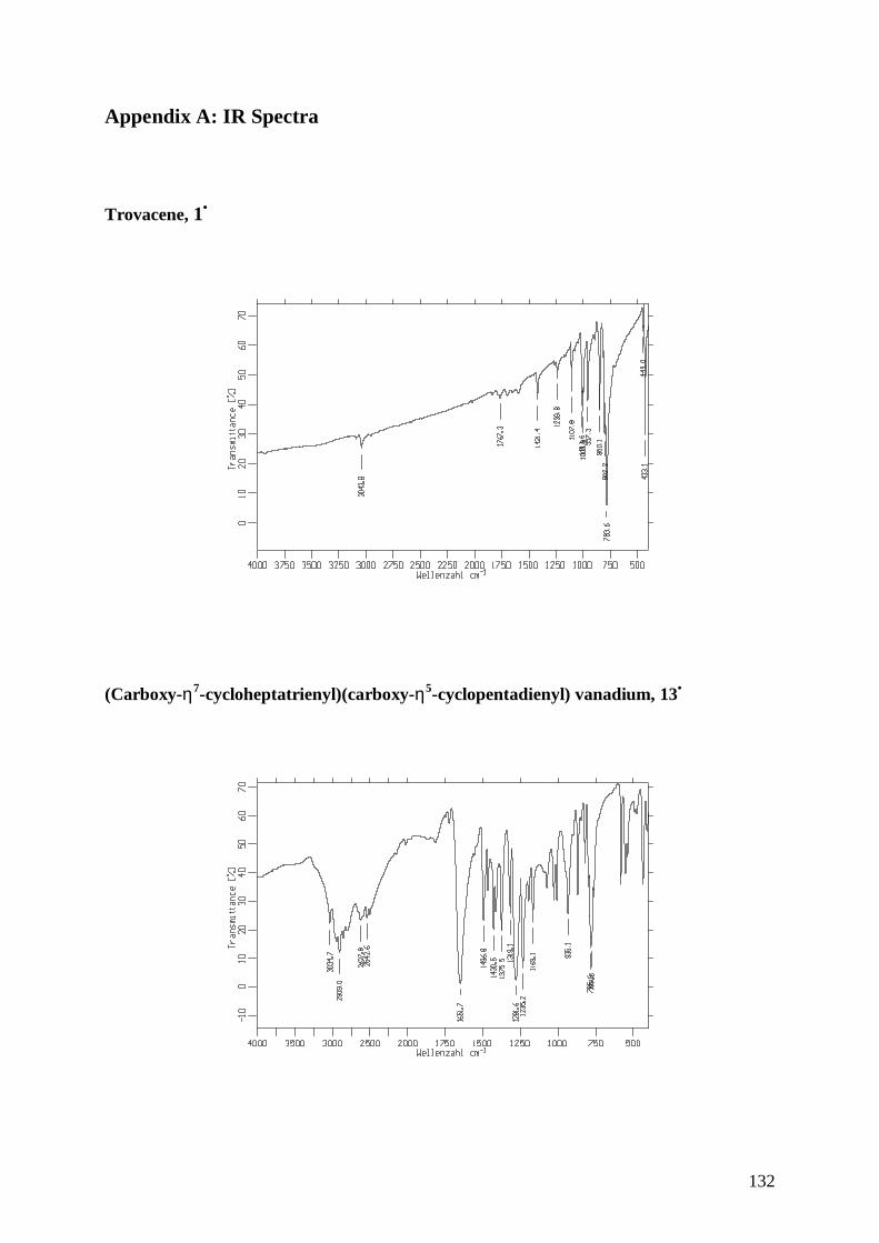
132
Appendix A: IR Spectra
Trovacene, 1•
(Carboxy-η7-cycloheptatrienyl)(carboxy-η5-cyclopentadienyl) vanadium, 13•
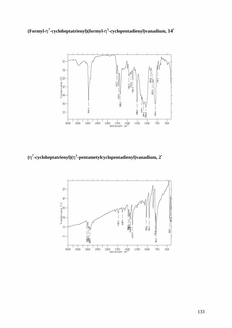
133
(Formyl-η7-cycloheptatrienyl)(formyl-η5-cyclopentadienyl)vanadium, 14•
(η7-cycloheptatrienyl)(η5-pentametylcyclopentadienyl)vanadium, 2•
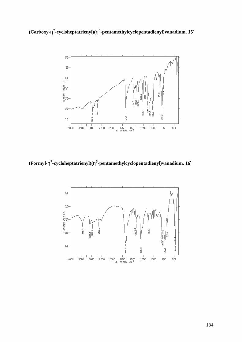
134
(Carboxy-η7-cycloheptatrienyl)(η5-pentamethylcyclopentadienyl)vanadium, 15•
(Formyl-η7-cycloheptatrienyl)(η5-pentamethylcyclopentadienyl)vanadium, 16•
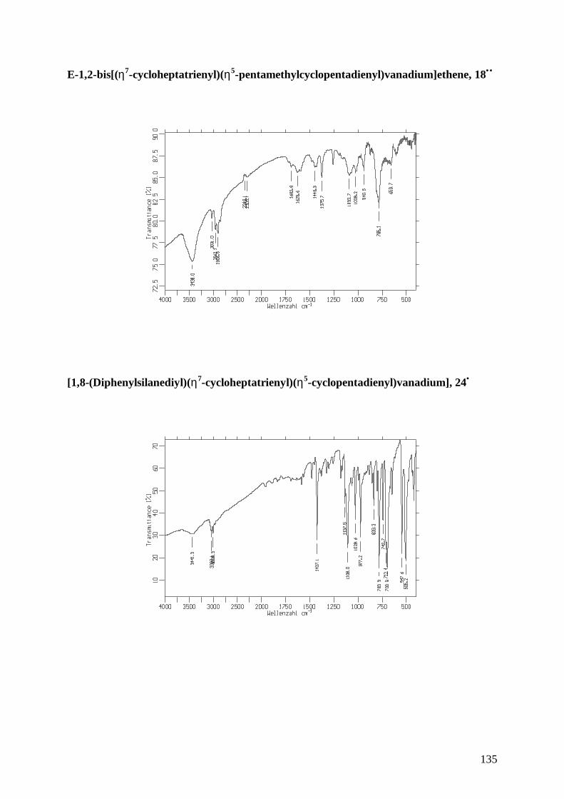
135
E-1,2-bis[(η7-cycloheptatrienyl)(η5-pentamethylcyclopentadienyl)vanadium]ethene, 18••
[1,8-(Diphenylsilanediyl)(η7-cycloheptatrienyl)(η5-cyclopentadienyl)vanadium], 24•
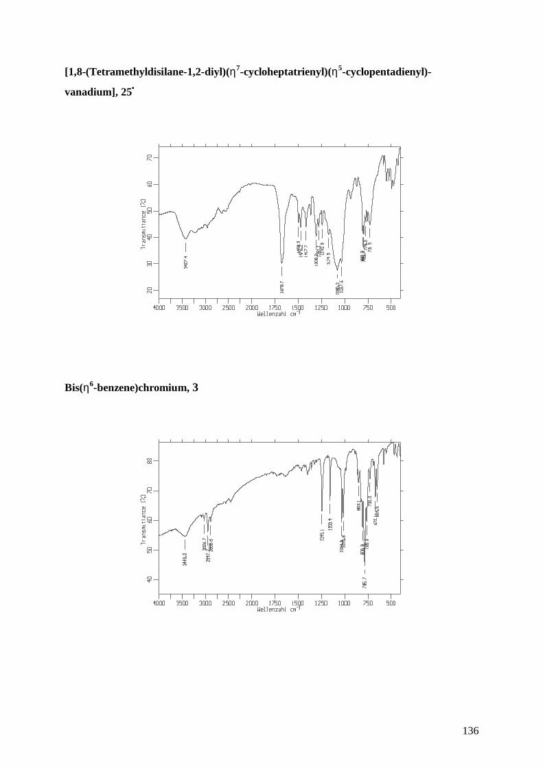
136
[1,8-(Tetramethyldisilane-1,2-diyl)(η7-cycloheptatrienyl)(η5-cyclopentadienyl)-
vanadium], 25•
Bis(η6-benzene)chromium, 3
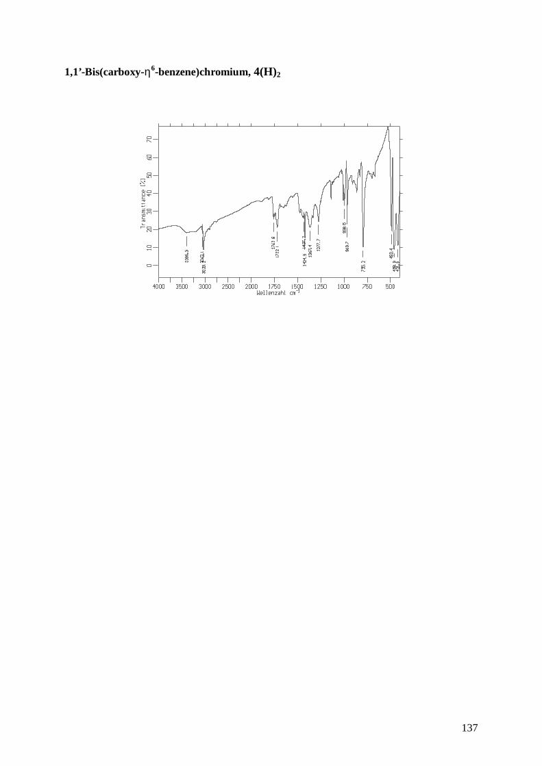
137
1,1’-Bis(carboxy-η6-benzene)chromium, 4(H)2
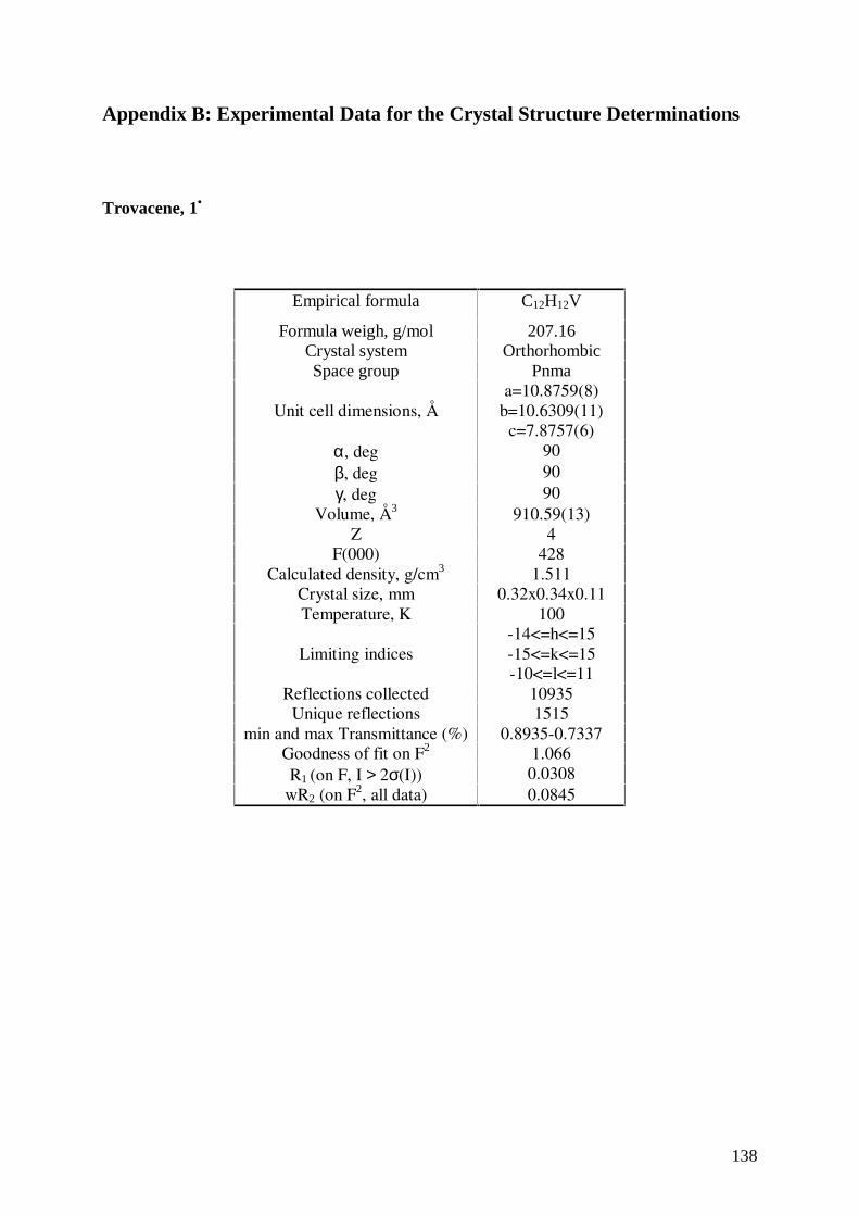
138
Appendix B: Experimental Data for the Crystal Structure Determinations
Trovacene, 1•
Empirical formula C12H12V
Formula weigh, g/mol 207.16Crystal system OrthorhombicSpace group Pnma
Unit cell dimensions, Åa=10.8759(8)
b=10.6309(11)c=7.8757(6)
α, deg 90β, deg 90γ, deg 90
Volume, Å3 910.59(13)Z 4
F(000) 428Calculated density, g/cm3 1.511
Crystal size, mm 0.32x0.34x0.11Temperature, K 100
Limiting indices-14<=h<=15-15<=k<=15-10<=l<=11
Reflections collected 10935Unique reflections 1515
min and max Transmittance (%) 0.8935-0.7337Goodness of fit on F2 1.066R1 (on F, I > 2σ(I)) 0.0308
wR2 (on F2, all data) 0.0845
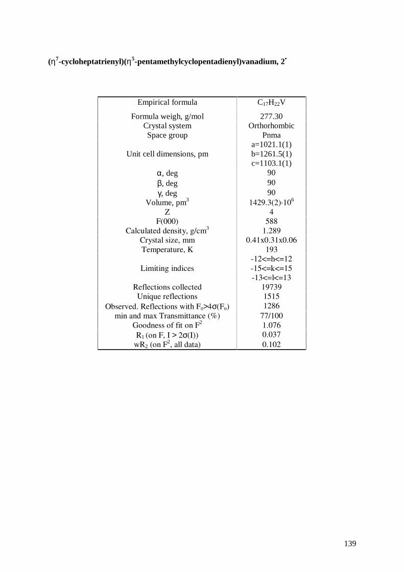
139
(η7-cycloheptatrienyl)(η5-pentamethylcyclopentadienyl)vanadium, 2•
Empirical formula C17H22V
Formula weigh, g/mol 277.30Crystal system OrthorhombicSpace group Pnma
Unit cell dimensions, pma=1021.1(1)b=1261.5(1)c=1103.1(1)
α, deg 90β, deg 90γ, deg 90
Volume, pm3 1429.3(2)·106
Z 4F(000) 588
Calculated density, g/cm3 1.289Crystal size, mm 0.41x0.31x0.06Temperature, K 193
Limiting indices-12<=h<=12-15<=k<=15-13<=l<=13
Reflections collected 19739Unique reflections 1515
Observed. Reflections with Fo>4σ(Fo) 1286min and max Transmittance (%) 77/100
Goodness of fit on F2 1.076R1 (on F, I > 2σ(I)) 0.037
wR2 (on F2, all data) 0.102
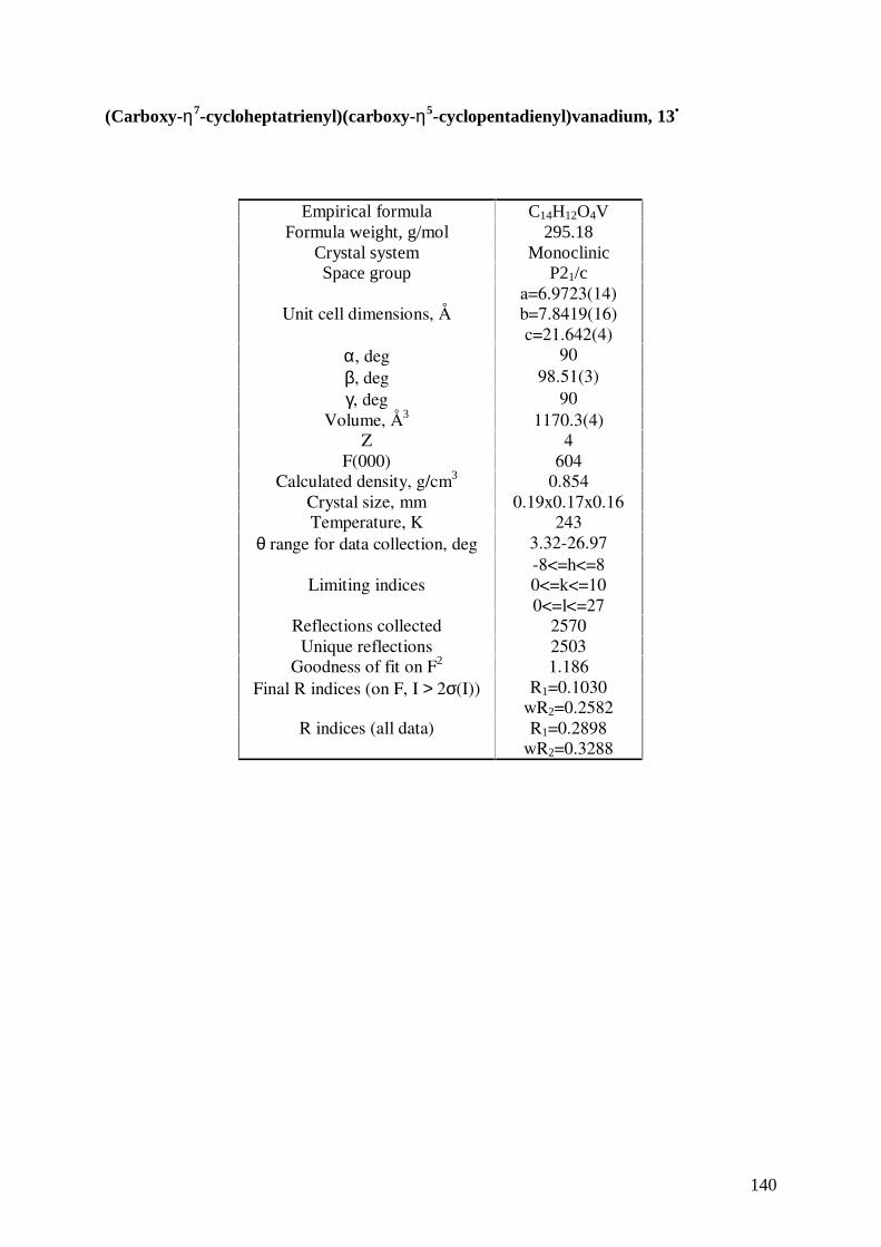
140
(Carboxy-η7-cycloheptatrienyl)(carboxy-η5-cyclopentadienyl)vanadium, 13•
Empirical formula C14H12O4VFormula weight, g/mol 295.18
Crystal system MonoclinicSpace group P21/c
Unit cell dimensions, Åa=6.9723(14)b=7.8419(16)c=21.642(4)
α, deg 90β, deg 98.51(3)γ, deg 90
Volume, Å3 1170.3(4)Z 4
F(000) 604Calculated density, g/cm3 0.854
Crystal size, mm 0.19x0.17x0.16Temperature, K 243
θ range for data collection, deg 3.32-26.97
Limiting indices-8<=h<=80<=k<=100<=l<=27
Reflections collected 2570Unique reflections 2503
Goodness of fit on F2 1.186Final R indices (on F, I > 2σ(I)) R1=0.1030
wR2=0.2582R indices (all data) R1=0.2898
wR2=0.3288
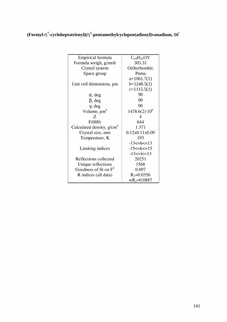
141
(Formyl-η7-cycloheptatrienyl)(η5-pentamethylcyclopentadienyl)vanadium, 16•
Empirical formula C18H22OVFormula weigh, g/molt 305.31
Crystal system OrthorhombicSpace group Pnma
Unit cell dimensions, pma=1061.7(1)b=1248.5(1)c=1115.5(1)
α, deg 90β, deg 90γ, deg 90
Volume, pm3 1478.6(2)·106
Z 4F(000) 644
Calculated density, g/cm3 1.371Crystal size, mm 0.12x0.11x0.09Temperature, K 193
Limiting indices-13<=h<=13-15<=k<=15-13<=l<=13
Reflections collected 20251Unique reflections 1568
Goodness of fit on F2 0.097R indices (all data) R1=0.0356
wR2=0.0887
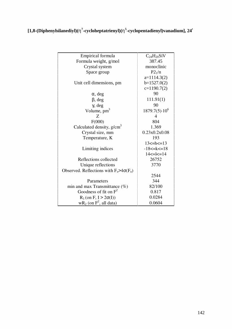
142
[1,8-(Diphenylsilanediyl)(η7-cycloheptatrienyl)(η5-cyclopentadienyl)vanadium], 24•
Empirical formula C24H20SiVFormula weight, g/mol 387.45
Crystal system monoclinicSpace group P21/n
Unit cell dimensions, pma=1114.3(2)b=1527.0(2)c=1190.7(2)
α, deg 90β, deg 111.91(1)γ, deg 90
Volume, pm3 1879.7(5)·106
Z 4F(000) 804
Calculated density, g/cm3 1.369Crystal size, mm 0.23x0.2x0.08Temperature, K 193
Limiting indices13<=h<=13-18<=k<=1814<=l<=14
Reflections collected 26752Unique reflections 3770
Observed. Reflections with Fo>4σ(Fo)2544
Parameters 344min and max Transmittance (%) 82/100
Goodness of fit on F2 0.817R1 (on F, I > 2σ(I)) 0.0284
wR2 (on F2, all data) 0.0604
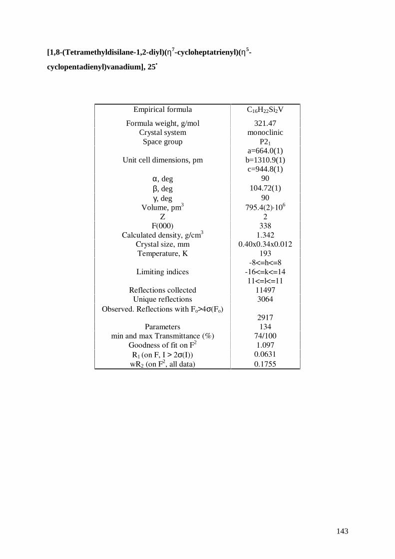
143
[1,8-(Tetramethyldisilane-1,2-diyl)(η7-cycloheptatrienyl)(η5-
cyclopentadienyl)vanadium], 25•
Empirical formula C16H22Si2V
Formula weight, g/mol 321.47Crystal system monoclinicSpace group P21
Unit cell dimensions, pma=664.0(1)
b=1310.9(1)c=944.8(1)
α, deg 90β, deg 104.72(1)γ, deg 90
Volume, pm3 795.4(2)·106
Z 2F(000) 338
Calculated density, g/cm3 1.342Crystal size, mm 0.40x0.34x0.012Temperature, K 193
Limiting indices-8<=h<=8
-16<=k<=1411<=l<=11
Reflections collected 11497Unique reflections 3064
Observed. Reflections with Fo>4σ(Fo)2917
Parameters 134min and max Transmittance (%) 74/100
Goodness of fit on F2 1.097R1 (on F, I > 2σ(I)) 0.0631
wR2 (on F2, all data) 0.1755
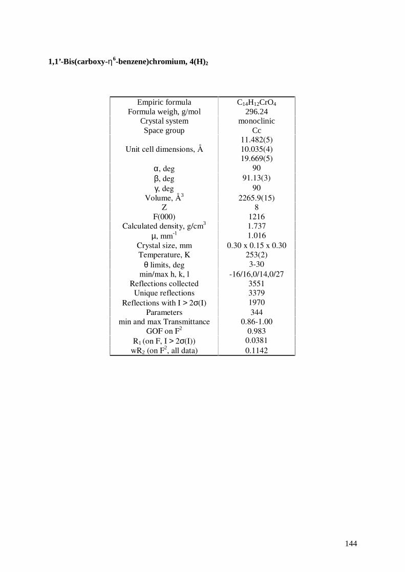
144
1,1’-Bis(carboxy-η6-benzene)chromium, 4(H)2
Empiric formula C14H12CrO4
Formula weigh, g/mol 296.24Crystal system monoclinicSpace group Cc
Unit cell dimensions, Å11.482(5)10.035(4)19.669(5)
α, deg 90β, deg 91.13(3)γ, deg 90
Volume, Å3 2265.9(15)Z 8
F(000) 1216Calculated density, g/cm3 1.737
µ, mm-1 1.016Crystal size, mm 0.30 x 0.15 x 0.30Temperature, K 253(2)
θ limits, deg 3-30min/max h, k, l -16/16,0/14,0/27
Reflections collected 3551Unique reflections 3379
Reflections with I > 2σ(I) 1970Parameters 344
min and max Transmittance 0.86-1.00GOF on F2 0.983
R1 (on F, I > 2σ(I)) 0.0381wR2 (on F2, all data) 0.1142
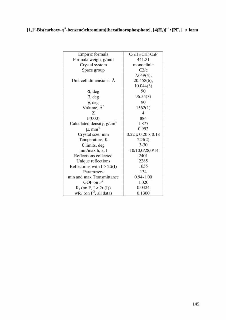
145
[1,1’-Bis(carboxy-η6-benzene)chromium][hexafluorophosphate], [4(H2)]• +• [PF6]
− α form
Empiric formula C14H12CrF6O4PFormula weigh, g/mol 441.21
Crystal system monoclinicSpace group C2/c
Unit cell dimensions, Å7.649(4);20.458(6);10.044(3)
α, deg 90β, deg 96.55(3)γ, deg 90
Volume, Å3 1562(1)Z 4
F(000) 884Calculated density, g/cm3 1.877
µ, mm-1 0.992Crystal size, mm 0.22 x 0.20 x 0.18Temperature, K 223(2)
θ limits, deg 3-30min/max h, k, l -10/10,0/28,0/14
Reflections collected 2401Unique reflections 2285
Reflections with I > 2σ(I) 1655Parameters 134
min and max Transmittance 0.94-1.00GOF on F2 1.020
R1 (on F, I > 2σ(I)) 0.0424wR2 (on F2, all data) 0.1300
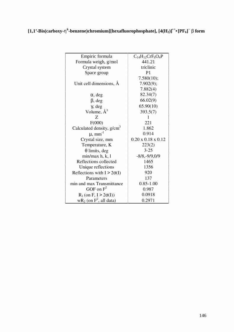
146
[1,1’-Bis(carboxy-η6-benzene)chromium][hexafluorophosphate], [4(H2)]• +• [PF6]
− β form
Empiric formula C14H12CrF6O4PFormula weigh, g/mol 441.21
Crystal system triclinicSpace group P1
Unit cell dimensions, Å7.580(10);7.902(9);7.882(4)
α, deg 82.34(7)β, deg 66.02(9)γ, deg 65.90(10)
Volume, Å3 393.5(7)Z 1
F(000) 221Calculated density, g/cm3 1.862
µ, mm-1 0.914Crystal size, mm 0.20 x 0.18 x 0.12Temperature, K 223(2)
θ limits, deg 3-25min/max h, k, l -8/8,-9/9,0/9
Reflections collected 1465Unique reflections 1356
Reflections with I > 2σ(I) 920Parameters 137
min and max Transmittance 0.85-1.00GOF on F2 0.987
R1 (on F, I > 2σ(I)) 0.0918wR2 (on F2, all data) 0.2971
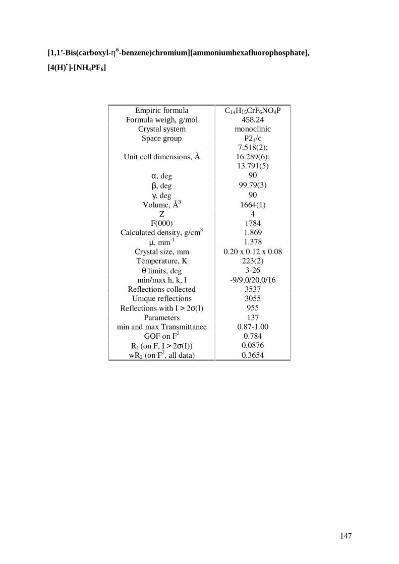
147
[1,1’-Bis(carboxyl-η6-benzene)chromium][ammoniumhexafluorophosphate],
[4(H)•]•[NH4PF6]
Empiric formula C14H15CrF6NO4PFormula weigh, g/mol 458.24
Crystal system monoclinicSpace group P21/c
Unit cell dimensions, Å7.518(2);
16.289(6);13.791(5)
α, deg 90β, deg 99.79(3)γ, deg 90
Volume, Å3 1664(1)Z 4
F(000) 1784Calculated density, g/cm3 1.869
µ, mm-1 1.378Crystal size, mm 0.20 x 0.12 x 0.08Temperature, K 223(2)
θ limits, deg 3-26min/max h, k, l -9/9,0/20,0/16
Reflections collected 3537Unique reflections 3055
Reflections with I > 2σ(I) 955Parameters 137
min and max Transmittance 0.87-1.00GOF on F2 0.784
R1 (on F, I > 2σ(I)) 0.0876wR2 (on F2, all data) 0.3654
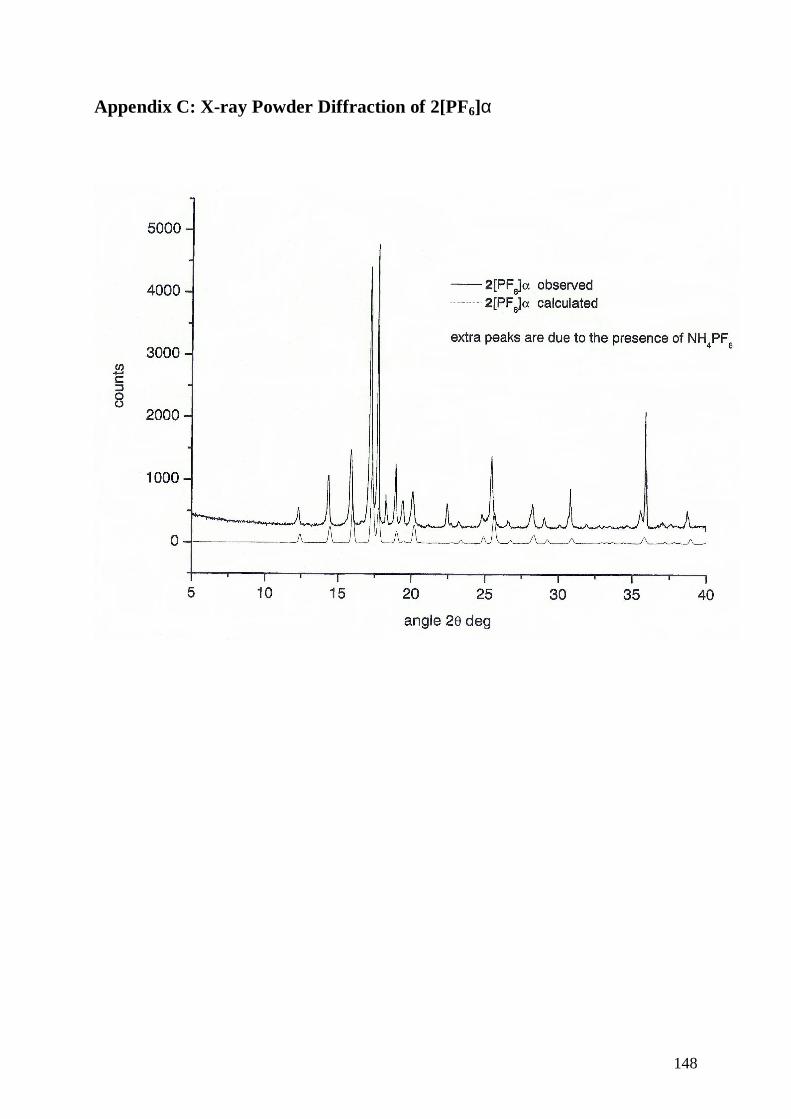
148
Appendix C: X-ray Powder Diffraction of 2[PF6]α
Top Related