


γλώσσες
Σελίδες
Νομικός

CroniconO P E N A C C E S S EC PHARMACEUTICAL SCIENCE
Research Article
In vitro Antimicrobial Activity of Essential Oils from the Lamiaceae and Rutaceae Plant Families Against β -Lactamse-Producing Clinical Isolates Of
Moraxella Catarrhalis
Ukpai Agwu Eze1,2*1Department of Medical Laboratory Sciences, College of Health Sciences, Ebonyi State University, Nigeria2Institute for Global Food Security, School of Biological Sciences, Queen’s University Belfast, United Kingdom
*Corresponding Author: Ukpai A Eze, Lecturer and Research Scientist, Department of Medical Laboratory Sciences, College of Health Sciences, Ebonyi State University, P. M. B. 053, Abakaliki, Nigeria.
Citation: Ukpai Agwu Eze. “In vitro Antimicrobial Activity of Essential Oils from the Lamiaceae and Rutaceae Plant Families Against β -Lactamse-Producing Clinical Isolates Of Moraxella Catarrhalis”. EC Pharmaceutical Seience 2.3 (2016): 325-337.
Received: March 10, 2016; Published: April 21, 2016.
AbstractMoraxella catarrhalis is the third common bacterial cause of otitis media, acute bronchitis, bronchopneumonia and acute exacer-
bations in chronic obstructive pulmonary disease. The emergence of M. catarrhalis strains producing β-lactamases and with high-level resistance to macrolides and lincosamides poses a great risk to public health in the treatment of respiratory tract infections, es-pecially in mixed infections. The increased bacterial resistance to conventional antibiotics has led to a search for alternative sources of antimicrobial agents for the treatment of infections. Essential oils are one natural source for the development of novel antibacterial therapies and complementary treatments. This study was designed to evaluate the in vitro antimicrobial efficacy of 9 essential oils from the Lamiaceae and Rutaceae plant families against 29 β-lactamase-producing clinical isolates and one [1] standard strain of M. catarrhalis using disc diffusion method. In the disc diffusion test, paraffin oil was used as diluents in 1:2 fashion with each essential oil, and 15 µl of each preparation was used for the test. Oregano oil had larger overall mean inhibition zone against all the 30 strains (65.65 mm), followed by thyme oil (62.65 mm), and peppermint oil (54.76 mm) while clary sage oil was the least active against the M. catarrhalis strains. This is the first report of a large scale evaluation of the in vitro antimicrobial efficacy of essential oils against M. catarrhalis and the results indicate that they could be used as inhalation therapy or nebulizer against respiratory tract infections caused by M. catarrhalis. Further studies should be carried out to ascertain the minimum inhibitory concentrations (MIC) of all the essential oils with high efficacy against all the M. catarrhalis strains. In addition, there is need to evaluate the toxicity of these essen-tial oils with high antimicrobial effect using suitable bioassays.
Keywords: Moraxella catarrhalis; Essential oils; Oregano oil; Peppermint oil; Antimicrobial resistance; Alternative therapy
Introduction
M. catarrhalis was previously regarded as a commensal of the upper respiratory tract [1,2]. However, it has regained prominence as a pathogenic bacterium in the last 30 years. M. catarrhalis is now regarded as the third most common bacterial cause of respiratory tract infections after Streptococcus pneumoniae and Haemophilus influenzae [3,4]. In children, M. catarrhalis causes sinusitis, otitis media, pneumonia, tracheitis, preseptal and periorbital cellulitis, pericarditis, osteomyelitis, ophthalmia neonatorum, keratitis and suppurative arthritis [5-7]. In their review, Ioannidis et al. [8] reported 58 cases of bacteraemia caused by M. catarrhalis, 28 in infants and 30 in ado-lescents and adults, including 5 patients with endocarditis. In addition, Yongshou [9] presented data of 40 cases of meningitis associated with M. catarrhalis. In adults, M. catarrhalis is a common cause of purulent tracheobronchitis, pneumonia, laryngitis and acute exacerba-tions in chronic obstructive pulmonary disease, COPD [10-12]. M. catarrhalis has also been reported as a cause of nosocomial respiratory tract infections [13,14].
326
In vitro Antimicrobial Activity of Essential Oils from the Lamiaceae and Rutaceae Plant Families Against β -Lactamse-Producing Clinical Isolates Of Moraxella Catarrhalis
Citation: Ukpai Agwu Eze. “In vitro Antimicrobial Activity of Essential Oils from the Lamiaceae and Rutaceae Plant Families Against β -Lactamse-Producing Clinical Isolates Of Moraxella Catarrhalis”. EC Pharmaceutical Seience 2.3 (2016): 326-337.
There are several reports on the increasing prevalence of β-lactamase-producing strains of M. catarrhalis globally [15]. The first re-port of β-lactamase-producing strains of M. catarrhalis was in Sweden in 1976, at a prevalence of 3.8% [16]. M. catarrhalis produce β-lactamases which have been designated as bro-1, bro-2 and bro-3 [11,17], but there is a probability that bro-3 may be a precursor for the bro-1 and/ or bro-2 [18]. Both the bro-1 and bro-2 enzymes are encoded by chromosomal genes and are phenotypically identical and membrane-associated [18]. Strains with bro-1 have a higher antibiotic resistance than strains with bro-2 or bro-3 [19,20]. Currently, over 95% of M. catarrhalis isolates are resistant to clarithromycin, erythromycin, trimethoprim, penicillin, ampicillin and amoxicillin and trimethoprim-sulfamethoxazole [15, 21- 24].
The increase in β-lactamase-producing strains of M. catarrhalis poses a serious global health challenge for the treatment of communi-ty-acquired infections, especially in mixed infections in which it has been involved in indirect pathogenicity [25]. M. catarrhalis can hinder antibiotic therapy of otherwise susceptible pathogens, including S. pneumoniae and H. influenzae culminating in treatment failure as their β-lactamase enzymes are also secreted by the outer membrane vesicles into the surrounding environment [26,27]. The major treatment option for patients with otitis media and exacerbation of COPD are the macrolides [7]. Recently, resistance of M. catarrhalis to macrolides was reported in China and Japan. In China, Liu et al. [28] demonstrated that mutations of A2982T (corresponding to A2058 in Escherichia coli numbering) and A2796T in the 23S rRNA gene were associated with high-level macrolide resistance in M. catarrhalis strains while Saito et al. [29] reported high-level resistance to the macrolides and lincosamides in an M. catarrhalis strain (NSH1) in Japan and attrib-uted the resistance to a single A2058T mutation in at least three of the four 23S rRNA alleles.
The increased bacterial resistance to conventional antibiotics has led to a search for new antimicrobial compounds from a variety of natural sources for the treatment of infections. Essential oils are one alternative source for the development of novel antibacterial thera-pies and complementary treatments [30-32]. According to Burt [30], essential oils are aromatic oily liquids obtained from the flowers, buds, seeds, leaves, twigs, bark, herbs, wood, fruits and roots of plants. They are mainly liquid, aromatic and exhibit pleasant odour and essence.
The antimicrobial effects of essential oils have been widely studied in food pathogens and have demonstrated in vitro antibacterial ac-tivity against Listeria monocytogenes, Salmonella enterica subspecies enterica serotype Typhimurium (formerly Salmonella typhymurium), E. coli O157:H7, Shigella dysenteriae, Bacillus cereus and Staphylococcus aureus [33,34]. They have also been shown to have antibacterial activity against other bacterial species, antiviral, and anti toxigenic properties [30,35-41]. Reports also indicate that essential oils are po-tent against some potential respiratory pathogens, including H. influenzae, Pseudomonas aeruginosa [42,43], S. pneumoniae and Klebsiella pneumoniae [44]. In their research, Inuonye et al. [45] evaluated a variety of essential oils on respiratory tract pathogens, and reported that H. influenzae, S. pneumoniae, Streptococcus pyogenes and S. aureus were susceptible to the essential oils tested, including thyme oil, cinnamon oil, lemon grass oil, tea tree oil and peppermint oil. Reduction of relapse frequency, maintenance of permanent ventilation and drainage of sinuses has been reported in experimental trials using essential oils for the treatment of respiratory infections [45].
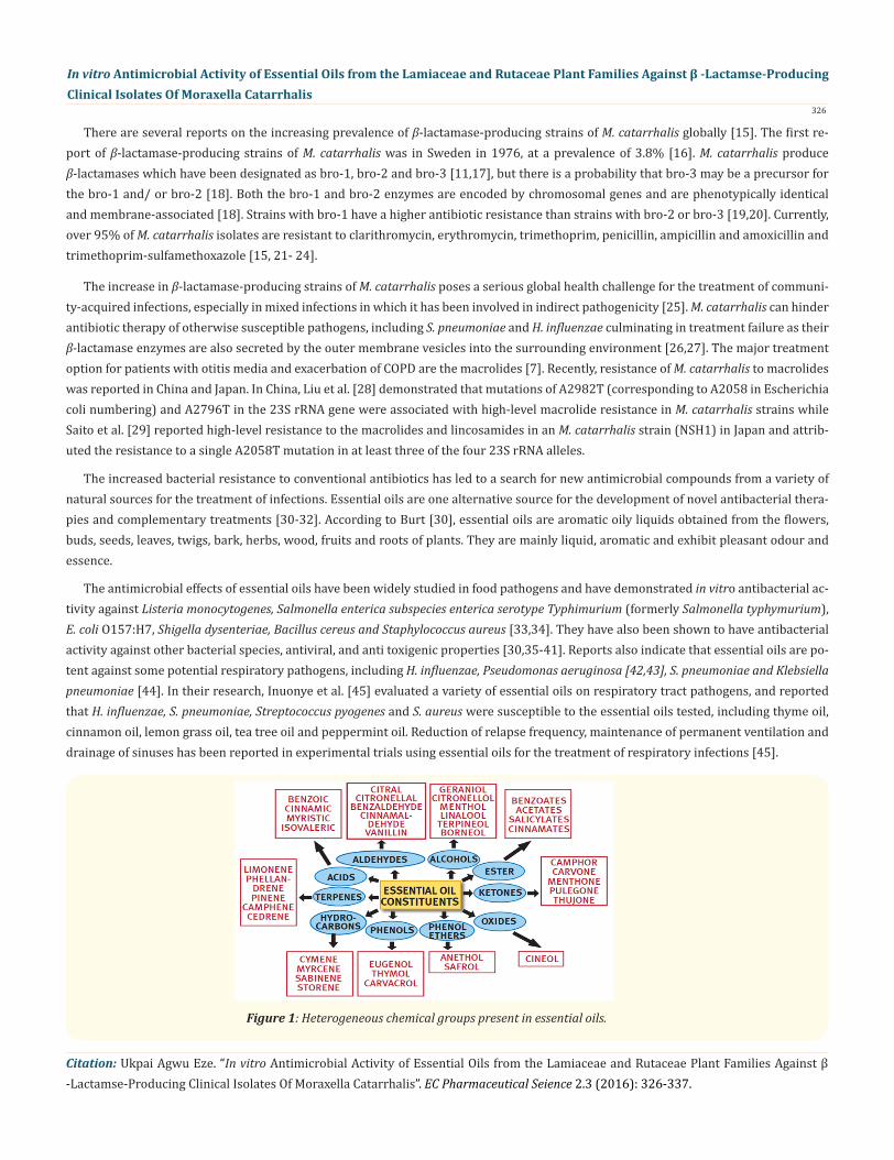
Figure 1: Heterogeneous chemical groups present in essential oils.
Citation: Ukpai Agwu Eze. “In vitro Antimicrobial Activity of Essential Oils from the Lamiaceae and Rutaceae Plant Families Against β -Lactamse-Producing Clinical Isolates Of Moraxella Catarrhalis”. EC Pharmaceutical Seience 2.3 (2016): 326-337.
In vitro Antimicrobial Activity of Essential Oils from the Lamiaceae and Rutaceae Plant Families Against β -Lactamse-Producing Clinical Isolates Of Moraxella Catarrhalis
327
Although the pharmacological effects of essential oils and other bacterial species have been thoroughly studied, to the best of my knowl-edge there are no earlier reports regarding the detailed study on M. catarrhalis and their sensitivity to the activity of essential oils. In ad-dition, the emergence of M. catarrhalis strains resistant to the β-lactams, macrolides and lincosamides warrants the evaluation of essential oils as alternative therapeutic agents. This research was therefore, designed to evaluate the in vitro antimicrobial efficacy of essential oils against β-lactamase-producing clinical isolates of M. catarrhalis.
Material and MethodsBacterial isolates
Twenty nine (29) clinical isolates and one standard strain (NCTC 11020) of M. catarrhalis were obtained from the Centre for Research in Biosciences (CRIBS), School of Health and Life Science, University of the West of England (UWE), Bristol, United Kingdom. All the 29 M. catarrhalis strains were clinical isolates from Weston General Hospital and Southmead Hospital, Bristol, United Kingdom and produce bro-1 and/or bro-2 beta-lactamases [20]. The isolates which were previously stored at -196°C in a liquid nitrogen were revived by in-oculating the thawed samples directly onto blood agar and incubating aerobically at 37°C for 72 h. Purity checks were performed on the isolates using Gram staining technique and oxidase test [46] to confirm the presence of Gram negative diplococci showing positive oxi-dase reaction which are consistent with M. catarrhalis. The 30 M. catarrhalis strains were then subcultured onto Mueller-Hinton agar and incubated aerobically at 37°C for 24 hours. Throughout the period of the research, the strains were continuously subcultured onto new Mueller-Hinton agar and incubated aerobically at 37°C for 24 hours weekly, and stored in the refrigerator at 4°C to avoid them reaching senescence stage.
Essential oilsThe essential oils used for this study are presented below (Table 1). They were procured from Vitamin World Inc., USA, except Ginger
oil and Thyme oil (procured from Sigma-Aldrich, Germany), and Coriander oil and Nutmeg oil (procured from Amphora Aromatics Ltd, Bristol, UK). These essential oils and their diluents are currently stored in the Microbiology Laboratory, UWE for further analysis.
S/No Name of oil Scientific name Product num-ber
1. Clary sage Salvia sclarea B601632. Peppermint Mentha piperita HB601393. Oregano Origanum vulgare B312934. Rosemary Rosmarinus officinalis B601245. Thyme Thymus vulgaris 1102530846. Grapefruit Citrus paradisi B290717. Lemon Citrus limonum B290718. Lime Citrus aurantifolia B290709. Orange Citrus aurantium dulcis B60193
Table 1: Essential oils used for the study.
Evaluation of the antimicrobial activities of the essential oilsIn this preliminary study, the antimicrobial activity of the selected essential oils was determined by disc diffusion method [47]. A
colony of each bacterial strain was first subcultured in Mueller-Hinton broth - MHB (Oxoid, UK; CM045; Lot No. 17803401) and incubated aerobically in Orbital incubator S150 (Stuart Scientific, UK) at 37°C for 24 hours. These were diluted with sterile phosphate buffered saline, 0.85% w/v (Oxoid, UK; BR0014G; Lot No. 17803401) to obtain an inoculum equivalent to 0.5 MacFarland standard (108 cfu/mL) by comparing with a MacFarland standard suspension (Pro-Lab Diagnostics, USA) and confirmed by reading spectrophotometrically (Sanyo SP50 Spectrophotometer, Gallenkamp, UK; Serial no: SP0106028) at 580 nm. Briefly, 100 μL of suspension containing 108 cfu/mL
Citation: Ukpai Agwu Eze. “In vitro Antimicrobial Activity of Essential Oils from the Lamiaceae and Rutaceae Plant Families Against β -Lactamse-Producing Clinical Isolates Of Moraxella Catarrhalis”. EC Pharmaceutical Seience 2.3 (2016): 326-337.
In vitro Antimicrobial Activity of Essential Oils from the Lamiaceae and Rutaceae Plant Families Against β -Lactamse-Producing Clinical Isolates Of Moraxella Catarrhalis
328
of bacteria cells were spread on Petri dishes containing 20 mL of Mueller-Hinton agar - MHA (Oxoid, UK; CM0337; Lot No. 1251584) with hockey spreader dipped in absolute ethanol (VWR International, France) flamed and allowed to cool. Then sterile paper discs measuring 6 mm in diameter (GE Healthcare, UK; Lot No. 4632053) were placed on the agar previously inoculated with the selected test strain using forceps dipped in ethanol and flamed, and were separately impregnated with 15 μL of essential oils diluted with paraffin oil in 1:2 dilution. Ciprofloxacin antibiotic discs - 5 μg (Oxoid, UK; GI290A) were used as positive controls while paper discs impregnated with or without the carrier oil (paraffin oil) were used as negative controls. Plates were sealed with parafilm “M” Laboratory film (Pechiney Plastic Packaging, Chicago, US) and kept for 30 minutes at room temperature to allow diffusion of the oils, and incubated aerobically at 35°C for 24 h. After the incubation period, the antimicrobial activity was assessed by measuring the diameter of the growth-inhibition zone in millimetres us-ing vernier caliper (Fischer, Loughbrough, UK) for the test organisms and the controls. In areas smaller than 6 mm, the inhibitory effects were classified as ‘zero’. Each essential oil was tested using a full plate of MHA to avoid the interference from other oils which is likely to occur if more than one is done on the same plate and tests were done in duplicate.
Statistical analysisThe essential oils were assayed individually in duplicate for their antimicrobial activity against each strain of M. catarrhalis. The data
presented in bar charts with error-bars are mean values ± standard deviation calculated from duplicate determinations and were de-signed using GraphPad Prism statistical software version 5.00 (GraphPad Software Incorporated, USA).
ResultsThe antimicrobial efficacy of 20 M. catarrhalis strains were evaluated using a modified disc diffusion method with the zone of inhibi-
tion indicating the strength of activity of each oil. All the clinical isolates had bro-1 gene, except A10, A11, and E10 that had bro-2 gene. The bro gene status of the standard strain (NCTC 11020) was unknown. The minimum zone of inhibition was taken as the size of the paper disc (6 mm) and zone sizes smaller than 6 mm were converted to zero millimetre (0 mm) in the graphs while the largest zone of inhibition was taken as the size of the Petri-dish (90 mm) .
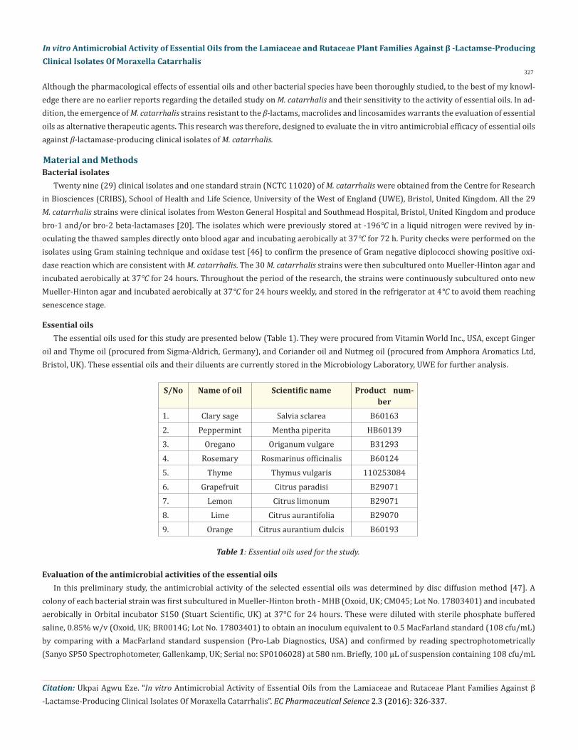
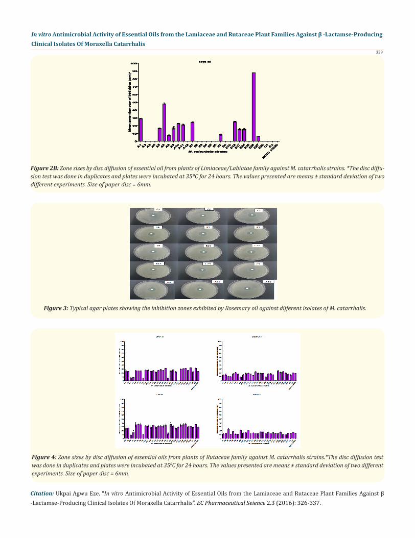
The essential oils from plants of Lamiaceae/Labiatae family (except sage oil) exhibited large zone sizes of inhibition against all the M. catarrhalis strains tested (Figure 2A and 2B). Among the five species, oregano oil had the largest mean zone of inhibition (52.40 - 87.90 mm), followed by thyme oil (45.58 - 87.90 mm) and peppermint oil (39.95 - 87.90 mm) while sage had the smallest zones of inhibition. Oregano oil was had high inhibition zones against A3, A6, E12 and E26 strains; thyme oil (A3, E26, C2 and NCTC 11020); peppermint oil (A3, E6, C2, and NCTC 11020) while rosemary oil had large inhibition zones against A6, E6, E26 and C2 strains. Sage oil had large inhibi-tion zones against A6 and E26 strains, but was unable to inhibit the growth of A2, A3, A4, A12, E2, E3, E5, E6, E8, E10, E25, E29, C1, C2 and NCTC 11020 strains.
Figure 2A: Zone sizes by disc diffusion of essential oils from plants of Lamiaceae/Labiatae family against M. catarrhalis strains. *The disc diffusion test was done in duplicates and plates were incubated at 35ºC for 24 hours. The values presented are means ± standard deviation of two different experiments and the size of paper disc was 6mm.
Citation: Ukpai Agwu Eze. “In vitro Antimicrobial Activity of Essential Oils from the Lamiaceae and Rutaceae Plant Families Against β -Lactamse-Producing Clinical Isolates Of Moraxella Catarrhalis”. EC Pharmaceutical Seience 2.3 (2016): 326-337.
In vitro Antimicrobial Activity of Essential Oils from the Lamiaceae and Rutaceae Plant Families Against β -Lactamse-Producing Clinical Isolates Of Moraxella Catarrhalis
329
Figure 2B: Zone sizes by disc diffusion of essential oil from plants of Limiaceae/Labiatae family against M. catarrhalis strains. *The disc diffu-sion test was done in duplicates and plates were incubated at 35ºC for 24 hours. The values presented are means ± standard deviation of two different experiments. Size of paper disc = 6mm.
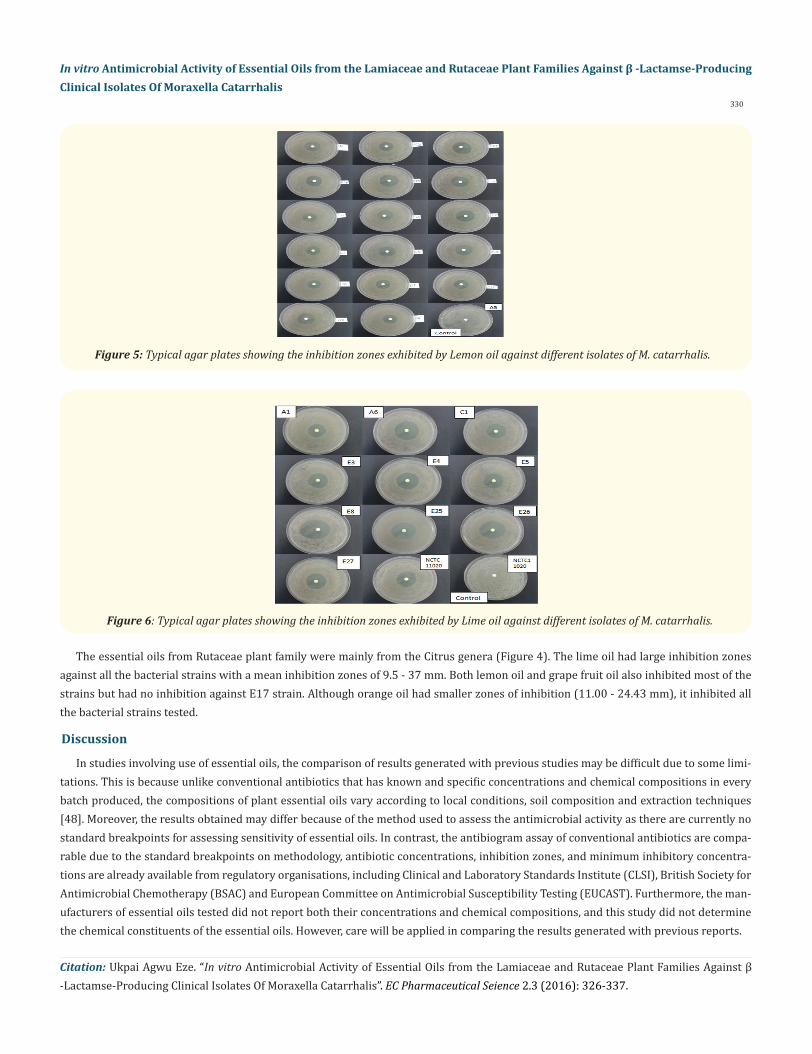
Figure 3: Typical agar plates showing the inhibition zones exhibited by Rosemary oil against different isolates of M. catarrhalis.
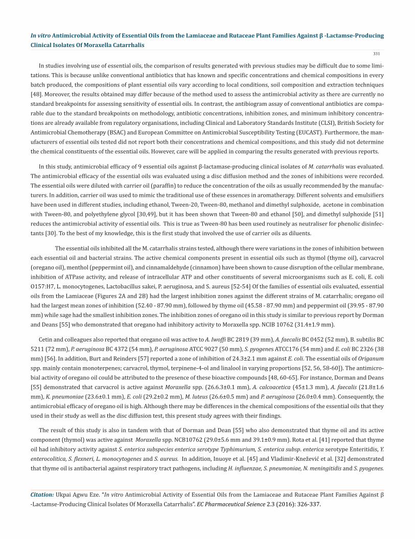
Figure 4: Zone sizes by disc diffusion of essential oils from plants of Rutaceae family against M. catarrhalis strains.*The disc diffusion test was done in duplicates and plates were incubated at 350C for 24 hours. The values presented are means ± standard deviation of two different experiments. Size of paper disc = 6mm.
330
In vitro Antimicrobial Activity of Essential Oils from the Lamiaceae and Rutaceae Plant Families Against β -Lactamse-Producing Clinical Isolates Of Moraxella Catarrhalis
Citation: Ukpai Agwu Eze. “In vitro Antimicrobial Activity of Essential Oils from the Lamiaceae and Rutaceae Plant Families Against β -Lactamse-Producing Clinical Isolates Of Moraxella Catarrhalis”. EC Pharmaceutical Seience 2.3 (2016): 326-337.
Figure 6: Typical agar plates showing the inhibition zones exhibited by Lime oil against different isolates of M. catarrhalis.
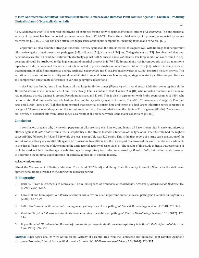
Figure 5: Typical agar plates showing the inhibition zones exhibited by Lemon oil against different isolates of M. catarrhalis.
The essential oils from Rutaceae plant family were mainly from the Citrus genera (Figure 4). The lime oil had large inhibition zones against all the bacterial strains with a mean inhibition zones of 9.5 - 37 mm. Both lemon oil and grape fruit oil also inhibited most of the strains but had no inhibition against E17 strain. Although orange oil had smaller zones of inhibition (11.00 - 24.43 mm), it inhibited all the bacterial strains tested.
Discussion
In studies involving use of essential oils, the comparison of results generated with previous studies may be difficult due to some limi-tations. This is because unlike conventional antibiotics that has known and specific concentrations and chemical compositions in every batch produced, the compositions of plant essential oils vary according to local conditions, soil composition and extraction techniques [48]. Moreover, the results obtained may differ because of the method used to assess the antimicrobial activity as there are currently no standard breakpoints for assessing sensitivity of essential oils. In contrast, the antibiogram assay of conventional antibiotics are compa-rable due to the standard breakpoints on methodology, antibiotic concentrations, inhibition zones, and minimum inhibitory concentra-tions are already available from regulatory organisations, including Clinical and Laboratory Standards Institute (CLSI), British Society for Antimicrobial Chemotherapy (BSAC) and European Committee on Antimicrobial Susceptibility Testing (EUCAST). Furthermore, the man-ufacturers of essential oils tested did not report both their concentrations and chemical compositions, and this study did not determine the chemical constituents of the essential oils. However, care will be applied in comparing the results generated with previous reports.
In studies involving use of essential oils, the comparison of results generated with previous studies may be difficult due to some limi-tations. This is because unlike conventional antibiotics that has known and specific concentrations and chemical compositions in every batch produced, the compositions of plant essential oils vary according to local conditions, soil composition and extraction techniques [48]. Moreover, the results obtained may differ because of the method used to assess the antimicrobial activity as there are currently no standard breakpoints for assessing sensitivity of essential oils. In contrast, the antibiogram assay of conventional antibiotics are compa-rable due to the standard breakpoints on methodology, antibiotic concentrations, inhibition zones, and minimum inhibitory concentra-tions are already available from regulatory organisations, including Clinical and Laboratory Standards Institute (CLSI), British Society for Antimicrobial Chemotherapy (BSAC) and European Committee on Antimicrobial Susceptibility Testing (EUCAST). Furthermore, the man-ufacturers of essential oils tested did not report both their concentrations and chemical compositions, and this study did not determine the chemical constituents of the essential oils. However, care will be applied in comparing the results generated with previous reports.
331
In vitro Antimicrobial Activity of Essential Oils from the Lamiaceae and Rutaceae Plant Families Against β -Lactamse-Producing Clinical Isolates Of Moraxella Catarrhalis
Citation: Ukpai Agwu Eze. “In vitro Antimicrobial Activity of Essential Oils from the Lamiaceae and Rutaceae Plant Families Against β -Lactamse-Producing Clinical Isolates Of Moraxella Catarrhalis”. EC Pharmaceutical Seience 2.3 (2016): 326-337.
In this study, antimicrobial efficacy of 9 essential oils against β-lactamase-producing clinical isolates of M. catarrhalis was evaluated. The antimicrobial efficacy of the essential oils was evaluated using a disc diffusion method and the zones of inhibitions were recorded. The essential oils were diluted with carrier oil (paraffin) to reduce the concentration of the oils as usually recommended by the manufac-turers. In addition, carrier oil was used to mimic the traditional use of these essences in aromatherapy. Different solvents and emulsifiers have been used in different studies, including ethanol, Tween-20, Tween-80, methanol and dimethyl sulphoxide, acetone in combination with Tween-80, and polyethylene glycol [30,49], but it has been shown that Tween-80 and ethanol [50], and dimethyl sulphoxide [51] reduces the antimicrobial activity of essential oils. This is true as Tween-80 has been used routinely as neutraliser for phenolic disinfec-tants [30]. To the best of my knowledge, this is the first study that involved the use of carrier oils as diluents.
The essential oils inhibited all the M. catarrhalis strains tested, although there were variations in the zones of inhibition between each essential oil and bacterial strains. The active chemical components present in essential oils such as thymol (thyme oil), carvacrol (oregano oil), menthol (peppermint oil), and cinnamaldehyde (cinnamon) have been shown to cause disruption of the cellular membrane, inhibition of ATPase activity, and release of intracellular ATP and other constituents of several microorganisms such as E. coli, E. coli O157:H7, L. monocytogenes, Lactobacillus sakei, P. aeruginosa, and S. aureus [52-54] Of the families of essential oils evaluated, essential oils from the Lamiaceae (Figures 2A and 2B) had the largest inhibition zones against the different strains of M. catarrhalis; oregano oil had the largest mean zones of inhibition (52.40 - 87.90 mm), followed by thyme oil (45.58 - 87.90 mm) and peppermint oil (39.95 - 87.90 mm) while sage had the smallest inhibition zones. The inhibition zones of oregano oil in this study is similar to previous report by Dorman and Deans [55] who demonstrated that oregano had inhibitory activity to Moraxella spp. NCIB 10762 (31.4±1.9 mm).
Cetin and colleagues also reported that oregano oil was active to A. Iwoffi BC 2819 (39 mm), A. faecalis BC 0452 (52 mm), B. subtilis BC 5211 (72 mm), P. aeruginosa BC 4372 (54 mm), P. aeruginosa ATCC 9027 (50 mm), S. pyogenes ATCC176 (54 mm) and E. coli BC 2326 (38 mm) [56]. In addition, Burt and Reinders [57] reported a zone of inhibition of 24.3±2.1 mm against E. coli. The essential oils of Origanum spp. mainly contain monoterpenes; carvacrol, thymol, terpinene-4-ol and linalool in varying proportions [52, 56, 58-60]). The antimicro-bial activity of oregano oil could be attributed to the presence of these bioactive compounds [48, 60-65]. For instance, Dorman and Deans [55] demonstrated that carvacrol is active against Moraxella spp. (26.6.3±0.1 mm), A. calcoacetica (45±1.3 mm), A. faecalis (21.8±1.6 mm), K. pneumoniae (23.6±0.1 mm), E. coli (29.2±0.2 mm), M. luteus (26.6±0.5 mm) and P. aeruginosa (26.0±0.4 mm). Consequently, the antimicrobial efficacy of oregano oil is high. Although there may be differences in the chemical compositions of the essential oils that they used in their study as well as the disc diffusion test, this present study agrees with their findings.
The result of this study is also in tandem with that of Dorman and Dean [55] who also demonstrated that thyme oil and its active component (thymol) was active against Moraxella spp. NCB10762 (29.0±5.6 mm and 39.1±0.9 mm). Rota et al. [41] reported that thyme oil had inhibitory activity against S. enterica subspecies enterica serotype Typhimurium, S. enterica subsp. enterica serotype Enteritidis, Y. enterocolitica, S. flexneri, L. monocytogenes and S. aureus. In addition, Inuoye et al. [45] and Vladimir-Knežević et al. [32] demonstrated that thyme oil is antibacterial against respiratory tract pathogens, including H. influenzae, S. pneumoniae, N. meningitidis and S. pyogenes.
Citation: Ukpai Agwu Eze. “In vitro Antimicrobial Activity of Essential Oils from the Lamiaceae and Rutaceae Plant Families Against β -Lactamse-Producing Clinical Isolates Of Moraxella Catarrhalis”. EC Pharmaceutical Seience 2.3 (2016): 326-337.
In vitro Antimicrobial Activity of Essential Oils from the Lamiaceae and Rutaceae Plant Families Against β -Lactamse-Producing Clinical Isolates Of Moraxella Catarrhalis
332
Peppermint oil also exhibited strong antibacterial activity against all the strains tested; this agrees well with findings that peppermint oil is active against respiratory tract pathogens [45]. Hili et al. [51], Iscan et a.l [74] and Yadegarinia et al. [75] also observed that pep-permint oil essential oil exhibited antimicrobial activity against both S. aureus and E. coli strains. The large inhibition zones found in pep-permint oil could be attributed to the high content of menthol present in it [76-78]. Essential oils rich in compounds such as, menthone, piperitone oxide, carvone and linalool are widely reported to possess high level of antimicrobial activity [79]. While this study revealed that peppermint oil had antimicrobial activity against S. pneumoniae and E. coli, Prabuseenivasan et al. [80] reported no such activity. This variation in the antimicrobial activity could be attributed to several factors such as genotype, stage of maturity, cultivation peculiarities, soil composition and climate differences in various geographical locations.
In the Rutaceae family, lime oil and lemon oil had large inhibition zones (Figure 4) with overall mean inhibition zones against all the Moraxella strains as 29.5 mm and 23.33 mm, respectively. This is similiar to that of Yadav et al. [81] who reported that lime and lemon oil had moderate activity against S. aureus, Pseudomonas spp. and E. coli. This is also in agreement with Prabu seenivasan et al. [80], who demonstrated that lime and lemon oils had excellent inhibitory activity against S. aureus, B. subtilis, K. pneumoniae, P. vulgaris, P. aerugi-nosa and E. coli. Javed et al. [82] also demonstrated that essential oils from lime and lemon oils had larger inhibition zones compared to orange oil. There are several reports on the antimicrobial activity of essential oils from the plants of Citrus genera [83-86]. The antimicro-bial activity of essential oils from Citrus spp. is as a result of dl-limonene which is the major constituent [86-89].
Conclusion
In conclusion, oregano oils, thyme oils, peppermint oil, rosemary oils, lime oil, and lemon oil have shown high in vitro antimicrobial efficacy against M. catarrhalis strains. The susceptibility of the strains tested is a function of the type of oil. The E6 strain had the highest susceptibility, followed by A3, and E26 while the least susceptible was E29 strain. This is the first report of a large scale evaluation of the antimicrobial efficacy of essential oils against M. catarrhalis. In addition, it is the first report that involved the use of carrier oils as diluents in the disc diffusion method of determining the antibacterial activity of essential oils. The results of this study indicate that essential oils could be used as inhalation therapy or nebuliser against respiratory tract infections caused by M. catarrhalis, but further work is needed to determine the minimal exposure time for efficacy, applicability, and the toxicity.
I thank the Management of Tertiary Education Trust Fund (TET Fund), and Ebonyi State University, Abakaliki, Nigeria for the staff devel-opment scholarship awarded to me during the research period.
Bibliography
Also, Łysakowska et al. [66] reported that thyme oil exhibited strong activity against 29 clinical strains of A. baumanii. The antimicrobial activity of thyme oil has been reported by several researchers [57, 67-71]. The antimicrobial activity of thyme oil, as reported by several researchers [30, 45, 72, 73], has been attributed to presence of phenolic compounds, including thymol and carvacrol [66].
Acknowledgements
1. Berk SL. “From Micrococcus to Moraxella. The re-emergence of Branhamella catarrhalis”. Archives of International Medicine 150 (1990): 2254-2257.
2. Karalus R and Campagnari A. “Moraxella catarrhalis: a review of an important human mucosal pathogen”. Microbes and Infection 2 (2000): 547-559.
3. Catlin BW. “Branhamella catarrhalis: an organism gaining respect as a pathogen”. Clinical Microbiology review 3 (1990): 293-320.
4. Verduin CM., et al. “Moraxella catarrhalis: from emerging to established pathogen”. Clinical Microbiology Reviews 15.1 (2012): 125-144.
5. Boyle FM., et al. “Branhamella (Moraxella) catarrhalis: pathogenic significance in respiratory infections”. Medical Journal of Australia 154 (1991): 592-596.
Citation: Ukpai Agwu Eze. “In vitro Antimicrobial Activity of Essential Oils from the Lamiaceae and Rutaceae Plant Families Against β -Lactamse-Producing Clinical Isolates Of Moraxella Catarrhalis”. EC Pharmaceutical Seience 2.3 (2016): 326-337.
In vitro Antimicrobial Activity of Essential Oils from the Lamiaceae and Rutaceae Plant Families Against β -Lactamse-Producing Clinical Isolates Of Moraxella Catarrhalis
333
6. Lutwick L and Fernandes L. “The other sibblings: respiratory infections caused by Moraxella catarrhalis and Haemophilus influen-zae”. Current Infectious Disease Reports 8 (2006): 215-221.
7. Murphy TF and Parameswaran GI. “Moraxella catarrhalis, a human respiratory tract pathogen”. Clinical Infectious Diseases 49 (2009): 124-131.
8. Ioannidis JPA., et al. “Spectrum and significance of bacteraemia due to Moraxella catarrhalis”. Clinical Infectious Disease 21 (1995): 390-397.
9. Yongshou JIN. “Moraxella catarrhalis meningitis: a case report”. Chinese Medical Journal 113.4 (2000): 381-382.
10. Hol C., et al. “Experimental evidence for Moraxella-induced penicillin neutralization in pneumococcal pneumonia”. Journal of Infec-tious Disease 170 (1994): 1613-1616.
11. McGregor K., et al. “Moraxella catarrhalis: clinical significance, antimicrobial susceptibility and BRO β-lactamases”. European Journal of Clinical Microbiology and Infectious Diseases 17 (1998): 219-234.
12. Prashanth HV., et al. “Moraxella catarrhalis - A rediscovered pathogen”. International Journal of Biological and Medical Research 2.4 (2011): 979-981.
13. Wirth T., et al. “The rise and spread of a new pathogen: seroresistant Moraxella catarrhalis”. Genome Research 17 (2007): 1647-1656.
14. Levy F., et al. “Nosocomial clusters and risk factors in Moraxella catarrhalis”. Epidemiology and Infection 137.4 (2009): 581-590.
15. Hsu SF., et al. “Antimicrobial resistance of Moraxella catarrhalis isolates in Taiwan”. Journal of Microbiology, Immunology and Infection 45 (2012): 134-140.
16. Malvmvall BE., et al. “In vitro sensitivity to penicillin v and β-lactamase production of Branhamella catarrhalis”. Journal of Antimicro-bial Chemotherapy 3 (1977): 374-375.
17. Kӧseoğlu O., et al. “Evaluation of restriction endonuclease analysis of BRO beta-lactamases in clinical and carrier isolates of Moraxella catarhalis”. Scandinavian Journal of Infectious Diseases 36.6-7 (2004): 431-434.
18. Bootsma HJ., et al. “Molecular characterization of the BRO β-lactamase of Moraxella (Branhamella) catarrhalis. Antimicrobial Agents and Chemotherapy 40 (1996): 966-972.
19. Paykel JM. “Moraxella (Branhamella) catarrhalis infections”. Primary Care Update in Obstetrics/Gynaecology 9 (2002): 33-35.
20. Moores, E. “Genotypic and phenotypic epidemiology of β-lactamases from clinical strains of Moraxella catarrhalis”. University of the West of England (UWE), Bristol, MSc project (2009).
21. Esel D., et al. “Evaluation of susceptibility patterns and BRO β-lactamase types among clinical isolates of Moraxella catarrhalis”. Clini-cal Microbiology and Infectious Diseases 13 (2007): 1023-1025.
22. Karpanoja P., et al. “Connection between trimethoprim-sulfamethoxazole use and resistance in Streptococcus pneumoniae, Haemoph-ilus influenzae and Moraxella catarrhalis”. Antimicrobial Agents Chemotherapy 5.7 (2008): 2480-2485.
23. Bell JM., et al. “Development of a disk diffusion method for testing Moraxella catarrhalis susceptibility using Clinical and Laboratory Standards Institute methods: a SENTRY antimicrobial surveillance program report”. Journal of Clinical Microbiology 47.7 (2009): 2187-2193.
24. Khan MA., et al. “bro β-lactamase resistance in a global cross-sectional study of Moraxella catarrhalis from children and adults”. Jour-nal of Antimicrobial Chemotherapy 65 (2010): 91-97.
Citation: Ukpai Agwu Eze. “In vitro Antimicrobial Activity of Essential Oils from the Lamiaceae and Rutaceae Plant Families Against β -Lactamse-Producing Clinical Isolates Of Moraxella Catarrhalis”. EC Pharmaceutical Seience 2.3 (2016): 326-337.
In vitro Antimicrobial Activity of Essential Oils from the Lamiaceae and Rutaceae Plant Families Against β -Lactamse-Producing Clinical Isolates Of Moraxella Catarrhalis
334
25. Johnson DM., et al. “Susceptibility trends of Haemophilus influenzae and Moraxella catarrhalis against orally administered antimi-crobial agents: five-year report from the SENTRY Antimicrobial Surveillance Programme”. Diagnostic Microbiology and Infectious Disease 47 (2003): 373-376.
26. Schaar V., et al. “Moraxella catarrhalis outer membrane vesicles carry beta-lactamase and promote survival of Streptococcus pneu-moniae and Haemophilus influenzae by inactivating amoxicillin”. Antimicrobial Agents and Chemotherapy 55.8 (2011):3845-3853.
27. Schaar V., et al. “Outer membrane vesicles shield Moraxella catarrhalis b-lactamase from neutralization by serum IgG”. Journal of Antimicrobial Chemotherapy 68 (2013): 593-600.
28. Liu Y., et al. “High prevalence and molecular analysis of macrolide-non susceptible Moraxella catarrhalis isolated from nasopharynx of healthy children in China”. Microbial Drug Resistance 18 (2012): 417-426.
29. Saito R., et al. “Molecular mechanism of macrolide-lincosamide resistance in Moraxella catarrhalis”. Journal of Medical Microbiology 61 (2012): 1435-1438.
30. Burt S. “Essential oils: their antibacterial properties and potential applications in foods - a review”. International of Food Microbiology 94 (2004): 223-253.
31. Eldris AE. “Pharmaceutical and therapeutic potentials of essential oils and their individual volatile constituents: a review”. Phyto-therapy Research (2007).
32. Vladimir-Knežević S., et al. “Antimicrobial activity of Thymus longicaulis C. Presyl essential oil against respiratory pathogens”. Central European Journal of Biology 7.6 (2012): 1109-1115.
33. Schmidt E., et al. “Antimicrobial testing and gas chromatographic analysis of aroma chemicals”. Journal of Essential Oil Bearing Plants 8 (2005): 99-106.
34. Jirovetz L., et al. “Antimicrobial testings and gas chromatographic analysis of pure oxygenated monoterpenes 1, 8-cineol, α-terpineol, terpen-4-ol and camphor as well as target compounds in essential oils of Pine (Pinus pinaster), Rosemary (Romarinus officinalis) and tea trees (Melaleuca alternifolia)”. Scientific Pharmacology 73 (2005): 27-39.
35. Manohar V., et al. “Antifungal activities of origanum oil against Candida albicans”. Molecular and Cellular Biochemistry 228 (2001): 111-117.
36. Hammer KA., et al. “In vitro activity of Melaleuca alternifolia (tea tree) oil against dermatophytes and other filamentous fungi”. Jour-nal of Antimicrobial Chemotherapy 50 (2002): 195-199.
37. Pandey AK., et al. “Chemical composition and antimycotic activity of the essential oils of corn mint (Mentha arvensis) and lemon grass (Cymbopogon flexuosus) against human pathogenic fungi”. Pharmaceutical Biology 41 (2003): 421-425.
38. Duschatzky CB., et al. “Evaluation of chemical and antiviral properties of essential oils from South American plants”. Antiviral Chem-istry and Chemotherapy 16 (2005): 247-251.
39. Singh G., et al. “Antioxidative and antibacterial potentials of essential oils and extracts isolated from various spice material”. Journal of Food Safety 25.2 (2005): 130-145.
40. Cavaleiro C. et al. “Antifungal activity of Juniperus essential oils against dermatophyte, Aspergillus and Candida strains”. Journal of Applied Microbiology 100 (2006): 1333-1338.
41. Rota MC., et al. “Antimicrobial activity and chemical composition of Thymus vulgaris, Thymus zygis and Thymus hyemalis essential oils”. Food Control 19 (2008): 681-687.
42. Skocibusic M., et al. “Antibacterial activity of Achillea clavennae essential oil against respiratory tract pathogens”. Fitoterapia 75 (2004): 733-736.
Citation: Ukpai Agwu Eze. “In vitro Antimicrobial Activity of Essential Oils from the Lamiaceae and Rutaceae Plant Families Against β -Lactamse-Producing Clinical Isolates Of Moraxella Catarrhalis”. EC Pharmaceutical Seience 2.3 (2016): 326-337.
In vitro Antimicrobial Activity of Essential Oils from the Lamiaceae and Rutaceae Plant Families Against β -Lactamse-Producing Clinical Isolates Of Moraxella Catarrhalis
335
43. Messager S., et al. “Assessment of the antibacterial activity of tea tree oil using the European EN 1276 and EN 12054 standard suspen-sion tests”. Journal of Hospital Infections 59 (2005): 113-125.
44. Rahman AKMS., et al. “Antibacterial activity of two limonoids from Swietenia mahagoni against multi-drug-resistant (MDR) bacterial strains”. Journal of National Medicine 63 (2009): 41-45.
45. Inuoye S., et al. “Screening of the bacterial effects of a variety of essential oils on respiratory pathogens, using a modified dilution as-say method”. Journal of Infection and Chemotherapy 7 (2001): 251-254.
46. Cheesbrough M. “District Laboratory Practice in Tropical Countries”. Part 2. Second Edition. Cambridge University Press. Cambridge, UK (2006).
47. Clinical Laboratory Standards Institute. “Performance for antimicrobial disk susceptibility tests”. Approved standard, 9th edition. CLSI Document M2-A9 (2006): 26:1.
48. Hussain AI., et al. “Chemical composition, antioxidant and antimicrobial activities of basil (Ocimum basilicum) essential oils depends on seasonal variations”. Food Chemistry 108 (2008): 986-995.
49. Mann CM and Markham JL. “A new method for determining the minimum inhibitory concentration of essential oils”. Journal of Applied Microbiology 84 (1998): 538-544.
50. Remmal A., et al. “Inhibition of antibacterial activity of essential oils by Tween-80 and ethanol in liquid medium”. Journal of Pharmacy in Belgium 48 (1993): 32-36.
51. Hili P., et al. “Antimicrobial action of essential oils: the effect of dimethylsulphoxide on the activity of cinnamon oil”. Letters in Applied Microbiology 24 (1997): 269-275.
52. Lambert R., et al. “A study of the minimum inhibitory concentration and mode of action of oregano essential oil, thymol and carva-crol”. Journal of Applied Microbiolology 91.3 (2001): 453-462.
53. Raybaudi-Massilia R., et al. “Control of pathogenic and spoilage microorganisms in fresh-cut fruits and Fruit 192 juices by traditional and alternative natural antimicrobials”. Comparative Review in Food Science and Food Safety 8.3 (2009): 157-180.
54. Lopez P., et al. “Vapor-phase activities of cinnamon, thyme, and oregano essential oils and key constituents against foodborne micro-organisms”. Journal of Agriculture and Food Chemistry 55 (2007): 4348-4356.
55. Dorman HJD and Deans SG. “Antimicrobial agents from plants: antibacterial activity of plant volatile oils”. Journal of Applied Microbiol-ogy 88 (2000): 308-316.
56. Çetin B., et al. “The investigation of antimicrobial activity of thyme and oregano essential oils”. Turkish Journal Agriculture and For-estry 35 (2011): 145-154.
57. Burt S and Reinders RD. “Antibacterial activity of selected plant essential oils against Escherichia coli O157:H7”. Letters in Applied Microbiology 36.3 (2003): 162-167.
58. Prudent D., et al. “Analysis of essential oil of wild oregano from Martinique (Coleus aromaticus Benth.) - evaluation of its bacterio-static and fungistatic properties”. Journal of Essential Oil Research 7 (1995): 165-173.
59. Sivropoulou A., et al. “Antimicrobial and cytotoxic activities of oreganum essential oils”. Journal of Agricultural and Food Chemistry 44 (1996): 1202-1205.
60. Bendahou M., et al. “Antimicrobial activity and chemical composition of Origanum glandulosum Desf. essential oil and extract ob-tained by microwave extraction: comparison with hydrodistillation”. Food Chemistry 106 (2008): 132-139.
Citation: Ukpai Agwu Eze. “In vitro Antimicrobial Activity of Essential Oils from the Lamiaceae and Rutaceae Plant Families Against β -Lactamse-Producing Clinical Isolates Of Moraxella Catarrhalis”. EC Pharmaceutical Seience 2.3 (2016): 326-337.
In vitro Antimicrobial Activity of Essential Oils from the Lamiaceae and Rutaceae Plant Families Against β -Lactamse-Producing Clinical Isolates Of Moraxella Catarrhalis
336
61. Muller-Riebau F., et al. “Chemical composition and fungitoxic properties to phytopathogenic fungi of essential oils of selected aro-matic plants growing wild in Turkey”. Journal of Agriculture and Food Chemistry 43 (1995): 2262-2266.
62. Bouchra C., et al. “Chemical composition and antifungal activity of essential oils of seven Moroccan Labiatae against Botrytis cinerea. Pers: Fr”. Journal of Ethnopharmacology 89 (2003): 165-169.
63. Daferera DJ., et al. “The effectiveness of plant essential oils on the growth of Botrytis cinerea, Fusarium sp. and Clavibacter michi-ganensis subsp. Michiganensis”. Crop Protectection 22 (2003): 39-44.
64. Sokmen M., et al. “In vitro antioxidant, antimicrobial, and antiviral activities of the essential oil and various extracts from herbal parts and callus cultures of Origanum acutidens”. Journal of Agriculture and Food Chemistry 52 (2004): 3309-3312.
65. Esen G., et al. “Essential oil and antimicrobial activity of wild and cultivated Origanum vulgare L. subsp. hirtum (Link) Ietswaart from the Marmara region, Turkey”. Flavour and Fragrance Journal 22 (2007): 371-376.
66. Łysakowska M., et al. “The activity of thyme essential oil against Acinetobacter spp”. Central European Journal Biology 6.3 (2011): 405-413.
67. Karaman S., et al. “Antibacterial and antifungal activity of the essential oils of Thymus revolutus Celak from Turkey”. Journal of Eth-nopharmacology 76.2 (2001): 183-186.
68. Rasooli I and Mirmostafa SA. “Bacterial susceptibility to and chemical composition of essential oils from Thymus kotschyanus and Thymus persicus”. Journal of Agricultural and Food Chemistry 51 (2003): 2200-2205.
69. Rota C., et al. “In vitro antimicrobial activity of essential oils from aromatic plants against selected foodborne pathogens”. Journal of Food Protection 67 (2004): 1252-1256.
70. Azaz AD., et al. “Composition and the in vitro antimicrobial activities of the essential oils of some Thymus species”. Z. Naturforsch. C. 59 (2004): 75-80.
71. Fabio A., et al. “Screening of the antibacterial effects of a variety of essential oils on microorganisms responsible for respiratory infec-tions”. Phytotherapy Research 21 (2007): 374-377.
72. Horváth G., et al. “Essential oil composition of three cultivated Thymus chemotypes from Hungary”. Journal of Essential Oil Research 18 (2006): 315-317.
73. Sharafzadeh S., et al. “Comparison of Essential Oil yield and Components in Two Parts of Garden Thyme Shoot”. Advances in Environ-mental Biology 5.10 (2011): 3179-3182.
74. Iscan G., et al. “Antimicrobial screening of Mentha piperita essential oils”. Journal of Agriculture and Food Chemistry 50 (2002): 3943-3946.
75. Yadegarinia D., et al. “Biochemical activities of Iranian Mentha piperita L. and Mentha communis L., essential oils”. Phytochemistry 67 (2006): 1249-1255.
76. Ashok K., et al. “Essential oil composition of Mentha x piperita L. from different environments of north India”. Flavour and Fragrance Journal 14.1 (1999): 5-8.
77. Arldogan BC., et al. “Antimicrobial Activity and Chemical Composition of Some Essential Oils”. Archives of Pharmaceutical Research 25.6 (2002): 860-864.
78. Derwich E., et al. “Aromatic Plants of Morocco: GC/MS Analysis of the Essential Oils of Leaves of Mentha piperita”. Advances in Envi-ronmental Biology 4.1 (2010): 80-85.
Citation: Ukpai Agwu Eze. “In vitro Antimicrobial Activity of Essential Oils from the Lamiaceae and Rutaceae Plant Families Against β -Lactamse-Producing Clinical Isolates Of Moraxella Catarrhalis”. EC Pharmaceutical Seience 2.3 (2016): 326-337.
In vitro Antimicrobial Activity of Essential Oils from the Lamiaceae and Rutaceae Plant Families Against β -Lactamse-Producing Clinical Isolates Of Moraxella Catarrhalis
337
Volume 2 Issue 3 April 2016© All rights are reserved by Ukpai Agwu Eze.
79. Vagionas K., et al. “Composition and antimicrobial activity of the essential oils of three Satureja species growing in Tanzania”. Food Chemistry 103 (2007): 319-324.
80. Prabuseenivasan S., et al. “In vitro antibacterial activity of some plant essential oils”. BMC Complementary and Alternative Medicine 6 (2006): 39.
81. Yadav N., et al. “Study of antimicrobial activity of natural plant oils against bacterial species isolated from hospital sample”. Interna-tional Journal of Pharmaceutical & Biological Archives 3.4 (2012): 789-791.
82. Javed S., et al. “Biocidal activity of citrus peel essential oils against some food spoilage bacteria”. Journal of Medicinal Plants Research 5.16 (2011): 3697-3701.
83. Ayoola GA., et al. “Evaluation of the chemical constituents and the antimicrobial activity of the volatile oil of Citrus reticulata fruit (Tangerine fruit peel) from South West Nigeria”. African Journal of Biotechnology 7 (2008): 2227-2231.
84. Chanthaphon S., et al. “Antimicrobial activities of essential oils and crude extracts from tropical Citrus spp. against food-related mi-croorganisms”. Songklanakarin Journal of Science and Technology 30.Suppl.1 (2008): 125-131.
85. Upadhyay RK., et al. “Screening of antibacterial activity of six plant essential oils against pathogenic bacterial strains”. Asian Journal of Medical Sciences 2 (2010): 152-158.
86. Vasudeva N and Sharma T. “Chemical Composition and Antimicrobial Activity of Essential Oil of Citrus limettioides Tanaka”. Journal of Pharmaceutical Technology & Drug Research (2012).
87. Minh NT., et al. “Volatile constituents of Vietnamese pummel, orange, tangrene and lime oils”. Flavour and Fragrance Journal 17 (2002): 169-174.
88. Vekiari SA., et al. “Composition and seasonal variation of the essential oil from leaves and peel of a Cretan lemon variety”. Journal of Agriculture and Food Chemistry 50.1 (2002): 147-53.
89. Bourgou S., et al. “Changes of Peel Essential Oil Composition of Four Tunisian Citrus during Fruit Maturation”. The Scientific World Journal (2012). 528593.
Top Related