


γλώσσες
Σελίδες
Νομικός

Amino acids, peptides, and Amino acids, peptides, and proteinsproteinsDr. Mamoun AhramNursingSummer semester, 2015
General structureGeneral structure
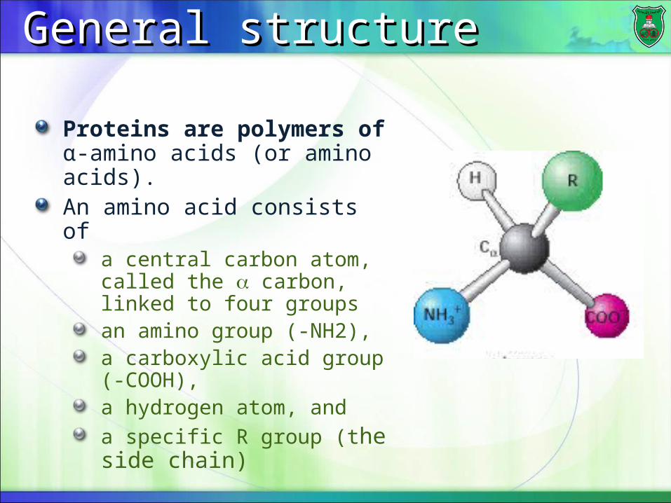
Proteins are polymers of α-amino acids (or amino acids).An amino acid consists of
a central carbon atom, called the carbon, linked to four groupsan amino group (-NH2), a carboxylic acid group (-COOH), a hydrogen atom, and a specific R group (the side chain)
L and D isomersL and D isomers
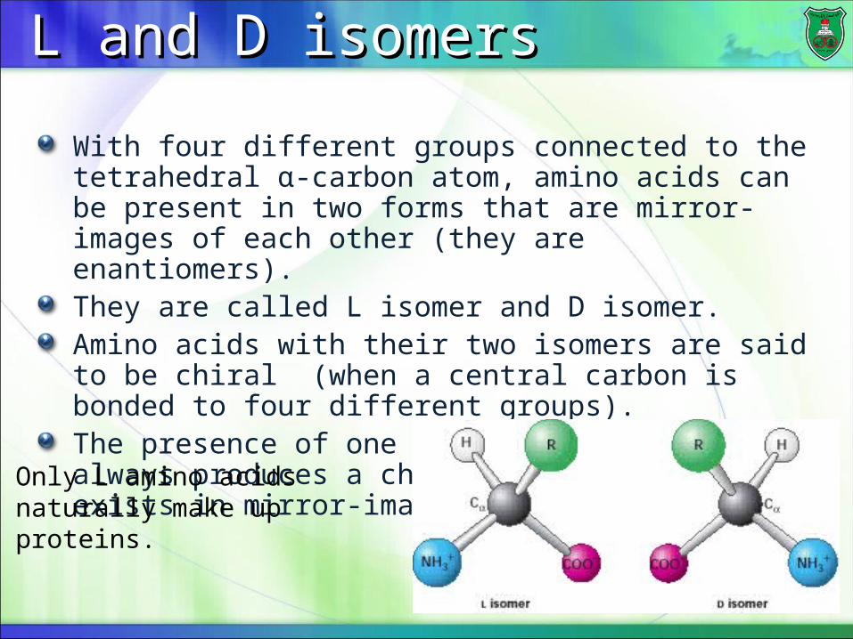
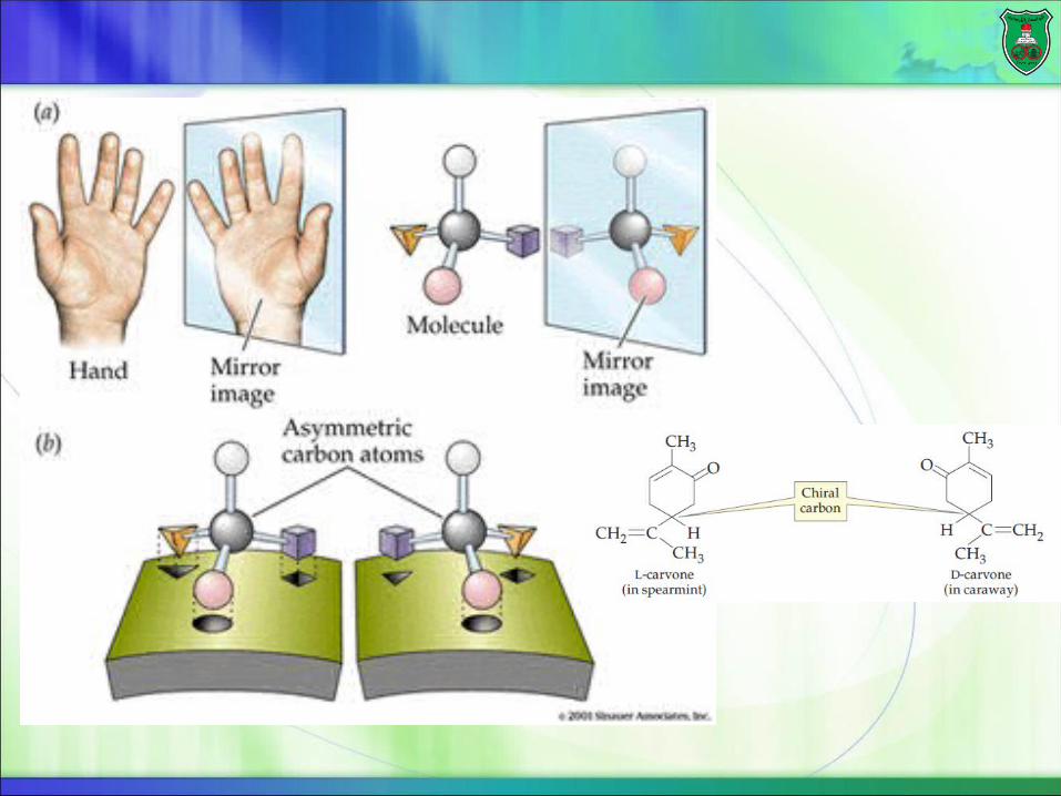
With four different groups connected to the tetrahedral α-carbon atom, amino acids can be present in two forms that are mirror-images of each other (they are enantiomers).They are called L isomer and D isomer.Amino acids with their two isomers are said to be chiral (when a central carbon is bonded to four different groups).The presence of one chiral carbon atom always produces a chiral molecule that exists in mirror-image forms.
Only L amino acids naturally make up proteins.
Types of amino acidsTypes of amino acids
There are twenty kinds of amino acids depending on the side chains varying in:
sizeShapeChargehydrogen-bonding capacityhydrophobic characterchemical reactivity
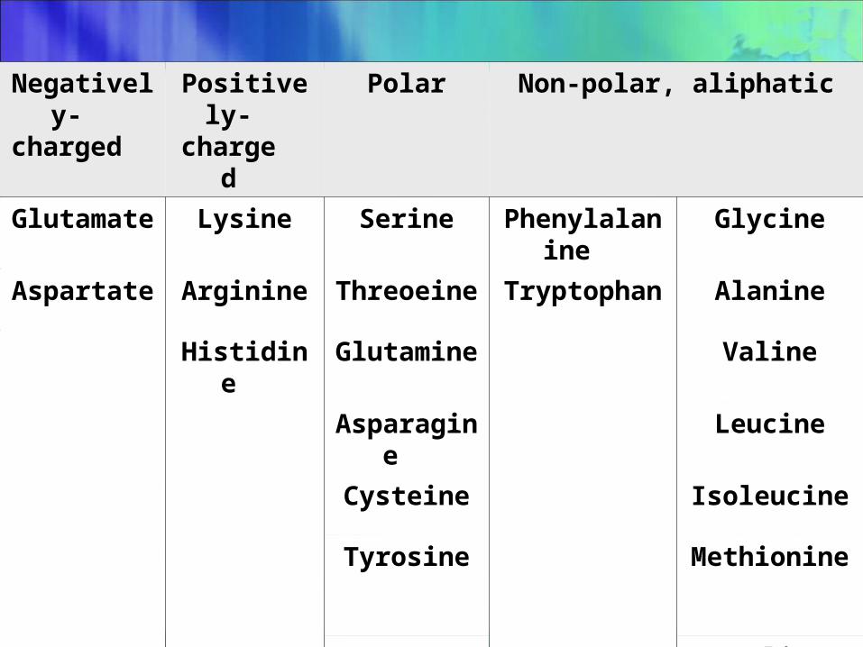
Non-polar, aliphaticPolarPositively-charged
Negatively-charged
GlycinePhenylalanineSerineLysineGlutamate
AlanineTryptophanThreoeineArginineAspartate
ValineGlutamineHistidine
LeucineAsparagine
IsoleucineCysteine
MethionineTyrosine
Proline
Non-polar, aliphatic amino Non-polar, aliphatic amino acidsacids
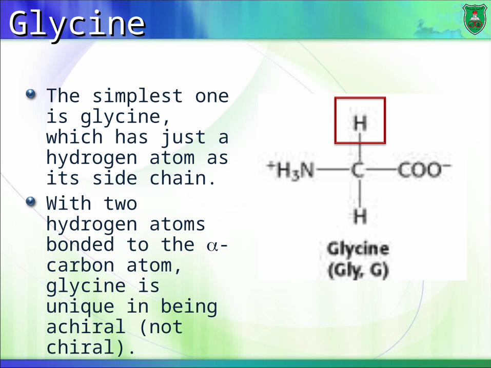
GlycineGlycine
The simplest one is glycine, which has just a hydrogen atom as its side chain.With two hydrogen atoms bonded to the -carbon atom, glycine is unique in being achiral (not chiral).
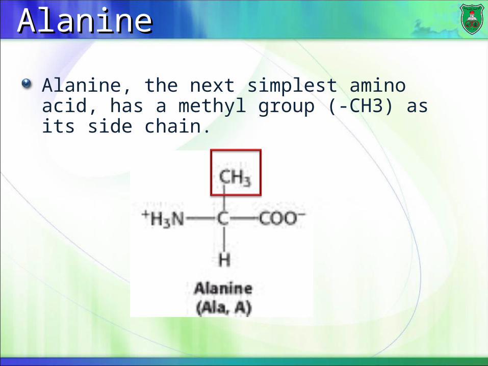
AlanineAlanine
Alanine, the next simplest amino acid, has a methyl group (-CH3) as its side chain.
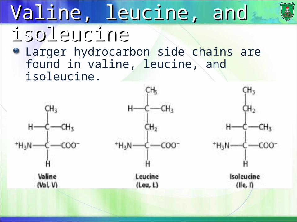
Valine, leucine, and isoleucineValine, leucine, and isoleucine
Larger hydrocarbon side chains are found in valine, leucine, and isoleucine.
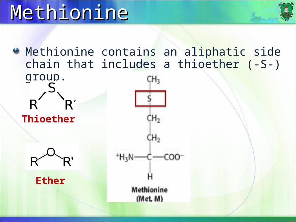
MethionineMethionine
Methionine contains an aliphatic side chain that includes a thioether (-S-) group.
Ether
Thioether
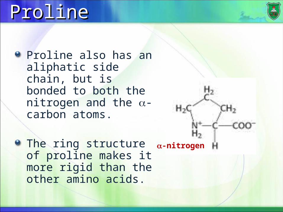
ProlineProline
Proline also has an aliphatic side chain, but is bonded to both the nitrogen and the -carbon atoms.
The ring structure of proline makes it more rigid than the other amino acids. -nitrogen
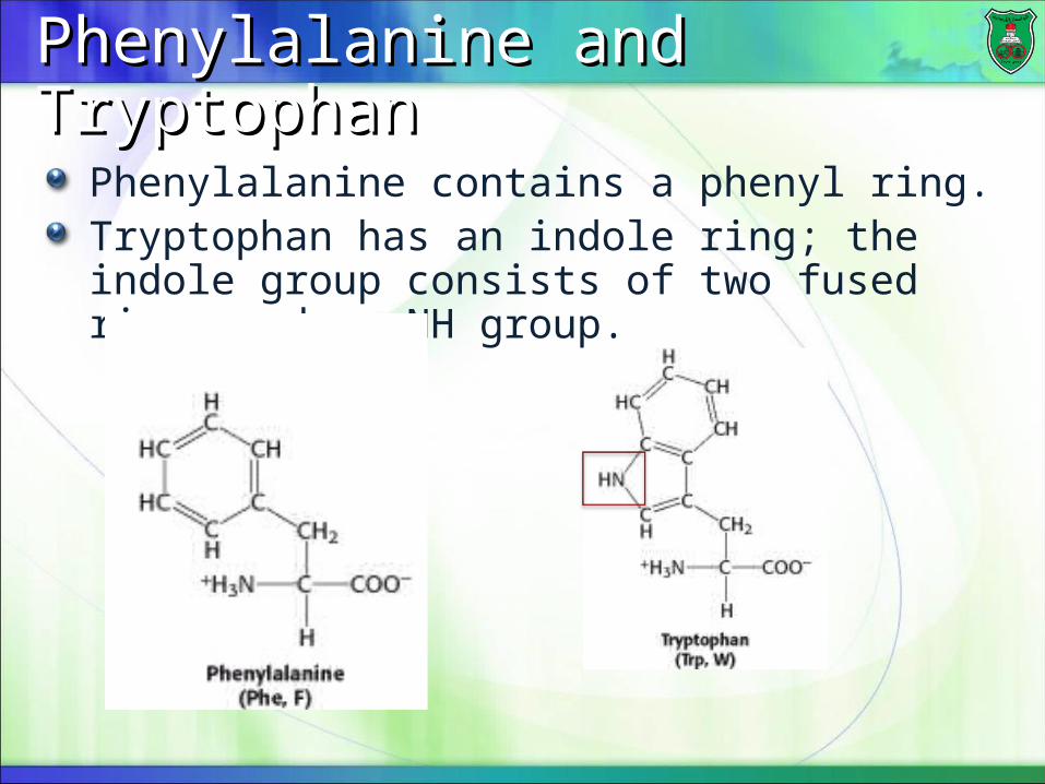
Phenylalanine and Tryptophan Phenylalanine and Tryptophan
Phenylalanine contains a phenyl ring.Tryptophan has an indole ring; the indole group consists of two fused rings and an NH group.
Positively-charged amino Positively-charged amino acidsacids
Lysine and arginineLysine and arginine
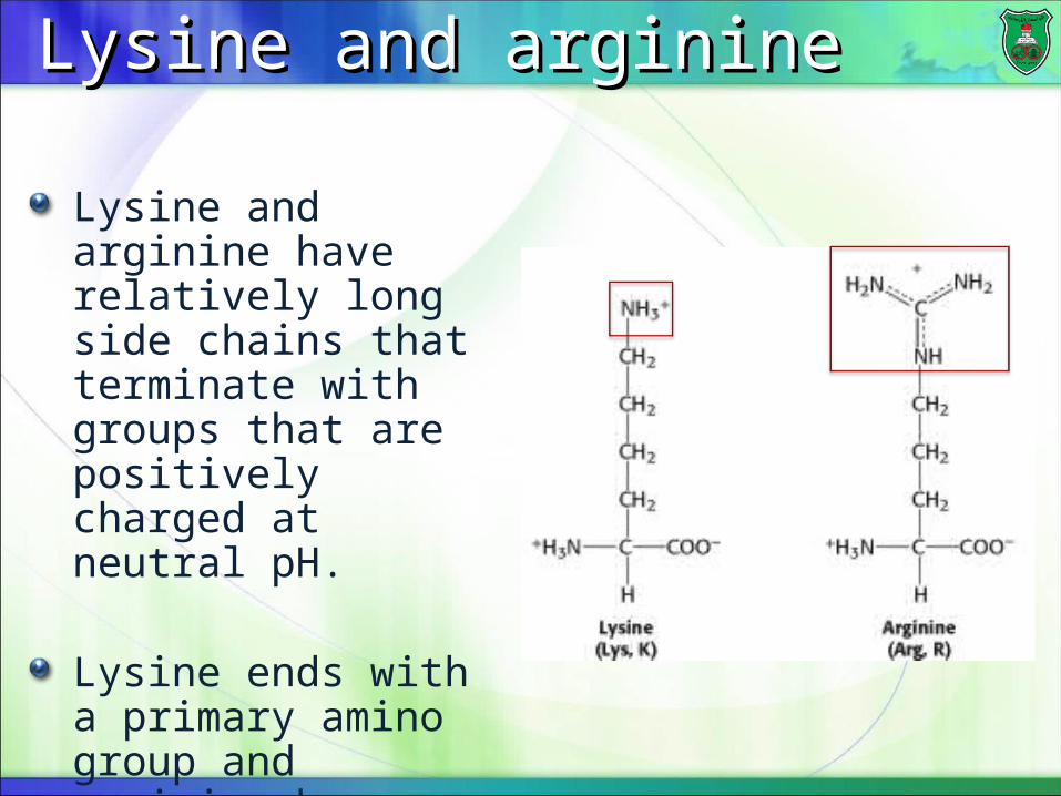
Lysine and arginine have relatively long side chains that terminate with groups that are positively charged at neutral pH.
Lysine ends with a primary amino group and arginine by a guanidinium group.
HistidineHistidine
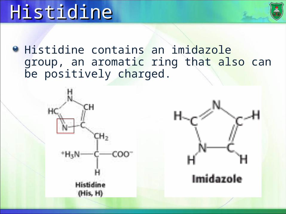
Histidine contains an imidazole group, an aromatic ring that also can be positively charged.
Negatively-charged amino Negatively-charged amino acidsacids
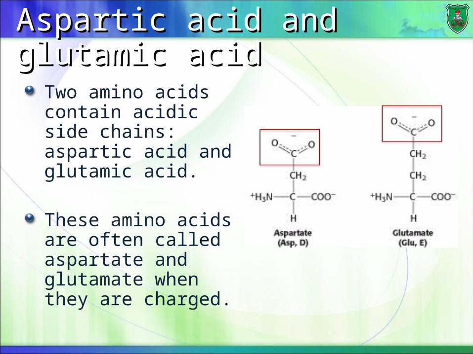
Aspartic acid and glutamic acidAspartic acid and glutamic acid
Two amino acids contain acidic side chains: aspartic acid and glutamic acid.
These amino acids are often called aspartate and glutamate when they are charged.
Polar amino acidsPolar amino acids
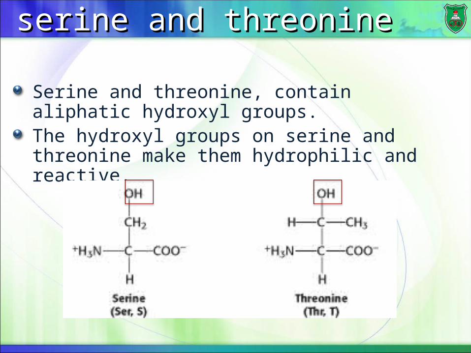
serine and threonineserine and threonine
Serine and threonine, contain aliphatic hydroxyl groups.The hydroxyl groups on serine and threonine make them hydrophilic and reactive.

CysteineCysteine
Cysteine contains a sulfhydryl or thiol (-SH), group. The sulfhydryl group is reactive.
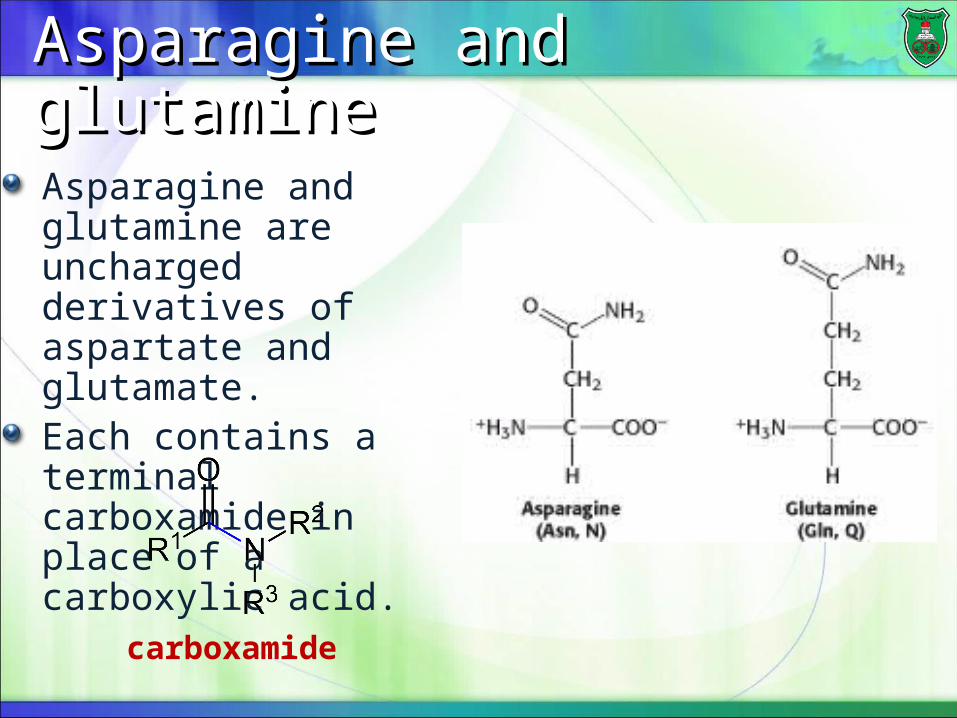
Asparagine and glutamineAsparagine and glutamine
Asparagine and glutamine are uncharged derivatives of aspartate and glutamate.Each contains a terminal carboxamide in place of a carboxylic acid.
carboxamide
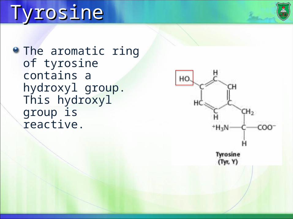
TyrosineTyrosine
The aromatic ring of tyrosine contains a hydroxyl group. This hydroxyl group is reactive.
Amino acidThree-letter abbreviation
AlanineAla
ArginineArg
AsparagineAsn
Aspartic AcidAsp
CysteineCys
GlutamineGln
Glutamic AcidGlu
GlycineGly
HistidineHis
IsoleucineIle
LeucineLeu
LysineLys
MethionineMet
PhenylalaninePhe
ProlinePro
SerineSer
ThreonineThr
TryptophanTrp
TyrosineTyr
ValineVal
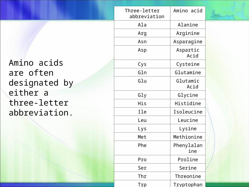
Amino acids are often designated by either a three-letter abbreviation.
Ionization of amino acidsIonization of amino acids
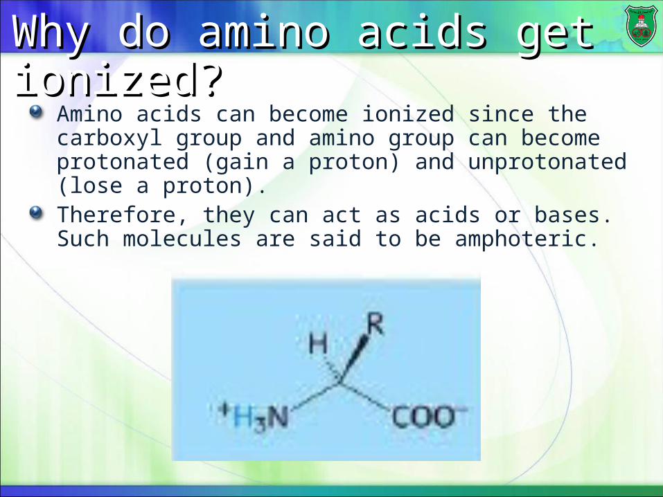
Why do amino acids get ionized?Why do amino acids get ionized?
Amino acids can become ionized since the carboxyl group and amino group can become protonated (gain a proton) and unprotonated (lose a proton).Therefore, they can act as acids or bases. Such molecules are said to be amphoteric.
Effect of pHEffect of pH
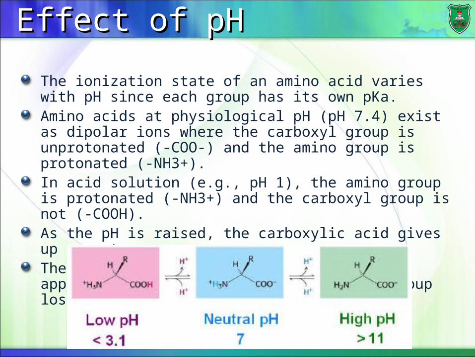
The ionization state of an amino acid varies with pH since each group has its own pKa.Amino acids at physiological pH (pH 7.4) exist as dipolar ions where the carboxyl group is unprotonated (-COO-) and the amino group is protonated (-NH3+).In acid solution (e.g., pH 1), the amino group is protonated (-NH3+) and the carboxyl group is not (-COOH).As the pH is raised, the carboxylic acid gives up a proton.The dipolar form persists until the pH approaches 9, when the protonated amino group loses a proton.
Zwitterion and isoelectric point Zwitterion and isoelectric point
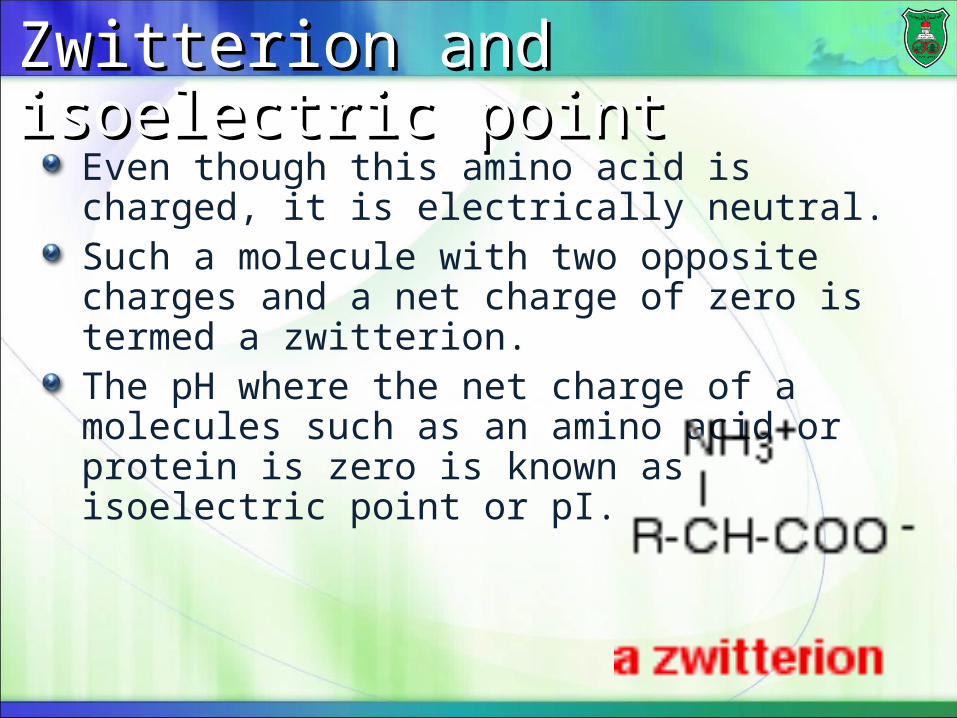
Even though this amino acid is charged, it is electrically neutral.Such a molecule with two opposite charges and a net charge of zero is termed a zwitterion.The pH where the net charge of a molecules such as an amino acid or protein is zero is known as isoelectric point or pI.
Ionization of side chainsIonization of side chains
Nine of the 20 amino acids have ionizable side chains.These amino acids are tyrosine, cysteine, arginine, lysine, histidine, serine, threonine, aspartic and glutamic acids.Each side chain has its own pKa values for ionization of the side chains.
At neutral pHaspartic acid and glutamic acid are negatively charged.Arginine and lysine are positively charged.
HistidineHistidine
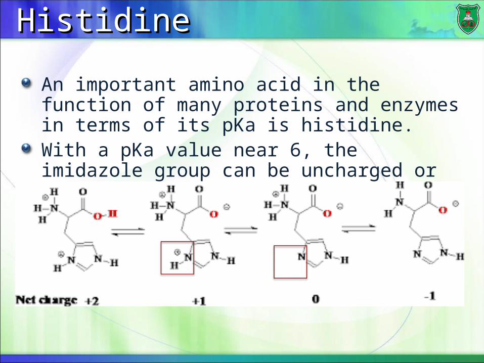
An important amino acid in the function of many proteins and enzymes in terms of its pKa is histidine.With a pKa value near 6, the imidazole group can be uncharged or positively charged near neutral pH.
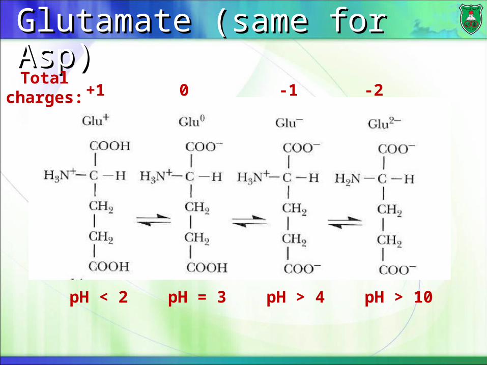
Glutamate (same for Asp)Glutamate (same for Asp)
pH < 2 pH = 3 pH > 4 pH > 10
+1 0 -1 -2Total
charges:
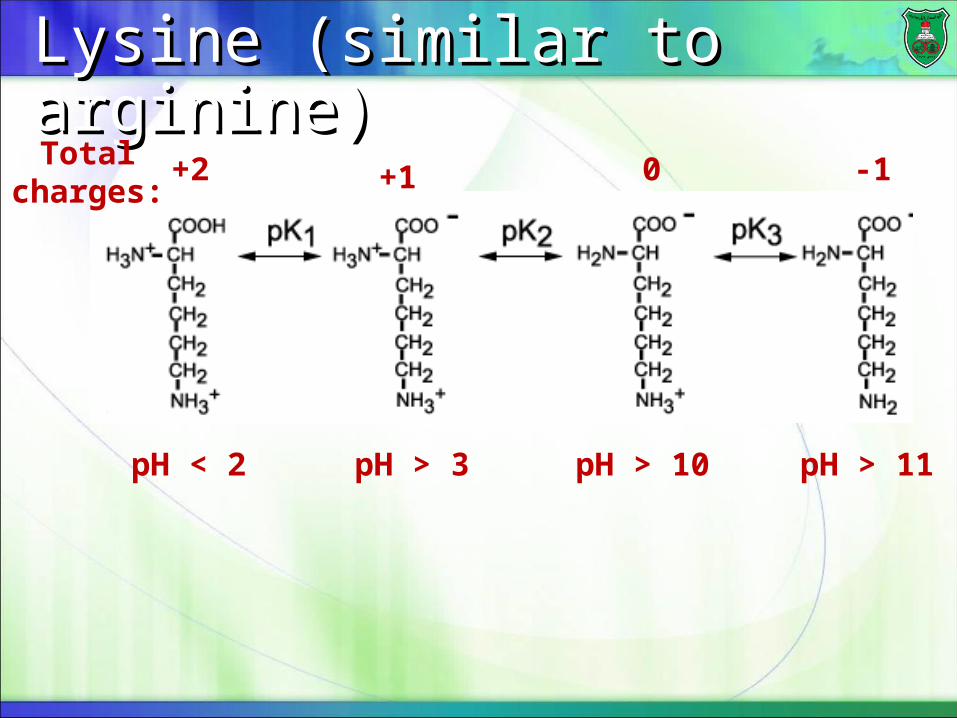
Lysine (similar to arginine)Lysine (similar to arginine)
pH < 2 pH > 3 pH > 10 pH > 11
+2 +1 0 -1Total charges:
NoteNote
You need to know the names of amino acids, the special structural features of amino acids, their abbreviations or designations, the pKa of groups (not exact numbers, but which ones are acidic, basic, or near neutral).
Essential amino acidsEssential amino acids
There are nine amino acids that are essential.Essential nutrients are those not made by the human body in significant amounts and must be derived from diet
These are: Histidine, Isoleucine, Leucine, Lysine, Methionine, Phenylalanine, Threonine, Tryptophan, and Valine.
The other 11 amino acids are non-essential amino acids.
Four Levels of Protein structureFour Levels of Protein structure
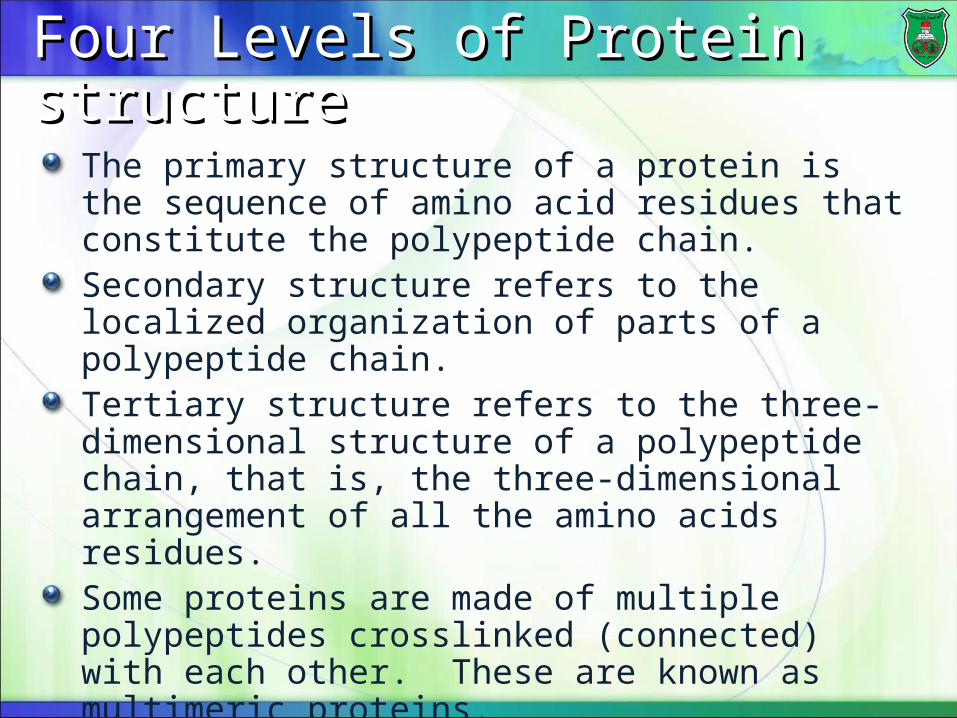
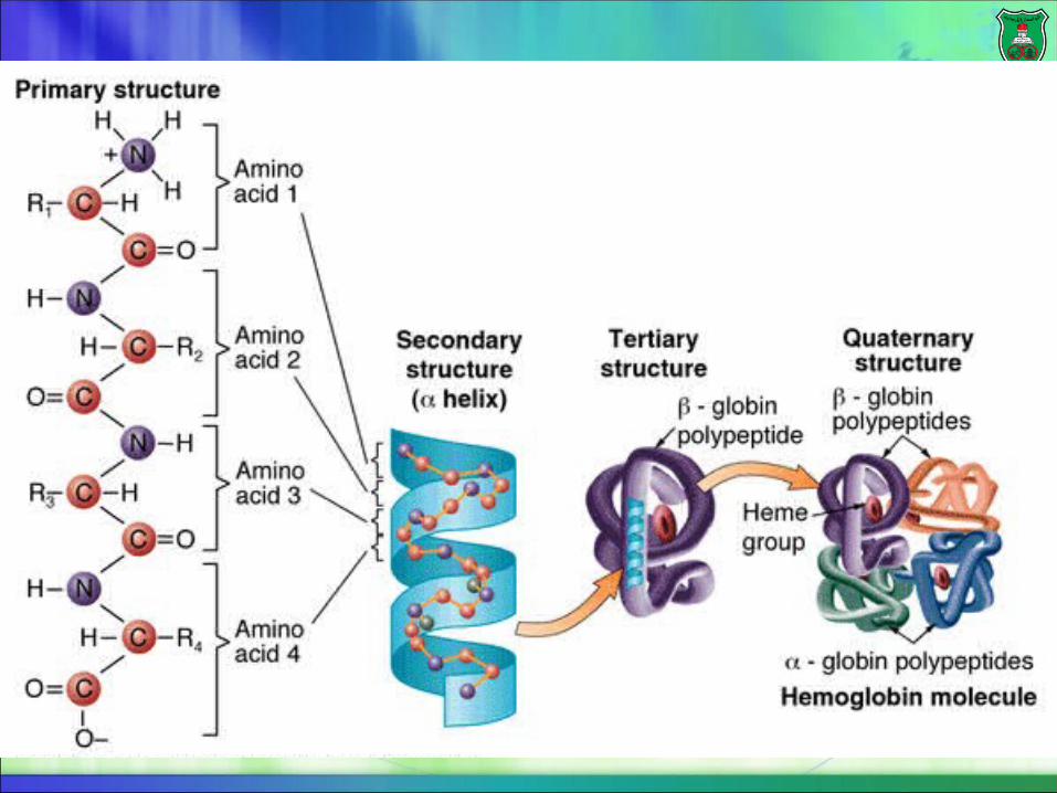
The primary structure of a protein is the sequence of amino acid residues that constitute the polypeptide chain.Secondary structure refers to the localized organization of parts of a polypeptide chain.Tertiary structure refers to the three-dimensional structure of a polypeptide chain, that is, the three-dimensional arrangement of all the amino acids residues.Some proteins are made of multiple polypeptides crosslinked (connected) with each other. These are known as multimeric proteins. Quaternary structure describes the number and relative positions of the subunits in a multimeric protein.

Peptide bondPeptide bond
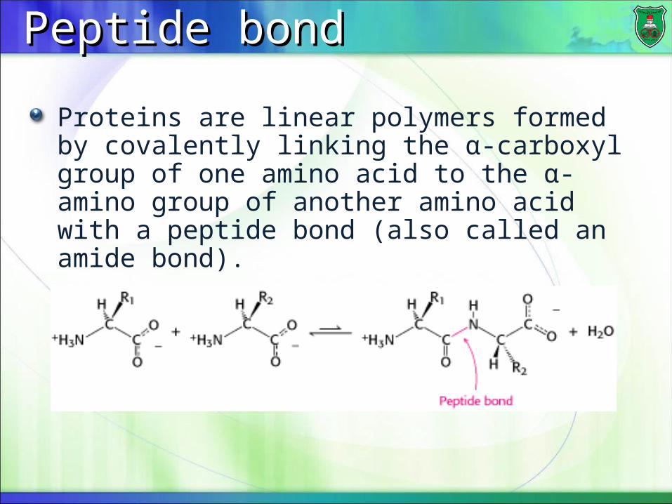
Proteins are linear polymers formed by covalently linking the α-carboxyl group of one amino acid to the α-amino group of another amino acid with a peptide bond (also called an amide bond).
A condensation reactionA condensation reaction
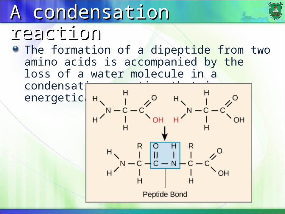
The formation of a dipeptide from two amino acids is accompanied by the loss of a water molecule in a condensation reaction that is energetically unfavorable.
DefinitionsDefinitions
The short chain of amino acids is known as an oligopeptides or just peptide.Each amino acid unit in a polypeptide is called a residue Longer peptides are referred to as polypeptides.Peptides generally contain fewer than 20-30 amino acid residues, whereas polypeptides contain as many as 4000 residues.Polypeptide chains that have organized three-dimensional structures are referred to as proteins .
Directionality of readingDirectionality of reading
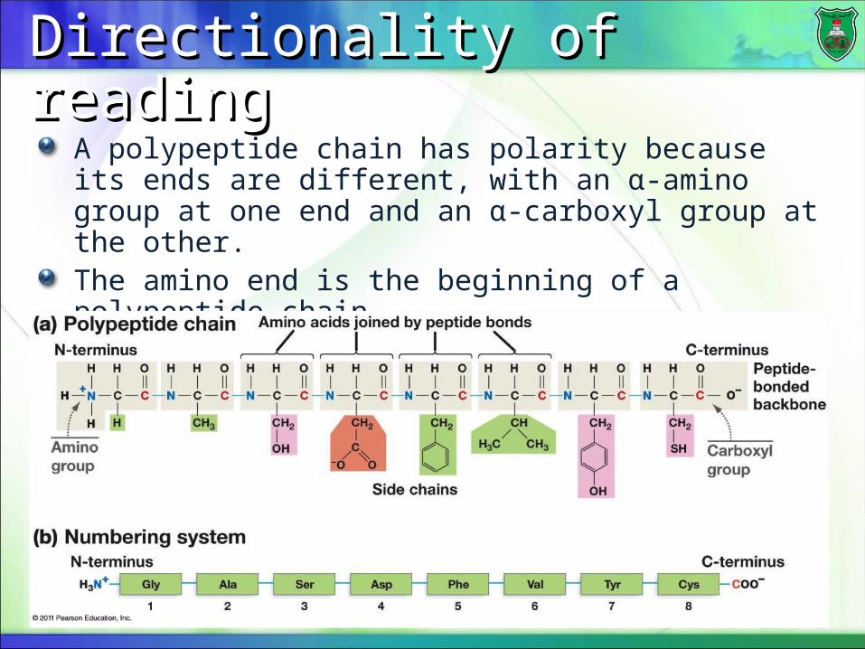
A polypeptide chain has polarity because its ends are different, with an α-amino group at one end and an α-carboxyl group at the other.The amino end is the beginning of a polypeptide chain.
Backbone and side chainsBackbone and side chains
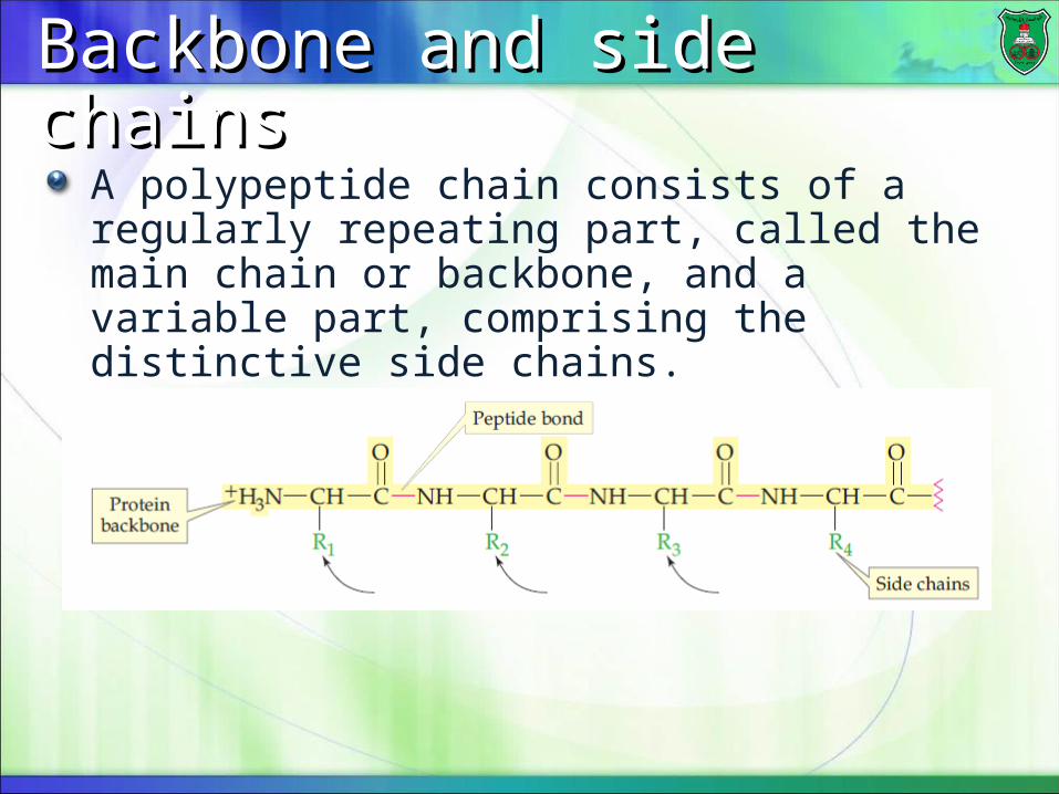
A polypeptide chain consists of a regularly repeating part, called the main chain or backbone, and a variable part, comprising the distinctive side chains.
Features of the backboneFeatures of the backbone
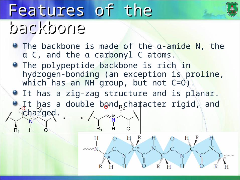
The backbone is made of the α-amide N, the α C, and the α carbonyl C atoms.The polypeptide backbone is rich in hydrogen-bonding (an exception is proline, which has an NH group, but not C=O).It has a zig-zag structure and is planar.It has a double bond character rigid, and charged.
Importance of peptide bondImportance of peptide bond
The primary structure of a protein determines the other levels of structure.A single amino acid substitution can give rise to a malfunctioning protein, as is the case with sickle-cell anemia.
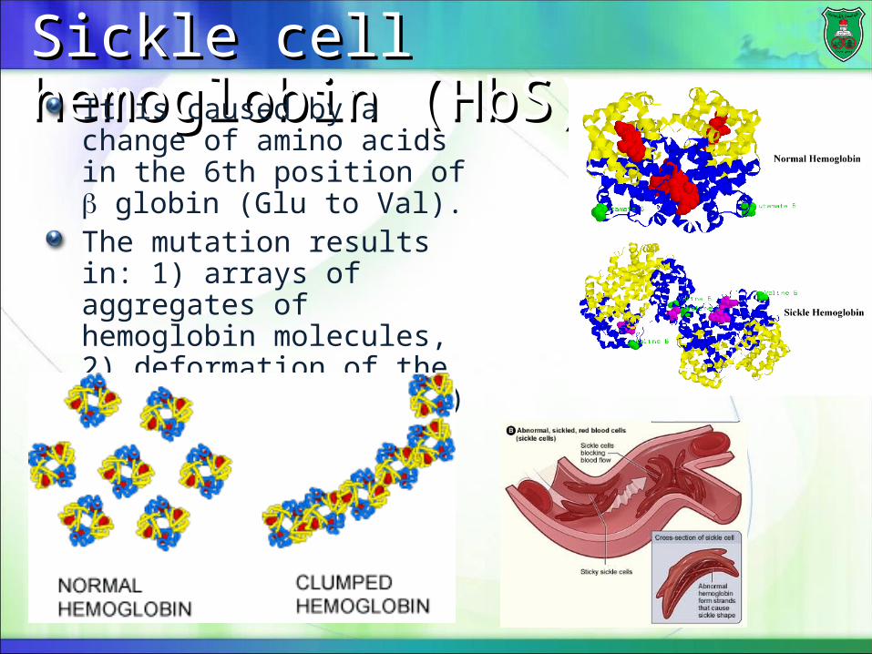
Sickle cell hemoglobin (HbS)Sickle cell hemoglobin (HbS)It is caused by a change of amino acids in the 6th position of globin (Glu to Val).The mutation results in: 1) arrays of aggregates of hemoglobin molecules, 2) deformation of the red blood cell, and 3) clotting in blood vessels and tissues.
How is protein structure How is protein structure determined?determined?
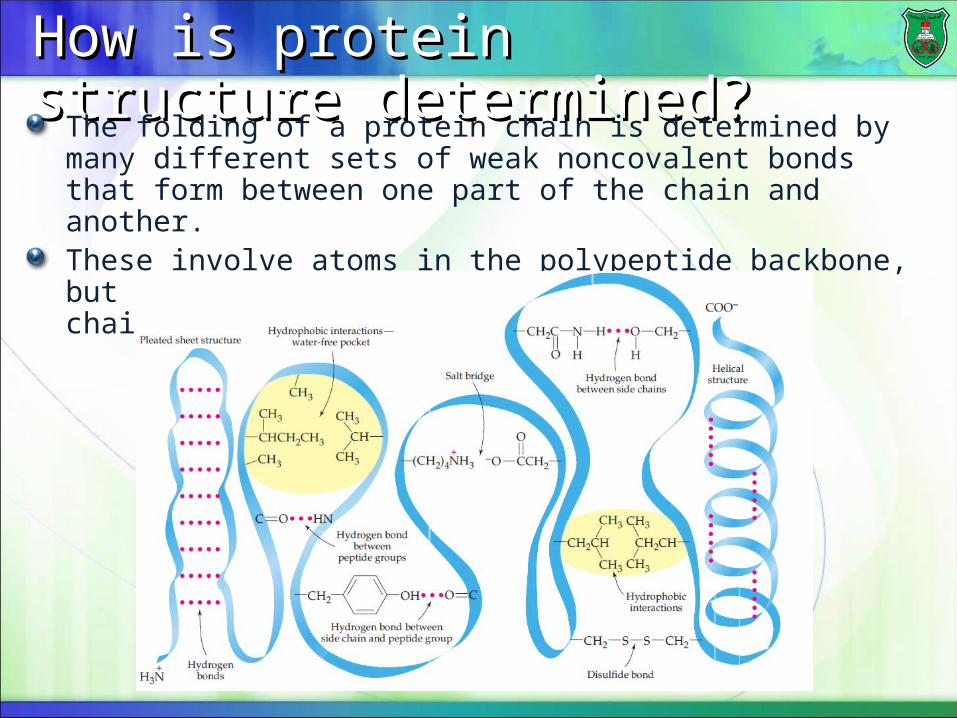
The folding of a protein chain is determined by many different sets of weak noncovalent bonds that form between one part of the chain and another.These involve atoms in the polypeptide backbone, but mainly by atoms in the amino acid side chains.
Hydrogen bondsHydrogen bonds
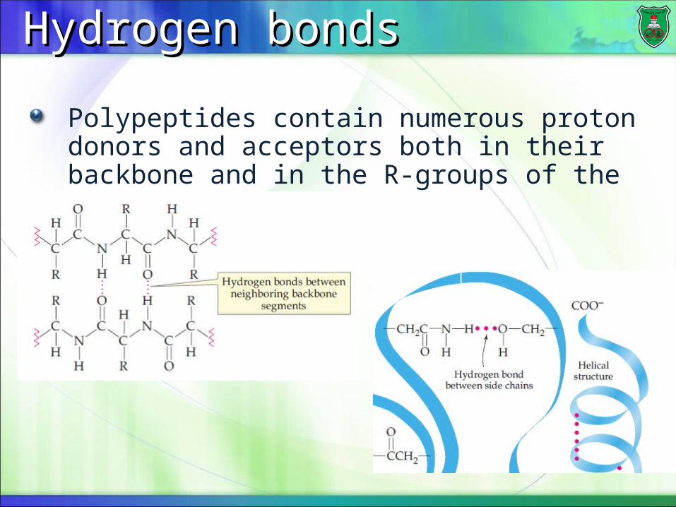
Polypeptides contain numerous proton donors and acceptors both in their backbone and in the R-groups of the amino acids.
Electrostatic interactionsElectrostatic interactions
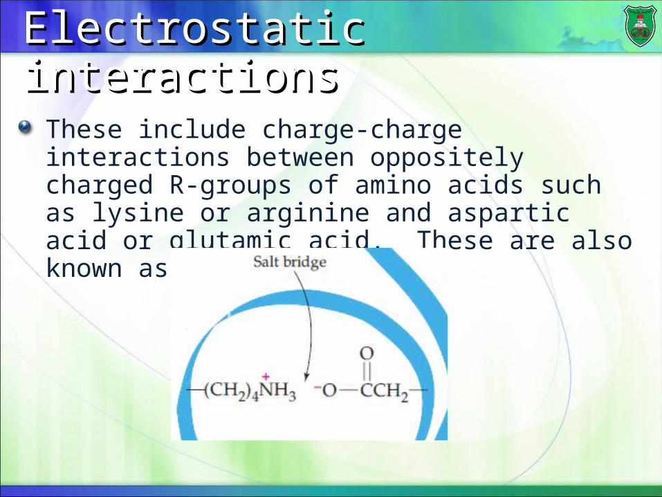
These include charge-charge interactions between oppositely charged R-groups of amino acids such as lysine or arginine and aspartic acid or glutamic acid. These are also known as salt bridges.
van der Waals attractionsvan der Waals attractions
Although van der Waals forces are extremely weak, it is the huge number of such interactions that occur in large protein molecules that make them significant to the folding of proteins.
hydrophobic interactionshydrophobic interactions
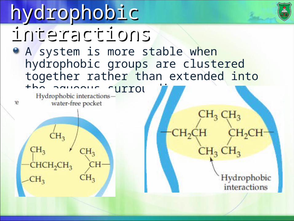
A system is more stable when hydrophobic groups are clustered together rather than extended into the aqueous surroundings.
Very importantVery important
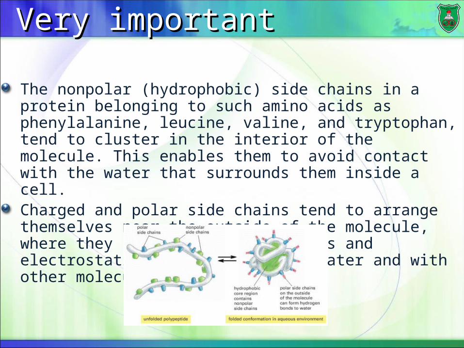
The nonpolar (hydrophobic) side chains in a protein belonging to such amino acids as phenylalanine, leucine, valine, and tryptophan, tend to cluster in the interior of the molecule. This enables them to avoid contact with the water that surrounds them inside a cell.Charged and polar side chains tend to arrange themselves near the outside of the molecule, where they can form hydrogen bonds and electrostatic interactions with water and with other molecules.
Disulfide bondsDisulfide bonds
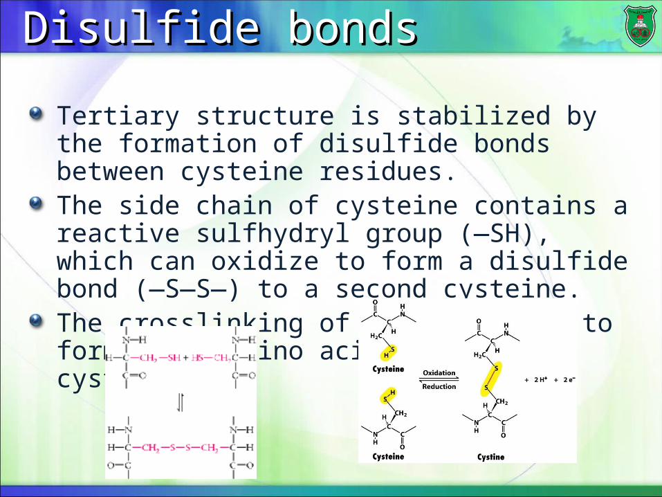
Tertiary structure is stabilized by the formation of disulfide bonds between cysteine residues.The side chain of cysteine contains a reactive sulfhydryl group (—SH), which can oxidize to form a disulfide bond (—S—S—) to a second cysteine.The crosslinking of two cysteines to form a new amino acid, called cystine.
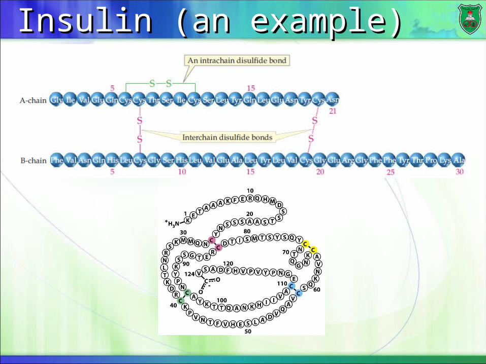
Insulin (an example)Insulin (an example)

Importance of amino acid sequence?Importance of amino acid sequence?
Certain sequence or order of amino acids within a small region of protein are organized in specific shapesThese conformations are determined by the primary sequence of the amino acids.Polypeptide chains can fold into regular structures such as:
the alpha helixthe beta sheetLoopsTurns
The The helix helix
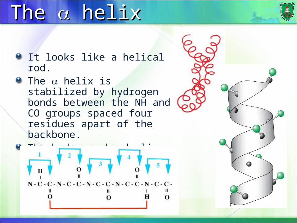
It looks like a helical rod.The helix is stabilized by hydrogen bonds between the NH and CO groups spaced four residues apart of the backbone.The hydrogen bonds lie vertically along the helix, and the R groups extend to the outside of the coil.
β pleated sheet (β sheet)β pleated sheet (β sheet)
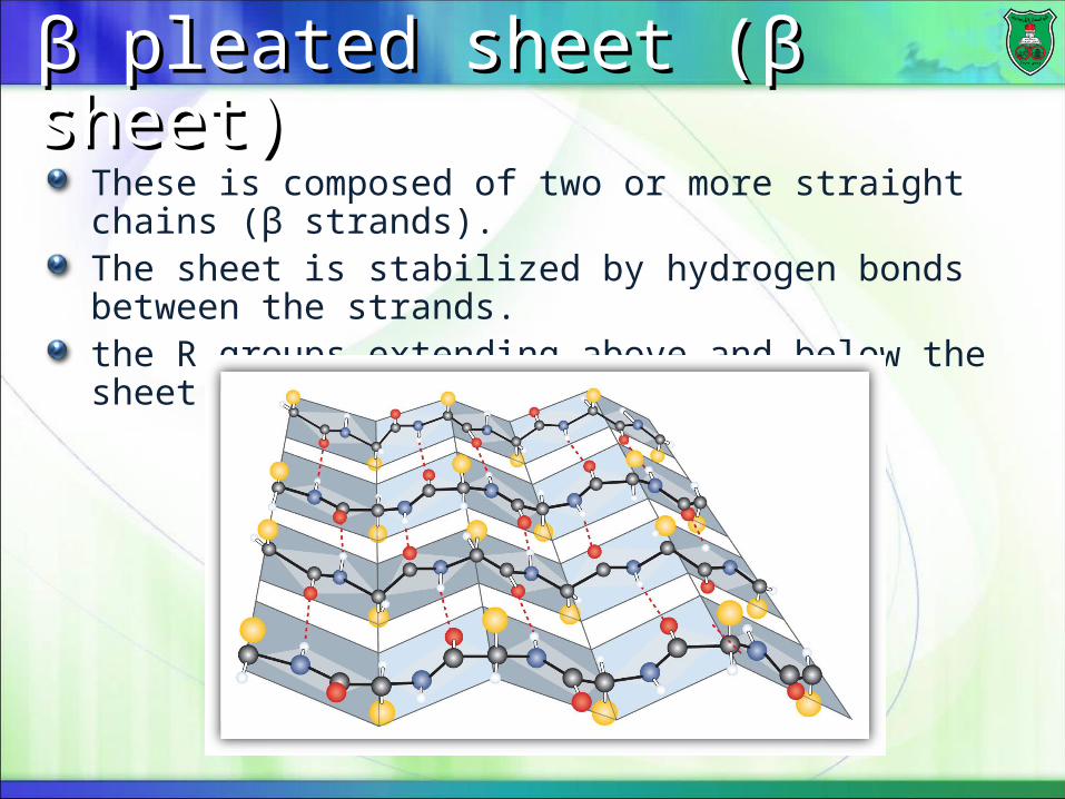
These is composed of two or more straight chains (β strands).The sheet is stabilized by hydrogen bonds between the strands.the R groups extending above and below the sheet.

What is tertiary structure?What is tertiary structure?
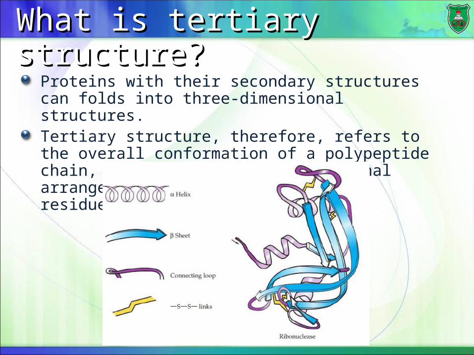
Proteins with their secondary structures can folds into three-dimensional structures.Tertiary structure, therefore, refers to the overall conformation of a polypeptide chain, that is, the three-dimensional arrangement of all the amino acids residues.

What is it?What is it?
Some proteins exhibit a fourth level of structural organization where proteins are composed of more than one polypeptide chain.This is the quaternary structure of proteins. Each polypeptide chain in such a protein is called a subunit.Quaternary structure, thus, refers to the spatial arrangement of multiple subunits of a protein and the nature of their interactions.Sometimes subunits are disulfide-bonded together, other times, noncovalent bonds stabilize interactions between subunit.
More on namingMore on naming
The simplest sort of quaternary structure is a dimer, consisting of two subunits:
If these two subunits are the same, the protein is said to be made of a homodimerIf the subunits are different, then it is a heterodimer
Proteins made of three subunits are trimers, and of four subunits are tetramers, and soon on.
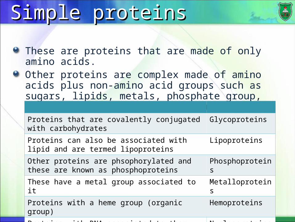
Simple proteinsSimple proteins
These are proteins that are made of only amino acids.Other proteins are complex made of amino acids plus non-amino acid groups such as sugars, lipids, metals, phosphate group, nucleic acids, and organic groups.
GlycoproteinsProteins that are covalently conjugated with carbohydrates
LipoproteinsProteins can also be associated with lipid and are termed lipoproteins
PhosphoproteinsOther proteins are phsophorylated and these are known as phosphoproteins
MetalloproteinsThese have a metal group associated to it
HemoproteinsProteins with a heme group (organic group)
NucleoproteinsProteins with RNA associated to them
Native protein
Each protein molecule folds in a distinctive manner that is determined by its primary structure and results in its maximum stability. A protein with the shape in which it functions in living systems is known as a native protein.Ribonuclease is classified as a simple protein because it is of one polypeptide.Myoglobin is a conjugated monomeric protein that is composed of one polypeptide and a nonprotein group.Hemoglobin is a conjugated protein with a quaternary structure.
Classes of proteinsClasses of proteins
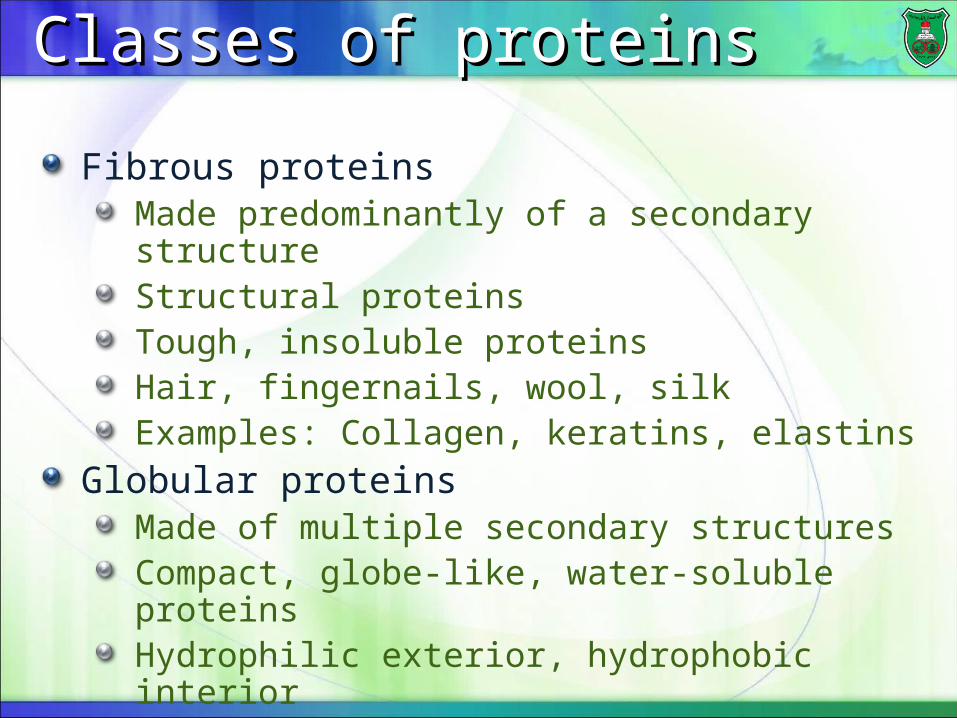
Fibrous proteinsMade predominantly of a secondary structureStructural proteinsTough, insoluble proteinsHair, fingernails, wool, silkExamples: Collagen, keratins, elastins
Globular proteinsMade of multiple secondary structuresCompact, globe-like, water-soluble proteinsHydrophilic exterior, hydrophobic interiorMultiple functions
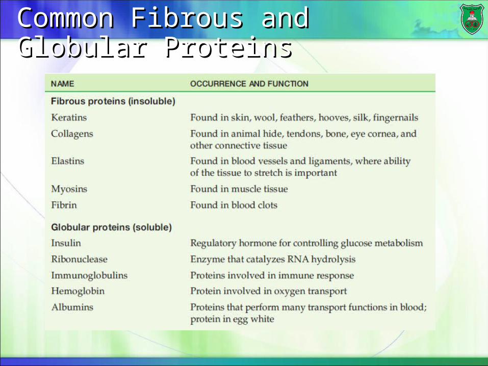
Common Fibrous and Globular Common Fibrous and Globular ProteinsProteins
Biological Functions of ProteinsBiological Functions of Proteins
Enzymes--catalysts for reactions Transport molecules--hemoglobin; lipoproteins, channel proteinsContractile/motion--myosin; actin Structural--collagen; keratin, actinDefense--antibodies Signaling—hormones, receptorsToxins--diphtheria; enterotoxins
CollagenCollagen
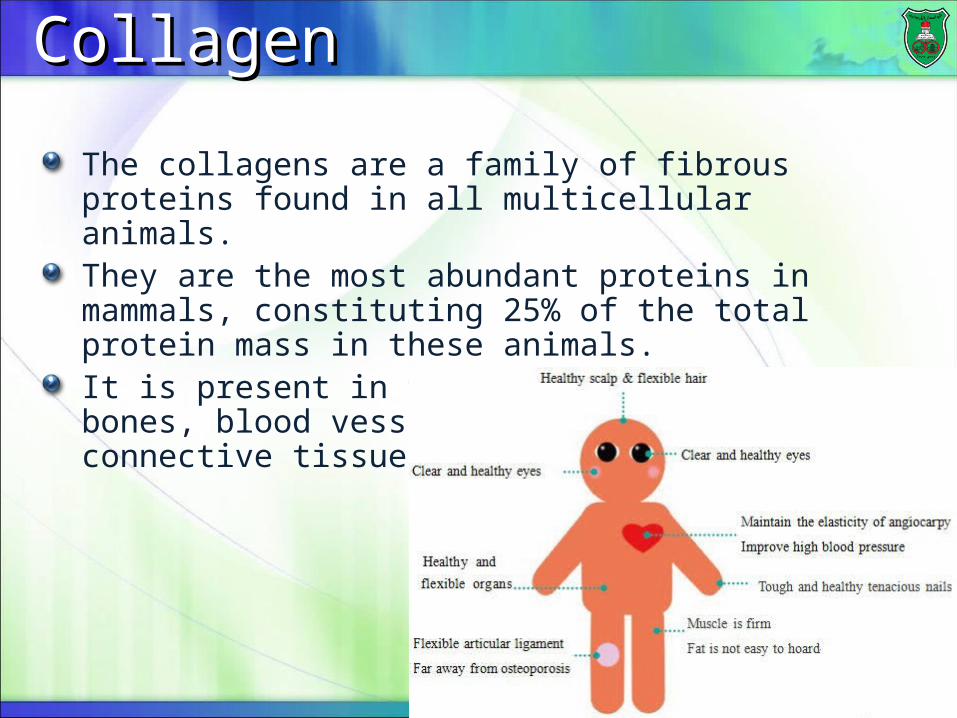
The collagens are a family of fibrous proteins found in all multicellular animals.They are the most abundant proteins in mammals, constituting 25% of the total protein mass in these animals.It is present in tissues such as skin, bones, blood vessels, tendons, and other connective tissues.
Overall functionOverall function
The main function of collagen molecules is to provide structural support to tissues.Therefore, the primary feature of a typical collagen molecule is its stiffness.
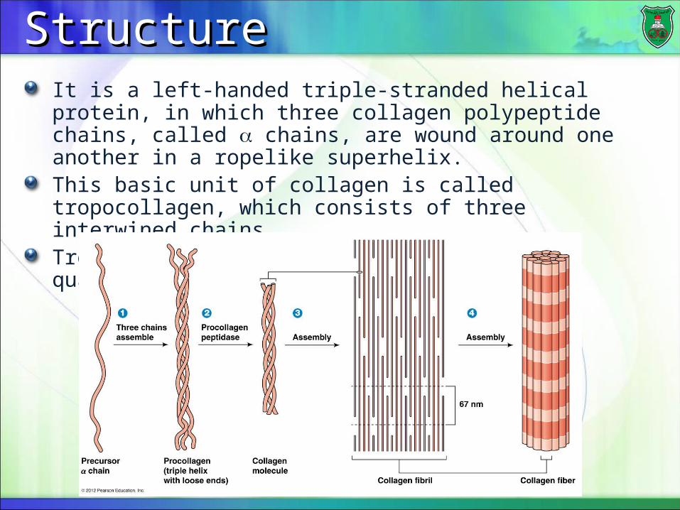
StructureStructureIt is a left-handed triple-stranded helical protein, in which three collagen polypeptide chains, called chains, are wound around one another in a ropelike superhelix.This basic unit of collagen is called tropocollagen, which consists of three interwined chains.Tropocollagen assembles into larger fibers quaternary structure).
Composition of collagensComposition of collagens
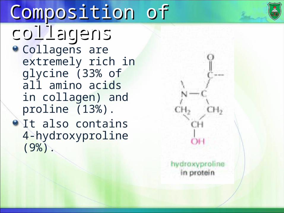
Collagens are extremely rich in glycine (33% of all amino acids in collagen) and proline (13%).It also contains 4-hydroxyproline (9%).
Functional purpose of amino acidsFunctional purpose of amino acids
Glycine allows the three helical a chains to pack tightly together to form the final collagen superhelix because it is small.Proline makes the structure rigid and creates the helical conformation in each chain.Hydroxyproline stabilizes the structure via formation of hydrogen bonds among them.Every third residue is glycine, which, with the preceding residue being proline or hydroxyproline in a repetitive fashion as follows:
Gly-pro-Y Gly-X-hydroxyproline
ScurvyScurvy
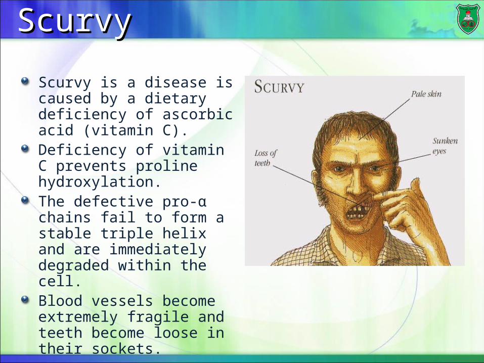
Scurvy is a disease is caused by a dietary deficiency of ascorbic acid (vitamin C).Deficiency of vitamin C prevents proline hydroxylation.The defective pro-α chains fail to form a stable triple helix and are immediately degraded within the cell.Blood vessels become extremely fragile and teeth become loose in their sockets.
Myoglobin and hemoglobinMyoglobin and hemoglobin
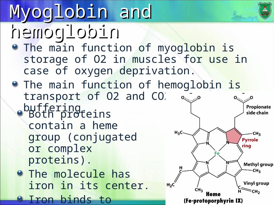
The main function of myoglobin is storage of O2 in muscles for use in case of oxygen deprivation.The main function of hemoglobin is transport of O2 and CO2 and blood buffering.
Both proteins contain a heme group (conjugated or complex proteins).The molecule has iron in its center.Iron binds to oxygen.
MyoglobinMyoglobin
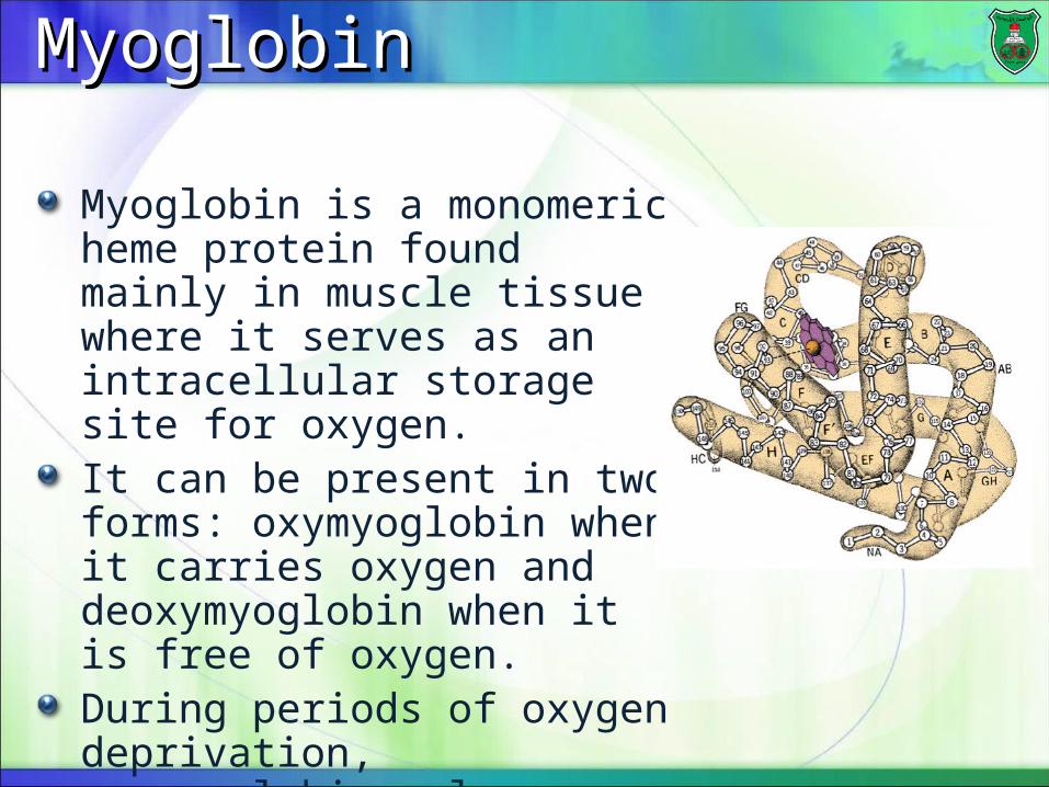
Myoglobin is a monomeric heme protein found mainly in muscle tissue where it serves as an intracellular storage site for oxygen.It can be present in two forms: oxymyoglobin when it carries oxygen and deoxymyoglobin when it is free of oxygen.During periods of oxygen deprivation, oxymyoglobin releases its bound oxygen.
Tertiary structure of myoglobinTertiary structure of myoglobin
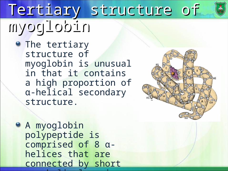
The tertiary structure of myoglobin is unusual in that it contains a high proportion of α-helical secondary structure.
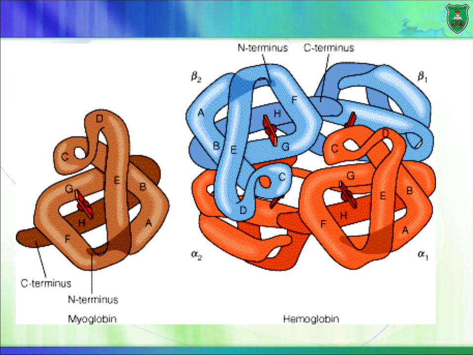
A myoglobin polypeptide is comprised of 8 α-helices that are connected by short non-helical regions.
HemoglobinHemoglobin structurestructure
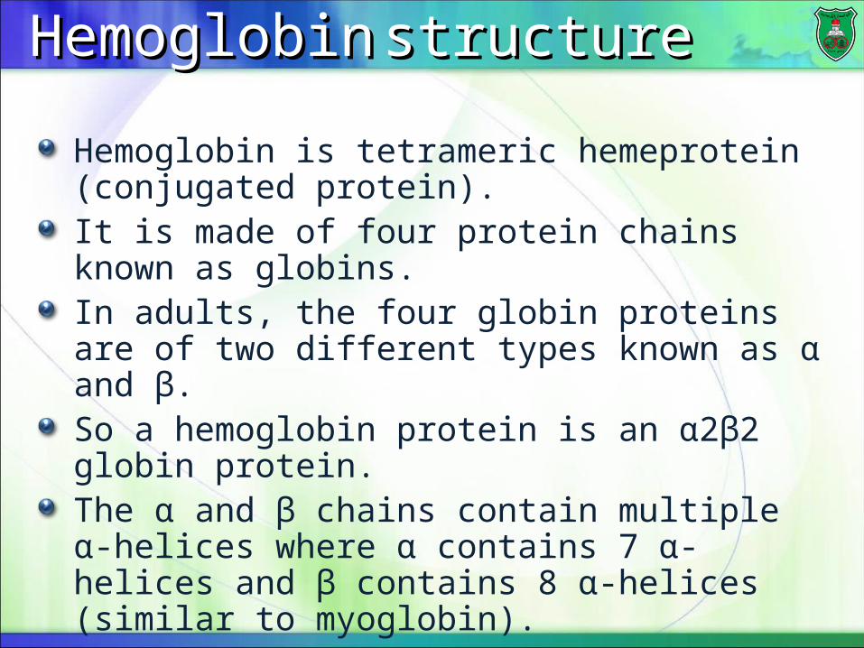
Hemoglobin is tetrameric hemeprotein (conjugated protein).It is made of four protein chains known as globins.In adults, the four globin proteins are of two different types known as α and β.So a hemoglobin protein is an α2β2 globin protein.The α and β chains contain multiple α-helices where α contains 7 α-helices and β contains 8 α-helices (similar to myoglobin).Each chain has a heme group, so it can carry four oxygen molecules.
Function of hemoglobinFunction of hemoglobin
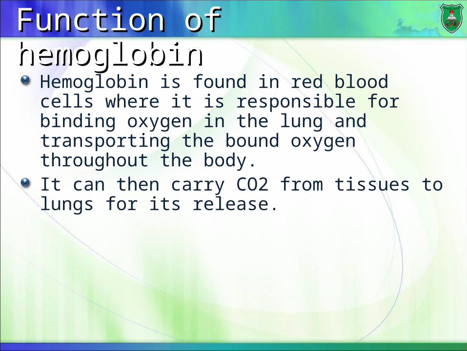
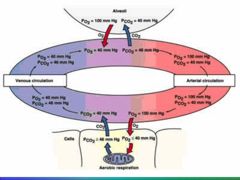
Hemoglobin is found in red blood cells where it is responsible for binding oxygen in the lung and transporting the bound oxygen throughout the body.It can then carry CO2 from tissues to lungs for its release.

DenaturationDenaturation
Denaturation is the disruption of the native conformation of a protein, the characteristic three-dimensional structure that it attains after synthesis, with no effect on the primary structure of the protein.Denaturation involves the breaking of the noncovalent bonds, which determine the structure of a protein.
Denaturing agentsDenaturing agents
Heat disrupts low energy van der Waals forces in proteins.Extremes of pH change the charge of the protein’s amino acid side chains and result in the disruption of electrostatic and hydrogen bonds.Detergents disrupt the hydrophobic forces which fold proteinsInorganic salts disrupt salt bridges.Organic compounds such as ethanol and acetone disrupt hydrogen bondings (disinfectants).Mechanical agitation such as beating an egg.
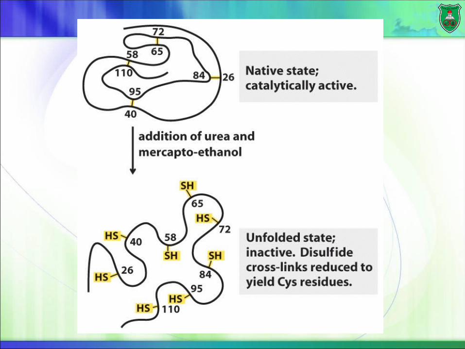
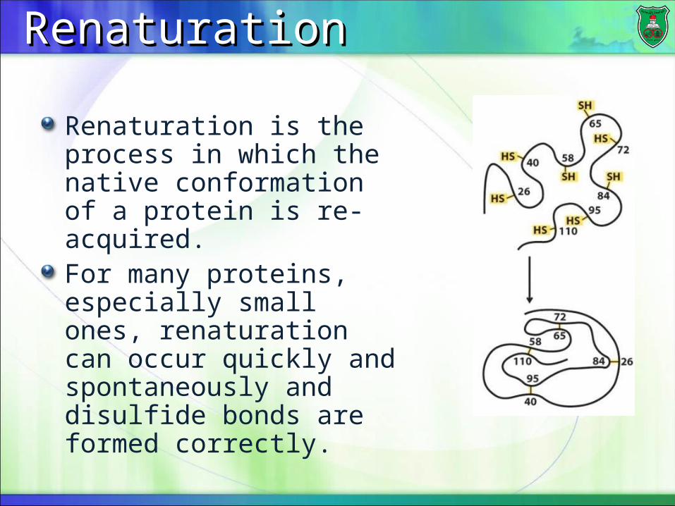
RenaturationRenaturation
Renaturation is the process in which the native conformation of a protein is re-acquired.For many proteins, especially small ones, renaturation can occur quickly and spontaneously and disulfide bonds are formed correctly.
ElectrophoresisElectrophoresis
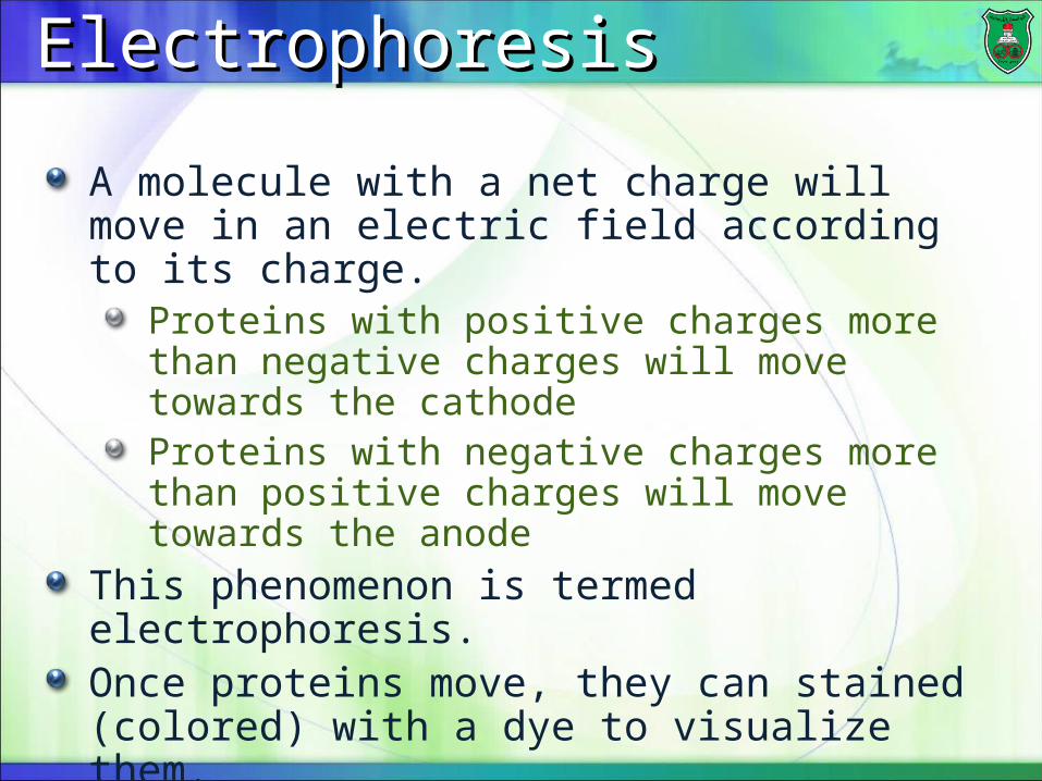
A molecule with a net charge will move in an electric field according to its charge.
Proteins with positive charges more than negative charges will move towards the cathodeProteins with negative charges more than positive charges will move towards the anode
This phenomenon is termed electrophoresis.Once proteins move, they can stained (colored) with a dye to visualize them.
The roles of pI and pHThe roles of pI and pH
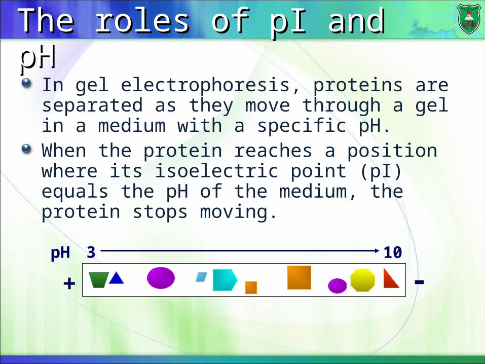
In gel electrophoresis, proteins are separated as they move through a gel in a medium with a specific pH.When the protein reaches a position where its isoelectric point (pI) equals the pH of the medium, the protein stops moving.
3pH 10
-+
Top Related