


γλώσσες
Σελίδες
Νομικός

SHORT REPORT Open Access
Alleviating toxic α-Synuclein accumulationby membrane depolarization: evidencefrom an in vitro model of Parkinson’sdiseaseAlysia Ross1, Viktoria Xing1, Ting Ting Wang1, Samantha C. Bureau1,2, Giovana A. Link1, Teresa Fortin1, Hui Zhang3,Shawn Hayley1* and Hongyu Sun1*
Abstract
Parkinson’s disease (PD) is characterized by the formation of toxic, fibrillar form alpha-synuclein (α-Syn) proteinaggregates in dopaminergic neurons. Accumulating evidence has shown a multifactorial interplay betweenthe intracellular calcium elevation and α-Syn dynamics. However, whether membrane depolarization regulatestoxic α-Syn aggregates remains unclear. To understand this better, we used an in vitro α-Syn preformed fibrils(PFF) model of PD in human neural cells. We demonstrated functional membrane depolarization indifferentiated SH-SY5Y cells induced by two independent treatments: high extracellular K+ and the GABAA
receptor blocker picrotoxin. We then observed that these treatments significantly alleviated toxic α-Synaggregation in PFF-treated SH-SY5Y cells. Moreover, clinically relevant direct current stimulation (DCS) alsoremarkably decreased toxic α-Syn aggregation in PFF-treated SH-SY5Y cells. Taken together, our findingssuggest that membrane depolarization plays an important role in alleviating PFF-induced toxic α-Synaggregates, and that it may represent a novel therapeutic mechanism for PD.
Keywords: Parkinson’s disease, Membrane depolarization, Preformed fibrils, GABAA receptor, α-Synuclein,Calcium channel, Direct current stimulation
IntroductionParkinson’s disease (PD) is characterized by the for-mation of insoluble toxic alpha-synuclein (α-Syn) pro-tein aggregates called amyloid fibrils in dopaminergicneurons [18, 19, 21, 38, 54]. α-Syn is a small solubleprotein that concentrates at presynaptic terminalsthroughout the brain [8, 51]. Under physiological con-ditions, α-Syn monomers facilitate neurotransmitterrelease through modulating synaptic vesicle matur-ation and release [6, 19, 44]. However, these soluble
α-Syn monomers are prone to undergoing posttrans-lational modifications under pathological conditionsand consequently, form insoluble toxic α-Syn oligo-mers and fibrils [37]. Toxic α-Syn fibrils aggregateinto Lewy bodies and Lewy neurites which representthe key trigger for the onset of synapse deconstruc-tion and subsequent neurodegeneration [5, 18, 43].Understandably, targeting the mechanisms regulatingand clearing toxic α-Syn aggregation has representeda promising disease-modifying strategy for PD treat-ment [23, 38, 51].Previous studies using in vivo animal models and
in vitro cell culture systems suggest an important roleof depolarization-induced disruption of calcium
© The Author(s). 2020 Open Access This article is licensed under a Creative Commons Attribution 4.0 International License,which permits use, sharing, adaptation, distribution and reproduction in any medium or format, as long as you giveappropriate credit to the original author(s) and the source, provide a link to the Creative Commons licence, and indicate ifchanges were made. The images or other third party material in this article are included in the article's Creative Commonslicence, unless indicated otherwise in a credit line to the material. If material is not included in the article's Creative Commonslicence and your intended use is not permitted by statutory regulation or exceeds the permitted use, you will need to obtainpermission directly from the copyright holder. To view a copy of this licence, visit http://creativecommons.org/licenses/by/4.0/.The Creative Commons Public Domain Dedication waiver (http://creativecommons.org/publicdomain/zero/1.0/) applies to thedata made available in this article, unless otherwise stated in a credit line to the data.
* Correspondence: [email protected]; [email protected] of Neuroscience, Carleton University, Ottawa, 1125 Colonel ByDrive, Ottawa, ON K1S 5B6, CanadaFull list of author information is available at the end of the article
Ross et al. Molecular Brain (2020) 13:108 https://doi.org/10.1186/s13041-020-00648-8
homeostasis in α-Syn monomer secretion and aggre-gation [25]. In fact, even transient elevation of intra-cellular calcium by increasing neuronal activitiesin vivo [52] and in vitro [17, 35] can stimulate the se-cretion of physiological α-Syn monomers in neurons.On the other hand, in the context of cells overex-pressing α-Syn, membrane depolarization leads to anincrease in α-Syn aggregation [15, 34]. Thus, it seemsthat a multifactorial interplay occurs between theintracellular calcium levels and α-Syn dynamics. How-ever, how intracellular toxic α-Syn fibril accumulationis affected by membrane depolarization or the disrup-tion of calcium homeostasis under pathological condi-tions is completely unknown.With aggregation, α-Syn fibrils can have toxic con-
sequences, leading to neuronal loss and characteristicPD pathology [9, 26, 30, 51]. To better understandthe relationship between membrane depolarizationand pathological α-Syn accumulation, we have usedan in vitro α-Syn Preformed fibrils (PFF) model ofPD in human neural cells. We first assessed whetherfunctional membrane depolarization, induced by treat-ment of high extracellular K+ or GABAA receptorblocker, would alleviate aggregation and promoteclearance of toxic α-Syn in PFF-treated SH-SY5Ycells. Secondly, we determined whether clinically rele-vant direct current stimulation (DCS) could decreaseα-Syn aggregation in PFF-treated SH-SY5Y cells. Ourfindings indicate that these depolarizing treatmentsdid indeed prevent α-Syn pathological aggregationthrough promoting α-Syn secretion into extracellularmedium. This points to the fundamental importanceof neuronal activity state in the processing of α-Synand hence, could lead to the development of novelclinical strategies for PD.
MethodsCell cultureSH-SY5Y cells, a human neuroblastoma cell line, weremaintained in Dulbecco’s Modified Eagle Media(DMEM) supplemented with 10% fetal bovine serum(FBS) and 1% penicillin/streptomycin (pen-strep) at37o in a humidified 90% air and 5% CO2 incubator.Cells were plated at a density of 1 × 10⁵ cells/well incell media, in either flat-bottomed 96 well plates(100 μL/well) for immunohistochemistry, or 24-wellplates with coverslips (500 μL/well) for electrophysi-ology and live-cell imaging. 24-well plates were cover-slipped and coated with Poly-D-Lysine (PDL) andLaminin (LM) at 1:1000 in DMEM for 2 h in incuba-tor before plating. Cells were cultured for 2 weeks incomplete media (DMEM supplemented with 10% FBSand 1% pen-strep) (week 1) or reduced media(DMEM supplemented with 1% FBS and 1% pen-
strep) (week 2) supplemented with 0.1% retinoic acid(RA). Half-media changes were performed every 2days.
Whole-cell patch clamp recordingsDifferentiated SH-SY5Y cells were collected from 24-well culture plates at 2–3 weeks in culture. Beforecells were transferred to the recording chamber underan upright Nikon Eclipse FN1 microscope, culturemedium was gradually changed to oxygenated artifi-cial cerebral spinal fluid (ACSF) containing (mM):124 NaCl, 5 KCl, 1.25 NaH2PO4, 1.2 MgSO4, 26NaHCO3, 2 CaCl2, and 10 glucose, in 30 mins atroom temperature. Whole-cell patch clamp recordingswere obtained from differentiated neurons at 2–3weeks following the initiation of the differentiationprocedure [46]. Patch electrodes with a resistance of5–10MΩ were prepared from borosilicate glass capil-laries with a Narishige micropipette puller (ModelPC-100, Tokyo, Japan). Pipette intracellular solutioncontains (mM): 130 K-Gluconate, 2 MgCl2, 0.6 EGTA,10 HEPES, 5 KCl, 2 ATP-Mg(Na2), pH 7.3. Data werecollected using MultiClamp 700B amplifier. To evokeaction potentials in these differentiated SH-SY5Y cells,depolarizing rectangular pulses of 500 ms duration(10pA/step) were applied. Signals were filtered at 2kHz, digitized at 20 kHz by a Digidata 1500 interface,acquired by the pClamp 10.7 software, and analyzedwith the Clampfit 10.7 program (Molecular Devices).
Live cell voltage sensitive dye imagingAs mentioned above, culture medium was graduallychanged to oxygenated ACSF containing (mM): 124NaCl, 5 KCl, 1.25 NaH2PO4, 1.2 MgSO4, 26 NaHCO3,2 CaCl2, and 10 glucose, in 30 mins before the startof the fast voltage-sensitive dye (Di-4-ANEPPS, Bio-tium, CA) staining procedure. Cells were stained with0.2 mM Di-4-ANEPPS for 30 min in the incubationchamber and then transferred into the recordingchamber. Imaging was performed at roomtemperature. Excitation light emitted by a shutteredgreen LED (LEX2, Brainvision, Tokyo, Japan) wasreflected toward the cells through an excitation filter(531 nm wavelength). Emitted fluorescence signalspassed through an absorption filter (580 nm wave-length) was imaged (0.5 ms frame rate; 6–8 min timelapse period) by a MiCam05 CMOS-based camera(SciMedia) with a Leica Plan APO 5x objective (NA:0.5, Leica Microsystems, Wetzlar, Germany). The im-aging data were acquired and analyzed using Brainvi-sion Analysis Software (Brainvision). Fluorescenceintensity changes (ΔF/F) were normalized to baselinefluorescence recorded during the initial 10 ms of eachrecording and represented by pseudo-colors. Red
Ross et al. Molecular Brain (2020) 13:108 Page 2 of 11
color indicated a membrane depolarization, while bluecolor indicated a membrane hyperpolarization.
Preparation of fibrilsHuman α-Syn monomer protein for making pre-formed fibrils (PFFs) (1 mg aliquots, Proteos, cat.RP003) were generated over a seven-day period. Ali-quots were thawed on ice for approximately 3 h. Oncethawed, aliquots were centrifuged at 4 °C and 14.8RPM. The supernatant was obtained and transferredinto an autoclaved 1.5 mL microcentrifuge tube. Pro-tein concentration of each aliquot was determined bya NanoDrop 2000 Spectrophotometer using the A280protein method. 2 μL of 10x DPBS was used as ablank, followed by 2 μL of sample. Beer’s law wasused to measure concentration (ε = 5960; kDa = 14.6).PFFs were diluted into 10x DPBS for a final concen-tration of 5 mg/mL. Tubes were vortexed for 3 s andlids were locked to prevent opening during the shak-ing process. Tubes were placed in an EppendorfThermomixer R at 37 °C. PFFs were shaken for 7 days(168 h) at 1000 RPM. Following shaking period, PFFswere aliquoted into 25 μL samples using gel loadingpipet tips. Aliquots were frozen on dry ice and storedat − 80 °C until use in experiments.
TreatmentsCells were exposed to PFFs (25 μg/ml), TTX (1 μM)),Picrotoxin (100 μM) or KCL (10 mM) alone or in com-bination in order to modulate cell activity. α-Syn PFFswere sonicated in pulses at approximately 1 pulse/sec-ond for 60 s before use. In some experiments, cultureswe pre-treated with TTX, Picrotoxin or KCL for onehour at 37 °C and 5% CO2, then PFFs for 3 days. Cellswere also co-incubated with TTX, Picrotoxin, KCL and/or PFFs for 3 days. Each experiment was performed intriplicate.
In vitro direct current stimulation (DCS)DCS was delivered to cells in each well of a custom-built24-well culture plate (see Fig. 4a-c) through two L-shaped Ag/AgCl electrodes (0.5 mm diameter, Sigma,Oakville, ON) that were submerged in culture mediumand connected to a Grass s8800 stimulator with aconstant-current stimulus isolation unit (Grass Instru-ment Co., USA). Before each use, electrodes were steril-ized using 70% ethanol for 15–20 min and washed withsterile culture medium. The stimulation intensity was setto achieve 50mV/mm electric field for 40 mins and wasapplied following the 3-day incubation with PFFs. Im-munohistochemistry, cell viability, and ELISA evaluationwere performed 24 h after the DCS.
Alpha-Synuclein ELISATo measure the amount of α-Syn found extracellu-larly in cell culture medium, a sandwich ELISA kit(AnaSpec, Fremont, CA) was used. Following treat-ments, 300 μL per well of whole cell media was col-lected in duplicates from SH-SY5Y cells and stored at-80 °C until use. Samples were thawed, vortexed, and100 μL of each diluted sample and standard was ap-plied to microtiter plates pre-coated with anti-α-Synuclein monoclonal antibodies. Then, a detectionantibody (Rabbit Polyclonal anti-α-Synuclein IgG-HRP, 10 μg/50 μl) was applied. After a four-hourincubation, wells were aspirated and washed with350 μl/well of 1x wash buffer 6 times. Each wash in-cluded a 10 s lag time and was dried by inverting theplate and hitting it until no moisture appeared. Thesubstrate was added and incubated at roomtemperature until a blue gradient was clearly observed(approximately 10 min). The colour reaction was mea-sured using a Molecular Devices SpectraMax micro-plate reader at 450 nm within 20 min of adding thestop solution. The software (Soft-Max Pro) was usedto create a standard curve and calculate the concen-tration of α-Syn in the samples.
Evaluation of cell viabilityTo ensure the toxins and DCS were modulating cell ac-tivity without causing substantial cell death, a cytotox-icity assay was used. Cells that were incubated withfibrils alone or in combination with TTX, Picrotoxin orKCL for 3 days as well as DCS treated cells were ana-lyzed. After incubation, cells had all media removed andreplaced with complete media containing 2 drops/mLHoeschst33342 (NucBlue™ live cell stain) and SYTOX(NucGreen™ dead cell indicator) (Invitrogen, Carlsbad,CA, USA). The plate was incubated at 37 °C with 5%CO2 for 15 min then imaged using the EVOS FL Im-aging System (Invitrogen, Carlsbad, CA, USA). Thecounted nuclei of both dead (green) and live (blue) cellswere averaged for six replicates and presented as a per-centage of dead/live cells.
ImmunohistochemistryCells were fixed with 4% PFA in PBS for 15 min, thenwashed 3 × 5 minutes in PBS. Next, the cells wereblocked with 2% BSA + 0.1% Triton-X in PBS for 30min. The cells were subsequently incubated with anti-alpha synuclein filament antibody (1:1000, Abcam,Cambridge, MA, USA) which is a conformation spe-cific antibody and specifically detects alpha-synucleinfilaments or an anti-GABAA receptor alpha 1 (Abcam,Cambridge, MA, USA) primary antibody at 1:250 in0.1% BSA in PBS. Cells were washed 3 × 5 minutes inPBS, after which they were labelled with Alexa 488
Ross et al. Molecular Brain (2020) 13:108 Page 3 of 11
anti-rabbit antibodies (Invitrogen, Carlsbad, CA, USA)at 1:1000 for 30 min at room temperature in 0.1%BSA in PBS. Cells were washed 3 × 5 minutes in PBSthen imaged using the EVOS FL Imaging System(Invitrogen, Carlsbad, CA, USA).
Fibril formationFibril formation was analyzed as total intensity usingImage J (NIH). 30 cells were randomly selected per ex-perimental condition, and total intensity was measuredbased on cell area and background. All evaluation andanalysis were performed blindly.
Data analysis and statisticsAll experimental data are presented as mean ± standarderror (S.E.M). Data were first analyzed for normalityusing the Shapiro-Wilk test and for equal variance using
Levene’s method. The two-tailed unpaired or paired ttest was performed for two-group comparisons. A one-way ANOVA followed by Tukey’s HSD post hoc testswas used for multi-group comparisons. Statistical signifi-cance was considered at p < 0.05.
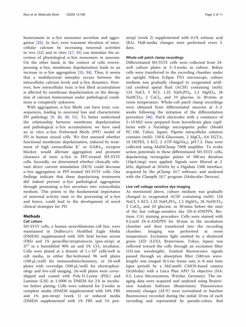
ResultsPreformed fibrils (PFF) treatment induced α-Syn inclusionaccumulation in differentiated SH-SY5Y cellsIn order to test the effects of membrane depolarizationon α-Syn accumulation, we first added 0.1% RA to theculture medium for two weeks to differentiate the SH-SY5Y neuroblastoma cells. Consistent with previousstudies [16, 22, 47], differentiated SH-SY5Y cells showeda variety of neuron-like phenotypes including long neur-ites and complex network connections (Fig. 1a1). Fur-thermore, whole-cell current clamp recordings
Fig. 1 Preformed fibrils (PFFs) treatment induced α-Syn inclusion accumulations in the differentiated SH-SY5Y cells. a1 and b1 Representative IR-DIC image of differentiated SH-SY5Y neuroblastoma cells treated with (B1) or without (A1) 25 μg/ml PFFs treatment for 3 days. Note differentiatedcells show long neurites and complex network connections. a2 and b2 Representative whole-cell voltage responses to 500ms rectangular currentinjections of 0, 50, 120pA under the current clamp mode in differentiated SH-SY5Y cells treated with (B2) or without (A2) 25 μg/ml PFFs for 3 days.Arrow points to the action potentials generated. c Summary of the mean resting membrane potentials in control and 25 μg/ml PFFs-treated SH-SY5Y cells. Error bars indicate SEM. d Summary of the mean membrane potential threshold in control and 25 μg/ml PFFs-treated SH-SY5Y cells.Error bars indicate SEM. Two-tailed unpaired t test, *p < 0.05. e Immunofluorescent staining of α-Syn (green) in the differentiated SH-SY5Y cellsfollowing the treatment of 0, 5, 10, 25 μg/ml PFFs for 3 days. Scale bar: 200 μm. f Summary of the mean integrated intensity per cell of α-Synstaining in the differentiated SH-SY5Y cells following the treatment of 0, 5, 10, 25 μg/ml PFFs for 3 days. Error bars indicate SEM. One-way ANOVA,Tukey’s HSD post-hoc test. **p < 0.01. ***p < 0.001
Ross et al. Molecular Brain (2020) 13:108 Page 4 of 11
demonstrated that while no spontaneous action poten-tials were detected, appropriate depolarizing current in-jections could evoke overshooting action potentials indifferentiated SH-SY5Y cells (Fig. 1a2). We next pro-ceeded to treat the differentiated SH-SY5Y cells with dif-ferent concentrations (5, 10, and 25 μg/ml) of PFFs orvehicle for 3 days, and subsequently, evaluated α-Syn ac-cumulation (Fig. 1e and f). The PFF treatments inducedsignificant increases in α-Syn accumulation (5 μg/ml:13.36 ± 5.89, n = 19, p < 0.001; 10 μg/ml: 27.58 ± 6.15,n = 19, p < 0.001; 25 μg/ml: 34.70 ± 6.47, n = 20, p <0.001) compared to control cells (6.92 ± 2.44, n = 20). α-Syn accumulation appeared to increase with PFFconcentrations and peaked in 25 μg/ml PFF-treated SH-SY5Y cells (25 μg/ml vs 5 μg/ml: p < 0.001; 25 μg/ml vs10 μg/ml, p < 0.001). In addition, PFFs (25 μg/ml) did notcause a significant change in resting membrane poten-tials (RMPs), but there was a trend towards hyperpolari-zation (− 46.7 ± 3.2 mV, n = 10, p = 0.18) compared tocontrol SH-SY5Y cells (− 42.3 ± 1.3 mV, n = 13, Fig. 1c.This was associated with an increase in the action poten-tial threshold in PFF-treated SH-SY5Y cells (PFF: 18.0 ±2.2 mV, n = 6 vs Control: − 23.9 ± 1.6 mV, n = 10, p =0.049, Fig. 1d). These results suggest that 3-day PFF
treatment induced significant pathological α-Syn accu-mulation in differentiated SH-SY5Y cells.
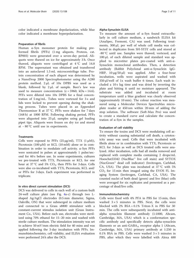
Membrane depolarization significantly reduces α-Synuclein fibril accumulation in differentiated SH-SY5YcellsWe next sought to assess the effects of membranedepolarization on α-Syn fibril accumulation in differenti-ated SH-SY5Y cells. To achieve membrane depolarization,we employed two independent strategies rather than sim-ply one in order to avoid possible stimulation-specific ef-fects. 1) By increasing extracellular K+ concentration inthe culture medium through addition of 10mM KCL. Fastvoltage-sensitive dye imaging of Di-4-ANEPPS-loadedSH-SY5Y cells confirmed high extracellular K+-inducedmembrane depolarization (Fig. 2a, n = 5). 2) Blockade ofthe inhibitory GABAA receptors by specific inhibitorPicrotoxin (100 μM) was applied to depolarize the cellmembrane. Immunohistochemistry with an antibodyagainst GABAA receptor α1subunit confirmed the expres-sion levels of GABAA receptors in differentiated SH-SY5Ycells (Fig. 2b, n = 4). Consistent with the expression ofGABAA receptors, fast voltage-sensitive dye imagingshowed GABAA receptor inhibitor Picrotoxin evoked
Fig. 2 Membrane depolarization induced by increasing extracellular K+ and blocking GABAA receptors in differentiated SH-SY5Y cells. aRepresentative responses (ΔF/F) of Fast voltage-sensitive dye imaging of Di-4-ANEPPS-loaded SH-SY5Y cells before, during, and after 10 mM KCL.Scale bar, 100 μm. b Immunofluorescent staining of GABAA α1(green) and DAPI (blue) in the differentiated SH-SY5Y cells. Scale bar: 200 μm. cRepresentative responses of Fast voltage-sensitive dye imaging of Di-4-ANEPPS-loaded SH-SY5Y cells before, during, and after 100 μM Picrotoxin.Scale bar, 100 μm
Ross et al. Molecular Brain (2020) 13:108 Page 5 of 11
membrane depolarization in differentiated SH-SY5Y cells(Fig. 2c, n = 4). These results provide functional evidenceof KCL and Picrotoxin induced membrane depolarizationin differentiated SH-SY5Y cells.We then examined whether these treatments affect
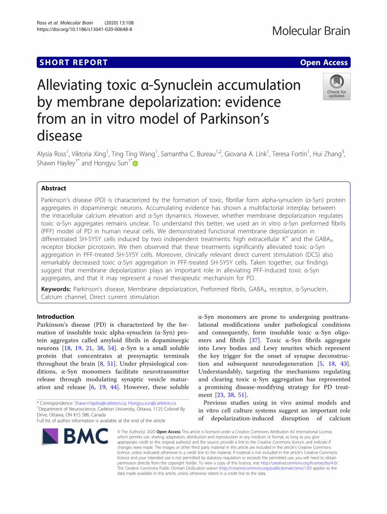
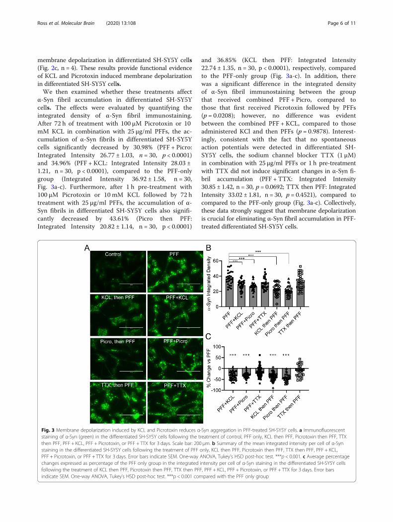
α-Syn fibril accumulation in differentiated SH-SY5Ycells. The effects were evaluated by quantifying theintegrated density of α-Syn fibril immunostaining.After 72 h of treatment with 100 μM Picrotoxin or 10mM KCL in combination with 25 μg/ml PFFs, the ac-cumulation of α-Syn fibrils in differentiated SH-SY5Ycells significantly decreased by 30.98% (PFF + Picro:Integrated Intensity 26.77 ± 1.03, n = 30, p < 0.0001)and 34.96% (PFF + KCL: Integrated Intensity 28.03 ±1.21, n = 30, p < 0.0001), compared to the PFF-onlygroup (Integrated Intensity 36.92 ± 1.58, n = 30,Fig. 3a-c). Furthermore, after 1 h pre-treatment with100 μM Picrotoxin or 10 mM KCL followed by 72 htreatment with 25 μg/ml PFFs, the accumulation of α-Syn fibrils in differentiated SH-SY5Y cells also signifi-cantly decreased by 43.61% (Picro then PFF:Integrated Intensity 20.82 ± 1.14, n = 30, p < 0.0001)
and 36.85% (KCL then PFF: Integrated Intensity22.74 ± 1.35, n = 30, p < 0.0001), respectively, comparedto the PFF-only group (Fig. 3a-c). In addition, therewas a significant difference in the integrated densityof α-Syn fibril immunostaining between the groupthat received combined PFF + Picro, compared tothose that first received Picrotoxin followed by PFFs(p = 0.0208); however, no difference was evidentbetween the combined PFF + KCL, compared to thoseadministered KCl and then PFFs (p = 0.9878). Interest-ingly, consistent with the fact that no spontaneousaction potentials were detected in differentiated SH-SY5Y cells, the sodium channel blocker TTX (1 μM)in combination with 25 μg/ml PFFs or 1 h pre-treatmentwith TTX did not induce significant changes in α-Syn fi-bril accumulation (PFF + TTX: Integrated Intensity30.85 ± 1.42, n = 30, p = 0.0692; TTX then PFF: IntegratedIntensity 33.02 ± 1.81, n = 30, p = 0.4521), compared tocompared to the PFF-only group (Fig. 3a-c). Collectively,these data strongly suggest that membrane depolarizationis crucial for eliminating α-Syn fibril accumulation in PFF-treated differentiated SH-SY5Y cells.
Fig. 3 Membrane depolarization induced by KCL and Picrotoxin reduces α-Syn aggregation in PFF-treated SH-SY5Y cells. a Immunofluorescentstaining of α-Syn (green) in the differentiated SH-SY5Y cells following the treatment of control, PFF only, KCL then PFF, Picrotoxin then PFF, TTXthen PFF, PFF + KCL, PFF + Picrotoxin, or PFF + TTX for 3 days. Scale bar: 200 μm. b Summary of the mean integrated intensity per cell of α-Synstaining in the differentiated SH-SY5Y cells following the treatment of PFF only, KCL then PFF, Picrotoxin then PFF, TTX then PFF, PFF + KCL,PFF + Picrotoxin, or PFF + TTX for 3 days. Error bars indicate SEM. One-way ANOVA, Tukey’s HSD post-hoc test. ***p < 0.001. c Average percentagechanges expressed as percentage of the PFF only group in the integrated intensity per cell of α-Syn staining in the differentiated SH-SY5Y cellsfollowing the treatment of KCL then PFF, Picrotoxin then PFF, TTX then PFF, PFF + KCL, PFF + Picrotoxin, or PFF + TTX for 3 days. Error barsindicate SEM. One-way ANOVA, Tukey’s HSD post-hoc test. ***p < 0.001 compared with the PFF only group
Ross et al. Molecular Brain (2020) 13:108 Page 6 of 11
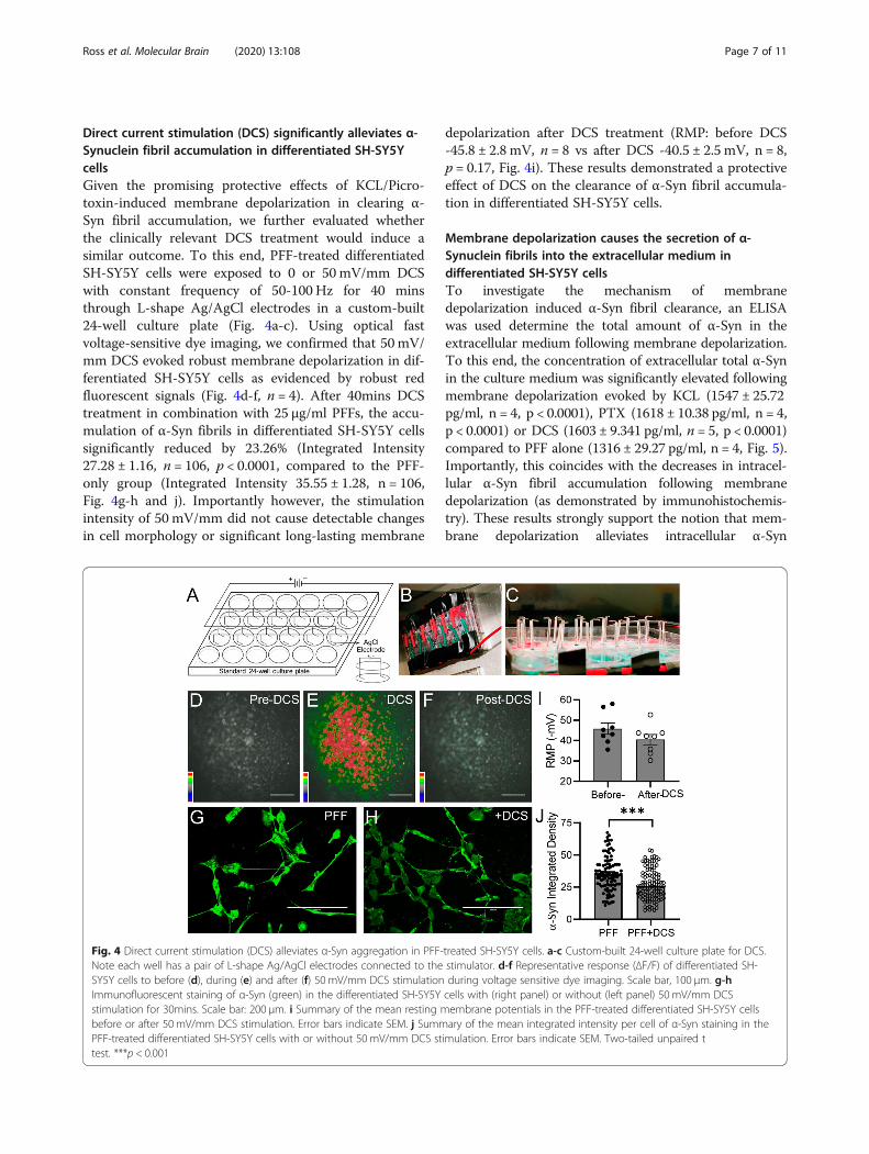
Direct current stimulation (DCS) significantly alleviates α-Synuclein fibril accumulation in differentiated SH-SY5YcellsGiven the promising protective effects of KCL/Picro-toxin-induced membrane depolarization in clearing α-Syn fibril accumulation, we further evaluated whetherthe clinically relevant DCS treatment would induce asimilar outcome. To this end, PFF-treated differentiatedSH-SY5Y cells were exposed to 0 or 50 mV/mm DCSwith constant frequency of 50-100 Hz for 40 minsthrough L-shape Ag/AgCl electrodes in a custom-built24-well culture plate (Fig. 4a-c). Using optical fastvoltage-sensitive dye imaging, we confirmed that 50 mV/mm DCS evoked robust membrane depolarization in dif-ferentiated SH-SY5Y cells as evidenced by robust redfluorescent signals (Fig. 4d-f, n = 4). After 40mins DCStreatment in combination with 25 μg/ml PFFs, the accu-mulation of α-Syn fibrils in differentiated SH-SY5Y cellssignificantly reduced by 23.26% (Integrated Intensity27.28 ± 1.16, n = 106, p < 0.0001, compared to the PFF-only group (Integrated Intensity 35.55 ± 1.28, n = 106,Fig. 4g-h and j). Importantly however, the stimulationintensity of 50 mV/mm did not cause detectable changesin cell morphology or significant long-lasting membrane
depolarization after DCS treatment (RMP: before DCS-45.8 ± 2.8 mV, n = 8 vs after DCS -40.5 ± 2.5 mV, n = 8,p = 0.17, Fig. 4i). These results demonstrated a protectiveeffect of DCS on the clearance of α-Syn fibril accumula-tion in differentiated SH-SY5Y cells.
Membrane depolarization causes the secretion of α-Synuclein fibrils into the extracellular medium indifferentiated SH-SY5Y cellsTo investigate the mechanism of membranedepolarization induced α-Syn fibril clearance, an ELISAwas used determine the total amount of α-Syn in theextracellular medium following membrane depolarization.To this end, the concentration of extracellular total α-Synin the culture medium was significantly elevated followingmembrane depolarization evoked by KCL (1547 ± 25.72pg/ml, n = 4, p < 0.0001), PTX (1618 ± 10.38 pg/ml, n = 4,p < 0.0001) or DCS (1603 ± 9.341 pg/ml, n = 5, p < 0.0001)compared to PFF alone (1316 ± 29.27 pg/ml, n = 4, Fig. 5).Importantly, this coincides with the decreases in intracel-lular α-Syn fibril accumulation following membranedepolarization (as demonstrated by immunohistochemis-try). These results strongly support the notion that mem-brane depolarization alleviates intracellular α-Syn
Fig. 4 Direct current stimulation (DCS) alleviates α-Syn aggregation in PFF-treated SH-SY5Y cells. a-c Custom-built 24-well culture plate for DCS.Note each well has a pair of L-shape Ag/AgCl electrodes connected to the stimulator. d-f Representative response (ΔF/F) of differentiated SH-SY5Y cells to before (d), during (e) and after (f) 50 mV/mm DCS stimulation during voltage sensitive dye imaging. Scale bar, 100 μm. g-hImmunofluorescent staining of α-Syn (green) in the differentiated SH-SY5Y cells with (right panel) or without (left panel) 50 mV/mm DCSstimulation for 30mins. Scale bar: 200 μm. i Summary of the mean resting membrane potentials in the PFF-treated differentiated SH-SY5Y cellsbefore or after 50 mV/mm DCS stimulation. Error bars indicate SEM. j Summary of the mean integrated intensity per cell of α-Syn staining in thePFF-treated differentiated SH-SY5Y cells with or without 50 mV/mm DCS stimulation. Error bars indicate SEM. Two-tailed unpaired ttest. ***p < 0.001
Ross et al. Molecular Brain (2020) 13:108 Page 7 of 11
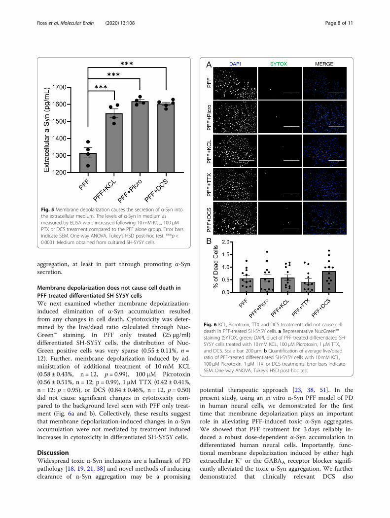
aggregation, at least in part through promoting α-Synsecretion.
Membrane depolarization does not cause cell death inPFF-treated differentiated SH-SY5Y cellsWe next examined whether membrane depolarization-induced elimination of α-Syn accumulation resultedfrom any changes in cell death. Cytotoxicity was deter-mined by the live/dead ratio calculated through Nuc-Green™ staining. In PFF only treated (25 μg/ml)differentiated SH-SY5Y cells, the distribution of Nuc-Green positive cells was very sparse (0.55 ± 0.11%, n =12). Further, membrane depolarization induced by ad-ministration of additional treatment of 10 mM KCL(0.58 ± 0.43%, n = 12, p = 0.99), 100 μM Picrotoxin(0.56 ± 0.51%, n = 12; p = 0.99), 1 μM TTX (0.42 ± 0.41%,n = 12; p = 0.95), or DCS (0.84 ± 0.46%, n = 12, p = 0.50)did not cause significant changes in cytotoxicity com-pared to the background level seen with PFF only treat-ment (Fig. 6a and b). Collectively, these results suggestthat membrane depolarization-induced changes in α-Synaccumulation were not mediated by treatment inducedincreases in cytotoxicity in differentiated SH-SY5Y cells.
DiscussionWidespread toxic α-Syn inclusions are a hallmark of PDpathology [18, 19, 21, 38] and novel methods of inducingclearance of α-Syn aggregation may be a promising
potential therapeutic approach [23, 38, 51]. In thepresent study, using an in vitro α-Syn PFF model of PDin human neural cells, we demonstrated for the firsttime that membrane depolarization plays an importantrole in alleviating PFF-induced toxic α-Syn aggregates.We showed that PFF treatment for 3 days reliably in-duced a robust dose-dependent α-Syn accumulation indifferentiated human neural cells. Importantly, func-tional membrane depolarization induced by either highextracellular K+ or the GABAA receptor blocker signifi-cantly alleviated the toxic α-Syn aggregation. We furtherdemonstrated that clinically relevant DCS also
Fig. 5 Membrane depolarization causes the secretion of α-Syn intothe extracellular medium. The levels of α-Syn in medium asmeasured by ELISA were increased following 10mM KCL, 100 μMPTX or DCS treatment compared to the PFF alone group. Error barsindicate SEM. One-way ANOVA, Tukey’s HSD post-hoc test. ***p <0.0001. Medium obtained from cultured SH-SY5Y cells
Fig. 6 KCL, Picrotoxin, TTX and DCS treatments did not cause celldeath in PFF-treated SH-SY5Y cells. a Representative NucGreen™staining (SYTOX, green; DAPI, blue) of PFF-treated differentiated SH-SY5Y cells treated with 10 mM KCL, 100 μM Picrotoxin, 1 μM TTX,and DCS. Scale bar: 200 μm. b Quantification of average live/deadratio of PFF-treated differentiated SH-SY5Y cells with 10 mM KCL,100 μM Picrotoxin, 1 μM TTX, or DCS treatments. Error bars indicateSEM. One-way ANOVA, Tukey’s HSD post-hoc test
Ross et al. Molecular Brain (2020) 13:108 Page 8 of 11
remarkably decreased toxic α-Syn aggregation in PFF-treated human neural cells. Furthermore, this membranedepolarization-induced decrease in intracellular α-Synobserved with KCL, PTX and DCS was coupled with anincrease in extracellular α-Syn. Importantly, thetreatments used in the present study did not induceconfounding cell cytotoxicity. Our results provide directevidence of an important interplay between membranedepolarization and clearance of intracellular α-Synaggregation.α-Syn PFFs have been repeatedly shown to initiate
Lewy body pathological changes through templating andrecruiting endogenous α-Syn to accumulate and form in-soluble inclusions in neurons in a variety of PD models[9, 14, 24, 26, 36, 47]. Here, we used an α-Syn PFFmodel in the differentiated SH-SY5Y cells which havealready been extensively used in PD research due totheir human-specific proteins [3], dopaminergic pheno-types [1, 3, 41], and neuronal electrophysiological prop-erties, i.e., overshooting action potentials [48] (Fig. 1b).Consistent with previous studies [9, 47], our resultsshowed that α-Syn PFF treatment effectively induced en-dogenous α-Syn inclusions and modulated membraneproperties in the differentiated SH-SY5Y cells. Prolongedexogenous PFF treatment for two weeks has previouslybeen reported to lead to neurotoxicity in primary hippo-campal neurons [50]. In the present study, to avoid thesustained PFF treatment-induced cell death, a higherconcentration (25 μg/ml) of α-Syn PFF was applied for arelatively short period of time (3 days). Under these con-ditions, toxic α-Syn inclusions were induced with no de-tectable cell toxicity in either PFF only or PFF incombination with KCL/TTX/Picrotoxin/DCS treatedSH-SY5Y cells (Fig. 6), supporting that changes in thelevels of α-Syn inclusions were not due to treatment-induced cell death.An important finding of our study is that functional
membrane depolarization has a crucial role in clearingPFF-induced toxic α-Syn aggregation. Previous in vivoand in vitro studies have shown that depolarization orincreased neuronal activity can increase the mobility ofphysiological α-Syn monomers and promote their disas-sociation from presynaptic membranes and release toextracellular spaces through a Ca2+ dependent mechan-ism [12, 13, 17, 52]. In undifferentiated human cell linesoverexpressing physiological α-Syn monomers, however,a transit (about an hour) elevation of intracellular Ca2+
through the addition of high extracellular KCL or Ca2+
levels can induce an increase in intracellular α-Syn ag-gregation [15, 34, 40], while this same treatment alsopromoted the extracellular secretion of α-Syn monomersafter longer elevations (6 h) of intracellular Ca2+ in dif-ferentiated human neuroblastoma cells [12]. These stud-ies support a potential multifactorial interplay between
the intracellular calcium levels and α-Syn dynamics. It isimportant to note that the high level of α-Syn monomersused in these studies can increase the potential mito-chondrial oxidative stress [10] and the vulnerability of α-Syn aggregation [18, 27] and thus may introduce con-founds while evaluating the α-Syn toxicity. Currently, itis still not entirely clear whether membranedepolarization can affect endogenous α-Syn fibrilizationduring pathological conditions.In the present study, we used human wild-type PFF-
treated human neural cells to seed endogenous α-Syn in-clusions without the additional complications of α-Synoverexpression. Two independent pharmacological strat-egies were used to evoke functional membranedepolarization to avoid the possibility of stimulation-specific effects. We revealed that membranedepolarization evoked by either KCL or PTX for 3 daysalleviated the PFF-induced toxic α-Syn aggregates in hu-man neural cells by significantly increasing the secretionof α-Syn into the extracellular media (Figs. 3 and 5).Whether or not the secreted α-Syn has undergone con-formational changes will require further investigation.Furthermore, while we found that chronic membranedepolarization for 3 days did not induce detectable cyto-toxicity, we cannot exclude the potential possibility ofexcitotoxicity due to prolonged Ca2+ influx. The timecourse of depolarization treatment may need to be fur-ther optimized to balance any potential excitotoxicitywith alterations in the clearance of α-Syn aggregates.Membrane depolarization might enhance the clearanceof the toxic α-Syn aggregates by upregulating the expres-sion of heat shock protein 70 (HSP70) and the activationof autophagy, both of which have been shown to pro-mote the clearance of α-Syn inclusions [18, 20, 29, 53].Interestingly, TTX did not cause a significant change inthe amount of α-Syn accumulation regardless of when itwas administered. The lack of response seen with TTXis in line with the perspective that SH-SY5Y cells do notshow spontaneous action potentials during the restingcondition. It is of interest that PFFs have been shown toimpair the initiation of synaptogenesis and synapticfunction in cultured excitatory hippocampal neurons[51]. This might, in turn, induce further α-Syn fibriliza-tion and synaptic dysfunction resulting in a vicious cycleto promoting toxic α-Syn aggregates in synucleinopa-thies. Further research is needed to explore themolecular mechanisms mediating the membranedepolarization-induced disaggregation. Although we cannot exclude the possibility that our alpha Synuclein fila-ment antibody may label PFFs in addition to endogenousalpha-Syn fibrils, our results clearly demonstrate thatmembrane depolarization does significantly decrease thelevels of intracellular alpha-Syn fibrils (PFF or endogen-ous alpha-syn fibrils), and importantly, also increased
Ross et al. Molecular Brain (2020) 13:108 Page 9 of 11
the extracellular secretion of a-Syn. Therefore, mem-brane depolorization might represent an effective strat-egy to interfere with the intitiation of synapticdysfunction-α-Syn fibrilization in PD pathophysiology.Finally, we induced membrane depolarization by ap-
plying an external electrical field using a clinically rele-vant DCS procedure. Physiological α-Syn contains twostructural domains, the N-terminal lipid-binding domainincluding a hydrophobic NAC (non-amyloid-β compo-nent) and the C-terminal domain [4, 28, 31]. Toxic α-Syn fibril formation requires significant α-Syn conform-ational changes from α-helix to β-sheet to expose hydro-phobic NAC, leading to the formation of α-Synprotofibrils and fibrils in neurons [7, 45]. Interestingly,the α-Syn N-terminal is positively charged, while the C-terminal is negatively charged [11, 31, 49]. Therefore,theoretically, an external electrical field could disrupt α-Syn aggregation by aligning α-Syn with induced dipolesfollowing the axis of the electric field or directly interact-ing with the α-Syn dipoles to induce a β-sheet to α-helixconformational switch [2, 42]. Indeed, using a novel,custom-built 24-well culture plate, we obtained directevidence for the first time to demonstrate that clinicallyrelevant DCS effectively alleviated the accumulation ofPFF-induced toxic α-Syn aggregates in differentiated hu-man neural cells, very much the same as we observedwith high extracellular K+ and the GABAA receptorblocker picrotoxin. Interestingly, Electroconvulsive ther-apy (ECT) has been shown to cause antiparkinsonian ef-fects in patients with advanced PD [32, 33].Furthermore, we have recently shown that epilepsy wasassociated with significant improvement in clinicalmotor symptoms and cognitive decline in PD patients[39]. While the mechanism behind these positive effectsremains elusive, our findings suggest this effect may bedue in part to the clearing of toxic α-Syn aggregatesfrom dopaminergic neurons and offers beneficial infor-mation that could provide therapeutic benefits in con-trolling PD pathology. Future in vivo studies may wishto investigate the mechanism responsible for the inter-play between membrane depolarization and α-Syn fibrilclearance and the relationship to clinical improvement.
ConclusionOverall, employing several novel approaches that utilizea new in vitro α-Syn PFF model of PD, we have shownthat membrane depolarization, through provocation ofα-Syn fibril secretion, can protect against intracellularaccumulation of toxic PFF-induced α-Syn aggregatesand importantly, that this occurs in the absence of anycell death. The present data could have important clin-ical implications for synucleinopathies, specifically PD,and may also help determine a mechanism of action forthe accumulation of α-Syn aggregates.
AbbreviationsPD: Parkinson’s disease; α-Syn: alpha-synuclein; PFF: Preformed fibril;DCS: Direct current stimulation; DMEM: Dulbecco’s Modified Eagle Media;FBS: Fetal bovine serum; PDL: Poly-D-Lysine; LM: Laminin; RA: Retinoic acid;ACSF: Artificial cerebral spinal fluid; TTX: Tetrodotoxin; KCL: Potassiumchloride; PFA: Paraformaldehyde; PBS: Phosphate-buffered saline; BSA: Bovineserum albumin; GABAA: γ-Aminobutyric acid type A; HSP70: Heat shockprotein 70; NAC: Non-amyloid-β component; ECT: Electroconvulsive therapy
AcknowledgementsWe thank all members of the Hayley lab and the Sun lab for valuablecomments.
Authors’ contributionsA.R., H.Z., S.H. and H.S. designed the study. A.R., V.X., T.T.W., S.C.B., T.F., G.A.L.and H.S. conducted the experiments. A.R. and H.S. analyzed the data. A.R.,H.Z., S.H. and H.S. wrote the manuscript. All authors have read and approvedthe final manuscript.
FundingThis work was supported by an NSERC Discovery Grant (to H.S.), and CIHRgrants (to H.S. and S.H.). H.S. holds a Canada Research Chair inDevelopmental Neuroscience. A.R. holds a Queen Elizabeth II Scholarship inScience and Technology.
Availability of data and materialsThe datasets used and/or analyzed during the current study available fromthe corresponding author on reasonable request.
Ethics approval and consent to participateNot applicable.
Consent for publicationNot applicable.
Competing interestsThe authors declare that they have no competing interests.
Author details1Department of Neuroscience, Carleton University, Ottawa, 1125 Colonel ByDrive, Ottawa, ON K1S 5B6, Canada. 2Department of Cellular and MolecularMedicine, University of Ottawa, Ottawa, ON, Canada. 3Department ofNeurology, SUNY Downstate Medical center, Brooklyn, NY 11226, USA.
Received: 29 April 2020 Accepted: 22 July 2020
References1. Arun P, Madhavarao CN, Moffett JR, Namboodiri AM. Antipsychotic drugs
increase N-acetylaspartate and N-acetylaspartylglutamate in SH-SY5Y humanneuroblastoma cells. J Neurochem. 2008;106(4):1669–80.
2. Baumketner A. Electric field as a disaggregating agent for amyloid fibrils. JPhys Chem B. 2014;118:14578–89.
3. Biedler JL, Roffler-Tarlov S, Schachner M, Freedman LS. Multipleneurotransmitter synthesis by human neuroblastoma cell lines and clones.Cancer Res. 1978;38(11 Pt 1):3751–7.
4. Breydo L, Wu JW, Uversky VN. Alpha-synuclein misfolding and Parkinson'sdisease. Biochim Biophys Acta. 2012;1822:261–85.
5. Bridi JC, Hirth F. Mechanisms of alpha-Synuclein induced Synaptopathy inParkinson’s disease. Front Neurosci. 2018;12:80.
6. Burre J, Sharma M, Sudhof TC. Alpha-Synuclein assembles into higher-ordermultimers upon membrane binding to promote SNARE complex formation.Proc Natl Acad Sci U S A. 2014;111:E4274–83.
7. Chandra S, Chen X, Rizo J, Jahn R, Sudhof TC. A broken alpha -helix infolded alpha -Synuclein. J Biol Chem. 2003;278:15313–8.
8. Chandra S, Gallardo G, Fernandez-Chacon R, Schluter OM, Sudhof TC. Alpha-synuclein cooperates with CSPalpha in preventing neurodegeneration. Cell.2005;123:383–96.
9. Chu Y, Muller S, Tavares A, Barret O, Alagille D, Seibyl J, Tamagnan G, MarekK, Luk KC, Trojanowski JQ, et al. Intrastriatal alpha-synuclein fibrils in
Ross et al. Molecular Brain (2020) 13:108 Page 10 of 11
monkeys: spreading, imaging and neuropathological changes. Brain. 2019;142:3565–79.
10. Dryanovski DI, Guzman JN, Xie Z, Galteri DJ, Volpicelli-Daley LA, Lee VM,Miller RJ, Schumacker PT, Surmeier DJ. Calcium entry and alpha-synucleininclusions elevate dendritic mitochondrial oxidant stress in dopaminergicneurons. J Neurosci. 2013;33:10154–64.
11. Emamzadeh FN. Alpha-synuclein structure, functions, and interactions. J ResMed Sci. 2016;21:29.
12. Emmanouilidou E, Melachroinou K, Roumeliotis T, Garbis SD, Ntzouni M,Margaritis LH, Stefanis L, Vekrellis K. Cell-produced alpha-synuclein issecreted in a calcium-dependent manner by exosomes and impactsneuronal survival. J Neurosci. 2010;30:6838–51.
13. Emmanouilidou E, Minakaki G, Keramioti MV, Xylaki M, Balafas E,Chrysanthou-Piterou M, Kloukina I, Vekrellis K. GABA transmission via ATP-dependent K+ channels regulates alpha-synuclein secretion in mousestriatum. Brain. 2016;139:871–90.
14. Fares MB, Maco B, Oueslati A, Rockenstein E, Ninkina N, Buchman VL, Masliah E,Lashuel HA. Induction of de novo alpha-synuclein fibrillization in a neuronalmodel for Parkinson’s disease. Proc Natl Acad Sci U S A. 2016;113:E912–21.
15. Follett J, Darlow B, Wong MB, Goodwin J, Pountney DL. Potassiumdepolarization and raised calcium induces alpha-synuclein aggregates.Neurotox Res. 2013;23:378–92.
16. Forster JI, Koglsberger S, Trefois C, Boyd O, Baumuratov AS, Buck L, Balling R,Antony PM. Characterization of differentiated SH-SY5Y as neuronalscreening model reveals increased oxidative vulnerability. J Biomol Screen.2016;21:496–509.
17. Fortin DL, Nemani VM, Voglmaier SM, Anthony MD, Ryan TA, Edwards RH.Neural activity controls the synaptic accumulation of alpha-synuclein. JNeurosci. 2005;25:10913–21.
18. Gao J, Perera G, Bhadbhade M, Halliday GM, Dzamko N. Autophagyactivation promotes clearance of alpha-synuclein inclusions in fibril-seededhuman neural cells. J Biol Chem. 2019;294:14241–56.
19. Ghiglieri V, Calabrese V, Calabresi P. Alpha-Synuclein: from early synapticdysfunction to Neurodegeneration. Front Neurol. 2018;9:295.
20. Hu F, Zhou J, Lu Y, Guan L, Wei NN, Tang YQ, Wang K. Inhibition of Hsp70suppresses neuronal Hyperexcitability and attenuates epilepsy by enhancingA-type potassium current. Cell Rep. 2019;26:168–81 e164.
21. Iarkov A, Barreto GE, Grizzell JA, Echeverria V. Strategies for the treatment ofParkinson’s disease: beyond dopamine. Front Aging Neurosci. 2020;12:4.
22. Ito K, Eguchi Y, Imagawa Y, Akai S, Mochizuki H, Tsujimoto Y. MPP+ inducesnecrostatin-1- and ferrostatin-1-sensitive necrotic death of neuronal SH-SY5Y cells. Cell Death Discov. 2017;3:17013.
23. Kalia LV, Lang AE. Parkinson’s disease. Lancet. 2015;386:896–912.24. Karampetsou M, Ardah MT, Semitekolou M, Polissidis A, Samiotaki M,
Kalomoiri M, Majbour N, Xanthou G, El-Agnaf OMA, Vekrellis K.Phosphorylated exogenous alpha-synuclein fibrils exacerbate pathology andinduce neuronal dysfunction in mice. Sci Rep. 2017;7:16533.
25. Leandrou E, Emmanouilidou E, Vekrellis K. Voltage-gated calcium channelsand alpha-Synuclein: implications in Parkinson's disease. Front Mol Neurosci.2019;12:237.
26. Luk KC, Kehm V, Carroll J, Zhang B, O'Brien P, Trojanowski JQ, Lee VM.Pathological alpha-synuclein transmission initiates Parkinson-likeneurodegeneration in nontransgenic mice. Science. 2012;338:949–53.
27. Luna E, Decker SC, Riddle DM, Caputo A, Zhang B, Cole T, Caswell C, Xie SX,Lee VMY, Luk KC. Differential alpha-synuclein expression contributes toselective vulnerability of hippocampal neuron subpopulations to fibril-induced toxicity. Acta Neuropathol. 2018;135:855–75.
28. Luna E, Luk KC. Bent out of shape: alpha-Synuclein misfolding and theconvergence of pathogenic pathways in Parkinson’s disease. FEBS Lett.2015;589:3749–59.
29. Lyamzaev KG, Tokarchuk AV, Panteleeva AA, Mulkidjanian AY, Skulachev VP,Chernyak BV. Induction of autophagy by depolarization of mitochondria.Autophagy. 2018;14:921–4.
30. Masuda-Suzukake M, Nonaka T, Hosokawa M, Oikawa T, Arai T, Akiyama H,Mann DM, Hasegawa M. Prion-like spreading of pathological alpha-synuclein in brain. Brain. 2013;136:1128–38.
31. McClendon S, Rospigliosi CC, Eliezer D. Charge neutralization and collapse ofthe C-terminal tail of alpha-synuclein at low pH. Protein Sci. 2009;18:1531–40.
32. Moellentine C, Rummans T, Ahlskog JE, Harmsen WS, Suman VJ, O'ConnorMK, Black JL, Pileggi T. Effectiveness of ECT in patients with parkinsonism. JNeuropsychiatry Clin Neurosci. 1998;10:187–93.
33. Narang P, Glowacki A, Lippmann S. Electroconvulsive therapy interventionfor Parkinson's disease. Innov Clin Neurosci. 2015;12:25–8.
34. Nath S, Goodwin J, Engelborghs Y, Pountney DL. Raised calcium promotesalpha-synuclein aggregate formation. Mol Cell Neurosci. 2011;46:516–26.
35. Paillusson S, Clairembault T, Biraud M, Neunlist M, Derkinderen P. Activity-dependent secretion of alpha-synuclein by enteric neurons. J Neurochem.2013;125:512–7.
36. Panattoni G, Rota L, Colla E. Exogenous Administration of Microsomes-associated Alpha-synuclein Aggregates to primary neurons as a powerfulcell model of fibrils formation. J Vis Exp. 2018;136:57884.
37. Peng C, Gathagan RJ, Covell DJ, Medellin C, Stieber A, Robinson JL, ZhangB, Pitkin RM, Olufemi MF, Luk KC, et al. Cellular milieu imparts distinctpathological alpha-synuclein strains in alpha-synucleinopathies. Nature.2018;557:558–63.
38. Pujols J, Pena-Diaz S, Lazaro DF, Peccati F, Pinheiro F, Gonzalez D, Carija A,Navarro S, Conde-Gimenez M, Garcia J, et al. Small molecule inhibits alpha-synuclein aggregation, disrupts amyloid fibrils, and prevents degenerationof dopaminergic neurons. Proc Natl Acad Sci U S A. 2018;115:10481–6.
39. Rawjani S, Ross A, Zhang H, Hayley S, Sun H. An unrecognized “excitement”of epilepsy in Parkinson disease: evidence of systemic biomarkers andcognitive function. J Psychiatry Neurosci. 2019;44:S14.
40. Rcom-H'cheo-Gauthier AN, Davis A, Meedeniya ACB, Pountney DL. Alpha-synuclein aggregates are excluded from calbindin-D28k-positive neurons indementia with Lewy bodies and a unilateral rotenone mouse model. MolCell Neurosci. 2016;77:65–75.
41. Ross RA, Biedler JL. Presence and regulation of tyrosinase activity in humanneuroblastoma cell variants in vitro. Cancer Res. 1985;45(4):1628–32.
42. Saikia J, Pandey G, Sasidharan S, Antony F, Nemade HB, Kumar S, ChaudharyN, Ramakrishnan V. Electric field disruption of amyloid aggregation:potential noninvasive therapy for Alzheimer's disease. ACS Chem Neurosci.2019;10:2250–62.
43. Schirinzi T, Madeo G, Martella G, Maltese M, Picconi B, Calabresi P, Pisani A.Early synaptic dysfunction in Parkinson's disease: insights from animalmodels. Mov Disord. 2016;31:802–13.
44. Scott D, Roy S. Alpha-Synuclein inhibits intersynaptic vesicle mobility andmaintains recycling-pool homeostasis. J Neurosci. 2012;32:10129–35.
45. Snead D, Eliezer D. Alpha-synuclein function and dysfunction on cellularmembranes. Exp Neurobiol. 2014;23:292–313.
46. Sun H, Takesian AE, Wang TT, Lippman-Bell JJ, Hensch TK, Jensen FE. Earlyseizures prematurely Unsilence auditory synapses to disrupt Thalamocorticalcritical period plasticity. Cell Rep. 2018;23:2533–40.
47. Taylor-Whiteley TR, Le Maitre CL, Duce JA, Dalton CF, Smith DP.Recapitulating Parkinson’s disease pathology in a three-dimensional humanneural cell culture model. Dis Model Mech. 2019;12:dmm038042.
48. Tosetti P, Taglietti V, Toselli M. Functional changes in potassiumconductances of the human neuroblastoma cell line SH-SY5Y during invitro differentiation. J Neurophysiol. 1998;79:648–58.
49. Vamvaca K, Volles MJ, Lansbury PT Jr. The first N-terminal amino acids ofalpha-synuclein are essential for alpha-helical structure formation in vitroand membrane binding in yeast. J Mol Biol. 2009;389:413–24.
50. Volpicelli-Daley LA, Luk KC, Patel TP, Tanik SA, Riddle DM, Stieber A, MeaneyDF, Trojanowski JQ, Lee VM. Exogenous alpha-synuclein fibrils induce Lewybody pathology leading to synaptic dysfunction and neuron death. Neuron.2011;72:57–71.
51. Wu Q, Takano H, Riddle DM, Trojanowski JQ, Coulter DA, Lee VM. Alpha-Synuclein (alphaSyn) preformed fibrils induce endogenous alphaSynaggregation, compromise synaptic activity and enhance synapse loss incultured excitatory hippocampal neurons. J Neurosci. 2019;39:5080–94.
52. Yamada K, Iwatsubo T. Extracellular alpha-synuclein levels are regulated byneuronal activity. Mol Neurodegener. 2018;13:9.
53. Yang T, Hsu C, Liao W, Chuang JS. Heat shock protein 70 expression inepilepsy suggests stress rather than protection. Acta Neuropathol. 2008;115:219–30.
54. Zhang W, Zhang L, Zhou N, Huang E, Li Q, Wang T, Ma C, Li B, Li C, Du Y, etal. Dysregulation of respiratory center drive (P0.1) and muscle strength inpatients with early stage idiopathic Parkinson’s disease. Front Neurol. 2019;10:724.
Publisher’s NoteSpringer Nature remains neutral with regard to jurisdictional claims inpublished maps and institutional affiliations.
Ross et al. Molecular Brain (2020) 13:108 Page 11 of 11
Top Related