


γλώσσες
Σελίδες
Νομικός

AngewandteChemie
Communications
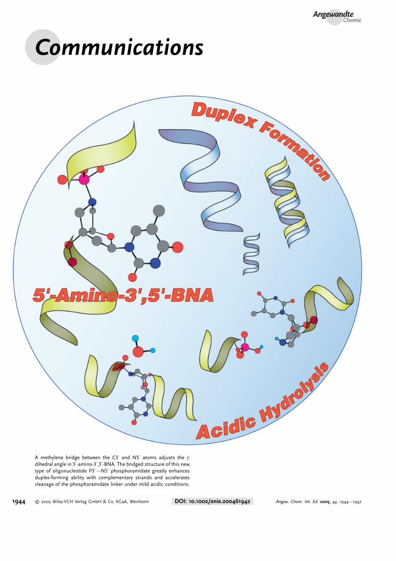
A methylene bridge between the C3’ and N5’ atoms adjusts the g
dihedral angle in 5’-amino-3’,5’-BNA. The bridged structure of this newtype of oligonucleotide P3’!N5’ phosphoramidate greatly enhancesduplex-forming ability with complementary strands and acceleratescleavage of the phosphoramidate linker under mild acidic conditions.
1944 � 2005 Wiley-VCH Verlag GmbH & Co. KGaA, Weinheim DOI: 10.1002/anie.200461942 Angew. Chem. Int. Ed. 2005, 44, 1944 – 1947
Bridged Nucleic Acids
Adjustment of the g Dihedral Angle of anOligonucleotide P3’!N5’ PhosphoramidateEnhances Its Binding Affinity towardsComplementary Strands**
Satoshi Obika, Mitsuaki Sekiguchi, Roongjang Somjing,and Takeshi Imanishi*
Chemical modification of oligodeoxynucleotides (ODNs) hasbeen receiving increasing attention in the fields of genetherapeutics and genetic diagnosis.[1,2] One promisingapproach is an internucleoside linkage modification of theODNs. An N3’!P5’-phosphoramidate-linked ODN, in whichthe 3’-oxygen atom is replaced with a nitrogen atom, forms astable duplex structure with its DNA or RNA complement.[3]
On the other hand, P3’!N5’-phosphoramidate-linked ODNs(5’-amino-DNA, Scheme 1a), with a 5’-nitrogen atom insteadof an oxygen atom, can be hydrolyzed at the phosphorami-date linkage under mild acidic conditions.[4] This property of5’-amino-DNA has attracted much attention and has beenapplied to a DNA-sequence determination.[5, 6] However, the5’-amino-DNA modification of ODNs decreases the hybrid-izing ability with its complementary strand.[7,8] This disad-vantage of 5’-amino-DNA may be caused by an inappropriateg dihedral angle (N5’–C5’–C4’–C3’). 1H NMR analysis of a 5’-amino-DNA dimer revealed that the orientation of the C4’–C5’ bond is predominantly + ap (g� 1808) or �sc (g��608),which is different from that in a typical DNA/DNA or RNA/RNA duplex (+ sc, g� 608).[9]
One promising strategy for restricting the conformationalflexibility of the nucleoside sugar moiety is to increase thebinding affinity of the ODNs. We have developed a series ofnovel nucleic acid analogues bearing a conformationallyrestricted sugar moiety, bridged nucleic acids (BNAs), andhave found that ODNs containing some kinds of BNAacquired extremely high binding affinity for their DNA orRNA complements.[10–12] One such nucleic acid analogue, 5’-amino-2’,4’-BNA (Scheme 1a), in which the sugar puckeringis exactly restricted to the C3’-endo conformation (a typicalN-type conformation), exhibited high binding affinity withcomplementary strands, although this nucleic acid analoguehas a P3’!N5’ phosphoramidate linkage.[12] Thus, the 5’-
amino-2’,4’-BNA may be one example of how to overcomethe drawback of 5’-amino-DNA; however, the effect of the g
dihedral angle of 5’-amino-DNA on its hybridizing propertiesis still unclear.
In this study, we have focused on the adjustment of the g
dihedral angle of 5’-amino-DNA. As DNA derivatives havinga restricted g dihedral angle, bicyclo-DNA[13] and tricyclo-DNA,[14] developed by Leumann et al., are well known. TheseDNA analogues showed interesting duplex- and triplex-forming properties, and the tricyclo-DNA was found to beuseful even as an antisense oligonucleotide.[15] However, the g
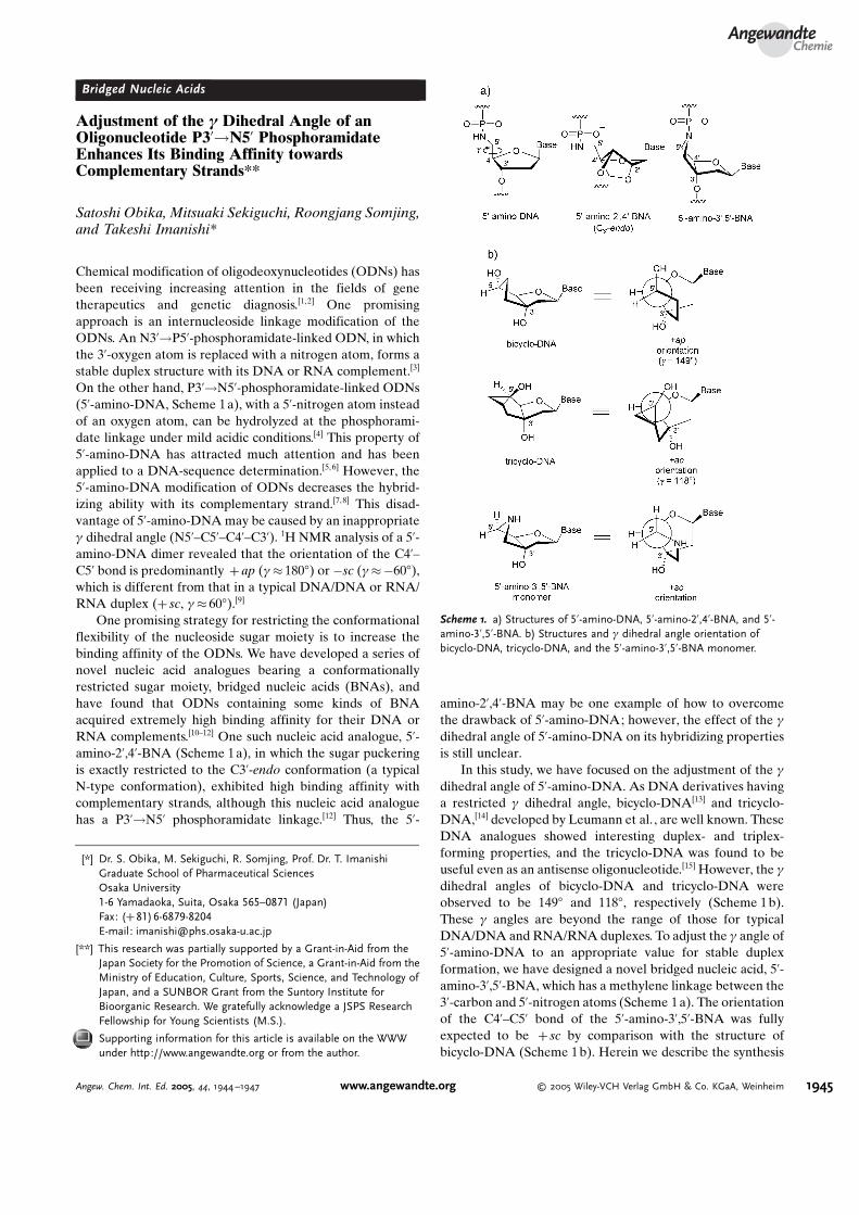
dihedral angles of bicyclo-DNA and tricyclo-DNA wereobserved to be 1498 and 1188, respectively (Scheme 1 b).These g angles are beyond the range of those for typicalDNA/DNA and RNA/RNA duplexes. To adjust the g angle of5’-amino-DNA to an appropriate value for stable duplexformation, we have designed a novel bridged nucleic acid, 5’-amino-3’,5’-BNA, which has a methylene linkage between the3’-carbon and 5’-nitrogen atoms (Scheme 1a). The orientationof the C4’–C5’ bond of the 5’-amino-3’,5’-BNA was fullyexpected to be + sc by comparison with the structure ofbicyclo-DNA (Scheme 1b). Herein we describe the synthesis
Scheme 1. a) Structures of 5’-amino-DNA, 5’-amino-2’,4’-BNA, and 5’-amino-3’,5’-BNA. b) Structures and g dihedral angle orientation ofbicyclo-DNA, tricyclo-DNA, and the 5’-amino-3’,5’-BNA monomer.
[*] Dr. S. Obika, M. Sekiguchi, R. Somjing, Prof. Dr. T. ImanishiGraduate School of Pharmaceutical SciencesOsaka University1-6 Yamadaoka, Suita, Osaka 565–0871 (Japan)Fax: (+ 81)6-6879-8204E-mail: [email protected]
[**] This research was partially supported by a Grant-in-Aid from theJapan Society for the Promotion of Science, a Grant-in-Aid from theMinistry of Education, Culture, Sports, Science, and Technology ofJapan, and a SUNBOR Grant from the Suntory Institute forBioorganic Research. We gratefully acknowledge a JSPS ResearchFellowship for Young Scientists (M.S.).
Supporting information for this article is available on the WWWunder http://www.angewandte.org or from the author.
AngewandteChemie
1945Angew. Chem. Int. Ed. 2005, 44, 1944 –1947 www.angewandte.org � 2005 Wiley-VCH Verlag GmbH & Co. KGaA, Weinheim
and conformation of the 5’-amino-3’,5’-BNA monomer andsome interesting properties of its ODN derivatives.
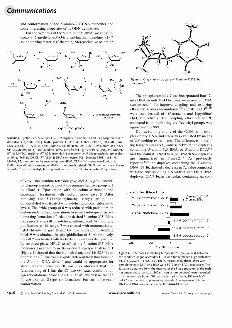
For the synthesis of the 5’-amino-3’,5’-BNA, we chose 3’-deoxy-3’-C-methylene-5’-O-triphenylmethylthymidine (2)[16]
as the starting material (Scheme 2). Stereoselective oxidation
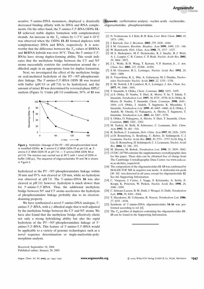
of 2 by using osmium tetroxide gave diol 3. A p-toluenesul-fonyl group was introduced at the primary hydroxy group of 3to afford 4. Epoxidation with potassium carbonate andsubsequent treatment with sodium azide gave 5. Afterremoving the 5’-O-triphenylmethyl (trityl) group, theobtained diol was treated with p-toluenesulfonyl chloride togive 6. The azide group of 6 was reduced with palladium oncarbon under a hydrogen atmosphere and subsequent pyrro-lidine ring formation afforded the desired 5’-amino-3’,5’-BNAmonomer 7 as a salt of p-toluenesulfonic acid. Without anypurification at this stage, 7 was treated with monomethoxy-trityl chloride to give 8, and the phosphoramidite buildingblock 9 was obtained by phosphitilation of 8. Alternatively,the salt 7 was treated with triethylamine and was then purifiedby reversed-phase HPLC to afford the 5’-amino-3’,5’-BNAmonomer 1 in a free form. X-ray crystallograpic analysis of 1(Figure 1) showed that the g dihedral angle of 1 is 28.38 (+ scorientation).[17] This value is quite different from that found inthe 5’-amino-DNA dimer[9] and would be appropriate forstable duplex formation. It was also observed that thefuranose ring of 1 has the C1’-exo-O4’-endo conformation(pseudorotational phase angle P = 115.48), which is neither anN-type nor an S-type conformation, but an in-betweenconformation.
The phosphoramidite 9 was incorporated into 12-mer DNA strands 11–13 by using an automated DNAsynthesizer.[18] To improve coupling and oxidizingefficiency, 4,5-dicyanoimidazole[19] and tBuOOH[20,21]
were used instead of 1H-tetrazole and I2/pyridine/H2O, respectively. The coupling efficiency for 9,estimated from monitoring the free trityl groups, wasapproximately 90%.
Duplex-forming ability of the ODNs with com-plementary DNA and RNA was evaluated by meansof UV melting experiments. The differences in melt-ing temperatures (DTm values) between the duplexescontaining 5’-amino-3’,5’-BNA or 5’-amino-DNA[22]
and the natural DNA/DNA or DNA/RNA duplexesare summarized in Figure 2.[23] As previouslyreported,[7,8] the duplexes comprising the 5’-amino-DNA, 14–16, showed a decrease in Tm value comparedwith the corresponding DNA/DNA and DNA/RNAduplexes. ODN 16, in particular, containing six con-
Scheme 2. Synthesis of 5’-amino-3’,5’-BNA-thymine monomer 1 and its phosphoramiditederivative 9. a) OsO4 (cat.), NMO, pyridine, H2O, tBuOH, 76 8C, 69%; b) TsCl, nBu2SnO,Et3N, CH2Cl2, RT, 72 %; c) K2CO3, MeOH, RT; d) NaN3, DMF, 90 8C, 98% from 4 ; e) CSA,CH2Cl2/MeOH, RT; f) TsCl, pyridine, 50 8C, 53% from 5 ; g) 10% Pd/C (wet), H2, MeOH,RT; h) MMTrCl, pyridine, RT, 69% from 6 ; i) 2-cyanoethyl N,N-diisopropylchlorophosphor-amidite, iPr2NEt, CH2Cl2, RT, 86%; j) DNA synthesizer (ABI Expedite 8909); k) Et3N,MeOH, RT, then purified by reversed-phase HPLC. CSA = (+ )-camphorsulfonic acid,DMF= N,N-dimethylformamide, MMTr = monomethoxytrityl, NMO=4-methylmorpholineN-oxide, Thy = thymin-1-yl, Tr = triphenylmethyl = trityl, Ts = toluene-4-sulfonyl= tosyl.
Figure 1. X-ray crystal structure of 5’-amino-3’,5’-BNAmonomer 1.
Figure 2. Differences in melting temperatures (DTm values) betweenthe modified oligonucleotides 11–16 and the reference oligonucleotide10, 5’-d(GCGTTTTTTGCT)-3’. The Tm values of duplexes of 10 withcomplementary DNA and RNA were 50 8C and 45 8C, respectively. TheTm values obtained from the maxima of the first derivatives of the melt-ing curves (absorbance at 260 nm versus temperature) were recordedin a medium salt buffer (10 mm sodium phosphate, 100 mm NaCl,pH 7.2) with 4 mm complementary strands. The sequence of targetDNA and RNA complement is 5’-AGCAAAAAACGC-3’.
Communications
1946 � 2005 Wiley-VCH Verlag GmbH & Co. KGaA, Weinheim www.angewandte.org Angew. Chem. Int. Ed. 2005, 44, 1944 –1947
secutive 5’-amino-DNA monomers, displayed a drasticallydecreased binding affinity with its DNA and RNA comple-ments. On the other hand, the 5’-amino-3’,5’-BNA ODNs 11–13 achieved stable duplex formation with complementarystrands. An increase in the Tm values by 1–3 8C and 4–10 8Cwas observed when the ODNs 11–13 formed duplexes withcomplementary DNA and RNA, respectively. It is note-worthy that the difference between the Tm values of 13/RNAand 16/RNA hybrids was over 30 8C. Thus, the 5’-amino-3’,5’-BNAs have strong duplex-forming ability. This result indi-cates that the methylene bridge between the C3’ and N5’atoms successfully restricts the conformation around the g
dihedral angle in an appropriate form for duplex formation.Next, we investigated the effect of the methylene bridge
on acid-mediated hydrolysis of the P3’!N5’-phosphorami-date linkage. The 5’-amino-3’,5’-BNA ODN 11 was treatedwith buffer (pH 3.0 or pH 7.0) to be hydrolyzed, and theamount of intact 11 was determined by reversed-phase HPLCanalysis (Figure 3). Under pH 3.0 conditions, 50 % of 11 was
hydrolyzed at the P3’!N5’-phosphoramidate linkage within30 min and 95% was cleaved at 120 min, while no hydrolysiswas observed at pH 7.0. The 5’-amino-DNA 16 was alsocleaved at pH 3.0; however, hydrolysis is much slower thanfor 5’-amino-3’,5’-BNA. Thus, the additional methylenebridge between N5’ and C3’ atoms accelerates the hydrolysisof phosphoramidate linkage probably due to its electron-donating property.
We have synthesized a novel 5’-amino-DNA analogue, 5’-amino-3’,5’-BNA, with a g dihedral angle that is well adjustedby the methylene bridge between the C3’ and N5’ atoms. Wehave also found that the methylene bridge effectively elicitsnot only a strong hybridizing ability but also the rapidhydrolysis of the P3’!N5’-phosphoramidate linkage of 5’-amino-3’,5’-BNA. This feature of 5’-amino-3’,5’-BNA wouldbe applicable to a variety of genome technologies, such as anovel sequence determination or single-nucleotide-poly-morphism analysis.
Received: September 10, 2004Published online: January 28, 2005
.Keywords: conformation analysis · nucleic acids · nucleosides ·oligonucleotides · phosphoramidates
[1] N. Venkatesan, S. J. Kim, B. H. Kim, Curr. Med. Chem. 2003, 10,1973 – 1991.
[2] J. Kurreck, Eur. J. Biochem. 2003, 270, 1628 – 1644.[3] S. M. Gryaznov, Biochim. Biophys. Acta 1999, 1489, 131 – 140.[4] W. Bannwarth, Helv. Chim. Acta 1988, 71, 1517 – 1527.[5] M. S. Shchepinov, M. F. Denissenko, K. J. Smylie, R. J. W�rl,
A. L. Leppin, C. R. Cantor, C. P. Rodi, Nucleic Acids Res. 2001,29, 3864 – 3872.
[6] J. L. Wolfe, B. H. Wang, T. Kawate, V. P. Stanton, Jr., J. Am.Chem. Soc. 2003, 125, 10500 – 10 501.
[7] S. M. Gryaznov, R. L. Letsinger, Nucleic Acids Res. 1992, 20,3403 – 3409.
[8] E. Viazovkina, K.-L. Min, A. Galarneau, M. J. Damha, Nucleo-sides Nucleotides Nucleic Acids 2003, 22, 1335 – 1338.
[9] E. M. Nottoli, J. B. Lambert, R. L. Letsinger, J. Am. Chem. Soc.1977, 99, 3486 – 3491.
[10] T. Imanishi, S. Obika, Chem. Commun. 2002, 1653 – 1659.[11] a) S. Obika, D. Nanbu, Y. Hari, K. Morio, Y. In, T. Ishida, T.
Imanishi, Tetrahedron Lett. 1997, 38, 8735 – 8738; b) S. Obika, K.Morio, D. Nanbu, T. Imanishi, Chem. Commun. 1998, 1643 –1644; c) S. Obika, J. Andoh, T. Sugimoto, K. Miyashita, T.Imanishi, Tetrahedron Lett. 1999, 40, 6465 – 6468; d) S. Obika, J.Andoh, M. Onoda, O. Nakagawa, A. Hiroto, T. Sugimoto, T.Imanishi, Tetrahedron Lett. 2003, 44, 5267 – 5270.
[12] S. Obika, O. Nakagawa, A. Hiroto, Y. Hari, T. Imanishi, Chem.Commun. 2003, 2202 – 2203.
[13] M. Tark�y, M. Bolli, B. Schweizer, C. Leumann, Helv. Chim.Acta 1993, 76, 481 – 510.
[14] R. Steffens, C. Leumann, Helv. Chim. Acta 1997, 80, 2426 – 2439.[15] a) D. Renneberg, E. Bouliong, U. Reber, D. Sch�mperli, C. J.
Leumann, Nucleic Acids Res. 2002, 30, 2751 – 2757; b) D. Ittig, S.Liu, D. Renneberg, D. Sch�mperli, C. J. Leumann, Nucleic AcidsRes. 2004, 32, 346 – 353.
[16] M. Sharma, M. Bobek, Tetrahedron Lett. 1990, 31, 5839 – 5842.[17] CCDC-247780 contains the supplementary crystallographic data
for this paper. These data can be obtained free of charge fromThe Cambridge Crystallographic Data Centre via www.ccdc.ca-m.ac.uk/data_request/cif.
[18] The composition of the oligonucleotides 11–13 was confirmed byMALDI-TOF MS in negative-ion mode. A molecular-ion peak[M�H]� was detected in all cases, except for oligonucleotide 12.See the Supporting Information.
[19] C. Vargeese, J. Carter, J. Yegge, S. Krivjansky, A. Settle, E.Kropp, K. Peterson, W. Pieken, Nucleic Acids Res. 1998, 26,1046 – 1050.
[20] C. Scheuer-Larsen, B. M. Dahl, J. Wengel, O. Dahl, TetrahedronLett. 1998, 39, 8361 – 8364.
[21] Y. Hayakawa, M. Uchiyama, R. Noyori, Tetrahedron Lett. 1986,27, 4191 – 4194.
[22] Synthesis of 5’-amino-DNA oligonucleotides 14–16 was per-formed according to ref. [4].
[23] The Tm profiles of duplexes containing the oligonucleotides 11–13 can be found in the Supporting Information.
Figure 3. Hydrolytic cleavage of the P3’!N5’-phosphoramidate bondin modified ODNs: &: 5’-amino-3’,5’-BNA ODN 11 at pH 3.0; ~: 5’-amino-3’,5’-BNA ODN 11 at pH 7.0; &: 5’-amino-DNA ODN 14 atpH 3.0. The reaction was carried out at 30 8C with 1 nmol of ODN inbuffer (100 mL). The sequence of oligonucleotides 11 and 14 is shownin Figure 2.
AngewandteChemie
1947Angew. Chem. Int. Ed. 2005, 44, 1944 –1947 www.angewandte.org � 2005 Wiley-VCH Verlag GmbH & Co. KGaA, Weinheim
Top Related