XI School on Synchrotron Radiation “Small Angle X-ray...
90
XI School on Synchrotron Radiation “Small Angle X-ray Scattering” S.A.X.S. STEFANO POLIZZI DIPARTIMENTO SCIENZE MOLECOLARI E NANOSITEMI UNIVERSITÀ CA’ FOSCARI VENEZIA
Transcript of XI School on Synchrotron Radiation “Small Angle X-ray...
XI School on Synchrotron Radiation
“Small Angle X-ray Scattering” S.A.X.S.
STEFANO POLIZZI
DIPARTIMENTO SCIENZE MOLECOLARI E NANOSITEMI
UNIVERSITÀ CA’ FOSCARI VENEZIA
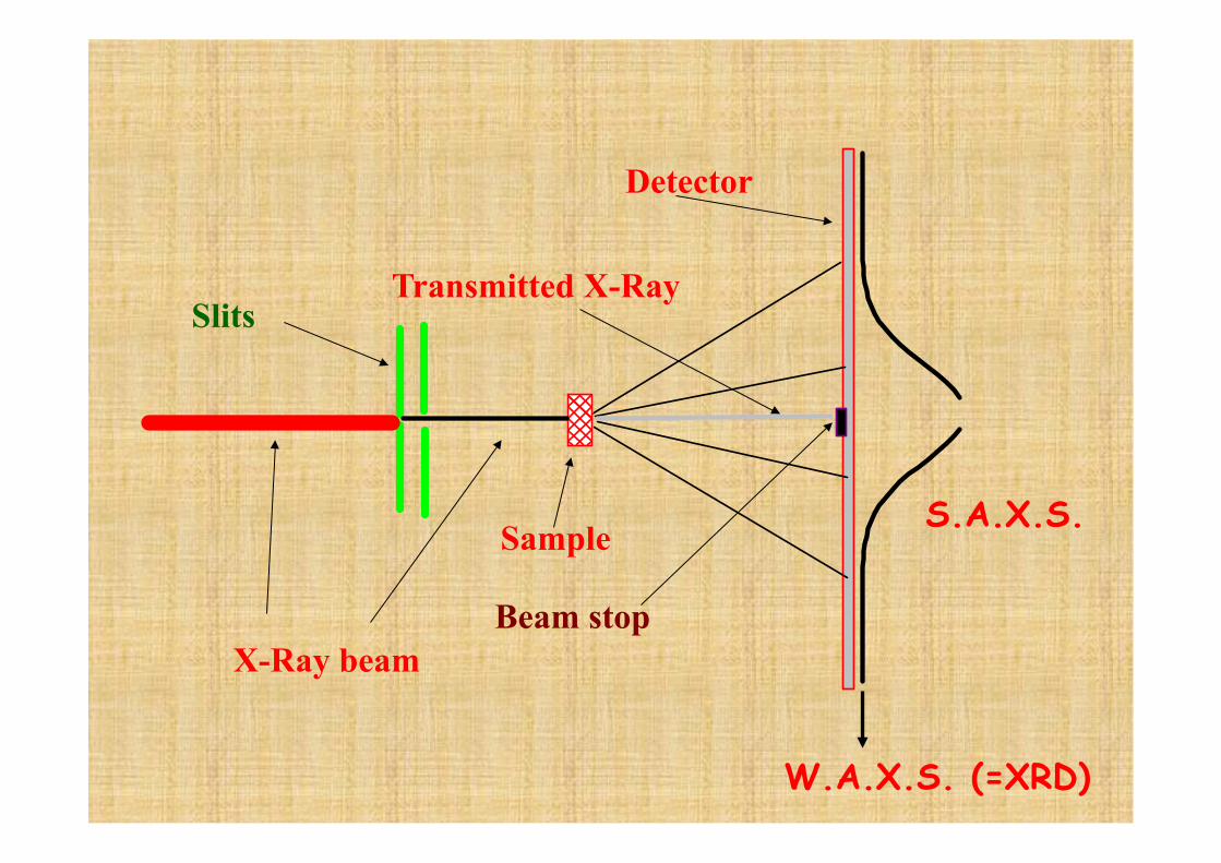
X-Ray beam
Slits
Sample
Transmitted X-Ray
Beam stop
Detector
S.A.X.S.
W.A.X.S. (=XRD)
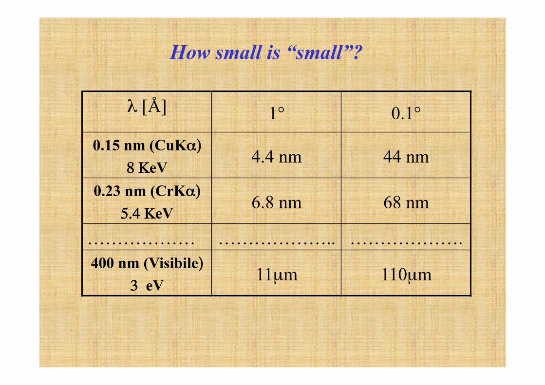
λ [Å] 1° 0.1°
0.15 nm (CuKα)
8 ΚeV 4.4 nm 44 nm
0.23 nm (CrKα)
5.4 ΚeV 6.8 nm 68 nm
……………… ……………….. ………………. 400 nm (Visibile)
3 eV 11µm 110µm
How small is “small”?
So, if one is able to measure scattered intensity below 1° from the incoming direction, one has a way to
investigate a range which spans from the atomic/molecular resolution of XRD to that of an optical
microscope.
Such dimensions are also called colloidal dimensions
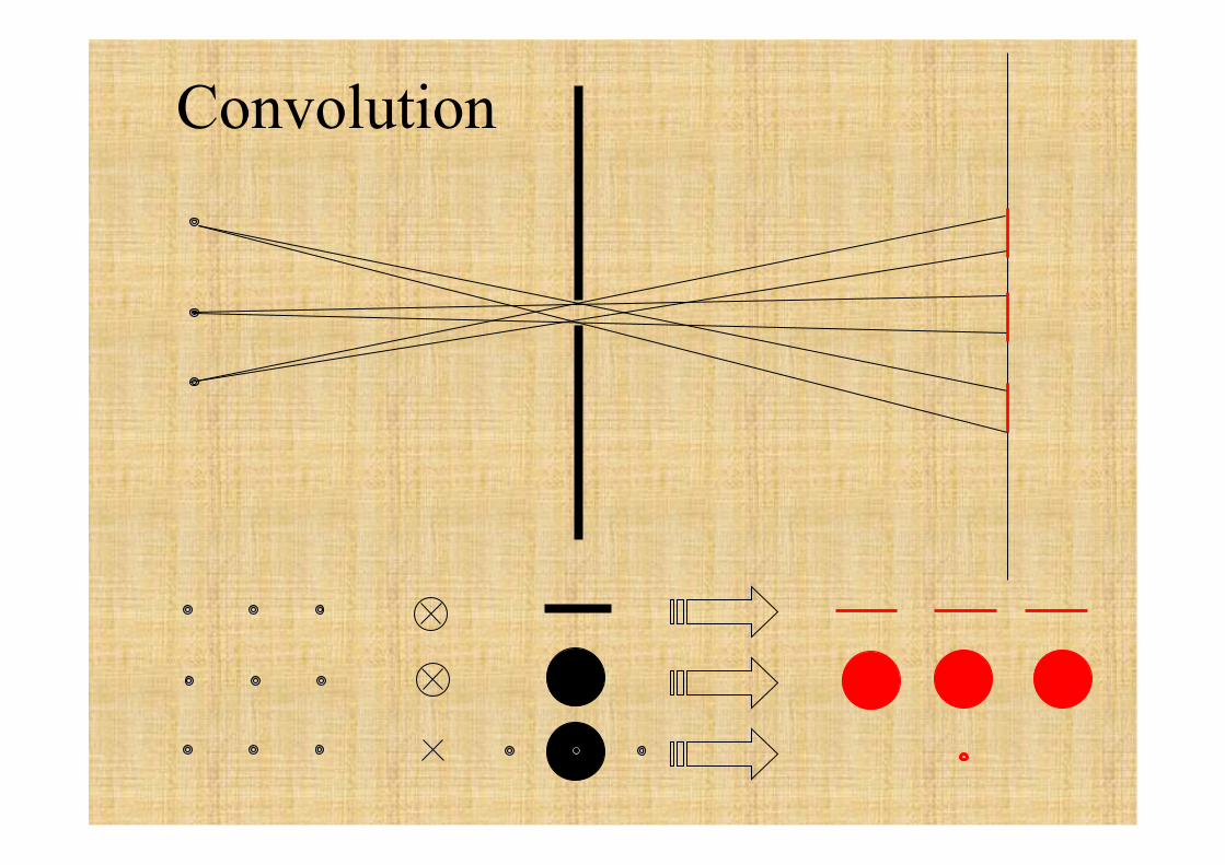
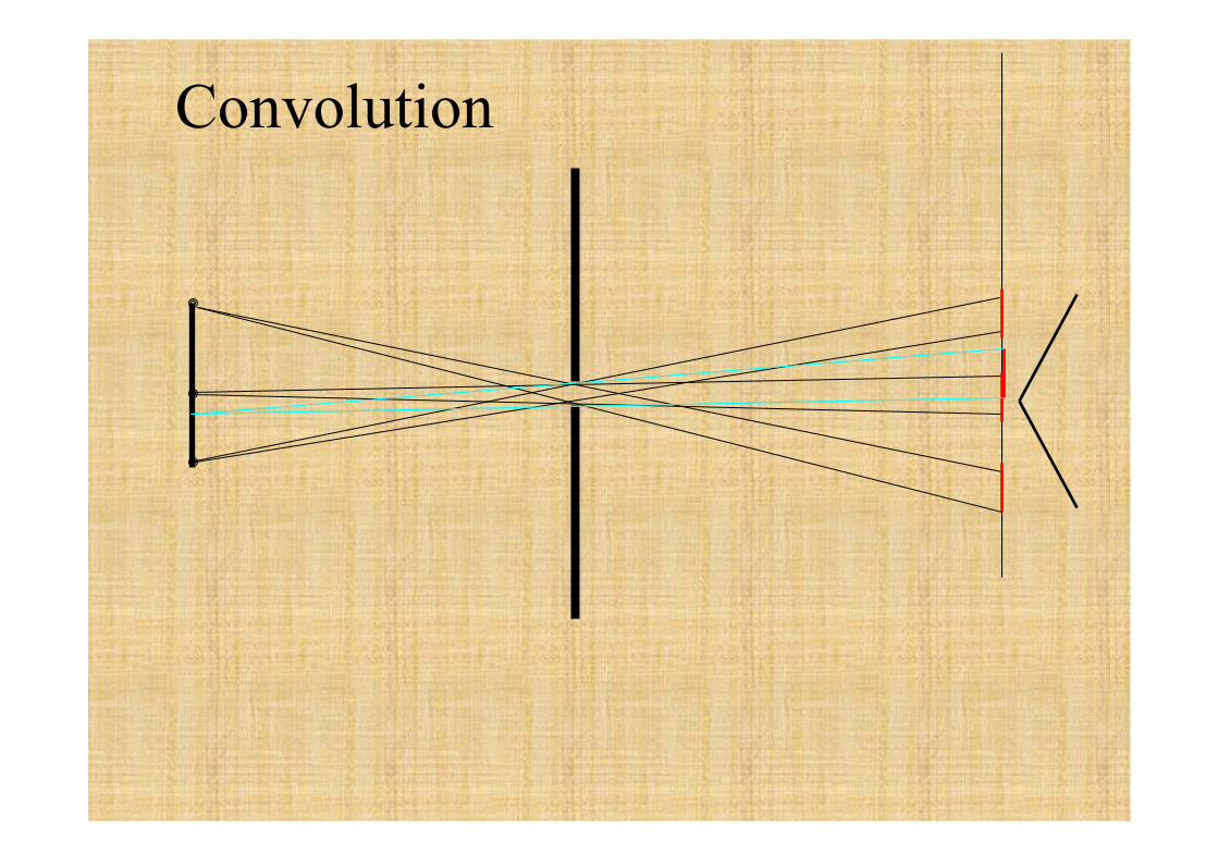
Convolution
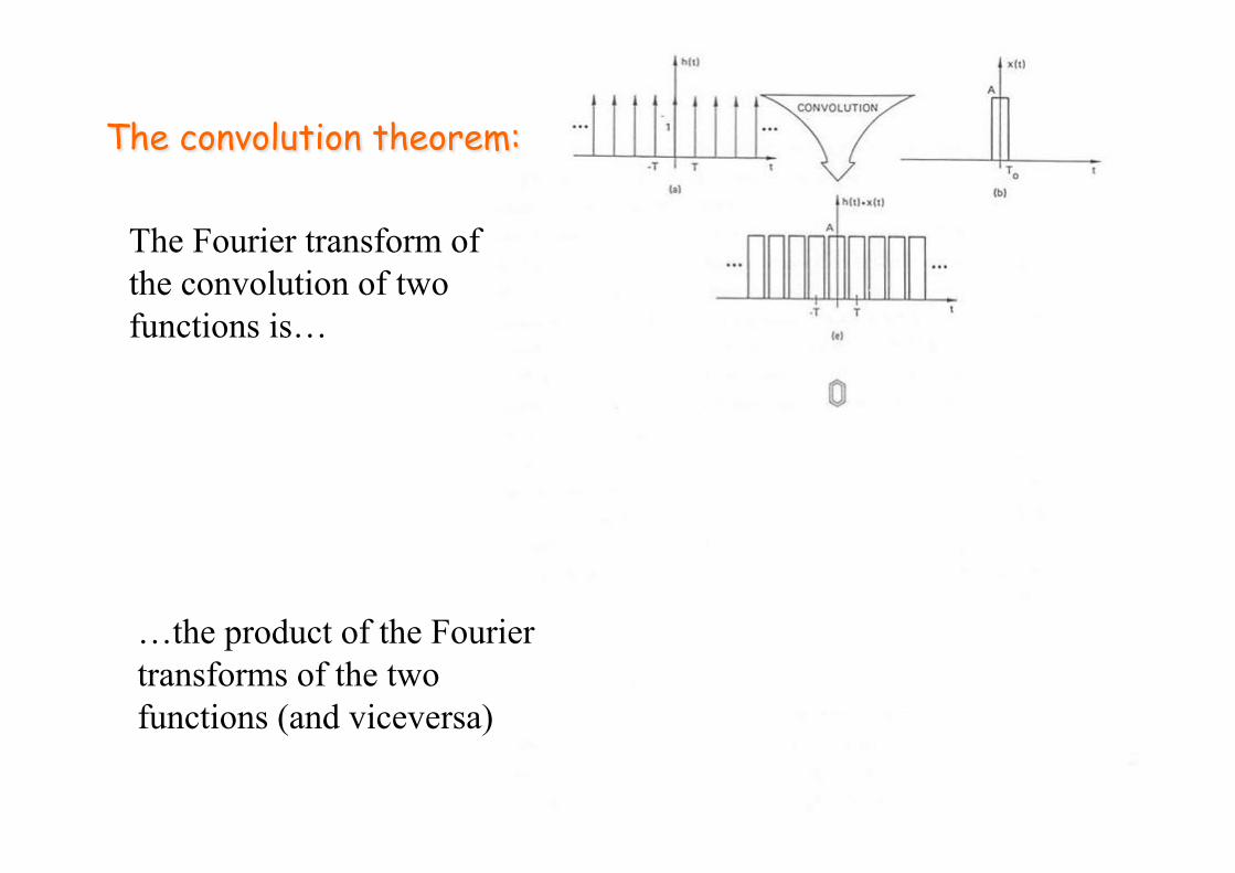
The Fourier transform of the convolution of two functions is…
…the product of the Fourier transforms of the two functions (and viceversa)
?
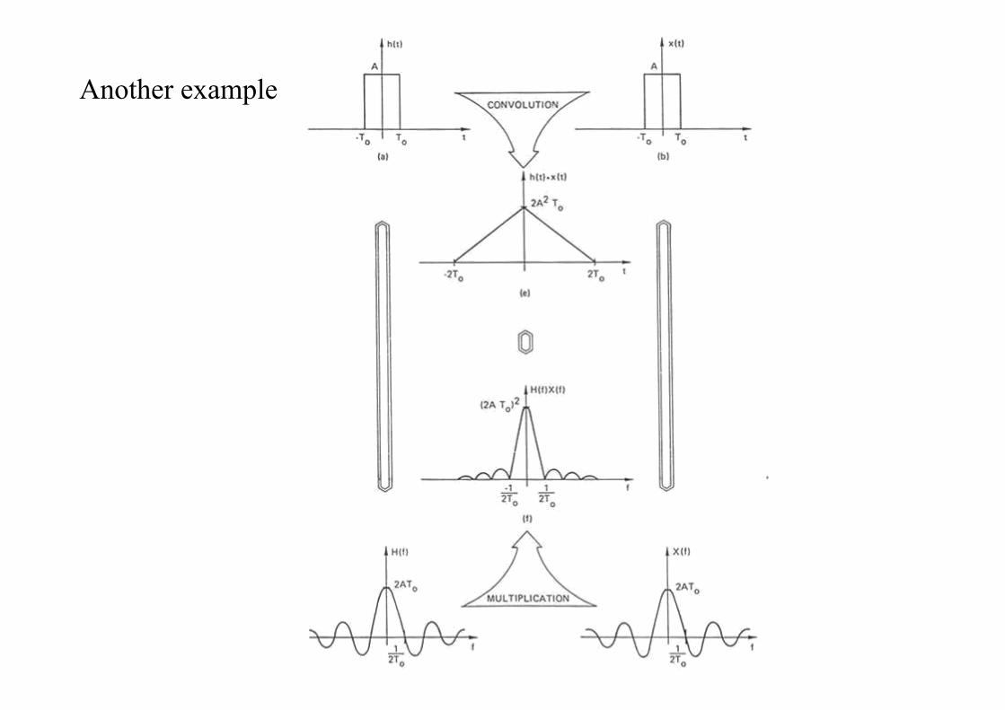
Another example
Convolution
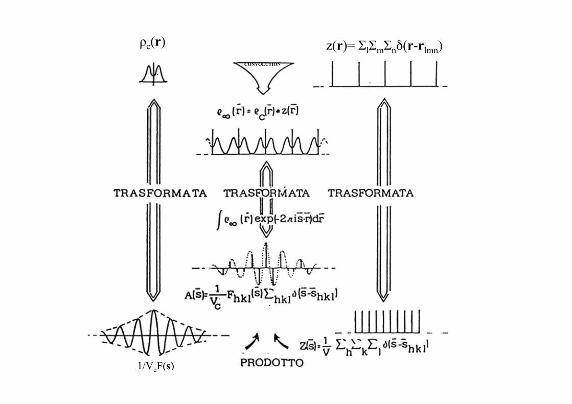
z(r)= ΣlΣmΣnδ(r-rlmn) ρc(r) CONVOLUTION
1/VcF(s)
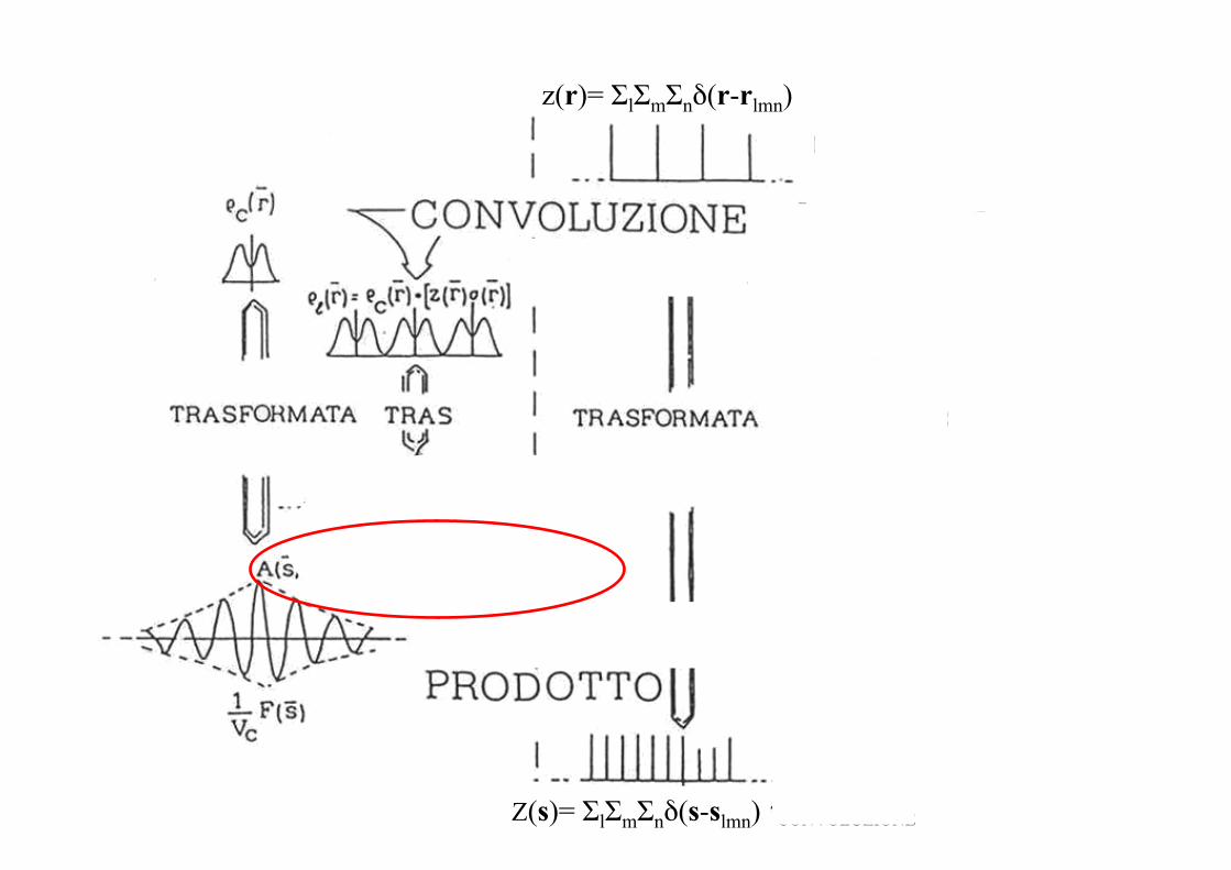
σ(r) 1 for |x|<L 0 for |x|>L z(r)= ΣlΣmΣnδ(r-rlmn)
Σ(s)
Z(s)= ΣlΣmΣnδ(s-slmn) CONVOLUZIONE
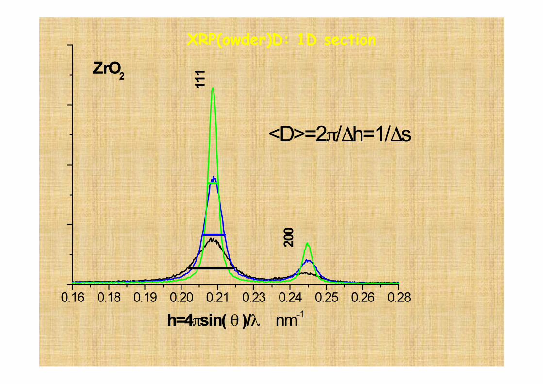
XRP(owder)D: 1D section
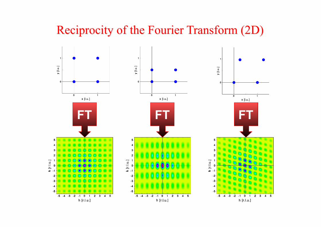
FT FT FT
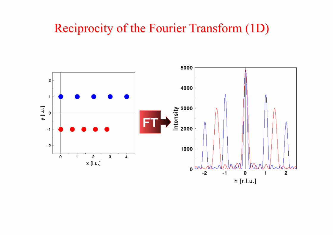
FT
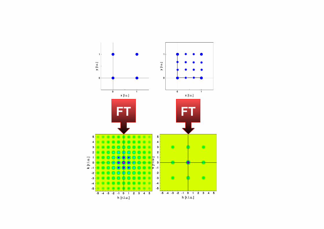
FT FT
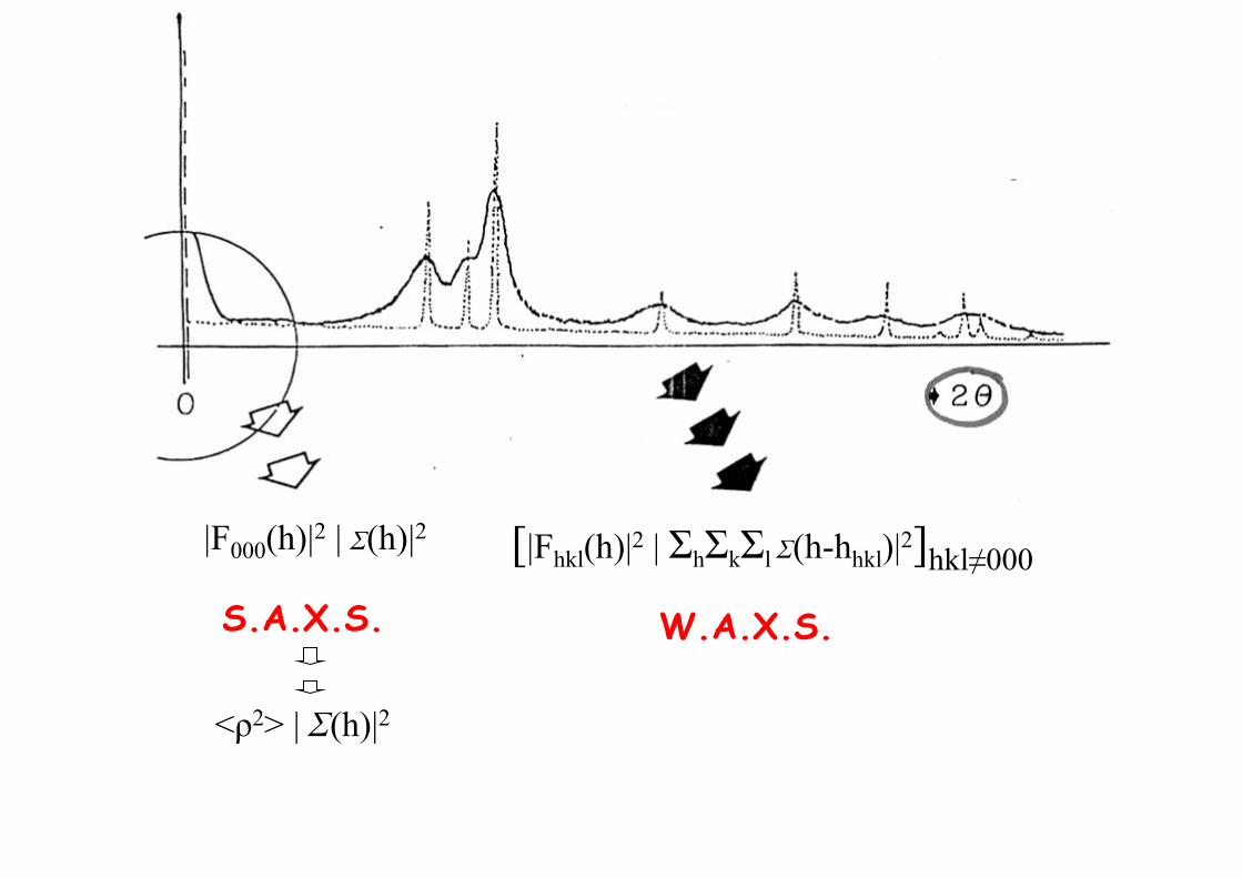
|F000(h)|2 | Σ(h)|2
S.A.X.S. W.A.X.S.
[|Fhkl(h)|2 | ΣhΣkΣl Σ(h-hhkl)|2]hkl≠000
<ρ2> | Σ(h)|2
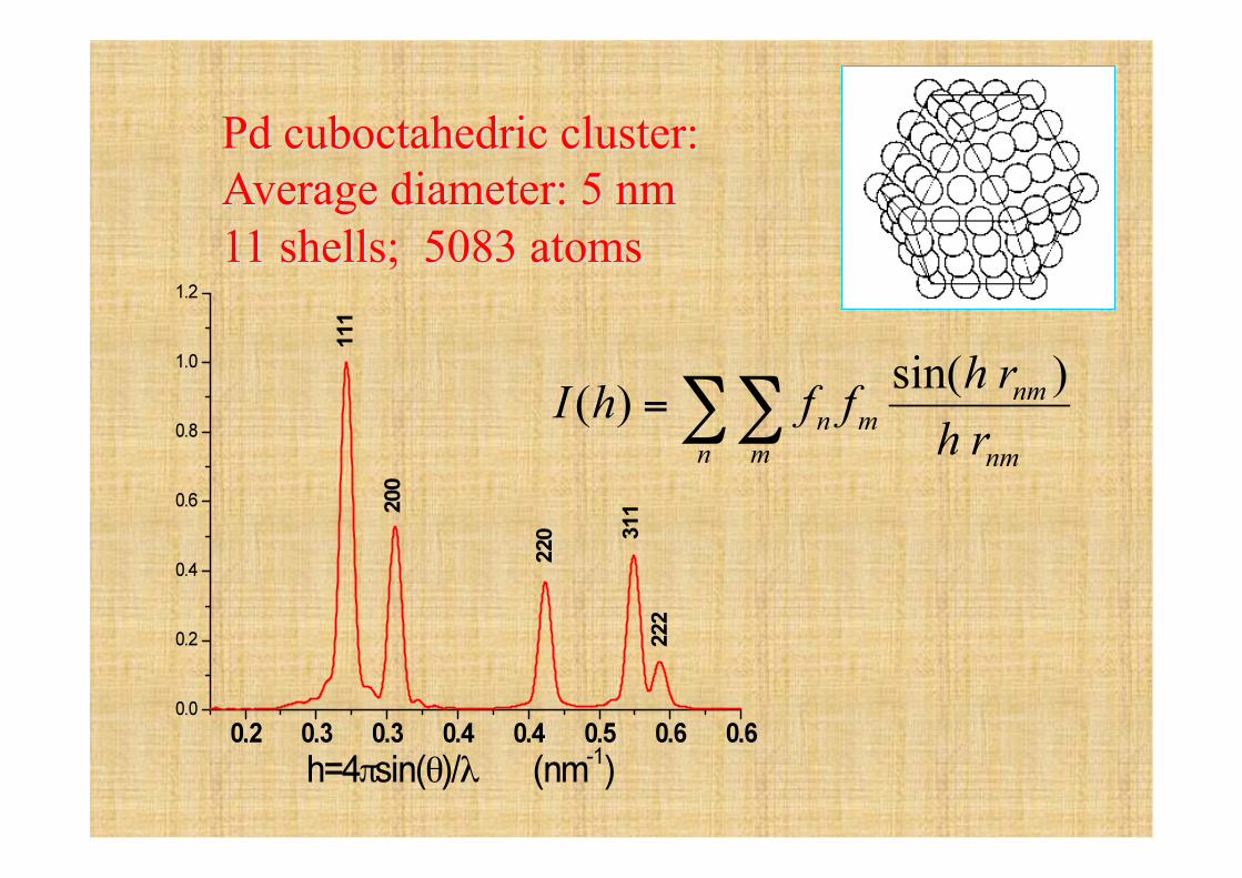
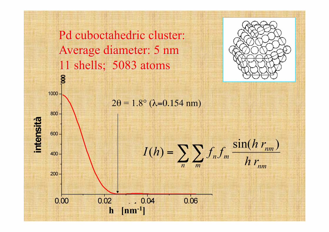
2θ = 1.8° (λ=0.154 nm)
h [nm-1]
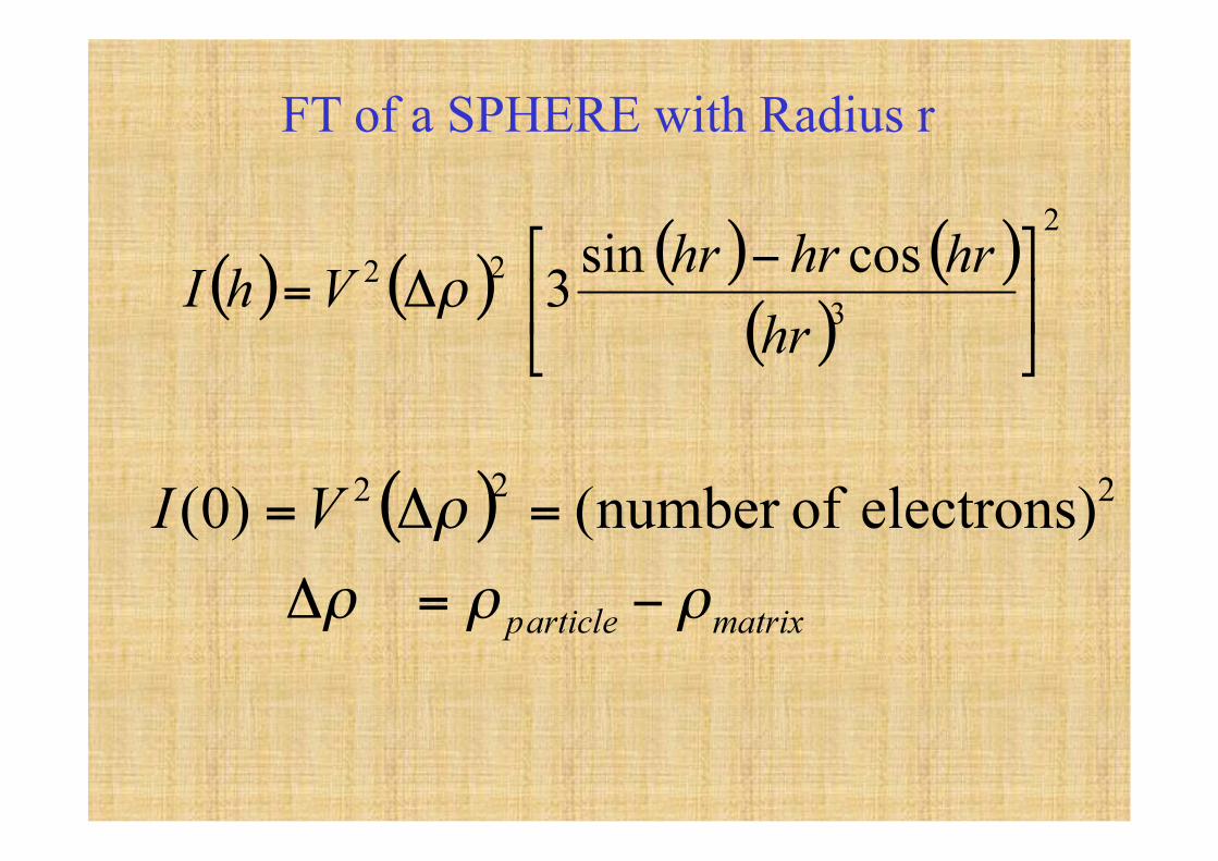
FT of a SPHERE with Radius r
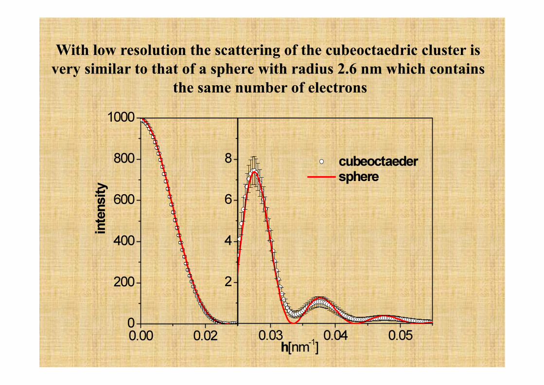
With low resolution the scattering of the cubeoctaedric cluster is very similar to that of a sphere with radius 2.6 nm which contains
the same number of electrons
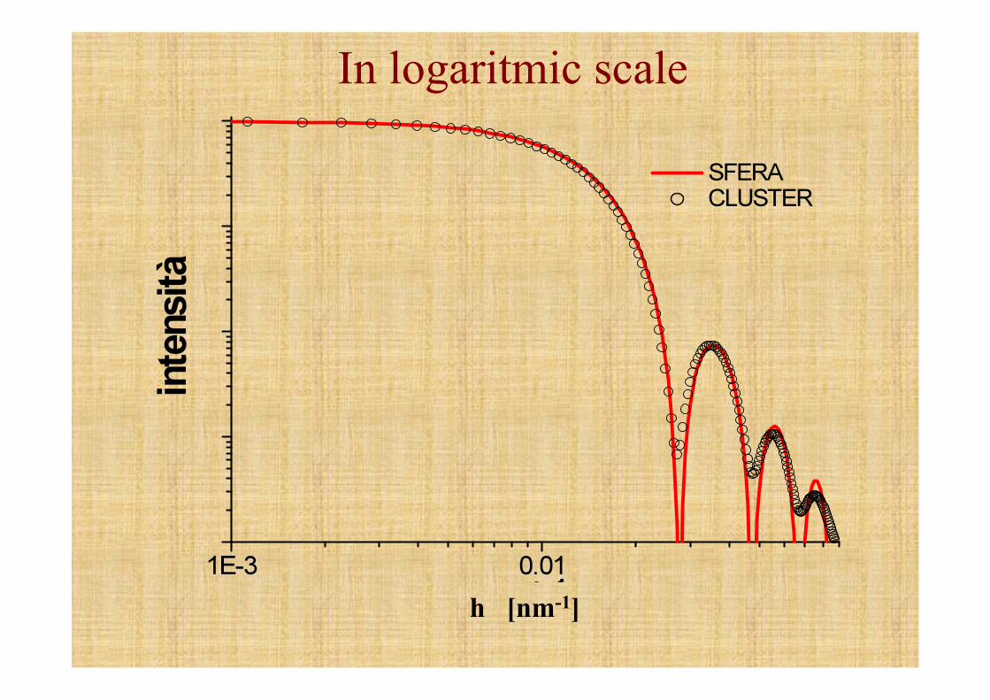
In logaritmic scale
h [nm-1]
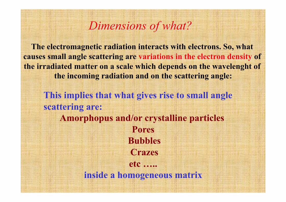
This implies that what gives rise to small angle scattering are:
Amorphopus and/or crystalline particles Pores
Bubbles Crazes etc …..
inside a homogeneous matrix
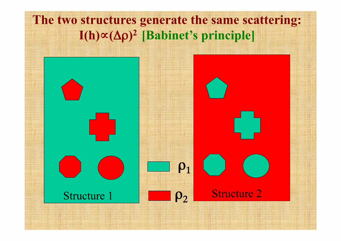
Structure 1 Structure 2
ρ1
ρ2
The two structures generate the same scattering: I(h)∝(Δρ)2 [Babinet’s principle]
h1
h2
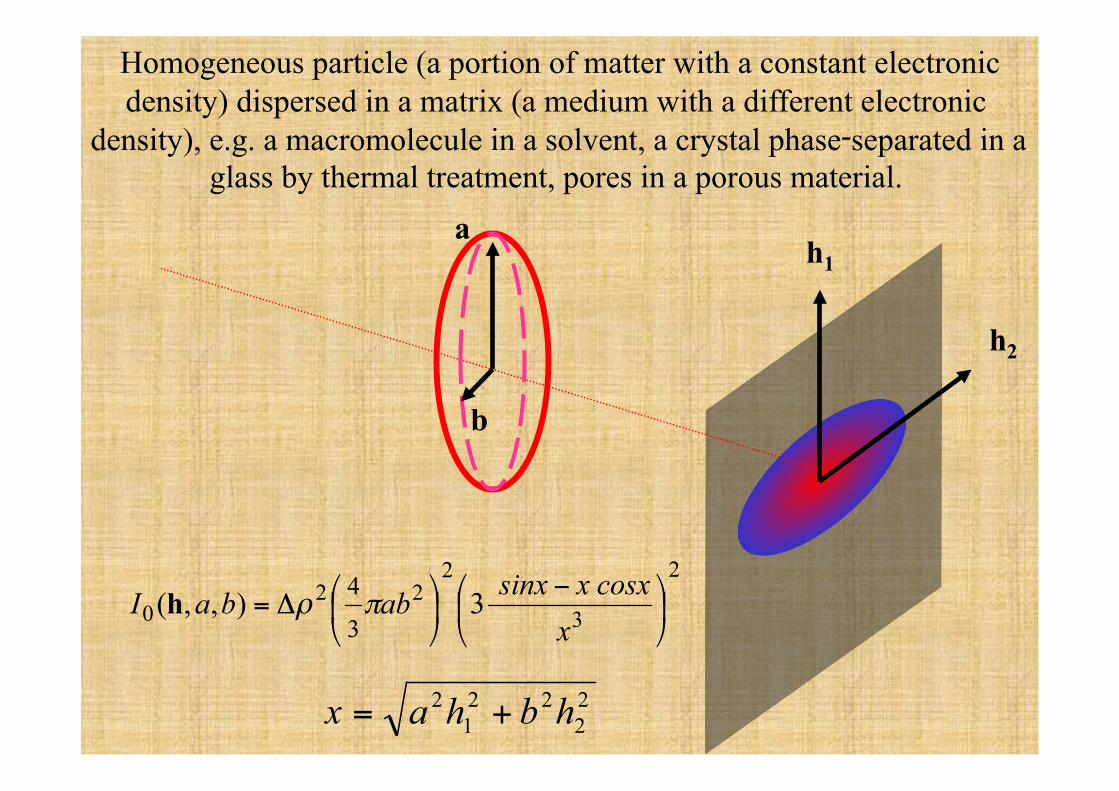
Homogeneous particle (a portion of matter with a constant electronic density) dispersed in a matrix (a medium with a different electronic
density), e.g. a macromolecule in a solvent, a crystal phase‑separated in a glass by thermal treatment, pores in a porous material.
a
b
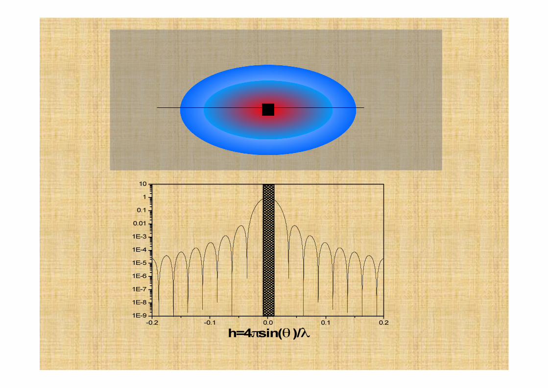
€
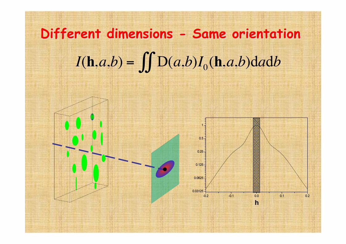
I(h,a,b) = D(a,b)I0(h,a,b)dadb∫∫
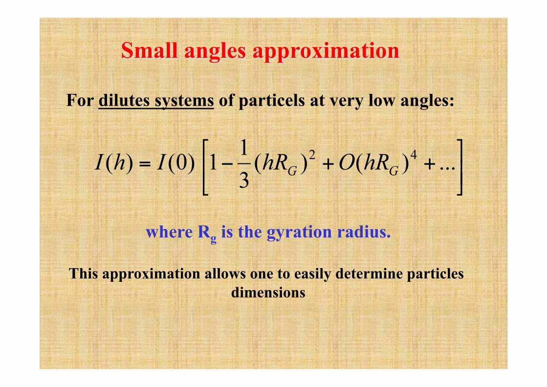
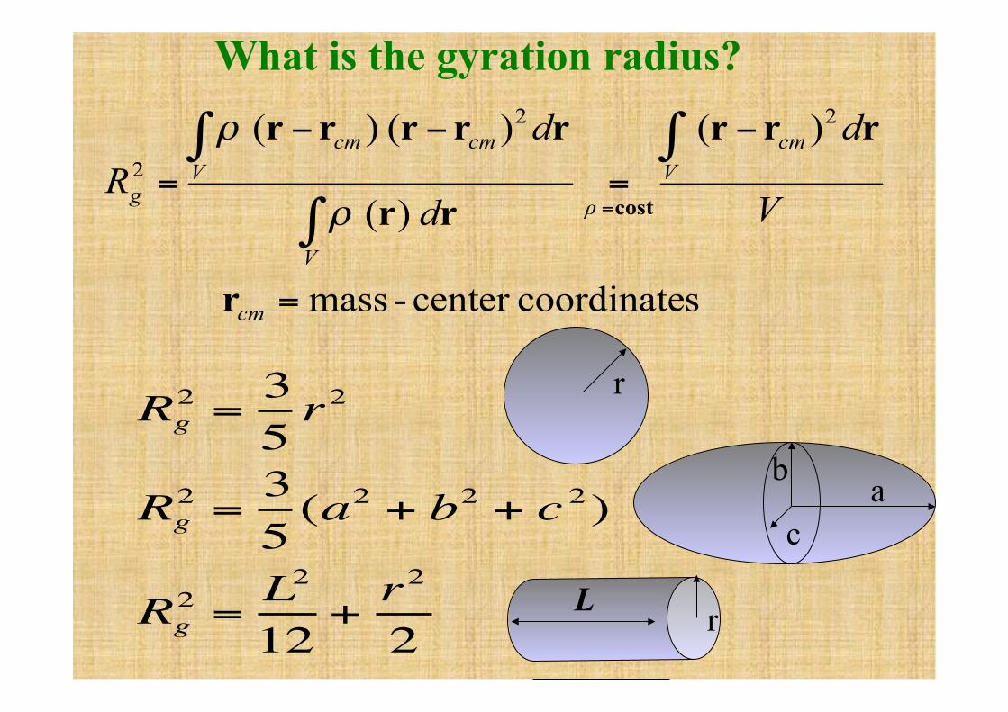
where Rg is the gyration radius.
This approximation allows one to easily determine particles dimensions
€
Rg2 =
35r2
Rg2 =
35
(a2 + b2 + c 2)
Rg2 =
L2
12+r2
2
What is the gyration radius?
r
a b
c
r L
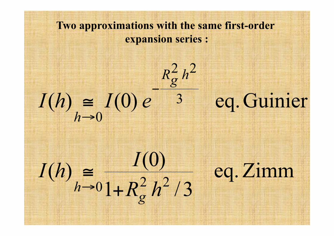
Two approximations with the same first-order expansion series :
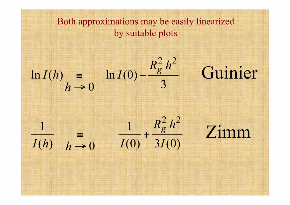
Both approximations may be easily linearized by suitable plots
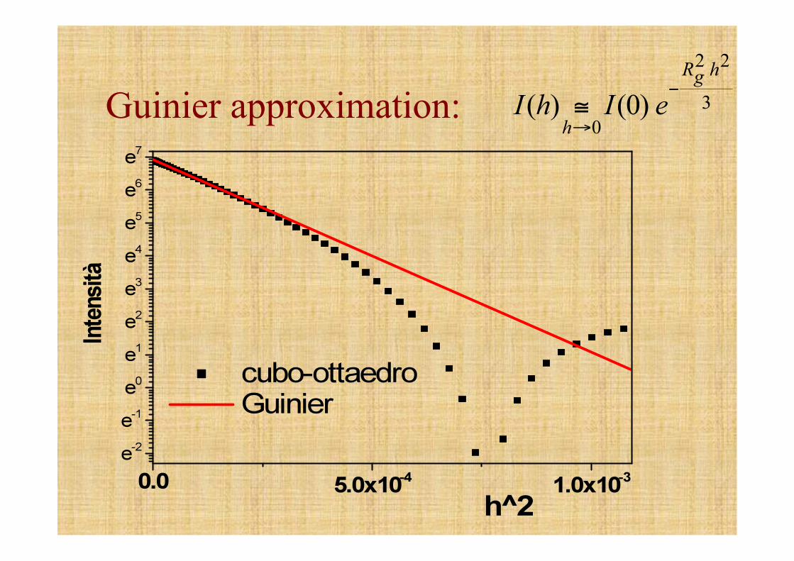
Guinier approximation:
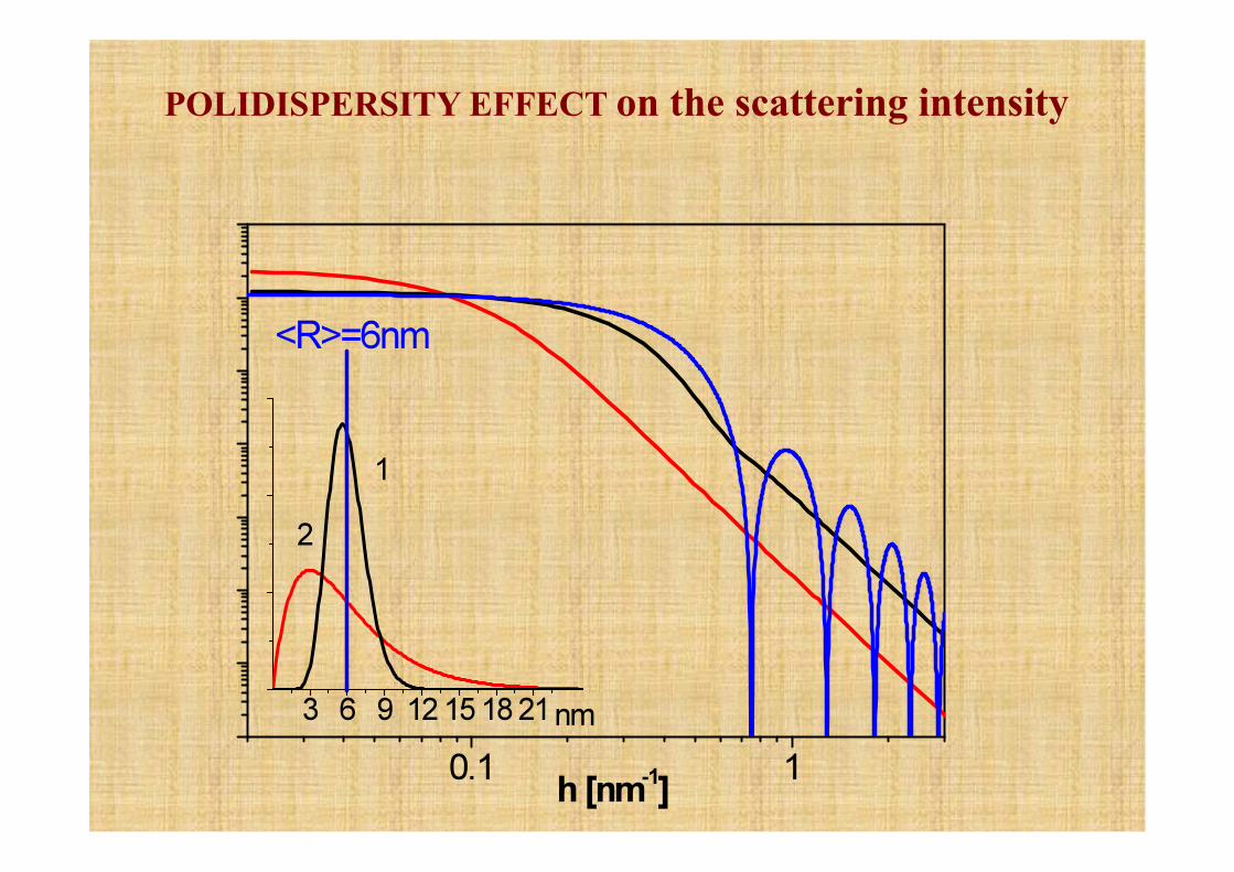
POLIDISPERSITY EFFECT on the scattering intensity
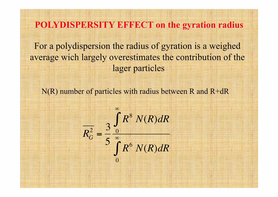
For a polydispersion the radius of gyration is a weighed average wich largely overestimates the contribution of the
lager particles
€
RG2 =
35
R8
0
∞
∫ N(R)dR
R6 N(R)dR0
∞
∫
N(R) number of particles with radius between R and R+dR
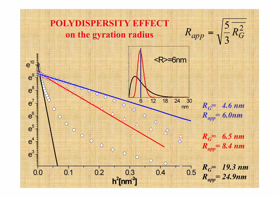
RG= 4.6 nm Rapp= 6.0nm
RG= 6.5 nm Rapp= 8.4 nm
RG= 19.3 nm Rapp= 24.9nm
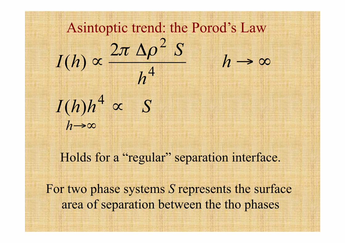
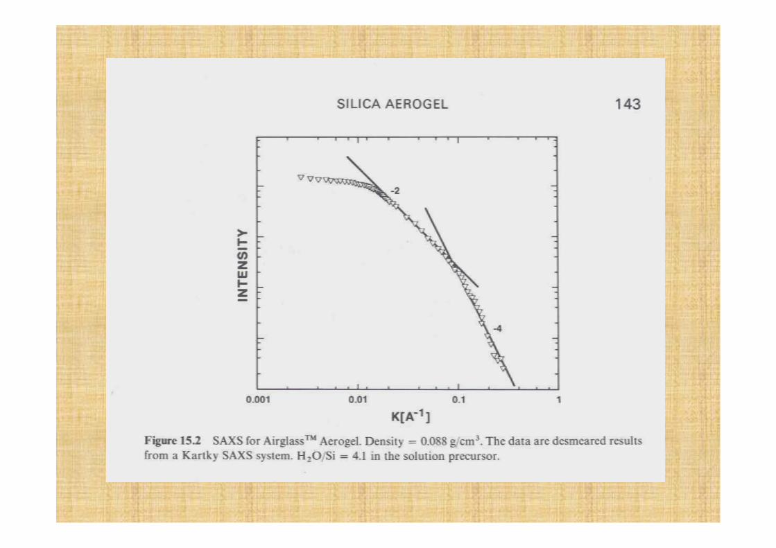
Asintoptic trend: the Porod’s Law
Holds for a “regular” separation interface.
For two phase systems S represents the surface area of separation between the tho phases
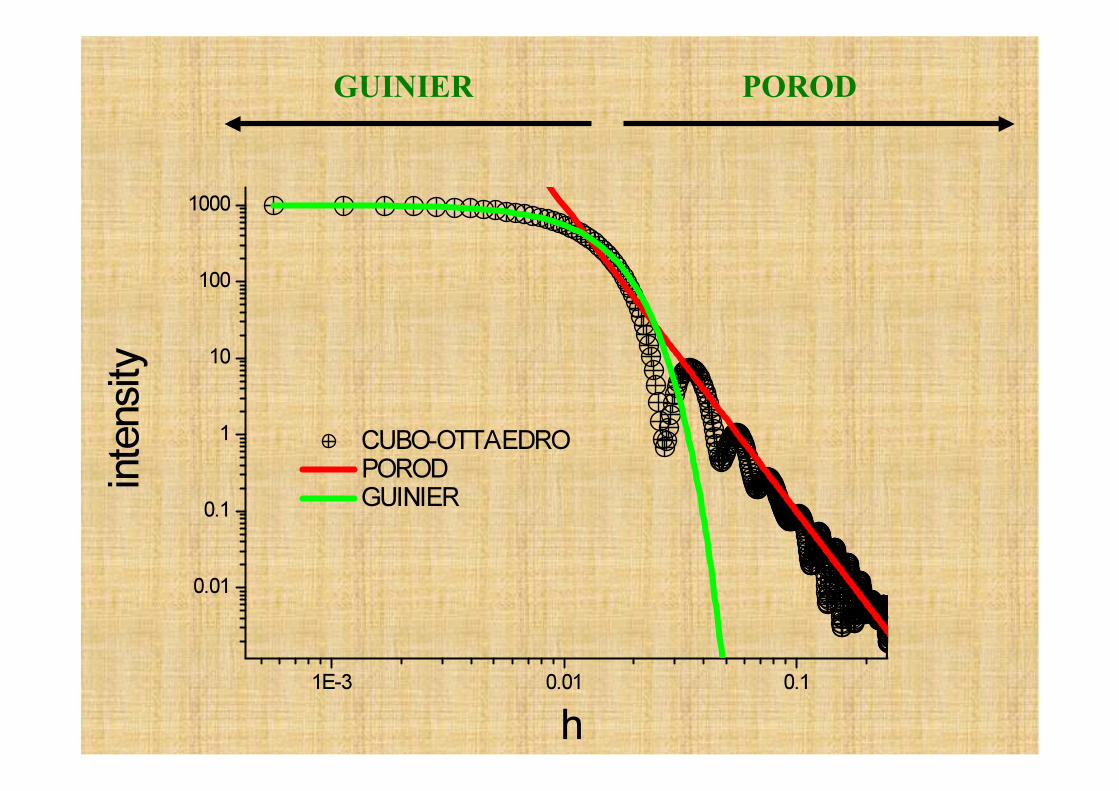
GUINIER POROD
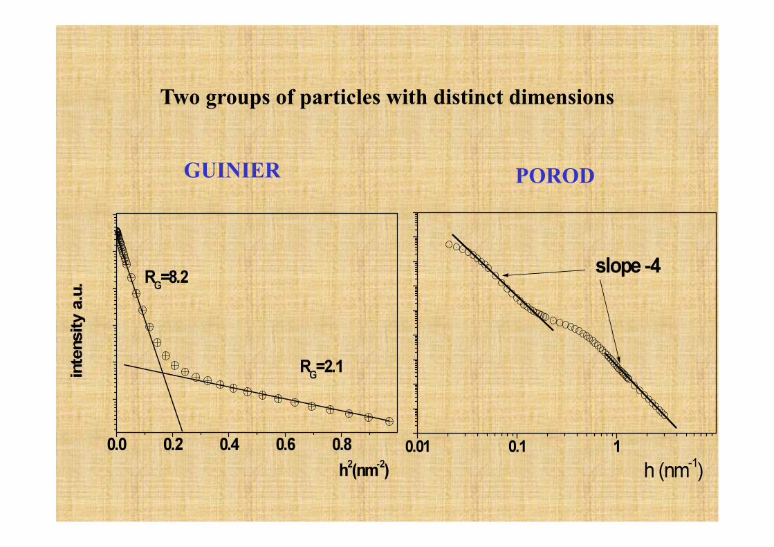
Two groups of particles with distinct dimensions
GUINIER POROD
€
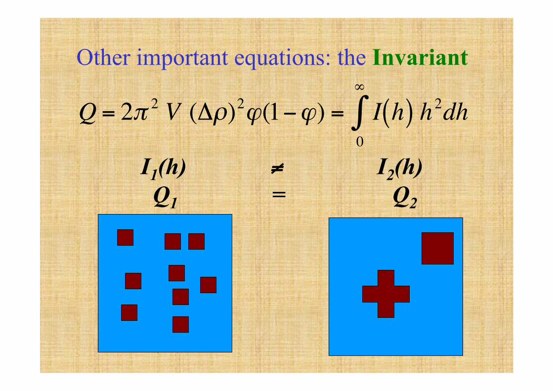
Q = 2π 2 V (Δρ)2ϕ(1−ϕ) = I h( )0
∞
∫ h2dh
Other important equations: the Invariant
I1(h) ≠ I2(h) Q1 = Q2
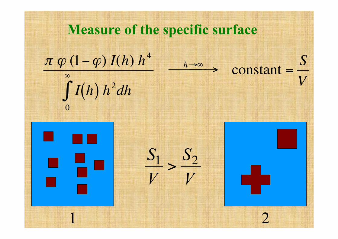
€
π ϕ (1−ϕ) I(h) h4
I h( )0
∞
∫ h2dh
h→∞⎯ → ⎯ ⎯ constant =SV
Measure of the specific surface
1 2
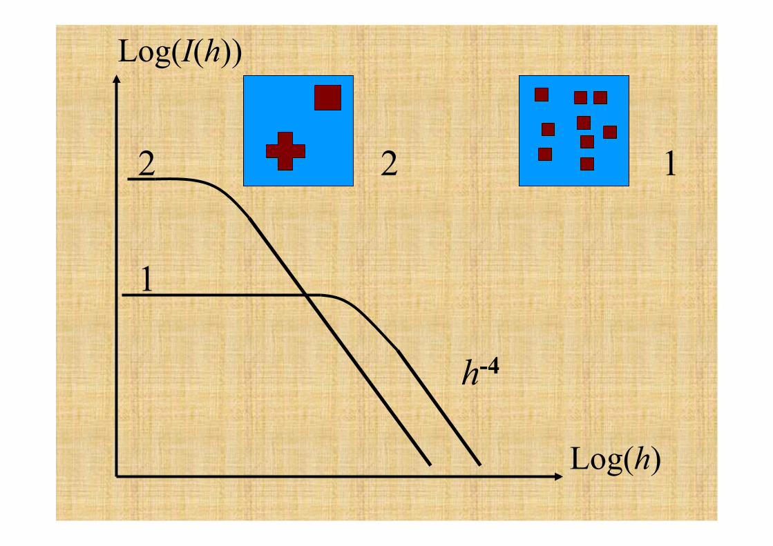
1 2
Log(I(h))
Log(h)
2
1
h-4
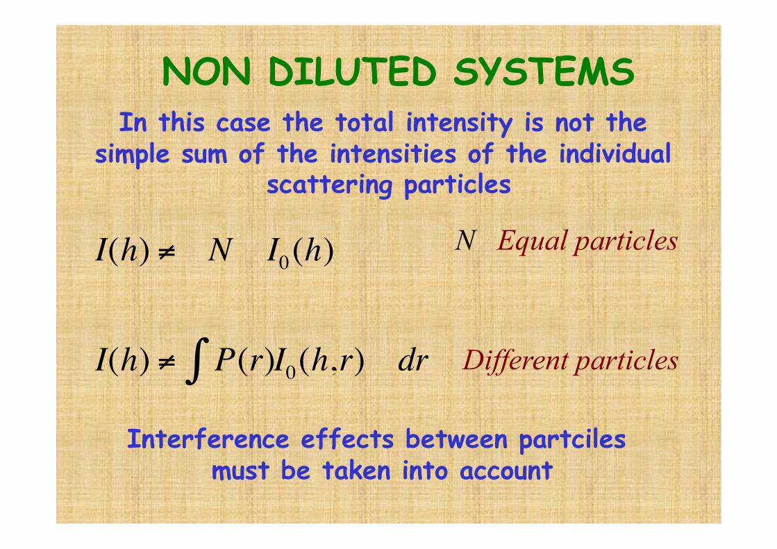
In this case the total intensity is not the simple sum of the intensities of the individual
scattering particles
€
I(h) ≠ N I0(h)
I(h) ≠ P(r)∫ I0(h,r) dr
Interference effects between partciles must be taken into account
N Equal particles
Different particles
NON DILUTED SYSTEMS
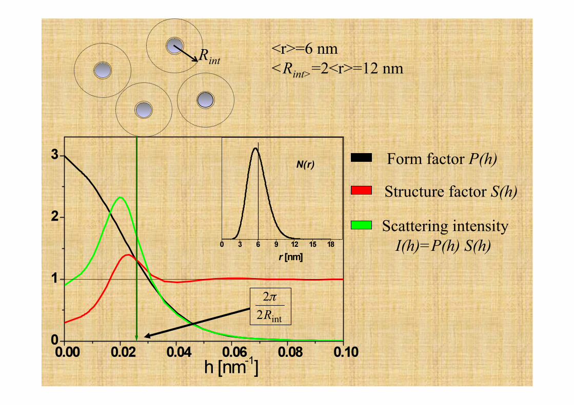
Rint <r>=6 nm <Rint>=2<r>=12 nm
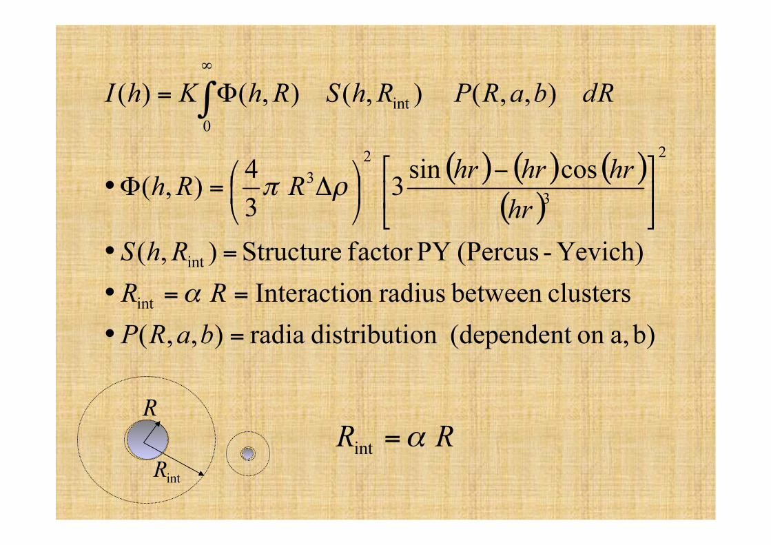
Form factor P(h)
Structure factor S(h)
Scattering intensity I(h)=P(h) S(h)
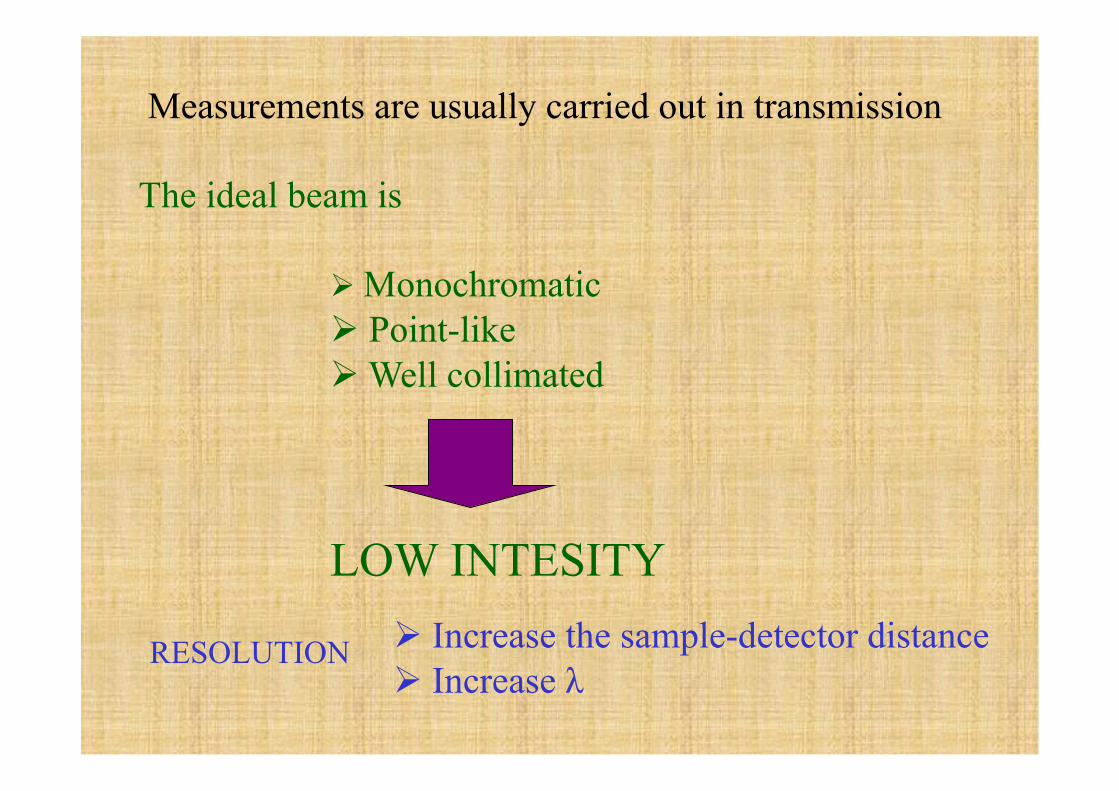
Measurements are usually carried out in transmission
The ideal beam is
Monochromatic Point-like Well collimated
LOW INTESITY Increase the sample-detector distance Increase λ
RESOLUTION
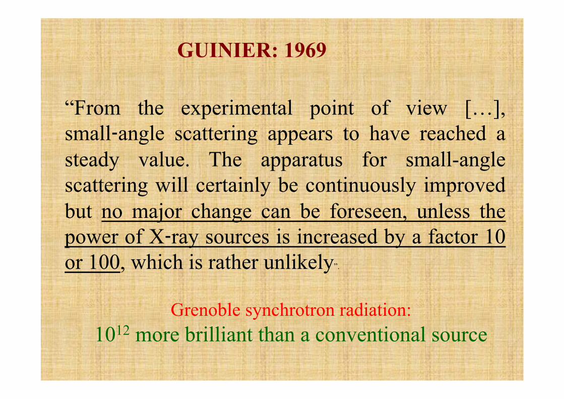
“From the experimental point of view […], small‑angle scattering appears to have reached a steady value. The apparatus for small-angle scattering will certainly be continuously improved but no major change can be foreseen, unless the power of X‑ray sources is increased by a factor 10 or 100, which is rather unlikely”.
GUINIER: 1969
Grenoble synchrotron radiation: 1012 more brilliant than a conventional source

This opens new frontiers
time-resolved measurements
2D- Detectors
Local measurements (microdiffusion)
Anomalous Scattering
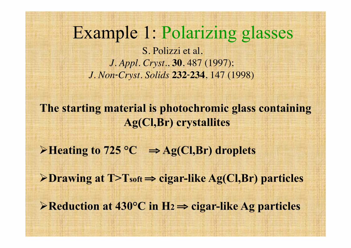
The starting material is photochromic glass containing Ag(Cl,Br) crystallites
Heating to 725 °C ⇒ Ag(Cl,Br) droplets
Drawing at T>Tsoft ⇒ cigar-like Ag(Cl,Br) particles
Reduction at 430°C in H2 ⇒ cigar-like Ag particles
Example 1: Polarizing glasses S. Polizzi et al,
J. Appl. Cryst., 30, 487 (1997); J. Non‑Cryst. Solids 232‑234, 147 (1998)

VETRI POLARIZZATORI
200nm
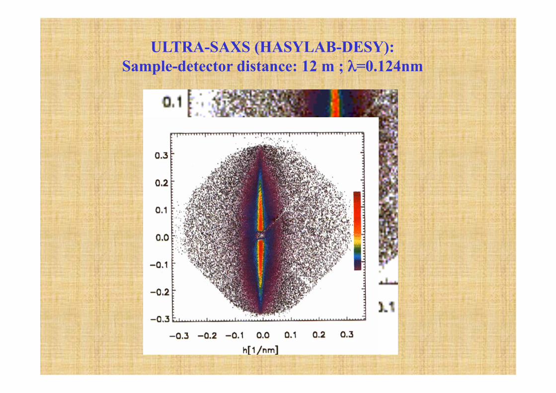
ULTRA-SAXS (HASYLAB-DESY): Sample-detector distance: 12 m ; λ=0.124nm

€
I(h,a,b) = D(a,b)I0(h,a,b)dadb∫∫
€
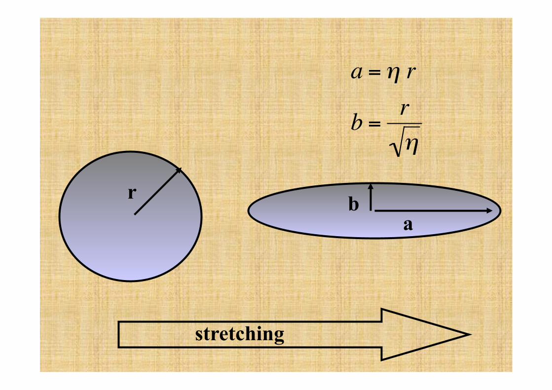
a =η r
b =rη
r a
b
stretching
€
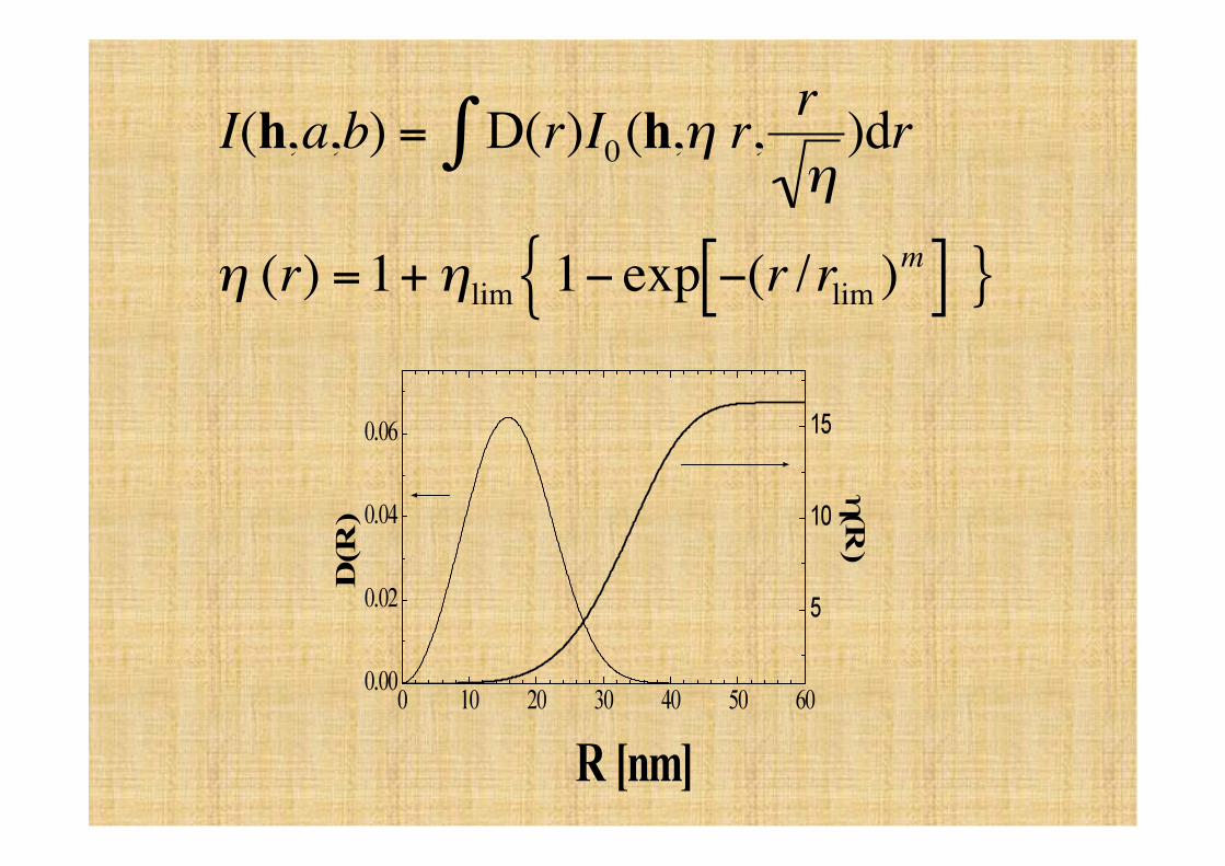
I(h,a,b) = D(r)I0(h,η r,rη)dr∫
η (r) =1+ηlim 1− exp −(r /rlim )m[ ]{ }
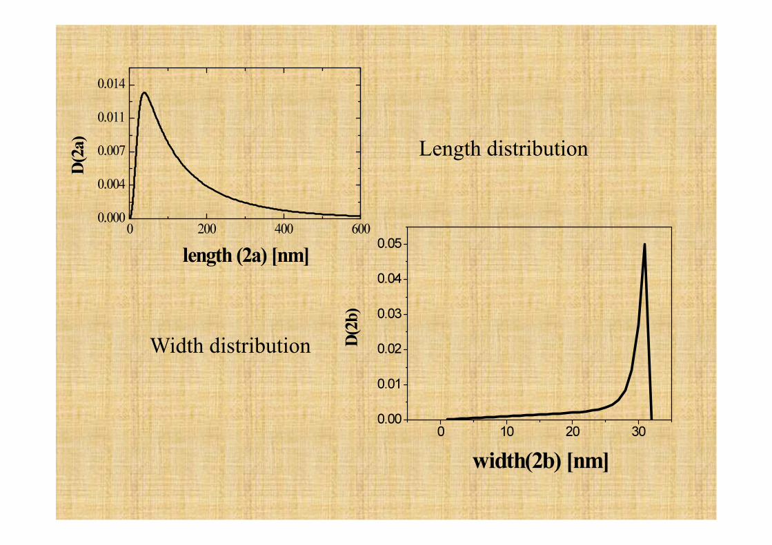
Length distribution
Width distribution
EXAMPLE 2: Aggregation of colloidal systems:
SULPHATE ZIRCONIA SOL-GEL
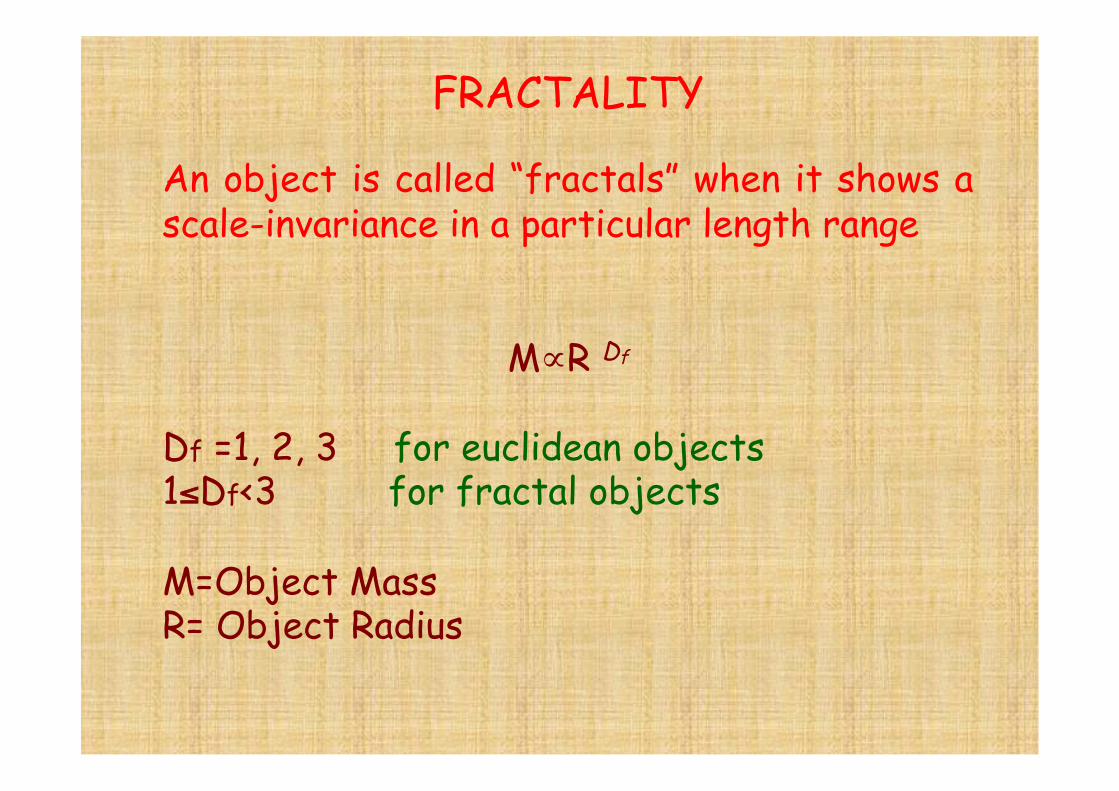
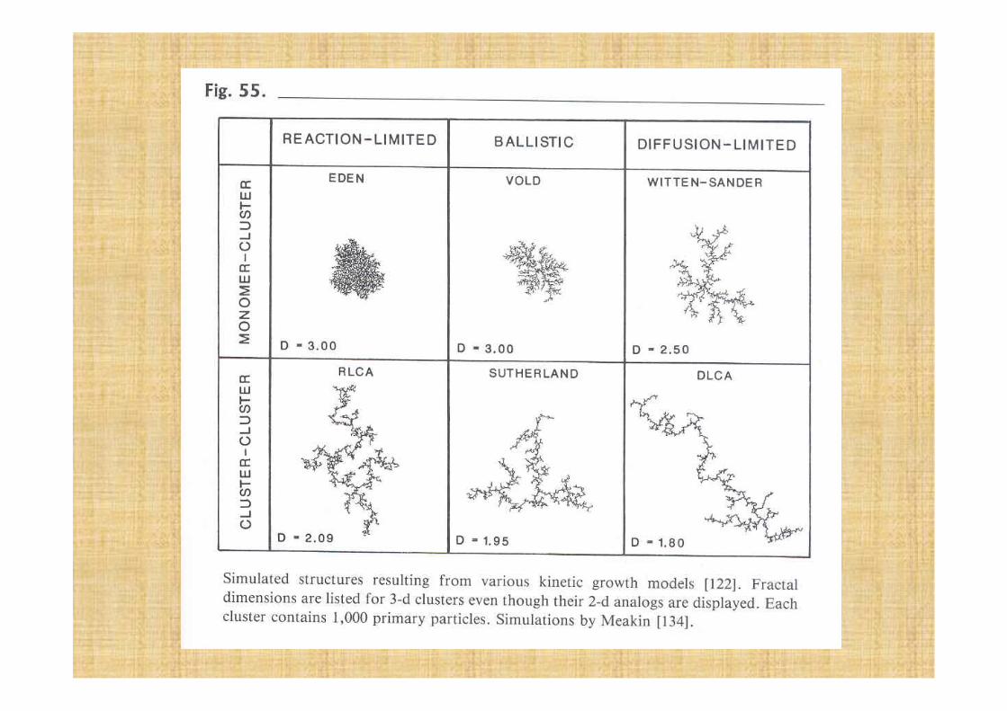
FRACTALITY
An object is called “fractals” when it shows a scale-invariance in a particular length range
M∝R Df
Df =1, 2, 3 for euclidean objects 1≤Df<3 for fractal objects
M=Object Mass R= Object Radius
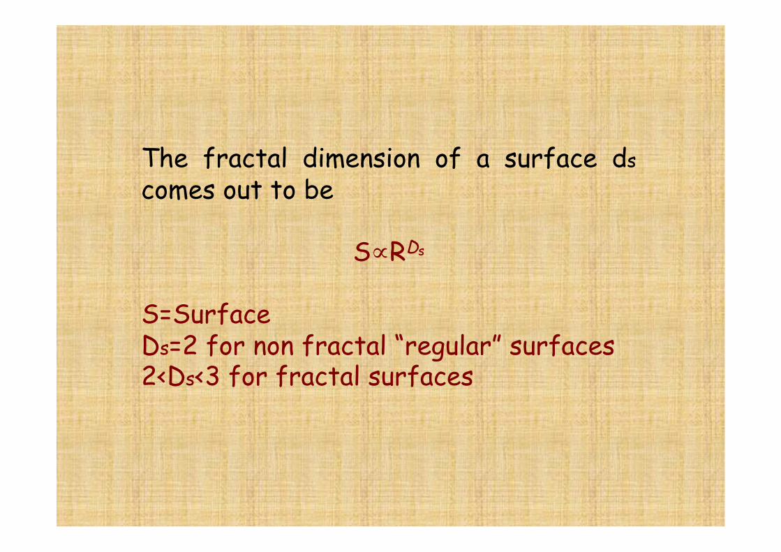
The fractal dimension of a surface ds comes out to be
S∝RDs
S=Surface Ds=2 for non fractal “regular” surfaces 2<Ds<3 for fractal surfaces
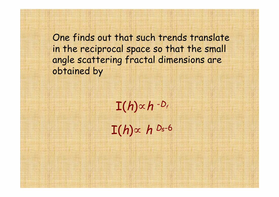
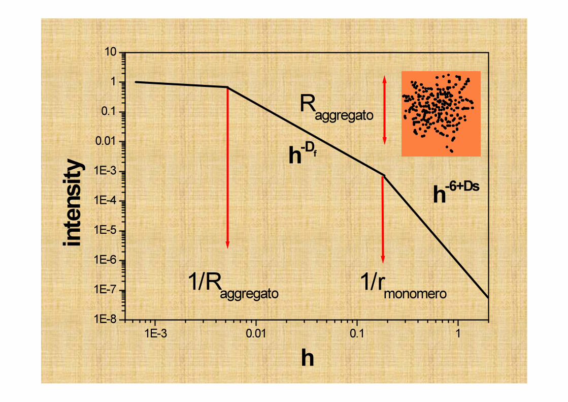
One finds out that such trends translate in the reciprocal space so that the small angle scattering fractal dimensions are obtained by
I(h)∝h -Df
I(h)∝ h Ds-6
€
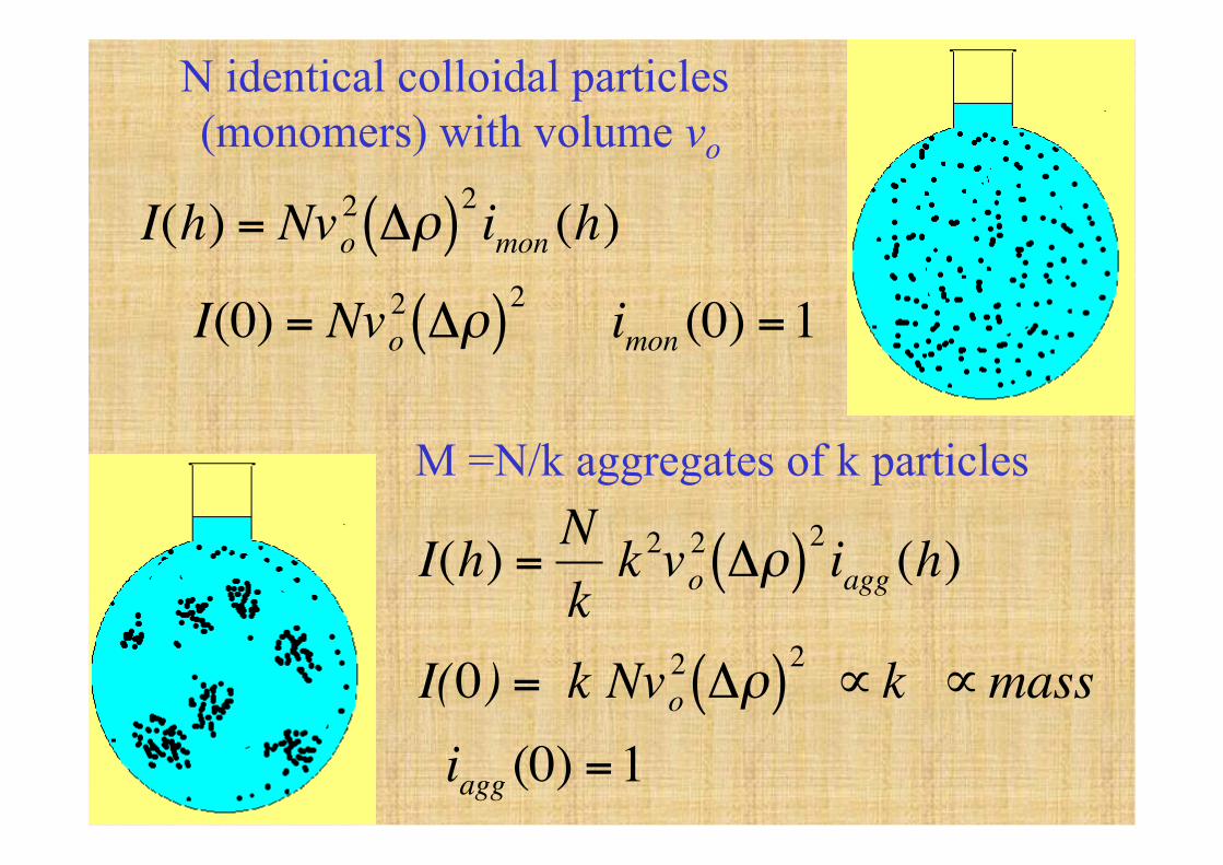
I(h) = Nvo2 Δρ( )2imon (h)
I(0) = Nvo2 Δρ( )2 imon (0) =1
€
I(h) =Nk
k 2vo2 Δρ( )2iagg (h)
I(0) = k Nvo2 Δρ( )2 ∝ k ∝mass
iagg (0) =1
N identical colloidal particles (monomers) with volume vo
M =N/k aggregates of k particles
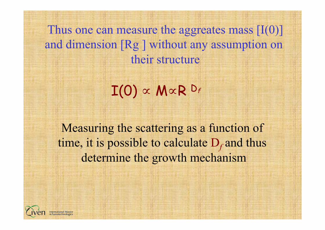
Thus one can measure the aggreates mass [I(0)] and dimension [Rg ] without any assumption on
their structure
I(0) ∝ M∝R Df
Measuring the scattering as a function of time, it is possible to calculate Df and thus
determine the growth mechanism
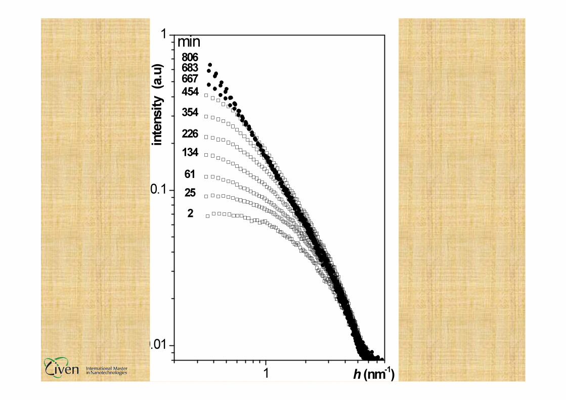
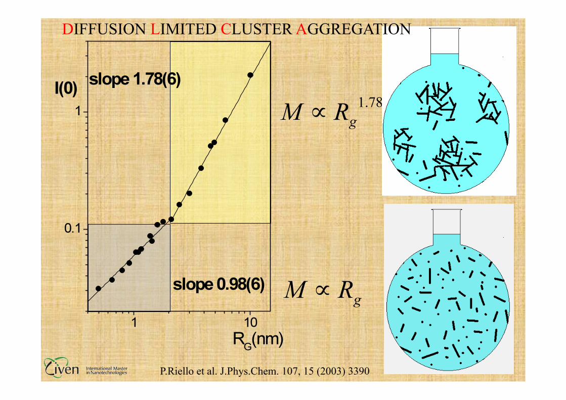
DIFFUSION LIMITED CLUSTER AGGREGATION
P.Riello et al. J.Phys.Chem. 107, 15 (2003) 3390
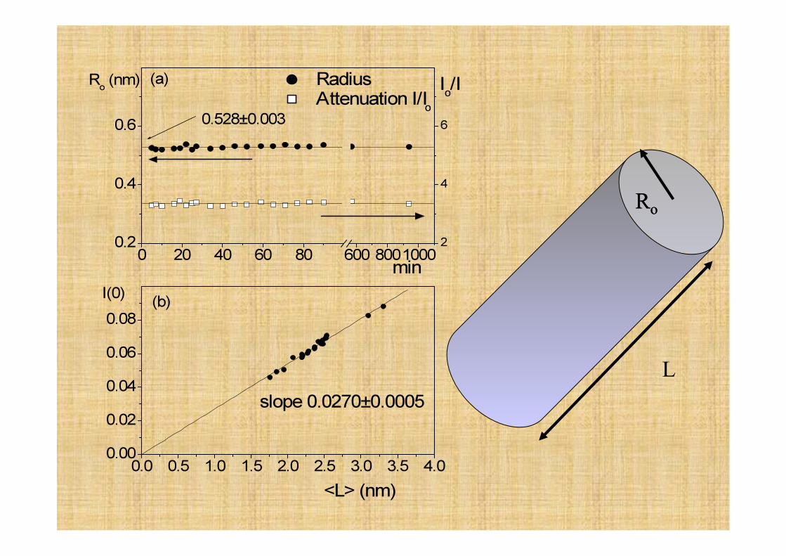
Ro Ro
L
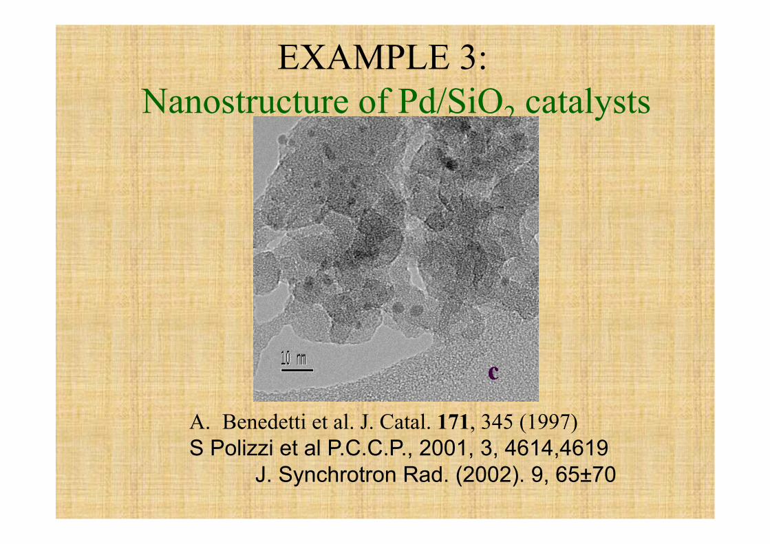
EXAMPLE 3: Nanostructure of Pd/SiO2 catalysts
A. Benedetti et al. J. Catal. 171, 345 (1997) S Polizzi et al P.C.C.P., 2001, 3, 4614,4619
J. Synchrotron Rad. (2002). 9, 65±70
Catalysis is a surface phenomenon
Efficient use of expensive metals
Different electronic structure
Increase of catalytical activity and selectivity
Small particles→ high surface/volume ratio
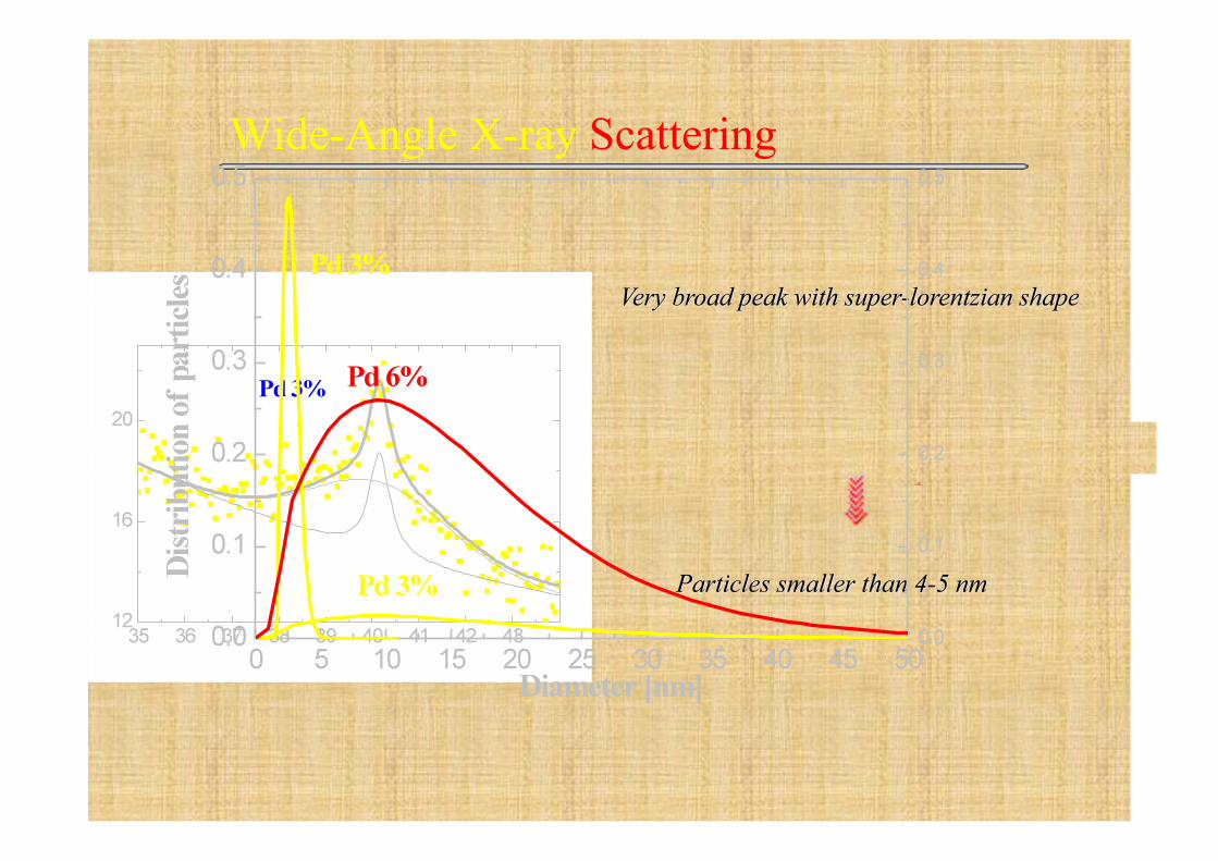
Wide-Angle X-ray Scattering
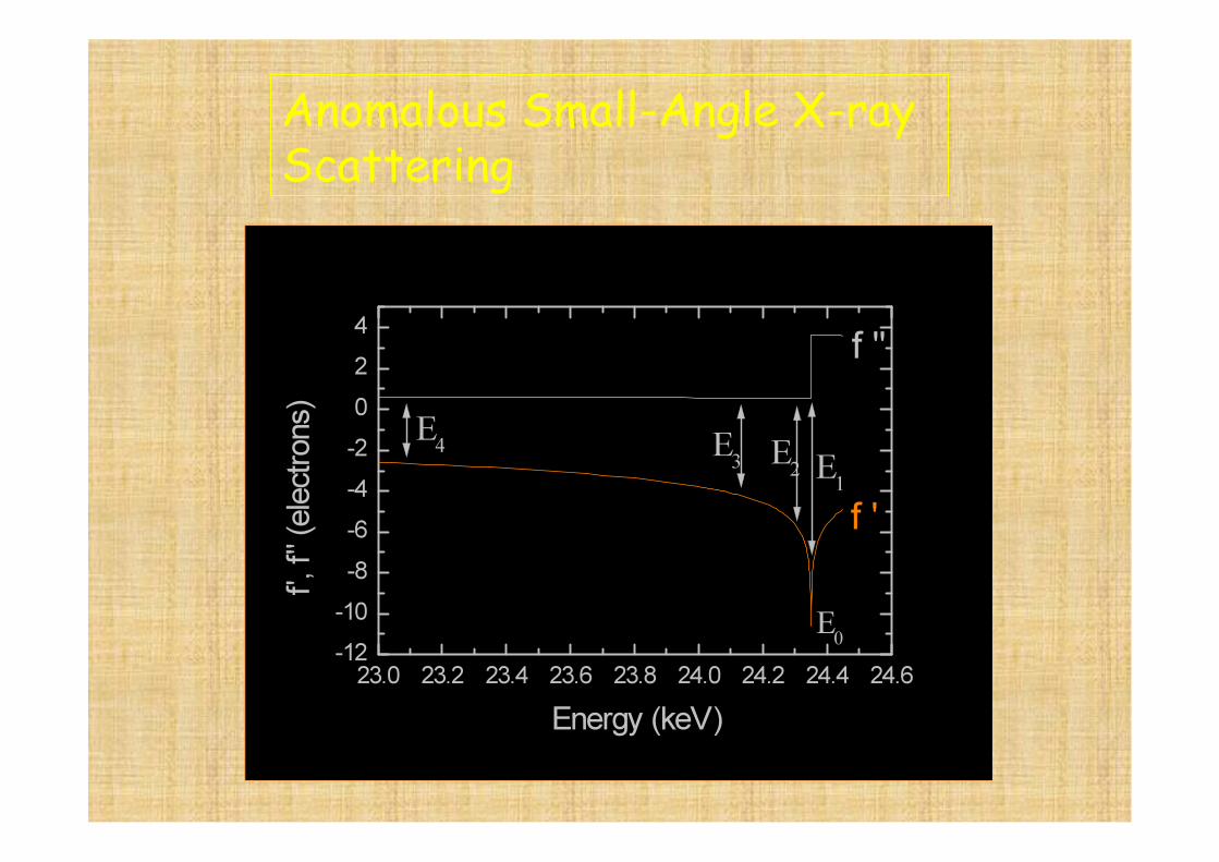
Anomalous Small-Angle X-ray Scattering
f = f0 + f ´ (E) + f ´´(E)
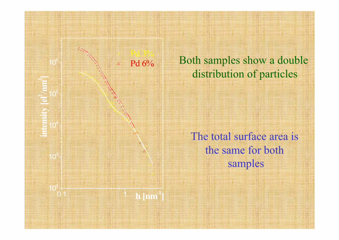
Both samples show a double distribution of particles
The total surface area is the same for both
samples
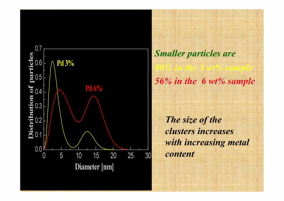
The size of the clusters increases with increasing metal content
EXAMPLE 4: SiO2-PEG Hybrid materials
Obtained by hidrolysis of the precursor (OEt)3Si-(PEG)-Si(OEt)3
Suitably doped they can be:
• Ionic conductors • Photocromic materials • Luminescent materials • ……..
Karim Dahmouche et al. J. Phys. Chem. B 1999, 103, 4937-4942
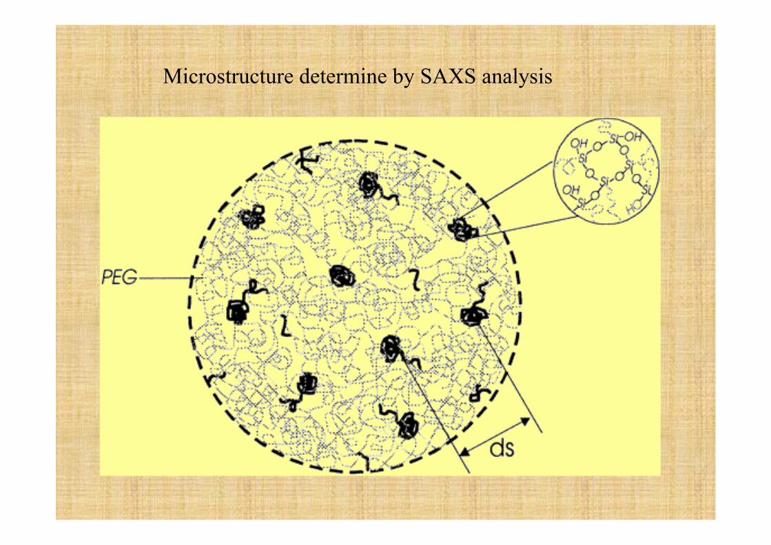
Microstructure determine by SAXS analysis
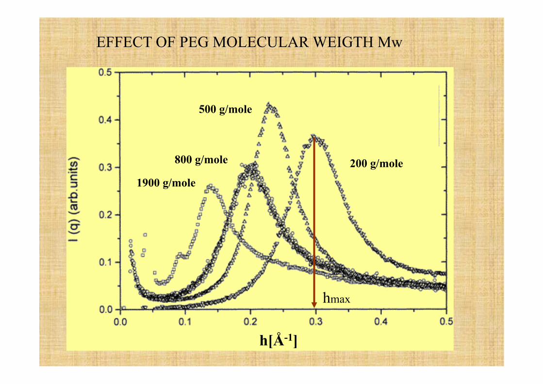
EFFECT OF PEG MOLECULAR WEIGTH Mw
1900 g/mole
800 g/mole
500 g/mole
200 g/mole
hmax
h[Å-1]
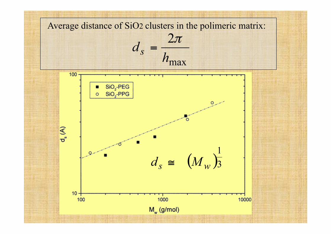
Average distance of SiO2 clusters in the polimeric matrix:
€
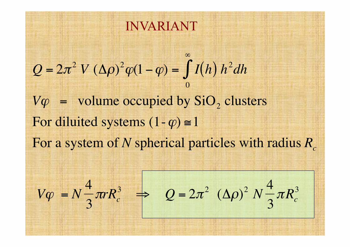
Q = 2π 2 V (Δρ)2ϕ(1−ϕ) = I h( )0
∞
∫ h2dh
Vϕ = volume occupied by SiO2 clusters For diluited systems (1-ϕ) ≅1For a system of N spherical particles with radius Rc
Vϕ = N 43πrRc
3 ⇒ Q = 2π 2 (Δρ)2 N 43πRc
3
INVARIANT
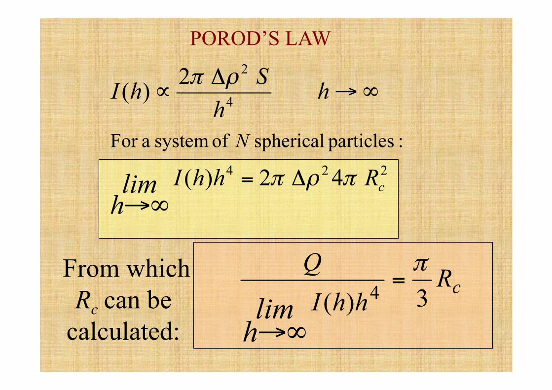
POROD’S LAW
From which Rc can be
calculated:
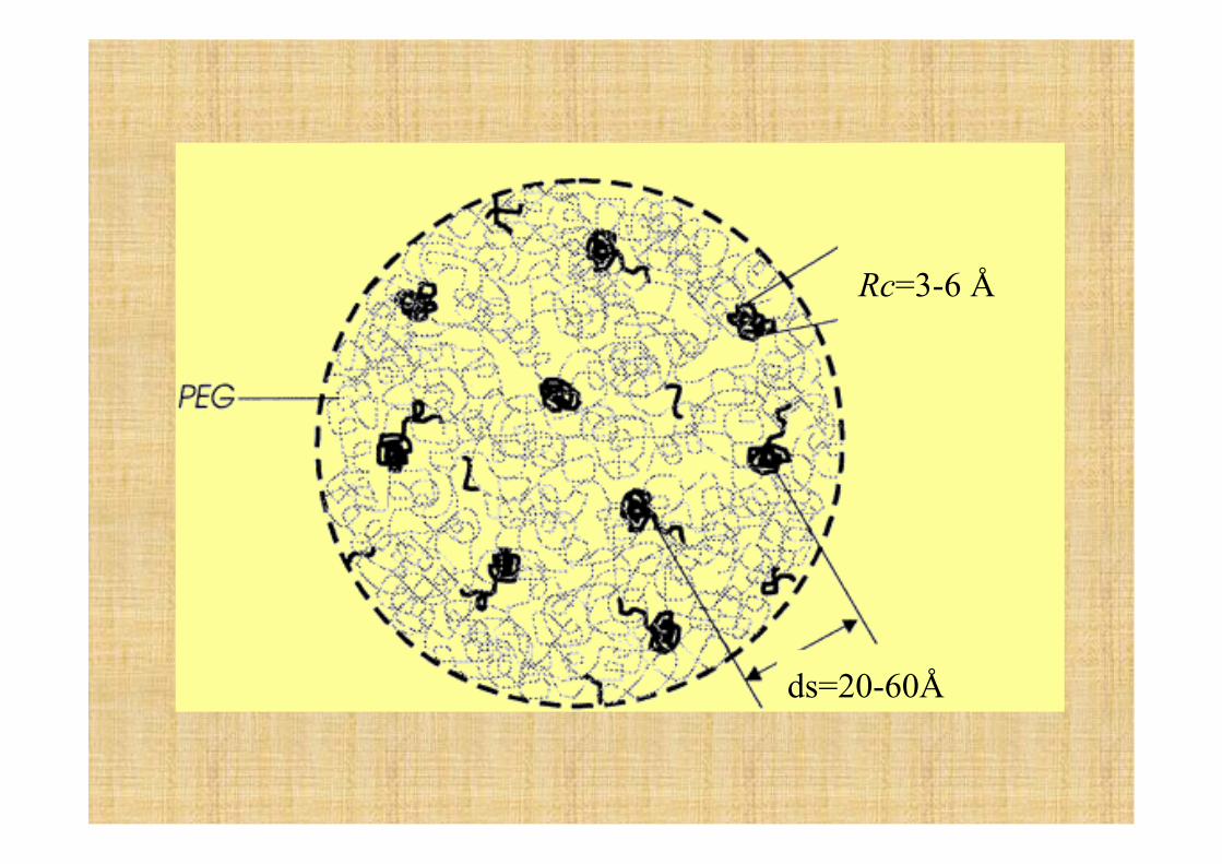
ds=20-60Å
Rc=3-6 Å
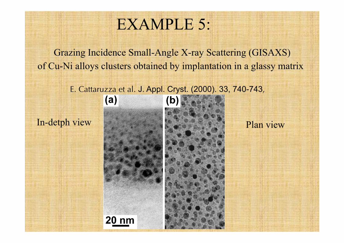
EXAMPLE 5: Grazing Incidence Small-Angle X-ray Scattering (GISAXS)
of Cu-Ni alloys clusters obtained by implantation in a glassy matrix
E. Cattaruzza et al. J. Appl. Cryst. (2000). 33, 740-743,
In-detph view Plan view
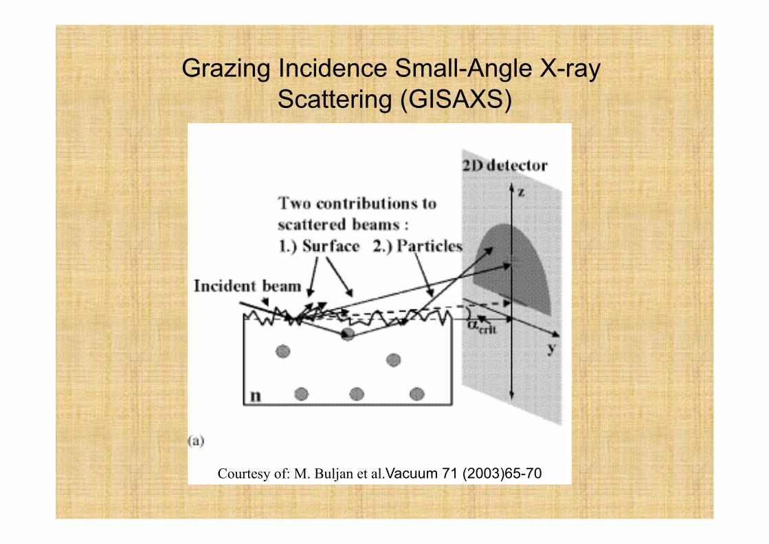
Courtesy of: M. Buljan et al.Vacuum 71 (2003)65-70
Grazing Incidence Small-Angle X-ray Scattering (GISAXS)
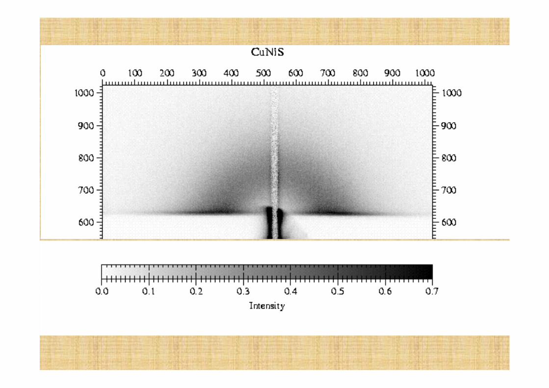
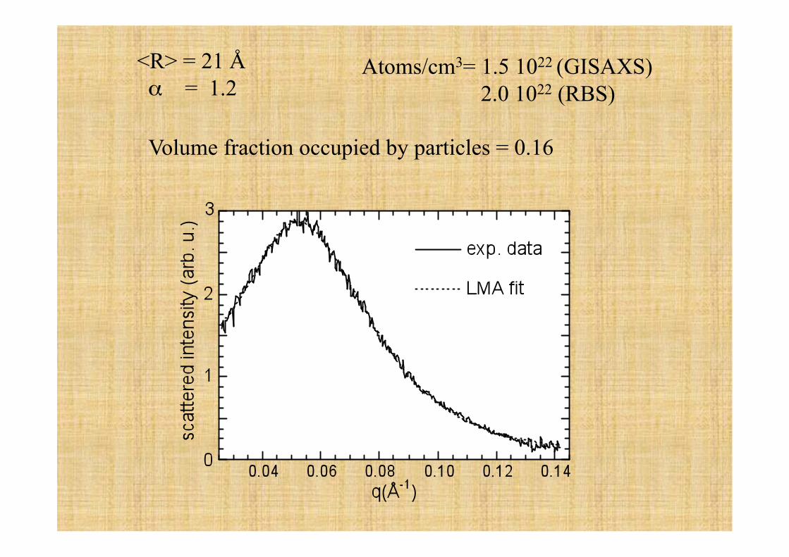
<R> = 21 Å α = 1.2
Atoms/cm3= 1.5 1022 (GISAXS) 2.0 1022 (RBS)
Volume fraction occupied by particles = 0.16
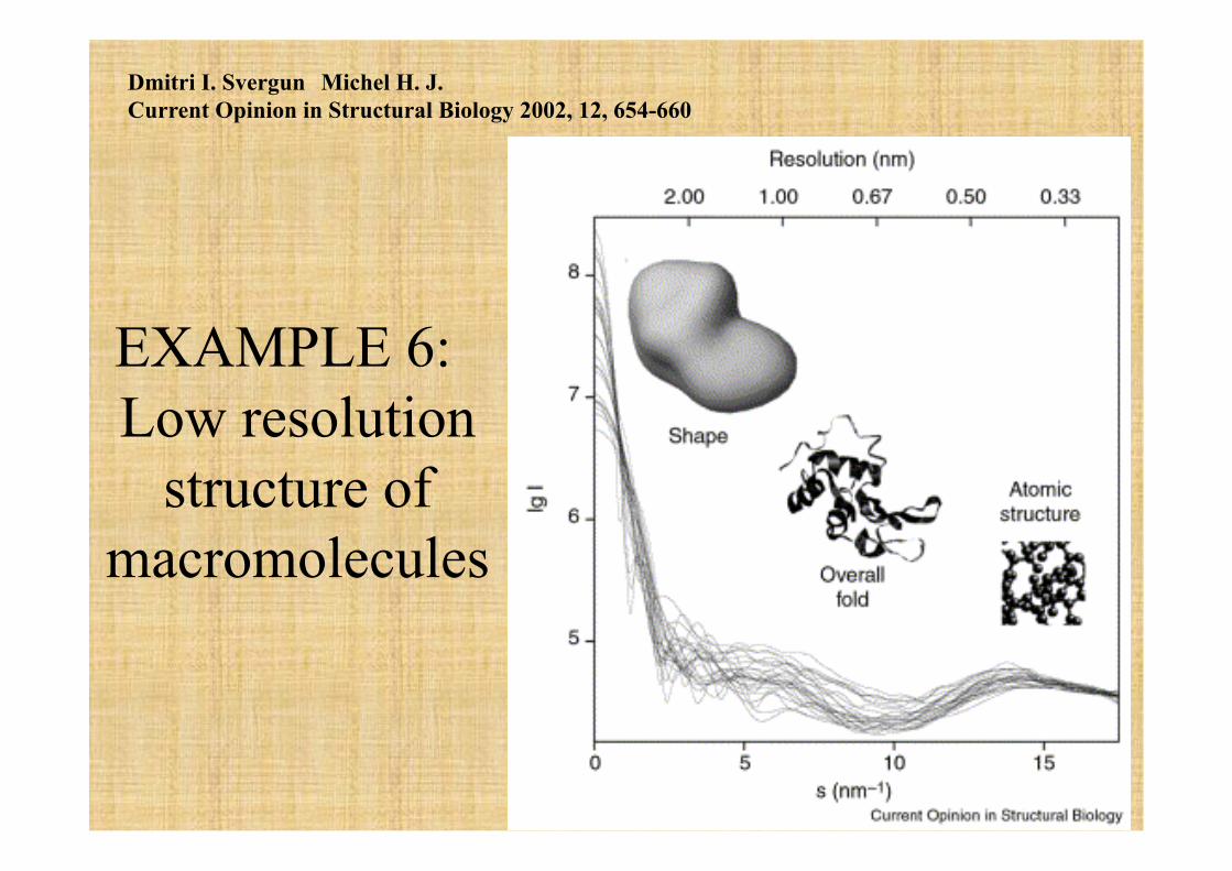
EXAMPLE 6: Low resolution
structure of macromolecules
Dmitri I. Svergun Michel H. J. Current Opinion in Structural Biology 2002, 12, 654-660
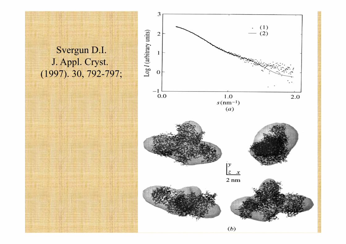
Svergun D.I. J. Appl. Cryst.
(1997). 30, 792-797;
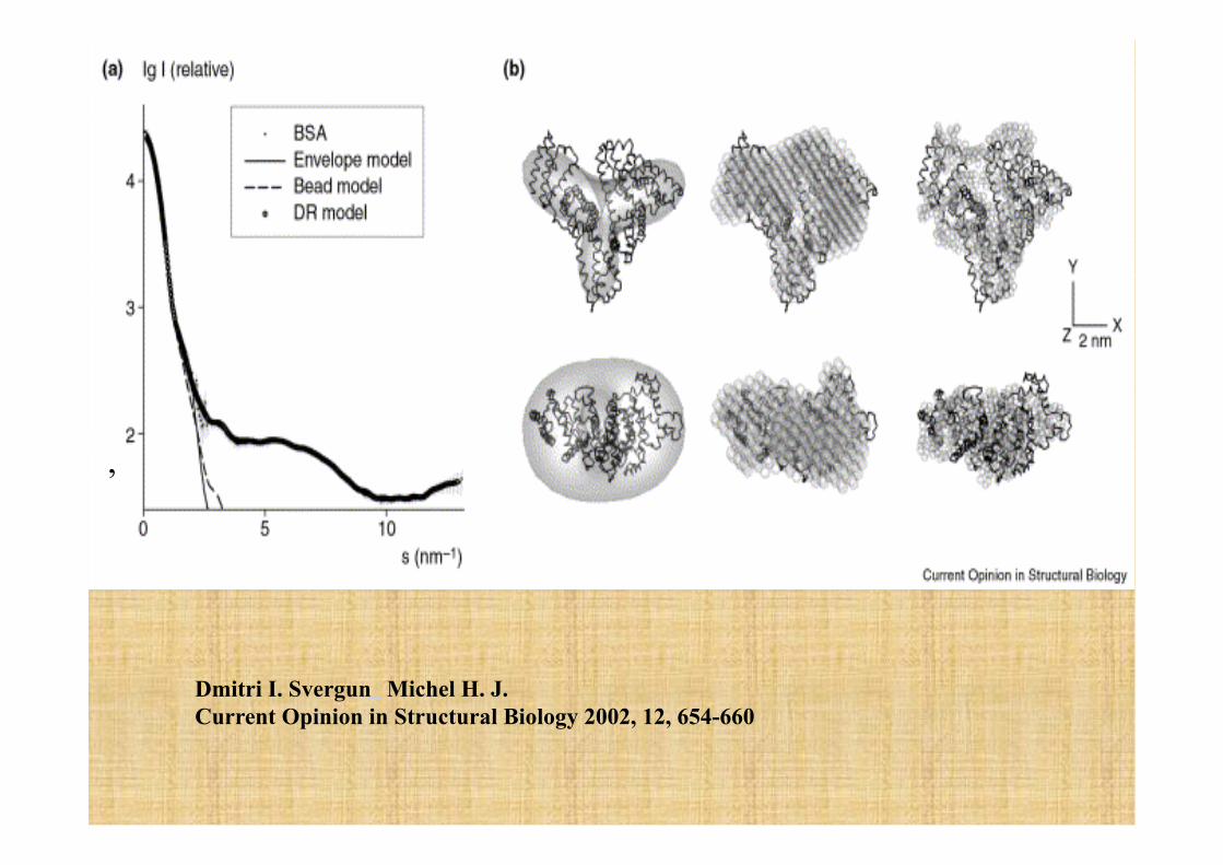
Dmitri I. Svergun Michel H. J. Current Opinion in Structural Biology 2002, 12, 654-660
,
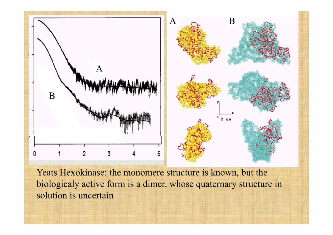
B
Yeats Hexokinase: the monomere structure is known, but the biologicaly active form is a dimer, whose quaternary structure in solution is uncertain
A B
A
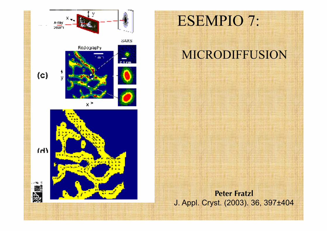
Peter Fratzl J. Appl. Cryst. (2003). 36, 397±404
ESEMPIO 7:
MICRODIFFUSION