XAS: X-ray Absorption Spectroscopy - University of...
26
1 1 XAS: X-ray Absorption Spectroscopy Hwo-Shuenn Sheu [email protected] NSRRC, Taiwan Malaya University, 2011/12/14-5 2 outline • Basic principles for XAS • Experimental setup for XAS • Applications
Transcript of XAS: X-ray Absorption Spectroscopy - University of...
1
1
XAS: X-ray AbsorptionSpectroscopy
Hwo-Shuenn Sheu
[email protected], Taiwan
Malaya University, 2011/12/14-5
2
outline
•Basic principles for XAS•Experimental setup for XAS•Applications
2
3
A
bsor
ptio
n
Photon energy(eV)
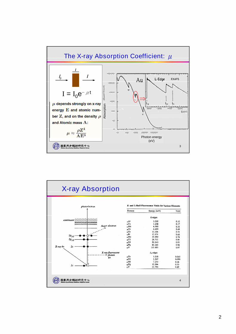
The X-ray Absorption Coefficient: μ
I = I0e−μt
4
X-ray Absorption
3
5
Why need SR X-ray sources?
6
**
*
RgDsMtHsBhSgDbRfLrRaFr
RnAtPoBiPbTlHgAuPtIrOsReWTaHfLuBaCs
XeITeSbSnInCdAgPdRhRuTcMoNbZrYSrRb
KrBrSeAsGeGaZnCuNiCoFeMnCrVTiScCaK
ArClSPSiAlMgNa
NeFONCBBeLi
HeH
**
*
NoMdFmEsCfBkCmAmPuNpUPaThAc
YbTmErHoDyTbGdEuSmPmNdPrCeLa
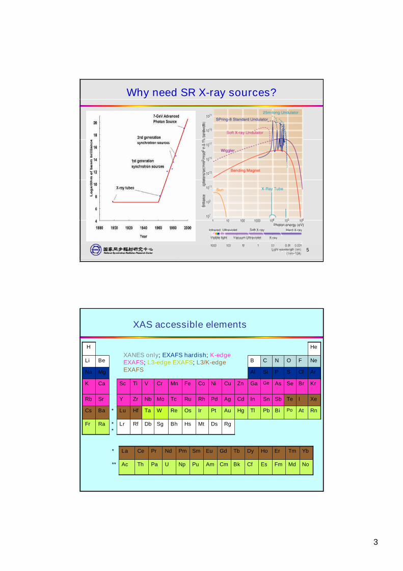
XAS accessible elements
XANES only; EXAFS hardish; K-edgeEXAFS; L3-edge EXAFS; L3/K-edgeEXAFS
26
4
7
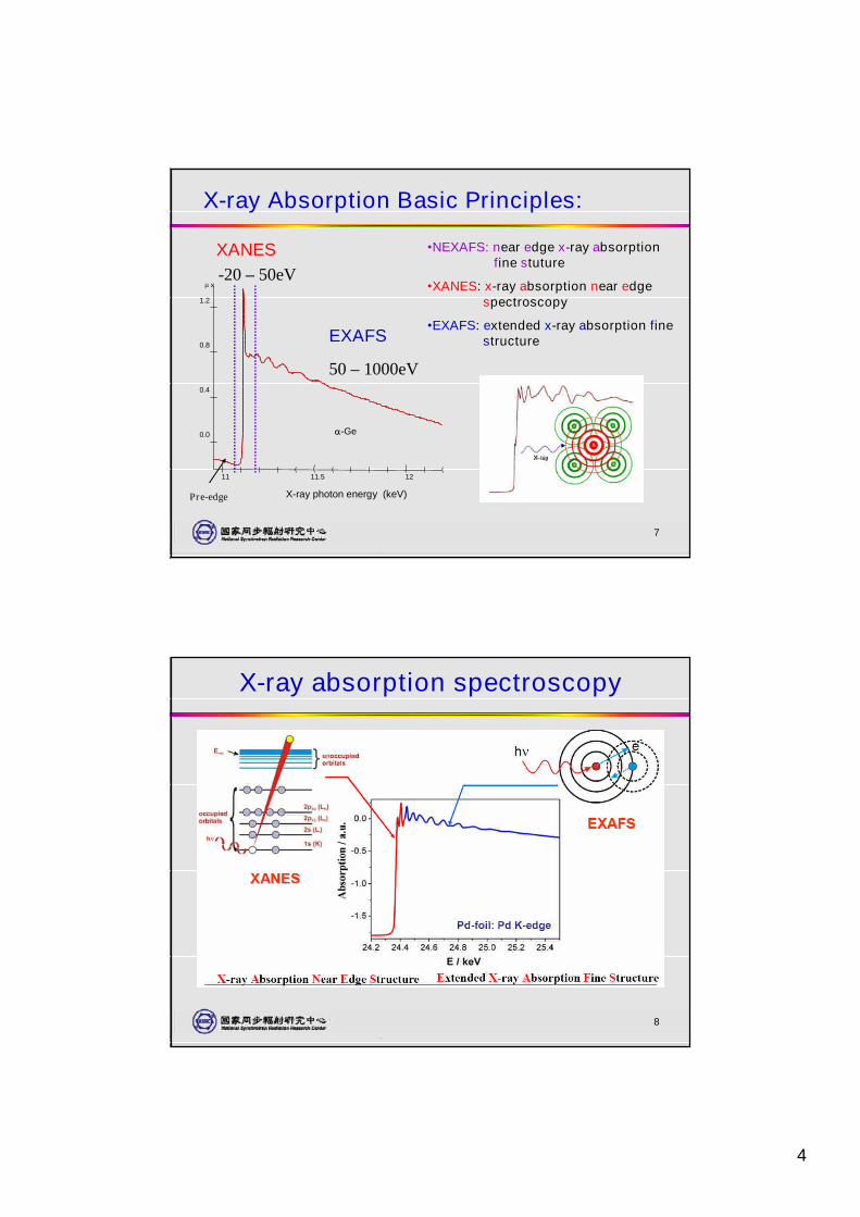
X-ray Absorption Basic Principles:
•NEXAFS: near edge x-ray absorptionfine stuture
•XANES: x-ray absorption near edgespectroscopy
•EXAFS: extended x-ray absorption finestructureEXAFS
XANES
x
X-ray photon energy (keV)
-Ge0.0
0.4
0.8
1.2
11 11.5 12
Pre-edge
-20 –50eV
50 –1000eV
8
X-ray absorption spectroscopy
5
9
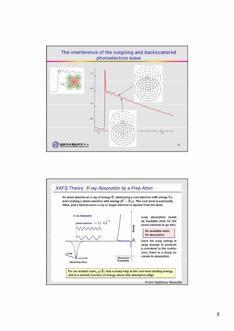
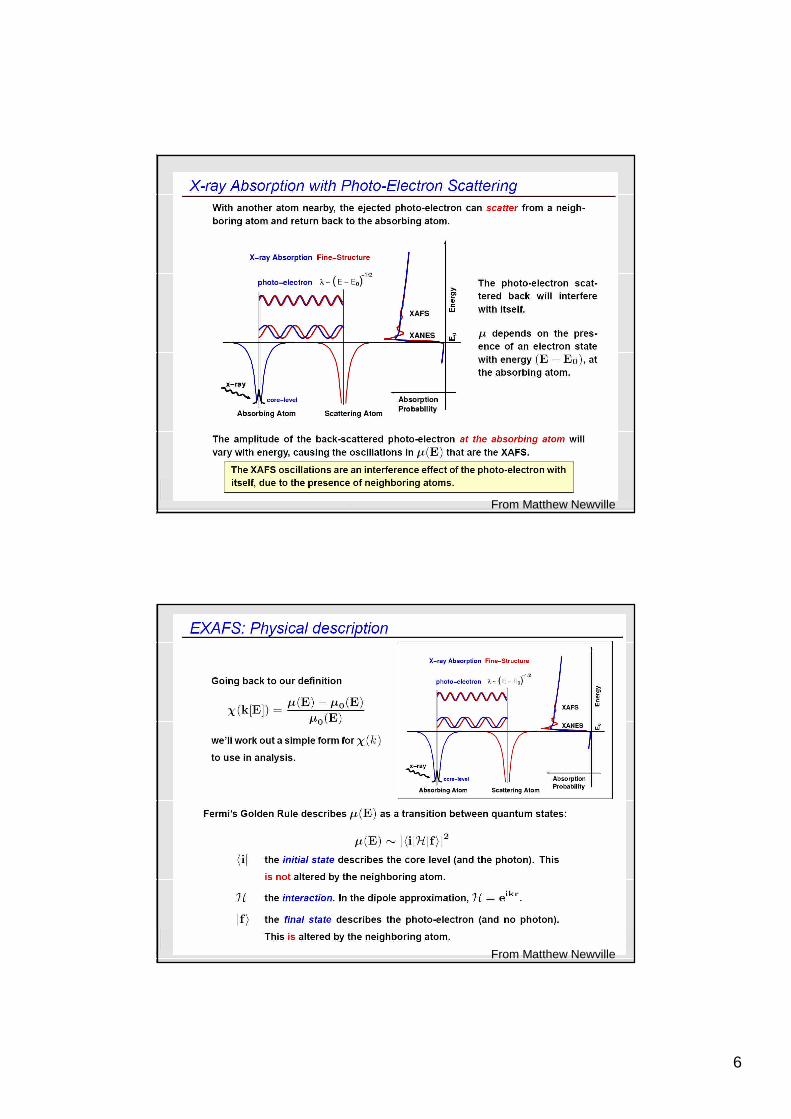
The interference of the outgoing and backscatteredphotoelectron wave
0.0
0.4
0.8
1.2
11 11.5 12
10
From Matthew Newville
6
11
From Matthew Newville
12
From Matthew Newville
7
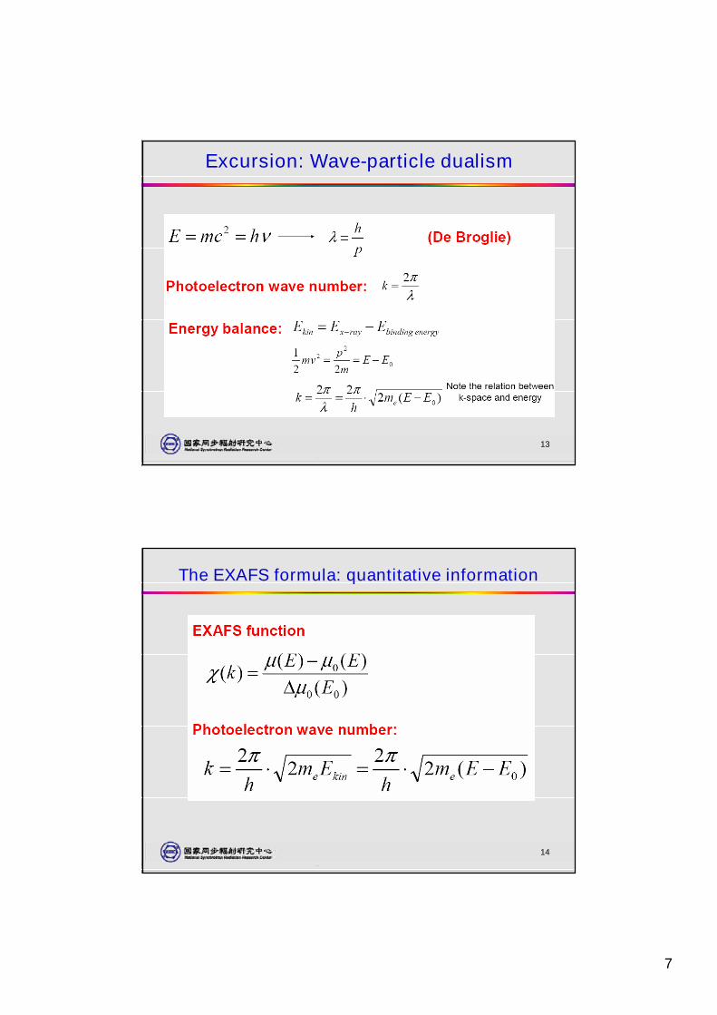
13
Excursion: Wave-particle dualism
14
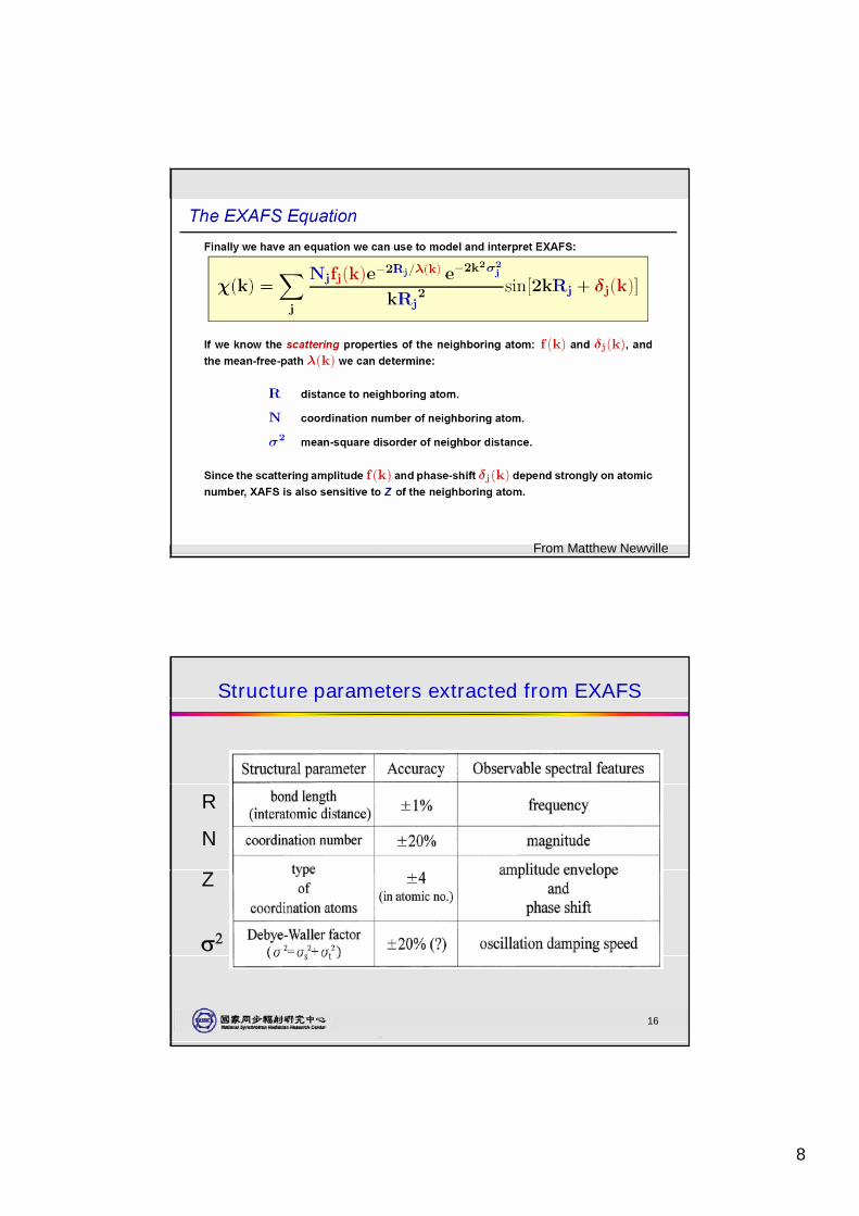
The EXAFS formula: quantitative information
8
15
From Matthew Newville
16
Structure parameters extracted from EXAFS
R
N
Z
9
17
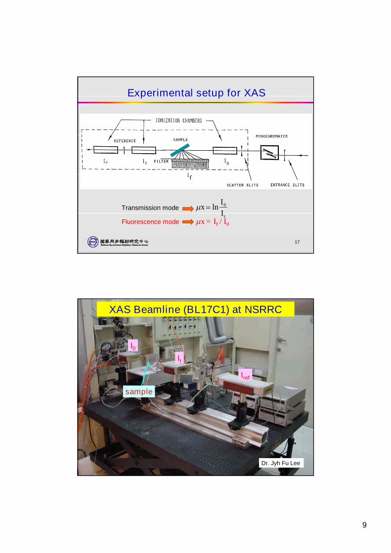
Experimental setup for XAS
tII
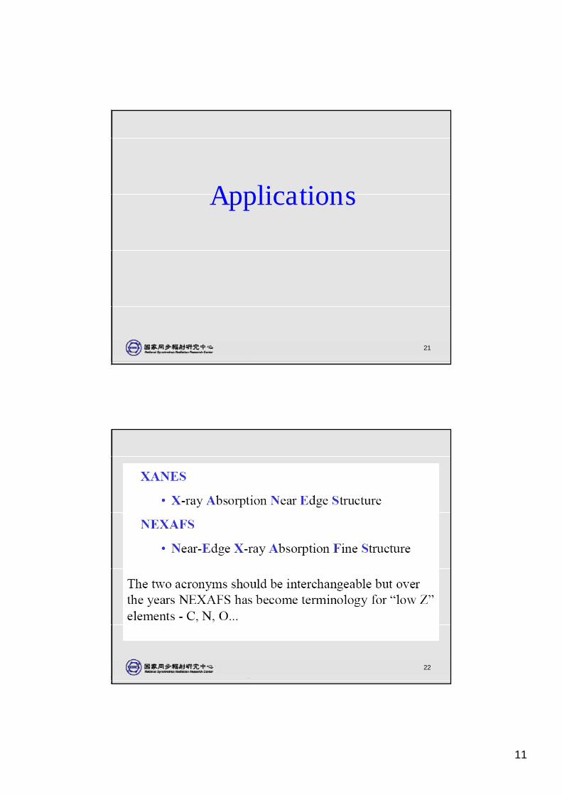
x 0lnTransmission mode
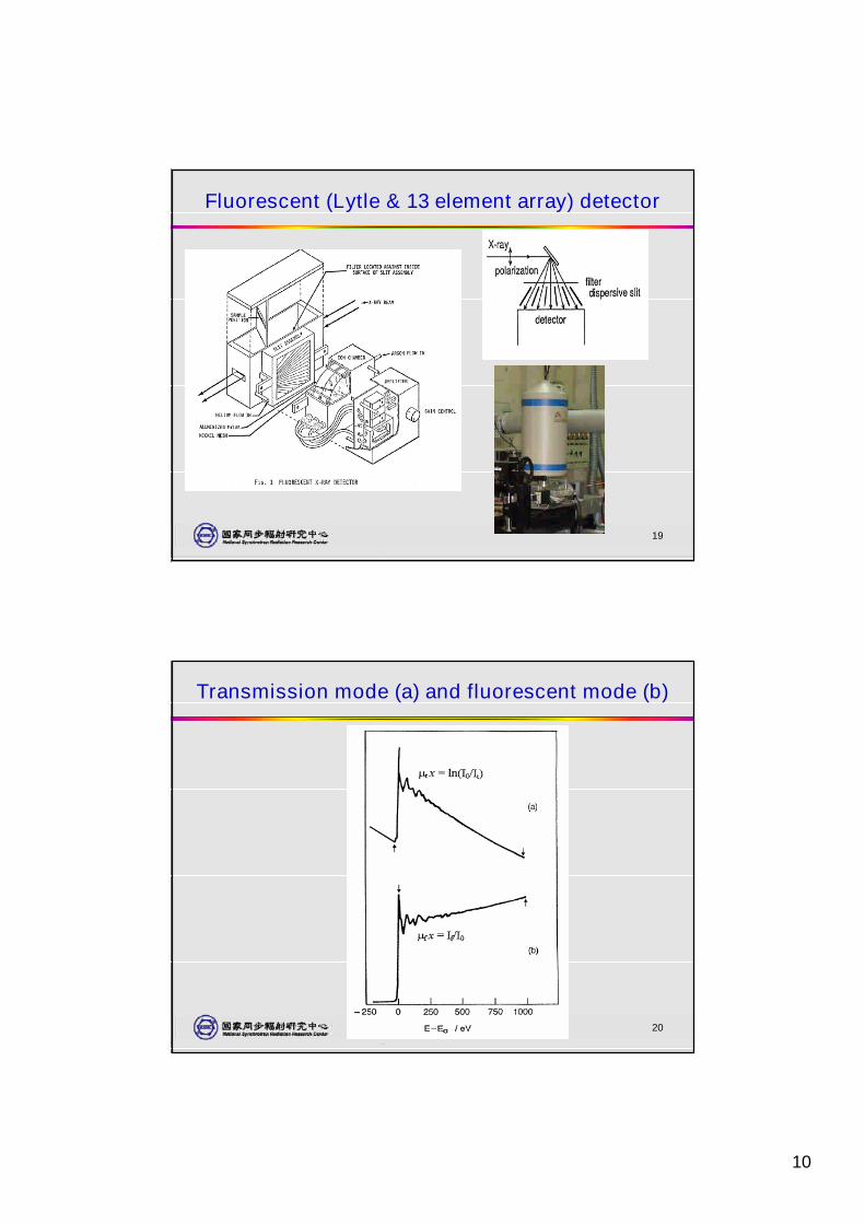
Fluorescence mode x = If / I0
18
XAS Beamline (BL17C1) at NSRRC
I0It
Iref
sample
Dr. Jyh Fu Lee
10
19
Fluorescent (Lytle & 13 element array) detector
20
Transmission mode (a) and fluorescent mode (b)
11
21
Applications
22
12
23
414 415
Double excitations
(c)
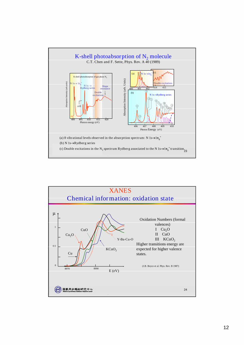
(c) Double excitations in the N2 spectrum Rydberg associated to the N 1s1g* transition.
(a)
400 401 402
N 1s1g*
(a) 8 vibrational levels observed in the absorpttion spectrum: N 1s1g*
Abs
orpt
ion
Inte
nsity
(arb
.Uni
ts)
406 407 408 409 410Photon Energy (eV)
(b)N 1sRydberg series
1
3
2
4
3
56
78
9
10
1112 13
(b) N 1sRydberg series
K-shell photoabsorption of N2 moleculeC.T. Chen and F. Sette, Phys. Rev. A 40 (1989)
K-shell photoabsorption of gas-phase N2
Abs
orpt
ion
Inte
nsity
(arb
.uni
ts)
N 1s 1g*
N 1s Rydberg series
Doubleexcitations
Shaperesonance
x10
400 405 410 415 420Photon energy (eV)
24
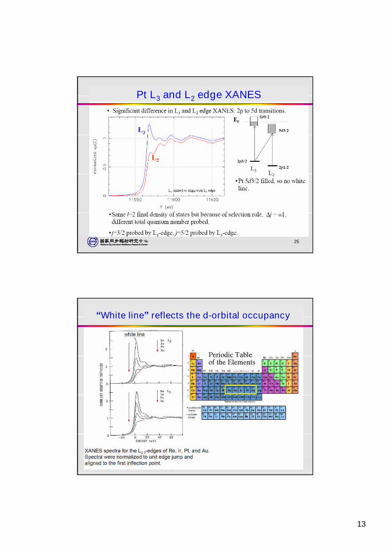
XANESChemical information: oxidation state
Oxidation Numbers (formalvalences)I Cu2OII CuOIII KCuO2
Higher transitions energy areexpected for higher valencestates.
KCuO2
CuOCu2O
Cu
E (eV)
8970 89900
0.5
1
Y-Ba-Cu-O
(J.B. Boyce et al. Phys. Rev. B 1987)
13
25
Pt L3 and L2 edge XANES
26
“White line”reflects the d-orbital occupancy
14
27
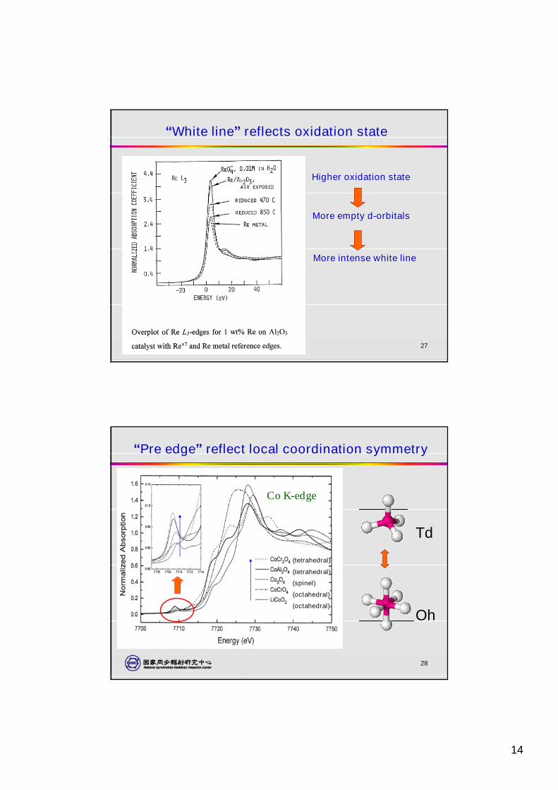
“White line”reflects oxidation state
Higher oxidation state
More empty d-orbitals
More intense white line
28
“Pre edge”reflect local coordination symmetry
Td
Oh
(tetrahedral)
(tetrahedral)
(spinel)
(octahedral)
(octahedral)
Co K-edge
15
29
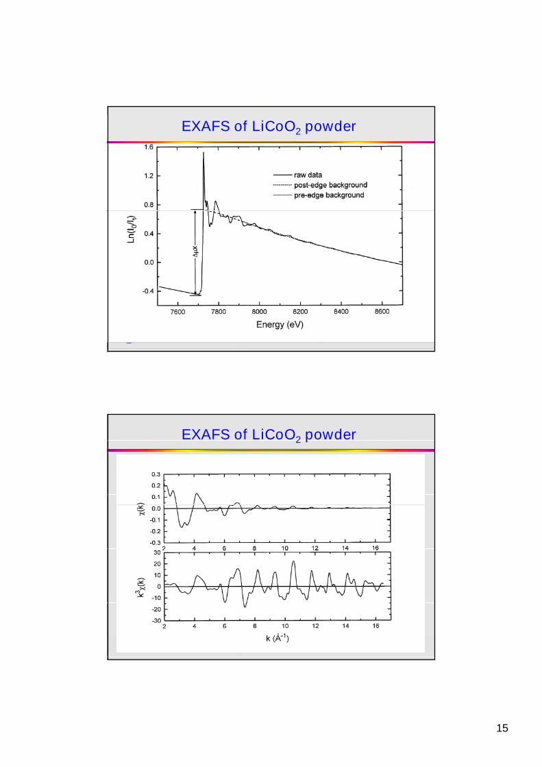
EXAFS of LiCoO2 powder
30
EXAFS of LiCoO2 powder
16
31
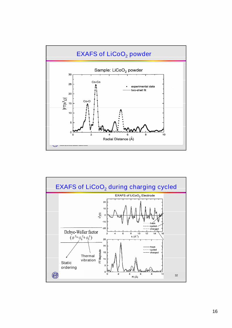
EXAFS of LiCoO2 powder
32
EXAFS of LiCoO2 during charging cycled
Staticordering
Thermalvibration
17
33
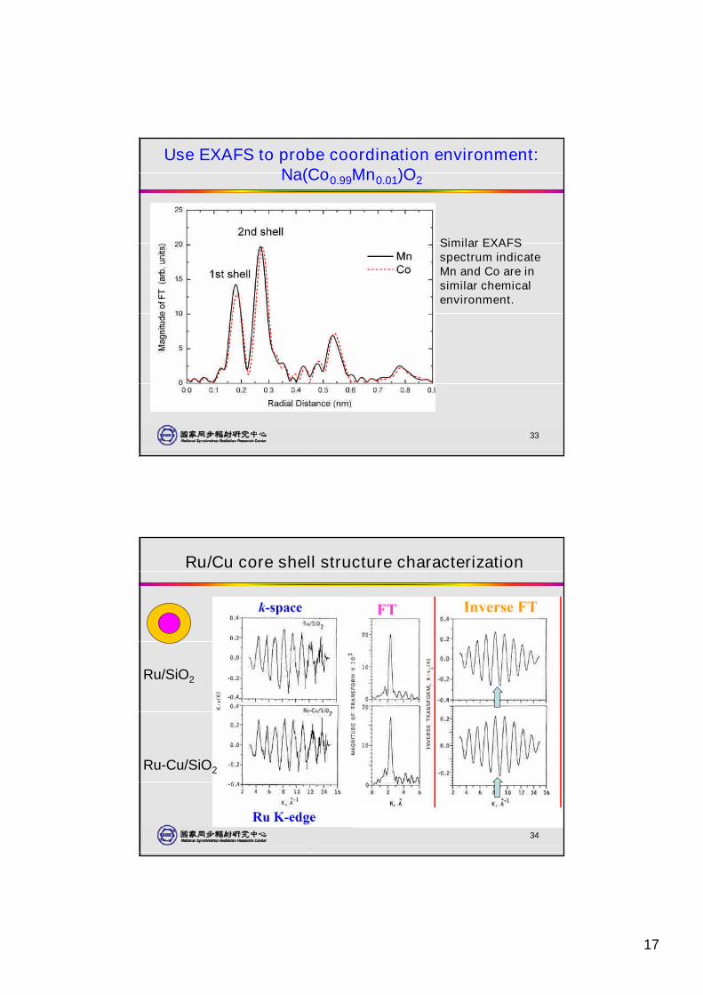
Use EXAFS to probe coordination environment:Na(Co0.99Mn0.01)O2
Similar EXAFSspectrum indicateMn and Co are insimilar chemicalenvironment.
34
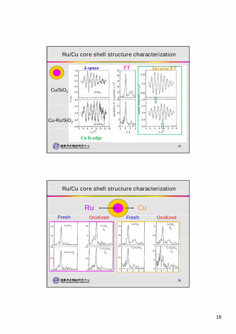
Ru/Cu core shell structure characterization
Ru/SiO2
Ru-Cu/SiO2
18
35
Ru/Cu core shell structure characterization
Cu/SiO2
Cu-Ru/SiO2
36
Ru/Cu core shell structure characterization
Fresh FreshOxidized Oxidized
Ru Cu
19
37
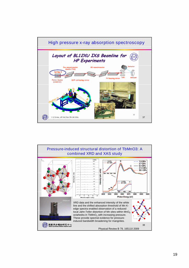
High pressure x-ray absorption spectroscopy
38
Pressure-induced structural distortion of TbMnO3: Acombined XRD and XAS study
Physical Review B 79, 165110 2009
XRD data and the enhanced intensity of the whiteline and the shifted absorption threshold of Mn K-edge spectra enabled observation of a reducedlocal Jahn-Teller distortion of Mn sites within MnO6octahedra in TbMnO3 with increasing pressure.These provide spectral evidence for pressure-induced bandwidth broadening for mangnites.
a
b1.889Å
2.243Å
145.7 º
Mn(1)
Mn(2)
Mn(1)
Mn(2)
J`
J
20
39
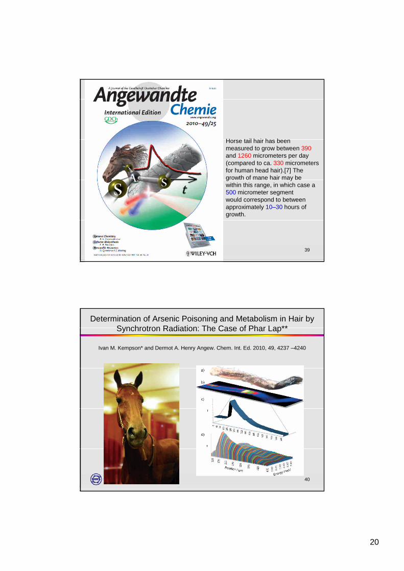
Horse tail hair has beenmeasured to grow between 390and 1260 micrometers per day(compared to ca. 330 micrometersfor human head hair).[7] Thegrowth of mane hair may bewithin this range, in which case a500 micrometer segmentwould correspond to betweenapproximately 10–30 hours ofgrowth.
40
Determination of Arsenic Poisoning and Metabolism in Hair bySynchrotron Radiation: The Case of Phar Lap**
Ivan M. Kempson* and Dermot A. Henry Angew. Chem. Int. Ed. 2010, 49, 4237 –4240
21
41
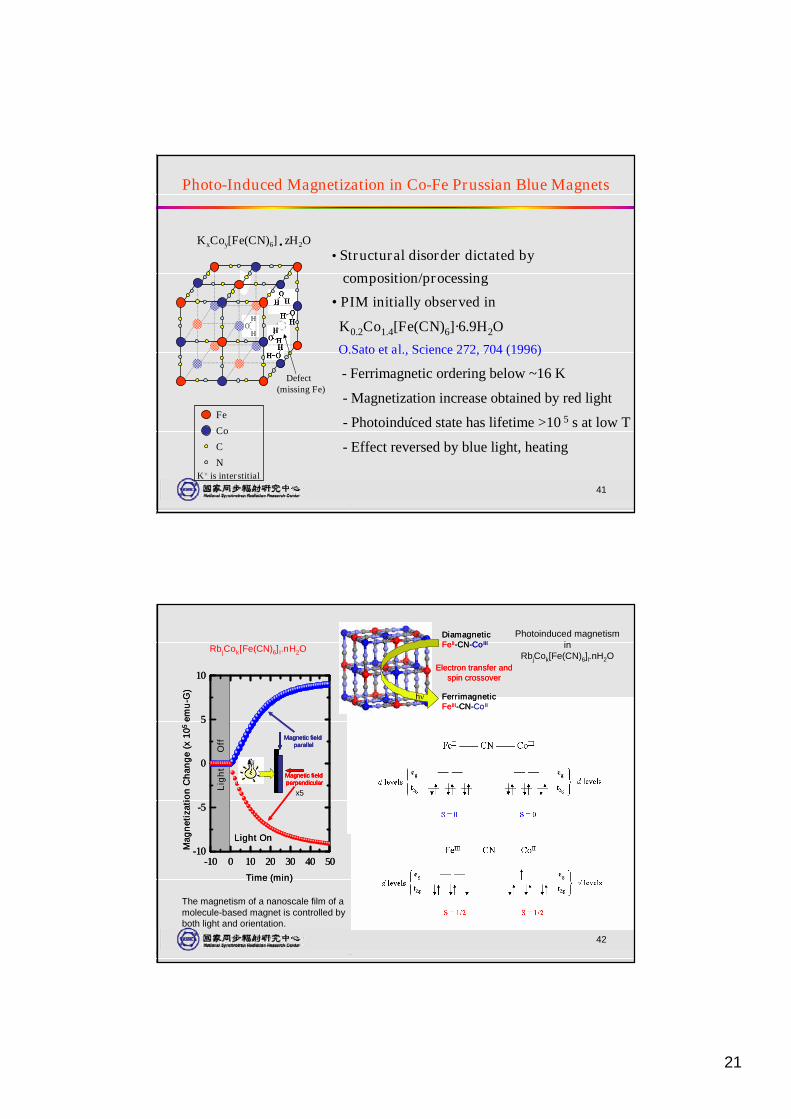
Photo-Induced Magnetization in Co-Fe Prussian Blue Magnets
•Structural disorder dictated by
composition/processing
•PIM initially observed in
K0.2Co1.4[Fe(CN)6]·6.9H2O
O.Sato et al., Science 272, 704 (1996)
- Ferrimagnetic ordering below ~16 K
- Magnetization increase obtained by red light
- Photoinduced state has lifetime >10 5 s at low T
- Effect reversed by blue light, heating
K+ is interstitial
Fe
Co
C
N
Defect(missing Fe)
KxCoy[Fe(CN)6]˙zH2O
42
Photoinduced magnetismin
RbjCok[Fe(CN)6]l.nH2O
DiamagneticFeII-CN-CoIII
FerrimagneticFeIII-CN-CoII
Electron transfer andspin crossover
h
DiamagneticFeII-CN-CoIII
FerrimagneticFeIII-CN-CoII
Electron transfer andspin crossover
h
-10 0 10 20 30 40 50-10
-5
0
5
10
Mag
net
izat
ion
Ch
ang
e(x
105
emu
-G)
Magnetic fieldparallel
Magnetic fieldperpendicular
Magnetic fieldparallel
Magnetic fieldperpendicular
Lig
ht
Off
Light On
Time (min)
-10 0 10 20 30 40 50-10
-5
0
5
10
Mag
net
izat
ion
Ch
ang
e(x
105
emu
-G)
Magnetic fieldparallel
Magnetic fieldperpendicular
Magnetic fieldparallel
Magnetic fieldperpendicular
Lig
ht
Off
Light On
Time (min)
The magnetism of a nanoscale film of amolecule-based magnet is controlled byboth light and orientation.
RbjCok[Fe(CN)6]l.nH2O
x5
22
43
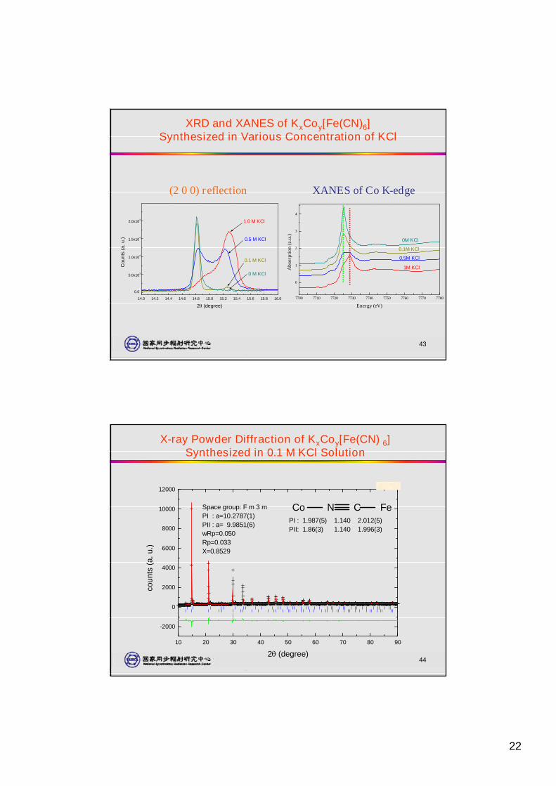
XRD and XANES of KxCoy[Fe(CN)6]Synthesized in Various Concentration of KCl
14.0 14.2 14.4 14.6 14.8 15.0 15.2 15.4 15.6 15.8 16.0
0.0
5.0x10³
1.0x10⁴
1.5x10⁴
2.0x10⁴
0 M KCl
0.1 M KCl
0.5 M KCl
1.0 M KCl
Cou
nts
(a.u
.)
2(degree)
(2 0 0) reflection
7700 7710 7720 7730 7740 7750 7760 7770 7780
0
1
2
3
4
1M KCl
0.5M KCl
0.1M KCl
0M KCl
Abs
orpt
ion
(a.u
.)
Energy (eV)
XANES of Co K-edge
44
X-ray Powder Diffraction of KxCoy[Fe(CN) 6]Synthesized in 0.1 M KCl Solution
10 20 30 40 50 60 70 80 90
-2000
0
2000
4000
6000
8000
10000
120005/13/00
Space group: F m 3 mPI : a=10.2787(1)PII : a= 9.9851(6)wRp=0.050Rp=0.033X=0.8529
PI : 1.987(5) 1.140 2.012(5)PII: 1.86(3) 1.140 1.996(3)
Co N C Fe
coun
ts(a
.u.)
2(degree)
23
45
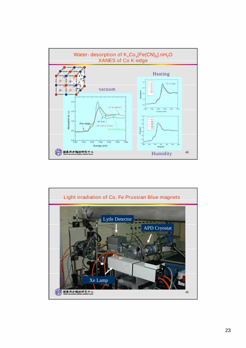
Water- desorption of KxCoy[Fe(CN)6].nH2OXANES of Co K-edge
7700 7710 7720 7730 7740 7750 77600.0
0.5
1.0
1.5
2.0
Pre-edge14K to RT in Vacuum
14K to RT then in air
10-3 torr (30min)
RT in air
Abs
orpt
ion
(a.u
.)
Energy (eV)
vacuum
7700 7710 7720 7730 7740 7750
0.5
1.0
1.5
2.0
2.5Co K-edge25 C
45 C55 C65 C75 C85 C95 C
105 C125 C
Abs
orpt
ion
Energy (eV)
Heating
7700 7710 7720 7730 7740 7750 77600.3
0.4
0.5
0.6
0.7
0.8
0.9
1.0
7%11%18%19%32%42%
Abs
orpt
ion
Energy (eV)
Humidity
46
Lytle Detector
APD Cryostat
Xe Lamp
Light irradiation of Co, Fe Prussian Blue magnets
24
47
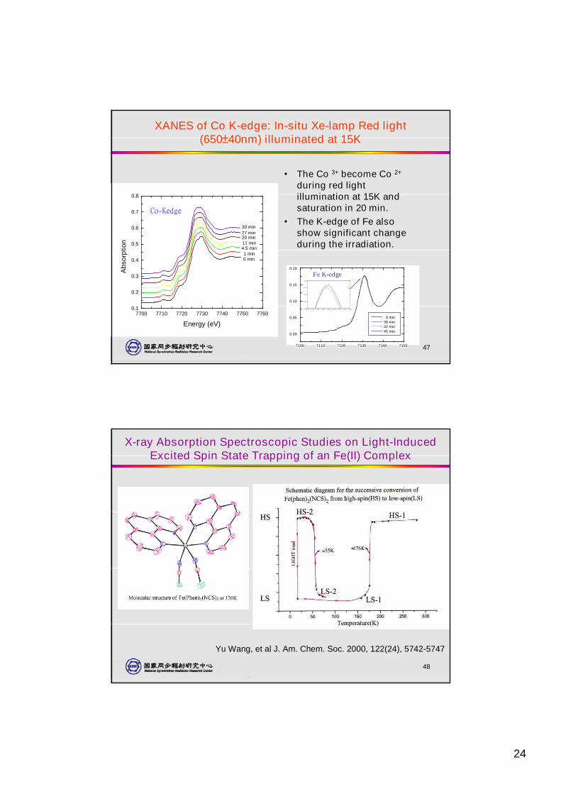
XANES of Co K-edge: In-situ Xe-lamp Red light(650±40nm) illuminated at 15K
• The Co 3+ become Co 2+
during red lightillumination at 15K andsaturation in 20 min.
• The K-edge of Fe alsoshow significant changeduring the irradiation.
7700 7710 7720 7730 7740 7750 77600.1
0.2
0.3
0.4
0.5
0.6
0.7
0.8
Co-Kedge
0 min1 min
4.5 min11 min20 min27 min30 min
Abs
orpt
ion
Energy (eV)
7100 7110 7120 7130 7140 7150
0.00
0.05
0.10
0.15
0.20
0 min38 min42 min45 min
7129.0 7129.5 7130.0 7130.5 7131.0 7131.5 7132.0 7132.5 7133.00.160
0.165
0.170
0.175
0.180
Fe K-edge
48
Yu Wang, et al J. Am. Chem. Soc. 2000, 122(24), 5742-5747
X-ray Absorption Spectroscopic Studies on Light-InducedExcited Spin State Trapping of an Fe(II) Complex
25
49
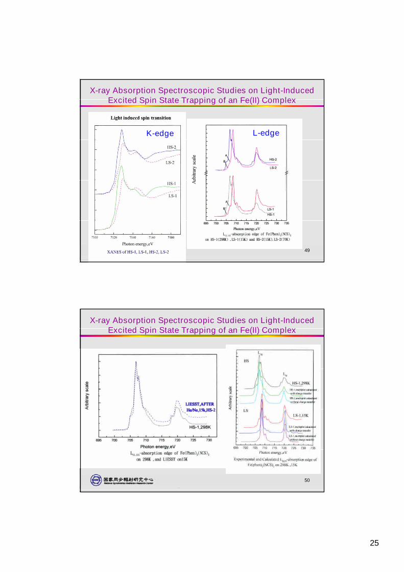
X-ray Absorption Spectroscopic Studies on Light-InducedExcited Spin State Trapping of an Fe(II) Complex
K-edge L-edge
50
X-ray Absorption Spectroscopic Studies on Light-InducedExcited Spin State Trapping of an Fe(II) Complex
26
51
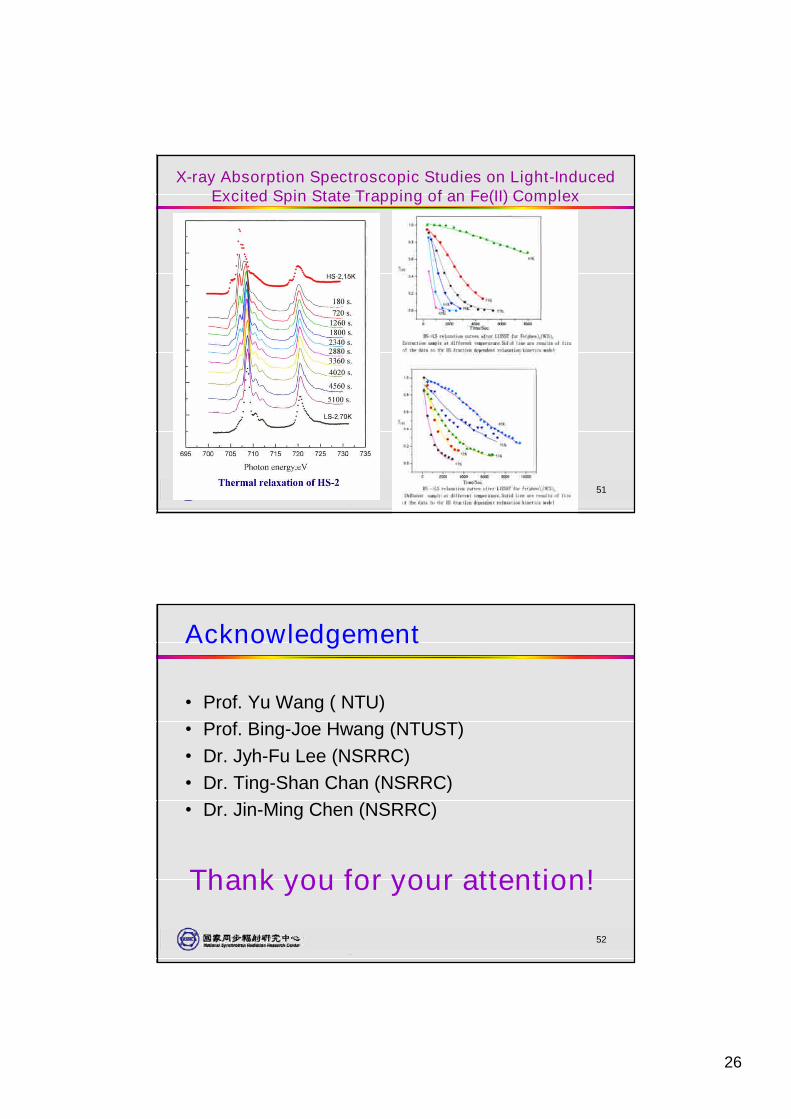
X-ray Absorption Spectroscopic Studies on Light-InducedExcited Spin State Trapping of an Fe(II) Complex
52
Acknowledgement
•Prof. Yu Wang ( NTU)•Prof. Bing-Joe Hwang (NTUST)•Dr. Jyh-Fu Lee (NSRRC)•Dr. Ting-Shan Chan (NSRRC)•Dr. Jin-Ming Chen (NSRRC)
Thank you for your attention!