Wnt/β-Catenin Signaling in Development and Disease fileexhibits alternating denticle and naked...
12
Leading Edge Review Cell 127, November 3, 2006 ©2006 Elsevier Inc. 469 The mouse wnt1 gene, originally named Int-1 , was iden- tified in 1982 by Nusse and Varmus as a preferential integration site for the Mouse Mammary Tumor Virus in virally induced breast tumors (Nusse and Varmus, 1982). When sequenced, the Wnt1 proto-oncogene was seen to encode a secreted protein that is cysteine rich. Subsequently, Drosophila wingless (wg), which controls segment polarity during larval development (Nüsslein- Volhard and Wieschaus, 1980), was shown to be a fly homolog of Wnt1 (Rijsewijk et al., 1987). Segmentation of the epidermis of wg mutant fly embryos is severely impaired as evidenced by abnormalities in the overlying ventral cuticle. In contrast to the wild-type cuticle, which exhibits alternating denticle and naked belts, the wg cuti- cle is completely covered with denticles. Fly embryos carrying mutations in the porcupine, dishevelled, and armadillo genes display similar cuticle abnormalities to wg mutant embryos, whereas mutations in shaggy/ zeste-white 3 cause the opposite phenotype, a naked cuticle. Epistatic analysis of cuticle structure in double mutants indicated that these genes constituted the core of a new signal transduction cascade (Siegfried et al., 1992; Noordermeer et al., 1994; Peifer et al., 1994). In 1989, McMahon and Moon (McMahon and Moon, 1989) observed a duplication of the body axis in Xeno- pus following injection of mouse Wnt1 mRNA into ventral blastomeres of embryos at the 4-cell stage. This observa- tion supported the notion that Wnt signaling was shared between vertebrates and invertebrates and, moreover, provided a rapid and convenient assay to study compo- nents of the Wnt pathway in vertebrates. Axis duplication was also induced by Dishevelled (Dsh), β-catenin (the vertebrate homolog of armadillo), and a dominant-nega- tive version of glycogen synthase kinase 3 (GSK3), the vertebrate homolog of shaggy/zeste-white 3 (Dominguez et al., 1995; Guger and Gumbiner, 1995; He et al., 1995). Although long elusive, the specific Wnt signal that trig- gers axis induction in Xenopus was identified as Wnt11 by Heasman and colleagues last year (Tao et al., 2005). The combined observations made in Drosophila and Xenopus delineated a highly conserved signaling path- way, activated by secreted Wnt proteins. Independent of these studies, the adenomatous polyposis coli (APC) gene was discovered in a hereditary cancer syndrome termed familial adenomatous polyposis (FAP) (Kinzler et al., 1991; Nishisho et al., 1991). Soon after, the large cytoplasmic APC protein was found to interact with β-catenin (Rubinfeld et al., 1993; Su et al., 1993). This observation provided the first connection between the Wnt pathway and human cancer. Genome sequencing has since revealed that mammalian species have roughly 20 secreted Wnt proteins, which can be divided into 12 conserved Wnt subfamilies. Of these, only 6 subfamilies have counterparts in ecdysozoan ani- mals such as Drosophila and Caenorhabditis. In contrast, at least 11 of the Wnt subfamilies occur in the genome of a cnidarian (the sea anemone Nematostella vectensis). This finding suggests that some Wnt subfamilies were lost during the evolution of the ecdysozoan lineage but more importantly reveals that a complex inventory of Wnt factors was present in multicellular animals well before the Cam- brian explosion (550 million years ago). Thus, comparative genomic analysis underscores the crucial role that Wnt genes play in organismal patterning throughout the animal kingdom (Kusserow et al., 2005). Currently, three different pathways are believed to be activated upon Wnt receptor activation: the canonical Wnt/β-catenin cascade, the noncanonical planar cell polarity (PCP) pathway, and the Wnt/Ca 2+ pathway. Of these three, the canonical pathway is best understood and is the primary subject of this review. For recent com- prehensive overviews on the other Wnt signaling path- ways, the reader is referred to Katoh (2005) and Kohn and Moon (2005). This review discusses how Wnt proteins are produced and secreted and how they activate the canoni- cal Wnt signaling pathway in recipient cells. Further, the review examines the roles of the canonical Wnt pathway in development, tissue self-renewal, and cancer. Wnt/ β-Catenin Signaling in Development and Disease Hans Clevers 1, * 1 Hubrecht Laboratory and Utrecht University, Uppsalalaan 8, 3584CT, Utrecht, the Netherlands *Contact: [email protected] DOI 10.1016/j.cell.2006.10.018 A remarkable interdisciplinary effort has unraveled the WNT (Wingless and INT-1) signal transduction cascade over the last two decades. Wnt genes encode small secreted proteins that are found in all animal genomes. Wnt signaling is involved in virtually every aspect of embryonic development and also controls homeostatic self-renewal in a number of adult tissues. Germline mutations in the Wnt pathway cause several hereditary diseases, and somatic mutations are associated with cancer of the intestine and a variety of other tissues.
-
Upload
truongthien -
Category
Documents
-
view
215 -
download
0
Transcript of Wnt/β-Catenin Signaling in Development and Disease fileexhibits alternating denticle and naked...
Leading Edge
Review
Wnt/β-Catenin Signaling in Development and DiseaseHans Clevers1,*1Hubrecht Laboratory and Utrecht University, Uppsalalaan 8, 3584CT, Utrecht, the Netherlands*Contact: [email protected] 10.1016/j.cell.2006.10.018
A remarkable interdisciplinary effort has unraveled the WNT (Wingless and INT-1) signal transduction cascade over the last two decades. Wnt genes encode small secreted proteins that are found in all animal genomes. Wnt signaling is involved in virtually every aspect of embryonic development and also controls homeostatic self-renewal in a number of adult tissues. Germline mutations in the Wnt pathway cause several hereditary diseases, and somatic mutations are associated with cancer of the intestine and a variety of other tissues.
The mouse wnt1 gene, originally named Int-1, was iden-tified in 1982 by Nusse and Varmus as a preferential integration site for the Mouse Mammary Tumor Virus in virally induced breast tumors (Nusse and Varmus, 1982). When sequenced, the Wnt1 proto-oncogene was seen to encode a secreted protein that is cysteine rich. Subsequently, Drosophila wingless (wg), which controls segment polarity during larval development (Nüsslein-Volhard and Wieschaus, 1980), was shown to be a fly homolog of Wnt1 (Rijsewijk et al., 1987). Segmentation of the epidermis of wg mutant fly embryos is severely impaired as evidenced by abnormalities in the overlying ventral cuticle. In contrast to the wild-type cuticle, which exhibits alternating denticle and naked belts, the wg cuti-cle is completely covered with denticles. Fly embryos carrying mutations in the porcupine, dishevelled, and armadillo genes display similar cuticle abnormalities to wg mutant embryos, whereas mutations in shaggy/zeste-white 3 cause the opposite phenotype, a naked cuticle. Epistatic analysis of cuticle structure in double mutants indicated that these genes constituted the core of a new signal transduction cascade (Siegfried et al., 1992; Noordermeer et al., 1994; Peifer et al., 1994).
In 1989, McMahon and Moon (McMahon and Moon, 1989) observed a duplication of the body axis in Xeno-pus following injection of mouse Wnt1 mRNA into ventral blastomeres of embryos at the 4-cell stage. This observa-tion supported the notion that Wnt signaling was shared between vertebrates and invertebrates and, moreover, provided a rapid and convenient assay to study compo-nents of the Wnt pathway in vertebrates. Axis duplication was also induced by Dishevelled (Dsh), β-catenin (the vertebrate homolog of armadillo), and a dominant-nega-tive version of glycogen synthase kinase 3 (GSK3), the vertebrate homolog of shaggy/zeste-white 3 (Dominguez et al., 1995; Guger and Gumbiner, 1995; He et al., 1995). Although long elusive, the specific Wnt signal that trig-gers axis induction in Xenopus was identified as Wnt11 by Heasman and colleagues last year (Tao et al., 2005).
The combined observations made in Drosophila and Xenopus delineated a highly conserved signaling path-way, activated by secreted Wnt proteins. Independent of these studies, the adenomatous polyposis coli (APC) gene was discovered in a hereditary cancer syndrome termed familial adenomatous polyposis (FAP) (Kinzler et al., 1991; Nishisho et al., 1991). Soon after, the large cytoplasmic APC protein was found to interact with β-catenin (Rubinfeld et al., 1993; Su et al., 1993). This observation provided the first connection between the Wnt pathway and human cancer.
Genome sequencing has since revealed that mammalian species have roughly 20 secreted Wnt proteins, which can be divided into 12 conserved Wnt subfamilies. Of these, only 6 subfamilies have counterparts in ecdysozoan ani-mals such as Drosophila and Caenorhabditis. In contrast, at least 11 of the Wnt subfamilies occur in the genome of a cnidarian (the sea anemone Nematostella vectensis). This finding suggests that some Wnt subfamilies were lost during the evolution of the ecdysozoan lineage but more importantly reveals that a complex inventory of Wnt factors was present in multicellular animals well before the Cam-brian explosion (550 million years ago). Thus, comparative genomic analysis underscores the crucial role that Wnt genes play in organismal patterning throughout the animal kingdom (Kusserow et al., 2005).
Currently, three different pathways are believed to be activated upon Wnt receptor activation: the canonical Wnt/β-catenin cascade, the noncanonical planar cell polarity (PCP) pathway, and the Wnt/Ca2+ pathway. Of these three, the canonical pathway is best understood and is the primary subject of this review. For recent com-prehensive overviews on the other Wnt signaling path-ways, the reader is referred to Katoh (2005) and Kohn and Moon (2005). This review discusses how Wnt proteins are produced and secreted and how they activate the canoni-cal Wnt signaling pathway in recipient cells. Further, the review examines the roles of the canonical Wnt pathway in development, tissue self-renewal, and cancer.
Cell 127, November 3, 2006 ©2006 Elsevier Inc. 469
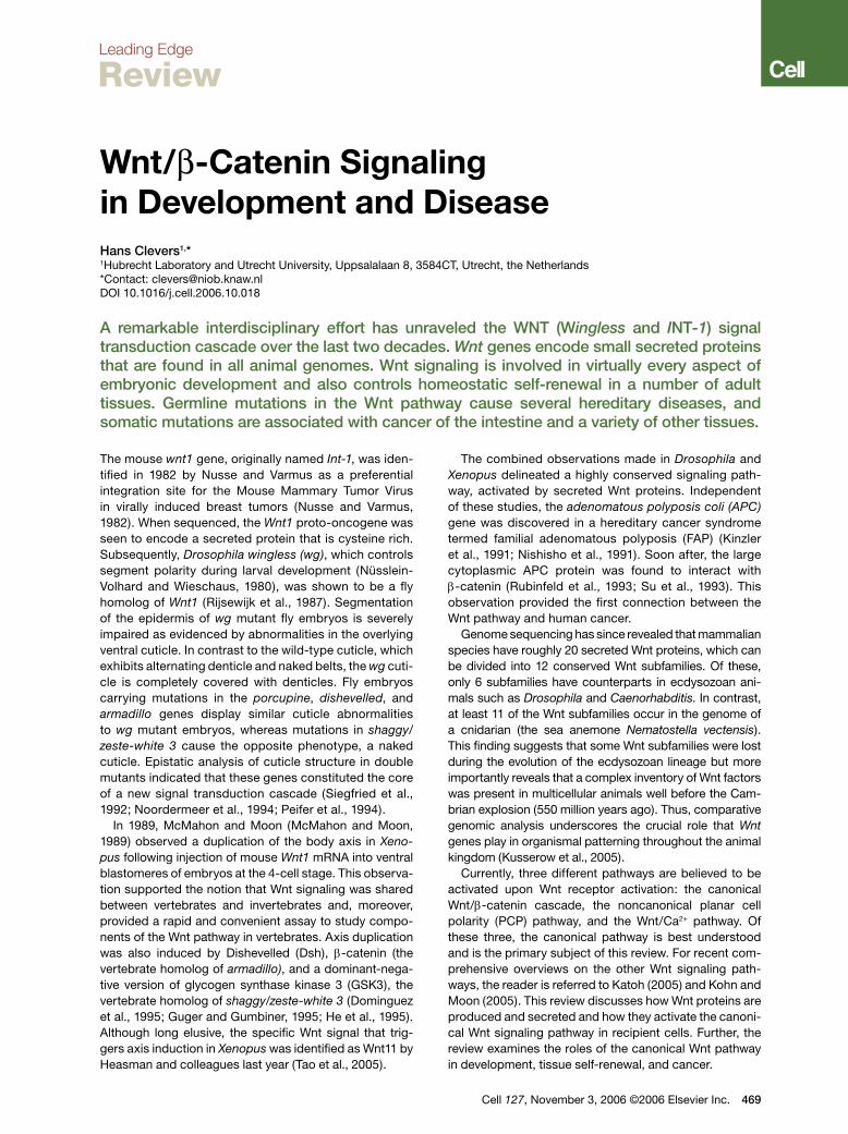
Wnt Protein SecretionWnt proteins are characterized by a high number of con-served cysteine residues. Although Wnt proteins carry an N-terminal signal peptide and are secreted, they are relatively insoluble. This insolubility has been attributed to a particular protein modification, cysteine palmitoyla-tion, which is essential for Wnt function (Willert et al., 2003). Hofmann (2000) reported that a Drosophila gene required in the Wnt-secreting cell, termed porcupine, displays homology to acyl-transferases, enzymes that acylate a variety of substrates in the endoplasmic reticu-lum. Thus, porcupine and its worm homolog mom-1 are believed to encode the enzyme that is responsible for Wnt palmitoylation (Zhai et al., 2004).
Recently, Banziger et al. (2006) and Bartscherer et al. (2006) uncovered in Drosophila another conserved gene that is essential for Wnt secretion, named wntless (wls) and evenness interrupted (evi), respectively. The gene encodes a seven-pass transmembrane protein that is conserved from worms (mom-3) to man (hWLS). In the absence of Wls/evi, Wnts are retained inside the cell that produces them. The Wntless protein resides pri-marily in the Golgi apparatus, where it colocalizes and physically interacts with Wnts. A genetic screen in C. elegans revealed that the retromer, a multiprotein com-plex involved in intracellular trafficking and conserved
Figure 1. Wnt SecretionTo be secreted, Wnt proteins in the endoplasmic reticulum (ER) need to be palmitoylated by the action of Porcupine. Wnt proteins also re-quire Wntless (Wls/Evi) in order to be routed to the outside of the cell. Loading onto lipoprotein particles may occur in a dedicated endo/ex-ocytic compartment. The retromer complex may shuttle Wls between the Golgi and the endo/exocytic compartment.
470 Cell 127, November 3, 2006 ©2006 Elsevier Inc.
from yeast to man, is also essential for Wnt secretion and for the generation of a Wnt gradient (Coudreuse et al., 2006). An attractive hypothesis is that the retromer complex is involved in recycling a Wnt cargo receptor (such as Wntless) between the default secretory path-way and a compartment dedicated to Wnt secretion (see Figure 1).
Wnt is thought to act as a morphogen (that is, a long-range signal whose activity is concentration depend-ent) (reviewed in Logan and Nusse, 2004). However, it is unclear how these long-range gradients are generated. It is conceivable that the palmitoyl moiety constrains movement away from membranes or lipid particles. Thus, Wnts may be tethered to intercellular transport vesicles or lipoprotein particles (Panakova et al., 2005). Alternatively, Wnts may be transported by cytonemes, which are long, thin filopodial processes. Additionally, studies in Drosophila suggest a role for extracellular heparan sulfate proteoglycans (HSPG) in the transport or stabilization of Wnt proteins. For instance, flies car-rying mutations in Dally, a GPI-anchored HSPG, or in genes encoding enzymes that modify HSPGs resemble wingless mutants (reviewed in Lin, 2004).
Receptors, Agonists, and Antagonists for WntWnts bind Frizzled (Fz) proteins, which are seven-pass transmembrane receptors with an extracellular N-ter-minal cysteine-rich domain (CRD) (Bhanot et al., 1996). The Wnt-Fz interaction appears promiscuous, in that a single Wnt can bind multiple Frizzled proteins (e.g., Bhanot et al., 1996) and vice versa. In binding Wnt, Fzs cooperate with a single-pass transmembrane molecule of the LRP family known as Arrow in Drosophila (Wehrli et al., 2000) and LRP5 and -6 in vertebrates (Pinson et al., 2000; Tamai et al., 2000). The transport of Arrow/LRP5/6 to the cell surface is dependent on a chaper-one called Boca in Drosophila and Mesd in mice (Culi and Mann, 2003; Hsieh et al., 2003). And consistent with a role of the Boca/Mesd chaperone in the transport of Arrow/LRP5/6 transport, mutations in Boca and Mesd resemble loss of Arrow/LRP5/6. Although it has not been formally demonstrated that Wnt molecules form trimeric complexes with LRP5/6 and Frizzled, surface expression of both receptors is required to initiate the Wnt signal.
Derailed, a transmembrane tyrosine kinase receptor from the RYK subfamily, is an unusual Wnt receptor. Dro-sophila Wnt5 controls axon guidance in the central nerv-ous system. Embryos lacking Dwnt-5 resemble those lacking Derailed, that is, they generate aberrant neuronal projections across the midline (Yoshikawa et al., 2003). Derailed binds DWnt-5 through its extracellular WIF (Wnt inhibitory factor) domain. Signaling events downstream of this alternative Wnt receptor remain unclear. Some-what unexpectedly, the Derailed kinase domain may be dispensable for signaling. Lu et al. (2004) propose that, unlike the Drosophila Ryk homolog Derailed, mammalian Ryk functions as a coreceptor along with Fz. Mammalian
Ryk binds Dishevelled to activate the canonical Wnt/β-catenin signaling pathway. Another tyrosine kinase receptor, Ror2, harbors a Wnt binding CRD motif. Wnt5a can engage Ror2 to inhibit the canonical Wnt signaling pathway, although paradoxically Wnt5a can also activate the canonical pathway by directly engaging Fz4 (Mikels and Nusse, 2006) and Fz5 (He et al., 1997).
At least two types of proteins that are unrelated to Wnt factors activate the Frizzled/LRP receptors. One of these factors is the cysteine-knot protein Norrin, which is mutated in Norrie disease, a developmental disorder characterized by vascular abnormalities in the eye and blindness. Norrin binds with high affinity to Frizzled-4 and activates the canonical signaling pathway in an LRP5/6-dependent fashion (Xu et al., 2004). Other fac-tors that activate the canonical Wnt signaling pathway are R-spondins, which are thrombospondin domain-containing proteins. In Xenopus, R-spondin-2 is a Wnt agonist that synergizes with Wnts to activate β-catenin (Kazanskaya et al., 2004). Human R-spondin-1 has been found to strongly promote the proliferation of intestinal crypt cells, a process which involves the stabilization of β-catenin (Kim et al., 2005). Indeed, studies in cultured cells demonstrate that R-spondins can physically inter-act with the extracellular domains of LRP6 and Fzd8 and activate Wnt reporter genes (Nam et al., 2006).
The secreted Dickkopf (Dkk) proteins inhibit Wnt signaling by direct binding to LRP5/6 (Glinka et al., 1998). Through this interaction, Dkk1 crosslinks LRP6 to another class of transmembrane molecules, the Kre-mens (Mao et al., 2002), thus promoting the internaliza-tion and inactivation of LRP6. An unrelated secreted Wnt inhibitor, Wise, also acts by binding to LRP (Itasaki et al., 2003), as does the WISE family member SOST (Li et al., 2005; Semenov et al., 2005).
Soluble Frizzled-Related Proteins (SFRPs) resemble the ligand-binding CRD domain of the Frizzled family of Wnt receptors (Hoang et al., 1996). WIF proteins are secreted molecules with similarity to the extracellular portion of the Derailed/RYK class of transmembrane Wnt receptors (Hsieh et al., 1999). SFRPs and WIFs are believed to function as extracellular Wnt inhibitors (reviewed in Logan and Nusse, 2004) but, depending on context, may also promote signaling by Wnt stabilization or by facilitating Wnt secretion or transport.
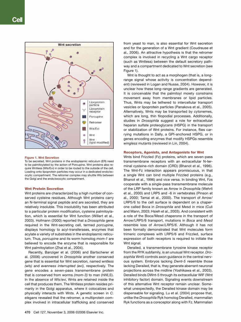
Canonical Wnt SignalingOnce bound by their cognate ligands, the Fz/LRP coreceptor complex activates the canonical signal-ing pathway (Figure 2). Fz can physically interact with Dsh, a cytoplasmic protein that functions upstream of β-catenin and the kinase GSK-3. Wnt signaling controls phosphorylation of Dsh (reviewed in Wallingford and Habas, 2005). However, it remains unclear whether the binding of Wnt to Fz regulates a direct Fz-Dsh interac-tion, nor is it known how Dsh phosphorylation is con-trolled or how phosphorylated Dsh functions in Wnt signal transduction.
Recent studies have indicated that the coreceptor LRP5/6 interacts with Axin through five phosphorylated PPP(S/T)P repeats in the cytoplasmic tail of LRP (David-son et al., 2005; Zeng et al., 2005). Wnts are thought to induce the phosphorylation of the cytoplasmic tail of LRP, thus regulating the docking of Axin. GSK3 phos-phorylates the PPP(S/T)P motif, whereas caseine kinase I-γ (CK1γ) phosphorylates multiple motifs close to the GSK3 sites. CK1γ is unique within the CK1 family in that it is anchored in the membrane through C-terminal palmi-toylation. Both kinases are essential for signal initia-tion. It remains presently debated whether Wnt controls GSK3-mediated phosphorylation of LRP5/6 (Zeng et al., 2005) or whether CK1γ is the kinase regulated by Wnt (Davidson et al., 2005). When bound to their respective membrane receptors, Dsh and Axin may cooperatively mediate downstream activation events by heterodimeri-zation through their respective DIX (Dishevelled-Axin) domains.The Cytoplasmic Destruction ComplexThe central player in the canonical Wnt cascade is β-catenin, a cytoplasmic protein whose stability is regu-lated by the destruction complex. The tumor suppressor protein Axin acts as the scaffold of this complex as it directly interacts with all other components—β-catenin, the tumor suppressor protein APC, and the two kinase
Figure 2. Canonical Wnt Signaling(Left panel) When Wnt receptor complexes are not bound by ligand, the serine/threonine kinases, CK1 and GSK3α/β, phosphorylate β-catenin. Phosphorylated β-catenin is recognized by the F box/WD repeat protein β-TrCP, a component of a dedicated E3 ubiquitin ligase complex. Following ubiquitination, β-catenin is targeted for rapid de-struction by the proteasome. In the nucleus, the binding of Groucho to TCF (T cell factor) inhibits the transcription of Wnt target genes. (Right panel) Once bound by Wnt, the Frizzled(Fz)/LRP coreceptor complex activates the canonical signaling pathway. Fz interacts with Dsh, a cy-toplasmic protein that functions upstream of β-catenin and the kinase GSK3β. Wnt signaling controls phosphorylation of Dishevelled (Dsh). Wnts are thought to induce the phosphorylation of LRP by GSK3β and casein kinase I-γ (CK1γ), thus regulating the docking of Axin. The recruitment of Axin away from the destruction complex leads to the stabilization of β-catenin. In the nucleus, β-catenin displaces Groucho from Tcf/Lef to promote the transcription of Wnt target genes.
Cell 127, November 3, 2006 ©2006 Elsevier Inc. 471
families (CK1α, -δ, -ε and GSK3α and -β [reviewed in Price, 2006]). When WNT receptor complexes are not engaged, CK1 and GSK3α/β sequentially phosphor-ylate β-catenin at a series of highly conserved Ser/Thr residues near its N terminus (Figure 2). Phosphorylated β-catenin is then recognized by the F box/WD repeat protein β-TrCP, a component of a dedicated E3 ubiq-uitin ligase complex. As a consequence, β-catenin is ubiquitinated and targeted for rapid destruction by the proteasome (Aberle et al., 1997). Note that the CK1 and GSK3 kinases perform paradoxical roles in the Wnt pathway. At the level of the LRP coreceptor they act as agonists, whereas in the destruction complex they act as antagonists
Although genetic observations imply an essential role for APC in the destruction complex, there is no consen-sus on its specific molecular activity. APC has a series of 15 and 20 amino acid repeats with which it interacts with β-catenin. Three Axin-binding motifs are interspersed between these β-catenin-binding motifs. Increasing the expression of Axin in cancer cells that lack APC restores the activity of the destruction complex, implying that APC is only essential when Axin levels are limiting. Quantita-tively, Axin indeed appears to be the limiting factor (Lee et al., 2003) and may be the key scaffolding molecule that promotes the rapid assembly and disassembly of the destruction complex.
Given that CK1, Dsh, β-TrCP, and GSK3 participate in other signaling pathways, low levels of Axin may insu-late the Wnt pathway from changes in the abundance or activity of these signaling components. It has been proposed that APC is required for efficient shuttling and loading/unloading of β-catenin onto the cytoplasmic destruction complex. Both APC and Axin can them-selves be phosphorylated by their associated kinases,
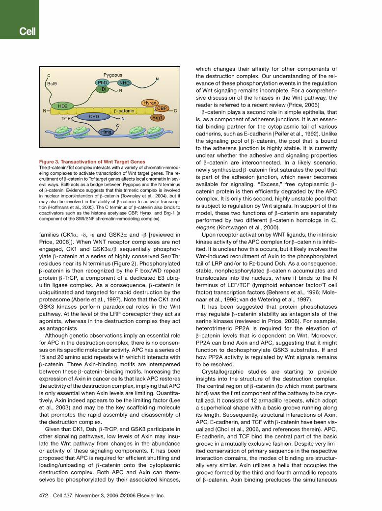
Figure 3. Transactivation of Wnt Target GenesThe β-catenin/Tcf complex interacts with a variety of chromatin-remod-eling complexes to activate transcription of Wnt target genes. The re-cruitment of β-catenin to Tcf target genes affects local chromatin in sev-eral ways. Bcl9 acts as a bridge between Pygopus and the N terminus of β-catenin. Evidence suggests that this trimeric complex is involved in nuclear import/retention of β-catenin (Townsley et al., 2004), but it may also be involved in the ability of β-catenin to activate transcrip-tion (Hoffmans et al., 2005). The C terminus of β-catenin also binds to coactivators such as the histone acetylase CBP, Hyrax, and Brg-1 (a component of the SWI/SNF chromatin-remodeling complex).
472 Cell 127, November 3, 2006 ©2006 Elsevier Inc.
which changes their affinity for other components of the destruction complex. Our understanding of the rel-evance of these phosphorylation events in the regulation of Wnt signaling remains incomplete. For a comprehen-sive discussion of the kinases in the Wnt pathway, the reader is referred to a recent review (Price, 2006)β-catenin plays a second role in simple epithelia, that
is, as a component of adherens junctions. It is an essen-tial binding partner for the cytoplasmic tail of various cadherins, such as E-cadherin (Peifer et al., 1992). Unlike the signaling pool of β-catenin, the pool that is bound to the adherens junction is highly stable. It is currently unclear whether the adhesive and signaling properties of β-catenin are interconnected. In a likely scenario, newly synthesized β-catenin first saturates the pool that is part of the adhesion junction, which never becomes available for signaling. “Excess,” free cytoplasmic β-catenin protein is then efficiently degraded by the APC complex. It is only this second, highly unstable pool that is subject to regulation by Wnt signals. In support of this model, these two functions of β-catenin are separately performed by two different β-catenin homologs in C. elegans (Korswagen et al., 2000).
Upon receptor activation by WNT ligands, the intrinsic kinase activity of the APC complex for β-catenin is inhib-ited. It is unclear how this occurs, but it likely involves the Wnt-induced recruitment of Axin to the phosphorylated tail of LRP and/or to Fz-bound Dsh. As a consequence, stable, nonphosphorylated β-catenin accumulates and translocates into the nucleus, where it binds to the N terminus of LEF/TCF (lymphoid enhancer factor/T cell factor) transcription factors (Behrens et al., 1996; Mole-naar et al., 1996; van de Wetering et al., 1997).
It has been suggested that protein phosphatases may regulate β-catenin stability as antagonists of the serine kinases (reviewed in Price, 2006). For example, heterotrimeric PP2A is required for the elevation of β-catenin levels that is dependent on Wnt. Moroever, PP2A can bind Axin and APC, suggesting that it might function to dephosphorylate GSK3 substrates. If and how PP2A activity is regulated by Wnt signals remains to be resolved.
Crystallographic studies are starting to provide insights into the structure of the destruction complex. The central region of β-catenin (to which most partners bind) was the first component of the pathway to be crys-tallized. It consists of 12 armadillo repeats, which adopt a superhelical shape with a basic groove running along its length. Subsequently, structural interactions of Axin, APC, E-cadherin, and TCF with β-catenin have been vis-ualized (Choi et al., 2006, and references therein). APC, E-cadherin, and TCF bind the central part of the basic groove in a mutually exclusive fashion. Despite very lim-ited conservation of primary sequence in the respective interaction domains, the modes of binding are structur-ally very similar. Axin utilizes a helix that occupies the groove formed by the third and fourth armadillo repeats of β-catenin. Axin binding precludes the simultaneous
interaction with other β-catenin partners in this region. Based on this observation, it is suggested that a key function of APC is to remove phosphorylated β-catenin from the active site of the complex (Xing et al., 2003). In a further study, the structure of Axin bound to APC (Spink et al., 2000) was solved. These studies form step-ping stones to a better understanding of the dynamics of the destruction complex. Unfortunately, biochemical studies of the destruction complex in its different activa-tion states are sorely lacking.Nuclear EventsUpon stabilization by Wnt signals, β-catenin enters the nucleus to reprogram the responding cell (Figure 3). There is no consensus on the mechanism by which β-catenin travels between the cytoplasm and the nucleus. In many cases, cells that undergo Wnt signaling may actually display an overall rise in β-catenin protein without a clear nuclear preference. β-catenin’s nuclear import is independent of the Nuclear Localization Sig-nal/importin machinery. β-catenin itself is a close rela-tive of importin/karyopherins and directly interacts with nuclear pore components. Two proteins, Tcf and Pygo-pus are proposed to anchor β-catenin in the nucleus, although β-catenin can still localize to the nucleus in the absence of either of the two (reviewed in Staedeli et al., 2006). β-catenin can also be actively transported back to the cytoplasm, by either an intrinsic export signal or as cargo of Axin (Cong and Varmus, 2004) or APC (Rosin-Arbesfeld et al., 2000) that shuttle between cyto-plasm and nucleus.
Whereas the fly and worm genomes both encode a single Tcf protein, the vertebrate genome harbors four Tcf/Lef genes. Tcf factors bind their cognate motif in an unusual fashion, i.e., in the minor groove of the DNA helix, while inducing a dramatic bend of over 90°. Tcf target sites are highly conserved between the four ver-tebrate Tcf/Lef proteins and Drosophila Tcf. These sites resemble AGATCAAAGG (van de Wetering et al., 1997). Wnt/TCF reporter plasmids such as pTOPflash (Korinek et al., 1997), widely used to measure Wnt pathway acti-vation, consist of concatamers of 3–10 of these binding motifs cloned upstream of a minimal promoter. The four vertebrate TCF/LEF differ dramatically in their embry-onic and adult expression domains, yet they are highly similar biochemically, explaining the extensive redun-dancy unveiled in double knockout experiments (as in Galceran et al., 1999).
In the absence of Wnt signals, Tcf acts as a tran-scriptional repressor by forming a complex with Grou-cho/Grg/TLE proteins (Cavallo et al., 1998; Roose et al., 1998). The interaction of β-catenin with the N terminus of Tcf (Behrens et al., 1996; Molenaar et al., 1996; van de Wetering et al., 1997) transiently converts it into an activator, translating the Wnt signal into the transient transcription of Tcf target genes. To accomplish this, β-catenin physically displaces Groucho from Tcf/Lef (Daniels and Weis, 2005). The recruitment of β-catenin to Tcf target genes affects local chromatin in several
ways. Its C terminus is a potent transcriptional activa-tor in transient reporter gene assays (van de Wetering et al., 1997). It binds coactivators such as the histone acetylase CBP and Brg-1, a component of the SWI/SNF chromatin remodeling complex (reviewed in Staedeli et al., 2006). A recent study implies that the human and fly homologs of yeast Cdc37 (Parafibromin and Hyrax, respectively) also interact with the C-terminal transacti-vation domain of β-catenin to activate target gene tran-scription (Mosimann et al., 2006). Cdc37 is a component of the PAF complex. In yeast the PAF complex directly interacts with RNA polymerase II to regulate transcrip-tion initiation and elongation.
Two dedicated, nuclear partners of the TCF/β-cat-enin complex, Legless/Bcl9 and Pygopus, were recently found in genetic screens in Drosophila (Kramps et al., 2002; Parker et al., 2002; Thompson et al., 2002). Muta-tions in these genes result in phenotypes similar to wingless, and overexpression of both genes promotes TCF/β-catenin activity in mammalian cells (Thompson et al., 2002). Bcl9 bridges Pygopus to the N terminus of β-catenin. The formation of this trimeric complex has been implicated in nuclear import/retention of β-catenin (Townsley et al., 2004) but may also directly contribute to the ability of β-catenin to transactivate transcription (Hoffmans et al., 2005). Although most if not all Wnt sig-naling events in Drosophila appear to be dependent on Bcl9 and Pygopus, it is currently unclear if this holds true in vertebrate development.
Tcf itself can be regulated by phosphorylation. The MAP kinase-related protein kinase NLK/Nemo (Ishitani et al., 1999) phosphorylates Tcf, thereby decreasing the DNA-binding affinity of the β-catenin/Tcf complex and inhibiting transcriptional regulation of Wnt target genes. In C. elegans, LIT-1/NLK-dependent phosphorylation results in PAR-5/14-3-3- and CRM-1-dependent nuclear export of POP-1/Tcf (Meneghini et al., 1999; Lo et al., 2004). And lastly, a recent study utilizing chromatin immunoprecipitations suggests that APC, independent of its role in the cytoplasmic destruction complex, acts on chromatin to facilitate CtBP-mediated repression of Wnt target genes in normal, but not in colorectal cancer cells (Sierra et al., 2006).
Wnt Target GenesLoss of components of the Wnt pathway can produce dramatic phenotypes that affect a wide variety of organs and tissues. A popular view equates Wnt signaling with maintenance or activation of stem cells (Reya and Cle-vers, 2005). It should be realized, however, that Wnt signals ultimately activate transcriptional programs and that there is no intrinsic restriction in the type of bio-logical event that may be controlled by these programs. Thus, Wnt signals may promote cell proliferation and tissue expansion but also control fate determination or terminal differentiation of postmitotic cells. Sometimes, these disparate events, proliferation and terminal differ-entiation, can be activated by Wnt in different cell types
Cell 127, November 3, 2006 ©2006 Elsevier Inc. 473
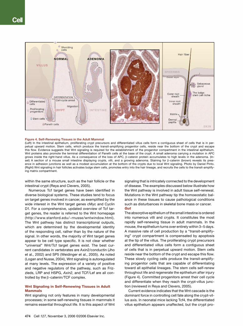
Figure 4. Self-Renewing Tissues in the Adult Mammal(Left) In the intestinal epithelium, proliferating crypt precursors and differentiated villus cells form a contiguous sheet of cells that is in per-petual upward motion. Stem cells, which produce the transit-amplifying progenitor cells, reside near the bottom of the crypt and escape this flow. Evidence suggests that Wnt signaling is required for the establishment of the progenitor compartment in the intestinal epithelium. Wnt proteins also promote the terminal differentiation of Paneth cells at the base of the crypt. A small adenoma carrying a mutation in APC grows inside the right-hand villus. As a consequence of the loss of APC, β-catenin protein accumulates to high levels in the adenoma. (In-set) A section of a mouse small intestine displaying crypts, villi, and a growing adenoma. Staining for β-catenin (brown) reveals its pres-ence in adhesion junctions as well as a modest accumulation at the bottom of the crypts due to local Wnt signaling. Photo by Daniel Pinto. (Right) Wnt signaling in hair follicles activates bulge stem cells, promotes entry into the hair lineage, and recruits the cells to the transit-amplify-ing matrix compartment.
within the same structure, such as the hair follicle or the intestinal crypt (Reya and Clevers, 2005).
Numerous Tcf target genes have been identified in diverse biological systems. These studies tend to focus on target genes involved in cancer, as exemplified by the wide interest in the Wnt target genes cMyc and Cyclin D1. For a comprehensive, updated overview of Tcf tar-get genes, the reader is referred to the Wnt homepage (http://www.stanford.edu/~rnusse/wntwindow.html). The Wnt pathway has distinct transcriptional outputs, which are determined by the developmental identity of the responding cell, rather than by the nature of the signal. In other words, the majority of Wnt target genes appear to be cell type specific. It is not clear whether “universal” Wnt/Tcf target genes exist. The best cur-rent candidates in vertebrates are Axin2/conductin (Jho et al., 2002) and SP5 (Weidinger et al., 2005). As noted (Logan and Nusse, 2004), Wnt signaling is autoregulated at many levels. The expression of a variety of positive and negative regulators of the pathway, such as Friz-zleds, LRP and HSPG, Axin2, and TCF/Lef are all con-trolled by the β-catenin/TCF complex.
Wnt Signaling in Self-Renewing Tissues in Adult MammalsWnt signaling not only features in many developmental processes; in some self-renewing tissues in mammals it remains essential throughout life. It is this aspect of Wnt
474 Cell 127, November 3, 2006 ©2006 Elsevier Inc.
signaling that is intricately connected to the development of disease. The examples discussed below illustrate how the Wnt pathway is involved in adult tissue self-renewal. Mutations in the Wnt pathway tip the homoeostatic bal-ance in these tissues to cause pathological conditions such as disturbances in skeletal bone mass or cancer.GutThe absorptive epithelium of the small intestine is ordered into numerous villi and crypts. It constitutes the most rapidly self-renewing tissue in adult mammals. In the mouse, the epithelium turns over entirely within 3–5 days. A massive rate of cell production by a “transit-amplify-ing” crypt compartment is compensated by apoptosis at the tip of the villus. The proliferating crypt precursors and differentiated villus cells form a contiguous sheet of cells that is in perpetual upward motion. Stem cells reside near the bottom of the crypt and escape this flow. These slowly cycling cells produce the transit-amplify-ing progenitor cells that are capable of differentiating toward all epithelial lineages. The stem cells self-renew throughout life and regenerate the epithelium after injury (Figure 4). Committed progenitors arrest their cell cycle and differentiate when they reach the crypt-villus junc-tion (reviewed in Reya and Clevers, 2005).
Current evidence indicates that the Wnt cascade is the dominant force in controlling cell fate along the crypt-vil-lus axis. In neonatal mice lacking Tcf4, the differentiated villus epithelium appears unaffected, but the crypt pro-
genitor compartment is entirely absent (Korinek et al., 1998). This implies that physiological Wnt signaling is required for the establishment of this progenitor com-partment. Inhibition of Wnt signaling by the transgenic expression of Dkk-1 in adult mice induces the complete loss of crypts, identifying Wnt as the dominant mitogen for crypt progenitor cells throughout life (reviewed in Reya and Clevers, 2005). Transgenic expression of the Wnt agonist R-spondin-1 results in a massive hyperpro-liferation of intestinal crypts, confirming this notion (Kim et al., 2005). Wnt proteins are physiologically expressed by the crypt epithelial cells rather than by the surround-ing mesenchyme (Gregorieff et al., 2005). These Wnt proteins not only stimulate the proliferation of crypt pro-genitors but also promote the terminal differentiation of Paneth cells, residing at the bottoms of the crypts (van Es et al., 2005).Hair FollicleMultipotent epidermal stem cells reside in the bulge region of the hair follicle (Figure 4). Bulge stem cells can generate all hair lineages but also the sebocytes and even the stem cells of the interfollicular epidermis (Alonso and Fuchs, 2003). To form a hair, cells migrate downward from the bulge through the outer root sheath. At the base of the hair, the cells enter a transit-amplifying compartment termed the germinative matrix where they undergo terminal differentiation in the precortex com-partment of the hair.
Wnt signaling is required for the establishment of the hair follicle. Mice with a mutation in Lef1 have few hair follicles (van Genderen et al., 1994). Conversely, trans-genic overexpression of Lef1 leads to de novo hair follicle formation in the epidermis. Similarly, transgenic overex-pression of a constitutively stable (“oncogenic”) form of β-catenin induces additional hair follicles (reviewed in Alonso and Fuchs, 2003).
Within the established hair follicle, the Wnt cascade remains crucial throughout life. Wnt signals play a key role in the activation of bulge stem cells to progress toward hair formation, and this signal is relayed by β-catenin and Lef1 (Lowry et al., 2005). Conditional dele-tion of the β-catenin gene or transgenic expression of dominant-negative Lef1 in established hair follicles dis-rupts the process by which bulge stem cells provide hair lineage precursors (Huelsken et al., 2001). Instead, the mutant cells adopt an epidermal or sebocyte fate. In sum, Wnt signaling in hair follicles, relayed through Lef1, activates bulge stem cells, promotes entry into the hair lineage, and recruits the cells to the transit-amplifying matrix compartment.
Multiple hair keratin genes carry Tcf binding sites in their promoters (Zhou et al., 1995), implying the coun-terintuitive notion that Wnt signaling in the matrix, trans-duced through β-catenin and Lef1, not only controls precursor cell expansion but also drives terminal dif-ferentiation of the hair lineage. This situation resembles the terminal differentiation of Paneth cells by Wnt in the crypt (see above).
Hematopoietic SystemHematopoietic stem cells (HSCs) are the best studied stem cells in mammals. A number of studies have impli-cated the Wnt signaling pathway as an important regu-lator of hematopoietic stem and progenitor cells. HSCs themselves as well as the bone marrow microenviron-ment can produce Wnt proteins. Indeed, Tcf reporters are active in HSCs in their native microenvironment. In vitro, soluble Wnt proteins promote the proliferation and inhibit the differentiation of murine hematopoietic progenitors. In addition, overexpressed β-catenin as well as stimulation with purified Wnt proteins sustain self-renewal of murine and human HSCs in vitro. Such treated HSCs are enhanced in their ability to reconsti-tute the hematopoietic system of lethally irradiated mice (Reya et al., 2003). Wnt signaling is also implicated in fate determination within the T lymphoid lineage as mice lacking either Tcf1 alone or both Tcf1 and Lef1 display severe reductions in the earliest thymocyte progenitor compartment (Okamura et al., 1998).
In the light of these observations it was unexpected that conditional deletion of β-catenin in mice did not affect the hematopoietic/lymphoid system (Cobas et al., 2004). However, it remains possible that the loss of β-catenin is compensated for in the hematopoietic system by its homolog γ-catenin/plakoglobin.BoneIn postnatal and adult life, osteoblasts produce bone matrix, whereas osteoclasts resorb the matrix. Bone den-sity is determined by the relative activities of these two cell types. Gain-of-function mutations in the human LRP5 gene occur in bone diseases, indicating that canonical Wnt signaling may regulate bone mass. This observation has motivated genetic studies in mouse models, which gen-erally confirm the importance of this signaling pathway in bone homeostasis, primarily as a positive regulator of the osteoblast lineage. Similar to humans carrying the gain-of-function LRP5G171V mutation, transgenic mice expressing this allele in osteoblasts display increased bone density and elevated numbers of active osteoblasts (reviewed in Hartmann, 2006). Consistent with this notion, mice lack-ing Lrp5 exhibit a reduction in bone mass and a defect in osteoblast proliferation and maturation (Kato et al., 2002). In addition, the loss of the SOST gene product sclerostin leads to sclerosteosis, a human disease characterized by high bone mass. Two groups recently demonstrated that SOST/Sclerostin is a secreted Wnt inhibitor, which binds and blocks LRP5/6 (Li et al., 2005; Semenov et al., 2005).
Several other mouse models support the notion that activated Wnt signaling leads to a postnatal increase in bone mass, such as transgenic mice expressing Wnt10b in adipose tissue and bone marrow and mice lacking the Wnt antagonist Sfrp1 (reviewed in Hartmann, 2006). Mice lacking Axin2 exhibit craniosynostosis as the result of increased proliferation of osteoblast progenitors in the skull sutures (Yu et al., 2005). These genetic stud-ies indicate that osteoblast maturation and activity is induced by the canonical Wnt pathway.
Cell 127, November 3, 2006 ©2006 Elsevier Inc. 475
Wnt Signaling in CancerColon CancerThe APC gene was among the first tumor suppressors to be cloned. A germline APC mutation is the genetic cause of a hereditary cancer syndrome termed Familiar Adenomatous Polyposis (FAP) (Kinzler et al., 1991; Nish-isho et al., 1991). FAP patients inherit one defective APC allele and as a consequence develop large numbers of colon adenomas, or polyps, in early adulthood. Polyps are benign, clonal outgrowths of epithelial cells in which the second APC allele is inactivated. Inevitably, some of these polyps progress into malignant adenocarcinoma. Loss of both APC alleles occurs in the large majority of sporadic colorectal cancers (Kinzler and Vogelstein, 1996). Mutational inactivation of APC leads to the inap-propriate stabilization of β-catenin (Rubinfeld et al., 1996; Figure 4). Indeed, Tcf reporter constructs, nor-mally transcribed only upon Wnt signaling, are inappro-priately transcribed in APC mutant cancer cells through the action of constitutive complexes between β-catenin and the intestinal TCF family member Tcf4 (Korinek et al., 1997). In rare cases of colorectal cancer where APC is not mutated, Axin2 is mutant (Liu et al., 2000), or activat-ing (oncogenic) point mutations in β-catenin remove its N-terminal Ser/Thr destruction motif (Morin et al., 1997). Of note, patients with hereditary Axin2 mutations display a predisposition to colon cancer (Lammi et al., 2004).
In intestinal epithelial cells in which APC is mutated, the constitutive β-catenin/Tcf4 complex activates a genetic program in crypt stem/progenitor cells (van de Wetering et al., 2002). In the crypt, the Wnt signal-ing gradient drives expression of this genetic program to maintain progenitor cell proliferation. The Wnt gra-dient also controls expression of the EphB/EphrinB sorting receptors and ligands (Battle et al., 2002). The resulting EphB/EphrinB countergradients establish crypt-villus boundaries as well as position the Paneth cells at the bottom of the crypt. Several EphB genes are initially upregulated as Wnt/Tcf4 target genes in early adenomas, but their expression is lost upon can-cer progression (Batlle et al., 2005) apparently as the result of a selection process. Activating Wnt pathway mutations are not restricted to cancer of the intestine. Loss-of-function mutations in Axin have also been found in hepatocellular carcinomas, whereas onco-genic β-catenin mutations occur in a wide variety of solid tumors (reviewed in Reya and Clevers, 2005).
Several animal models exist for FAP. Dove and col-leagues first described the multiple intestinal neoplasia (min) mouse, which carries a stop codon in APC (Apc-min). Unlike FAP patients, Apcmin mice develop adenomas predominantly in the small intestine (Su et al., 1992). Several additional Apc knockout models have been generated in mice. Invariably, these mice develop neo-plastic lesions but they may differ in tumor incidence and tissue type in which tumors first appear. In a recent elegant study, the Wnt cascade was mutationally acti-vated in adult mice by conditional deletion of Apc (San-
476 Cell 127, November 3, 2006 ©2006 Elsevier Inc.
som et al., 2004). Within days, villi were entirely popu-lated by crypt-like cells, demonstrating the direct link between active Wnt signaling and the proliferation of crypt progenitors, which when unrestrained results in cancer. Zebrafish that are mutant in Apc resemble the mouse models in that heterozygous mutants develop adenomas in organs of endodermal origin including the intestine. These fish may prove useful for genetic screens for genes that modify cancer risk (Haramis et al., 2006).Hair Follicle TumorsThe transit-amplifying matrix compartment of the hair follicle appears to be the target for malignant transfor-mation by mutational activation of the Wnt cascade. A constitutively active, oncogenic β-catenin transgene induces pilomatricoma-like lesions (Gat et al., 1998), tumors whose exterior zone of densely packed cells resembles the hair follicle matrix. A tamoxifen-inducible β-catenin transgene induces another hair follicle tumor, a so-called trichofolliculoma (Lo Celso et al., 2004). Moreover, most spontaneous pilomatricomas in humans carry activating mutations in β-catenin (Chan et al., 1999). Like adenomas in the gut, these pilomatricomas and trichofolliculomas subvert the process by which Wnt drives the physiological expansion of hair precursors in the matrix/precortex region of the hair.
Watt and colleagous recently described the first inac-tivating mutations in the Wnt pathway in human cancer. One-third of a series of human sebaceous tumors car-ried LEF1 mutations, impairing LEF1 binding to β-cat-enin and transcriptional activation. This observation agrees with the model that entry of early progenitors into a hair fate requires Wnt signals transduced through Lef1. In the absence of such signaling, progenitors may be erroneously redirected toward a sebocyte fate (Takeda et al., 2006).LeukemiaDrawing from the parallels between self-renewal and cancer in the gut and hair follicle, the effects of Wnt pathway components on hematopoietic progeni-tors predict that Wnt deregulation may contribute to hematological malignancies. Indeed, a recent report suggests that leukemic growth of both myeloid and lymphoid lineages is dependent on Wnt signaling. Granulocyte-macrophage progenitors from Chronic Myelogenous Leukemia patients and blast crisis cells from patients resistant to therapy display active Wnt signaling as demonstrated by Tcf reporter activity and the accumulation of nuclear β-catenin (Jamieson et al., 2004). The nature of the aberrant Wnt pathway activ-ity is unresolved; no Wnt pathway mutations have been detected in hematological malignancies.
Future ChallengesOver the last 20 years, a detailed outline of the canonical Wnt pathway has emerged. Although it is likely that most core components of the pathway have now been identi-fied, much remains to be learned about the biochemi-
cal events that connect these components. Many of the gaps in our knowledge are due to the notorious dif-ficulties in the production of purified Wnt proteins. Few good Wnt antibodies exist and, 25 years after the clon-ing of Wnt1, its structure remains unknown. The rout-ing and the coincident posttranslational modifications of Wnt proteins in the secreting cell are incompletely understood. And the rules that dictate the movement of Wnt proteins between cells remain uncertain. However, a procedure to produce soluble Wnt has recently been developed (Willert et al., 2003), which creates avenues to address many of these issues.
The components of the destruction complex have been long known, yet the biochemistry of its activity has remained elusive. APC is an essential component of the destruction complex, but what is its biochemical activ-ity? How relevant is Dsh for the coupling of Wnt recep-tors to the destruction complex? And what mechanism inhibits the phosphorylation of β-catenin by the destruc-tion complex when a Wnt signal is being transduced?
In addition, a multitude of proposed pathway com-ponents, not discussed here, may activate, modify, or inhibit Wnt signaling or may be involved in crosstalk to other pathways. An updated, comprehensive list of these putative components and interactions appears on http://www.stanford.edu/~rnusse/wntwindow.html. Often based on single studies, these candidate compo-nents remain to be independently confirmed.
Wnt signaling ultimately controls developmental fates through the transcription of cell type-specific programs of Tcf target genes. Recent developments in array-based technology allow detailed analysis of the nuclear transcriptional response to Wnt signals. With these technologies, it is expected that the dissection of the gene programs in various developmental or pathologi-cal events will provide a wealth of insight into the biol-ogy of these processes. Such insights will be crucial for the development of targeted therapies for diseases related to the Wnt pathway. To date, the development of such therapies has unfortunately been lagging behind the rapid progress made in our fundamental under-standing of this pathway. Small-molecule inhibitors of the Wnt pathway have proven difficult to develop but should hold great promise for the treatment of cancer. Agonists of the pathway have become available, such as small-molecule inhibitors of GSK-3 and soluble Wnt or R-spondin proteins. These may be used to support renewal or repair of tissues such as bone, hair, or dam-aged intestinal epithelia.
The rapidly maturing Wnt field unites an unusually broad and effective spectrum of scientific disciplines. It can be expected that many of the outstanding questions in the field will be answered in the foreseeable future.
ACknoWleDGMenTS
The author thanks Xi He, R. Korswagen, E. Verheyen, and C. Niehrs for comments and J. van Eldik for secretarial help with the manuscript.
ReFeRenCeS
Aberle, H., Bauer, A., Stappert, J., Kispert, A., and Kemler, R. (1997). beta-catenin is a target for the ubiquitin-proteasome pathway. EMBO J. 16, 3797–3804.
Alonso, L., and Fuchs, E. (2003). Stem cells in the skin: waste not, Wnt not. Genes Dev. 17, 1189–1200.
Banziger, C., Soldini, D., Schutt, C., Zipperlen, P., Hausmann, G., and Basler, K. (2006). Wntless, a conserved membrane protein dedi-cated to the secretion of wnt proteins from signaling cells. Cell 125, 509–522.
Bartscherer, K., Pelte, N., Ingelfinger, D., and Boutros, M. (2006). Se-cretion of wnt ligands requires evi, a conserved transmembrane pro-tein. Cell 125, 523–533.
Batlle, E., Bacani, J., Begthel, H., Jonkheer, S., Gregorieff, A., van de Born, M., Malats, N., Sancho, E., Boon, E., Pawson, T., et al. (2005). EphB activity suppresses colorectal cancer progression. Nature 435, 1126–1130.
Battle, E., Henderson, J.T., Beghtel, H., van den Born, M., Sancho, E., Huls, G., Meeldijk, J., Robertson, J., van de Wetering, M., Pawson, T., et al. (2002). Beta- catenin and TCF mediate cell positioning in the intestinal epithelium by controlling the expression of EphB/ephrinB. Cell 111, 251–263.
Bhanot, P., Brink, M., Samos, C.H., Hsieh, J.C., Wang, Y., Macke, J.P., Andrew, D., Nathans, J., and Nusse, R. (1996). A new member of the frizzled family from Drosophila functions as a Wingless receptor. Na-ture 382, 225–230.
Behrens, J., von Kries, J.P., Kuhl, M., Bruhn, L., Wedlich, D., Gross-chedl, R., and Birchmeier, W. (1996). Functional interaction of beta-catenin with the transcription factor LEF-1. Nature 382, 638–642.
Cavallo, R.A., Cox, R.T., Moline, M.M., Roose, J., Polevoy, G.A., Clevers, H., Peifer, M., and Bejsovec, A. (1998). Drosophila Tcf and Groucho interact to repress Wingless signaling activity. Nature 395, 604–608.
Chan, E.F., Gat, U., McNiff, J.M., and Fuchs, E. (1999). A common human skin tumour is caused by activating mutations in beta-catenin. Nat. Genet. 21, 410–413.
Choi, H.J., Huber, A.H., and Weis, W.I. (2006). Thermodynamics of beta-catenin-ligand interactions: the roles of the N- and C-terminal tails in modulating binding affinity. J. Biol. Chem. 281, 1027–1038.
Cobas, M., Wilson, A., Ernst, B., Mancini, S.J., MacDonald, H.R., Ke-mler, R., and Radtke, F. (2004). Beta-catenin is dispensable for hema-topoiesis and lymphopoiesis. J. Exp. Med. 199, 221–229.
Cong, F., and Varmus, H. (2004). Nuclear-cytoplasmic shuttling of Axin regulates subcellular localization of beta-catenin. Proc. Natl. Acad. Sci. USA 101, 2882–2887.
Coudreuse, D.Y., Roel, G., Betist, M.C., Destree, O., and Korswagen, H.C. (2006). Wnt gradient formation requires retromer function in Wnt-producing cells. Science 312, 921–924.
Culi, J., and Mann, R.S. (2003). Boca, an endoplasmic reticulum pro-tein required for wingless signaling and trafficking of LDL receptor family members in Drosophila. Cell 112, 343–354.
Daniels, D.L., and Weis, W.I. (2005). Beta-catenin directly displaces Groucho/TLE repressors from Tcf/Lef in Wnt-mediated transcription activation. Nat. Struct. Mol. Biol. 12, 364–371.
Davidson, G., Wu, W., Shen, J., Bilic, J., Fenger, U., Stannek, P., Glin-ka, A., and Niehrs, C. (2005). Casein kinase 1 gamma couples Wnt receptor activation to cytoplasmic signal transduction. Nature 438, 867–872.
Dominguez, I., Itoh, K., and Sokol, S.Y. (1995). Role of glycogen syn-thase kinase 3 beta as a negative regulator of dorsoventral axis forma-
Cell 127, November 3, 2006 ©2006 Elsevier Inc. 477
tion in Xenopus embryos. Proc. Natl. Acad. Sci. USA 92, 8498–8502.
Galceran, J., Farinas, I., Depew, M.J., Clevers, H., and Grosschedl, R. (1999). Wnt3a-/--like phenotype and limb deficiency in Lef1(-/-)Tcf1(-/-) mice. Genes Dev. 13, 709–717.
Gat, U., Dasgupta, R., Degenstein, L., and Fuchs, E. (1998). De Novo hair follicle morphogenesis and hair tumors in mice expressing a trun-cated beta-catenin in skin. Cell 95, 605–614.
Glinka, A., Wu, W., Delius, H., Monaghan, A.P., Blumenstock, C., and Niehrs, C. (1998). Dickkopf-1 is a member of a new family of secreted proteins and functions in head induction. Nature 391, 357–362.
Gregorieff, A., Pinto, D., Begthel, H., Destree, O., Kielman, M., and Clevers, H. (2005). Expression pattern of Wnt signaling components in the adult intestine. Gastroenterology 129, 626–638.
Guger, K.A., and Gumbiner, B.M. (1995). Beta-Catenin has Wnt-like activity and mimics the Nieuwkoop signaling center in Xenopus dor-sal-ventral patterning. Dev. Biol. 172, 115–125.
Haramis, A.P., Hurlstone, A., van der Velden, Y., Begthel, H., van den Born, M., Offerhaus, G.J., and Clevers, H.C. (2006). Adenomatous polyposis coli-deficient zebrafish are susceptible to digestive tract neoplasia. EMBO Rep. 7, 444–449.
Hartmann, C. (2006). A Wnt canon orchestrating osteoblastogenesis. Trends Cell Biol. 16, 151–158.
He, X., Saint-Jeannet, J.P., Woodgett, J.R., Varmus, H.E., and Dawid, I.B. (1995). Glycogen synthase kinase-3 and dorsoventral patterning in Xenopus embryos. Nature 374, 617–622.
He, X., Saint-Jeannet, J.P., Wang, Y., Nathans, J., Dawid, I., and Var-mus, H. (1997). A member of the Frizzled protein family mediating axis induction by Wnt-5A. Science 275, 1652–1654.
Hoang, B., Moos, M., Jr., Vukicevic, S., and Luyten, F.P. (1996). Pri-mary structure and tissue distribution of FRZB, a novel protein related to Drosophila frizzled, suggest a role in skeletal morphogenesis. J. Biol. Chem. 271, 26131–26137.
Hoffmans, R., Stadeli, R., and Basler, K. (2005). Pygopus and legless provide essential transcriptional coactivator functions to armadillo/beta-catenin. Curr. Biol. 15, 1207–1211.
Hofmann, K. (2000). A superfamily of membrane-bound O-acyltrans-ferases with implications for wnt signaling. Trends Biochem. Sci. 25, 111–112.
Hsieh, J.C., Kodjabachian, L., Rebbert, M.L., Rattner, A., Smallwood, P.M., Samos, C.H., Nusse, R., Dawid, I.B., and Nathans, J. (1999). A new secreted protein that binds to Wnt proteins and inhibits their activities. Nature 398, 431–436.
Hsieh, J.C., Lee, L., Zhang, L., Wefer, S., Brown, K., DeRossi, C., Wines, M.E., Rosenquist, T., and Holdener, B.C. (2003). Mesd encodes an LRP5/6 chaperone essential for specification of mouse embryonic polarity. Cell 112, 355–367.
Huelsken, J., Vogel, R., Erdmann, B., Cotsarelis, G., and Birchmeier, W. (2001). beta-Catenin controls hair follicle morphogenesis and stem cell differentiation in the skin. Cell 105, 533–545.
Ishitani, T., Ninomiya-Tsuji, J., Nagai, S., Nishita, M., Meneghini, M., Barker, N., Waterman, M., Bowerman, B., Clevers, H., Shibuya, H., and Matsumoto, K. (1999). The TAK1-NLK-MAPK-related pathway antagonizes signaling between beta-catenin and transcription factor TCF. Nature 399, 798–802.
Itasaki, N., Jones, C.M., Mercurio, S., Rowe, A., Domingos, P.M., Smith, J.C., and Krumlauf, R. (2003). Wise, a context-dependent acti-vator and inhibitor of Wnt signaling. Development 130, 4295–4305.
Jamieson, C.H., Ailles, L.E., Dylla, S.J., Muijtjens, M., Jones, C., Zehnder, J.L., Gotlib, J., Li, K., Manz, M.G., Keating, A., et al. (2004). Granulocyte-macrophage progenitors as candidate leukemic stem cells in blast-crisis CML. N. Engl. J. Med. 351, 657–667.
478 Cell 127, November 3, 2006 ©2006 Elsevier Inc.
Jho, E.H., Zhang, T., Domon, C., Joo, C.K., Freund, J.N., and Costan-tini, F. (2002). Wnt/beta-catenin/Tcf signaling induces the transcription of Axin2, a negative regulator of the signaling pathway. Mol. Cell. Biol. 22, 1172–1183.
Kato, M., Patel, M.S., Levasseur, R., Lobov, I., Chang, B.H., Glass, D.A., 2nd, Hartmann, C., Li, L., Hwang, T.H., Brayton, C.F., et al. (2002). Cbfa1-independent decrease in osteoblast proliferation, osteopenia, and persistent embryonic eye vascularization in mice deficient in Lrp5, a Wnt coreceptor. J. Cell Biol. 157, 303–314.
Katoh, M. (2005). WNT/PCP signaling pathway and human cancer (re-view). Oncol. Rep. 14, 1583–1588.
Kazanskaya, O., Glinka, A., del Barco Barrantes, I., Stannek, P., Nieh-rs, C., and Wu, W. (2004). R-Spondin2 is a secreted activator of Wnt/beta-catenin signaling and is required for Xenopus myogenesis. Dev. Cell 7, 525–534.
Kim, K.A., Kakitani, M., Zhao, J., Oshima, T., Tang, T., Binnerts, M., Liu, Y., Boyle, B., Park, E., Emtage, P., et al. (2005). Mitogenic influ-ence of human R-spondin1 on the intestinal epithelium. Science 309, 1256–1259.
Kinzler, K.W., and Vogelstein, B. (1996). Lessons from hereditary colorectal cancer. Cell 87, 159–170.
Kinzler, K.W., Nilbert, M.C., Su, L.K., Vogelstein, B., Bryan, T.M., Levy, D.B., Smith, K.J., Preisinger, A.C., Hedge, P., McKechnie, D., et al. (1991). Identification of FAP locus genes from chromosome 5q21. Sci-ence 253, 661–665.
Kohn, A.D., and Moon, R.T. (2005). Wnt and calcium signaling: β-Catenin-independent pathways. Cell Calcium 38, 439–446.
Korswagen, R., Herman, M., and Clevers, H. (2000). Distinct beta-catenins mediate adhesion and signaling functions in C. elegans. Na-ture 406, 527–532.
Korinek, V., Barker, N., Morin, P.J., van Wichen, D., de Weger, R., Kinzler, K.W., Vogelstein, B., and Clevers, H. (1997). Constitutive tran-scriptional activation by a beta-catenin-Tcf complex in APC-/- colon carcinoma. Science 275, 1784–1787.
Korinek, V., Barker, N., Moerer, P., van Donselaar, E., Huls, G., Peters, P.J., and Clevers, H. (1998). Depletion of epithelia stem-cell compart-ments in the small intestine of mice lacking Tcf-4. Nat. Genet. 19, 1–5.
Kramps, T., Peter, O., Brunner, E., Nellen, D., Froesch, B., Chatterjee, S., Murone, M., Zullig, S., and Basler, K. (2002). Wnt/wingless sig-naling requires BCL9/legless-mediated recruitment of pygopus to the nuclear beta-catenin-TCF complex. Cell 109, 47–60.
Kusserow, A., Pang, K., Sturm, C., Hrouda, M., Lentferm, J., Schmidt, H.A., Technau, U., von Haeseler, A., Hobmayer, B., Martindale, M.Q., and Holstein, T.W. (2005). Unexpected complexity of the Wnt gene family in a sea anemone. Nature 433, 156–160.
Lammi, L., Arte, S., Somer, M., Jarvinen, H., Lahermo, P., Thesleff, I., Pirinen, S., and Nieminen, P. (2004). Mutations in AXIN2 cause famil-ial tooth agenesis and predispose to colorectal cancer. Am. J. Hum. Genet. 74, 1043–1050.
Lee, E., Salic, A., Kruger, R., Heinrich, R., and Kirschner, M.W. (2003). The roles of APC and Axin derived from experimental and theoreti-cal analysis of the Wnt pathway. PLoS Biol. 1, E10. 10.1371/journal.pbio.0000010.
Li, X., Zhang, Y., Kang, H., Liu, W., Liu, P., Zhang, J., Harris, S.E., and Wu, D. (2005). Sclerostin binds to LRP5/6 and antagonizes canonical Wnt signaling. J. Biol. Chem. 280, 19883–19887.
Lin, X. (2004). Functions of heparan sulfate proteoglycans in cell sig-naling during development. Development 131, 6009–6021.
Liu, W., Dong, X., Mai, M., Seelan, R.S., Taniguchi, K., Krishnadath, K.K., Halling, K.C., Cunningham, J.M., Boardman, L.A., Qian, C., et
al. (2000). Mutations in AXIN2 cause colorectal cancer with defective mismatch repair by activating beta-catenin/TCF signaling. Nat. Genet. 26, 146-147.
Lo Celso, C., Prowse, D.M., and Watt, F.M. (2004). Transient activa-tion of beta-catenin signaling in adult mouse epidermis is sufficient to induce new hair follicles but continuous activation is required to maintain hair follicle tumours. Development 131, 1787–1799.
Lo, M.C., Gay, F., Odom, R., Shi, Y., and Lin, R. (2004). Phosphoryla-tion by the beta-catenin/MAPK complex promotes 14-3-3-mediated nuclear export of TCF/POP-1 in signal-responsive cells in C. elegans. Cell 117, 95–106.
Logan, C.Y., and Nusse, R. (2004). The Wnt signaling pathway in de-velopment and disease. Annu. Rev. Cell Dev. Biol. 20, 781–810.
Lowry, W.E., Blanpain, C., Nowak, J.A., Guasch, G., Lewis, L., and Fuchs, E. (2005). Defining the impact of beta-catenin/Tcf transactiva-tion on epithelial stem cells. Genes Dev. 19, 1596–1611.
Lu, W., Yamamoto, V., Ortega, B., and Baltimore, D. (2004). Mamma-lian Ryk is a Wnt coreceptor required for stimulation of neurite out-growth. Cell 119, 97–108.
Mao, B., Wu, W., Davidson, G., Marhold, J., Li, M., Mechler, B.M., Delius, H., Hoppe, D., Stannek, P., Walter, C., et al. (2002). Kremen proteins are Dickkopf receptors that regulate Wnt/beta-catenin signal-ing. Nature 417, 664–667.
McMahon, A.P., and Moon, R.T. (1989). Ectopic expression of the proto-oncogene int-1 in Xenopus embryos leads to duplication of the embryonic axis. Cell 58, 1075–1084.
Meneghini, M.D., Ishitani, T., Carter, J.C., Hisamoto, N., Ninomiya-Tsuji, J., Thorpe, C.J., Hamill, D.R., Matsumoto, K., and Bowerman, B. (1999). MAP kinase and Wnt pathways converge to downregulate an HMG-domain repressor in Caenorhabditis elegans. Nature 399, 793–797.
Mikels, A.J., and Nusse, R. (2006). Purified Wnt5a protein activates or inhibits beta-catenin-TCF signaling depending on receptor context. PLoS Biol. 4, e115 10.1371/journal.pbio.0040115.
Molenaar, M., Van de Wetering, M., Oosterwegel, M., Peterson-Mad-uro, J., Godsave, S., Korinek, V., Roose, J., Destrée, O., and Clevers, H. (1996). Xtcf-3 Transcription factor mediates beta-catenin-induced axis formation in xenopus embryos. Cell 86, 391–399.
Morin, P.J., Sparks, A.B., Korinek, V., Barker, N., Clevers, H., Vogel-stein, B., and Kinzler, K.W. (1997). Activation of beta-catenin-Tcf sig-naling in colon cancer by mutations in beta-catenin or APC. Science 275, 1787–1790.
Mosimann, C., Hausmann, G., and Basler, K. (2006). Parafibromin/Hy-rax activates Wnt/Wg target gene transcription by direct association with beta-catenin/Armadillo. Cell 125, 327–341.
Nam, J.S., Turcotte, T.J., Smith, P.F., Choi, S., and Yoon, J.K. (2006). Mouse Cristin/R-spondin family proteins are novel ligands for the friz-zled 8 and LRP6 receptors and activate beta-catenin-dependent gene expression. J. Biol. Chem. 281, 13247–13257.
Nishisho, I., Nakamura, Y., Miyoshi, Y., Miki, Y., Ando, H., Horii, A., Koyama, K., Utsunomiya, J., Baba, S., and Hedge, P. (1991). Muta-tions of chromosome 5q21 genes in FAP and colorectal cancer pa-tients. Science 253, 665–669.
Noordermeer, J., Klingensmith, J., Perrimon, N., and Nusse, R. (1994). Dishevelled and armadillo act in the wingless signaling pathway in Drosophila. Nature 367, 80–83.
Nusse, R., and Varmus, H.E. (1982). Many tumors induced by the mouse mammary tumor virus contain a provirus integrated in the same region of the host genome. Cell 31, 99–109.
Nüsslein-Volhard, C., and Wieschaus, E. (1980). Mutations affecting segment number and polarity in Drosophila. Nature 287, 795–801.
Okamura, R.M., Sigvardsson, M., Galceran, J., Verbeek, S., Clevers, H., and Grosschedl, R. (1998). Redundant regulation of T cell differ-entiation and TCRalpha gene expression by the transcription factors LEF-1 and TCF-1. Immunity 8, 11–20.
Panakova, D., Sprong, H., Marois, E., Thiele, C., and Eaton, S. (2005). Lipoprotein particles are required for Hedgehog and Wingless signal-ing. Nature 435, 58–65.
Parker, D.S., Jemison, J., and Cadigan, K.M. (2002). Pygopus, a nu-clear PHD-finger protein required for Wingless signaling in Drosophila. Development 129, 2565–2576.
Peifer, M., McCrea, P.D., Green, K.J., Wieschaus, E., and Gumbiner, B.M. (1992). The vertebrate adhesive junction proteins beta-catenin and plakoglobin and the Drosophila segment polarity gene armadil-lo form a multigene family with similar properties. J. Cell Biol. 118, 681–691.
Peifer, M., Sweeton, D., Casey, M., and Wieschaus, E. (1994). Wing-less signal and Zeste-white 3 kinase trigger opposing changes in the intracellular distribution of Armadillo. Development 120, 369–380.
Pinson, K.I., Brennan, J., Monkley, S., Avery, B.J., and Skarnes, W.C. (2000). An LDL-receptor-related protein mediates Wnt signaling in mice. Nature 407, 535–538.
Price, M.A. (2006). CKI, there’s more than one: casein kinase I family members in Wnt and Hedgehog signaling. Genes Dev. 20, 399–410.
Reya, T., Duncan, A.W., Ailles, L., Domen, J., Scherer, D.C., Willert, K., Hintz, L., Nusse, R., and Weissman, I.L. (2003). A role for Wnt signaling in self-renewal of haematopoietic stem cells. Nature 423, 409–414.
Reya, T., and Clevers, H. (2005). Wnt signaling in stem cells and can-cer. Nature 434, 843–850.
Rijsewijk, F., Schuermann, M., Wagenaar, E., Parren, P., Weigel, D., and Nusse, R. (1987). The Drosophila homolog of the mouse mam-mary oncogene int-1 is identical to the segment polarity gene wing-less. Cell 50, 649–657.
Roose, J., Molenaar, M., Peterson, J., Hurenkamp, J., Brantjes, H., Moerer, P., van de Wetering, M., Destree, O., and Clevers, H. (1998). The Xenopus Wnt effector XTcf-3 interacts with Groucho-related tran-scriptional repressors. Nature 395, 608–612.
Rosin-Arbesfeld, R., Townsley, F., and Bienz, M. (2000). The APC tu-mour suppressor has a nuclear export function. Nature 406, 1009–1012.
Rubinfeld, B., Souza, B., Albert, I., Muller, O., Chamberlain, S.H., Ma-siarz, F.R., Munemitsu, S., and Polakis, P. (1993). Association of the APC gene product with beta-catenin. Science 262, 1731–1734.
Rubinfeld, B., Albert, I., Porfiri, E., Fiol, C., Munemitsu, S., and Polakis, P. (1996). Binding of GSK3beta to the APC-beta-catenin complex and regulation of complex assembly. Science 272, 1023–1026.
Sansom, O.J., Reed, K.R., Hayes, A.J., Ireland, H., Brinkmann, H., Newton, I.P., Battle, E., Simon-Assmann, P., Clevers, H., Nathke, I.S., et al. (2004). Loss of Apc in vivo immediately perturbs Wnt signaling, differentiation, and migration. Genes Dev. 18, 1385–1390.
Semenov, M., Tamai, K., and He, X. (2005). SOST is a ligand for LRP5/LRP6 and a Wnt signaling inhibitor. J. Biol. Chem. 280, 26770–26775.
Siegfried, E., Chou, T.B., and Perrimon, N. (1992). Wingless signal-ing acts through zeste-white 3, the Drosophila homolog of glycogen synthase kinase-3, to regulate engrailed and establish cell fate. Cell 71, 1167–1179.
Sierra, J., Yoshida, T., Joazeiro, C.A., and Jones, K.A. (2006). The APC tumor suppressor counteracts beta-catenin activation and H3K4 methylation at Wnt target genes. Genes Dev. 20, 586–600.
Spink, K.E., Polakis, P., and Weis, W.I. (2000). Structural basis of the Axin-adenomatous polyposis coli interaction. EMBO J. 19, 2270–2279.
Cell 127, November 3, 2006 ©2006 Elsevier Inc. 479
Staedeli, R., Hoffmans, R., and Basler, K. (2006). Transcription under the control of nuclear Arm/b-catenin. Curr. Biol. 16, R378–R385.
Su, L.K., Kinzler, K.W., Vogelstein, B., Preisinger, A.C., Moser, A.R., Luongo, C., Gould, K.A., and Dove, W.F. (1992). Multiple intestinal neoplasia caused by a mutation in the murine homolog of the APC gene. Science 256, 668–670.
Su, L.K., Vogelstein, B., and Kinzler, K.W. (1993). Association of the APC tumor suppressor protein with catenins. Science 262, 1734–1737.
Takeda, H., Lyle, S., Lazar, A.J., Zouboulis, C.C., Smyth, I., and Watt, F.M. (2006). Human sebaceous tumors harbor inactivating mutations in LEF1. Nat. Med. 12, 395–397.
Tamai, K., Semenov, M., Kato, Y., Spokony, R., Liu, C., Katsuyama, Y., Hess, F., Saint-Jeannet, J.P., and He, X. (2000). LDL-receptor-related proteins in Wnt signal transduction. Nature 407, 530–535.
Tao, Q., Yokota, C., Puck, H., Kofron, M., Birsoy, B., Yan, D., Asashi-ma, M., Wylie, C.C., Lin, X., and Heasman, J. (2005). Maternal wnt11 activates the canonical wnt signaling pathway required for axis forma-tion in Xenopus embryos. Cell 120, 857–871.
Thompson, B., Townsley, F., Rosin-Arbesfeld, R., Musisi, H., and Bi-enz, M. (2002). A new nuclear component of the Wnt signaling path-way. Nat. Cell Biol. 4, 367–373.
Townsley, F.M., Cliffe, A., and Bienz, M. (2004). Pygopus and Legless target Armadillo/beta-catenin to the nucleus to enable its transcrip-tional co-activator function. Nat. Cell Biol. 6, 626–633.
Wallingford, J.B., and Habas, R. (2005). The developmental biology of Dishevelled: an enigmatic protein governing cell fate and cell polarity. Development 132, 4421–4436.
Wehrli, M., Dougan, S.T., Caldwell, K., O’Keefe, L., Schwartz, S., Vai-zel-Ohayon, D., Schejter, E., Tomlinson, A., and DiNardo, S. (2000). Arrow encodes an LDL-receptor-related protein essential for Wingless signaling. Nature 407, 527–530.
Weidinger, G., Thorpe, C.J., Wuennenberg-Stapleton, K., Ngai, J., and Moon, R.T. (2005). The Sp1-related transcription factors sp5 and sp5-like act downstream of Wnt/beta-catenin signaling in mesoderm and neuroectoderm patterning. Curr. Biol. 15, 489–500.
van de Wetering, M., Cavallo, R., Dooijes, D., van Beest, M., van Es, J., Loureiro, J., Ypma, A., Hursh, D., Jones, T., Bejsovec, A., et al. (1997). Armadillo co-activates transcription driven by the product of the Drosophila segment polarity gene dTCF. Cell 88, 789–799.
480 Cell 127, November 3, 2006 ©2006 Elsevier Inc.
van de Wetering, M., Sancho, E., Verweij, C., de Lau, W., Oving, I., Hurlstone, A., van der Horn, K., Batlle, E., Coudreuse, D., Hara-mis, A.P., et al. (2002). The beta-catenin/TCF4 complex imposes a crypt progenitor phenotype on colorectal cancer cells. Cell 111, 241–250.
van Es, J.H., van Gijn, M.E., Riccio, O., van den Born, M., Vooijs, M., Begthel, H., Cozijnsen, M., Robine, S., Winton, D.J., Radtke, F., and Clevers, H. (2005). Notch pathway/γ-secretase inhibition turns prolif-erative cells in intestinal crypts and neoplasia into Goblet cells. Nature 435, 959–963.
van Genderen, C., Okamura, R.M., Farinas, I., Quo, R.G., Parslow, T.G., Bruhn, L., and Grosschedl, R. (1994). Development of several organs that require inductive epithelial-mesenchymal interactions is impaired in LEF-1-deficient mice. Genes Dev. 8, 2691–2703.
Willert, K., Brown, J.D., Danenberg, E., Duncan, A.W., Weissman, I.L., Reya, T., Yates, J.R., 3rd, and Nusse, R. (2003). Wnt proteins are lipid-modified and can act as stem cell growth factors. Nature 423, 448–452.
Xing, Y., Clements, W.K., Kimelman, D., and Xu, W. (2003). Crystal structure of a beta-catenin/axin complex suggests a mechanism for the beta-catenin destruction complex. Genes Dev. 17, 2753–2764.
Xu, Q., Wang, Y., Dabdoub, A., Smallwood, P.M., Williams, J., Woods, C., Kelley, M.W., Jiang, L., Tasman, W., Zhang, K., and Nathans, J. (2004). Vascular development in the retina and inner ear: control by Norrin and Frizzled-4, a high-affinity ligand-receptor pair. Cell 116, 883–895.
Yoshikawa, S., McKinnon, R.D., Kokel, M., and Thomas, J.B. (2003). Wnt-mediated axon guidance via the Drosophila Derailed receptor. Nature 422, 583–588.
Yu, H.M., Jerchow, B., Sheu, T.J., Liu, B., Costantini, F., Puzas, J.E., Birchmeier, W., and Hsu, W. (2005). The role of Axin2 in calvarial mor-phogenesis and craniosynostosis. Development 132, 1995–2005.
Zeng, X., Tamai, K., Doble, B., Li, S., Huang, H., Habas, R., Okamura, H., Woodgett, J., and He, X. (2005). A dual-kinase mechanism for Wnt co-receptor phosphorylation and activation. Nature 438, 873–877.
Zhai, L., Chaturvedi, D., and Cumberledge, S. (2004). Drosophila wnt-1 undergoes a hydrophobic modification and is targeted to lipid rafts, a process that requires porcupine. J. Biol. Chem. 279, 33220–33227.
Zhou, P., Byrne, C., Jacobs, J., and Fuchs, E. (1995). Lymphoid en-hancer factor 1 directs hair follicle patterning and epithelial cell fate. Genes Dev. 9, 700–713.


![Respiratory Research BioMed Central · cle [1-3], activation of ion and fluid transport in epithelial cells [4], inhibition of mediator release from mast cells [5], stimulation of](https://static.fdocument.org/doc/165x107/5c8b31f009d3f22c4e8ba411/respiratory-research-biomed-central-cle-1-3-activation-of-ion-and-fluid-transport.jpg)