· Web view: Myocardial vulnerability to ischemia/reperfusion (I/R) injury is strictly regulated...
50
Branched chain amino acids exacerbate myocardial ischemia/reperfusion vulnerability via enhancing GCN2/ATF6/PPAR-α pathway-dependent fatty acid oxidation Yueyang Li 1 *, Zhenyu Xiong 2 *, Wenjun Yan 1 , Erhe Gao 3 , Hexiang Cheng 1 , Guiling Wu 4 , Yi Liu 1 , Ling Zhang 1 , Congye Li 1 , Shan Wang 1 , Miaomiao Fan 1 , Huishou Zhao 1 , Fuyang Zhang 1,5† , and Ling Tao 1† 1. Department of Cardiology, Xijing Hospital, Air Force Medical University, Xi’an, China. 2. Department of Cardiology, Tangdu Hospital, Air Force Medical University, Xi’an, China. 3. Center for Translational Medicine, Temple University, Philadelphia, USA. 4. School of Aerospace Medicine, Air Force Medical University, Xi’an, China. 5. Department of Physiology and Pathophysiology, School of Basic Medicine, Air Force Medical University, Xi’an, China. Word Count: 7910 Figure Count: 8 Running title: BCAA Modulate I/R via FAO. *These authors contributed equally to this work. †Corresponding authors: Ling Tao, PhD, MD or Fuyang Zhang, MD. Department of Cardiology, Xijing Hospital Air Force Medical University 127 West Changle Road, Xi’an, 710032, China. Tel/Fax: 029-84771692 1 2 3 4 5 6 7 8 9 10 11 12 13 14 15 16 17 18 19 20 21 22 23 24 25 26 27
Transcript of · Web view: Myocardial vulnerability to ischemia/reperfusion (I/R) injury is strictly regulated...
Branched chain amino acids exacerbate myocardial ischemia/reperfusion vulnerability via
enhancing GCN2/ATF6/PPAR-α pathway-dependent fatty acid oxidation
Yueyang Li1*, Zhenyu Xiong2*, Wenjun Yan1, Erhe Gao3, Hexiang Cheng1, Guiling Wu4, Yi Liu1,
Ling Zhang1, Congye Li1, Shan Wang1, Miaomiao Fan1, Huishou Zhao1, Fuyang Zhang1,5†, and
Ling Tao1†
1. Department of Cardiology, Xijing Hospital, Air Force Medical University, Xi’an, China.
2. Department of Cardiology, Tangdu Hospital, Air Force Medical University, Xi’an, China.
3. Center for Translational Medicine, Temple University, Philadelphia, USA.
4. School of Aerospace Medicine, Air Force Medical University, Xi’an, China.
5. Department of Physiology and Pathophysiology, School of Basic Medicine, Air Force Medical
University, Xi’an, China.
Word Count: 7910
Figure Count: 8
Running title: BCAA Modulate I/R via FAO.
*These authors contributed equally to this work.
†Corresponding authors:Ling Tao, PhD, MD
or Fuyang Zhang, MD.
Department of Cardiology, Xijing Hospital
Air Force Medical University
127 West Changle Road, Xi’an, 710032, China.
Tel/Fax: 029-84771692
E-mail: [email protected] (to Dr. Tao) or [email protected] (to Dr. Zhang).
1
2
3
4
5
6
7
8
9
10
11
12
13
14
15
16
17
18
19
20
21
22
23

Abstract:
Rationale: Myocardial vulnerability to ischemia/reperfusion (I/R) injury is strictly regulated by
energy substrate metabolism. Branched chain amino acids (BCAA), consisting of valine, leucine
and isoleucine, are a group of essential amino acids that are highly oxidized in the heart. Elevated
levels of BCAA have been implicated in the development of cardiovascular diseases; however, the
role of BCAA in I/R process is not fully understood. The present study aims to determine how
BCAA influence myocardial energy substrate metabolism and to further clarify the
pathophysiological significance during cardiac I/R injury.
Methods: Parameters of glucose and fatty acid metabolism were measured by seahorse metabolic
flux analyzer in adult mouse cardiac myocytes with or without BCAA incubation. Chronic
accumulation of BCAA was induced in mice receiving oral BCAA administration. A genetic
mouse model with defective BCAA catabolism was also utilized. Mice were subjected to MI/R
and the injury was assessed extensively at the whole-heart, cardiomyocyte, and molecular levels.
Results: We confirmed that chronic accumulation of BCAA enhanced glycolysis and fatty acid
oxidation (FAO) but suppressed glucose oxidation in adult mouse ventricular cardiomyocytes.
Oral gavage of BCAA enhanced FAO in cardiac tissues, exacerbated lipid peroxidation toxicity
and worsened myocardial vulnerability to I/R injury. Etomoxir, a specific inhibitor of FAO,
rescued the deleterious effects of BCAA on I/R injury. Mechanistically, valine, leucine and their
corresponding branched chain α-keto acid (BCKA) derivatives, but not isoleucine and its BCKA
derivative, transcriptionally upregulated peroxisome proliferation-activated receptor alpha (PPAR-
α). BCAA/BCKA induced PPAR-α upregulation through the general control nonderepresible-2
(GCN2)/ activating transcription factor-6 (ATF6) pathway. Finally, in a genetic mouse model with
BCAA catabolic defects, chronic accumulation of BCAA increased FAO in myocardial tissues and
sensitized the heart to I/R injury, which could be reversed by adenovirus-mediated PPAR-α
silencing.
Conclusions: We identify BCAA as an important nutrition regulator of myocardial fatty acid
metabolism through transcriptional upregulation of PPAR-α. Chronic accumulation of BCAA,
caused by either dietary or genetic factors, renders the heart vulnerable to I/R injury via
exacerbating lipid peroxidation toxicity. These data support the notion that BCAA lowering
24
25
26
27
28
29
30
31
32
33
34
35
36
37
38
39
40
41
42
43
44
45
46
47
48
49
50
51
52
methods might be potentially effective cardioprotective strategies, especially among patients with
diseases characterized by elevated levels of BCAA, such as obesity and diabetes.
Key words: Branched chain amino acids; Fatty acid metabolism; Ischemia/reperfusion injury;
Peroxisome proliferation-activated receptor-α; Vulnerability.
53
54
55
56
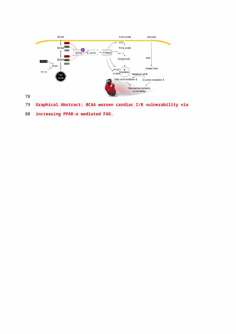
Graphical Abstract: BCAA worsen cardiac I/R vulnerability via increasing PPAR-α
mediated FAO.
57
58
59
Introduction
Ischemic heart disease (IHD) remains a leading cause of death in developed as well as
developing countries [1]. Although percutaneous coronary intervention can rapidly open the
occluded coronary arteries, it paradoxically causes myocardial ischemia/reperfusion (I/R) injury,
which is characterized by oxidative stress, cardiomyocyte death and contraction dysfunction [2-3].
Unfortunately, effective therapeutic interventions for myocardial I/R injury are still lacking. The
heart is a high-energy-consuming organ and energy substrate metabolism is essential to maintain
the normal cardiac structure and function [4-6]. Alterations in energy substrate utilization, in
particular, glucose and fatty acid (FA) metabolism, have been recognized as critical determinants
of myocardial vulnerability to I/R damage [7-8]. Thus, modulation of cardiac substrate
metabolism might be a promising therapeutic strategy for the management of I/R injury.
In addition to glucose and FAs, amino acids are important energy substrates to regulate
cardiovascular pathophysiology. However, unlike glucose and FA metabolism, the regulatory role
of amino acid metabolism in I/R process remains largely unknown. Branched chain amino acids
(BCAA), consisting of valine, leucine and isoleucine, are a group of essential amino acids that can
be oxidized in myocardial tissues [9]. In mammalian animals, the metabolic homeostasis of BCAA
is controlled by a series of BCAA catabolic enzymes. BCAA are firstly converted into
corresponding branched chain α-keto acids (BCKA), which are subsequently decarboxylated by
branched chain α-keto acid dehydrogenase (BCKDH) complex in the mitochondrion [9]. As the
rate-limiting enzyme of BCAA degradation, BCKDH complex is specifically dephosphorylated
and activated by a mitochondrion-localized protein phosphatase-2C (PP2Cm) [10-11]. Recently,
numerous studies have revealed that elevation of BCAA/BCKA levels, due to dietary BCAA
intake or PP2Cm genetic knockout (KO), directly contributes to the pathogenesis of a variety of
cardiometabolic diseases, including heart failure [12], diabetes [13], and non-alcoholic fatty liver
disease [14]; however, the underlying mechanisms remain not fully understood.
It has recently been recognized that BCAA and their intermediate metabolites play important
roles in the regulation of glucose and FA metabolism [14-15]. For example, 3-hydroxyisobutyrate
(a valine-derived metabolite) promotes trans-endothelial FA transport and causes lipid deposition
in skeletal muscles [15]. BCAA also stimulate triglyceride lipolysis in adipocytes and exacerbate
60
61
62
63
64
65
66
67
68
69
70
71
72
73
74
75
76
77
78
79
80
81
82
83
84
85
86
87
88
high fat diet-induced hyperlipidemia [14]. In the heart, chronic accumulation of BCAA disrupts
glucose oxidation via suppressing mitochondrial pyruvate dehydrogenase (PDH) activity [8].
These findings lead to a speculation that BCAA may affect cardiac vulnerability to I/R injury via
modulating of myocardial substrate metabolism. Therefore, the present study aimed to: 1)
systemically evaluate the impact of BCAA on cardiac energy substrate metabolism; 2) evaluate
whether BCAA influence cardiac I/R vulnerability through modulation of cardiac energy substrate
utilization; 3) if so, clarify the underlying molecular mechanisms involved.
Materials and methods
Animals and drugs
All animal studies were carried out in accordance with the National Institutes of Health
Guidelines on the Use of Laboratory Animals and were approved by the Animal Care Committee
of Air Force Medical University. PP2Cm global knockout (KO) mice are widely used as animal
models with BCAA catabolic defects [10]. KO mice were obtained and maintained as we
previously described [13]. Both KO and their wild-type (WT) littermates (aged from 10-12 weeks)
were housed in a constant-temperature vivarium at 22°C with a 12-h light/dark cycle. Food and
water were available ad libitum. BCAA mixture (weight ratio, leucine: valine: isoleucine=2:1:1;
Sigma-Aldrich, St. Louis, MO, USA) were given into mice by oral gavage (1.5 mg/g/day) for 7 d
before these mice received sham or I/R operation, as described by Li et al [8]. Vehicle or Etomoxir
(Eto) (5 mg/kg body weight, Sigma-Aldrich) was intraperitoneally injected at 15 min before I/R
procedure [16]. BCKA, including α-ketoisovaleric acid (αKIV), α-ketoisocaproate (αKIC) and α-
keto-β-methylvalerate (αKMV), were commercially obtained from Sigma-Aldrich. A customized
BCAA-free DMEM was used to exclude the impact of culture medium-contained BCAA. The
detailed composition of BCAA-free DMEM is showed in Table S4 .
Myocardial I/R models
Myocardial I/R injury was induced as we previously performed [17]. Briefly, mice were
anesthetized with 2% isoflurane throughout the procedure. After a left thoracic incision and ribs
exposure, the heart was mildly pumped out. A slipknot was tied on the left descending coronary
artery to induce myocardial ischemia. After 30 min ischemia, the slipknot was loosened to
reperfuse the myocardium for 3 h (to detect caspase-3 activation, superoxide production and LDH
release) or 24 h (to detect cardiac function, infarct size and cardiac apoptosis). Mice in sham group
89
90
91
92
93
94
95
96
97
98
99
100
101
102
103
104
105
106
107
108
109
110
111
112
113
114
115
116
117
118
underwent all the same procedure, except the knot tying.
Intra-myocardial adenovirus injection and gene delivery
Adenovirus vectors carrying peroxisome proliferator-activated receptor alpha (Ppara)-
specific short hairpin RNA (Ad-shPpara) or scramble control vectors (Ad-scramble) were
constructed by Hanbio Co., Ltd (Shanghai, China). The sequences of shPpara and scramble were
available in Table S1. Ad-shPpara and Ad-scramble vectors were delivered into the heart via intra-
myocardial injection as we previously described [18]. In brief, adenovirus vectors were diluted to
2.5×10¹¹ particles/ml in phosphatase buffer (PBS). 25 μL adenovirus solution was then injected
into the left ventricular (LV) free wall using a Hamilton syringe (Hamilton Co. Reno, NV, USA).
The intra-myocardial adenovirus injection was conducted as follows: 1) starting from apex and
moving toward to the base in LV anterior wall; 2) at the upper part of LV anterior wall; 3) starting
from apex and moving toward to base in LV posterior wall. After the injection, the heart was
gently returned back to the thoracic cavity and carefully closed the incision. At 7 d after
adenovirus injection, mice underwent sham or I/R operation as described above.
Echocardiography
Mice were anesthetized by 2% isoflurane and ventricular function was determined by an
echocardiographic imaging system (Vevo 2100, VisualSonics, Toronto, ON, Canada) at 24 h after
sham or I/R operation. Two-dimensional echocardiographic views of the mid-ventricular short
axis were collected at the section of the papillary muscle tips below the mitral valve. Subsequent
calculation of the left ventricular ejection fraction (LVEF) and left ventricular fractional
shortening (LVFS) were performed [19].
Evaluation of cardiomyocyte apoptosis
To evaluate in vivo cardiomyocyte apoptosis, TdT-mediated dUTP nick-end labeling
(TUNEL) staining (Roche, USA) was performed in heart tissue sections as the manual described.
TUNEL/DAPI double-positive nuclei were counted as apoptotic cardiac myocytes. In vitro
cardiomyocyte apoptosis was assessed by flow cytometry using the Annexin V-FITC/propidium
iodide (PI) Apoptosis Detection kit (Beyotime, Beijing, China) according to the manufacturer’s
instructions.
Evaluation of infarction size
At the end of a 24-h reperfusion, mice were anesthetized by 2% isoflurane and hearts were
119
120
121
122
123
124
125
126
127
128
129
130
131
132
133
134
135
136
137
138
139
140
141
142
143
144
145
146
147
148

excised. The infarct size induced by I/R was measured by 5-triphenyltetrazolium chloride (TTC)
and Evans blue double staining as we previously described [17].
Adult mouse cardiac myocyte isolation
To isolate adult mouse cardiac myocytes, mice aged 10-12 weeks were anesthetized with 2%
isoflurane and were fully heparinized with heparin. Hearts were carefully removed and placed into
ice-cold PBS. Next, the heart was attached to the Langendorff system via the aorta and was fully
perfused with perfusion solution (126 mmol/l NaCl, 4.4 mmol/l KCl, 18 mmol/l NaHCO3, 1
mmol/l MgCl2, 11 mmol/l glucose, 10 mmol/l 2,3-butanedione monoxime, 30 mmol/l taurine and
4 mmol/l HEPES) for 5 min. Thereafter, the heart was perfused by collagenase solution (perfusion
solution with 0.1% bovine serum albumin, 0.025 mmol/l CaCl2 and 0.1% type II collagenase) for
another 10 min. After digestion, the ventricle was triturated with 10 ml pipette at a slow speed and
was filtered through a 100 μm filter. At last, cardiac myocytes were slowly adjusted to increasing
concentrations of CaCl2 solution (range from 0.05 to 0.525 mmol/l) for 2 h before plating. Cardiac
myocytes were seeded onto the laminin-coated plates overnight.
Seahorse analysis
Metabolic flux experiments were performed in adult mouse cardiac myocytes because the
metabolism of adult cardiac myocytes is largely different from neonatal cardiac myocytes. Briefly,
adult mouse cardiac myocytes were seeded into laminin-coated XF24 Seahorse plates at a density
of 10, 000 cells per well. Cardiac myocytes were cultured in a BCAA-free substrate-limited
medium overnight before the analysis. The detailed composition of the BCAA-free substrate-
limited medium is showed in Table S5 . Oxygen consumption rate (OCR) and extracellular
acidification rate (ECAR) were used to evaluate fatty acid oxidation (FAO), glucose oxidation and
glycolysis in real-time when the appropriate substrates were added into or included in the assay
medium. OCR and ECAR were measured using the Seahorse XF24 Extracellular Flux Analyzer
(Seahorse Bioscience, North Billerica, MA, USA). Substrates or perturbation compounds were
prepared in the assay medium as in the corresponding well and were added from the reagent ports
automatically to the wells at the time as indicated.
Preparation of palmitate-BSA conjugates
Palmitate was solubilized in 150 mmol/l sodium chloride by heating up to 65 °C in the water
bath. BSA was dissolved in PBS and warmed up to 37 °C. Palmitate was then added into BSA at
149
150
151
152
153
154
155
156
157
158
159
160
161
162
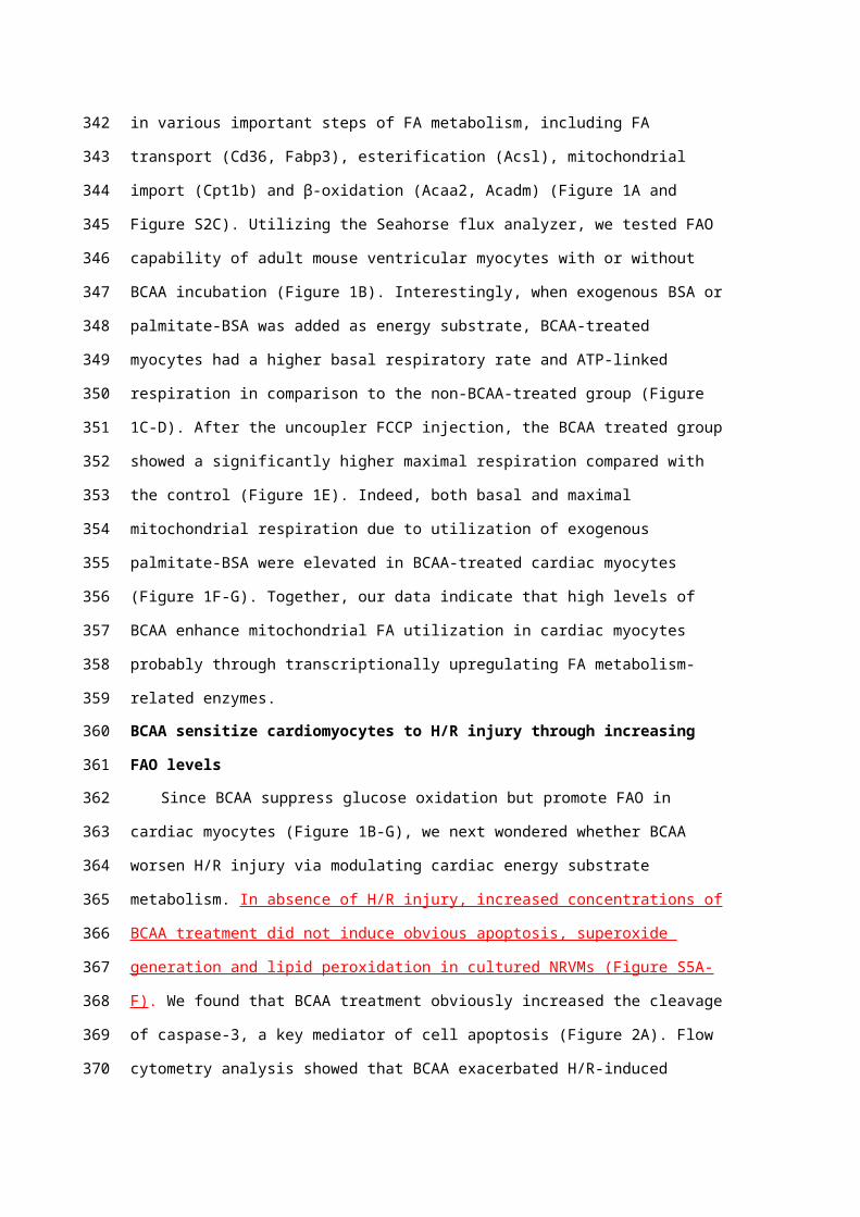
163
164
165
166
167
168
169
170
171
172
173
174
175
176
177
178
37 °C with continuous stirring. When exogenous FAO flux was assessed by Seahorse, palmitate-
BSA (0.175 mM) conjugate or BSA (0.03 mM) was added to appropriate wells.
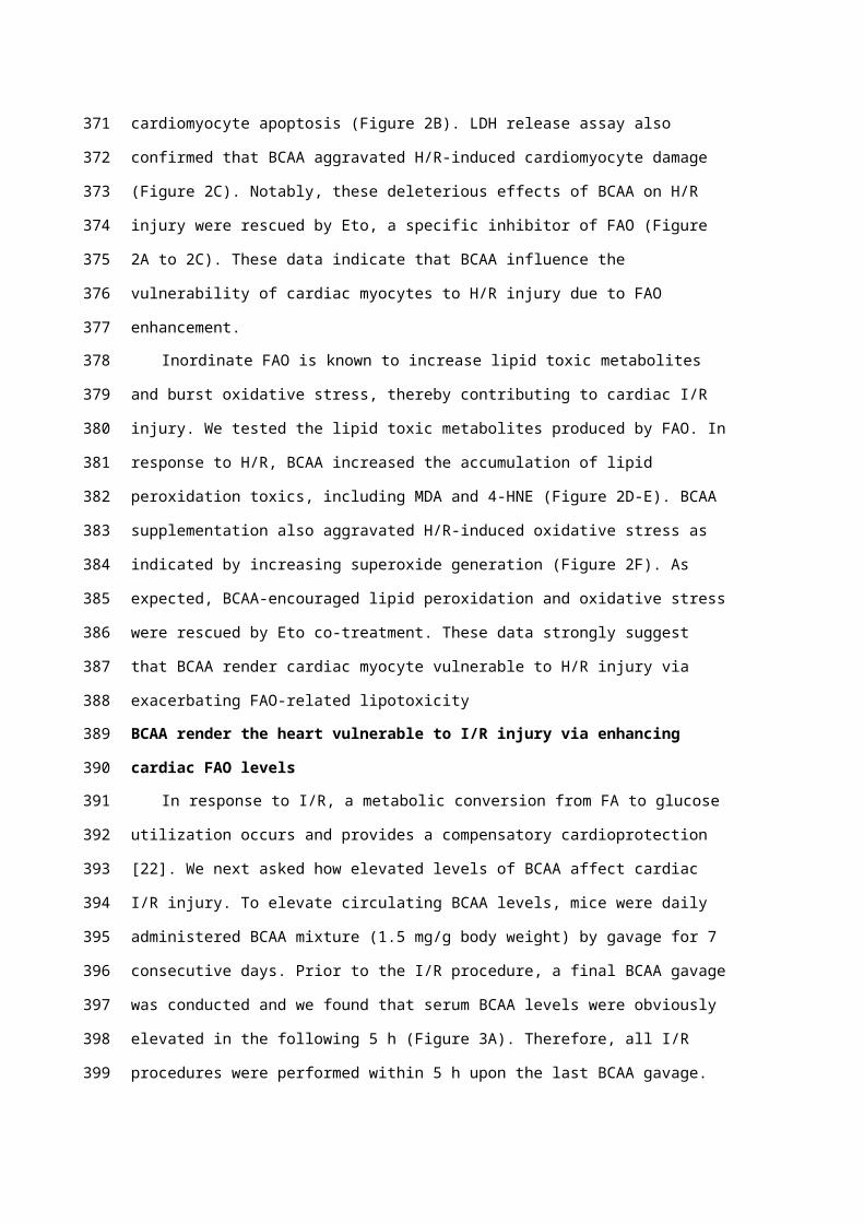
In vitro hypoxia/reoxygenation (H/R)
To simulate in vivo I/R injury, in vitro H/R models was established in neonatal rat ventricular
myocytes (NRVMs). Briefly, NRVMs were isolated from Sprague-Dawley rats aged 1-3 d as we
previously described [19]. NRVMs were subjected to 9 h hypoxia and the following 3 h
reoxygenation. To induce hypoxia, NRVMs were cultured in a hypoxia chamber with 95% N2 and
5% CO2 at 37 °C. H/R-induced lipid peroxidation, superoxide generation and cardiomyocyte
apoptosis were systemically evaluated as the methods described.
Determination of superoxide generation and lipid peroxidation
Superoxide generation was assessed by dihydroethidium (DHE, Sigma-Aldrich) staining in
cultured NRVMs and freshly frozen heart tissue sections. Briefly, heart tissue sections or NRVMs
were incubated with 40 μmol/l DHE for 1 h at room temperature, protected from light. The images
were then obtained using a Leica laser scanning confocal microscope. Lipid peroxidation was
determined by measuring malondialdehyde (MDA) and 4-hydroxynonenal (4-HNE) generation in
NRVMs or heart tissues. MDA was measured using a commercial detection kit (S0131, Beyotime,
Beijing, China) according to the instructions. 4-HNE content was measured using a commercially
available kit (ab238538, Abcam, USA).
Small interfering RNA (siRNA) transfection
NRVMs were routinely cultured. Specific siRNA of ATF6 (GenePharma, Shanghai, China)
was obtained to knockdown ATF6 protein expression. Scrambled siRNA was also obtained from
Genepharma. siRNA was transfected into cells by using Lipofectamine 2000 (Invitrogen, USA)
according to manufacturers' instructions. Silencing efficacy was evaluated by immunoblotting.
The sequences of siRNA were provided in Table S2.
Real-time polymerase chain reaction (RT-PCR)
Total RNA was extracted from frozen heart tissues or cultured cells and RNA reverse-
transcription were performed as we previously described [20]. RT-PCR was conducted using a
SYBR Green Master Mix (Cowin, Beijing, China). Primer sequences were listed in Table S3.
Western blotting
Protein extraction and western blotting were performed as we previously described [21]. In
179
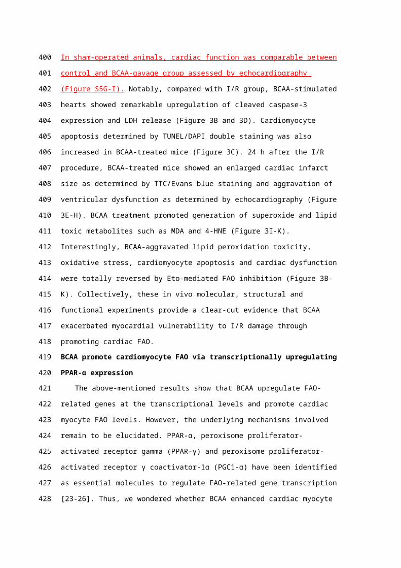
180
181
182
183
184
185
186
187
188
189
190
191
192
193
194
195
196
197
198
199
200
201
202
203
204
205
206
207
208
brief, proteins were electrophoresed on SDS-PAGE gels and transferred onto nitrocellulose
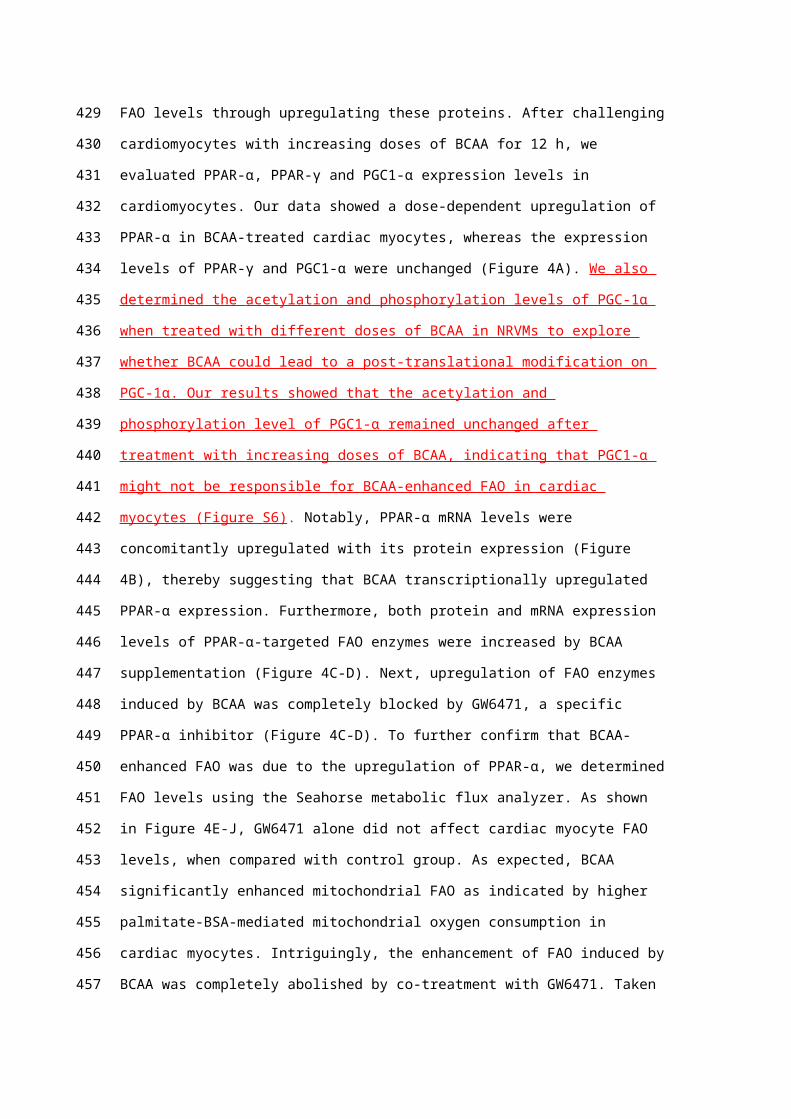
membranes. Membranes were blocked in 5% non-fat milk and incubated with the following
primary antibodies overnight at 4 °C. The primary antibodies were as follows: ACAA2 (1:1000,
Abclonal, A15778), ACADM (1:1000, Abclonal, A1873), ATF6 (1:1000, Affinity, DF6009), β-
actin (1:1000, Abclonal, AC004), Caspase-3 (1:1000, Cell Signaling Technology, 9662), CD36
(1:1000, Abclonal, A5792), Cleaved caspase-3 (1:1000, Cell Signaling Technology, 9661), CPT1B
(1:1000, Abclonal, A6796), GCN2 (1:1000, Affinity, DF7801), p-GCN2 (1:1000, Affinity,
AF8154) and PPAR-α (1:1000, Abclonal, A6697). After incubation in the secondary antibodies,
blots were visualized using enhanced chemiluminescence (Millipore, USA) and scanned by
ChemiDocXRS system (Bio-Rad Laboratory, Hercules, CA, USA).
BCAA concentration measurement
Serum samples and cell supernatants were collected and immediately restored at -80 °C.
BCAA concentration was determined with a commercially available BCAA detection kit (K564-
100, Biovision, USA) as the protocol described. The absorbance value of each sample was
measured at 450 nm in a microplate reader and calculated based on the standard curve.
Statistical analysis
Data were analyzed with GraphPad Prism 8 statistic software and were presented as mean ±
SEM. Statistical comparisons between groups were performed by Student’s t-test for two groups
and one-way ANOVA followed by Bonferroni Post-Hoc analysis for more than two groups. P <
0.05 was considered statistically significant.
Results
BCAA promote FAO in cardiac myocytes
Significant elevation of circulating BCAA levels has been observed in obese/diabetic mice,
which are at high risk of cardiovascular diseases [13-14]. Therefore, we attempted to determine
how BCAA influence glucose and FA metabolism in cardiac myocytes. Adult mouse cardiac
myocytes were incubated with increasing concentrations of BCAA (ranging from 0 to 3.432 mM).
We found that high levels of BCAA inhibited mRNA expression of glucose oxidation enzymes
and suppressed glucose-dependent oxygen consumption (Figure S1A, S2A and S3). Moreover,
BCAA supplementation upregulated mRNA levels of glycolytic enzymes and augmented
glycolysis in cardiac myocytes, which might be a compensatory phenomenon for BCAA-mediated
209
210
211
212
213
214
215
216
217
218
219
220
221
222
223
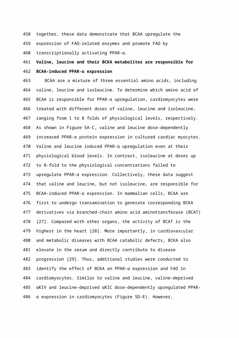
224
225
226
227
228
229
230
231
232
233
234
235
236
237
238
glucose oxidation suppression (Figure S1B, S2B and S4). FAs are the predominant energy
substrate in normal conditions. We next evaluated the impact of BCAA on FA metabolism in
cardiac myocytes. Excessive BCAA remarkably increased mRNA levels of FA metabolism-related
enzymes in a dose-dependent manner. These enzymes were involved in various important steps of
FA metabolism, including FA transport (Cd36, Fabp3), esterification (Acsl), mitochondrial import
(Cpt1b) and β-oxidation (Acaa2, Acadm) (Figure 1A and Figure S2C). Utilizing the Seahorse flux
analyzer, we tested FAO capability of adult mouse ventricular myocytes with or without BCAA
incubation (Figure 1B). Interestingly, when exogenous BSA or palmitate-BSA was added as
energy substrate, BCAA-treated myocytes had a higher basal respiratory rate and ATP-linked
respiration in comparison to the non-BCAA-treated group (Figure 1C-D). After the uncoupler
FCCP injection, the BCAA treated group showed a significantly higher maximal respiration
compared with the control (Figure 1E). Indeed, both basal and maximal mitochondrial respiration
due to utilization of exogenous palmitate-BSA were elevated in BCAA-treated cardiac myocytes
(Figure 1F-G). Together, our data indicate that high levels of BCAA enhance mitochondrial FA
utilization in cardiac myocytes probably through transcriptionally upregulating FA metabolism-
related enzymes.
BCAA sensitize cardiomyocytes to H/R injury through increasing FAO levels
Since BCAA suppress glucose oxidation but promote FAO in cardiac myocytes (Figure 1B-
G), we next wondered whether BCAA worsen H/R injury via modulating cardiac energy substrate
metabolism. In absence of H/R injury, increased concentrations of BCAA treatment did not induce
obvious apoptosis, superoxide generation and lipid peroxidation in cultured NRVMs (Figure S5A-
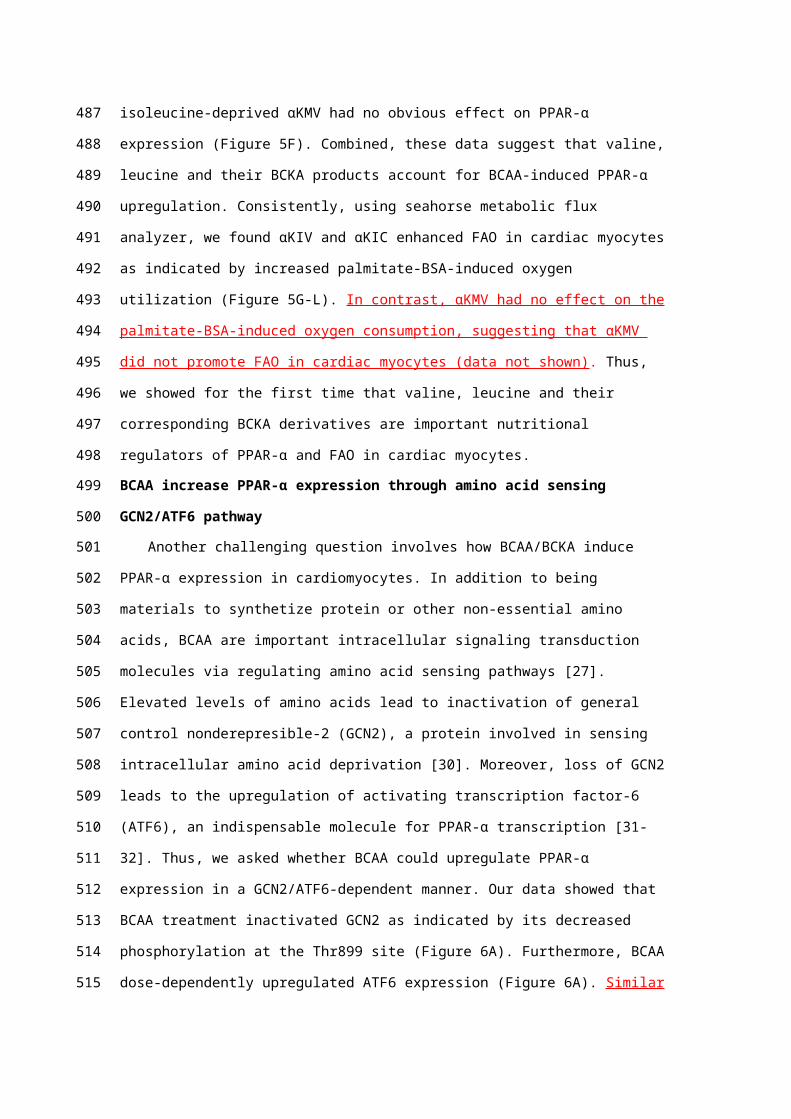
F). We found that BCAA treatment obviously increased the cleavage of caspase-3, a key mediator
of cell apoptosis (Figure 2A). Flow cytometry analysis showed that BCAA exacerbated H/R-
induced cardiomyocyte apoptosis (Figure 2B). LDH release assay also confirmed that BCAA
aggravated H/R-induced cardiomyocyte damage (Figure 2C). Notably, these deleterious effects of
BCAA on H/R injury were rescued by Eto, a specific inhibitor of FAO (Figure 2A to 2C). These
data indicate that BCAA influence the vulnerability of cardiac myocytes to H/R injury due to FAO
enhancement.
Inordinate FAO is known to increase lipid toxic metabolites and burst oxidative stress,
239
240
241
242
243
244
245
246
247
248
249
250
251
252
253
254
255
256
257
258
259
260
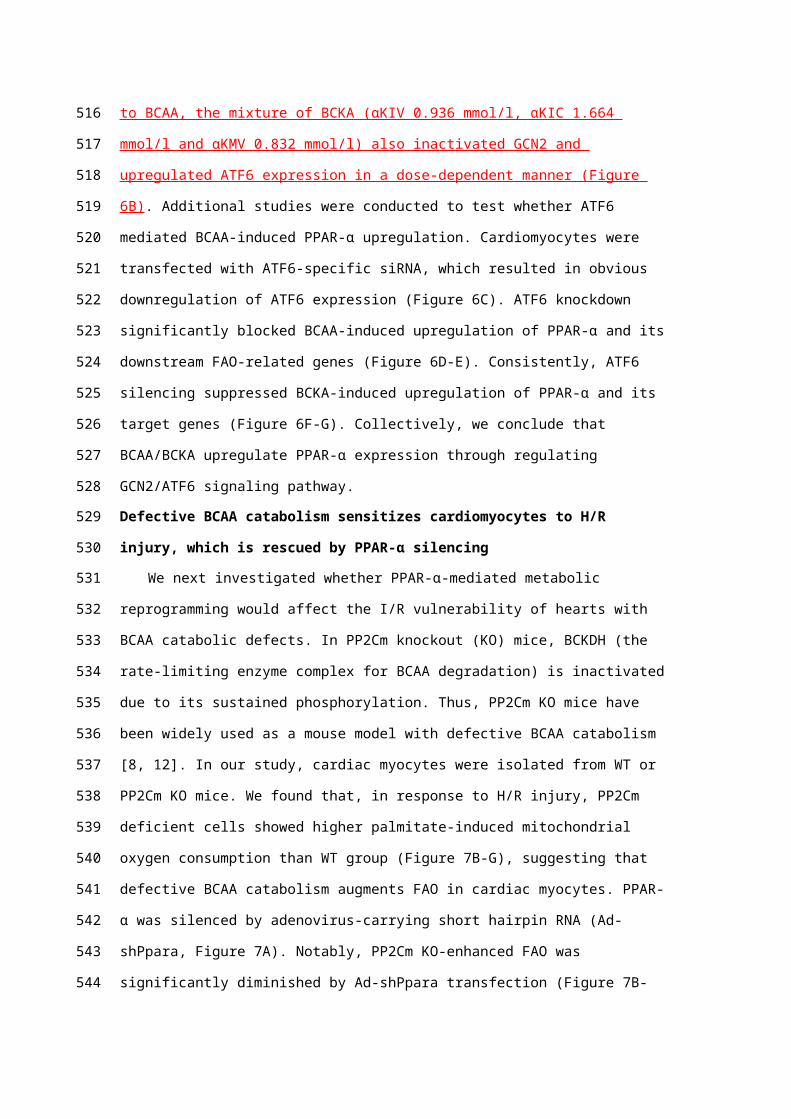
261
262
263
264
265
266
267
thereby contributing to cardiac I/R injury. We tested the lipid toxic metabolites produced by FAO.
In response to H/R, BCAA increased the accumulation of lipid peroxidation toxics, including
MDA and 4-HNE (Figure 2D-E). BCAA supplementation also aggravated H/R-induced oxidative
stress as indicated by increasing superoxide generation (Figure 2F). As expected, BCAA-
encouraged lipid peroxidation and oxidative stress were rescued by Eto co-treatment. These data
strongly suggest that BCAA render cardiac myocyte vulnerable to H/R injury via exacerbating
FAO-related lipotoxicity
BCAA render the heart vulnerable to I/R injury via enhancing cardiac FAO levels
In response to I/R, a metabolic conversion from FA to glucose utilization occurs and provides
a compensatory cardioprotection [22]. We next asked how elevated levels of BCAA affect cardiac
I/R injury. To elevate circulating BCAA levels, mice were daily administered BCAA mixture (1.5
mg/g body weight) by gavage for 7 consecutive days. Prior to the I/R procedure, a final BCAA
gavage was conducted and we found that serum BCAA levels were obviously elevated in the
following 5 h (Figure 3A). Therefore, all I/R procedures were performed within 5 h upon the last
BCAA gavage. In sham-operated animals, cardiac function was comparable between control and
BCAA-gavage group assessed by echocardiography (Figure S5G-I). Notably, compared with I/R
group, BCAA-stimulated hearts showed remarkable upregulation of cleaved caspase-3 expression
and LDH release (Figure 3B and 3D). Cardiomyocyte apoptosis determined by TUNEL/DAPI
double staining was also increased in BCAA-treated mice (Figure 3C). 24 h after the I/R
procedure, BCAA-treated mice showed an enlarged cardiac infarct size as determined by
TTC/Evans blue staining and aggravation of ventricular dysfunction as determined by
echocardiography (Figure 3E-H). BCAA treatment promoted generation of superoxide and lipid
toxic metabolites such as MDA and 4-HNE (Figure 3I-K). Interestingly, BCAA-aggravated lipid
peroxidation toxicity, oxidative stress, cardiomyocyte apoptosis and cardiac dysfunction were
totally reversed by Eto-mediated FAO inhibition (Figure 3B-K). Collectively, these in vivo
molecular, structural and functional experiments provide a clear-cut evidence that BCAA
exacerbated myocardial vulnerability to I/R damage through promoting cardiac FAO.
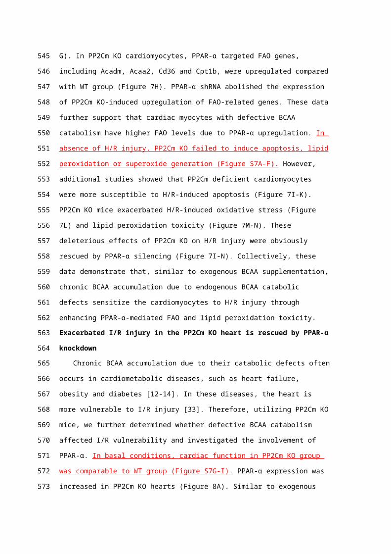
BCAA promote cardiomyocyte FAO via transcriptionally upregulating PPAR-α expression
The above-mentioned results show that BCAA upregulate FAO-related genes at the
268
269
270
271
272
273
274
275
276
277
278
279
280
281
282
283
284
285
286
287
288
289
290
291
292
293
294
295
296
transcriptional levels and promote cardiac myocyte FAO levels. However, the underlying
mechanisms involved remain to be elucidated. PPAR-α, peroxisome proliferator-activated receptor
gamma (PPAR-γ) and peroxisome proliferator-activated receptor γ coactivator-1α (PGC1-α) have
been identified as essential molecules to regulate FAO-related gene transcription [23-26]. Thus,
we wondered whether BCAA enhanced cardiac myocyte FAO levels through upregulating these
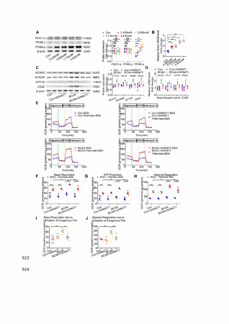
proteins. After challenging cardiomyocytes with increasing doses of BCAA for 12 h, we evaluated
PPAR-α, PPAR-γ and PGC1-α expression levels in cardiomyocytes. Our data showed a dose-
dependent upregulation of PPAR-α in BCAA-treated cardiac myocytes, whereas the expression
levels of PPAR-γ and PGC1-α were unchanged (Figure 4A). We also determined the acetylation
and phosphorylation levels of PGC-1α when treated with different doses of BCAA in NRVMs to
explore whether BCAA could lead to a post-translational modification on PGC-1α. Our results
showed that the acetylation and phosphorylation level of PGC1-α remained unchanged after
treatment with increasing doses of BCAA, indicating that PGC1-α might not be responsible for
BCAA-enhanced FAO in cardiac myocytes (Figure S6). Notably, PPAR-α mRNA levels were
concomitantly upregulated with its protein expression (Figure 4B), thereby suggesting that BCAA
transcriptionally upregulated PPAR-α expression. Furthermore, both protein and mRNA
expression levels of PPAR-α-targeted FAO enzymes were increased by BCAA supplementation
(Figure 4C-D). Next, upregulation of FAO enzymes induced by BCAA was completely blocked
by GW6471, a specific PPAR-α inhibitor (Figure 4C-D). To further confirm that BCAA-enhanced
FAO was due to the upregulation of PPAR-α, we determined FAO levels using the Seahorse
metabolic flux analyzer. As shown in Figure 4E-J, GW6471 alone did not affect cardiac myocyte
FAO levels, when compared with control group. As expected, BCAA significantly enhanced
mitochondrial FAO as indicated by higher palmitate-BSA-mediated mitochondrial oxygen
consumption in cardiac myocytes. Intriguingly, the enhancement of FAO induced by BCAA was
completely abolished by co-treatment with GW6471. Taken together, these data demonstrate that
BCAA upregulate the expression of FAO-related enzymes and promote FAO by transcriptionally
activating PPAR-α.
Valine, leucine and their BCKA metabolites are responsible for BCAA-induced PPAR-α
expression
297
298
299
300
301
302
303
304
305
306
307
308
309
310
311
312
313
314
315
316
317
318
319
320
321
322
323
324
325
BCAA are a mixture of three essential amino acids, including valine, leucine and isoleucine.
To determine which amino acid of BCAA is responsible for PPAR-α upregulation, cardiomyocytes
were treated with different doses of valine, leucine and isoleucine, ranging from 1 to 8 folds of
physiological levels, respectively. As shown in Figure 5A-C, valine and leucine dose-dependently
increased PPAR-α protein expression in cultured cardiac myocytes. Valine and leucine induced
PPAR-α upregulation even at their physiological blood levels. In contrast, isoleucine at doses up to
8-fold to the physiological concentrations failed to upregulate PPAR-α expression. Collectively,
these data suggest that valine and leucine, but not isoleucine, are responsible for BCAA-induced
PPAR-α expression. In mammalian cells, BCAA are first to undergo transamination to generate
corresponding BCKA derivatives via branched-chain amino acid aminotransferase (BCAT) [27].
Compared with other organs, the activity of BCAT is the highest in the heart [28]. More
importantly, in cardiovascular and metabolic diseases with BCAA catabolic defects, BCKA also
elevate in the serum and directly contribute to disease progression [29]. Thus, additional studies
were conducted to identify the effect of BCKA on PPAR-α expression and FAO in
cardiomyocytes. Similar to valine and leucine, valine-deprived αKIV and leucine-deprived αKIC
dose-dependently upregulated PPAR-α expression in cardiomyocytes (Figure 5D-E). However,
isoleucine-deprived αKMV had no obvious effect on PPAR-α expression (Figure 5F). Combined,
these data suggest that valine, leucine and their BCKA products account for BCAA-induced
PPAR-α upregulation. Consistently, using seahorse metabolic flux analyzer, we found αKIV and
αKIC enhanced FAO in cardiac myocytes as indicated by increased palmitate-BSA-induced
oxygen utilization (Figure 5G-L). In contrast, αKMV had no effect on the palmitate-BSA-induced
oxygen consumption, suggesting that αKMV did not promote FAO in cardiac myocytes (data not
shown). Thus, we showed for the first time that valine, leucine and their corresponding BCKA
derivatives are important nutritional regulators of PPAR-α and FAO in cardiac myocytes.
BCAA increase PPAR-α expression through amino acid sensing GCN2/ATF6 pathway
Another challenging question involves how BCAA/BCKA induce PPAR-α expression in
cardiomyocytes. In addition to being materials to synthetize protein or other non-essential amino
acids, BCAA are important intracellular signaling transduction molecules via regulating amino
acid sensing pathways [27]. Elevated levels of amino acids lead to inactivation of general control
326
327
328
329
330
331
332
333
334
335
336
337
338
339
340
341
342
343
344
345
346
347
348
349
350
351
352
353
354
nonderepresible-2 (GCN2), a protein involved in sensing intracellular amino acid deprivation [30].
Moreover, loss of GCN2 leads to the upregulation of activating transcription factor-6 (ATF6), an
indispensable molecule for PPAR-α transcription [31-32]. Thus, we asked whether BCAA could
upregulate PPAR-α expression in a GCN2/ATF6-dependent manner. Our data showed that BCAA
treatment inactivated GCN2 as indicated by its decreased phosphorylation at the Thr899 site
(Figure 6A). Furthermore, BCAA dose-dependently upregulated ATF6 expression (Figure 6A).
Similar to BCAA, the mixture of BCKA (αKIV 0.936 mmol/l, αKIC 1.664 mmol/l and αKMV
0.832 mmol/l) also inactivated GCN2 and upregulated ATF6 expression in a dose-dependent
manner (Figure 6B). Additional studies were conducted to test whether ATF6 mediated BCAA-
induced PPAR-α upregulation. Cardiomyocytes were transfected with ATF6-specific siRNA,
which resulted in obvious downregulation of ATF6 expression (Figure 6C). ATF6 knockdown
significantly blocked BCAA-induced upregulation of PPAR-α and its downstream FAO-related
genes (Figure 6D-E). Consistently, ATF6 silencing suppressed BCKA-induced upregulation of
PPAR-α and its target genes (Figure 6F-G). Collectively, we conclude that BCAA/BCKA
upregulate PPAR-α expression through regulating GCN2/ATF6 signaling pathway.
Defective BCAA catabolism sensitizes cardiomyocytes to H/R injury, which is rescued by
PPAR-α silencing
We next investigated whether PPAR-α-mediated metabolic reprogramming would affect the
I/R vulnerability of hearts with BCAA catabolic defects. In PP2Cm knockout (KO) mice, BCKDH
(the rate-limiting enzyme complex for BCAA degradation) is inactivated due to its sustained
phosphorylation. Thus, PP2Cm KO mice have been widely used as a mouse model with defective
BCAA catabolism [8, 12]. In our study, cardiac myocytes were isolated from WT or PP2Cm KO
mice. We found that, in response to H/R injury, PP2Cm deficient cells showed higher palmitate-
induced mitochondrial oxygen consumption than WT group (Figure 7B-G), suggesting that
defective BCAA catabolism augments FAO in cardiac myocytes. PPAR-α was silenced by
adenovirus-carrying short hairpin RNA (Ad-shPpara, Figure 7A). Notably, PP2Cm KO-enhanced
FAO was significantly diminished by Ad-shPpara transfection (Figure 7B-G). In PP2Cm KO
cardiomyocytes, PPAR-α targeted FAO genes, including Acadm, Acaa2, Cd36 and Cpt1b, were
upregulated compared with WT group (Figure 7H). PPAR-α shRNA abolished the expression of
355
356
357
358
359
360
361
362
363
364
365
366
367
368
369
370
371
372
373
374
375
376
377
378
379
380
381
382
383
PP2Cm KO-induced upregulation of FAO-related genes. These data further support that cardiac
myocytes with defective BCAA catabolism have higher FAO levels due to PPAR-α upregulation.
In absence of H/R injury, PP2Cm KO failed to induce apoptosis, lipid peroxidation or superoxide
generation (Figure S7A-F). However, additional studies showed that PP2Cm deficient
cardiomyocytes were more susceptible to H/R-induced apoptosis (Figure 7I-K). PP2Cm KO mice
exacerbated H/R-induced oxidative stress (Figure 7L) and lipid peroxidation toxicity (Figure 7M-
N). These deleterious effects of PP2Cm KO on H/R injury were obviously rescued by PPAR-α
silencing (Figure 7I-N). Collectively, these data demonstrate that, similar to exogenous BCAA
supplementation, chronic BCAA accumulation due to endogenous BCAA catabolic defects
sensitize the cardiomyocytes to H/R injury through enhancing PPAR-α-mediated FAO and lipid
peroxidation toxicity.
Exacerbated I/R injury in the PP2Cm KO heart is rescued by PPAR-α knockdown
Chronic BCAA accumulation due to their catabolic defects often occurs in cardiometabolic
diseases, such as heart failure, obesity and diabetes [12-14]. In these diseases, the heart is more
vulnerable to I/R injury [33]. Therefore, utilizing PP2Cm KO mice, we further determined
whether defective BCAA catabolism affected I/R vulnerability and investigated the involvement
of PPAR-α. In basal conditions, cardiac function in PP2Cm KO group was comparable to WT
group (Figure S7G-I). PPAR-α expression was increased in PP2Cm KO hearts (Figure 8A).
Similar to exogenous BCAA accumulation by oral gavage, PP2Cm KO resulted in endogenous
BCAA accumulation as indicated by higher serum BCAA levels (Figure 8B). Chronic
accumulation of BCAA exacerbated I/R-induced caspase-3 activation, cardiomyocyte apoptosis
and LDH release (Figure 8C-E). Consistently, PP2Cm KO hearts had larger infarct sizes and
worse ventricular function in response to I/R injury (Figure 8F-I). These data establish a causative
role of defective BCAA catabolism in the regulation of myocardial I/R vulnerability.
Next, we asked whether defective BCAA catabolism could render the heart vulnerable to I/R
injury due to upregulation of PPAR-α. WT and PP2Cm KO hearts were transfected with
adenovirus vectors carrying PPAR-α shRNA (Ad-shPpara). We found that intra-cardiac Ad-
shPpara injection obviously reduced PPAR-α expression in cardiac tissue (Figure S8). Moreover,
compared with the KO heart, PPAR-α silencing rescued BCAA catabolic defects-aggravated
384
385
386
387
388
389
390
391
392
393
394
395
396
397
398
399
400
401
402
403
404
405
406
407
408
409
410
411
412
cardiomyocyte apoptosis, infarction and ventricular dysfunction (Figure 8C-I). In PP2Cm KO
hearts, chronic accumulation of BCAA exacerbated superoxide generation and lipid peroxidation
toxicity (Figure 8J-L), which was reversed by adenovirus-mediated PPAR-α knockdown. Taken
together, these data demonstrate that defective BCAA catabolism sensitize the heart to I/R injury
via upregulating PPAR-α expression.
Discussion
In the present study, we have several important observations. First, we confirm that BCAA
are important endogenous nutrition regulator of cardiac FAO. It has been reported that BCAA
suppress cardiac glucose oxidation via inhibiting PDH activity [8]. However, FAs are the
dominant energy substrate and provide 50-70% of total ATP for the heart in normal conditions
[34-35]. Therefore, it is of great significance to clarify the impact of BCAA on cardiac FA
metabolism. Utilizing the Seahorse metabolic flux analyzer, we for the first time reveal that
BCAA enhance endogenous and exogenous FA utilization in adult cardiac myocytes. We also
confirm that valine and leucine, but not isoleucine, are responsible for BCAA-induced FAO
enhancement. Most amino acids are mainly degraded in the liver whereas BCAA are first
degraded in extra-hepatic organs, such as the heart and the skeletal muscle [28, 36]. We next
evaluate the impact of BCAA downstream metabolites BCKA on cardiac FAO. The present study
reveals that valine-derived α-KIV and leucine-derived αKIC, but not isoleucine-derived α-KMV,
enhance FAO in cardiac myocytes. Collectively, these data reveal that BCAA and their metabolites
BCKA act as an important endogenous nutrition regulator of FAO in cardiac myocytes.
Secondly, we have identified PPAR-α as an indispensable transcriptional factor for BCAA-
enhanced FAO. PPAR-α is a critical regulator of FA metabolism via transcriptionally regulating
the expression levels of FAO-related enzymes [24]. In the present study, we confirm that
BCAA/BCKA transcriptionally upregulate PPAR-α expression. Pharmacological inhibition of
PPAR-α by GW-6471 blocks BCAA/BCKA-induced FAO enhancement, revealing an
indispensable role of PPAR-α in this process. Furthermore, we also clarify that BCAA/BCKA
upregulate PPAR-α expression through the amino acid sensing GCN2/ATF6 pathway. Amino acid
starvation leads to phosphorylation and activation of GCN2 [30]. Therefore, elevated levels of
intracellular amino acids cause dephosphorylation and inactivation of GCN2. ATF6 is a
413
414
415
416
417
418
419
420
421
422
423
424
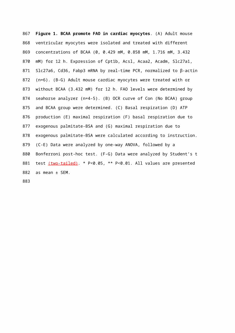
425
426
427
428
429
430
431
432
433
434
435
436
437
438
439
440
441
transcription factor activated by endoplasmic reticulum stress and is essential for PPAR-α
transcription [32, 37]. In the present study, we find that BCAA/BCKA inactivate GCN2 and
upregulate ATF6, thereby promoting PPAR-α transcription. This novel signaling pathway links
intracellular amino acid sensing and FA metabolism in cardiac myocytes. Collectively, these data
for the first time demonstrate that BCAA/BCKA promote FAO through the GCN2/ATF6/ PPAR-α
pathway.
Thirdly, we have provided a clear-cut evidence that chronic accumulation of BCAA/BCKA,
no matter induced by dietary or genetic factors, increase the vulnerability to I/R injury due to the
enhancement of FAO. Compared with glucose oxidation, FAO results in higher oxygen
consumption, lower ATP production efficacy and much more lipotoxic product accumulation.
Thus, in response to I/R injury, the metabolic shift from FA to glucose utilization is considered as
a compensatory protection [5]. In the heart, downregulation of PPAR-α has been shown to exert
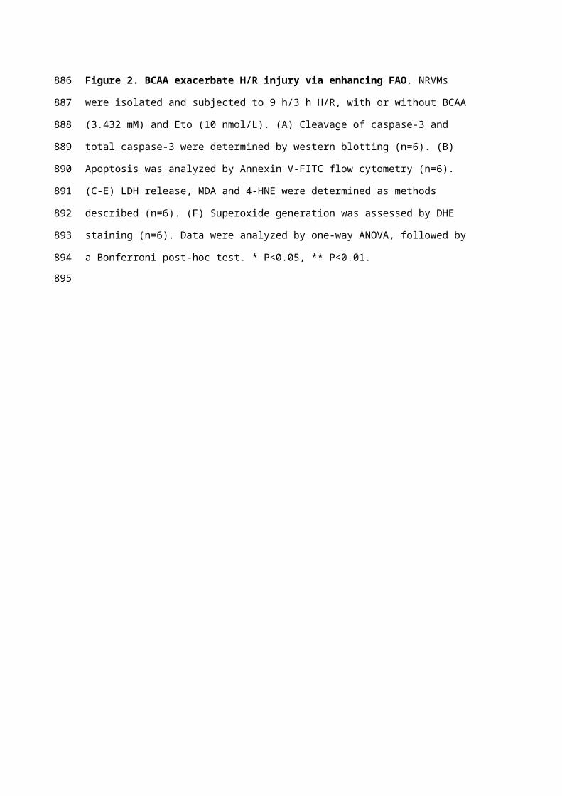
cardioprotection against I/R injury through preventing lipid peroxidation toxicity [38-39]. We
have recently observed that PP2Cm deficient heart is more susceptible to I/R injury due to
increased mitochondrial superoxide generation; however, the underlying mechanisms are totally
unknown [40]. Here we further demonstrate that chronic accumulation of BCAA renders the heart
vulnerable to I/R injury, which could be rescued by FAO inhibition or PPAR-α silencing. These
results reveal that elevated levels of BCAA due to excessive BCAA intake or defective BCAA
catabolism regulate metabolic substrate preference and determine cardiac vulnerability to I/R.
Defective BCAA catabolism and elevated BCAA/BCKA levels are often seen in the development
of cardiometabolic diseases, such as heart failure, diabetes, obesity and non-alcoholic fatty liver
disease [12-14]. Patients with these above diseases are more susceptible to myocardial I/R injury.
Therefore, the present study calls for the caution that elevated levels of BCAA should be
intervened to reduce myocardial vulnerability to I/R injury in these patients.
In summary, the present study for the first time reveal that BCAA/BCKA are important
nutrition regulators of cardiac energy substrate preference. BCAA/BCKA enhance cardiac FAO
levels via transcriptionally upregulating PPAR-α expression, thereby exacerbating lipid
peroxidation toxicity and cardiac vulnerability to I/R injury. Given the fact that BCAA catabolism
is impaired in multiple cardiometabolic diseases, this study suggests that targeting BCAA
442
443
444
445
446
447
448
449
450
451
452
453
454
455
456
457
458
459
460
461
462
463
464
465
466
467
468
469
470
catabolism might be a brand-new strategy to manage cardiac vulnerability to I/R injury among
patients with diseases characterized by defective BCAA catabolism.
Abbreviations
Acaa: acetyl-CoA acetyltransferase; Acadm: medium-chain specific acyl-CoA dehydrogenase,
mitochondrial; Acsl: long-chain acyl-CoA synthetase; ATF6: activating transcription factor 6;
BCAA: branched chain amino acids; BCAT: branched-chain amino acid aminotransferase; BCKA:
branched chain α-keto acid; BCKDH: branched chain α-keto acid dehydrogenase; Cd36: fatty acid
translocase; Cpt1b: carnitine palmitoyltransferase 1B; DHE: dihydroethidium; ECAR:
extracellular acidification rate; Eto: etomoxir; FA: fatty acid; FAO: fatty acid oxidation; GCN2:
general control nonderepresible-2; H/R: hypoxia/reoxygenation; HNE: hydroxynonenal; IHD:
ischemia heart disease; I/R: ischemia/reperfusion; KO: knockout; LVEF: left ventricular ejection
fraction; LVFS: left ventricular fractional shortening; MDA: malondialdehyde; NRVMs: neonatal
rat ventricular myocytes; OCR: oxygen consumption rate; PDH: pyruvate dehydrogenase; PGC1-
α: peroxisome proliferator-activated receptor γ coactivator-1α; PP2Cm: mitochondrion-localized
protein phosphatase-2C; PPAR-α: peroxisome proliferation-activated receptor alpha; PPAR-γ:
peroxisome proliferator-activated receptor gamma; TTC: 5-triphenyltetrazolium chloride;
TUNEL: TdT-mediated dUTP nick-end labeling; WT: wild type.
471
472
473
474
475
476
477
478
479
480
481
482
483
484
485
486
487
Acknowledgements
This work was financially supported by the National Key R&D Program of China (grant
number 2018YFA0107400), Program for National Science Funds of China (grants numbers
81730011, 81970721, 81927805 and 81800326), Program for Changjiang Scholars and Innovative
Research Team in University (Grant No. PCSIRT-14R08), China Post-Doctoral Science
Foundation Grant (No.2017M613347), and Natural Science Basic Research Plan in Shaanxi
Province of China (2018JQ8062).
Competing interests
The authors have declared that no competing interest exists.
References
1. Moran AE, Forouzanfar MH, Roth GA, Mensah GA, Ezzati M, Murray CJ, et al. Temporal
trends in ischemic heart disease mortality in 21 world regions, 1980 to 2010: the global burden of
disease 2010 study. Circulation. 2014; 129: 1483-92.
2. Ziegler M, Wang X, Lim B, Leitner E, Klingberg F, Ching V, et al. Platelet-targeted delivery of
peripheral blood mononuclear cells to the ischemic heart restores cardiac function after ischemia-
reperfusion injury. Theranostics. 2017; 7: 3192-206.
3. Kawaguchi M, Takahashi M, Hata T, Kashima Y, Usui F, Morimoto H, et al. Inflammasome
activation of cardiac fibroblasts is essential for myocardial ischemia/reperfusion injury.
Circulation. 2011; 123: 594-604.
4. Stanley WC, Recchia FA, Lopaschuk GD. Myocardial substrate metabolism in the normal and
failing heart. Physiol Rev. 2005; 85: 1093-129.
5. Dyck JR, Hopkins TA, Bonnet S, Michelakis ED, Young ME, Watanabe M, et al. Absence of
malonyl coenzyme A decarboxylase in mice increases cardiac glucose oxidation and protects the
heart from ischemic injury. Circulation. 2006; 114: 1721-8.
6. Ashrafian H, Frenneaux MP, Opie LH. Metabolic mechanisms in heart failure. Circulation.
2007; 116: 434-48.
7. Turer AT, Stevens RD, Bain JR, Muehlbauer MJ, van der Westhuizen J, Mathew JP, et al.
Metabolomic profiling reveals distinct patterns of myocardial substrate use in humans with
coronary artery disease or left ventricular dysfunction during surgical ischemia/reperfusion.
Circulation. 2009; 119: 1736-46.
488
489
490
491
492
493
494
495
496
497
498
499
500
501
502
503
504
505
506
507
508
509
510
511
512
513
514
515
516
517
8. Li T, Zhang Z, Kolwicz SC, Abell L, Roe ND, Kim M, et al. Defective branched-chain amino
acid catabolism disrupts glucose metabolism and sensitizes the heart to ischemia-reperfusion
injury. Cell Metab. 2017; 25: 374-85.
9. Huang Y, Zhou M, Sun H, Wang Y. Branched-chain amino acid metabolism in heart disease: an
epiphenomenon or a real culprit? Cardiovasc Res. 2011; 90: 220-3.
10. Lu G, Sun H, She P, Youn JY, Warburton S, Ping P, et al. Protein phosphatase 2Cm is a critical
regulator of branched-chain amino acid catabolism in mice and cultured cells. J Clin Invest. 2009;
119: 1678-87.
11. Shao D, Villet O, Zhang Z, Choi SW, Yan J, Ritterhoff J, et al. Glucose promotes cell growth
by suppressing branched-chain amino acid degradation. Nat Commun. 2018; 9: 2935.
12. Sun H, Olson KC, Gao C, Prosdocimo DA, Zhou M, Wang Z, et al. Catabolic defect of
branched-chain amino acids promotes heart failure. Circulation. 2016; 133: 2038-49.
13. Lian K, Du C, Liu Y, Zhu D, Yan W, Zhang H, et al. Impaired adiponectin signaling
contributes to disturbed catabolism of branched-chain amino acids in diabetic mice. Diabetes.
2015; 64: 49-59.
14. Zhang F, Zhao S, Yan W, Xia Y, Chen X, Wang W, et al. Branched chain amino acids cause
liver injury in obese/diabetic mice by promoting adipocyte lipolysis and inhibiting hepatic
autophagy. EBioMedicine. 2016; 13: 157-67.
15. Jang C, Oh SF, Wada S, Rowe GC, Liu L, Chan MC, et al. A branched-chain amino acid
metabolite drives vascular fatty acid transport and causes insulin resistance. Nat Med. 2016; 22:
421-6.
16. Schonke M, Massart J, Zierath JR. Effects of high-fat diet and AMP-activated protein kinase
modulation on the regulation of whole-body lipid metabolism. J Lipid Res. 2018; 59: 1276-82.
17. Gao E, Lei YH, Shang X, Huang ZM, Zuo L, Boucher M, et al. A novel and efficient model of
coronary artery ligation and myocardial infarction in the mouse. Circ Res. 2010; 107: 1445-53.
18. Zhao S, Xia Y, Zhang F, Xiong Z, Li Y, Yan W, et al. Nucleostemin dysregulation contributes
to ischemic vulnerability of diabetic hearts: Role of ribosomal biogenesis. J Mol Cell Cardiol.
2017; 108: 106-13.
518
519
520
521
522
523
524
525
526
527
528
529
530
531
532
533
534
535
536
537
538
539
540
541
542
543
544
545
19. Yan W, Zhang F, Zhang R, Zhang X, Wang Y, Zhou F, et al. Adiponectin regulates SR Ca(2+)
cycling following ischemia/reperfusion via sphingosine 1-phosphate-CaMKII signaling in mice. J
Mol Cell Cardiol. 2014; 74: 183-92.
20. Yan W, Zhang H, Liu P, Wang H, Liu J, Gao C, et al. Impaired mitochondrial biogenesis due to
dysfunctional adiponectin-AMPK-PGC-1alpha signaling contributing to increased vulnerability in
diabetic heart. Basic Res Cardiol. 2013; 108: 329.
21. Xia Y, Zhang F, Zhao S, Li Y, Chen X, Gao E, et al. Adiponectin determines farnesoid X
receptor agonism-mediated cardioprotection against post-infarction remodelling and dysfunction.
Cardiovasc Res. 2018; 114: 1335-49.
22. Ussher JR, Wang W, Gandhi M, Keung W, Samokhvalov V, Oka T, et al. Stimulation of
glucose oxidation protects against acute myocardial infarction and reperfusion injury. Cardiovasc
Res. 2012; 94: 359-69.
23. Haemmerle G, Moustafa T, Woelkart G, Buttner S, Schmidt A, van de Weijer T, et al. ATGL-
mediated fat catabolism regulates cardiac mitochondrial function via PPAR-alpha and PGC-1. Nat
Med. 2011; 17: 1076-85.
24. Banke NH, Wende AR, Leone TC, O'Donnell JM, Abel ED, Kelly DP, et al. Preferential
oxidation of triacylglyceride-derived fatty acids in heart is augmented by the nuclear receptor
PPAR alpha. Circ Res. 2010; 107: 233-41.
25. Gilde AJ, van der Lee KA, Willemsen PH, Chinetti G, van der Leij FR, van der Vusse GJ, et
al. Peroxisome proliferator-activated receptor (PPAR) alpha and PPARbeta/delta, but not
PPARgamma, modulate the expression of genes involved in cardiac lipid metabolism. Circ Res.
2003; 92: 518-24.
26. Luo J, Wu S, Liu J, Li Y, Yang H, Kim T, et al. Conditional PPARgamma knockout from
cardiomyocytes of adult mice impairs myocardial fatty acid utilization and cardiac function.
American journal of translational research. 2010, 3: 61-72.
27. White PJ, Newgard CB. Branched-chain amino acids in disease. Science. 2019; 363: 582-3.
28. Brand K. Metabolism of 2-oxoacid analogues of leucine, valine and phenylalanine by heart
muscle, brain and kidney of the rat. Biochim Biophys Acta. 1981; 677: 126-32.
546
547
548
549
550
551
552
553
554
555
556
557
558
559
560
561
562
563
564
565
566
567
568
569
570
571
572
573
29. Guo X, Huang C, Lian K, Wang S, Zhao H, Yan F, et al. BCKA down-regulates mTORC2-Akt
signal and enhances apoptosis susceptibility in cardiomyocytes. Biochem Biophys Res Commun.
2016; 480: 106-13.
30. Gallinetti J, Harputlugil E, Mitchell JR. Amino acid sensing in dietary-restriction-mediated
longevity: roles of signal-transducing kinases GCN2 and TOR. Biochem J. 2013; 449: 1-10.
31. Jiang HY, Wek RC. Phosphorylation of the alpha-subunit of the eukaryotic initiation factor-2
(eIF2alpha) reduces protein synthesis and enhances apoptosis in response to proteasome
inhibition. J Biol Chem. 2005; 280: 14189-202.
32. Chen X, Zhang F, Gong Q, Cui A, Zhuo S, Hu Z, et al. Hepatic ATF6 increases fatty acid
oxidation to attenuate hepatic steatosis in mice through peroxisome proliferator-activated receptor
α. Diabetes. 2016; 65: 1904-15.
33. Ferdinandy P, Schulz R, Baxter GF. Interaction of cardiovascular risk factors with myocardial
ischemia/reperfusion injury, preconditioning, and postconditioning. Pharmacol Rev. 2007; 59:
418-58.
34. Lopaschuk GD, Ussher JR, Folmes CD, Jaswal JS, Stanley WC. Myocardial fatty acid
metabolism in health and disease. Physiol Rev. 2010; 90: 207-58.
35. Li X, Wu Y, Zhao J, Wang H, Tan J, Yang M, et al. Distinct cardiac energy metabolism and
oxidative stress adaptations between obese and non-obese type 2 diabetes mellitus. Theranostics.
2020; 10: 2675–95.
36. Thackeray JT, Bankstahl JP, Wang Y, Wollert KC, Bengel FM. Targeting amino acid
metabolism for molecular imaging of inflammation early after myocardial infarction.
Theranostics. 2016; 6: 1768-79.
37. Yamamoto K, Sato T, Matsui T, Sato M, Okada T, Yoshida H, et al. Transcriptional induction
of mammalian ER quality control proteins is mediated by single or combined action of ATF6alpha
and XBP1. Dev Cell. 2007; 13: 365-76.
38. Dewald O, Sharma S, Adrogue J, Salazar R, Duerr GD, Crapo JD, et al. Downregulation of
peroxisome proliferator-activated receptor-alpha gene expression in a mouse model of ischemic
cardiomyopathy is dependent on reactive oxygen species and prevents lipotoxicity. Circulation.
2005; 112: 407-15.
39. Matsushima S, Kuroda J, Ago T, Zhai P, Ikeda Y, Oka S, et al. Broad suppression of NADPH
574
575
576
577
578
579
580
581
582
583
584
585
586
587
588
589
590
591
592
593
594
595
596
597
598
599
600
601
602
603
oxidase activity exacerbates ischemia/reperfusion injury through inadvertent downregulation of
hypoxia-inducible factor-1alpha and upregulation of peroxisome proliferator-activated receptor-
alpha. Circ Res. 2013; 112: 1135-49.
40. Lian K, Guo X, Wang Q, Liu Y, Wang RT, Gao C, et al. PP2Cm overexpression alleviates
MI/R injury mediated by a BCAA catabolism defect and oxidative stress in diabetic mice. Eur J
Pharmacol. 2020; 866: 172796.
604
605
606
607
608
609
610
611
612
Figure 1. BCAA promote FAO in cardiac myocytes. (A) Adult mouse ventricular myocytes
were isolated and treated with different concentrations of BCAA (0, 0.429 mM, 0.858 mM, 1.716
mM, 3.432 mM) for 12 h. Expression of Cpt1b, Acsl, Acaa2, Acadm, Slc27a1, Slc27a6, Cd36,
Fabp3 mRNA by real-time PCR, normalized to β-actin (n=6). (B-G) Adult mouse cardiac
myocytes were treated with or without BCAA (3.432 mM) for 12 h. FAO levels were determined
by seahorse analyzer (n=4-5). (B) OCR curve of Con (No BCAA) group and BCAA group were
determined. (C) Basal respiration (D) ATP production (E) maximal respiration (F) basal
respiration due to exogenous palmitate-BSA and (G) maximal respiration due to exogenous
palmitate-BSA were calculated according to instruction. (C-E) Data were analyzed by one-way
ANOVA, followed by a Bonferroni post-hoc test. (F-G) Data were analyzed by Student’s t test
(two-tailed). * P<0.05, ** P<0.01. All values are presented as mean ± SEM.
613
614
615
616
617
618
619
620
621
622
623
624
625
626
Figure 2. BCAA exacerbate H/R injury via enhancing FAO. NRVMs were isolated and
subjected to 9 h/3 h H/R, with or without BCAA (3.432 mM) and Eto (10 nmol/L). (A) Cleavage
of caspase-3 and total caspase-3 were determined by western blotting (n=6). (B) Apoptosis was
analyzed by Annexin V-FITC flow cytometry (n=6). (C-E) LDH release, MDA and 4-HNE were
determined as methods described (n=6). (F) Superoxide generation was assessed by DHE staining
(n=6). Data were analyzed by one-way ANOVA, followed by a Bonferroni post-hoc test. *
P<0.05, ** P<0.01.
627
628
629
630
631
632
633
634
635
636
Figure 3. BCAA worsen I/R injury, which can be rescued by inhibiting FAO. (A) Serum
BCAA concentrations at different time points after gavage (weight ratio, leucine: valine:
isoleucine=2:1:1, 1.5 mg/g/day, n=6-8). (B-K) Vehicle or BCAA-supplemented (1.5 mg/g/day, 7
days) mice were treated with or without Eto (20 mg/kg body weight, i.p. injection 15 min before
I/R surgery) under basal or I/R conditions. (B) Cardiac cleaved and non-cleaved caspase-3 by
western blotting (n=6). (C) Representative cardiac apoptosis determined by TUNEL staining.
Green fluorescence indicated TUNEL-positive cardiomyocyte nuclei; blue fluorescence showed
total cardiomyocytes nuclei (n=10-15). Scale bar: 50 μm. (D) Cardiac apoptosis by LDH release
assay (n=6). (E) Infarct area of heart tissue by Evans blue and tetrazolium chloride (TTC). The
blue area represented unaffected heart tissue; white area showed infarcted tissue; red pus white
area indicated tissue at risk (n=10-15). Scale bar: 2 mm. (F) Representative M-Mode
echocardiographic images. (G and H) Echocardiographic assessment of LV ejection fraction and
LV fractional shortening (n=10-15). (I) Superoxide production detected by DHE staining (n=6).
Scale bar: 50 μm. (J and K) Lipid peroxidation determined by MDA and 4-HNE contents (n=6).
(A) Data were analyzed by Student’s t test. (B-K) Data were analyzed by one-way ANOVA,
followed by a Bonferroni post-hoc test. * P<0.05, ** P<0.01. All values are presented as mean ±
SEM.
637
638
639
640
641
642
643
644
645
646
647
648
649
650
651
652
653
654
655
656
Figure 4. BCAA upregulate PPAR-α and PPAR-α targeted genes. (A to B) Adult mouse
cardiac myocytes were treated with different concentrations of BCAA (0, 0.429 mM, 0.858 mM,
1.716 mM, 3.432 mM) for 12 h (n=6). (A) Expression of PGC1-α, PPAR-γ, PPAR-α in
cardiomyocytes by western blotting. (B) Expression of Ppara in cardiomyocytes by real-time PCR.
(C-J) Adult cardiac myocytes were treated with Vehicle (Con), GW6471, BCAA,
BCAA+GW6471 for 12 h. (C and D) Expression of ACAA2, ACADM, CD36, CPT1B in
cardiomyocytes by western blotting and real-time PCR (n=6). (E) OCR curve of adult cardiac
myocytes treated with Vehicle (Con), GW6471, BCAA, BCAA+GW6471 were determined (n=5).
(F) Basal respiration (G) ATP production (H) maximal respiration (I) basal respiration due to
exogenous palmitate-BSA and (J) maximal respiration due to exogenous palmitate-BSA were
calculated according to instruction. N=5 per group. * P<0.05, ** P<0.01. Data were analyzed by
one-way ANOVA, followed by a Bonferroni post-hoc test. All values are presented as mean ±
SEM.
657
658
659
660
661
662
663
664
665
666
667
668
669
670
671
672
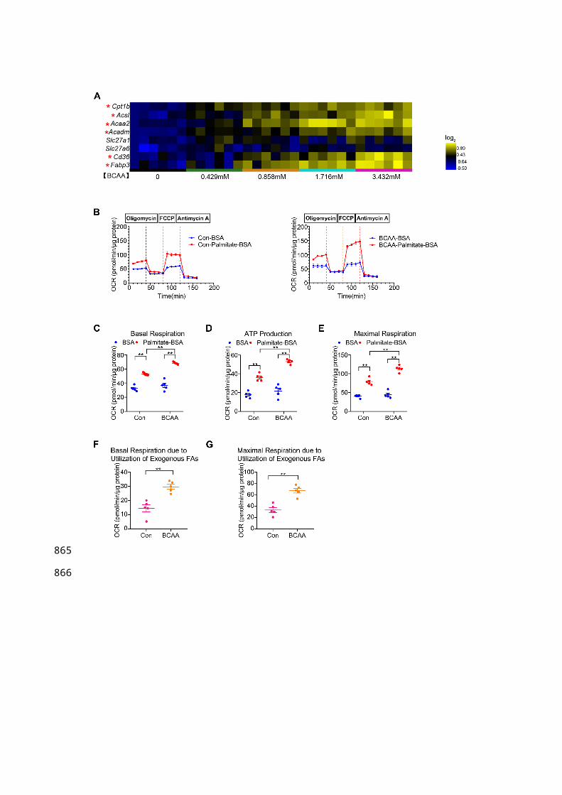
Figure 5. The effects of BCAA/BCKA on FAO level and PPAR-α expression in cardiac
myocytes. (A) Expression of PPAR-α in the presence of increasing concentrations of valine (0,
0.117 mM, 0.234 mM, 0.468 mM, 0.936 mM) by western blotting (n=5-6). (B) Expression of
PPAR-α at different concentrations of leucine (0, 0.208 mM, 0.416 mM, 0.832 mM, 1.664 mM)
(n=5-6). (C) PPAR-α expression at different concentrations of isoleucine (0, 0.104 mM, 0.208
mM, 0.416 mM, 0.832 mM) (n=5-6). (D) Expression of PPAR-α in the presence of increasing
concentrations of a-ketoisovaleric acid (αKIV) (0, 0.117 mM, 0.234 mM, 0.468 mM, 0.936 mM)
(n=5-6). (E) Expression of PPAR-α at increasing concentrations of α-ketoisocaproic acid (αKIC)
(0, 0.208 mM, 0.416 mM, 0.832 mM, 1.664 mM) (n=5-6). (F) PPAR-α expression at increasing
concentrations of α-keto-β-methylvaleric acid (αKMV) (0, 0.104 mM, 0.208 mM, 0.416 mM,
0.832 mM) (n=5-6). (G to L) Adult mouse cardiac myocytes were treated with vehicle (Con),
αKIV (0.936 mM) and αKIC (1.664 mM) for 12 h. FAO levels were determined by seahorse
analyzer (n=5). (G) OCR curve of Con group, αKIV group and αKIC group were determined. (H)
Basal respiration (I) ATP production (J) maximal respiration (K) basal respiration due to
exogenous FAs and (L) maximal respiration due to exogenous FAs were calculated according to
instruction. Data were analyzed by one-way ANOVA, followed by a Bonferroni post-hoc test. *
P<0.05. ** P<0.01. All values are presented as mean ± SEM.
673
674
675
676
677
678
679
680
681
682
683
684
685
686
687
688
689
690
691
692
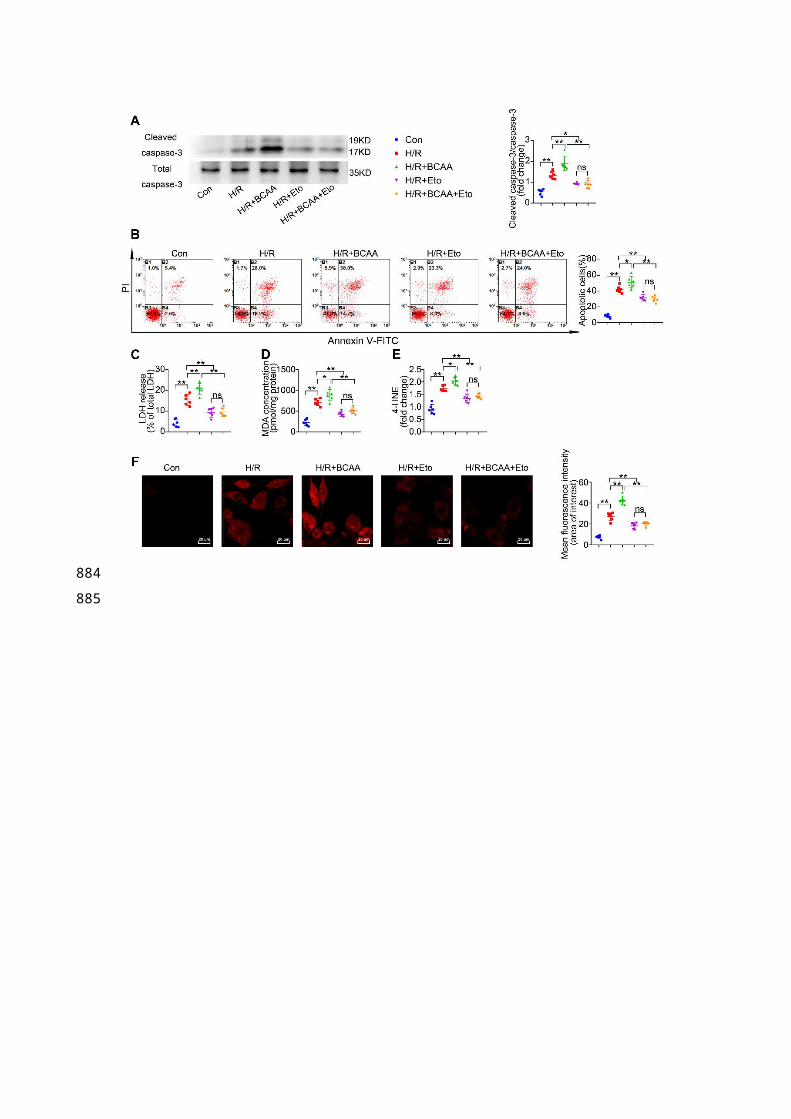
Figure 6. BCAA increase PPAR-α expression in a GCN2/ATF6 pathway-dependent manner.
(A) Expression of p-GCN2, GCN2 and ATF6 in the presence of increasing concentrations of
BCAA (0, 0.429 mM, 0.858 mM, 1.716 mM, 3.432 mM) by western blotting (n=6). (B)
Expression of p-GCN2, GCN2 and ATF6 in the presence of increasing concentrations of BCKA
(0, 0.429 mM, 0.858 mM, 1.716 mM, 3.432 mM) by western blotting (n=6). BCKA mixture is
composed of αKIC, αKIV and αKMV (weight ratio, αKIC: αKIV: αKMV= 2:1:1). (C) NRVMs
were treated with control siRNA and ATF6 siRNA. 48 h after transfection, expression of ATF6
was determined by western blotting (n=4). (D-E) ATF6 siRNA transferred NRVMs were treated
with or without BCAA (3.432 mM) (n=6). (D) PPAR-α expression was determined by western
blotting. (E) Expression of Acaa2, Acadm, Cd36 and Cpt1b by real-time PCR. (F-G) ATF6 siRNA
transferred NRVMs were treated with or without BCKA (3.432 mM) (n=6). (F) PPAR-α
expression was determined by western blotting. (G) Expression of Acaa2, Acadm, Cd36 and
Cpt1b by real-time PCR. (C) Data were analyzed by Student’s t test. (A-B) and (D-G) Data were
analyzed by one-way ANOVA, followed by a Bonferroni post-hoc test. * P<0.05, ** P<0.01. All
values are presented as mean ± SEM.
693
694
695
696
697
698
699
700
701
702
703
704
705
706
707
708
709
710
711
Figure 7. PPAR-α knockdown suppresses myocardial FAO levels and protects PP2Cm KO
cardiomyocytes against injury following H/R. (A) Expression of PPAR-α by western blotting in
cardiomyocytes with or without shPpara adenovirus infection (n=6). (B to N) Ventricular
myocytes isolated from WT mice or PP2Cm KO mice were infected with scrambled or shPpara
adenovirus for 48 h with or without H/R injury. (B to G) FAO levels were determined by seahorse
analyzer (n=4-5). (B) OCR curve treated as mentioned above were determined. (C) Basal
respiration (D) ATP production (E) maximal respiration (F) basal respiration due to exogenous
FAs and (G) maximal respiration due to exogenous FAs were calculated according to instruction.
(H) Expression of ACAA2, ACADM, CD36, CPT1B in cardiomyocytes by western blotting
(n=6). (I) Annexin V and propidium iodide (PI) staining by flow cytometry for cardiomyocyte
apoptosis determination (n=6). (J) Cleaved and non-cleaved caspase-3 by western blotting (n=6).
(K) Cell death assessed by LDH release (n=6). (L) Superoxide production detected by DHE
staining (n=6). Scale bar: 20 μm. (M and N) Lipid peroxidation determined by MDA and 4-HNE
contents (n=6). Data were analyzed by one-way ANOVA, followed by a Bonferroni post-hoc test.
* P<0.05, ** P<0.01. All values are presented as mean ± SEM.
712
713
714
715
716
717
718
719
720
721
722
723
724
725
726
727
728
729
Figure 8. Exacerbated I/R injury in the PP2Cm KO heart is rescued by PPAR-α knockdown.
(A) Expression of PPAR-α were determined by western blotting in WT mice and PP2Cm KO mice
(n=6). (B) WT and PP2Cm KO serum BCAA were determined (n=6-8). (C to L) WT mice or
PP2Cm KO mice were received intra-myocardial injection with scrambled or shPpara adenovirus
7 days before sham or I/R operation. (C) Cleaved and non-cleaved caspase-3 by western blotting
(n=6). (D) Cardiac apoptosis determined by TUNEL staining (n=10-15). Scale bar: 50 μm. (E)
Cardiac death by LDH release assay (n=6). (F) Infarct area of heart tissue by Evans blue and TTC
(n=10-15). Scale bar: 2 mm. (G) Representative M-Mode echocardiographic images. (H and I)
Echocardiographic assessment of LV ejection fraction and LV fractional shortening (n=10-15). (J)
Superoxide production detected by DHE staining (n=6). Scale bar: 50 μm. (K and L) Lipid
peroxidation determined by MDA and 4-HNE (n=6). (A-B) Data were analyzed by Student’s t test.
(C-L) Data were analyzed by one-way ANOVA, followed by a Bonferroni post-hoc test. * P<0.05,
** P<0.01. All values are presented as mean ± SEM.
730
731
732
733
734
735
736
737
738
739
740
741
742