Vitamin E δ-tocotrienol prolongs survival in the LSL...
30
1 Vitamin E δ-tocotrienol prolongs survival in the LSL-Kras G12D/+ ;LSL-Trp53 R172H/+ ;Pdx-1-Cre (KPC) transgenic mouse model of pancreatic cancer Kazim Husain 1 , Barbara A. Centeno 2 , Dung-Tsa Chen 4 , Sunil R. Hingorani 5 , Said M. Sebti 3 , and Mokenge P. Malafa 1, 3 Departments of 1 Gastrointestinal Oncology, 2 Anatomic Pathology, 3 Drug Discovery, and 4 Biostatistics Core, H. Lee Moffitt Cancer Center and Research Institute, Tampa, FL; and 5 Translational Research Program, Fred Hutchinson Cancer Research Center, Seattle, WA Key-words: δ-tocotrienol; gemcitabine, pancreatic cancer; Kras G12D -p53 R172H mouse model; survival Running title: δ-tocotrienol prolongs survival in KPC mice Financial support: The study was supported in part by National Cancer Institute/USPHS Grant 1RO1 CA-129227-01A1. Corresponding author: Mokenge P. Malafa, MD, FACS, Department of Gastrointestinal Oncology, Moffitt Cancer Center , 12902 Magnolia Drive, Tampa, FL 33612, USA (Phone: 813- 745-1432; Fax: 813-745-7229; Email: [email protected]) Disclosure of potential conflicts of interest: Drs. Malafa and Sebti are named as inventors on US Patent “Delta-Tocotrienol Treatment and Prevention of Pancreatic Cancer (June 26, 2007; OTML docket number 06A069) but do not have financial interest in the companies that have licensed this patent. Text word count: 4228; Abstract word count: 249 Total number of figures: 5; Total number of tables: 0 Cancer Research. on February 1, 2019. © 2013 American Association for cancerpreventionresearch.aacrjournals.org Downloaded from Author manuscripts have been peer reviewed and accepted for publication but have not yet been edited. Author Manuscript Published OnlineFirst on August 20, 2013; DOI: 10.1158/1940-6207.CAPR-13-0157
Transcript of Vitamin E δ-tocotrienol prolongs survival in the LSL...
1
Vitamin E δ-tocotrienol prolongs survival in the LSL-KrasG12D/+;LSL-Trp53R172H/+;Pdx-1-Cre (KPC) transgenic mouse model of pancreatic cancer Kazim Husain1, Barbara A. Centeno2, Dung-Tsa Chen4, Sunil R. Hingorani5, Said M. Sebti3, and Mokenge P. Malafa1, 3
Departments of 1Gastrointestinal Oncology, 2Anatomic Pathology, 3Drug Discovery, and 4Biostatistics Core, H. Lee Moffitt Cancer Center and Research Institute, Tampa, FL; and 5Translational Research Program, Fred Hutchinson Cancer Research Center, Seattle, WA
Key-words: δ-tocotrienol; gemcitabine, pancreatic cancer; KrasG12D-p53R172H mouse model;
survival
Running title: δ-tocotrienol prolongs survival in KPC mice
Financial support: The study was supported in part by National Cancer Institute/USPHS Grant
1RO1 CA-129227-01A1.
Corresponding author: Mokenge P. Malafa, MD, FACS, Department of Gastrointestinal
Oncology, Moffitt Cancer Center , 12902 Magnolia Drive, Tampa, FL 33612, USA (Phone: 813-
745-1432; Fax: 813-745-7229; Email: [email protected])
Disclosure of potential conflicts of interest: Drs. Malafa and Sebti are named as inventors on
US Patent “Delta-Tocotrienol Treatment and Prevention of Pancreatic Cancer (June 26, 2007;
OTML docket number 06A069) but do not have financial interest in the companies that have
licensed this patent.
Text word count: 4228; Abstract word count: 249
Total number of figures: 5; Total number of tables: 0
Cancer Research. on February 1, 2019. © 2013 American Association forcancerpreventionresearch.aacrjournals.org Downloaded from
Author manuscripts have been peer reviewed and accepted for publication but have not yet been edited. Author Manuscript Published OnlineFirst on August 20, 2013; DOI: 10.1158/1940-6207.CAPR-13-0157
2
Abstract
Previous work has shown that vitamin E δ-tocotrienol (VEDT) prolongs survival and delays progression
of pancreatic cancer in the LSL-KrasG12D/+;Pdx-1-Cre mouse model of pancreatic cancer. However, the
effect of VEDT alone or in combination with gemcitabine in the more aggressive LSL-KrasG12D/+;LSL-
Trp53R172H/+;Pdx-1-Cre (KPC) mouse model is unknown. Here, we studied the effects of VEDT and the
combination of VEDT and gemcitabine in the KPC mice. KPC mice were randomized into 4 groups: 1)
vehicle (olive oil, 1.0 mL/kg PO twice/day and PBS 1.0 mL/kg IP twice/week), 2) gemcitabine (100
mg/kg IP twice/week), 3) VEDT (200 mg/kg PO twice/day), and 4) gemcitabine + VEDT. Mice received
treatment until they displayed symptoms of impending death from pancreatic cancer, at which point
animals were euthanized. At 16 weeks, survival was 10% in the vehicle group, 30% in the gemcitabine
group, 70% in the VEDT group (P<0.01), and 90% in the VEDT combined with gemcitabine group
(P<0.05). VEDT alone and combined with gemcitabine resulted in reversal of epithelial-to-mesenchymal
transition in tumors. Biomarkers of apoptosis (plasma CK18), PARP1 cleavage, and Bax expression were
more greatly induced in tumors subjected to combined treatment versus individual treatment. Combined
treatment induced cell cycle inhibitors (p27Kip1 and p21Cip1) and inhibited VEGF, vascularity (CD31), and
oncogenic signaling (pAKT, pMEK, and pERK) greater than individual drugs. No significant differences
in body weight gain between drug treatment and control mice were observed. These results strongly
support further investigation of VEDT alone and in combination with gemcitabine for pancreatic cancer
prevention and treatment.
Cancer Research. on February 1, 2019. © 2013 American Association forcancerpreventionresearch.aacrjournals.org Downloaded from
Author manuscripts have been peer reviewed and accepted for publication but have not yet been edited. Author Manuscript Published OnlineFirst on August 20, 2013; DOI: 10.1158/1940-6207.CAPR-13-0157
3
Introduction
Advanced pancreatic ductal adenocarcinoma is a lethal disease with approximately 6
months median survival (1). Gemcitabine is the only approved single agent, with a median
survival of 5.7 months and 20% 1-year survival (2, 3). After many phase III trials exploring
gemcitabine-based combinations failed to improve overall survival in patients, two recent trials
with erlotinib and nab-paclitaxel (Abraxane) showed modest but significant improvements in
survival (4, 5). Erlotinib improved median survival to 6.24 months, whereas nab-paclitaxel
improved median survival to 8.5 months. At the end of 1 year, 35% of patients who received
nab-paclitaxel with gemcitabine were alive, compared with 22% of patients who received only
gemcitabine. Because of the moderate activity of the present gemcitabine-based regimens,
improved prevention and therapeutic options for pancreatic cancer are a high priority.
There has been intense interest in the role of natural nutritional/dietary factors in the
prevention and treatment of pancreatic cancer (6, 7). Several reports have shown that increasing
vegetable, fruit, and cereal consumption may impact protection against the development of
pancreatic cancer (8, 9). One of the most compelling groups of anti-tumor bioactive compounds
in cereal grains are vitamin E tocotrienols (10). Tocotrienols are unsaturated, naturally occurring
vitamin E compounds, which exist as four isoforms: α-, β-, δ-, and γ-tocotrienol (11). We have
shown that, for pancreatic cancer, vitamin E δ-tocotrienol (VEDT) is the most potent anticancer
agent among the four isomers, both in vitro and in vivo (12). We have also shown that oral
administration of 100 mg/kg/day of VEDT to mice resulted in satisfactory bioavailability in
mouse pancreas tissue with no significant toxicity (13). In a recent study, we also demonstrated
that VEDT, when administered for almost 1 year, prolonged the survival and delayed pancreatic
Cancer Research. on February 1, 2019. © 2013 American Association forcancerpreventionresearch.aacrjournals.org Downloaded from
Author manuscripts have been peer reviewed and accepted for publication but have not yet been edited. Author Manuscript Published OnlineFirst on August 20, 2013; DOI: 10.1158/1940-6207.CAPR-13-0157
4
intraepithelial neoplasia lesions in the LSL-KRASG12D/PDX-1-Cre genetic mouse model of
pancreatic cancer (14).
Activating mutations in the Kras oncogene are found in over 90% of human pancreatic
cancers (15). Conditionally targeting an activating point mutation in Kras (G12D) with a Pdx-1
pancreas-specific promoter resulted in pancreatic lesions that display a full spectrum of
pancreatic intraepithelial neoplasias in mice (16). These lesions progress to fully invasive and
metastatic adenocarcinomas; thus, the LSL-KrasG12D; Pdx-1-Cre transgenic mouse model mimics
both the genetic and histologic changes observed in human pancreatic cancer. When a point
mutation (R172H) in the p53 tumor suppressor gene, which is mutated in 75% of human
pancreatic tumors, was introduced into the pancreas of mice with a KrasG12D/+ mutation, these
triple transgenic LSL-KrasG12D;LSL-Trp53R127H; Pdx-1-Cre (KPC) mice developed pancreatic
cancer more rapidly than the mice with just the Kras mutation (17). The clinical symptoms,
including cachexia and abdominal distension, the defined histopathologic progression, and the
genomic instability found in human pancreatic cancer are all replicated in the KPC transgenic
mice, making it one of the most relevant animal models for preclinical evaluation of new drugs
for pancreatic cancer (18). We hypothesized that VEDT, alone and in combination with
gemcitabine, could significantly delay tumor growth and prolong survival in the KPC mice.
Based on the previous tumor effects of tocotrienols observed by us and others, we hypothesized
that VEDT would inhibit tumor angiogenesis, induce apoptosis, and alter oncogenic signaling.
MATERIALS AND METHODS
Reagents and animals
Cancer Research. on February 1, 2019. © 2013 American Association forcancerpreventionresearch.aacrjournals.org Downloaded from
Author manuscripts have been peer reviewed and accepted for publication but have not yet been edited. Author Manuscript Published OnlineFirst on August 20, 2013; DOI: 10.1158/1940-6207.CAPR-13-0157
5
All chemicals and reagents were purchased from Sigma-Aldrich (St. Louis, MO) unless
otherwise specified. VEDT was obtained from Davos Life Ltd (Helios, Singapore). Gemcitabine-HCl was purchased from Eli Lilly and Company. Pairs of male and female LSL-
KRASG12D, LSL-Trp53R127H, and PDX-1-Cre mice were obtained from the National Cancer
Institute Mouse Models of Human Cancers Consortium (Frederick, MD). The animal protocol
used in the study was approved by our Institutional Animal Care and Use Committee.
To study the effects of VEDT and gemcitabine on pancreatic tumor development, the
KPC mouse model from Hingorani et al. (17) was used. LSL-KRASG12D, LSL-Trp53R127H, and
PDX-1-Cre mice were maintained as heterozygous lines. They were crossed and bred in the
vivarium of our center. Tail snips were harvested from offspring of LSL-KRASG12D, LSL-
Trp53R127H, and PDX-1-Cre mice and allowed to digested overnight; genomic DNA was then
extracted and estimated using the DNAeasy Blood & Tissue (250) kit from Qiagen Inc, as per
the manufacturer’s instructions.
Genotyping analysis
PCR primer sequences used to detect PDX-1-Cre, LSL-KRASG12D, and LSL-Trp53R127H were as
follows: 5’-CTGGACTACATCTTGAGTTGC-3’ and 5’-GGTGTACGGTCAGTAAATTTG-3’
(PDX-1-Cre forward and reverse); 5’-AGGTAGCCACCATGGCTTGAGTAAGTCTGCA-3’
and 5’-CCTTTACAAGCGCACGCAGACTGTAGA-3’ (LSL-KRAS612D forward and reverse);
and 5’-AGCTAGCCACCATGGCTTGAGTAAGTCTGCA-3’ and 5’-
CTTGGAGACATAGCCACACTG-3’ (LSL-Trp53R127H forward and reverse) (all from IDT
Technologies). The PCR reaction of 25 µL was composed of buffer, 2.0 mM MgCl2, 0.2 mM
dNTP mix, 0.025 U/µL DNA Taq polymerase, 0.5 µM primers, and 50 ng DNA. The reaction
Cancer Research. on February 1, 2019. © 2013 American Association forcancerpreventionresearch.aacrjournals.org Downloaded from
Author manuscripts have been peer reviewed and accepted for publication but have not yet been edited. Author Manuscript Published OnlineFirst on August 20, 2013; DOI: 10.1158/1940-6207.CAPR-13-0157
6
was carried out in the PTC-200 thermal cycler programmed as preheat at 94°C for 3 minutes and
then 35 cycles of (30 seconds at 94°C, 30 seconds at 60°C, and 30 seconds at 72°C) followed by
72°C for 3 minutes. PCR products were mixed with 5 µL of loading dye and separated on a 2%
agarose gel containing ethidium bromide. The electrophoresis was run for 1 hour at 100 volts
using Tris base, acetic acid, and EDTA buffer. A 650-bp product (PDX), a 550-bp product
(KRAS), and 270-bp product (p53) were identified using DNA ladder, and the bands were
imaged using AlphaImage analysis.
Drug treatments
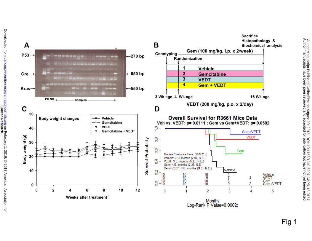
Offspring of LSL-KRASG12D, LSL-Trp53R127H, and PDX-1-Cre mice (triple gene positive), as
shown in Figure 1A, were randomized into four groups: 1) vehicle (ethanol extracted olive oil,
1.0 ml/kg twice/day by oral gavage) (n = 10), 2) gemcitabine (100 mg/kg ip twice/week) (n = 10),
3) VEDT (200 mg/kg twice/day by oral gavage) (n = 10), and 4) gemcitabine + VEDT (n = 10).
The treatment was started at the age of 4 weeks and continued for 12 weeks, as shown in Figure
1B. The body weights of the mice were recorded twice weekly, mortality was noted, and survival
curves were plotted (see Figure 1C). When the mice displayed symptoms of impending death
such as cachexia, abdominal distension, rapid weight loss, or labored breathing, animals were
euthanized. Blood was collected in heparinized tubes, and the entire tumor tissues were
harvested and weighed. Half of the tissues were fixed in buffered formalin for histological
analysis, with the remainder snap frozen in liquid nitrogen and kept at -80°C for protein
extraction and Western blot analysis.
Histologic evaluation
Cancer Research. on February 1, 2019. © 2013 American Association forcancerpreventionresearch.aacrjournals.org Downloaded from
Author manuscripts have been peer reviewed and accepted for publication but have not yet been edited. Author Manuscript Published OnlineFirst on August 20, 2013; DOI: 10.1158/1940-6207.CAPR-13-0157
7
Formalin-fixed, paraffin-embedded tissues were sectioned (4 µm) and stained with hematoxylin-
eosin. Immunohistochemistry was performed using the Ventana Discovery XT automated system
(Ventana Medical Systems, Tucson, AZ) per manufacturer's protocol with proprietary reagents.
Briefly, slides were deparaffinized on the automated system with EZ Prep solution. Sections
were heated for antigen retrieval. For immunohistochemistry, tissue sections were incubated with
anti-caspase 3, Ki-67, and CD31 at 1:4000 dilutions for 60 minutes. Detection was performed
using the Ventana OmniMap kit.
Assessment of immunohistochemical expression
All stained tissues were examined by one independent observer (BAC). Caspase 3 and Ki-67
stained tissues were assessed for expression in neoplastic and non-neoplastic areas. Percent
expression was recorded for each area and then averaged for each mouse. For CD31, sections
were examined at low power to identify cancers and associated hot spots. The vessels per x400
field were counted manually. Single cells and groups expressing CD31 were counted as vessels
in addition to groups with lumens. Sections not showing a cancer were assessed for hot spots at
low power.
Apoptosis marker CK18 ELISA
Heparinized blood from mice was centrifuged at 5000 rpm for 5 minutes, and plasma was
carefully isolated and stored at -80°C until analysis. The apoptosis marker cytokeratin-18
(CK18) was assayed using the M30-Apoptosense ELISA kit (PEVIVA, Bromma, Sweden).
Western blot analysis
Cancer Research. on February 1, 2019. © 2013 American Association forcancerpreventionresearch.aacrjournals.org Downloaded from
Author manuscripts have been peer reviewed and accepted for publication but have not yet been edited. Author Manuscript Published OnlineFirst on August 20, 2013; DOI: 10.1158/1940-6207.CAPR-13-0157
8
Proteins were extracted from pancreatic tumor tissues using RIPA lysis buffer containing
protease inhibitors (Thermo Scientific, Rockford, IL). Extracted proteins (40 µg) were resolved
on 12.5% SDS-polyacrylamide gel (SDS-PAGE) running gel and a 5% stacking gel. Proteins
were then electrotransferred onto nitrocellulose membranes. After blocking in 5% nonfat
powdered milk for 1 hour, membranes were washed and then treated with antibodies to PARP-1,
E-cadherin, vimentin, pAKT, pMEK, pERK, VEGF, Bax, p21Cip1, p27kip-1, and β-actin (1:1000)
overnight at 4oC (Santa Cruz Biotechnology, Santa Cruz, CA; Cell Signaling, Danvers, MA).
After blots were washed, they were incubated with horseradish peroxidase-conjugated secondary
antibody IgG (1:5000) for 1 hour at room temperature. The washed blot was then treated with
SuperSignal West Pico chemiluminescent substrate (Pierce) for positive antibody reaction.
Membranes were exposed to X-ray film (KODAK) for visualization and densitometric
quantization of protein bands using AlphaEaseFC software (Alpha Innotech).
Statistical analysis
Data are expressed as means ± SEM, analyzed statistically using one-way analysis of
variance (ANOVA) followed by Duncan's multiple range tests using SAS statistical software for
comparison between different treatment groups. Significance was set at P < 0.05.
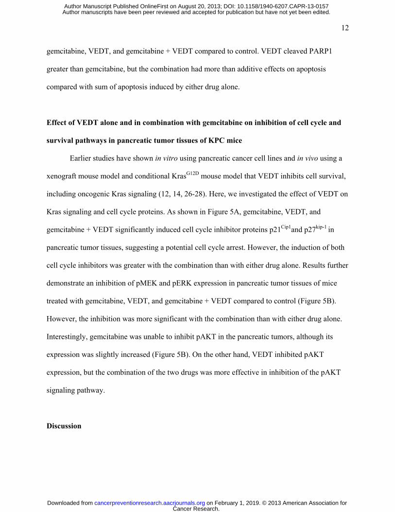
RESULTS Effects of VEDT alone and in combination with gemcitabine on survival and body weight
in KPC mice
To assess whether VEDT alone and in combination with gemcitabine affected survival of
our mice, we analyzed the effects of the drugs using Kaplan-Meier survival curves. We found
that the overall survival of vehicle-treated mice from the onset of treatment was 2.18 months
Cancer Research. on February 1, 2019. © 2013 American Association forcancerpreventionresearch.aacrjournals.org Downloaded from
Author manuscripts have been peer reviewed and accepted for publication but have not yet been edited. Author Manuscript Published OnlineFirst on August 20, 2013; DOI: 10.1158/1940-6207.CAPR-13-0157
9
(Figure 1D), which is in agreement with earlier reports of survival/death of KPC mice (17, 19).
Only 10% of the vehicle-treated mice were alive at 4 months of age. VEDT treatment
significantly increased the percentage of mice that were alive at 4 months to 70% (P<0.01); 30%
of gemcitabine-treated mice were alive at 4 months of age. However, VEDT treatment combined
with gemcitabine resulted in 90% (P<0.05) of mice being alive at 4 months of age. It is of great
interest that, at 5 months of age, 40% of VEDT-treated mice and 20% of VEDT combined with
gemcitabine-treated mice were alive without symptoms of impending death, whereas none of the
vehicle or gemcitabine-treated mice were alive (Figure 1D). There was no significant difference
in food intake and body weight gain between drug treatment groups and the control vehicle
treatment group during the study period (Figure 1C). Our data indicated no obvious toxicity or
side effects of the drugs alone and in combination after chronic dosing for 12 weeks in mice with
pancreatic cancer, indicating that symptoms of impending death were entirely related to tumor
burden. This finding was confirmed at autopsy in all of the mice.
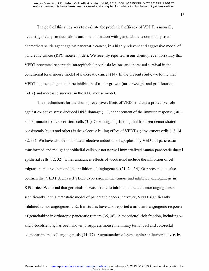
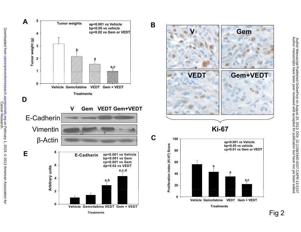
Effects of VEDT alone and in combination with gemcitabine on tumor weight, EMT, and
proliferation index in KPC mice
Mice treated with gemcitabine and VEDT alone and in combination had significantly
reduced tumor weights of 32% (P<0.05), 51% (P<0.001), and 69% (P<0.001), respectively,
versus vehicle (Figure 2A). The combination resulted in a significant reduction of tumor weight
compared to either drug alone (P<0.02). Interestingly, VEDT treatment resulted in a greater
decrease in tumor weight than gemcitabine. Gemcitabine alone, VEDT alone, and combined
gemcitabine and VEDT significantly decreased Ki-67 staining (a marker of proliferation index)
in tumors at 27% (P<0.05), 39% (P<0.001), and 61% (P<0.001), respectively, compared with
Cancer Research. on February 1, 2019. © 2013 American Association forcancerpreventionresearch.aacrjournals.org Downloaded from
Author manuscripts have been peer reviewed and accepted for publication but have not yet been edited. Author Manuscript Published OnlineFirst on August 20, 2013; DOI: 10.1158/1940-6207.CAPR-13-0157
10
vehicle (Figure 2B and C). Interestingly the combination of the two drugs resulted in a greater
inhibition of proliferation than either drug alone. Because recent studies revealed that
tocotrienols inhibit cancer cell invasion through reversal of epithelial-to-mesenchymal transition
(EMT), we evaluated the effects of VEDT on EMT in the mouse tumors (20, 21). E-cadherin, a
marker of epithelial phenotype, slightly increased after gemcitabine treatment, whereas with
VEDT alone and in combination with gemcitabine it was profoundly increased (Figure 2D and
E). In contrast, vimentin, a marker of mesenchymal phenotype, decreased after treatment with
gemcitabine or VEDT alone; however, when the two drugs were combined, expression was
almost completely abolished (Figure 2D). These finding clearly support the reversal of EMT by
VEDT in the pancreatic tumors.
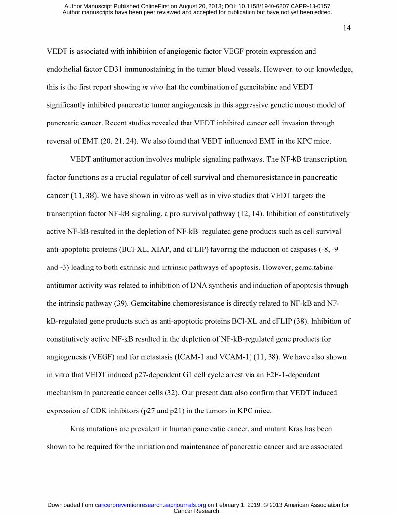
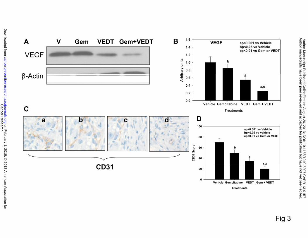
Effect of VEDT alone and in combination with gemcitabine on VEGF expression and
tumor angiogenesis in KPC mice
The antiangiogenic properties of tocotrienols have been demonstrated in several studies
(22-25). Specifically, VEGF has been implicated as a major target of tocotrienol antitumor
activity. Since the effect of VEDT on VEGF expression and angiogenesis in pancreatic cancer is
unknown, we examined the effect of VEDT on VEGF expression and angiogenesis in our mouse
tumors. Mice treated with gemcitabine alone, VEDT alone, and the combination showed
significantly reduced VEGF expression: 21% (P<0.05), 55% (P<0.001), and 78% (P<0.001),
respectively, compared with vehicle (Figure 3B). The tumor antiangiogenic effects of VEDT are
far greater than gemcitabine. On the other hand, when the two drugs are combined, there is a
greater inhibition of VEGF expression than with either drug alone. The tumor blood vessel
counts (CD31 immunoreactivity) were significantly decreased with gemcitabine alone (29%,
Cancer Research. on February 1, 2019. © 2013 American Association forcancerpreventionresearch.aacrjournals.org Downloaded from
Author manuscripts have been peer reviewed and accepted for publication but have not yet been edited. Author Manuscript Published OnlineFirst on August 20, 2013; DOI: 10.1158/1940-6207.CAPR-13-0157
11
P<0.02), VEDT alone (57%, P<0.001), and gemcitabine + VEDT combined (97%, P<0.001)
compared to vehicle (Figure 3D). The combination of the two drugs resulted in greater inhibition
of CD31 immunostaining than either drug alone.
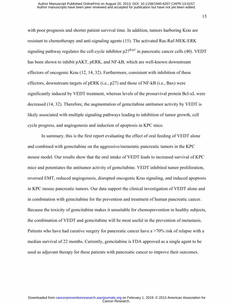
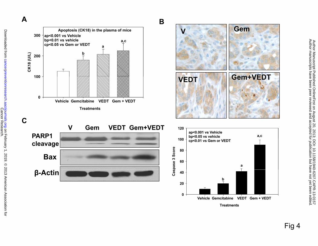
Effect of VEDT alone and in combination with gemcitabine on induction of apoptosis in
pancreatic tumor tissues of KPC mice
Earlier studies have shown in vitro using pancreatic cancer cell lines and in vivo using a
xenograft mouse model and a conditional KrasG12D mouse model that VEDT induces apoptosis
and inhibits cell survival (12, 26-28). Therefore, we evaluated the effect VEDT on apoptosis in
the KPC mouse tumors. As shown in Figure 4A, gemcitabine, VEDT, and the combination of the
two drugs significantly elicited apoptotic cell death in the circulating tumor cells (CK18) in the
blood: 45% (P<0.01), 71% (P<0.001), and 84% (P<0.001), respectively, compared to vehicle,
suggesting that loss of viable cells is due to the induction of the cell death pathway. We
confirmed this by more intense and significant immunostaining of cleaved caspase 3 in
gemcitabine (P<0.05), VEDT (P<0.001), and gemcitabine + VEDT (P<0.001) groups,
respectively, compared with the vehicle group (Figure 4B). Further Western immunoblotting in
the pancreatic tumor tissues of mice showed enhanced protein expression of the proapoptotic
protein Bax in gemcitabine, VEDT, and gemcitabine + VEDT-treated groups compared to
control (Figure 4C). The combination of the two drugs resulted in more than additive effect on
Bax expression when compared with sum of Bax expression induced by either drug alone.
PARP-1, a 116-kDa nuclear poly(ADP-ribose) polymerase 1, is one of the main cleavage targets
of caspase 3 in vivo, serving as a marker of cells undergoing apoptosis (29). Our Western blot
results clearly showed the PARP1 cleavage in pancreatic cancer tissues of mice treated with
Cancer Research. on February 1, 2019. © 2013 American Association forcancerpreventionresearch.aacrjournals.org Downloaded from
Author manuscripts have been peer reviewed and accepted for publication but have not yet been edited. Author Manuscript Published OnlineFirst on August 20, 2013; DOI: 10.1158/1940-6207.CAPR-13-0157
12
gemcitabine, VEDT, and gemcitabine + VEDT compared to control. VEDT cleaved PARP1
greater than gemcitabine, but the combination had more than additive effects on apoptosis
compared with sum of apoptosis induced by either drug alone.
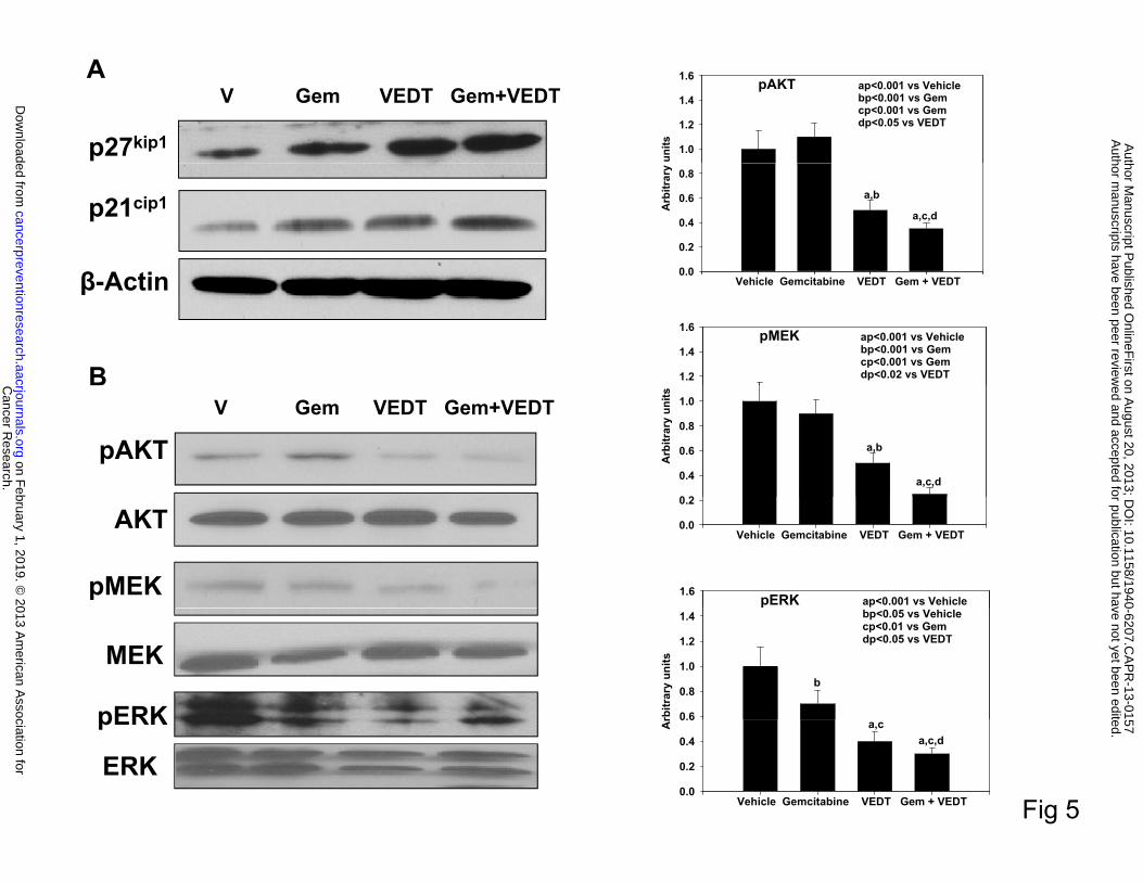
Effect of VEDT alone and in combination with gemcitabine on inhibition of cell cycle and
survival pathways in pancreatic tumor tissues of KPC mice
Earlier studies have shown in vitro using pancreatic cancer cell lines and in vivo using a
xenograft mouse model and conditional KrasG12D mouse model that VEDT inhibits cell survival,
including oncogenic Kras signaling (12, 14, 26-28). Here, we investigated the effect of VEDT on
Kras signaling and cell cycle proteins. As shown in Figure 5A, gemcitabine, VEDT, and
gemcitabine + VEDT significantly induced cell cycle inhibitor proteins p21Cip1and p27kip-1 in
pancreatic tumor tissues, suggesting a potential cell cycle arrest. However, the induction of both
cell cycle inhibitors was greater with the combination than with either drug alone. Results further
demonstrate an inhibition of pMEK and pERK expression in pancreatic tumor tissues of mice
treated with gemcitabine, VEDT, and gemcitabine + VEDT compared to control (Figure 5B).
However, the inhibition was more significant with the combination than with either drug alone.
Interestingly, gemcitabine was unable to inhibit pAKT in the pancreatic tumors, although its
expression was slightly increased (Figure 5B). On the other hand, VEDT inhibited pAKT
expression, but the combination of the two drugs was more effective in inhibition of the pAKT
signaling pathway.
Discussion
Cancer Research. on February 1, 2019. © 2013 American Association forcancerpreventionresearch.aacrjournals.org Downloaded from
Author manuscripts have been peer reviewed and accepted for publication but have not yet been edited. Author Manuscript Published OnlineFirst on August 20, 2013; DOI: 10.1158/1940-6207.CAPR-13-0157
13
The goal of this study was to evaluate the preclinical efficacy of VEDT, a naturally
occurring dietary product, alone and in combination with gemcitabine, a commonly used
chemotherapeutic agent against pancreatic cancer, in a highly relevant and aggressive model of
pancreatic cancer (KPC mouse model). We recently reported in our chemoprevention study that
VEDT prevented pancreatic intraepithelial neoplasia lesions and increased survival in the
conditional Kras mouse model of pancreatic cancer (14). In the present study, we found that
VEDT augmented gemcitabine inhibition of tumor growth (tumor weight and proliferation
index) and increased survival in the KPC mouse model.
The mechanisms for the chemopreventive effects of VEDT include a protective role
against oxidative stress-induced DNA damage (11), enhancement of the immune response (30),
and elimination of cancer stem cells (31). One intriguing finding that has been demonstrated
consistently by us and others is the selective killing effect of VEDT against cancer cells (12, 14,
32, 33). We have also demonstrated selective induction of apoptosis by VEDT of pancreatic
transformed and malignant epithelial cells but not normal immortalized human pancreatic ductal
epithelial cells (12, 32). Other anticancer effects of tocotrienol include the inhibition of cell
migration and invasion and the inhibition of angiogenesis (21, 24, 34). Our present data also
confirm that VEDT decreased VEGF expression in the tumors and inhibited angiogenesis in
KPC mice. We found that gemcitabine was unable to inhibit pancreatic tumor angiogenesis
significantly in this metastatic model of pancreatic cancer; however, VEDT significantly
inhibited tumor angiogenesis. Earlier studies have also reported a mild anti-angiogenic response
of gemcitabine in orthotopic pancreatic tumors (35, 36). A tocotrienol-rich fraction, including γ-
and δ-tocotrienols, has been shown to suppress mouse mammary tumor cell and colorectal
adenocarcinoma cell angiogenesis (34, 37). Augmentation of gemcitabine antitumor activity by
Cancer Research. on February 1, 2019. © 2013 American Association forcancerpreventionresearch.aacrjournals.org Downloaded from
Author manuscripts have been peer reviewed and accepted for publication but have not yet been edited. Author Manuscript Published OnlineFirst on August 20, 2013; DOI: 10.1158/1940-6207.CAPR-13-0157
14
VEDT is associated with inhibition of angiogenic factor VEGF protein expression and
endothelial factor CD31 immunostaining in the tumor blood vessels. However, to our knowledge,
this is the first report showing in vivo that the combination of gemcitabine and VEDT
significantly inhibited pancreatic tumor angiogenesis in this aggressive genetic mouse model of
pancreatic cancer. Recent studies revealed that VEDT inhibited cancer cell invasion through
reversal of EMT (20, 21, 24). We also found that VEDT influenced EMT in the KPC mice.
VEDT antitumor action involves multiple signaling pathways. The NF-kB transcription factor functions as a crucial regulator of cell survival and chemoresistance in pancreatic cancer (11, 38). We have shown in vitro as well as in vivo studies that VEDT targets the
transcription factor NF-kB signaling, a pro survival pathway (12, 14). Inhibition of constitutively
active NF-kB resulted in the depletion of NF-kB–regulated gene products such as cell survival
anti-apoptotic proteins (BCl-XL, XIAP, and cFLIP) favoring the induction of caspases (-8, -9
and -3) leading to both extrinsic and intrinsic pathways of apoptosis. However, gemcitabine
antitumor activity was related to inhibition of DNA synthesis and induction of apoptosis through
the intrinsic pathway (39). Gemcitabine chemoresistance is directly related to NF-kB and NF-
kB-regulated gene products such as anti-apoptotic proteins BCl-XL and cFLIP (38). Inhibition of
constitutively active NF-kB resulted in the depletion of NF-kB-regulated gene products for
angiogenesis (VEGF) and for metastasis (ICAM-1 and VCAM-1) (11, 38). We have also shown
in vitro that VEDT induced p27-dependent G1 cell cycle arrest via an E2F-1-dependent
mechanism in pancreatic cancer cells (32). Our present data also confirm that VEDT induced
expression of CDK inhibitors (p27 and p21) in the tumors in KPC mice.
Kras mutations are prevalent in human pancreatic cancer, and mutant Kras has been
shown to be required for the initiation and maintenance of pancreatic cancer and are associated
Cancer Research. on February 1, 2019. © 2013 American Association forcancerpreventionresearch.aacrjournals.org Downloaded from
Author manuscripts have been peer reviewed and accepted for publication but have not yet been edited. Author Manuscript Published OnlineFirst on August 20, 2013; DOI: 10.1158/1940-6207.CAPR-13-0157
15
with poor prognosis and shorter patient survival time. In addition, tumors harboring Kras are
resistant to chemotherapy and anti-signaling agents (15). The activated Ras-Raf-MEK-ERK
signaling pathway regulates the cell-cycle inhibitor p27Kip1 in pancreatic cancer cells (40). VEDT
has been shown to inhibit pAKT, pERK, and NF-kB, which are well-known downstream
effectors of oncogenic Kras (12, 14, 32). Furthermore, consistent with inhibition of these
effectors, downstream targets of pERK (i.e., p27) and those of NF-kB (i.e., Bax) were
significantly induced by VEDT treatment, whereas levels of the prosurvival protein Bcl-xL were
decreased (14, 32). Therefore, the augmentation of gemcitabine antitumor activity by VEDT is
likely associated with multiple signaling pathways leading to inhibition of tumor growth, cell
cycle progress, and angiogenesis and induction of apoptosis in KPC mice.
In summary, this is the first report evaluating the effect of oral feeding of VEDT alone
and combined with gemcitabine on the aggressive/metastatic pancreatic tumors in the KPC
mouse model. Our results show that the oral intake of VEDT leads to increased survival of KPC
mice and potentiates the antitumor activity of gemcitabine. VEDT inhibited tumor proliferation,
reversed EMT, reduced angiogenesis, disrupted oncogenic Kras signaling, and induced apoptosis
in KPC mouse pancreatic tumors. Our data support the clinical investigation of VEDT alone and
in combination with gemcitabine for the prevention and treatment of human pancreatic cancer.
Because the toxicity of gemcitabine makes it unsuitable for chemoprevention in healthy subjects,
the combination of VEDT and gemcitabine will be most useful in the prevention of metastasis.
Patients who have had curative surgery for pancreatic cancer have a >70% risk of relapse with a
median survival of 22 months. Currently, gemcitabine is FDA approved as a single agent to be
used as adjuvant therapy for these patients with pancreatic cancer to improve their outcomes.
Cancer Research. on February 1, 2019. © 2013 American Association forcancerpreventionresearch.aacrjournals.org Downloaded from
Author manuscripts have been peer reviewed and accepted for publication but have not yet been edited. Author Manuscript Published OnlineFirst on August 20, 2013; DOI: 10.1158/1940-6207.CAPR-13-0157
16
Based on our results, the combination of VEDT and gemcitabine should be investigated with the
goal of increasing the disease-free interval, thereby prolonging the survival of these patients.
ACKNOWLEDGMENTS
We thank Rasa Hamilton (Moffitt Cancer Center) for editorial assistance.
Cancer Research. on February 1, 2019. © 2013 American Association forcancerpreventionresearch.aacrjournals.org Downloaded from
Author manuscripts have been peer reviewed and accepted for publication but have not yet been edited. Author Manuscript Published OnlineFirst on August 20, 2013; DOI: 10.1158/1940-6207.CAPR-13-0157
17
References
1. Siegel R, Naishadham D, Jemal A. Cancer statistics, 2013. CA Cancer J Clin 2013;63:11-30.
2. Vickers MM, Powell ED, Asmis TR, Jonker DJ, Hilton JF, O'Callaghan CJ, et al. Comorbidity,
age and overall survival in patients with advanced pancreatic cancer - results from NCIC CTG PA.3: a
phase III trial of gemcitabine plus erlotinib or placebo. Eur J Cancer 2012;48:1434-42.
3. Arshad A, Al-Leswas D, Al-Taan O, Stephenson J, Metcalfe M, Steward WP, et al. Pooled
survival and response data from phase III randomized controlled trials for gemcitabine-based regimes in
the treatment of advanced pancreatic cancer. Am J Clin Oncol 2011.
4. Moore MJ, Goldstein D, Hamm J, Figer A, Hecht JR, Gallinger S, et al. Erlotinib plus
gemcitabine compared with gemcitabine alone in patients with advanced pancreatic cancer: a phase III
trial of the National Cancer Institute of Canada Clinical Trials Group. J Clin Oncol 2007;25:1960-6.
5. Drug Combo Effective against Pancreatic Cancer. Cancer Discov 2013;3:OF8.
6. Taylor PR, Greenwald P. Nutritional interventions in cancer prevention. J Clin Oncol
2005;23:333-45.
7. Stolzenberg-Solomon RZ, Albanes D, Nieto FJ, Hartman TJ, Tangrea JA, Rautalahti M, et al.
Pancreatic cancer risk and nutrition-related methyl-group availability indicators in male smokers. J Natl
Cancer Inst 1999;91:535-41.
8. Chan JM, Wang F, Holly EA. Vegetable and fruit intake and pancreatic cancer in a population-
based case-control study in the San Francisco bay area. Cancer Epidemiol Biomarkers Prev
2005;14:2093-7.
Cancer Research. on February 1, 2019. © 2013 American Association forcancerpreventionresearch.aacrjournals.org Downloaded from
Author manuscripts have been peer reviewed and accepted for publication but have not yet been edited. Author Manuscript Published OnlineFirst on August 20, 2013; DOI: 10.1158/1940-6207.CAPR-13-0157

18
9. Silverman DT, Swanson CA, Gridley G, Wacholder S, Greenberg RS, Brown LM, et al. Dietary
and nutritional factors and pancreatic cancer: a case-control study based on direct interviews. J Natl
Cancer Inst 1998;90:1710-9.
10. Aggarwal B, Nesaretnam K. Vitamin E tocotrienols: life beyond tocopherols. Genes Nutr
2012;7:1.
11. Aggarwal BB, Sundaram C, Prasad S, Kannappan R. Tocotrienols, the vitamin E of the 21st
century: its potential against cancer and other chronic diseases. Biochem Pharmacol 2010;80:1613-31.
12. Husain K, Francois RA, Yamauchi T, Perez M, Sebti SM, Malafa MP. Vitamin E delta-
tocotrienol augments the antitumor activity of gemcitabine and suppresses constitutive NF-kappaB
activation in pancreatic cancer. Mol Cancer Ther 2011;10:2363-72.
13. Husain K, Francois RA, Hutchinson SZ, Neuger AM, Lush R, Coppola D, et al. Vitamin E delta-
tocotrienol levels in tumor and pancreatic tissue of mice after oral administration. Pharmacology
2009;83:157-63.
14. Husain K, Centeno BA, Chen DT, Fulp WJ, Perez M, Zhang Lee G, et al. Prolonged survival and
delayed progression of pancreatic intraepithelial neoplasia in LSL-KrasG12D/+;Pdx-1-Cre mice by
vitamin E delta-tocotrienol. Carcinogenesis 2013.
15. Almoguera C, Shibata D, Forrester K, Martin J, Arnheim N, Perucho M. Most human carcinomas
of the exocrine pancreas contain mutant c-K-ras genes. Cell 1988;53:549-54.
16. Hingorani SR, Petricoin EF, Maitra A, Rajapakse V, King C, Jacobetz MA, et al. Preinvasive and
invasive ductal pancreatic cancer and its early detection in the mouse. Cancer Cell 2003;4:437-50.
Cancer Research. on February 1, 2019. © 2013 American Association forcancerpreventionresearch.aacrjournals.org Downloaded from
Author manuscripts have been peer reviewed and accepted for publication but have not yet been edited. Author Manuscript Published OnlineFirst on August 20, 2013; DOI: 10.1158/1940-6207.CAPR-13-0157
19
17. Hingorani SR, Wang L, Multani AS, Combs C, Deramaudt TB, Hruban RH, et al. Trp53R172H
and KrasG12D cooperate to promote chromosomal instability and widely metastatic pancreatic ductal
adenocarcinoma in mice. Cancer Cell 2005;7:469-83.
18. Schutte U, Bisht S, Brossart P, Feldmann G. Recent developments of transgenic and xenograft
mouse models of pancreatic cancer for translational research. Expert Opin Drug Discov 2011;6:33-48.
19. Liby KT, Royce DB, Risingsong R, Williams CR, Maitra A, Hruban RH, et al. Synthetic
triterpenoids prolong survival in a transgenic mouse model of pancreatic cancer. Cancer Prev Res (Phila)
2010;3:1427-34.
20. Chang PN, Yap WN, Lee DT, Ling MT, Wong YC, Yap YL. Evidence of gamma-tocotrienol as
an apoptosis-inducing, invasion-suppressing, and chemotherapy drug-sensitizing agent in human
melanoma cells. Nutrition and cancer 2009;61:357-66.
21. Yap WN, Chang PN, Han HY, Lee DT, Ling MT, Wong YC, et al. Gamma-tocotrienol
suppresses prostate cancer cell proliferation and invasion through multiple-signalling pathways. British
journal of cancer 2008;99:1832-41.
22. Inokuchi H, Hirokane H, Tsuzuki T, Nakagawa K, Igarashi M, Miyazawa T. Anti-angiogenic
activity of tocotrienol. Bioscience, biotechnology, and biochemistry 2003;67:1623-7.
23. Wells SR, Jennings MH, Rome C, Hadjivassiliou V, Papas KA, Alexander JS. Alpha-, gamma-
and delta-tocopherols reduce inflammatory angiogenesis in human microvascular endothelial cells. The
Journal of nutritional biochemistry 2010;21:589-97.
24. Weng-Yew W, Selvaduray KR, Ming CH, Nesaretnam K. Suppression of tumor growth by palm
tocotrienols via the attenuation of angiogenesis. Nutrition and cancer 2009;61:367-73.
Cancer Research. on February 1, 2019. © 2013 American Association forcancerpreventionresearch.aacrjournals.org Downloaded from
Author manuscripts have been peer reviewed and accepted for publication but have not yet been edited. Author Manuscript Published OnlineFirst on August 20, 2013; DOI: 10.1158/1940-6207.CAPR-13-0157
20
25. Yam ML, Abdul Hafid SR, Cheng HM, Nesaretnam K. Tocotrienols suppress proinflammatory
markers and cyclooxygenase-2 expression in RAW264.7 macrophages. Lipids 2009;44:787-97.
26. Shibata A, Nakagawa K, Sookwong P, Tsuzuki T, Oikawa S, Miyazawa T. Tumor anti-
angiogenic effect and mechanism of action of delta-tocotrienol. Biochem Pharmacol 2008;76:330-9.
27. Hussein D, Mo H. d-delta-Tocotrienol-mediated suppression of the proliferation of human
PANC-1, MIA PaCa-2, and BxPC-3 pancreatic carcinoma cells. Pancreas 2009;38:e124-36.
28. Shin-Kang S, Ramsauer VP, Lightner J, Chakraborty K, Stone W, Campbell S, et al. Tocotrienols
inhibit AKT and ERK activation and suppress pancreatic cancer cell proliferation by suppressing the
ErbB2 pathway. Free Radic Biol Med 2011;51:1164-74.
29. Fulda S, Debatin KM. Extrinsic versus intrinsic apoptosis pathways in anticancer chemotherapy.
Oncogene 2006;25:4798-811.
30. Hafid SR, Radhakrishnan AK, Nesaretnam K. Tocotrienols are good adjuvants for developing
cancer vaccines. BMC Cancer 2010;10:5.
31. Luk SU, Yap WN, Chiu YT, Lee DT, Ma S, Lee TK, et al. Gamma-tocotrienol as an effective
agent in targeting prostate cancer stem cell-like population. Int J Cancer 2011;128:2182-91.
32. Hodul PJ, Dong Y, Husain K, Pimiento JM, Chen J, Zhang A, et al. Vitamin E delta-tocotrienol
induces p27(Kip1)-dependent cell-cycle arrest in pancreatic cancer cells via an E2F-1-dependent
mechanism. PloS one 2013;8:e52526.
33. Srivastava JK, Gupta S. Tocotrienol-rich fraction of palm oil induces cell cycle arrest and
apoptosis selectively in human prostate cancer cells. Biochem Biophys Res Commun 2006;346:447-53.
Cancer Research. on February 1, 2019. © 2013 American Association forcancerpreventionresearch.aacrjournals.org Downloaded from
Author manuscripts have been peer reviewed and accepted for publication but have not yet been edited. Author Manuscript Published OnlineFirst on August 20, 2013; DOI: 10.1158/1940-6207.CAPR-13-0157
21
34. Shibata A, Nakagawa K, Sookwong P, Tsuduki T, Tomita S, Shirakawa H, et al. Tocotrienol
inhibits secretion of angiogenic factors from human colorectal adenocarcinoma cells by suppressing
hypoxia-inducible factor-1alpha. J Nutr 2008;138:2136-42.
35. Kunnumakkara AB, Sung B, Ravindran J, Diagaradjane P, Deorukhkar A, Dey S, et al.
Zyflamend suppresses growth and sensitizes human pancreatic tumors to gemcitabine in an orthotopic
mouse model through modulation of multiple targets. Int J Cancer 2012;131:E292-303.
36. Suzuki K, Aiura K, Matsuda S, Itano O, Takeuchi O, Umezawa K, et al. Combined effect of
dehydroxymethylepoxyquinomicin and gemcitabine in a mouse model of liver metastasis of pancreatic
cancer. Clin Exp Metastasis 2012.
37. Selvaduray KR, Radhakrishnan AK, Kutty MK, Nesaretnam K. Palm tocotrienols decrease levels
of pro-angiogenic markers in human umbilical vein endothelial cells (HUVEC) and murine mammary
cancer cells. Genes Nutr 2012;7:53-61.
38. Kunnumakkara AB, Sung B, Ravindran J, Diagaradjane P, Deorukhkar A, Dey S, et al.
{Gamma}-tocotrienol inhibits pancreatic tumors and sensitizes them to gemcitabine treatment by
modulating the inflammatory microenvironment. Cancer Res 2010;70:8695-705.
39. Schniewind B, Christgen M, Kurdow R, Haye S, Kremer B, Kalthoff H, et al. Resistance of
pancreatic cancer to gemcitabine treatment is dependent on mitochondria-mediated apoptosis. Int J
Cancer 2004;109:182-8.
40. Gysin S, Lee SH, Dean NM, McMahon M. Pharmacologic inhibition of RAF-->MEK-->ERK
signaling elicits pancreatic cancer cell cycle arrest through induced expression of p27Kip1. Cancer Res
2005;65:4870-80.
Cancer Research. on February 1, 2019. © 2013 American Association forcancerpreventionresearch.aacrjournals.org Downloaded from
Author manuscripts have been peer reviewed and accepted for publication but have not yet been edited. Author Manuscript Published OnlineFirst on August 20, 2013; DOI: 10.1158/1940-6207.CAPR-13-0157
22
FIGURE LEGENDS
Fig. 1. Vitamin E δ-tocotrienol (VEDT), alone and in combination with gemcitabine,
increases median survival of LSL-KrasG12D/+;LSL-Trp53R172H/+;Pdx-1-Cre (KPC) mice. A,
Genotyping of Pdx-1-Cre + KrasG12D and Trp53R172H offspring by PCR. Top arrow indicates
triple-positive gene. B, Experimental design. C, Body weights over 12 weeks by treatment group.
Body weight gain was not significantly altered by VEDT treatment (by ANOVA), shown as
means and SE (bars) (n = 3-10 mice). D, Kaplan-Meier survival curves. Survival curves were
significantly different among the 4 groups by log-rank test (P = 0.0002). CI, confidence interval.
Pairwise comparison with Bonferroni correction of P value showed significant survival increase
by VEDT treatment versus vehicle treatment (P = 0.01) and gemcitabine treatment versus
gemcitabine + VEDT treatment (P = 0.05). NE, not evaluable.
Fig. 2. VEDT, alone and in combination with gemcitabine (Gem), inhibits tumor growth
and epithelial-to-mesenchymal transition (EMT) in KPC mice. A, Mean pancreatic tumor
weight changes in mice after drug treatment. Gemcitabine, VEDT, and gemcitabine + VEDT
treatment significantly inhibited tumor growth by 32% (bP<0.05), 51% (aP<0.001), and 69%
(aP<0.001) compared to vehicle treatment. Bars indicate SE (n=5). B, Proliferation index (Ki-67)
immunostaining in pancreatic tumor after drug treatment. C, Semiquantitative analysis
(histogram) shows that gemcitabine (bP<0.05), VEDT (aP<0.001), and gemcitabine + VEDT
(aP<0.001) treatment significantly inhibited tumor cell proliferation compared to vehicle
treatment. Gemcitabine + VEDT significantly inhibited tumor cell proliferation (cP<0.01)
compared to gemcitabine or VEDT alone. Results are mean and SE (bars; n=5). D, Western blot
of E-cadherin and vimentin in tumor tissues of KPC mice (n = 5). E-cadherin expression in the
Cancer Research. on February 1, 2019. © 2013 American Association forcancerpreventionresearch.aacrjournals.org Downloaded from
Author manuscripts have been peer reviewed and accepted for publication but have not yet been edited. Author Manuscript Published OnlineFirst on August 20, 2013; DOI: 10.1158/1940-6207.CAPR-13-0157
23
tumors was inversely related to vimentin expression, and EMT was profoundly inhibited when
gemcitabine was combined with VEDT. E, Semiquantitative analysis (histogram) shows that
VEDT (aP<0.001) and gemcitabine + VEDT (aP<0.001) treatment significantly increased E-
cadherin expression compared to vehicle treatment. Gemcitabine + VEDT significantly increased
E-cadherin expression compared to Gemcitabine (cP<0.001) and VEDT (dP<0.02). Results are
mean and SE (bars; n=5).All statistical analyses were performed using ANOVA with Duncan
test.
Fig. 3. VEDT, alone and in combination with gemcitabine (Gem), inhibits tumor
angiogenesis in KPC mice. A, Western blot of VEGF in tumors of KPC mice treated with drugs
and vehicle (V) over 12 weeks (n = 5). Gemcitabine and VEDT alone and in combination
decreased VEGF expression. B, Semiquantitative analysis shows significant reduction of VEGF
expression of 21% (bP<0.05), 55% (aP<0.001), and 78% (aP<0.001) with gemcitabine, VEDT
alone, and gemcitabine + VEDT, respectively, compared with vehicle. C, Effect of vehicle (a),
gemcitabine (b), VEDT (c), and gemcitabine + VEDT (d) on CD31 immunostaining in tumors of
KPC mice. D, Semiquantitative analysis shows that gemcitabine, VEDT, and gemcitabine +
VEDT significantly decreased CD31 immunostaining by 29% (bP<0.02), 57% (aP<0.001), and
97% (aP<0.001), respectively, compared to vehicle. Results are means and SE (n=5). All
statistical analyses were performed using ANOVA with Duncan test.
Fig. 4. VEDT, alone and in combination with gemcitabine (Gem), induces apoptosis in
plasma and tumors of KPC mice. A, Gemcitabine, VEDT, and combination of the 2 drugs
significantly induced apoptotic tumor cells (CK18) in the plasma by 45% (bP<0.01), 71%
Cancer Research. on February 1, 2019. © 2013 American Association forcancerpreventionresearch.aacrjournals.org Downloaded from
Author manuscripts have been peer reviewed and accepted for publication but have not yet been edited. Author Manuscript Published OnlineFirst on August 20, 2013; DOI: 10.1158/1940-6207.CAPR-13-0157
24
(aP<0.001), and 84% (aP<0.001), respectively, compared to vehicle. Results are mean and SE
(n=5). B, Cleaved caspase 3 immunostaining in pancreatic tumors after drug treatment (top).
Semiquantitative analysis (histogram at bottom) shows that gemcitabine (bP<0.05), VEDT
(aP<0.001), and gemcitabine + VEDT (aP<0.001) treatment significantly increased
immunostaining of cleaved caspase 3 versus vehicle. Results are means and SE (n=5). C,
Western blot of PARP1 cleavage and pro-apoptotic protein Bax expression in tumor tissues of
drug-treated KPC mice. Gemcitabine, VEDT, and gemcitabine + VEDT increased apoptosis
marker PARP1 cleavage and Bax expression compared to vehicle (n =5). All statistical analyses
were performed using ANOVA with Duncan test.
Fig. 5. VEDT, alone and in combination with gemcitabine (Gem), induces cell cycle arrest
and inhibits survival pathway in tumors of KPC mice. A, Western blot of p27Kip1 and p21Cip1
in tumors of drug-treated KPC mice. Gemcitabine, VEDT, and gemcitabine + VEDT treatment
induced cell cycle inhibitor proteins p21Cip1and p27kip-1 in pancreatic tumor tissues (n = 5). B,
Western blots of pAKT, pMEK, and pERK in tumors of KPC mice (n = 5). A significant
inhibition of pAKT and pMEK (aP<0.001) was noted in VEDT- and gemcitabine + VEDT-
treated mice compared to vehicle treatment. Gemcitabine (bP < 0.05), VEDT (aP < 0.001), and
gemcitabine + VEDT (aP < 0.001) significantly inhibited pERK expression in tumor tissues of
KPC mice (right). All statistical analyses were performed using ANOVA with Duncan test.
Cancer Research. on February 1, 2019. © 2013 American Association forcancerpreventionresearch.aacrjournals.org Downloaded from
Author manuscripts have been peer reviewed and accepted for publication but have not yet been edited. Author Manuscript Published OnlineFirst on August 20, 2013; DOI: 10.1158/1940-6207.CAPR-13-0157
A BSacrificeHistopathology &Bi h i l l i
P53
Cre 650 bp
270 bp
A BGenotyping
Randomization
Biochemical analysis
Vehicle Gemcitabine
12
Gem (100 mg/kg, i.p. x 2/week)
Cre
Kras 550 bp
650 bp
PC NC Samples3 Wk age 4 Wk age 16 Wk age
GemcitabineVEDTGem + VEDT
234
VEDT (200 mg/kg, p.o. x 2/day)
Body weight changes
)
40
50Vehicle GemcitabineVEDTGemcitabine + VEDT
C DVEDT (200 mg/kg, p.o. x 2/day)
Bod
y w
eigh
t (g)
20
30
Weeks after treatment
0 2 4 6 8 10 120
10
Weeks after treatment
Fig 1
Cancer R
esearch. on F
ebruary 1, 2019. © 2013 A
merican A
ssociation forcancerpreventionresearch.aacrjournals.org
Dow
nloaded from
Author m
anuscripts have been peer reviewed and accepted for publication but have not yet been edited.
Author M
anuscript Published O
nlineFirst on A
ugust 20, 2013; DO
I: 10.1158/1940-6207.CA
PR
-13-0157
Tumor weights
(g)
4
5
ap<0.001 vs Vehiclebp<0.05 vs vehiclecp<0.02 vs Gem or VEDT
AV Gem
BTu
mor
wei
ght
1
2
3
a
a,c
b
Treatments
0
1
Vehicle Gemcitabine VEDT Gem + VEDT
VEDT Gem+VEDT
D
E-Cadherin
Vimentin
DV Gem VEDT Gem+VEDT
Ki-67
Ki-6
7) S
core
80
100
ap<0.001 vs Vehiclebp<0.05 vs vehiclecp<0.01 vs Gem or VEDT
Vimentinβ-Actin
Ki 67C
8ap<0.001 vs Vehiclebp<0.001 vs Gem
E-CadherinE
rolif
erat
ion
inde
x (K
20
40
60
a
a,c
b
Arb
itrar
y un
its
4
6
pcp<0.001 vs Gemdp<0.02 vs VEDT
a,b
a,c,d
Fig 2Treatments
Pr
0 Vehicle Gemcitabine VEDT Gem + VEDT
Treatments
A
0
2
Vehicle Gemcitabine VEDT Gem + VEDT
Cancer R
esearch. on F
ebruary 1, 2019. © 2013 A
merican A
ssociation forcancerpreventionresearch.aacrjournals.org
Dow
nloaded from
Author m
anuscripts have been peer reviewed and accepted for publication but have not yet been edited.
Author M
anuscript Published O
nlineFirst on A
ugust 20, 2013; DO
I: 10.1158/1940-6207.CA
PR
-13-0157
A B 1.6ap<0.001 vs VehicleVEGFV Gem VEDT Gem+VEDT
VEGF
β A ti
A
trar
y un
its
0.8
1.0
1.2
1.4
ap 0.001 vs Vehiclebp<0.05 vs Vehiclecp<0.01 vs Gem or VEDT
a
b
VEGF
β-Actin
Arb
it
0.0
0.2
0.4
0.6
Vehicle Gemcitabine VEDT Gem + VEDT
a
a,c
a b c d D100
ap<0.001 vs Vehiclebp<0.02 vs vehicle
Treatments
Vehicle Gemcitabine VEDT Gem + VEDT C
CD
31 S
core
40
60
80p
cp<0.01 vs Gem or VEDT
a
a c
b
CD31
Treatments
0
20
Vehicle Gemcitabine VEDT Gem + VEDT
a,c
Fig 3
Cancer R
esearch. on F
ebruary 1, 2019. © 2013 A
merican A
ssociation forcancerpreventionresearch.aacrjournals.org
Dow
nloaded from
Author m
anuscripts have been peer reviewed and accepted for publication but have not yet been edited.
Author M
anuscript Published O
nlineFirst on A
ugust 20, 2013; DO
I: 10.1158/1940-6207.CA
PR
-13-0157
Apoptosis (CK18) in the plasma of mice 300
ap<0.001 vs Vehiclebp<0.01 vs vehiclecp<0.05 vs Gem or VEDT
a,c
A BV Gem
CK
18 (U
/L)
100
200
cp<0.05 vs Gem or VEDT a
b
Gem+VEDT
T t t
0
100
Vehicle Gemcitabine VEDT Gem + VEDT
VEDT Gem+VEDT
Treatments
PARP1
CV Gem VEDT Gem+VEDT
100
120ap<0.001 vs Vehiclebp<0.05 vs vehicle a,c
cleavage
Bax
spas
e 3
Scor
e
60
80
100 cp<0.01 vs Gem or VEDT
a
β-Actin Cas
0
20
40
Vehicle Gemcitabine VEDT Gem + VEDT
b
Fig 4
Treatments
Cancer R
esearch. on F
ebruary 1, 2019. © 2013 A
merican A
ssociation forcancerpreventionresearch.aacrjournals.org
Dow
nloaded from
Author m
anuscripts have been peer reviewed and accepted for publication but have not yet been edited.
Author M
anuscript Published O
nlineFirst on A
ugust 20, 2013; DO
I: 10.1158/1940-6207.CA
PR
-13-0157
p27kip1
V Gem VEDT Gem+VEDTA
units 1.0
1.2
1.4
1.6
ap<0.001 vs Vehiclebp<0.001 vs Gemcp<0.001 vs Gemdp<0.05 vs VEDT
pAKT
p21cip1
β A ti
Arb
itrar
y
0.0
0.2
0.4
0.6
0.8
a,b
a,c,d
β-Actin
B
0.0 Vehicle Gemcitabine VEDT Gem + VEDT
1.2
1.4
1.6
ap<0.001 vs Vehiclebp<0.001 vs Gemcp<0.001 vs Gemdp<0.02 vs VEDT
pMEK
pAKTV Gem VEDT Gem+VEDT
Arb
itrar
y un
its
0 2
0.4
0.6
0.8
1.0
a,b
a,c,d
AKT
pMEK
0.0
0.2
Vehicle Gemcitabine VEDT Gem + VEDT
1.6ap<0.001 vs VehiclepERK
MEK
pERK rbitr
ary
units
0.6
0.8
1.0
1.2
1.4 bp<0.05 vs Vehiclecp<0.01 vs Gemdp<0.05 vs VEDT
b
Fig 5
pERK
ERK
Ar
0.0
0.2
0.4
0.6
Vehicle Gemcitabine VEDT Gem + VEDT
a,ca,c,d
Cancer R
esearch. on F
ebruary 1, 2019. © 2013 A
merican A
ssociation forcancerpreventionresearch.aacrjournals.org
Dow
nloaded from
Author m
anuscripts have been peer reviewed and accepted for publication but have not yet been edited.
Author M
anuscript Published O
nlineFirst on A
ugust 20, 2013; DO
I: 10.1158/1940-6207.CA
PR
-13-0157
Published OnlineFirst August 20, 2013.Cancer Prev Res Kazim Husain, Barbara A. Centeno, Dung-Tsa Chen, et al. mouse model of pancreatic cancer
transgenicLSL-KrasG12D/+;LSL-Trp53R172H/+;Pdx-1-Cre (KPC) -tocotrienol prolongs survival in theδVitamin E
Updated version
10.1158/1940-6207.CAPR-13-0157doi:
Access the most recent version of this article at:
Manuscript
Authoredited. Author manuscripts have been peer reviewed and accepted for publication but have not yet been
E-mail alerts related to this article or journal.Sign up to receive free email-alerts
Subscriptions
Reprints and
To order reprints of this article or to subscribe to the journal, contact the AACR Publications
Permissions
Rightslink site. Click on "Request Permissions" which will take you to the Copyright Clearance Center's (CCC)
.57http://cancerpreventionresearch.aacrjournals.org/content/early/2013/08/20/1940-6207.CAPR-13-01To request permission to re-use all or part of this article, use this link
Cancer Research. on February 1, 2019. © 2013 American Association forcancerpreventionresearch.aacrjournals.org Downloaded from
Author manuscripts have been peer reviewed and accepted for publication but have not yet been edited. Author Manuscript Published OnlineFirst on August 20, 2013; DOI: 10.1158/1940-6207.CAPR-13-0157