Vitamin D/VDR signaling inhibits LPS-induced IFNγ and IL ...
10
RESEARCH Open Access Vitamin D/VDR signaling inhibits LPS- induced IFNγ and IL-1β in Oral epithelia by regulating hypoxia-inducible factor-1α signaling pathway Xuejun Ge 1 , Lixiang Wang 1 , Mengdi Li 1 , Na Xu 1 , Feiyan Yu 1 , Fang Yang 1 , Ran Li 1 , Fang Zhang 1 , Bin Zhao 1 and Jie Du 1,2* Abstract Background: Oral lichen planus (OLP) is known as a chronic inflammatory disease. Our recent studies have suggested that vitamin D/vitamin D receptor (VDR) signaling exerts its protective effects on oral keratinocyte apoptosis by regulating microRNA-802 and p53-upregulated modulator of apoptosis (PUMA), but its roles in oral epithelial inflammatory responses in OLP are still unknown. Herein, we identify lipopolysaccharide (LPS) is able to enhance interferon gamma (IFNγ) and interleukin-1 beta (IL-1β) productions in human oral keratinocytes (HOKs) dependent on hypoxia-inducible factor-1α (HIF-1α). Methods: HIF-1α and cytokines levels in HOKs were investigated by real-time PCR and western blotting after LPS challenge. The effects of 1,25(OH) 2 D 3 on LPS-induced HIF-1α and cytokines were tested by real-time PCR, western blotting, siRNA-interference and plasmids transfection techniques. The roles of 1,25(OH) 2 D 3 in regulating HIF-1α levels were investigated using western blotting, siRNA-interference, plasmids transfection and Chromatin Immunoprecipitation (ChIP) assays. Finally, HIF-1α, IFNγ and IL-1β expressions in oral epithelia derived from mice and individuals were measured by real-time PCR, western blotting and immunohistochemical staining. Results: As a critical regulator, vitamin D suppresses LPS-induced HIF-1α to block IFNγ and IL-1β productions. Mechanistically, vitamin D inactivates nuclear factor-κB (NF-κB) pathway and up-regulates von Hippel-Lindau (VHL) levels, leading to HIF-1α reduction. Moreover, HIF-1α status of oral epithelia is elevated in VDR -/- mie as well as in VDR-deficient human biopsies, accompanied with increased IFNγ and IL-1β. Conclusion: Collectively, this study uncovers an unrecognized roles of vitamin D/VDR signaling in regulating cytokines in oral keratinocytes and reveals the molecular basis of it. Keywords: Vitamin D, Vitamin D receptor, HIF-1α, Cytokines, Oral lichen planus Background Oral lichen planus (OLP) is identified to be a chronic mu- cosal inflammatory condition [1]. The prevalence of this disorder is approximately 0.5–3% in adult and is expected to continue unabated in the coming decades [2]. OLP is considered as potentially malignant disorder and the malignant transformation rate is 0.4–5% as reported previ- ously [2]. Although the pathogenesis remains largely a mystery, OLP is characterized by T cell-infiltrated band in lamina propria in oral mucosa [3]. In a broad consensus, a cause of disease progression in OLP is inflammatory re- sponse due to autoimmune reaction [4]. Furthermore, re- cently studies recapitulated that the invasion of bacteria of oral cavity through mucosal epithelium is also a contribu- tor factor for inflammation in OLP, and interest in bacter- ial infection in the context of OLP continues to grow [5]. * Correspondence: [email protected] 1 Department of Oral Medicine, Shanxi Medical University School and Hospital of Stomatology, NO. 56 Xinjian South Road, Taiyuan 030001, Shanxi, China 2 Institute of Biomedical Research, Shanxi Medical University, Taiyuan, Shanxi, China © The Author(s). 2019 Open Access This article is distributed under the terms of the Creative Commons Attribution 4.0 International License (http://creativecommons.org/licenses/by/4.0/), which permits unrestricted use, distribution, and reproduction in any medium, provided you give appropriate credit to the original author(s) and the source, provide a link to the Creative Commons license, and indicate if changes were made. The Creative Commons Public Domain Dedication waiver (http://creativecommons.org/publicdomain/zero/1.0/) applies to the data made available in this article, unless otherwise stated. Ge et al. Cell Communication and Signaling (2019) 17:18 https://doi.org/10.1186/s12964-019-0331-9
Transcript of Vitamin D/VDR signaling inhibits LPS-induced IFNγ and IL ...
RESEARCH Open Access
Vitamin D/VDR signaling inhibits LPS-induced IFNγ and IL-1β in Oral epithelia byregulating hypoxia-inducible factor-1αsignaling pathwayXuejun Ge1, Lixiang Wang1, Mengdi Li1, Na Xu1, Feiyan Yu1, Fang Yang1, Ran Li1, Fang Zhang1, Bin Zhao1 andJie Du1,2*
Abstract
Background: Oral lichen planus (OLP) is known as a chronic inflammatory disease. Our recent studies havesuggested that vitamin D/vitamin D receptor (VDR) signaling exerts its protective effects on oral keratinocyteapoptosis by regulating microRNA-802 and p53-upregulated modulator of apoptosis (PUMA), but its roles in oralepithelial inflammatory responses in OLP are still unknown. Herein, we identify lipopolysaccharide (LPS) is able toenhance interferon gamma (IFNγ) and interleukin-1 beta (IL-1β) productions in human oral keratinocytes (HOKs)dependent on hypoxia-inducible factor-1α (HIF-1α).Methods: HIF-1α and cytokines levels in HOKs were investigated by real-time PCR and western blotting after LPSchallenge. The effects of 1,25(OH)2D3 on LPS-induced HIF-1α and cytokines were tested by real-time PCR, westernblotting, siRNA-interference and plasmids transfection techniques. The roles of 1,25(OH)2D3 in regulating HIF-1αlevels were investigated using western blotting, siRNA-interference, plasmids transfection and ChromatinImmunoprecipitation (ChIP) assays. Finally, HIF-1α, IFNγ and IL-1β expressions in oral epithelia derived from miceand individuals were measured by real-time PCR, western blotting and immunohistochemical staining.
Results: As a critical regulator, vitamin D suppresses LPS-induced HIF-1α to block IFNγ and IL-1β productions.Mechanistically, vitamin D inactivates nuclear factor-κB (NF-κB) pathway and up-regulates von Hippel-Lindau (VHL)levels, leading to HIF-1α reduction. Moreover, HIF-1α status of oral epithelia is elevated in VDR−/− mie as well as inVDR-deficient human biopsies, accompanied with increased IFNγ and IL-1β.Conclusion: Collectively, this study uncovers an unrecognized roles of vitamin D/VDR signaling in regulatingcytokines in oral keratinocytes and reveals the molecular basis of it.
Keywords: Vitamin D, Vitamin D receptor, HIF-1α, Cytokines, Oral lichen planus
BackgroundOral lichen planus (OLP) is identified to be a chronic mu-cosal inflammatory condition [1]. The prevalence of thisdisorder is approximately 0.5–3% in adult and is expectedto continue unabated in the coming decades [2]. OLP isconsidered as potentially malignant disorder and the
malignant transformation rate is 0.4–5% as reported previ-ously [2]. Although the pathogenesis remains largely amystery, OLP is characterized by T cell-infiltrated band inlamina propria in oral mucosa [3]. In a broad consensus, acause of disease progression in OLP is inflammatory re-sponse due to autoimmune reaction [4]. Furthermore, re-cently studies recapitulated that the invasion of bacteria oforal cavity through mucosal epithelium is also a contribu-tor factor for inflammation in OLP, and interest in bacter-ial infection in the context of OLP continues to grow [5].
* Correspondence: [email protected] of Oral Medicine, Shanxi Medical University School andHospital of Stomatology, NO. 56 Xinjian South Road, Taiyuan 030001, Shanxi,China2Institute of Biomedical Research, Shanxi Medical University, Taiyuan, Shanxi,China
© The Author(s). 2019 Open Access This article is distributed under the terms of the Creative Commons Attribution 4.0International License (http://creativecommons.org/licenses/by/4.0/), which permits unrestricted use, distribution, andreproduction in any medium, provided you give appropriate credit to the original author(s) and the source, provide a link tothe Creative Commons license, and indicate if changes were made. The Creative Commons Public Domain Dedication waiver(http://creativecommons.org/publicdomain/zero/1.0/) applies to the data made available in this article, unless otherwise stated.
Ge et al. Cell Communication and Signaling (2019) 17:18 https://doi.org/10.1186/s12964-019-0331-9
Hypoxia-inducible factor 1 (HIF-1), a member of thebasic helix-loop-helix (bHLH) family, is activated in vari-ous kinds of cells under the inflammatory and hypoxicconditions [6, 7]. HIF-1 is known as a heterodimerictranscription factor, which is comprised of a regulated αsubunit and a constitutively expressed β subunit [6]. Innormoxia, HIF-1α subunits are hydroxylated by threeprolyl hydroxylases (PHD1–3) on proline residues, andthen targeted by the von Hippel-Lindau (VHL) for sub-sequent proteasomal degradation [8]. Under hypoxiacondition, HIF-1α cannot be hydroxylated by PHDs,leading to its stabilization [8, 9]. Furthermore, α subunitis able to be induced in inflammatory microenvironmentin an oxygen-independent manner [6]. It is reported thatLipopolysaccharide (LPS) is able to promote HIF-1α sta-tus by increasing succinate levels in macrophages andactivating nuclear factor-κB (NF-κB) pathway in myeloidcells [10, 11], and conserved κB sites are located in thepromoters of human and mouse HIF-1α genes [12]. Inturn, the induced HIF-1α is claimed to enhance tran-scription of cytokines such as interleukin-1 beta (IL-1β)and interferon gamma (IFNγ) in immune cells, accelerat-ing inflammatory response [13, 14].Vitamin D is a pleiotropic hormone with a broad range
of biological activities and its active form is 1,25-dihydrox-yvitamin D (1,25(OH)2D3) [15]. 1,25(OH)2D3 exerts itsbiological functions dependent on the vitamin D receptor(VDR), which is a member of nuclear hormone receptorsuperfamily and markedly expressed in a wide variety ofcells [16]. While vitamin D/VDR signaling is known for itseffect on bone homeostasis, its suppressive regulationconcerning inflammatory response is becoming more at-tractive [17, 18]. Some studies have confirmed that vita-min D/VDR signaling suppresses the activation of NF-κBpathway in embryonic fibroblasts and intestinal epithelialcells [19, 20]. Furthermore, vitamin D is reported to sup-press HIF-1α expression in osteoclasts [21] and tumor ne-crosis factor alpha (TNFα) plays a role in downregulatingVDR level [22]. Although vitamin D/VDR signaling hasbeen confirmed to play a protective role in the progressionof OLP by mediating cytokines [23], the molecular under-pinnings have yet to be clearly explained. To better under-stand it, we set out to explore the mechanism by whichvitamin D/VDR signaling regulates inflammation in oralkeratinocytes. In this study, we demonstrate that vitaminD/VDR signaling suppresses IFNγ and IL-1β productionsby regulating LPS-induced HIF-1α and offer a unique win-dow into the understanding of OLP pathogenesis.
Methods and materialsCell cultureThe cell line, human oral keratinocyte (HOK), was gotfrom Chinese Beijing North Institution and cultured at37 °C and 5% CO2 with oral keratinocyte medium
(ScienCell Research Laboratories). Cells were stimulatedwith 100 ng/ml LPS (Sigma, MO) for 24 h after 12-hpre-treatment of 20 nM 1,25(OH)2D3 (Yanchun Li lab,University of Chicago) or 20 nM BAY 11–7082 (Biyun-tian, China). In separate experiments, VDR, IκB Kinase β(IKKβ) and HIF-1α plasmids (4 μg) were transfectedusing Lipofectamine 2000 (Invitrogen, Grand Island, NY),following 12-h LPS (100 ng/ml) or 1,25(OH)2D3 (20 nM)pre-treatment. Scramble or human specific siRNA (40 μM,GenePharma, China) was transfected by Lipofectamine2000 (Invitrogen, NY) as well for HIF-1α or VHL silencing.The target sequences of hHIF-1α-siRNA and hVHL-siRNAwere 5′-AGAGGUGGAUAUGUGUGGGdTdT-3′ and 5′-GGACACACGATGGGCTTCTGGTTAAC-3′, respect-ively [24, 25]. VDR and IKKβ plasmids were obtained fromYanchun Li lab (University of Chicago), HIF-1α plasmidwas got from Juanjuan Xiang (Central South University,China). All the HOKs used in this study were the third pas-sage when cells became stable. In addition, cell viabilityand proliferation rates were not affected by any of thetreatments.
Animal studiesVDR−/− and VDR+/+ C57BL/6 male mice (6–8-week old)were used in this study. VDR−/− mice were generated interm of the approach described previously [26]. 2 mmtails of mice were obtained and DNA was isolated forgenotyping. To deplete bacteria in oral cavity, mice weresubjected to an antibiotic cocktail (Sigma) in drinkingwater (1300 mg/l of metronidazole and 660 mg/l of levo-floxacin) for 2 weeks, and then 1-day treatment withoutantibiotics according to previous studies [27]. After sac-rifice, buccal epithelial tissues were got for RNA or pro-tein isolation. All animal studies were approved by theInstitutional Ethical Committee of Shanxi MedicalUniversity.
Human biopsiesSnap-frozen human buccal mucosal samples were ob-tained from patients suffered with OLP at the Stoma-tological Hospital of Shanxi Medical University asdescribed previously [23]. Patient inclusion criteriaand identification of OLP were established based onthe WHO diagnostic criteria and published investiga-tions [28, 29]. Healthy control tissues were got fromretained wisdom teeth extraction operations withoutany inflammation and other kinds of diseases. In-formed consent including signature was collectedfrom each participant. This study was approved bythe Institutional Ethical Committee of Shanxi MedicalUniversity. The size calculations of samples were all3 mm × 3 mm or so. OLP patients denied other local(e.g. periodontitis) or systemic diseases, and they did
Ge et al. Cell Communication and Signaling (2019) 17:18 Page 2 of 10
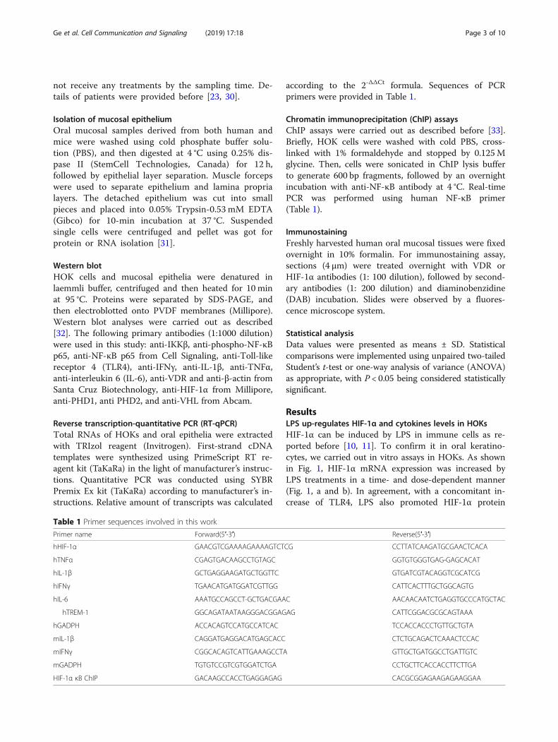
not receive any treatments by the sampling time. De-tails of patients were provided before [23, 30].
Isolation of mucosal epitheliumOral mucosal samples derived from both human andmice were washed using cold phosphate buffer solu-tion (PBS), and then digested at 4 °C using 0.25% dis-pase II (StemCell Technologies, Canada) for 12 h,followed by epithelial layer separation. Muscle forcepswere used to separate epithelium and lamina proprialayers. The detached epithelium was cut into smallpieces and placed into 0.05% Trypsin-0.53 mM EDTA(Gibco) for 10-min incubation at 37 °C. Suspendedsingle cells were centrifuged and pellet was got forprotein or RNA isolation [31].
Western blotHOK cells and mucosal epithelia were denatured inlaemmli buffer, centrifuged and then heated for 10 minat 95 °C. Proteins were separated by SDS-PAGE, andthen electroblotted onto PVDF membranes (Millipore).Western blot analyses were carried out as described[32]. The following primary antibodies (1:1000 dilution)were used in this study: anti-IKKβ, anti-phospho-NF-κBp65, anti-NF-κB p65 from Cell Signaling, anti-Toll-likereceptor 4 (TLR4), anti-IFNγ, anti-IL-1β, anti-TNFα,anti-interleukin 6 (IL-6), anti-VDR and anti-β-actin fromSanta Cruz Biotechnology, anti-HIF-1α from Millipore,anti-PHD1, anti PHD2, and anti-VHL from Abcam.
Reverse transcription-quantitative PCR (RT-qPCR)Total RNAs of HOKs and oral epithelia were extractedwith TRIzol reagent (Invitrogen). First-strand cDNAtemplates were synthesized using PrimeScript RT re-agent kit (TaKaRa) in the light of manufacturer’s instruc-tions. Quantitative PCR was conducted using SYBRPremix Ex kit (TaKaRa) according to manufacturer’s in-structions. Relative amount of transcripts was calculated
according to the 2-ΔΔCt formula. Sequences of PCRprimers were provided in Table 1.
Chromatin immunoprecipitation (ChIP) assaysChIP assays were carried out as described before [33].Briefly, HOK cells were washed with cold PBS, cross-linked with 1% formaldehyde and stopped by 0.125Mglycine. Then, cells were sonicated in ChIP lysis bufferto generate 600 bp fragments, followed by an overnightincubation with anti-NF-κB antibody at 4 °C. Real-timePCR was performed using human NF-κB primer(Table 1).
ImmunostainingFreshly harvested human oral mucosal tissues were fixedovernight in 10% formalin. For immunostaining assay,sections (4 μm) were treated overnight with VDR orHIF-1α antibodies (1: 100 dilution), followed by second-ary antibodies (1: 200 dilution) and diaminobenzidine(DAB) incubation. Slides were observed by a fluores-cence microscope system.
Statistical analysisData values were presented as means ± SD. Statisticalcomparisons were implemented using unpaired two-tailedStudent’s t-test or one-way analysis of variance (ANOVA)as appropriate, with P < 0.05 being considered statisticallysignificant.
ResultsLPS up-regulates HIF-1α and cytokines levels in HOKsHIF-1α can be induced by LPS in immune cells as re-ported before [10, 11]. To confirm it in oral keratino-cytes, we carried out in vitro assays in HOKs. As shownin Fig. 1, HIF-1α mRNA expression was increased byLPS treatments in a time- and dose-dependent manner(Fig. 1, a and b). In agreement, with a concomitant in-crease of TLR4, LPS also promoted HIF-1α protein
Table 1 Primer sequences involved in this work
Primer name Forward(5′-3′) Reverse(5′-3′)
hHIF-1α GAACGTCGAAAAGAAAAGTCTCG CCTTATCAAGATGCGAACTCACA
hTNFα CGAGTGACAAGCCTGTAGC GGTGTGGGTGAG-GAGCACAT
hIL-1β GCTGAGGAAGATGCTGGTTC GTGATCGTACAGGTCGCATCG
hIFNγ TGAACATGATGGATCGTTGG CATTCACTTTGCTGGCAGTG
hIL-6 AAATGCCAGCCT-GCTGACGAAC AACAACAATCTGAGGTGCCCATGCTAC
hTREM-1 GGCAGATAATAAGGGACGGAGAG CATTCGGACGCGCAGTAAA
hGADPH ACCACAGTCCATGCCATCAC TCCACCACCCTGTTGCTGTA
mIL-1β CAGGATGAGGACATGAGCACC CTCTGCAGACTCAAACTCCAC
mIFNγ CGGCACAGTCATTGAAAGCCTA GTTGCTGATGGCCTGATTGTC
mGADPH TGTGTCCGTCGTGGATCTGA CCTGCTTCACCACCTTCTTGA
HIF-1α κB ChIP GACAAGCCACCTGAGGAGAG CACGCGGAGAAGAGAAGGAA
Ge et al. Cell Communication and Signaling (2019) 17:18 Page 3 of 10
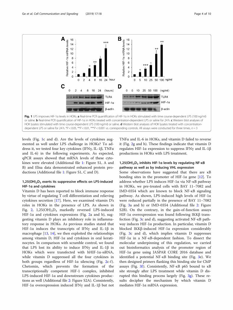
levels (Fig. 1c and d). Are the levels of cytokines aug-mented as well under LPS challenge in HOKs? To ad-dress it, we tested four key cytokines (IFNγ, IL-1β, TNFαand IL-6) in the following experiments. As expected,qPCR assays showed that mRNA levels of these cyto-kines were elevated (Additional file 1: Figure S1, A andB) and Elisa data demonstrated enhanced protein pro-ductions (Additional file 1: Figure S1, C and D).
1,25(OH)2D3 exerts its suppressive effects on LPS-inducedHIF-1α and cytokinesVitamin D has been reported to block immune responseby virtue of regulating T cell differentiation and relievingcytokines secretion [17]. Here, we examined vitamin D’sroles in HOKs in the presence of LPS. As shown inFig. 2, 1,25(OH)2D3 markedly reversed LPS-inducedHIF-1α and cytokines expressions (Fig. 2a and b), sug-gesting vitamin D plays an inhibitory role in inflamma-tory response in HOKs. As previous studies stated thatHIF-1α induces the transcripts of IFNγ and IL-1β inmacrophage [13, 14], we then exploited the relationshipsamong vitamin D, HIF-1α and cytokines in oral kerati-nocytes. In comparison with scramble control, we foundthat LPS lost its ability to induce IFNγ and IL-1β inHOKs which were transfected with hHIF-1α-siRNA,while vitamin D suppressed all the four cytokines inboth groups regardless of HIF-1α silencing (Fig. 2c-f ).Chetomin, which prevents the formation of thetranscriptionally competent HIF-1 complex, inhibitedLPS-induced HIF-1α and downstream cytokines produc-tions as well (Additional file 2: Figure S2A). Consistently,HIF-1α overexpression induced IFNγ and IL-1β but not
TNFα and IL-6 in HOKs, and vitamin D failed to reverseit (Fig. 2g and h). These findings indicate that vitamin Dregulates HIF-1α expression to suppress IFNγ and IL-1βproductions in HOKs with LPS treatment.
1,25(OH)2D3 inhibits HIF-1α levels by regulating NF-κBpathway as well as by inducing VHL expressionSome observations have suggested that there are κBbonding sites in the promoter of HIF-1α gene [12]. Toaddress whether LPS induces HIF-1α via NF-κB pathwayin HOKs, we pre-treated cells with BAY 11–7082 andIMD-0354 which are known to block NF-κB signalingpathway. As shown, LPS-induced high levels of HIF-1αwere reduced partially in the presence of BAY 11–7082(Fig. 3a and b) or IMD-0354 (Additional file 2: FigureS2B). On the contrary, in the gain-of-function assaysHIF-1α overexpression was found following IKKβ trans-fection (Fig. 3c and d), suggesting activated NF-κB path-way induces HIF-1α production. In particular, vitamin Dblocked IKKβ-induced HIF-1α expression considerably(Fig. 3c and d), which implies vitamin D suppressesHIF-1α in a NF-κB-dependent fashion. To dissect themolecular underpinning of this regulation, we carriedout bioinformatics analysis of the promoter region ofHIF-1α gene using JASPAR CORE 2016 database andidentified a potential NF-κB binding site (Fig. 3e). Wethen designed primers flanking this binding site for ChIPassays (Fig. 3f ). Consistently, NF-κB p65 bound to κBsite strongly after LPS treatment while vitamin D dis-rupted this binding process largely (Fig. 3g). These re-sults decipher the mechanism by which vitamin Dmediates HIF-1α mRNA expression.
Fig. 1 LPS improves HIF-1α levels in HOKs. a Real-time PCR quantification of HIF-1α in HOKs stimulated with time course-dependent LPS (100 ng/ml)or saline. b Real-time PCR quantification of HIF-1α in HOKs treated with concentration-dependent LPS or saline for 24 h. c Western blot analyses ofHOK lysates stimulated with time course-dependent LPS (100 ng/ml) or saline. d Western blot analyses of HOK lysates treated with concentration-dependent LPS or saline for 24 h. *P < 0.05, **P < 0.01, ***P < 0.001 vs. corresponding controls. All assays were conducted for three times, n = 3
Ge et al. Cell Communication and Signaling (2019) 17:18 Page 4 of 10
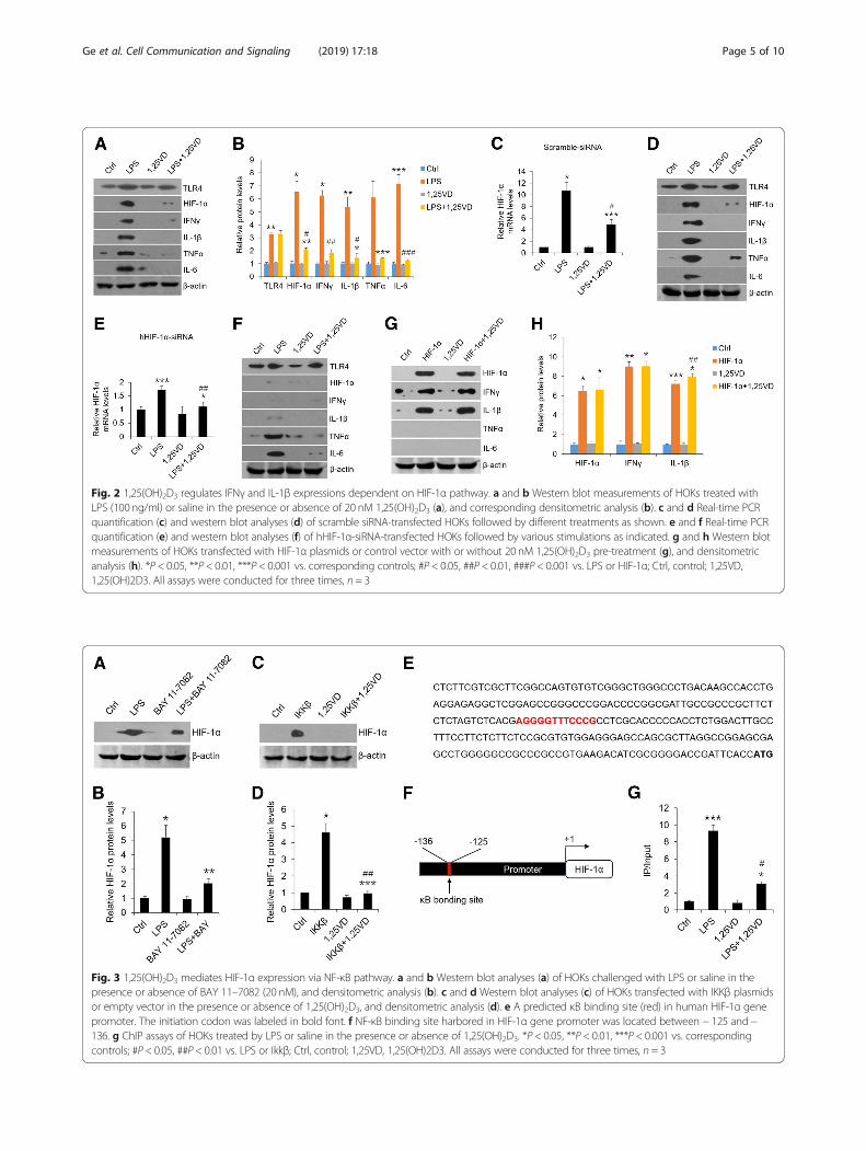
Fig. 2 1,25(OH)2D3 regulates IFNγ and IL-1β expressions dependent on HIF-1α pathway. a and b Western blot measurements of HOKs treated withLPS (100 ng/ml) or saline in the presence or absence of 20 nM 1,25(OH)2D3 (a), and corresponding densitometric analysis (b). c and d Real-time PCRquantification (c) and western blot analyses (d) of scramble siRNA-transfected HOKs followed by different treatments as shown. e and f Real-time PCRquantification (e) and western blot analyses (f) of hHIF-1α-siRNA-transfected HOKs followed by various stimulations as indicated. g and h Western blotmeasurements of HOKs transfected with HIF-1α plasmids or control vector with or without 20 nM 1,25(OH)2D3 pre-treatment (g), and densitometricanalysis (h). *P < 0.05, **P < 0.01, ***P < 0.001 vs. corresponding controls; #P < 0.05, ##P < 0.01, ###P < 0.001 vs. LPS or HIF-1α; Ctrl, control; 1,25VD,1,25(OH)2D3. All assays were conducted for three times, n = 3
Fig. 3 1,25(OH)2D3 mediates HIF-1α expression via NF-κB pathway. a and b Western blot analyses (a) of HOKs challenged with LPS or saline in thepresence or absence of BAY 11–7082 (20 nM), and densitometric analysis (b). c and d Western blot analyses (c) of HOKs transfected with IKKβ plasmidsor empty vector in the presence or absence of 1,25(OH)2D3, and densitometric analysis (d). e A predicted κB binding site (red) in human HIF-1α genepromoter. The initiation codon was labeled in bold font. f NF-κB binding site harbored in HIF-1α gene promoter was located between − 125 and−136. g ChIP assays of HOKs treated by LPS or saline in the presence or absence of 1,25(OH)2D3. *P < 0.05, **P < 0.01, ***P < 0.001 vs. correspondingcontrols; #P < 0.05, ##P < 0.01 vs. LPS or Ikkβ; Ctrl, control; 1,25VD, 1,25(OH)2D3. All assays were conducted for three times, n = 3
Ge et al. Cell Communication and Signaling (2019) 17:18 Page 5 of 10
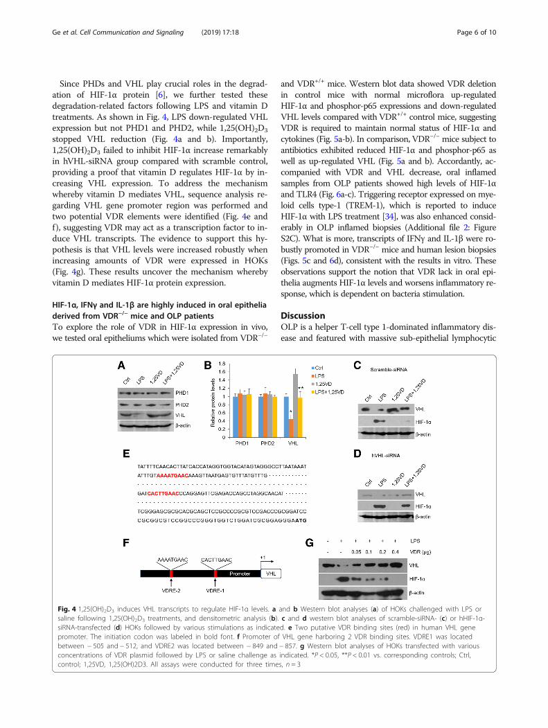
Since PHDs and VHL play crucial roles in the degrad-ation of HIF-1α protein [6], we further tested thesedegradation-related factors following LPS and vitamin Dtreatments. As shown in Fig. 4, LPS down-regulated VHLexpression but not PHD1 and PHD2, while 1,25(OH)2D3
stopped VHL reduction (Fig. 4a and b). Importantly,1,25(OH)2D3 failed to inhibit HIF-1α increase remarkablyin hVHL-siRNA group compared with scramble control,providing a proof that vitamin D regulates HIF-1α by in-creasing VHL expression. To address the mechanismwhereby vitamin D mediates VHL, sequence analysis re-garding VHL gene promoter region was performed andtwo potential VDR elements were identified (Fig. 4e andf), suggesting VDR may act as a transcription factor to in-duce VHL transcripts. The evidence to support this hy-pothesis is that VHL levels were increased robustly whenincreasing amounts of VDR were expressed in HOKs(Fig. 4g). These results uncover the mechanism wherebyvitamin D mediates HIF-1α protein expression.
HIF-1α, IFNγ and IL-1β are highly induced in oral epitheliaderived from VDR−/− mice and OLP patientsTo explore the role of VDR in HIF-1α expression in vivo,we tested oral epitheliums which were isolated from VDR−/−
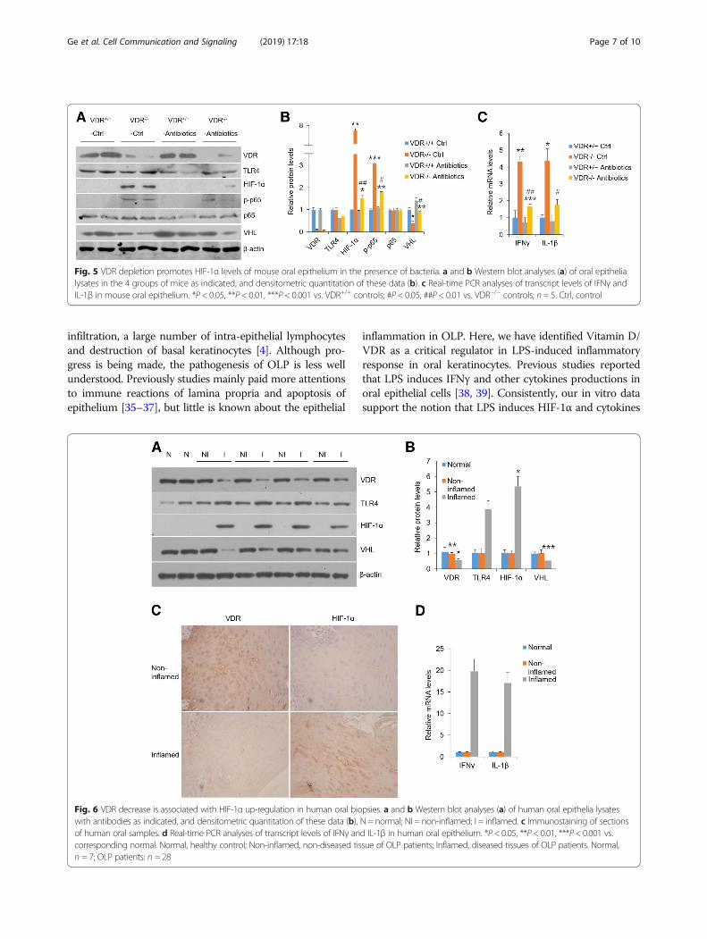
and VDR+/+ mice. Western blot data showed VDR deletionin control mice with normal microflora up-regulatedHIF-1α and phosphor-p65 expressions and down-regulatedVHL levels compared with VDR+/+ control mice, suggestingVDR is required to maintain normal status of HIF-1α andcytokines (Fig. 5a-b). In comparison, VDR−/− mice subject toantibiotics exhibited reduced HIF-1α and phosphor-p65 aswell as up-regulated VHL (Fig. 5a and b). Accordantly, ac-companied with VDR and VHL decrease, oral inflamedsamples from OLP patients showed high levels of HIF-1αand TLR4 (Fig. 6a-c). Triggering receptor expressed on mye-loid cells type-1 (TREM-1), which is reported to induceHIF-1α with LPS treatment [34], was also enhanced consid-erably in OLP inflamed biopsies (Additional file 2: FigureS2C). What is more, transcripts of IFNγ and IL-1β were ro-bustly promoted in VDR−/− mice and human lesion biopsies(Figs. 5c and 6d), consistent with the results in vitro. Theseobservations support the notion that VDR lack in oral epi-thelia augments HIF-1α levels and worsens inflammatory re-sponse, which is dependent on bacteria stimulation.
DiscussionOLP is a helper T-cell type 1-dominated inflammatory dis-ease and featured with massive sub-epithelial lymphocytic
Fig. 4 1,25(OH)2D3 induces VHL transcripts to regulate HIF-1α levels. a and b Western blot analyses (a) of HOKs challenged with LPS orsaline following 1,25(OH)2D3 treatments, and densitometric analysis (b). c and d western blot analyses of scramble-siRNA- (c) or hHIF-1α-siRNA-transfected (d) HOKs followed by various stimulations as indicated. e Two putative VDR binding sites (red) in human VHL genepromoter. The initiation codon was labeled in bold font. f Promoter of VHL gene harboring 2 VDR binding sites. VDRE1 was locatedbetween − 505 and − 512, and VDRE2 was located between − 849 and − 857. g Western blot analyses of HOKs transfected with variousconcentrations of VDR plasmid followed by LPS or saline challenge as indicated. *P < 0.05, **P < 0.01 vs. corresponding controls; Ctrl,control; 1,25VD, 1,25(OH)2D3. All assays were conducted for three times, n = 3
Ge et al. Cell Communication and Signaling (2019) 17:18 Page 6 of 10
infiltration, a large number of intra-epithelial lymphocytesand destruction of basal keratinocytes [4]. Although pro-gress is being made, the pathogenesis of OLP is less wellunderstood. Previously studies mainly paid more attentionsto immune reactions of lamina propria and apoptosis ofepithelium [35–37], but little is known about the epithelial
inflammation in OLP. Here, we have identified Vitamin D/VDR as a critical regulator in LPS-induced inflammatoryresponse in oral keratinocytes. Previous studies reportedthat LPS induces IFNγ and other cytokines productions inoral epithelial cells [38, 39]. Consistently, our in vitro datasupport the notion that LPS induces HIF-1α and cytokines
Fig. 5 VDR depletion promotes HIF-1α levels of mouse oral epithelium in the presence of bacteria. a and b Western blot analyses (a) of oral epithelialysates in the 4 groups of mice as indicated, and densitometric quantitation of these data (b). c Real-time PCR analyses of transcript levels of IFNγ andIL-1β in mouse oral epithelium. *P < 0.05, **P < 0.01, ***P < 0.001 vs. VDR+/+ controls; #P < 0.05, ##P < 0.01 vs. VDR−/− controls; n = 5. Ctrl, control
Fig. 6 VDR decrease is associated with HIF-1α up-regulation in human oral biopsies. a and b Western blot analyses (a) of human oral epithelia lysateswith antibodies as indicated, and densitometric quantitation of these data (b), N = normal; NI = non-inflamed; I = inflamed. c Immunostaining of sectionsof human oral samples. d Real-time PCR analyses of transcript levels of IFNγ and IL-1β in human oral epithelium. *P< 0.05, **P< 0.01, ***P< 0.001 vs.corresponding normal. Normal, healthy control; Non-inflamed, non-diseased tissue of OLP patients; Inflamed, diseased tissues of OLP patients. Normal,n = 7; OLP patients: n = 28
Ge et al. Cell Communication and Signaling (2019) 17:18 Page 7 of 10
expressions in HOKs. Importantly, our western blot datashow that vitamin D plays a suppressive role in HIF-1α aswell as cytokines in HOKs, demonstrating its novel func-tion on OLP. To explore the molecular mechanism of vita-min D’s action, we transfected siRNA and HIF-1α plasmidsinto HOKs and found that vitamin D inhibits IFNγ andIL-1β expressions in a HIF-1α-dependent way. These find-ings are in agreement with the point that IFNγ and IL-1βbinding sites are harbored in the promoter region ofHIF-1α gene [13, 14]. Interestingly, our results manifestthat LPS-induced enhanced levels of TNFα and IL-6,which are suppressed by vitamin D, appear to be not regu-lated by HIF-1α. The reason underling the discrepancyshould be that TNFα and IL-6 are mediated by NF-κB sig-naling pathway directly as described previously [23].HIF-1α is very hard to be detected in normal condition,
but highly expressed in hypoxic or inflammatory situationin a great variety of cells [6]. HIF-1α activity in the epithe-lium is also important in O2 sensing and epithelial innateimmunity [40]. Many studies have demonstrated that it canbe induced by activated NF-κB pathway [12, 41]. Consist-ently, our loss-of-function and gain-of-function data sup-port a regulatory role of NF-κB pathway in HIF-1αexpression in oral epithelial cells. Previous studies have re-vealed that vitamin D blocks NF-κB activity through pro-moting the interaction between VDR and IKKβ [20]. In thisstudy, we identified a putative κB bonding site in the pro-moter region of HIF-1α gene and found that 1,25(OH)2D3disrupts LPS-induced p65 binding to the element ofHIF-1α gene, resulting in the reduction of HIF-1α expres-sion. 1,25(OH)2D3 has been stated to upregulates HIF-1α
via mTOR signaling in human monocytes and breast epi-thelial cells [42, 43], while other studies uncovered thatHIF-1α status is attenuated by 1,25(OH)2D3 treatment inhuman cancer cells [44]. This discrepancy may result fromdifferent types of cell lines used in these studies. In this in-vestigation, we have confirmed that vitamin D reversesLPS-induced overexpression of HIF-1α in HOKs by imped-ing NF-κB pathway (Fig. 7).PHDs and VHL are crucial factors which are required
for HIF-1α proteasomal degradation [6]. In the followingsection, we found LPS exerts suppressive effects on VHLexpression rather than PHD1 and PHD2 (PHD3 is notdetectable) in HOKs, which is consistent with the notionthat LPS down-regulates VHL expression in human hep-atic stellate cells [45]. These results note that VHL likelyplays a protective role in inflammatory diseases, inagreement with previous studies concerning hepatic fi-brosis [46]. Furthermore, we have confirmed that vita-min D reverses VHL expression in HOKs with LPSchallenge, and siRNA interference data show that vita-min D dose regulate HIF-1α dependent in part on VHL.The new finding above raises a crucial question regard-ing the molecular basis of vitamin D’s mediation onVHL. We then identified two VDR elements in the pro-moter region of VHL gene, and proved that overexpres-sion of VDR increases VHL and decreases HIF-1α usingplasmids transfection assays. These observations providea novel notion that vitamin D weakens LPS-inducedHIF-1α in a VHL-dependent manner (Fig. 7). The directmediation of vitamin D/VDR on HIF-1α requires moreexploration (Fig. 7).
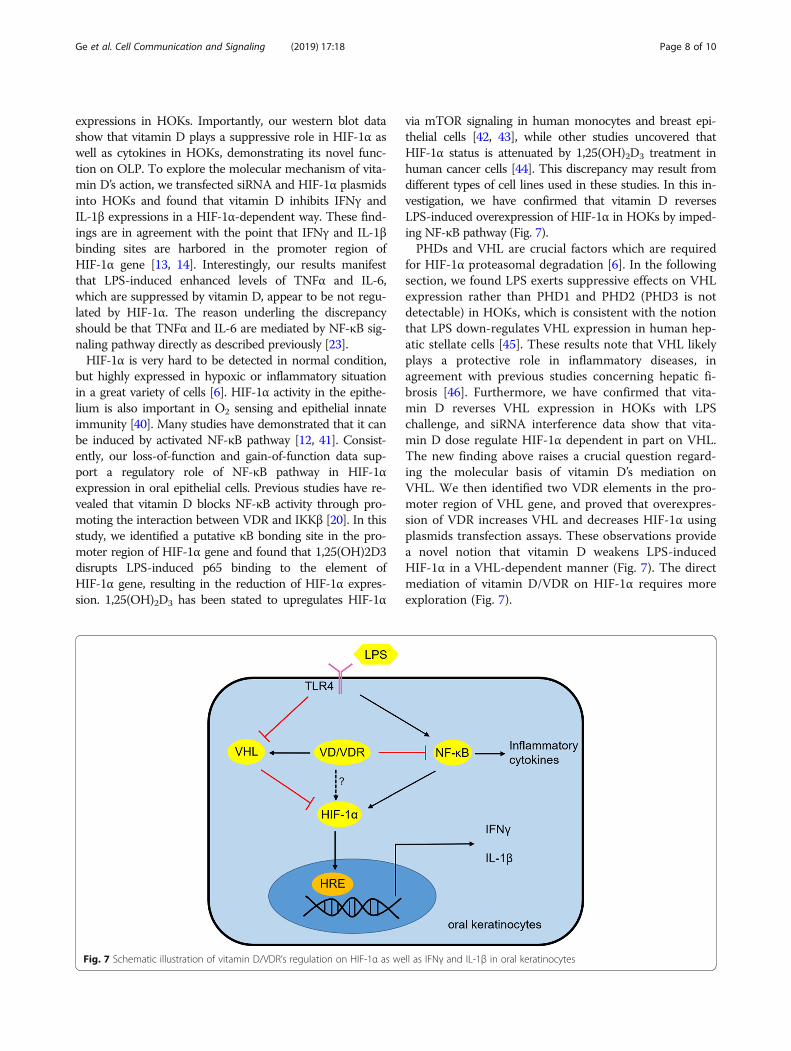
Fig. 7 Schematic illustration of vitamin D/VDR’s regulation on HIF-1α as well as IFNγ and IL-1β in oral keratinocytes
Ge et al. Cell Communication and Signaling (2019) 17:18 Page 8 of 10
Our previous works have suggested that oral mucosalVDR signaling protects epithelium against apoptosis byregulating microRNA-802 and p53-upregulated modula-tor of apoptosis (PUMA) in OLP [30, 35]. In this investi-gation, we have provided evidence that VDR signalingcontrols HIF-1α, phospho-p65 and VHL in mouse oralepithelia. We further eliminated bacteria in mouse oralcavity using antibiotics and found that VDR signalingfails to regulate HIF-1α expression without bacteriastimulation, suggesting the indispensable role of LPS inHIF-1α induction. Consistently, TLR4 and HIF-1α levelsare robustly elevated in human lesion samples of OLP,whereas VDR and VHL expressions are down-regulated.The status of phospho-p65 in lesion epithelium of OLPhas been detected in previous studies [35]. Meanwhile,related cytokines such as IFNγ and IL-1β are also in-duced largely in the diseased biopsies. These in vivo datareveal critical functions of VDR signaling on HIF-1α ex-pression. As there is no well-established animal modelfor resembling OLP, we can not offer more in vivo dataof mice.
ConclusionTaken together, we provide a novel insight into the effectof vitamin D/VDR signaling on oral epithelial inflamma-tion and decipher the molecular basis of this regulation.Despite increasing evidence supports vitamin D’s pro-tective roles in inflammatory diseases, more experimen-tal and clinical studies are needed to ensure vitamin Dand its analogs are effective for the management of OLP.
Additional files
Additional file 1: Figure S1. LPS induces cytokines expression in HOKs.(A and B) Real-time PCR quantification of HOKs treated with time course-dependent LPS (100 ng/ml) or saline (A) and concentration-dependent LPSor saline (B) as indicated. (C and D) Elisa measurements of HOKs treated withtime course-dependent LPS (100 ng/ml) or saline (C) and concentration-dependent LPS or saline (D) as indicated. *P < 0.05, **P < 0.01, ***P < 0.001vs. corresponding controls; All assays were conducted for three times, n = 3.(TIF 634 kb)
Additional file 2: Figure S2. The effects of chetomin and IMD-0354 onHIF-1α in HOKs and TREM-1 expression in human biopsies. (A and B)Western blot analyses of HOKs challenged with LPS or saline in thepresence or absence of chetomin (A) and IMD-0354 (B), n = 3. (C) TREM-1levels in human samples. *P < 0.05, **P < 0.01 vs. corresponding normal;Normal, healthy control; Non-inflamed, non-diseased tissue of OLPpatients; Inflamed, diseased tissues of OLP patients. Normal, n = 7; OLPpatients: n = 28. (TIF 699 kb)
AbbreviationsHIF-1α: Hypoxia-Inducible Factor-1α; HOK: Human oral keratinocyte; IKKβ: IκBkinase β; IL-1β: Interleukin-1 beta; IL-6: Interleukin 6; LPS: Lipopolysaccharide;OLP: Oral lichen planus; PHD: Prolyl hydroxylases; PUMA: p53-upregulatedmodulator of apoptosis; TLR4: Toll-like receptor 4; TNFα: Tumor necrosisfactor alpha; VDR: Vitamin D receptor; VHL: Von Hippel-Lindau
AcknowledgementsWe thank Juanjuan Xiang (Central South University, China) and Yanchun Li(University of Chicago) for providing us with plasmids.
Ethical approval and consent to participateThis study was approved by the Institutional Ethical Committee of ShanxiMedical University. Informed consent including signature was collected fromeach participant.
FundingThis study was supported by National Natural Science Foundation of Chinagrant (81800499), and a funding from Stomatological hospital of ShanxiMedical University.
Availability of data and materialsThese are part of the additional figures.
Authors’ contributionsDJ designed the research; GXJ, WLX and XN performed the study; ZF, LMDand YF provided technical assistance; YFY performed data analysis; DJdrafted the manuscript; DJ, GXJ and ZB acquired funding and supervised theresearch. All authors read and approved the final manuscript.
Consent for publicationAll authors have read the manuscript and approved of the final version.
Competing interestsThe authors declare that they have no competing interests.
Publisher’s NoteSpringer Nature remains neutral with regard to jurisdictional claims inpublished maps and institutional affiliations.
Received: 12 December 2018 Accepted: 20 February 2019
References1. De Rossi SS, Ciarrocca K. Oral lichen planus and lichenoid mucositis. Dent
Clin N Am. 2014;58:299–313.2. Zhao Z, Han Y, Zhang Z, Li W, Ji X, Liu X, et al. Total glucosides of paeony
improves the immunomodulatory capacity of MSCs partially via the miR-124/STAT3 pathway in oral lichen planus. Biomed Pharmacother. 2018;105:151–8.
3. Gorouhi F, Davari P, Fazel N. Cutaneous and mucosal lichen planus: acomprehensive review of clinical subtypes, risk factors, diagnosis, andprognosis. ScientificWorldJournal. 2014;2014:742826.
4. Cheng YS, Gould A, Kurago Z, Fantasia J, Muller S. Diagnosis of oral lichenplanus: a position paper of the American Academy of Oral and maxillofacialpathology. Oral Surg Oral Med Oral Pathol Oral Radiol. 2016;122:332–54.
5. Choi YS, Kim Y, Yoon H-J, Baek KJ, Alam J, Park HK, et al. The presence ofbacteria within tissue provides insights into the pathogenesis of oral lichenplanus. Sci Rep. 2016;6:29186.
6. Palazon A, Goldrath AW, Nizet V, Johnson RS. HIF transcription factors,inflammation, and immunity. Immunity. 2014;41:518–28.
7. Goggins BJ, Chaney C, Radford-Smith GL, Horvat JC, Keely S. Hypoxia andintegrin-mediated epithelial restitution during mucosal inflammation. FrontImmunol. 2013;4:272.
8. Lin N, Simon MC. Hypoxia-inducible factors: key regulators of myeloid cellsduring inflammation. J Clin Invest. 2016;126:3661–71.
9. Taylor CT, Doherty G, Fallon PG, Cummins EP. Hypoxia-dependentregulation of inflammatory pathways in immune cells. J Clin Invest. 2016;126:3716–24.
10. Mills EL, Kelly B, Logan A, Costa ASH, Varma M, Bryant CE, et al. Succinatedehydrogenase supports metabolic repurposing of mitochondria to driveinflammatory macrophages. Cell. 2016;167:457–70.
11. Fan D, Coughlin LA, Neubauer MM, Kim J, Kim MS, Zhan XW, et al.Activation of HIF-1 alpha and LL-37 by commensal bacteria inhibits Candidaalbicans colonization. Nat Med. 2015;21:808–14.
12. Rius J, Guma M, Schachtrup C, Akassoglou K, Zinkernagel AS, Nizet V, et al.NF-kappa B links innate immunity to the hypoxic response throughtranscriptional regulation of HIF-1 alpha. Nature. 2008;453:807–9.
Ge et al. Cell Communication and Signaling (2019) 17:18 Page 9 of 10
13. Corcoran SE, O'Neill LA. HIF1α and metabolic reprogramming ininflammation. J Clin Invest. 2016;126:3699–707.
14. Lee JH, Elly C, Park Y, Liu YC. E3 ubiquitin ligase VHL regulates hypoxia-inducible factor-1α to maintain regulatory T cell stability and suppressivecapacity. Immunity. 2015;42:1062–74.
15. Bouillon R, Carmeliet G, Verlinden L, van Etten E, Verstuyf A, Luderer HF, etal. Vitamin D and human health: lessons from vitamin D receptor null mice.Endocr Rev. 2008;29:726–76.
16. Haussler MR, Whitfield GK, Haussler CA, Hsieh JC, Thompson PD, SelznickSH, et al. The nuclear vitamin D receptor: biological and molecularregulatory properties revealed. J Bone Miner Res. 1998;13:325–49.
17. Lim WC, Hanauer SB, Li YC. Mechanisms of disease: vitamin D and inflammatorybowel disease. Nat Clin Pract Gastroenterol Hepatol. 2005;2:308–15.
18. Liu W, Chen Y, Golan MA, Annunziata ML, Du J, Dougherty U, et al.Intestinal epithelial vitamin D receptor signaling inhibits experimental colitis.J Clin Invest. 2013;123:3983–96.
19. Du J, Chen Y, Shi Y, Liu T, Cao Y, Tang Y, et al. 1,25-Dihydroxyvitamin Dprotects intestinal epithelial barrier by regulating the myosin light chainkinase signaling pathway. Inflamm Bowel Dis. 2015;21:2495–506.
20. Chen Y, Zhang J, Ge X, Du J, Deb DK, Li YC. Vitamin D receptor inhibitsnuclear factor kappaB activation by interacting with IkappaB kinase betaprotein. J Biol Chem. 2013;288:19450–8.
21. Sato Y, Miyauchi Y, Yoshida S, Morita M, Kobayashi T, Kanagawa H, et al. Thevitamin D analogue ED71 but not 1,25(OH)2D3 targets HIF1α protein inosteoclasts. PLoS One. 2014;9:e111845.
22. Chen Y, Du J, Zhang Z, Liu T, Shi Y, Ge X, et al. MicroRNA-346 mediatestumor necrosis factor alpha-induced downregulation of gut epithelialvitamin D receptor in inflammatory bowel diseases. Inflamm Bowel Dis.2014;20:1910–8.
23. Du J, Li R, Yu F, Yang F, Wang J, Chen Q, et al. Experimental study on1,25(OH)2D3 amelioration of oral lichen planus through regulating NF-κBsignaling pathway. Oral Dis. 2017;23:770–8.
24. Yin H, Zheng L, Liu W, Zhang D, Li W, Yuan L. Rootletin prevents Cep68from VHL-mediated proteasomal degradation to maintain centrosomecohesion. Biochim Biophys Acta. 2017;1864:645–54.
25. Chen WH, Lecaros RLG, Tseng YC, Huang L, Hsu YC. Nanoparticle delivery ofHIF1 alpha siRNA combined with photodynamic therapy as a potentialtreatment strategy for head-and-neck cancer. Cancer Lett. 2015;359:65–74.
26. Li YC, Pirro AE, Amling M, Delling G, Baron R, Bronson R, et al.Targeted ablation of the vitamin D receptor: an animal model ofvitamin D-dependent rickets type II with alopecia. Proc Natl Acad Sci US A. 1997;94:9831–5.
27. Su YY, Chen CD, Guo LJ, Du J, Li XY, Liu Y. Ecological balance of oralmicrobiota is required to maintain oral mesenchymal stem cell homeostasis.Stem Cells. 2018;36:551–61.
28. van der Meij EH, van der Waal I. Lack of clinicopathologic correlation in thediagnosis of oral lichen planus based on the presently available diagnosticcriteria and suggestions for modifications. J Oral Pathol Med. 2003;32:507–12.
29. Cheng YS, Rees T, Jordan L, Oxford L, O'Brien J, Chen HS, et al. Salivaryendothelin-1 potential for detecting oral cancer in patients with oral lichenplanus or oral cancer in remission. Oral Oncol. 2011;47:1122–6.
30. Zhao B, Xu N, Li R, Yu F, Zhang F, Yang F, et al. Vitamin D/VDR signalingsuppresses microRNA-802-induced apoptosis of keratinocytes in oral lichenplanus. FASEB J. 2019;33:1042–50.
31. Wang Y, Zhang H, Du G, Wang Y, Cao T, Luo Q, et al. Total glucosides ofpaeony (TGP) inhibits the production of inflammatory cytokines in orallichen planus by suppressing the NF-kappaB signaling pathway. IntImmunopharmacol. 2016;36:67–72.
32. Li YC, Bolt MJ, Cao LP, Sitrin MD. Effects of vitamin D receptor inactivationon the expression of calbindins and calcium metabolism. Am J PhysiolEndocrinol Metab. 2001;281:558–64.
33. Yuan W, Pan W, Kong J, Zheng W, Szeto FL, Wong KE, et al. 1,25-dihydroxyvitamin d3 suppresses renin gene transcription by blocking theactivity of the cyclic AMP response element in the renin gene promoter. JBiol Chem. 2007;282:29821–30.
34. Tu C, Wang S, Hu X, Wang W, Dong Y, Xiao S, et al. Lipopolysaccharideinduces TREM-1-dependent HIF-1alpha expression in human keratinocytecell line. Cell Biol Int. 2016;40:1357–65.
35. Zhao B, Li R, Yang F, Yu FY, Xu N, Zhang F, et al. LPS-induced vitamin Dreceptor decrease in oral keratinocytes is associated with oral lichen planus.Sci Rep. 2018;8:763.
36. Fathi MS, El Dessouky HF, Breni HA. CD4+CD25+ T regulatory cells andMMP-9 as diagnostic salivary biomarkers in oral lichen planus. Egypt JImmunol. 2013;20:39–53.
37. Wang H, Zhang D, Han Q, Zhao X, Zeng X, Xu Y, et al. Role of distinctCD4(+) T helper subset in pathogenesis of oral lichen planus. J Oral PatholMed. 2016;45:385–93.
38. Rouabhia M, Ross G, Page N, Chakir J. Interleukin-18 and gamma interferonproduction by oral epithelial cells in response to exposure to Candidaalbicans or lipopolysaccharide stimulation. Infect Immun. 2002;70:7073–80.
39. Ohno T, Yamamoto G, Hayashi JI, Nishida E, Goto H, Sasaki Y, et al.Angiopoietin-like protein 2 regulates Porphyromonas gingivalislipopolysaccharide-induced inflammatory response in human gingivalepithelial cells. PLoS One. 2017;12:e0184825.
40. Polke M, Seiler F, Lepper PM, Kamyschnikow A, Langer F, Monz D, et al.Hypoxia and the hypoxia-regulated transcription factor HIF-1alpha suppressthe host defence of airway epithelial cells. Innate Immun. 2017;23:373–80.
41. Taylor CT, Colgan SP. Regulation of immunity and inflammation by hypoxiain immunological niches. Nat Rev Immunol. 2017;17:774–85.
42. Jiang Y, Zheng W, Teegarden D. 1alpha, 25-Dihydroxyvitamin D regulateshypoxia-inducible factor-1alpha in untransformed and Harvey-rastransfected breast epithelial cells. Cancer Lett. 2010;298:159–66.
43. Lee B, Kwon E, Kim Y, Kim JH, Son SW, Lee JK, et al. 1alpha,25-Dihydroxyvitamin D3 upregulates HIF-1 and TREM-1 via mTOR signaling.Immunol Lett. 2015;163:14–21.
44. Ben-Shoshan M, Amir S, Dang DT, Dang LH, Weisman Y, Mabjeesh NJ.1alpha,25-dihydroxyvitamin D3 (Calcitriol) inhibits hypoxia-inducible factor-1/vascular endothelial growth factor pathway in human cancer cells. MolCancer Ther. 2007;6:1433–9.
45. Wu N, McDaniel K, Zhou T, Ramos-Lorenzo S, Wu C, Huang L, et al.Knockout of microRNA-21 attenuates alcoholic hepatitis through VHL/NF-κBsignaling pathway in hepatic stellate cells. Am J Physiol Gastrointest LiverPhysiol. 2018;315:385–98.
46. Wang J, Lu Z, Xu Z, Tian P, Miao H, Pan S, et al. Reduction of hepatic fibrosisby overexpression of von Hippel-Lindau protein in experimental models ofchronic liver disease. Sci Rep. 2017;7:41038.
Ge et al. Cell Communication and Signaling (2019) 17:18 Page 10 of 10