Vibrational Spectroscopy (Typical for IR and Raman) 14(Vib Spec)-07-12.pdfVibrational Spectroscopy...
28
XIV–67 Spectroscopy: Tinoco Chapter 10 (but vibration, Ch.9) Vibrational Spectroscopy (Typical for IR and Raman) Born-Oppenheimer separate electron-nuclear motion ψ (rR) = χ υ (R) φ el (r,R) -- product fct. solves sum H electronic Schrödinger Equation H el φ el (r,R) = U el (R) φ e (r,R) eigen value → U el (R) parametric on R potential energy for nuclear motion (see below) Nuclear Schrödinger Equation H n χ (R) = E υ χ υ (R) H n = - ∑ /2M α 2 h α ∇ α 2 + V n (R) V n (R) = U el (R) + Z ∑ β α , α Z β e 2 /R αβ Solving this is 3N dimensional – N atom Simplify → Remove (a) Center of Mass (Translate) (b) Orientation of molecule (Rotate) Results in (3N – 6) coordinates - called internal coord. – motion of nuclei w/r/t each other a) Translation — like atoms – no impact on spectra since continuous (no potential – plane wave) b) Rotation — also no potential but have angular momentum kinetic energy associated with rotation – quantized - no potential, but angular momentum restricted 1. Diatomics (linear) sol'n Y JM (θ,φ) – same as H-atom E JM = ћ 2 J(J+1)/2I I= M ∑ α α R α 2 (diatomic I=µ R α 2 ) (spectra ∆J = ±1, ∆M J = 0, ±1 ∆E + = (J + 1) ћ 2 /I ) 2. Polyatomics – add coordinate (ω) and quantum number (K) for angular momentum – previously refer J,M J to a lab axis, now complex (this K is projection of angular momentum onto molecular axis, so internal orientation of molecule) Rotational Spectra (aside – little impact on Biology) Diatomic: E JM rot =J(J+1)ћ 2 /2I I=µ R e µ=M A M B /M A +M B if B e = h/(8π 2 I c ) E JM = J(J + 1) B e in cm -1 or E JM rot = (hc) J (J + 1)B e Note: levels increase separation as J inc.
Transcript of Vibrational Spectroscopy (Typical for IR and Raman) 14(Vib Spec)-07-12.pdfVibrational Spectroscopy...
XIV–67 Spectroscopy: Tinoco Chapter 10 (but vibration, Ch.9) Vibrational Spectroscopy (Typical for IR and Raman) Born-Oppenheimer separate electron-nuclear motion ψ (rR) = χυ (R) φel (r,R) -- product fct. solves sum H electronic Schrödinger Equation Hel φel (r,R) = Uel (R) φe (r,R) eigen value → Uel(R) parametric on R potential energy for nuclear motion (see below) Nuclear Schrödinger Equation Hn χ (R) = Eυ χυ (R)
Hn = -∑ /2Mα
2 hα ∇α
2 + Vn (R)
Vn(R) = Uel(R) + Z∑βα,
α Zβ e2/Rαβ
Solving this is 3N dimensional – N atom Simplify → Remove (a) Center of Mass (Translate) (b) Orientation of molecule (Rotate) Results in (3N – 6) coordinates - called internal coord. – motion of nuclei w/r/t each other a) Translation — like atoms – no impact on spectra since continuous (no potential – plane wave) b) Rotation — also no potential but have angular momentum kinetic energy associated with rotation – quantized - no potential, but angular momentum restricted 1. Diatomics (linear) sol'n YJM (θ,φ) – same as H-atom
EJM = ћ2J(J+1)/2I I= M∑α
α Rα2 (diatomic I=µ Rα
2)
(spectra ∆J = ±1, ∆MJ = 0, ±1 ∆E+ = (J + 1) ћ2/I ) 2. Polyatomics – add coordinate (ω) and quantum number (K) for angular momentum – previously refer J,MJ to a lab axis, now complex (this K is projection of angular momentum onto molecular axis, so internal orientation of molecule) Rotational Spectra (aside – little impact on Biology) Diatomic: EJM
rot=J(J+1)ћ2/2I I=µ Re µ=MAMB/MA+MB if Be= h/(8π2Ic) EJM = J(J + 1) Be in cm-1 or EJM
rot = (hc) J (J + 1)BeNote: levels increase separation as J inc.
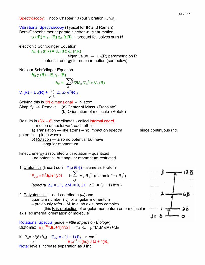
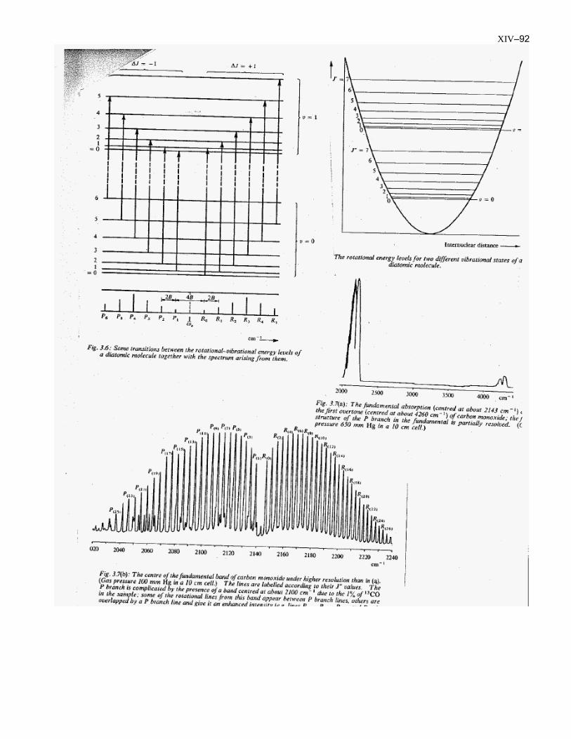
XIV–68 Transitions allowed by absorption (far-IR or µ-wave / typical Be<10cm-1). Selection rules: ∆J=±1 ∆M=0,±1 ∆EJ→J+1=(J+1)2B (absorb ∆J=+1) IR or µ-wave
regularly spaced lines, intensity reflect rise → degeneracy (δJ) fall → exponential depopulation Boltzmann: nJ = δJ n0 exp [– J(J+1)B/kT] Pure Rotational Far-IR spectrum of CO -- note 1st transition (23 cm-1) is for J=5 --> J=6 (I think)
XIV–69
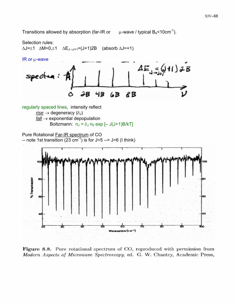
∆E = -2B (2J + 3) (laser) ∆E = +2B (2J + 3)
Raman – light scattering experiment νs = ν0 – νJ∆J = 0, ±1, ±2 in general but ∆J = ±2 diatomic (K = 0)
diatomic (linear) spacing ~4B
Rotational Raman spect. of N2 (left) anti-Stokes: ∆J = -2 (right) Stokes: ∆J = 2
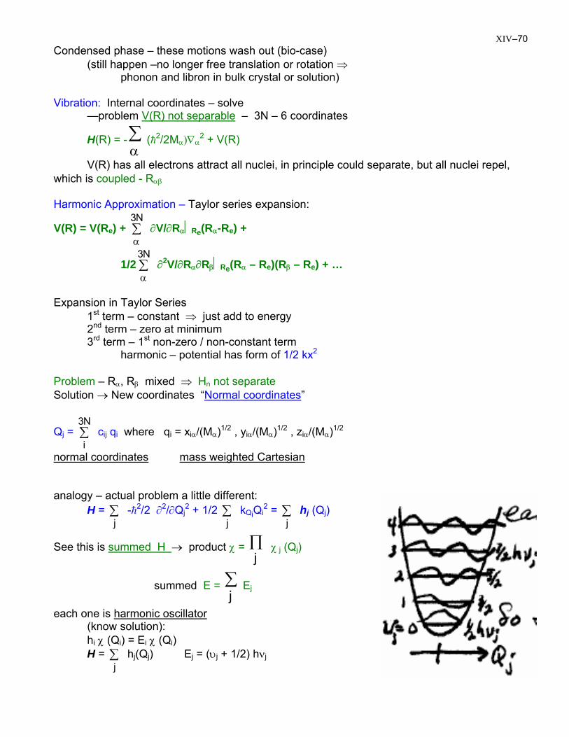
XIV–70 Condensed phase – these motions wash out (bio-case)
(still happen –no longer free translation or rotation ⇒ phonon and libron in bulk crystal or solution)
Vibration: Internal coordinates – solve
—problem V(R) not separable – 3N – 6 coordinates
H(R) = - (h∑α
2/2Mα)∇α2 + V(R)
V(R) has all electrons attract all nuclei, in principle could separate, but all nuclei repel, which is coupled - Rαβ
Harmonic Approximation – Taylor series expansion:
V(R) = V(Re) + ∂V/∂R∑α
N3α⏐Re(Rα-Re) +
1/2 ∂∑α
N32V/∂Rα∂Rβ⏐Re(Rα – Re)(Rβ – Re) + …
Expansion in Taylor Series
1st term – constant ⇒ just add to energy 2nd term – zero at minimum 3rd term – 1st non-zero / non-constant term
harmonic – potential has form of 1/2 kx2
Problem – Rα, Rβ mixed ⇒ Hn not separate Solution → New coordinates “Normal coordinates”
Qj = c∑N3
iij qi where qi = xiα/(Mα)1/2 , yiα/(Mα)1/2 , ziα/(Mα)1/2
normal coordinates mass weighted Cartesian analogy – actual problem a little different:
H = -h∑j
2/2 ∂2/∂Qj2 + 1/2 ∑ k
jQiQi
2 = ∑j
hj (Qj)
See this is summed H → product χ = ∏j
χ j (Qj)
summed E = ∑ Ej
j
each one is harmonic oscillator (know solution):
hi χ (Qi) = Ei χ (Qi) H = h∑
jj(Qj) Ej = (υj + 1/2) hνj
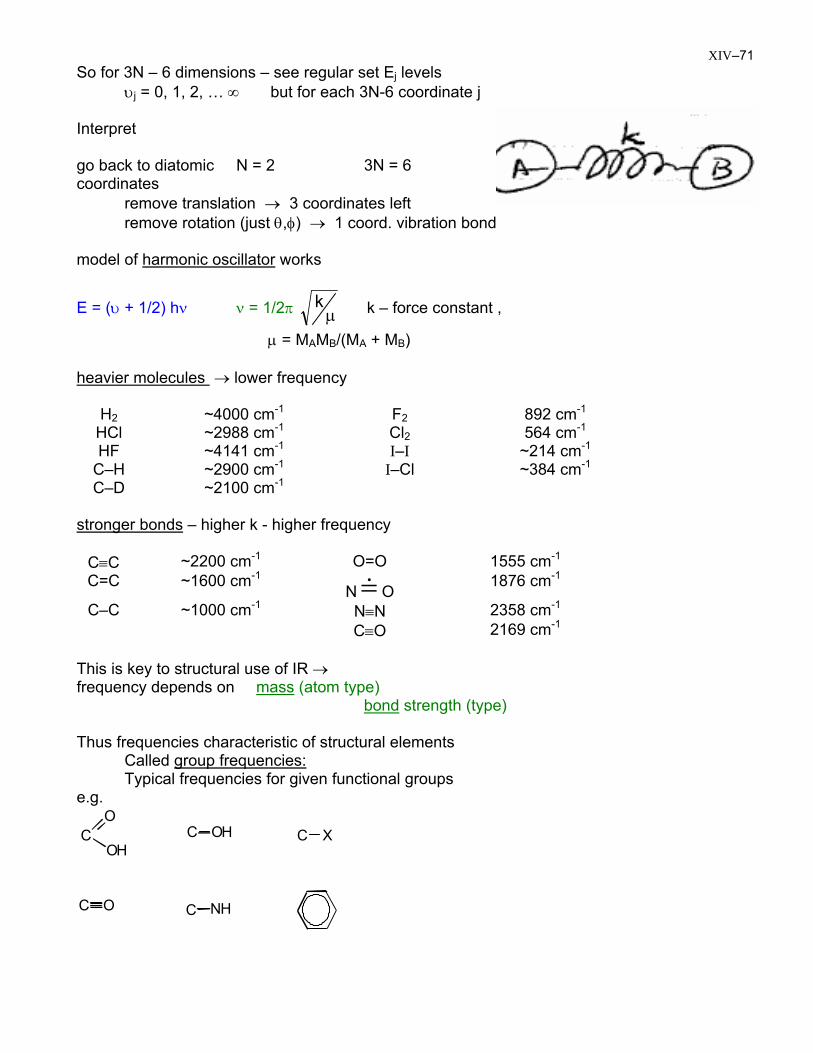
XIV–71 So for 3N – 6 dimensions – see regular set Ej levels
υj = 0, 1, 2, … ∞ but for each 3N-6 coordinate j
Interpret go back to diatomic N = 2 3N = 6 coordinates
remove translation → 3 coordinates left remove rotation (just θ,φ) → 1 coord. vibration bond
model of harmonic oscillator works
E = (υ + 1/2) hν ν = 1/2π µk k – force constant ,
µ = MAMB/(MA + MB) heavier molecules → lower frequency
H2 ~4000 cm-1 F2 892 cm-1
HCl ~2988 cm-1 Cl2 564 cm-1
HF ~4141 cm-1 I–I ~214 cm-1
C–H ~2900 cm-1 I–Cl ~384 cm-1
C–D ~2100 cm-1 stronger bonds – higher k - higher frequency
C≡C ~2200 cm-1 O=O 1555 cm-1
C=C ~1600 cm-1
N =& O 1876 cm-1
C–C ~1000 cm-1 N≡N 2358 cm-1
C≡O 2169 cm-1
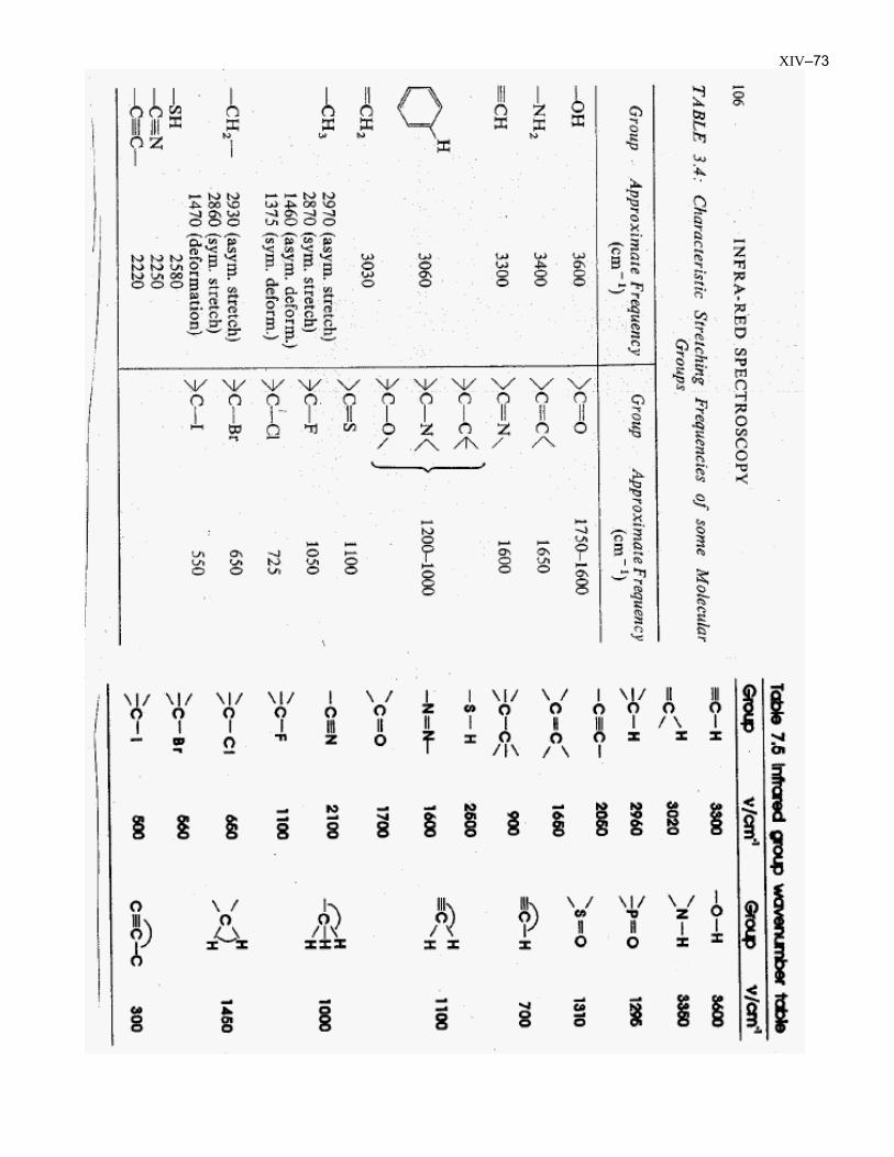
This is key to structural use of IR → frequency depends on mass (atom type) bond strength (type) Thus frequencies characteristic of structural elements
Called group frequencies:Typical frequencies for given functional groups
e.g.
COH
OC OH C X
C O C NH
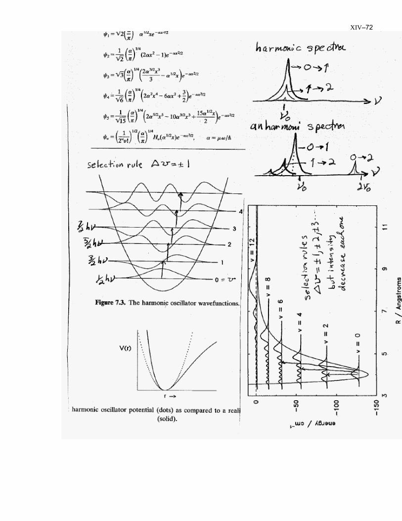
XIV–72
XIV–73
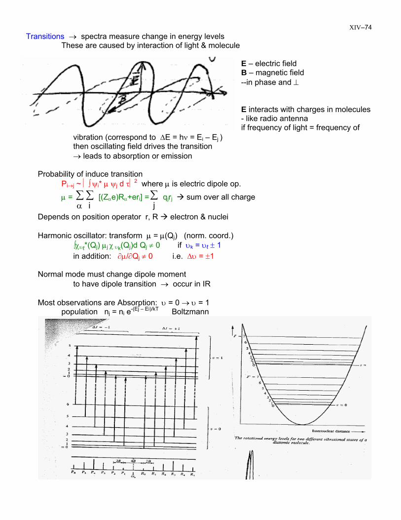
XIV–74 Transitions → spectra measure change in energy levels
These are caused by interaction of light & molecule E – electric field B – magnetic field --in phase and ⊥ E interacts with charges in molecules - like radio antenna if frequency of light = frequency of
vibration (correspond to ∆E = hν = Ei – Ej ) then oscillating field drives the transition → leads to absorption or emission Probability of induce transition Pi→j ~ ⏐∫ ψi* µ ψj d τ⏐2 where µ is electric dipole op.
µ = ∑ ∑ [(Zα i
αe)Rα+eri] = q∑j
jrj sum over all charge
Depends on position operator r, R electron & nuclei Harmonic oscillator: transform µ = µ(Qj) (norm. coord.) ∫χυℓ*(Qj) µj χ υk(Qj)d Qj ≠ 0 if υk = υℓ ± 1 in addition: ∂µ/∂Qj ≠ 0 i.e. ∆υ = ±1 Normal mode must change dipole moment to have dipole transition → occur in IR Most observations are Absorption: υ = 0 → υ = 1
population nj = ni e-(Ej – Ei)/kT Boltzmann
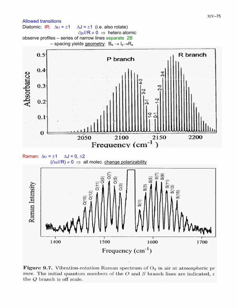
XIV–75 Allowed transitions Diatomic: IR: ∆υ = ±1 ∆J = ±1 (i.e. also rotate)
∂µ/∂R ≠ 0 ⇒ hetero atomic observe profiles – series of narrow lines separate 2B – spacing yields geometry: Be → Ie→Re
Raman: ∆υ = ±1 ∆J = 0, ±2
(∂α/∂R) ≠ 0 ⇒ all molec. change polarizability
XIV–76
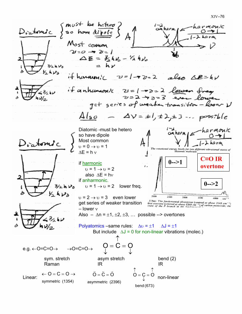
Diatomic -must be hetero so have dipole Most common υ = 0 → υ = 1 ∆E = h ν if harmonic υ = 1 → υ = 2 also ∆E = hν if anharmonic. υ = 1 → υ = 2 lower freq. υ = 2 → υ = 3 even lower get series of weaker transition – lower ν
C≡O IR overtone
0-->2
0-->1
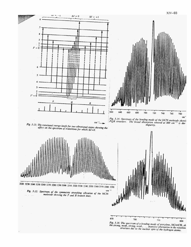
Also – ∆n = ±1, ±2, ±3, … possible --> overtones Polyatomics –same rules: ∆υ = ±1 ∆J = ±1 But include ∆J = 0 for non-linear vibrations (molec.)
e.g. ←O=C=O→ →O=C=O→ ↓
↑
↓== OCO
sym. stretch asym stretch bend (2) Raman IR IR
Linear: (1354) symmetric
OCO →==←
(2396) asymmetric
OCO rsr
== non-linear
)673( bend
OCO↑
↓
↑==
XIV–77
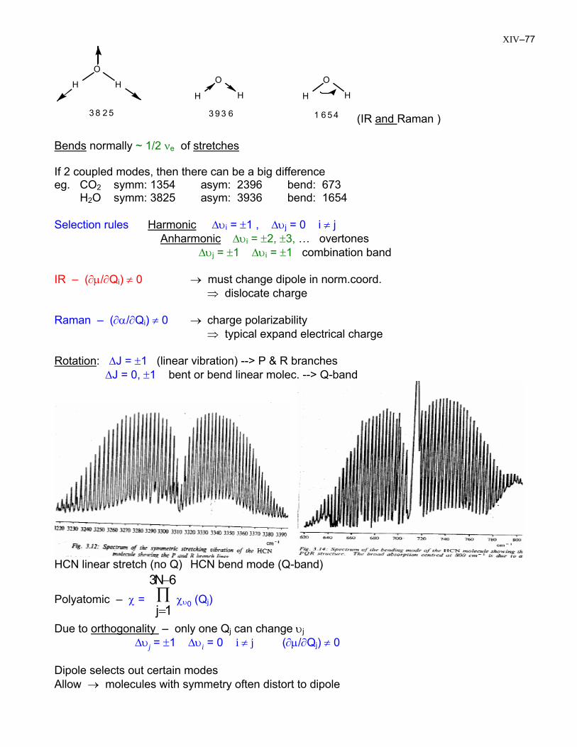
3 8 2 5 3 9 3 6 1 6 5 4
O
H HH
O
H
O
H H
(IR and Raman ) Bends normally ~ 1/2 νe of stretches If 2 coupled modes, then there can be a big difference eg. CO2 symm: 1354 asym: 2396 bend: 673 H2O symm: 3825 asym: 3936 bend: 1654 Selection rules Harmonic ∆υi = ±1 , ∆υj = 0 i ≠ j Anharmonic ∆υi = ±2, ±3, … overtones ∆υj = ±1 ∆υi = ±1 combination band IR – (∂µ/∂Qi) ≠ 0 → must change dipole in norm.coord. ⇒ dislocate charge Raman – (∂α/∂Qi) ≠ 0 → charge polarizability ⇒ typical expand electrical charge Rotation: ∆J = ±1 (linear vibration) --> P & R branches ∆J = 0, ±1 bent or bend linear molec. --> Q-band
HCN linear stretch (no Q) HCN bend mode (Q-band)
Polyatomic – χ = χ∏−
=
6N3
1jυ0 (Qj)
Due to orthogonality – only one Qj can change υj ∆υj = ±1 ∆υi = 0 i ≠ j (∂µ/∂Qj) ≠ 0 Dipole selects out certain modes Allow → molecules with symmetry often distort to dipole
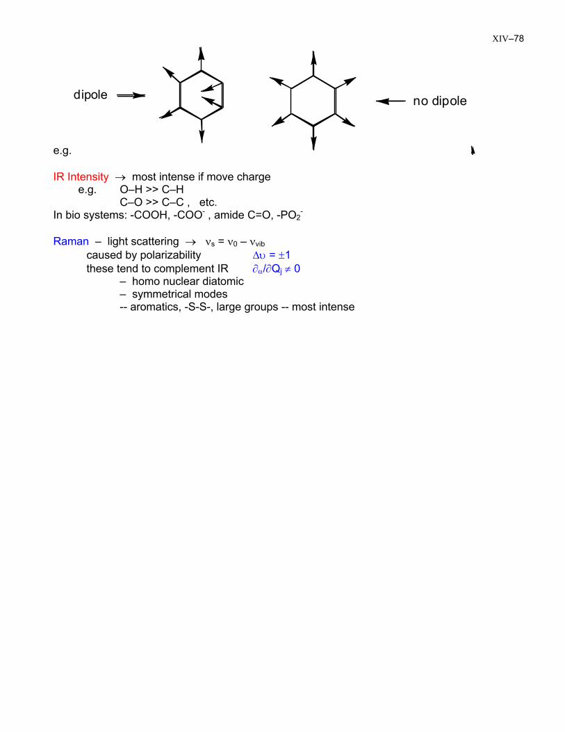
XIV–78
e.g.
dipole
no dipole
IR Intensity → most intense if move charge e.g. O–H >> C–H C–O >> C–C , etc. In bio systems: -COOH, -COO- , amide C=O, -PO2
-
Raman – light scattering → νs = ν0 – νvib caused by polarizability ∆υ = ±1 these tend to complement IR ∂α/∂Qj ≠ 0 – homo nuclear diatomic – symmetrical modes -- aromatics, -S-S-, large groups -- most intense
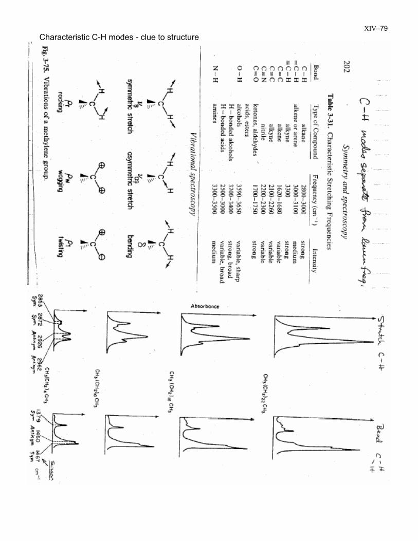
XIV–79 Characteristic C-H modes - clue to structure
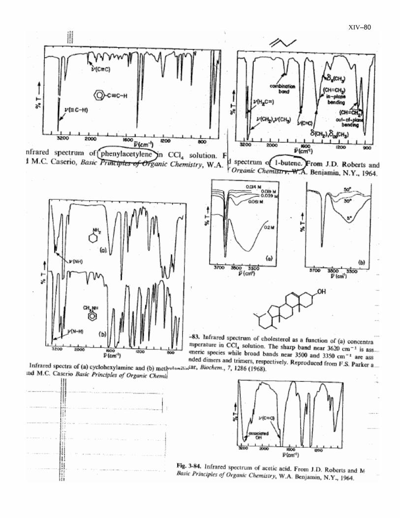
XIV–80
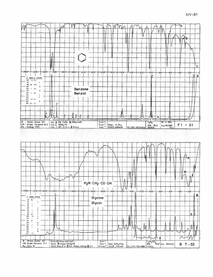
XIV–81
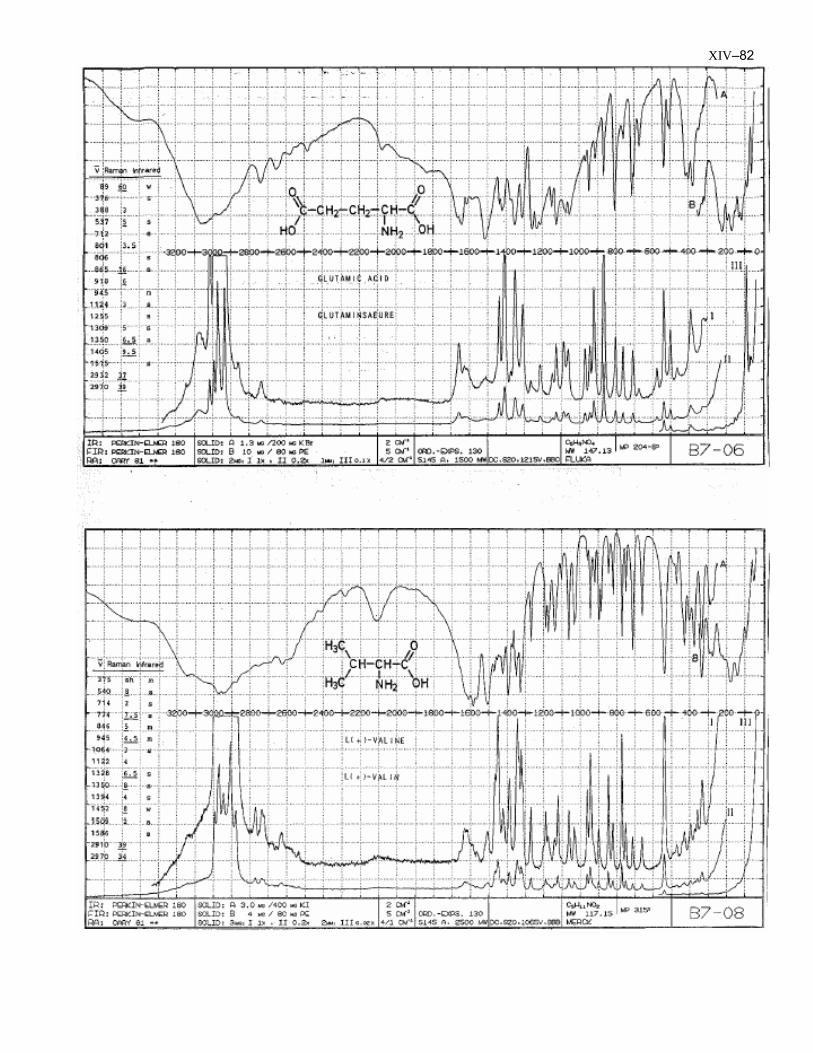
XIV–82
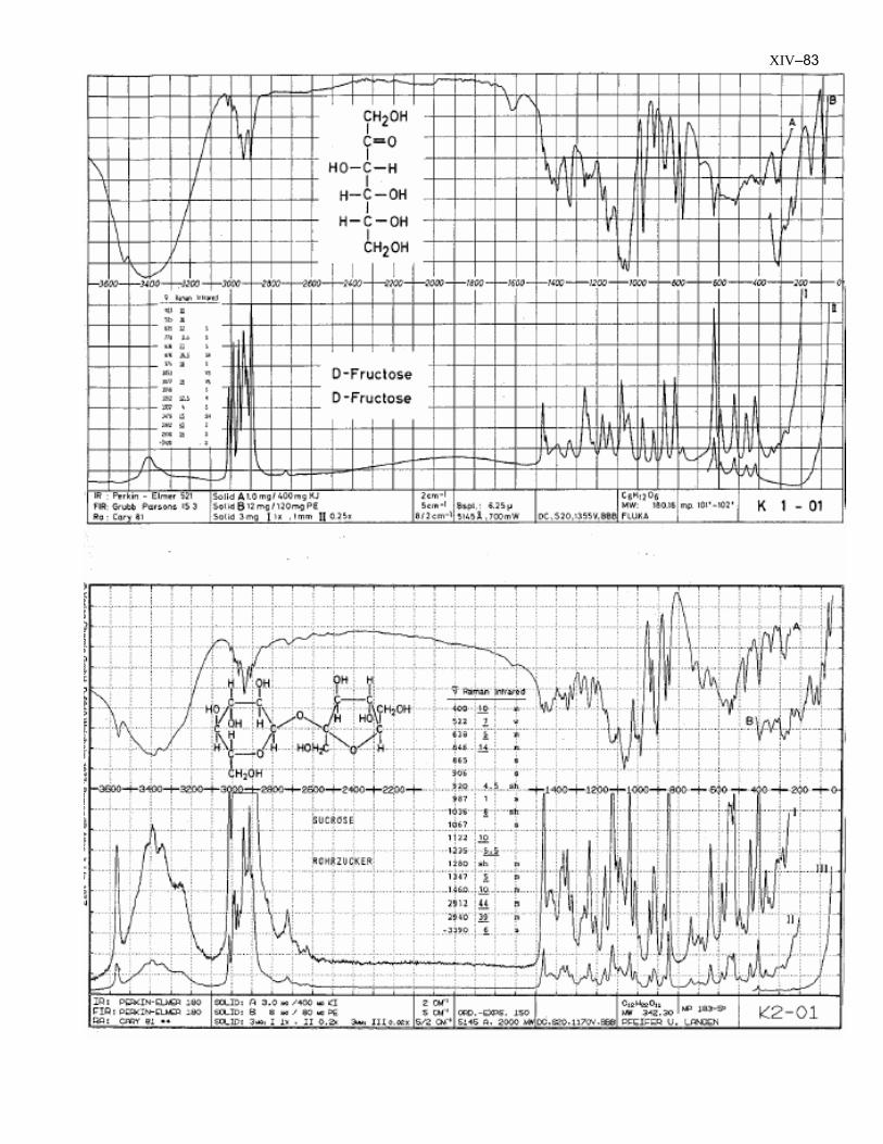
XIV–83
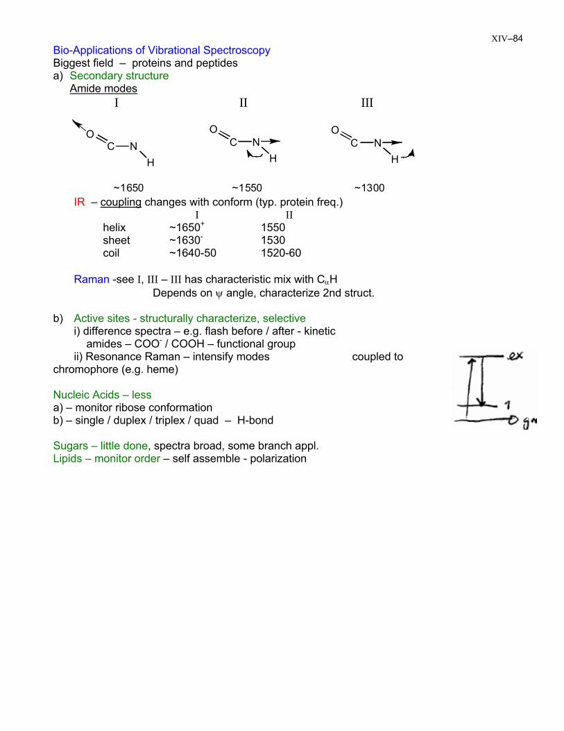
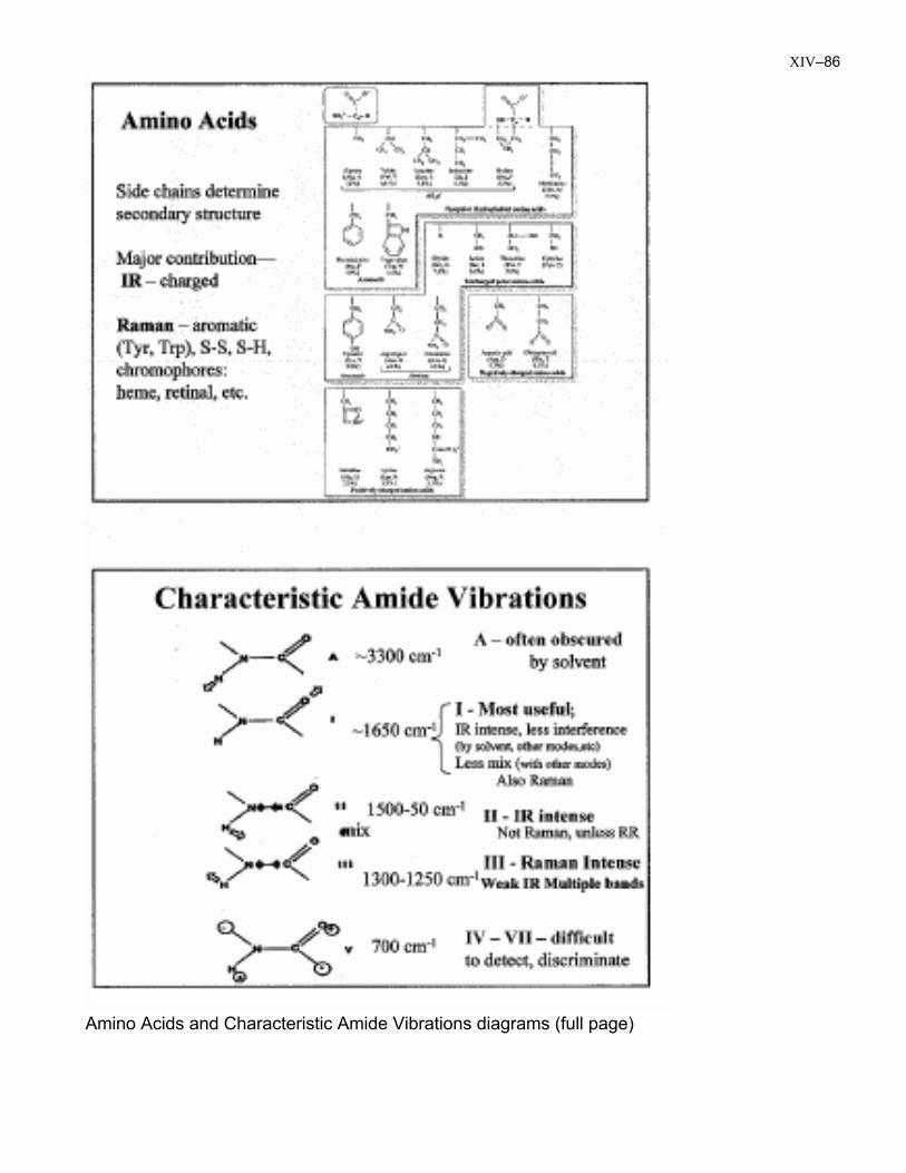
XIV–84 Bio-Applications of Vibrational Spectroscopy Biggest field – proteins and peptides a) Secondary structure Amide modes
C NO
H
C NO
HC N
O
H
I II III
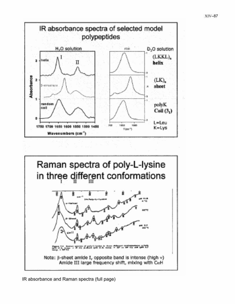
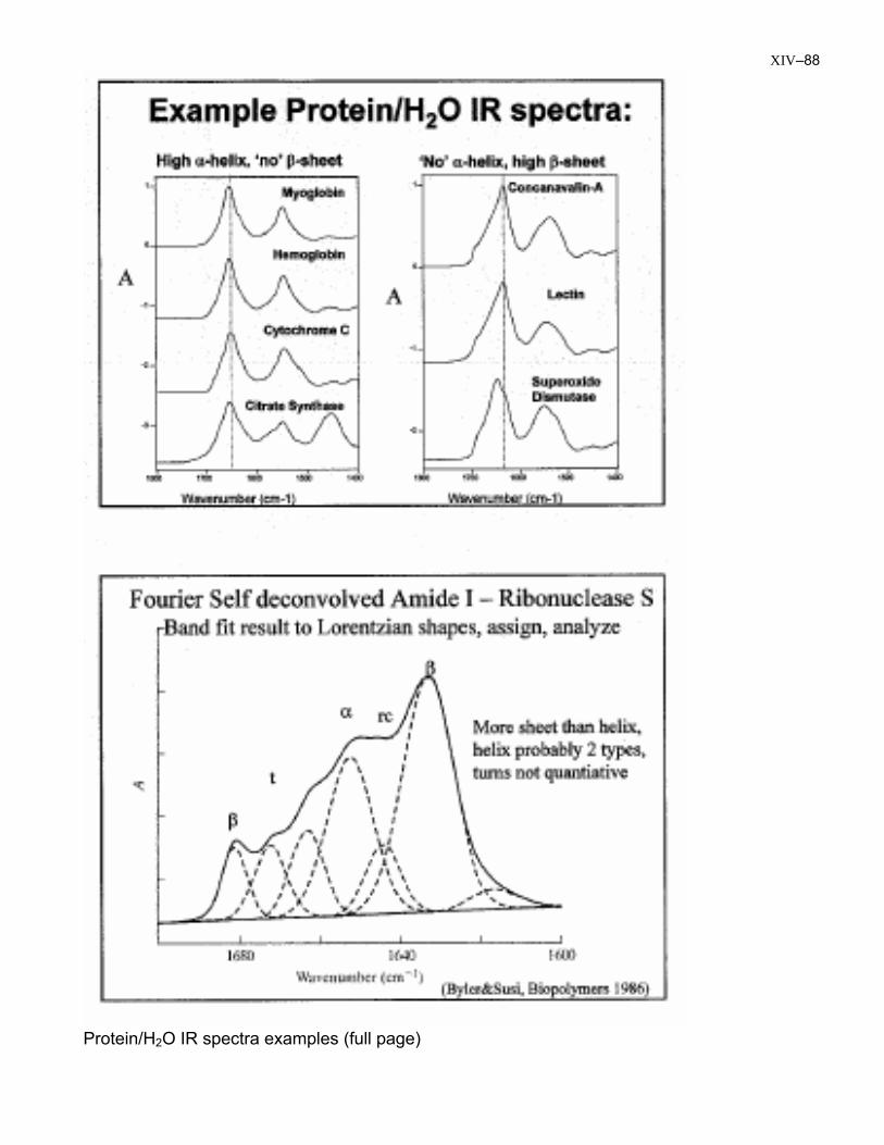
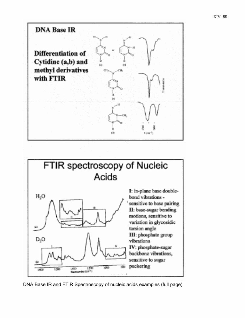
~1650 ~1550 ~1300 IR – coupling changes with conform (typ. protein freq.) I II helix ~1650+ 1550 sheet ~1630- 1530 coil ~1640-50 1520-60 Raman -see I, III – III has characteristic mix with CαH Depends on ψ angle, characterize 2nd struct. b) Active sites - structurally characterize, selective i) difference spectra – e.g. flash before / after - kinetic amides – COO- / COOH – functional group ii) Resonance Raman – intensify modes coupled to chromophore (e.g. heme) Nucleic Acids – less a) – monitor ribose conformation b) – single / duplex / triplex / quad – H-bond Sugars – little done, spectra broad, some branch appl. Lipids – monitor order – self assemble - polarization
XIV–85
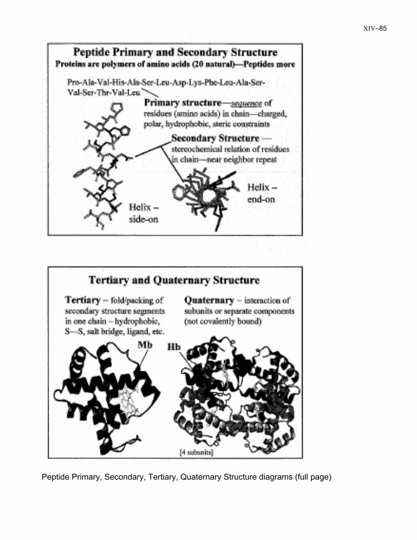
Peptide Primary, Secondary, Tertiary, Quaternary Structure diagrams (full page)
XIV–86
Amino Acids and Characteristic Amide Vibrations diagrams (full page)
XIV–87
IR absorbance and Raman spectra (full page)
XIV–88
Protein/H2O IR spectra examples (full page)
XIV–89
DNA Base IR and FTIR Spectroscopy of nucleic acids examples (full page)
XIV–90
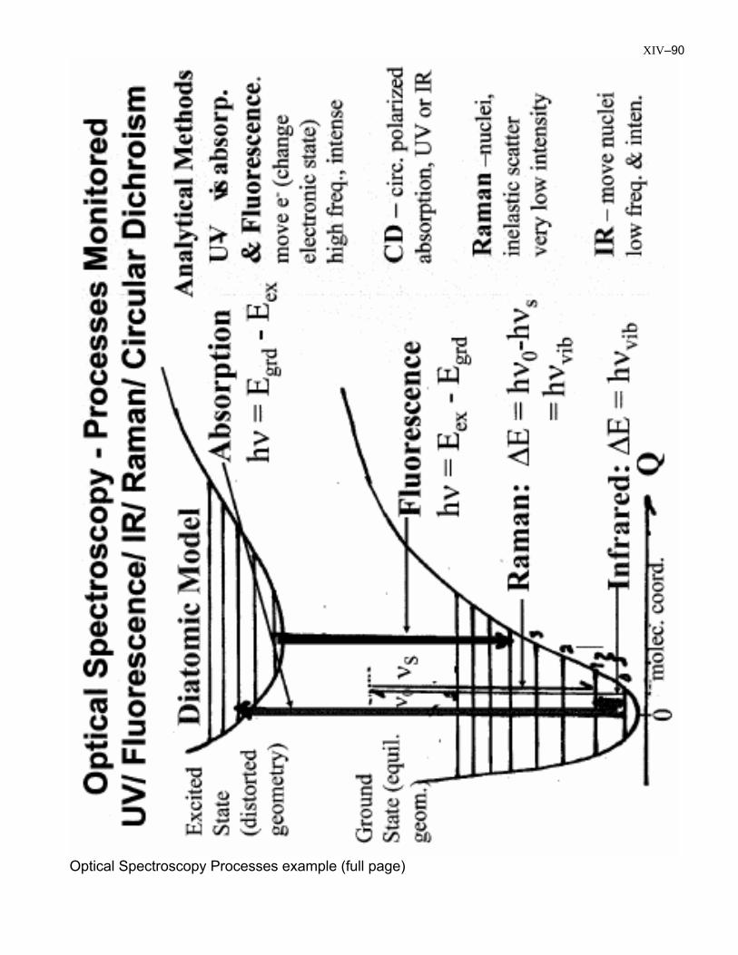
Optical Spectroscopy Processes example (full page)
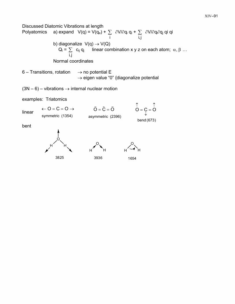
XIV–91 Discussed Diatomic Vibrations at length Polyatomics a) expand V(q) = V(qe) + ∑
i∂V/∂qi qi + ∑
j,i∂V/∂qi∂qj qi qi
b) diagonalize V(q) → V(Q) Qi = c∑
j,iij qj linear combination x y z on each atom; α, β …
Normal coordinates 6 – Transitions, rotation → no potential E → eigen value “0” {diagonalize potential (3N – 6) – vibrations → internal nuclear motion examples: Triatomics
linear (1354) symmetric
OCO →==←
(2396) asymmetric
OCO rsr
==
)673( bend
OCO↑
↓
↑==
bent
3825 3936 1654
O
H HH
O
H
O
H H
XIV–92
XIV–93

XIV–94