Usefulness and limitations of E-cadherin and -catenin in the ...
9
RESEARCH Open Access Usefulness and limitations of E-cadherin and β-catenin in the classification of breast carcinomas in situ with mixed pattern Douglas S Gomes 1 , Simone S Porto 1 , Rafael M Rocha 2 and Helenice Gobbi 1* Abstract Background: The distinction between lobular neoplasia of the breast and ductal carcinoma in situ has important therapeutic implications. In some cases, it is very difficult to determine whether the morphology of the lesion is ductal or lobular. The aim of this study was to evaluate the value of E-cadherin and β-catenin expression through the immunophenotypical characterization of carcinoma in situ with mixed pattern (CISM). Methods: A total of 25 cases of CISM were analyzed considering cytology/mixed architecture (ductal and lobular), nuclear pleomorphism, loss of cell cohesion, and presence of comedonecrosis. The immunophenotype pattern was considered E-cadherin positive and β-catenin positive, or negative. Results: Nineteen (76%) cases presented a mixed cytology and / or architectural pattern, two (8%) presented nuclear pleomorphism, two (8%) presented mixed cytology and nuclear pleomorphism, and two (8%) presented comedonecrosis and nuclear pleomorphism. A complete positivity for E-cadherin and β-catenin was observed in 11 cases (44%). In one case, the lesion was negative for both markers and showed nuclear pleomorphis. Thirteen lesions showed negative staining in areas of lobular cytology and positive staining in cells presenting the ductal pattern. Conclusions: The expression of E-cadherin and β-catenin, combined with cytological and architectural analysis, may highlight different immunophenotypes and improve classification of CISM. Virtual Slides: The virtual slide(s) for this article can be found here: http://www.diagnosticpathology.diagnomx.eu/vs/ 1693384202970681 Keywords: E-cadherin, β-catenin, Breast cancer, Lobular neoplasia, Ductal carcinoma in situ, Immunohistochemistry Additional non-English language abstract - Portuguese Introdução: A distinção entre neoplasia lobular e carcinoma ductal in situ tem importantes implicações terapêuticas. No entanto, em alguns casos, é muito difícil determinar se a morfologia da lesão é ductal ou lobular. O objetivo deste estudo foi avaliar a expressão de E-caderina e β-catenina na caracterização imunofenotípica dos carcinomas in situ de padrão misto (CISM). Métodos: Um total de vinte e cinco casos de CISM foram analisados considerando a citologia/arquitetura mista (ductal e lobular), pleomorfismo nuclear e presença de comedonecrose. A expressão imuno-histoquímica foi considerada positiva para E-caderina e β-catenina, ou negativa. (Continued on next page) * Correspondence: [email protected] 1 Breast Pathology Laboratory, School of Medicine, Federal University of Minas Gerais (UFMG), Av. Professor Alfredo Balena, 190, Belo Horizonte, Minas Gerais 30130-100, Brazil Full list of author information is available at the end of the article © 2013 Gomes et al.; licensee BioMed Central Ltd. This is an Open Access article distributed under the terms of the Creative Commons Attribution License (http://creativecommons.org/licenses/by/2.0), which permits unrestricted use, distribution, and reproduction in any medium, provided the original work is properly cited. Gomes et al. Diagnostic Pathology 2013, 8:114 http://www.diagnosticpathology.org/content/8/1/114
Transcript of Usefulness and limitations of E-cadherin and -catenin in the ...
Gomes et al. Diagnostic Pathology 2013, 8:114http://www.diagnosticpathology.org/content/8/1/114
RESEARCH Open Access
Usefulness and limitations of E-cadherinand β-catenin in the classification of breastcarcinomas in situ with mixed patternDouglas S Gomes1, Simone S Porto1, Rafael M Rocha2 and Helenice Gobbi1*
Abstract
Background: The distinction between lobular neoplasia of the breast and ductal carcinoma in situ has importanttherapeutic implications. In some cases, it is very difficult to determine whether the morphology of the lesion isductal or lobular. The aim of this study was to evaluate the value of E-cadherin and β-catenin expression throughthe immunophenotypical characterization of carcinoma in situ with mixed pattern (CISM).
Methods: A total of 25 cases of CISM were analyzed considering cytology/mixed architecture (ductal and lobular),nuclear pleomorphism, loss of cell cohesion, and presence of comedonecrosis. The immunophenotype pattern wasconsidered E-cadherin positive and β-catenin positive, or negative.
Results: Nineteen (76%) cases presented a mixed cytology and / or architectural pattern, two (8%) presented nuclearpleomorphism, two (8%) presented mixed cytology and nuclear pleomorphism, and two (8%) presentedcomedonecrosis and nuclear pleomorphism. A complete positivity for E-cadherin and β-catenin was observed in 11cases (44%). In one case, the lesion was negative for both markers and showed nuclear pleomorphis. Thirteen lesionsshowed negative staining in areas of lobular cytology and positive staining in cells presenting the ductal pattern.
Conclusions: The expression of E-cadherin and β-catenin, combined with cytological and architectural analysis, mayhighlight different immunophenotypes and improve classification of CISM.
Virtual Slides: The virtual slide(s) for this article can be found here: http://www.diagnosticpathology.diagnomx.eu/vs/1693384202970681
Keywords: E-cadherin, β-catenin, Breast cancer, Lobular neoplasia, Ductal carcinoma in situ, Immunohistochemistry
Additional non-English language abstract - Portuguese
Introdução: A distinção entre neoplasia lobular e carcinoma ductal in situ tem importantes implicaçõesterapêuticas. No entanto, em alguns casos, é muito difícil determinar se a morfologia da lesão é ductal ou lobular.O objetivo deste estudo foi avaliar a expressão de E-caderina e β-catenina na caracterização imunofenotípica doscarcinomas in situ de padrão misto (CISM).
Métodos: Um total de vinte e cinco casos de CISM foram analisados considerando a citologia/arquitetura mista(ductal e lobular), pleomorfismo nuclear e presença de comedonecrose. A expressão imuno-histoquímica foiconsiderada positiva para E-caderina e β-catenina, ou negativa.(Continued on next page)
* Correspondence: [email protected] Pathology Laboratory, School of Medicine, Federal University of MinasGerais (UFMG), Av. Professor Alfredo Balena, 190, Belo Horizonte, MinasGerais 30130-100, BrazilFull list of author information is available at the end of the article
© 2013 Gomes et al.; licensee BioMed Central Ltd. This is an Open Access article distributed under the terms of the CreativeCommons Attribution License (http://creativecommons.org/licenses/by/2.0), which permits unrestricted use, distribution, andreproduction in any medium, provided the original work is properly cited.
Gomes et al. Diagnostic Pathology 2013, 8:114 Page 2 of 9http://www.diagnosticpathology.org/content/8/1/114
(Continued from previous page)
Resultados: Dezenove (76%) casos apresentavam somente citologia e/ou padrão arquitetural misto (ductale lobular), dois casos (8%) apresentaram somente pleomorfismo nuclear, dois casos (8%) apresentavam citologiamista e pleomorfismo nuclear, e dois casos (8%) tinham comedonecrose e pleomorfismo nuclear. Uma positividadecompleta para E-caderina e β-catenina foi observada em 11 casos (44%). Em um caso a lesão foi negativa paraambos marcadores e apresentava pleomorfismo nuclear e comedonecrose. Em 13 lesões o imunofenótipo foinegativo em áreas lobulares e positivo em áreas ductais.
Conclusão: A caracterização imunofenotípica com E-caderina e β-catenina, combinada com a análise citológicae arquitetural, pode destacar diferentes imunofenótipos e auxiliar na classificação dos CISM.
BackgroundIn situ breast carcinomas are classified, according totheir morphology, as ductal carcinoma in situ (DCIS) orlobular neoplasia (LN), which includes lobular carcin-oma in situ (LCIS) and atypical lobular hyperplasia(ALH). According to the 2012 WHO classification of tu-mors of the breast, classic LCIS is diagnosed when morethan half of the acini of a lobular unit are distended anddistorted by a dyshesive proliferation of cells with small,uniform nuclei. Lesser involvement by the characteristiccells is diagnosed as ALH. Lesions that show marked nu-clear pleomorphism, with or without apocrine featuresand comedonecrosis are referred as pleomorphic LCIS(PLCIS) [1].In some cases, the diagnostic criteria based on the
morphology of LN is not clear, leading to mistaken diag-nosis of intraductal proliferative lesions. The main differ-ential diagnoses of lobular neoplasia are: LN with solidlow-grade DCIS, PLCIS and high-grade DCIS. Some insitu carcinomas present unusual cytological and / orarchitectural features, making it difficult to determinewhether the proliferation is lobular or ductal. This grouphas been called carcinomas in situ with a mixed or inde-terminate pattern (CISM) [2,3].The differential diagnosis of the CISM carries some im-
portant implications. Patients with LN are usually clinic-ally monitored and can be offered tamoxifen as aprophylactic therapy to prevent the development of inva-sive carcinoma [4,5]. On other hand, patients with DCISshould be treated by surgical removal of the lesion, withclear margins followed by radiotherapy, or mastectomy[6]. When diagnosed by core biopsy, DCIS should betreated with complete excision of the lesion. However, theclinical significance and therapeutic implications of findingLN in core biopsy specimens are still controversial [7,8].The diagnosis of CISM is extremely rare and studies
assessing the differential diagnosis of these lesions arescarce and include only a few patients. The largest seriesreported between 12 and 28 cases [9,10]. Previous studiesby our group identified 0.08% of CISM among breast bi-opsies performed in our general hospital [11]. Althoughrare, when analyzed under light microscope, the CISM
lesions are difficult to diagnose and there is lack ofepidemiological data linked to their biological behavior.A great progress in the diagnosis of these lesions came
with the observation that almost all cases of LN and in-vasive lobular carcinoma (ILC) lose the immunohisto-chemistry (IHC) signal for E-cadherin and β-cateninexpression in the cytoplasm membrane, whereas the ex-pression of these proteins is maintained in both in situand invasive ductal carcinomas [3,12,13]. The cadherinscomprise a large number of adhesion molecules local-ized in the intercellular junctions, keeping cells connec-ted through homophilic protein-protein interactions.The observation that cadherins play an important role inthe establishment of the epithelial phenotype, cell migra-tion, cell differentiation, and tumor dissemination hasstimulated great interest in this family of adhesion mole-cules. Among them is the Human Epithelial Cadherin(E-cadherin), a calcium-dependent transmembrane glyco-protein directly involved in the process of cell adhesion[14]. The α, β, p, and γ catenins play important roles inintercellular signal transduction. The β-catenin, specific-ally, binds to the cytoplasmatic portion of the E-cadherinand to the structure of actin microfilaments of the cyto-skeleton as well, being involved in cell adhesion [15,16].The E-cadherin gene mutation is the major mechanismresponsible for its inactivation in cancer cells and is asso-ciated with other carcinomas, such as hepatocellular car-cinoma, diffuse-type gastric cancer, thyroid and colorectalcancer [16,17]. Another route resulting in inactivation ofE-cadherin is attributed to dysfunctional promoter activityor DNA methylation at the promoter region [17,18].The aim of this study was to evaluate the expression
of E-cadherin and β-catenin for the immunopheno-typical characterization of carcinomas in situ with mixedpattern, and identify potential morphological patternsthat could assist in the diagnosis of the different types ofCISM lesions.
MethodsThis is a retrospective, descriptive study that analyzed 25cases of breast biopsies (wide local excision or mastec-tomy) performed between 1999 and 2011. The study
Gomes et al. Diagnostic Pathology 2013, 8:114 Page 3 of 9http://www.diagnosticpathology.org/content/8/1/114
analyzed the histopathological reports and slides avail-able at the Laboratory of Breast Pathology at the Schoolof Medicine at the Federal University of Minas Gerais(UFMG), Brazil. We selected one or more slides stainedwith hematoxylin and eosin (HE) and which were repre-sentative of each diagnosis of in situ carcinoma withmixed pattern. The slides were analyzed simultaneouslyby two of the authors (HG and DSG) using a dual-viewmicroscope and classified according to the standardmorphological patterns and immunohistochemical ex-pression of E-cadherin and β-catenin. Cases withoutavailable slides and / or paraffin blocks were excludedfrom the study. The study was approved by the UFMGresearch ethics committee.
Morphological evaluationIn situ lesions matching the LN morphological patternwere characterized according to the proliferation ofgenerally small, dyshesive cells, with uniform roundnuclei and clear cytoplasm. The acini are partially orcompletely expanded, but the lobular architecture ismaintained [1]. Lesions classified as DCIS presentedproliferation of monomorphic cells with regular distribu-tion, and hyperchromatic nuclei forming regular second-ary, rounded, and uniform lumens. The in situ lesionscharacterized as mixed pattern presented cytological orarchitectural features common to the ductal and lobularlesions and were classified into three main patternsaccording to criteria previously described by Jacobs et al.[3]: Group 1 – those presenting architectural and
A
C
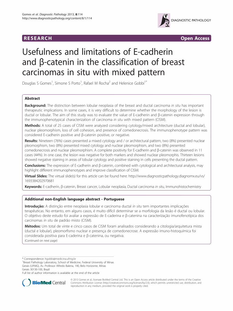
Figure 1 Case # 1: Lobular neoplasia (arrows) and ductal carcinoma inbreast field (hematoxylin and eosin; A – 100×). Cells of ductal carcinomnegative for E-cadherin (B, 100× and C, 400×).
cytological findings of LN but with areas of comedo-typenecrosis; Group 2 – those with CISM lesions character-ized by small and uniform cells, either growing in a solidpattern with focal microacinar-like structures but withcellular dyshesion, or growing in a mosaic pattern withoccasional intracytoplasmic vacuoles; Group 3 – thosewith marked cellular pleomorphism and nuclear atypia,however, the LN discohesive pattern remains.
Immunohistochemical evaluationSequential 5 μm thick histological sections were obtainedfrom the paraffin blocks from the breast specimens andmounted on silanized slides. Sections were deparaffinizedin two consecutive baths of xylene, for 20 minutes each,and rehydrated in ethanol series with decreasing concen-trations and finally in distilled water. For antigen retrieval,a buffer solution of 10 mM citrate pH 6.0 was used in anelectrical pressure-cooker. Immunohistochemistry wasperformed automatically using Ventana Benchmark XTequipment (Ventana Medical Systems Inc., Tucson, AZ,USA). E-cadherin (clone 36) and β-catenin (clone 18) anti-bodies were used, at titers of 1:600 and 1:800, respect-ively. Antibodies were purchased from BD Transduction(cat# C19220), San Jose, CA, USA.The non-biotinylated polymer system (Novolink®,
Leica Microsystens) technique was used for reactionamplification. A diaminobenzidine (DAB) solution wasused as chromogen and the slides were counter-stainedusing Harris hematoxylin. External positive and negativecontrols were used. Normal ducts and lobules, adjacent
B
D
situ of cribriform type (arrowheads) are present in the samea stain positive (B,100× and D, 400×) and cells of lobular neoplasia are
Gomes et al. Diagnostic Pathology 2013, 8:114 Page 4 of 9http://www.diagnosticpathology.org/content/8/1/114
to the lesions and expressing E-cadherin and β-cateninin the epithelium, were used as internal controls.Staining for E-cadherin and β-catenin was considered
positive when the staining intensity around the entirecircumference of the membrane was similar to that seenin the normal luminal epithelial cells. No staining wasconsidered as negative (Figure 1).
ResultsTwenty-five cases of CISM were identified from theBreast Pathology Laboratory during the study period.The average patient age was 52.7 (± 11.5) years. Nine-teen cases (76%) presented morphological pattern show-ing cytology and / or architectural mixed pattern (ductaland lobular), two cases (8%) showed lobular architec-tural pattern with nuclear pleomorphism, two cases (8%)showed mixed cytology and nuclear pleomorphism, and
Table 1 Morphology and immunophenotype of 25 breast carbiopsies
Morphological patternof in situ lesion
Morphology of in situ lesion w
Case # Lobular Ductal Mixed Nuclearpleomorphism
Comedonecros
1 Yes Yes Yes No No
2 No No Yes No No
3 No No Yes No No
4 No No Yes No No
5 No No Yes Yes No
6 No No Yes No No
7 Yes No Yes No No
8 No No Yes No No
9 No No Yes Yes Yes
10 No No Yes No No
11 No Yes Yes No No
12 No No Yes No No
13 No No Yes Yes Yes
14 Yes Yes Yes No No
15 No Yes Yes No No
16 No No Yes Yes No
17 Yes No No Yes No
18 Yes Yes Yes No No
19 No Yes Yes No No
20 No No Yes No No
21 No No Yes No No
22 Yes Yes Yes No No
23 No No Yes No No
24 No No Yes Yes No
25 No Yes Yes No No
NA Not applied.
two cases (8%) showed comedonecrosis and nuclearpleomorphism (Table 1).Immunohistochemistry for E-cadherin was performed
in all 25 cases. Eleven cases (44%) were positive for E-cadherin (Figure 2). Thirteen cases (52%) showed mixedimmunophenotype with positive E-cadherin staining theductal cells and negative in the lobular areas. In onecase, the cells were completely negative for E-cadherin.In all cases in which both markers were analyzed (20
cases) the immunohistochemical results agreed withboth E-cadherin and β-catenin. Immunohistochemistryfor β-catenin was not performed in five cases due tosample processing artifacts and insufficient material forthe preparation of new slides.Nineteen cases were composed by small, uniform cells
varying from low to intermediate nuclear grade, growing insolid pattern, with some microacinar-like structures admi-
cinomas in situ with mixed pattern analyzed from breast
ith mixed pattern E-cadherinimmunophenotype
β-cateninimmunophenotype
is Mixed cytologyor architecture
Lobularareas
Ductalareas
Lobularareas
Ductalareas
Yes - + - +
Yes - + - +
Yes + + + +
Yes + + + +
No - + - +
Yes + + + +
Yes + + + +
Yes + + + +
No - + - +
Yes - + - +
Yes + + + +
Yes + + NA NA
No - - - -
Yes - + - +
Yes + + + +
Yes - + NA NA
Yes - + - +
Yes - + NA NA
Yes + + + +
Yes + + + +
Yes - + - +
Yes - + NA NA
Yes - + - +
No - + NA NA
Yes + + + +
A B
C D
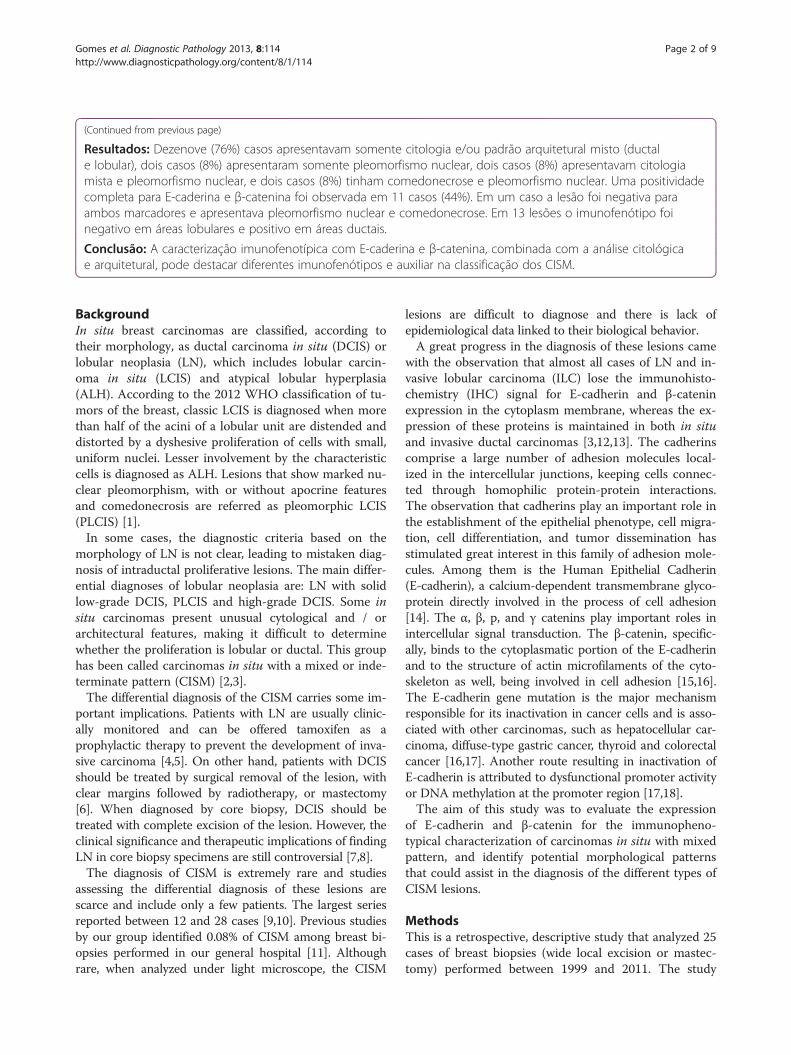
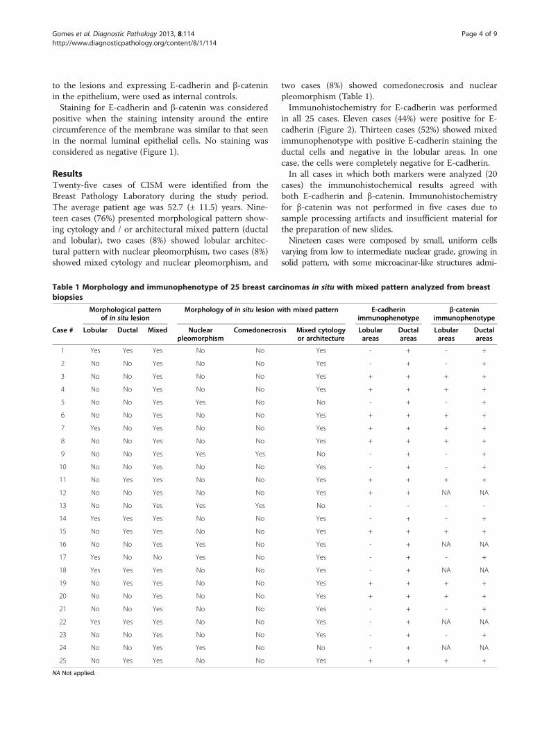
Figure 2 Case # 8: Carcinoma in situ with mixed pattern (CISM) showing dual cell population (Group 2) stained for hematoxylin andeosin (A – 100×, B – 400×). A stronger positive membrane staining for E-cadherin (C – 400×) and β-catenin (D – 400×) can be appreciated inthe outer layer of cells of ductal pattern. A weaker staining is seen in cells of lobular pattern present in the center of the units.
Gomes et al. Diagnostic Pathology 2013, 8:114 Page 5 of 9http://www.diagnosticpathology.org/content/8/1/114
xed with groups of low nuclear grade dyshesive cells, in amosaic pattern. Of these, 11 (57.9%) presented positive im-munohistochemistry for E-cadherin and β-catenin. In thesecases, solid architecture with low-grade cytology was themost common morphological pattern. Eight of these cases(42.1%) presented the mixed immunophenotype and in fourof them, the mixed pattern resulted from a “collision” of thelesions showing areas positive and areas negative for bothmarkers in the same duct-lobular unit (Figure 3).Two cases presented the lobular architectural pattern
with nuclear pleomorphism and two cases presentedmixed cytology and nuclear pleomorphism. The last twocases were considered as mixed immunophenotype. Ofthe two cases that presented comedonecrosis and nuclearpleomorphism, one was completely negative for bothmarkers (Figure 4) and the other presented cells positiveand cells negative for both markers (Table 2).
DiscussionIn this study, we sought to evaluate the expression of E-cadherin and β-catenin for the immunophenotypicalcharacterization of CISM. We also searched for potentialmorphological patterns that could help in the diagnosisof different types of CISM lesions. We adopted the mor-phological classification described by Jacobs et al. thatdefines CISM as “carcinomas with indeterminate fea-tures”[3]. According to this classification CISM lesionsare divided in three main groups, namely: (I) presence ofnecrosis, (II) cytology and / or mixed architecture, and(III) nuclear polymorphism. This classification is highly
reproducible and addresses the main morphologicalgroups described in our study.A total of 25 CISM cases were evaluated in this study.
The most common morphological pattern of lesionsidentified belonged to group (II) with 19 out of 25 cases(76%), followed by group (III) (2/25 cases - 8%), andoverlapping patterns between groups (I) and (II) (2/25cases - 8%) and groups (II) and (III) (2/25 cases - 8%).Our findings are in agreement with those reported byJacobs et al. who observed, in 28 cases of CISM, 60% ofthe lesions in group (II) (17/28 cases), 21% in group (I)(5/28 cases) , and 18% in group (III) (5/28 cases).However, it is noteworthy that the terminology and mor-
phologic criteria used for the diagnosis of CISM are hetero-geneous. Fisher et al. termed it as “ductolobular carcinomain situ” lesions with monomorphic cells with foci of necro-sis or cribriform pattern [4]. Acs et al. described 14 cases ofCISM referring to the lesions as “with ductal carcinoma insitu and lobular features” and adopted, as a diagnostic cri-teria, LN in situ lesions with cytological and architecturalpatterns, with central areas of comedonecrosis or lobules,or large duct units populated by non-cohesive cells withmarked nuclear pleomorphism [19]. Maluf et al. analyzed12 cases of “solid low grade carcinoma in situ of the breast”and included “low-grade solid DCIS, LCIS and DCIS andLCIS associated with invasive carcinomas of any type.Cases showing only unequivocal areas of LCIS or DCIS ofnonsolid type were excluded” [20].Even among experts in the pathology of breast tumors,
the descriptions of these lesions are divergent. Page and
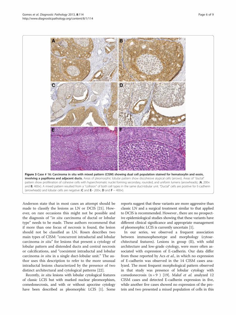
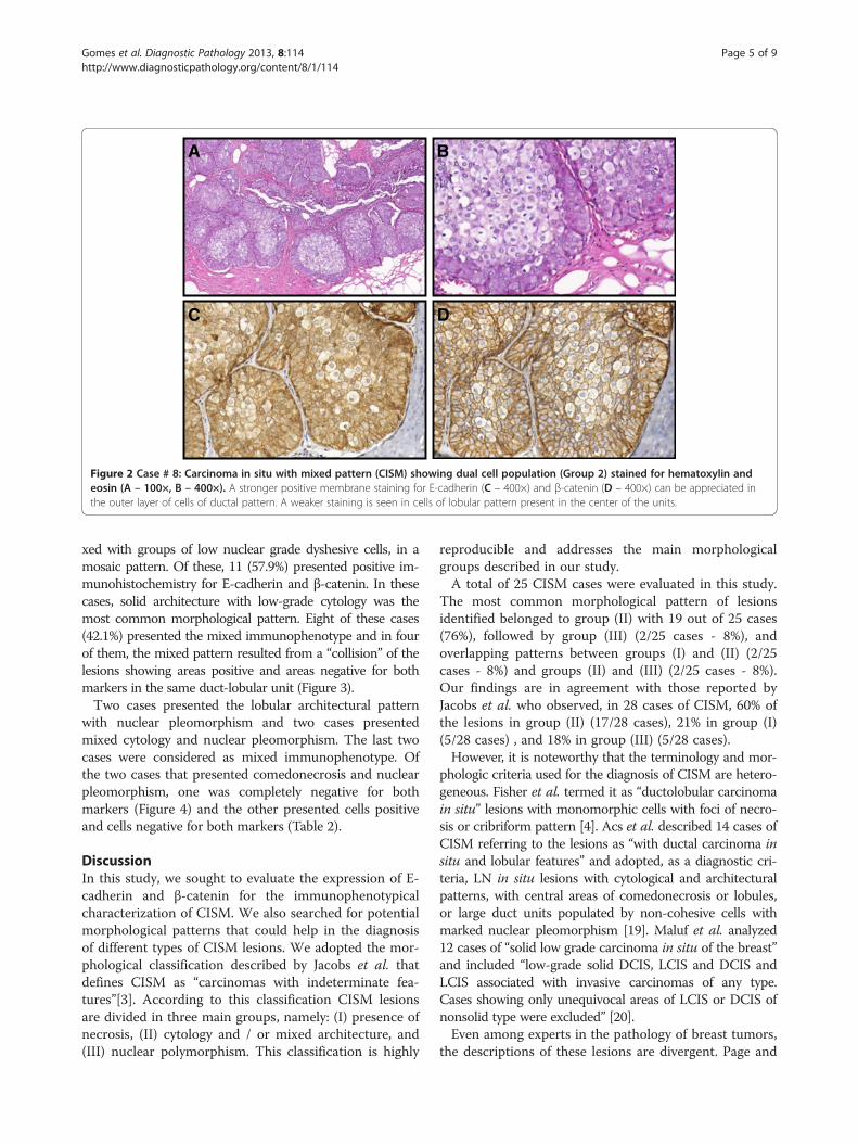
Figure 3 Case # 16: Carcinoma in situ with mixed pattern (CISM) showing dual cell population stained for hematoxylin and eosin,involving a papilloma and adjacent ducts. Areas of pleomorphic lobular pattern show discohesive atypical cells (arrows). Areas of “ductal”pattern show proliferation of cohesive cells with hyperchromatic nuclei forming secondary, rounded, and uniform lumens (arrowheads), (A, 200×and B, 400×). A mixed pattern resulted from a “collision” of both cell types in the same duct-lobular unit. “Ductal” cells are positive for E-cadherin(arrowheads) and lobular cells are negative (C and E– 200×, D and F – 400×).
Gomes et al. Diagnostic Pathology 2013, 8:114 Page 6 of 9http://www.diagnosticpathology.org/content/8/1/114
Anderson state that in most cases an attempt should bemade to classify the lesions as LN or DCIS [21]. How-ever, on rare occasions this might not be possible andthe diagnosis of “in situ carcinoma of ductal or lobulartype” needs to be made. These authors recommend thatif more than one focus of necrosis is found, the lesionshould not be classified as LN. Rosen describes twomain types of CISM: “concurrent intraductal and lobularcarcinoma in situ” for lesions that present a cytology oflobular pattern and distended ducts and central necrosisor calcifications, and “coexistent intraductal and lobularcarcinoma in situ in a single duct-lobular unit.” The au-thor uses this description to refer to the more unusualintraductal lesions characterized by the presence of twodistinct architectural and cytological patterns [22].Recently, in situ lesions with lobular cytological features
of classic LCIS but with marked nuclear pleomorphism,comedonecrosis, and with or without apocrine cytologyhave been described as pleomorphic LCIS [1]. Some
reports suggest that these variants are more aggressive thanclassic LN and a surgical treatment similar to that appliedto DCIS is recommended. However , there are no prospect-ive epidemiological studies showing that these variants havedifferent clinical significance and appropriate managementof pleomorphic LCIS is currently uncertain [1].In our series, we observed a frequent association
between immunophenotype and morphology (cytoar-chitectural features). Lesions in group (II), with solidarchitecture and low-grade cytology, were more often as-sociated with expression of E-cadherin. Our data differfrom those reported by Acs et al., in which no expressionof E-cadherin was observed in the 14 CISM cases ana-lyzed. The most frequent morphological pattern observedin that study was presence of lobular cytology withcomedonecrosis (n = 9 ) [19]. Maluf et al. analyzed 12CISM cases and detected E-cadherin expression in five,while another five cases showed no expression of the pro-tein and two presented a mixed population of cells in this
B
C D
A
Figure 4 Case # 13: Pleomorphic lobular carcinoma in situ showing discohesive atypical cells, with nuclear pleomorphism andcomedonecrosis. Hematoxylin and eosin (A – 100×). Lobular cells are negative for E-cadherin (B – 100×; D – 400×) and for β-catenin (C – 100×).
Gomes et al. Diagnostic Pathology 2013, 8:114 Page 7 of 9http://www.diagnosticpathology.org/content/8/1/114
regard. These authors did not observe the prevalence of aspecific morphological pattern over others [20]. Similarlyto the study by Jacobs et al., lesions in group (II) werethe most frequent lesion associated with expression ofE-cadherin in our study, however we observed them at ahigher frequency. A dual cell population in the same ter-minal duct-lobular unit was observed in four cases. This islikely due to the coexistence of LN and DCIS in the sameterminal duct-lobular unit.Since first reported, the immunohistochemical reac-
tion of E-cadherin has been proposed as an aiding toolin the differential diagnosis between ductal and lobularlesions, either invasive or in situ. However, it should benoted that up to 15% of lobular lesions may exhibit aber-rant expression of E-cadherin and thus, the lack ofE-cadherin expression should not be used as the sole cri-terion for LN diagnosis [12]. Choi et al. observedvariability in the immunohistochemical staining of
Table 2 Category morphology and immunophenotype forE-cadherin of carcinoma in situ with mixed pattern
Morphology Cases E-cadherin
Positive Negative Mixed
Group 1 0 0 0 0
Group 2 19 11 0 8
Group 3 2 0 0 2
Group 2 and 3 2 0 0 2
Group 1 and 3 2 0 1 0
Total 25 11 1 13
E-cadherin, and detected abnormal staining patterns,both in ductal and lobular lesions, making the differen-tial diagnosis between in situ lobular and invasive lesionsvery difficult through immunohistochemistry [10]. Analternative to reduce this interference and improve diag-nosis is the combined use of immunohistochemicalmarkers of the catenin pathway. Using IHC and molecu-lar biology techniques Da Silva et al. analyzed threecases presenting morphological characteristics and geno-typing that agreed with invasive lobular carcinoma, withnonetheless aberrant expression of E-cadherin. Of thesethree cases, two did not express β-catenin, indicatingthat the formation of the cadherin-catenin complex,which is required for the normal function of the cell andmaintenance of tissue architecture, including cell adhe-sion, failed [12,23]. In our study, we observed thatexpression of E-cadherin agreed with expression ofβ-catenin in all cases here observed.Other explanations for the abnormal expression of
E-cadherin found in other studies may be related totechnical difficulties and pitfalls that may occur duringthe different stages in the immunohistochemical reac-tion. In our study, we had some difficulties in thepre-analytical reaction such as material loss and weakstaining in some cases. This may reflect the fact thatwe used specimens coming from the routine diagnosis la-boratory of a general hospital; and other cases were sentto us for a second opinion. In many cases, there was nocontrol of the pre-analytical phase or standardization oftime of formalin fixation and unbuffered formalin wasused. Goldstein et al. showed that the reactivity level varies
Gomes et al. Diagnostic Pathology 2013, 8:114 Page 8 of 9http://www.diagnosticpathology.org/content/8/1/114
with the number of blocks and thickness of the samplesections in the pre-analytical process [2]. Differentclones of antibodies against E-cadherin and different an-tigens may also have an effect on the quality of the im-munohistochemical staining. A comparison between twotypes of antibodies revealed discrepancy in the stainingof lobular lesions in 6.4% of the cases [7]. Finally, thereis a lack of consensus regarding the interpretationof the positivity of immunohistochemical staining ofE-cadherin. The established cutoff of a positive signalvaries between basal membrane expression and presenceof any positivity to 20% of expressing cells [12,23]. Semi-quantitative evaluations of the intensity of staining andassociation of different criteria forming scores of stainingintensity have also been proposed [19].Other immunohistochemical markers have been sug-
gested to aid in the diagnosis of CISM. The p120 cateninis an intracellular protein that promotes the binding be-tween the complex of catenins and cell cytoskeleton.When E-cadherin expression is absent, p120 catenin isdispersed in the cytoplasm, which explains its expres-sion in the cytoplasm in LN, and in the membrane inDCIS [23,24].
ConclusionsThe immunophenotypic characterization of carcinomasin situ using E-cadherin and β-catenin, combined withthe analysis of cytological and architectural patterns, is auseful tool for the morphological and immunophe-notypical classification of CISM. However, a negativestaining for these markers should not be used as the solecriterion of lobular phenotype because aberrant expres-sion in lobular neoplasia and loss of expression in ductalcancers can both occur.
AbbreviationsALH: Atypical lobular hyperplasia; CISM: Carcinoma In Situ with mixedpattern; DCIS: Ductal carcinoma In Situ; LN: Lobular neoplasia; LCIS: Lobularcarcinoma In Situ; IHC: Immunohistochemistry; E-cadherin: Human epithelialcadherin; PLCIS: Pleomorphic lobular carcinoma In Situ; UFMG: FederalUniversity of Minas Gerais.
Competing interestsThe authors declare that they have no competing interests.
Authors’ contributionsDSG conceived the study, participated in the histological review, and draftedthe manuscript. SSP participated in the design of the study. RMG: Performedthe immunohistochemistry reactions. HG participated in design andcoordination of the study, participated in the histological review, and draftedand reviewed the manuscript. All authors have read and approved the finalmanuscript.
AcknowledgmentsThis work was supported in part by Fundação de Amparo à Pesquisa deMinas Gerais (FAPEMIG), Conselho Nacional de Desenvolvimento Científico eTecnológico (CNPq), and Coordenação de Aperfeiçoamento de Pessoal deNível Superior (CAPES).This manuscript was reviewed by a professional science editor and by anative English-speaking copy editor to improve readability.
Author details1Breast Pathology Laboratory, School of Medicine, Federal University of MinasGerais (UFMG), Av. Professor Alfredo Balena, 190, Belo Horizonte, MinasGerais 30130-100, Brazil. 2Laboratory of Investigative Pathology, A.C. CamargoHospital, São Paulo-SP, Brazil.
Received: 17 April 2013 Accepted: 12 June 2013Published: 9 July 2013
References1. Lakhani SR, Ellis IO, Schnitt SJ, Tan PH, van de Vijver MJ, World Health
Organization, International Agency for Research on Cancer: WHOclassification of tumours of the breast. 4th edition. Lyon: IARC; 2012.
2. Goldstein NS, Bassi D, Watts JC, Layfield LJ, Yaziji H, Gown AM: E-cadherinreactivity of 95 noninvasive ductal and lobular lesions of the breast.Implications for the interpretation of problematic lesions. Am J ClinPathol 2001, 115:534–542.
3. Jacobs TW, Pliss N, Kouria G, Schnitt SJ: Carcinomas in situ of the breastwith indeterminate features: role of E-cadherin staining incategorization. Am J Surg Pathol 2001, 25:229–236.
4. Fisher B, Costantino JP, Wickerham DL, Redmond CK, Kavanah M, CroninWM, Vogel V, Robidoux A, Dimitrov N, Atkins J, et al: Tamoxifen forprevention of breast cancer: report of the national surgical adjuvantbreast and bowel project P-1 study. J Natl Cancer Inst 1998, 90:1371–1388.
5. Schnitt SJ, Morrow M: Lobular carcinoma in situ: current concepts andcontroversies. Semin Diagn Pathol 1999, 16:209–223.
6. Lakhani SR, Audretsch W, Cleton-Jensen AM, Cutuli B, Ellis I, Eusebi V, GrecoM, Houslton RS, Kuhl CK, Kurtz J, et al: The management of lobularcarcinoma in situ (LCIS). Is LCIS the same as ductal carcinoma in situ(DCIS)? Eur J Cancer 2006, 42:2205–2211.
7. Liberman L: Clinical management issues in percutaneous core breastbiopsy. Radiol Clin North Am 2000, 38:791–807.
8. Nagi CS, O’Donnell JE, Tismenetsky M, Bleiweiss IJ, Jaffer SM: Lobularneoplasia on core needle biopsy does not require excision. Cancer 2008,112:2152–2158.
9. Bratthauer GL, Moinfar F, Stamatakos MD, Mezzetti TP, Shekitka KM, Man YG,Tavassoli FA: Combined E-cadherin and high molecular weightcytokeratin immunoprofile differentiates lobular, ductal, and hybridmammary intraepithelial neoplasias. Hum Pathol 2002, 33:620–627.
10. Choi YJ, Pinto MM, Hao L, Riba AK: Interobserver variability and aberrantE-cadherin immunostaining of lobular neoplasia and infiltrating lobularcarcinoma. Mod Pathol 2008, 21:1224–1237.
11. Gomes DS, Balabram D, Porto SS, Gobbi H: Lobular neoplasia: frequencyand association with other breast lesions. Diagn Pathol 2011, 6:74.
12. Da Silva L, Parry S, Reid L, Keith P, Waddell N, Kossai M, Clarke C, Lakhani SR,Simpson PT: Aberrant expression of E-cadherin in lobular carcinomas ofthe breast. Am J Surg Pathol 2008, 32:773–783.
13. Goldstein NS, Kestin LL, Vicini FA: Clinicopathologic implications of E-cadherin reactivity in patients with lobular carcinoma in situ of thebreast. Cancer 2001, 92:738–747.
14. Baranwal S, Alahari SK: Molecular mechanisms controlling E-cadherinexpression in breast cancer. Biochem Biophys Res Commun 2009, 384:6–11.
15. Andrews JL, Kim AC, Hens JR: The role and function of cadherins in themammary gland. Breast cancer res 2012, 14:203.
16. Wangefjord S, Brandstedt J, Lindquist KE, Nodin B, Jirstrom K, Eberhard J:Associations of beta-catenin alterations and MSI screening status withexpression of key cell cycle regulating proteins and survival fromcolorectal cancer. Diagn Pathol 2013, 8:10.
17. Cheng H, Liang H, Qin Y, Liu Y: Nuclear beta-catenin overexpression inmetastatic sentinel lymph node is associated with synchronous livermetastasis in colorectal cancer. Diagn Pathol 2011, 6:109.
18. Tsanou E, Peschos D, Batistatou A, Charalabopoulos A, Charalabopoulos K:The E-cadherin adhesion molecule and colorectal cancer. A globalliterature approach. Anticancer Res 2008, 28:3815–3826.
19. Acs G, Lawton TJ, Rebbeck TR, LiVolsi VA, Zhang PJ: Differential expressionof E-cadherin in lobular and ductal neoplasms of the breast and itsbiologic and diagnostic implications. Am J Clin Pathol 2001, 115:85–98.
20. Maluf HM, Swanson PE, Koerner FC: Solid low-grade in situ carcinoma ofthe breast: role of associated lesions and E-cadherin in differentialdiagnosis. Am J Surg Pathol 2001, 25:237–244.
Gomes et al. Diagnostic Pathology 2013, 8:114 Page 9 of 9http://www.diagnosticpathology.org/content/8/1/114
21. Page DL, Anderson TJ: Diagnostic histopathology of the breast. Edinburgh;New York: Churchill Livingstone; 1987.
22. Rosen PP: Rosen’s breast pathology. 3rd edition. Philadelphia: LippincottWilliams & Wilkins; 2008.
23. Dabbs DJ, Bhargava R, Chivukula M: Lobular versus ductal breastneoplasms: the diagnostic utility of p120 catenin. Am J Surg Pathol 2007,31:427–437.
24. Sarrio D, Perez-Mies B, Hardisson D, Moreno-Bueno G, Suarez A, Cano A,Martin-Perez J, Gamallo C, Palacios J: Cytoplasmic localization of p120ctnand E-cadherin loss characterize lobular breast carcinoma frompreinvasive to metastatic lesions. Oncogene 2004, 23:3272–3283.
doi:10.1186/1746-1596-8-114Cite this article as: Gomes et al.: Usefulness and limitations ofE-cadherin and β-catenin in the classification of breast carcinomas insitu with mixed pattern. Diagnostic Pathology 2013 8:114.
Submit your next manuscript to BioMed Centraland take full advantage of:
• Convenient online submission
• Thorough peer review
• No space constraints or color figure charges
• Immediate publication on acceptance
• Inclusion in PubMed, CAS, Scopus and Google Scholar
• Research which is freely available for redistribution
Submit your manuscript at www.biomedcentral.com/submit
![On the Relative Usefulness of Fireballssacerdot/PAPERS/lics15.pdf · 2016-07-28 · arXiv:1505.03791v1 [cs.LO] 14 May 2015 On the Relative Usefulness of Fireballs Beniamino Accattoli](https://static.fdocument.org/doc/165x107/5f085be17e708231d4219e2e/on-the-relative-usefulness-of-sacerdotpaperslics15pdf-2016-07-28-arxiv150503791v1.jpg)