Up-regulation of IL-6 and TNF-α induced by SARS-coronavirus spike protein in murine macrophages via...
8
Virus Research 128 (2007) 1–8 Up-regulation of IL-6 and TNF- induced by SARS-coronavirus spike protein in murine macrophages via NF-B pathway Wei Wang, Linbai Ye ∗ , Li Ye, Baozong Li, Bo Gao, Yingchun Zeng, Lingbao Kong, Xiaonan Fang, Hong Zheng, Zhenghui Wu, Yinglong She State Key Laboratory of Virology, College of Life Science, Wuhan University, Wuhan, Hubei 430072, China Received 3 November 2006; received in revised form 6 February 2007; accepted 8 February 2007 Available online 25 May 2007 Abstract The clinical picture of severe acute respiratory syndrome (SARS) is characterized by an over-exuberant immune response with lung lym- phomononuclear cells infilteration and proliferation that may account for tissue damage more than the direct effect of viral replication. To understand how cells response in the early stage of virus–host cell interaction, in this study, a purified recombinant S protein was studied for stimulating murine macrophages (RAW264.7) to produce proinflammatory cytokines (IL-6 and TNF-) and chemokine IL-8. We found that direct induction of IL-6 and TNF- release in the supernatant in a dose-, time-dependent manner and highly spike protein-specific, but no induction of IL-8 was detected. Further experiments showed that IL-6 and TNF- production were dependent on NF-B, which was activated through I-B degradation. These results suggest that SARS-CoV spike protein may play an important role in the pathogenesis of SARS, especially in inflammation and high fever. © 2007 Published by Elsevier B.V. Keywords: SARS-coronavirus; Spike protein; IL-6; TNF-; NF-B; Macrophages 1. Introduction Severe acute respiratory syndrome (SARS) caused by a novel human coronavirus (CoV), designated SARS-CoV, is a highly contagious respiratory disease with the lungs as a major target (Peiris et al., 2003). A histology of lung necropsy from SARS patients showed that abundant foamy macrophages and multi- nucleated syncytial cells were demonstrated (Lee et al., 2003; Nicholls et al., 2003). Thus, cytokines storm has been proposed to be involved in the rapid course of adult respiratory distress syndrome (ARDS) in SARS patients. The likelihood of SARS being an immune-mediated disease is supported by the observa- tion of a transient lymphopenia, especially in CD4 + and CD8 + T cells early in the SARS episode, and elevated serum levels of inflammatory cytokines in SARS patients (Li et al., 2003b; Wong et al., 2004a; Xie et al., 2003). SARS-CoV consists of a nucleocapsid (N) core and three membrane proteins of spike (S), membrane (M), and envelope ∗ Corresponding author. Tel.: +86 27 6875 2372; fax: +86 27 6876 4763. E-mail addresses: [email protected], [email protected] (L. Ye). (E). S protein of SARS-CoV binds to the host receptor, which was identified as angiotensin-converting enzyme 2 (ACE2) (Li et al., 2003a) and mediates membrane fusion. The protein can also induce neutralizing antibodies against SARS-CoV (Chen et al., 2005; He et al., 2004). NF-B is a family of Rel domain-containing proteins present in the cytoplasm of all cells, which includes Rel A, Rel B, c-Rel, p105/p50 and p100/p52. Under resting conditions, NF-B con- sists of a heterotrimer of p50, p65, and I-B in the cytoplasm, after activation by a large number of inducers, the I-B pro- teins become phosphorylated, ubiquitylated and, subsequently, degraded by the proteasome. The degradation of I-B allows NF-B proteins to translocate to the nucleus and bind their cog- nate DNA binding sites to regulate the transcription of a large number of genes, including antimicrobial peptides, cytokines, chemokines, stress-response proteins and anti-apoptotic pro- teins. I-B degradation is triggered by activation of a complex of multiple kinases that is composed of IKK and IKK and a structural regulatory subunit, IKK or NEMO. Phosphoryla- tion of IKK and IKK is activated by their upstream kinases including NF-B-inducing kinase (Ghosh et al., 1998; Karin and Ben-Neriah, 2000; Li and Verma, 2002; Verma et al., 1995). NIK 0168-1702/$ – see front matter © 2007 Published by Elsevier B.V. doi:10.1016/j.virusres.2007.02.007
Transcript of Up-regulation of IL-6 and TNF-α induced by SARS-coronavirus spike protein in murine macrophages via...

A
pusioIi©
K
1
hc(pnNtsbtToW
m
0d
Virus Research 128 (2007) 1–8
Up-regulation of IL-6 and TNF-� induced by SARS-coronavirus spikeprotein in murine macrophages via NF-�B pathway
Wei Wang, Linbai Ye ∗, Li Ye, Baozong Li, Bo Gao, Yingchun Zeng, Lingbao Kong,Xiaonan Fang, Hong Zheng, Zhenghui Wu, Yinglong She
State Key Laboratory of Virology, College of Life Science, Wuhan University, Wuhan, Hubei 430072, China
Received 3 November 2006; received in revised form 6 February 2007; accepted 8 February 2007Available online 25 May 2007
bstract
The clinical picture of severe acute respiratory syndrome (SARS) is characterized by an over-exuberant immune response with lung lym-homononuclear cells infilteration and proliferation that may account for tissue damage more than the direct effect of viral replication. Tonderstand how cells response in the early stage of virus–host cell interaction, in this study, a purified recombinant S protein was studied fortimulating murine macrophages (RAW264.7) to produce proinflammatory cytokines (IL-6 and TNF-�) and chemokine IL-8. We found that direct
nduction of IL-6 and TNF-� release in the supernatant in a dose-, time-dependent manner and highly spike protein-specific, but no inductionf IL-8 was detected. Further experiments showed that IL-6 and TNF-� production were dependent on NF-�B, which was activated through-�B� degradation. These results suggest that SARS-CoV spike protein may play an important role in the pathogenesis of SARS, especially innflammation and high fever.2007 Published by Elsevier B.V.
ges
(weaa
ipsatdNnn
eywords: SARS-coronavirus; Spike protein; IL-6; TNF-�; NF-�B; Macropha
. Introduction
Severe acute respiratory syndrome (SARS) caused by a noveluman coronavirus (CoV), designated SARS-CoV, is a highlyontagious respiratory disease with the lungs as a major targetPeiris et al., 2003). A histology of lung necropsy from SARSatients showed that abundant foamy macrophages and multi-ucleated syncytial cells were demonstrated (Lee et al., 2003;icholls et al., 2003). Thus, cytokines storm has been proposed
o be involved in the rapid course of adult respiratory distressyndrome (ARDS) in SARS patients. The likelihood of SARSeing an immune-mediated disease is supported by the observa-ion of a transient lymphopenia, especially in CD4+ and CD8+
cells early in the SARS episode, and elevated serum levelsf inflammatory cytokines in SARS patients (Li et al., 2003b;
ong et al., 2004a; Xie et al., 2003).SARS-CoV consists of a nucleocapsid (N) core and threeembrane proteins of spike (S), membrane (M), and envelope
∗ Corresponding author. Tel.: +86 27 6875 2372; fax: +86 27 6876 4763.E-mail addresses: [email protected], [email protected] (L. Ye).
ctoatiB
168-1702/$ – see front matter © 2007 Published by Elsevier B.V.oi:10.1016/j.virusres.2007.02.007
E). S protein of SARS-CoV binds to the host receptor, whichas identified as angiotensin-converting enzyme 2 (ACE2) (Li
t al., 2003a) and mediates membrane fusion. The protein canlso induce neutralizing antibodies against SARS-CoV (Chen etl., 2005; He et al., 2004).
NF-�B is a family of Rel domain-containing proteins presentn the cytoplasm of all cells, which includes Rel A, Rel B, c-Rel,105/p50 and p100/p52. Under resting conditions, NF-�B con-ists of a heterotrimer of p50, p65, and I-�B in the cytoplasm,fter activation by a large number of inducers, the I-�B pro-eins become phosphorylated, ubiquitylated and, subsequently,egraded by the proteasome. The degradation of I-�B allowsF-�B proteins to translocate to the nucleus and bind their cog-ate DNA binding sites to regulate the transcription of a largeumber of genes, including antimicrobial peptides, cytokines,hemokines, stress-response proteins and anti-apoptotic pro-eins. I-�B degradation is triggered by activation of a complexf multiple kinases that is composed of IKK� and IKK� and
structural regulatory subunit, IKK� or NEMO. Phosphoryla-ion of IKK� and IKK� is activated by their upstream kinasesncluding NF-�B-inducing kinase (Ghosh et al., 1998; Karin anden-Neriah, 2000; Li and Verma, 2002; Verma et al., 1995). NIK
2 Rese
atv
atipatcmtafetoaTi
aadICmtSotsRihphacma�raddsf
2
2
T1
(1
2
TtS
-wDC
2p
Scftp(fsci
tt
2s
ot1Ism
2(
attSd
W. Wang et al. / Virus
s a potential activator of the I-�B�-kinase complex appearedo have been identified an important element of the NF-�B acti-ation pathway (Smith et al., 2001).
Interleukin-6 (IL-6) plays a central role in both innate andcquired immune response. IL-6 is the predominate inducer ofhe acute-phase response, an innate immune mechanism whichs triggered by infection and inflammation. IL-6 also plays multi-le roles during subsequent development of acquired immunitygainst incoming pathogens, stimulation of antibody produc-ion by B cells, regulation of macrophage (M�) and dendriticell (DC) differentiation, and response of regulatory T cells toicrobial infection. Up-regulation of IL-6 was induced by infec-
ion of many viruses, including MHV, HRV, HCMV (Banerjee etl., 2002; Gealy et al., 2005; Griego et al., 2000). Tumor necrosisactor alpha (TNF-�) has multiple biologic effects including thenhanced release of other proinflammatory/chemotactic media-ors, up-regulation of adhesion molecules, enhanced migrationf eosinophils and neutrophils. TNF-� was detected in increasedmounts in sera of patients with SARS (Zhang et al., 2004).hese data implicate TNF-� is a potent inflammatory cytokine
n the pathogenesis of SARS.Macrophages are central immunocytes in innate immunity
nd produce non-specific inflammatory mediators. Many virusesnd their membrane proteins can activate macrophages to pro-uce cytokines. Varicella-zoster virus (VZV) induced IL-6 andL-8 in human monocytes (Wang et al., 2005). Hepatitis B virusapsid strongly induced TNF-�, IL-6 and IL-12p40 in THP-1acrophages (Cooper et al., 2005). SARS-CoV failed to produc-
ively infect human macrophages, but the interaction betweenARS-CoV and human macrophage triggered the productionf cytokine (IL-6, IL-12) efficiently (Tseng et al., 2005). Andhe proinflammatory cytokine IL-6 and the chemokine IL-8 aretrongly up-regulated in SARS patients (Hsueh et al., 2004).ecombinant baculovirus displaying SARS-CoV spike protein
nduced IL-8 release (Chang et al., 2004). Then, we had theypothesis that the spike protein plays an important role in theroduction of proinflammatory cytokine at the stage of virus-ost cell interaction. In the present study, we examined thebility of S protein of SARS to modulate the proinflammatoryytokines (IL-6, TNF-�) and chemokine (IL-8) production. Theurine macrophages cell line (RAW264.7) was incubated withpurified recombinant S protein, then the levels of IL-6, TNF-and IL-8 in the supernatant were measured by ELISA. Our
esults showed that S protein induced massive release of IL-6nd TNF-� in an S protein-specific, as well as time- and dose-ependent manner. However, there was no induction of IL-8 wasetected. Furthermore, we found that S protein activated NF-�Bignaling pathway via I-�B� degradation, which was essentialor up-regulation of IL-6 and TNF-�.
. Materials and methods
.1. Cell culture
RAW264.7 cells were obtained from China Center forype Culture Collection (CCTCC) and cultured in RPMI640 (Gibco/BRL) supplemented with 10% fetal bovine serum
spcs
arch 128 (2007) 1–8
FBS; Hyclone), and antibiotics (100 �g/ml streptomycin and00 U/ml penicillin) at 37 ◦C in 5% CO2.
.2. Antibodies and plasmid
Anti-S Abs (IMG-542) was from IMGENEX Corporation.he antibody was developed by immunizing rabbits with syn-
hetic peptides corresponding to amino acids 288–303 of theARS Spike glycoprotein.
Expression vector for dominant-negative NIK (pcDNA3.1(+)dn-NIK), which has a kinase-dead mutation of K429A/K430A,as kindly provided by Prof. Brian M.J. Foxwell and Alisonavis (Kennedy Institute of Rheumatologh Division, Imperialollege School of Medicine, London).
.3. Expression and purification of recombinant spikerotein
The spike gene encoding from 262nd to 606th amino acid ofARS-CoV S protein was cloned into pET-his expression vectorontaining T7 promoter and six histidine fusion tag. After trans-ormation of E. coli BL21 (DE3) with the constructed vector,he S proteins were produced by 1 mM IPTG. Cells were resus-ended in a buffer containing 50 mM Tris-Cl and 500 mM NaClpH 8.0), disrupted by ultrasonic and centrifuged at 12,000 × gor 15 min. The pellet was dissolved with 8 M urea and theix-histidine-tagged protein was purified by Ni-NTA affinityhromatography. After dialysis against deionized water remov-ng the urea, the purified S protein was performed.
Some soluble S protein was treated by Detoxi-GelTM Endo-oxin Removing Gel (Pierce) to remove the endotoxin accordingo the manufacturer’s instruction.
.4. Measurement of IL-6, TNF-α and IL-8 in theupernatant of cultured cells
RAW264.7 cells were seeded into 6-well plates at a densityf 5 × 10⁵ cells/well, and untreated or incubated for indicatedimes (0, 3, 6, 9, 12, 24 h) or at indicated concentrations (0,, 5, 10, 20 �g/ml) with purified S protein. IL-6, TNF-� andL-8 in the culture supernatant of RAW264.7 cells were mea-ured by ELISA kits (Bender MedSystems) according to theanufacturer’s instructions.
.5. Reverse transcription-polymerase chain reactionRT-PCR)
RAW264.7 cells were incubated with the purified S proteint different concentrations (0, 1, 5, 10, 20 �g/ml) for 12 h, thenotal RNA were extracted and treated with DNase I. For reverseranscription (RT), 1 �g of RNA was incubated with 200 U ofuperscript II reverse transcriptase (Invitrogen) and 100 ng ran-om hexanucleotides in 20 �l of 1× RT buffer (Invitrogen)
upplied with 1 mM each of the four dexynuleotide triphos-hates, 20 U of RNasin, and 10 mM dithiothreitol. The resultingDNA were amplified by 35 cycles of PCR, with each cycle con-isting of 30 s at 94 ◦C, 30 s at 52 ◦C, and 30 s at 72 ◦C. The prime
Research 128 (2007) 1–8 3
sTACCle
2
Nt2Twsu
2
cudTcapCns(p0nd
3
3
Euwpfie
3R
sat
Fig. 1. Expression and purification of recombinant S protein of SARS-CoV.Rpi
artetsmri5eitoantk(twimsba
a
W. Wang et al. / Virus
equences were as follows: for IL-6, forward (5′-GTT GCC TTCTG GGA CTG ATG-3′) and reverse (5′-CAT ACA ATC AGATT GCC ATT GC-3′); for actin, forward (5′-ACA ACG GCTCG GCA TGT GCA A-3′) and reverse (5′-CCA TGT CGTCC AGT TGG TGA C-3′). The RT-PCR products were ana-
yzed by electrophoresis through 2% agarose gels containingthidium bromide.
.6. Transfection and NF-κB reporter assay
RAW264.7 cells in 96-well plates were cotransfected withF-�B firefly luciferase reporter plasmid (0.1 �g/well) and con-
rol Renila luciferase plasmid (5 ng/well) using lipofectamine000 (Invitrogen) according to the manufacturer’s instructions.wenty-four hours after transfection, the cells were stimulatedith the purified S protein or untreated for 12 h. Cells were sub-
equently harvested, lysed and luciferase activity were measuredsing the dual luciferase reporter assay kit (Promega).
.7. Western blotting analysis of I-κBα
Cell lysates were separated by 12% denaturing sodium dode-yl sulfate-polyacrylamide gel electrophoresis (SDS-PAGE)sing standard techniques and electroblotted onto polyvinyli-ene difluoride membranes. The membranes were blocked inris-buffered saline (10 mM Tris-HCl [pH 8.0], 150 mM NaCl)ontaining 5% (w/v) dried milk for 2 h at room temperaturend then probed with the first antibodies, rabbit anti-I-�B�olyclonal antibodies (sc-371; Santa Cruz Biotechnology, Santaruz, USA) or anti-actin antibody (sc-1616; Santa Cruz Biotech-ology) for 2 h at room temperature. After extensive washing,econdary antibodies conjugated with horseradish peroxidaseHRP) were applied onto the blots for at least 1 h at room tem-erature. The blots were washed four times with TBS containing.1% Tween-20. Reagents (Pierce) for enhanced chemilumi-escence were applied to the blots and the light signals wereetected by X-ray film.
. Results
.1. SARS-CoV Spike protein was expressed and purified
The recombinant S protein of SARS-CoV was expressed in. coli strain BL21(DE3) and purified by Ni-NTA affinity col-mn chromatography. We found that expression of S proteinas at very high level after 5 h induction with IPTG, and therotein formed inclusion bodies. Our data showed that the puri-ed S protein (38 kDa) was clearly observed in the SDS-PAGElectrophoresis analysis (Fig. 1).
.2. Spike protein stimulated IL-6 and TNF-α release fromAW264.7 cells
To investigate whether S protein can induce cytokine expres-ion in macrophages, we first measured the level of IL-6, TNF-�nd IL-8 in the supernatant of RAW264.7 treated with S pro-ein. After exposing RAW264.7 cells to the purified S protein
tAs6
ecombinant S protein was expressed in E. coli after 5 h induction with IPTG,urified by Ni-NTA affinity column, separated by 12% SDS-PAGE and visual-zed by Coomassie Brilliant Blue staining.
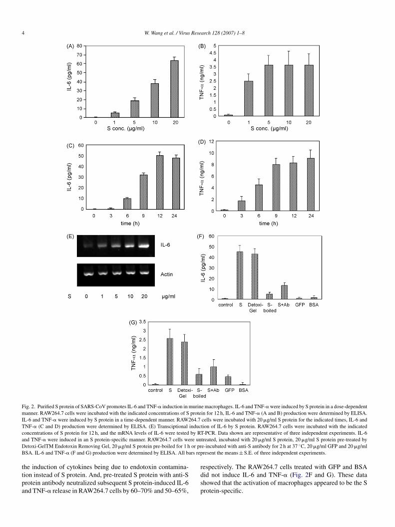
t a concentration ranging from 0 to 20 �g/ml and at a timeanging from 0 to 24 h, the levels of IL-6, TNF-� and IL-8 inhe culture supernatant of RAW264.7 cells were measured usingnzyme-linked immunosorbent assay kits. Recombinant S pro-ein induced the production of IL-6 and TNF-� in the cultureupernatant of RAW264.7 cells in a dose- and time-dependentanner (Fig. 2A–D). Increasing concentrations of S protein
esulted in release of increasing amounts of IL-6 and TNF-�nto the supernatant. Treatment of RAW264.7 cells with about�g/ml S protein was sufficient for full induction of TNF-�, andven 1 �g/ml S protein significantly induced TNF-� productionn RAW264.7 cells. IL-6 release was detectable at 6 h and sus-ained throughout the experiment (up to 24 h), and inductionf TNF-� was observed as early as 3 h and increased furthert 9 h to remain stable for up to 24 h of treatment. There waso up-regulation of IL-8 in RAW264.7 incubated with S pro-ein, and it was not due to the problem of the IL-8 ELISAit because of the positive control of IL-8 standard substancedata not shown). To further determine whether IL-6 induc-ion occurred at the transcriptional level, IL-6 mRNA levelsere evaluated by semiquantitative RT-PCR. S protein strongly
nduced the transcription level of IL-6 in a dose-dependenceanner, which was consistent with the release of IL-6 in the
upernatants (Fig. 2E). These results demonstrate that the recom-inant SARS-CoV S protein is a highly potent inducer of IL-6nd TNF-� in RAW264.7 macrophages.
We found that the IL-6 and TNF-� production were notffected in RAW264.7 cells stimulated with S protein pre-
reated by Detoxi-GelTM Endotoxin Removing Gel (Pierce).nd RAW264.7 cells were treated with pre-boiled S protein,ubsequent IL-6 and TNF-� release decreased by 85–90% and5–80%, respectively. These results ruled out the possibility of
4 W. Wang et al. / Virus Research 128 (2007) 1–8
Fig. 2. Purified S protein of SARS-CoV promotes IL-6 and TNF-� induction in murine macrophages. IL-6 and TNF-� were induced by S protein in a dose-dependentmanner. RAW264.7 cells were incubated with the indicated concentrations of S protein for 12 h, IL-6 and TNF-� (A and B) production were determined by ELISA.IL-6 and TNF-� were induced by S protein in a time-dependent manner. RAW264.7 cells were incubated with 20 �g/ml S protein for the indicated times, IL-6 andTNF-� (C and D) production were determined by ELISA. (E) Transciptional induction of IL-6 by S protein. RAW264.7 cells were incubated with the indicatedconcentrations of S protein for 12 h, and the mRNA levels of IL-6 were tested by RT-PCR. Data shown are representative of three independent experiments. IL-6a re unD or prB ars rep
ttpa
nd TNF-� were induced in an S protein-specific manner. RAW264.7 cells weetoxi-GelTM Endotoxin Removing Gel, 20 �g/ml S protein pre-boiled for 1 hSA. IL-6 and TNF-� (F and G) production were determined by ELISA. All b
he induction of cytokines being due to endotoxin contamina-ion instead of S protein. And, pre-treated S protein with anti-Srotein antibody neutralized subsequent S protein-induced IL-6nd TNF-� release in RAW264.7 cells by 60–70% and 50–65%,
rdsp
treated, incubated with 20 �g/ml S protein, 20 �g/ml S protein pre-treated bye-incubated with anti-S antibody for 2 h at 37 ◦C, 20 �g/ml GFP and 20 �g/mlresent the means ± S.E. of three independent experiments.
espectively. The RAW264.7 cells treated with GFP and BSAid not induce IL-6 and TNF-� (Fig. 2F and G). These datahowed that the activation of macrophages appeared to be the Srotein-specific.
W. Wang et al. / Virus Research 128 (2007) 1–8 5
Fig. 3. NF-�B activation by purified S protein of SARS-CoV. (A) RAW264.7cells in a 96-well plate were transfected with NF-�B-luciferase reporter plas-mid (0.1 �g/well). A Renila reporter plasmid (5 ng/well) was cotransfected fornormalization. Twenty-four hour after transfection, the cells were either treatedwith S protein (10 �g/ml) or left untreated. After 12 h, the cells were lysed, andluciferase activity was quantified. The results were expressed as the mean ± S.E.of three independent experiments. (B) RAW264.7 cells in a 6-well plate weretreated with S protein (10 �g/ml) or left untreated for 1.5 h. Total cell lysates wereanalyzed by Western blotting using anti-I-�B� polyclonal antibodies (upperpanel) and anti-actin polyclonal antibodies (lower panel). The density of thepTo
3
iptp(wsc
ad
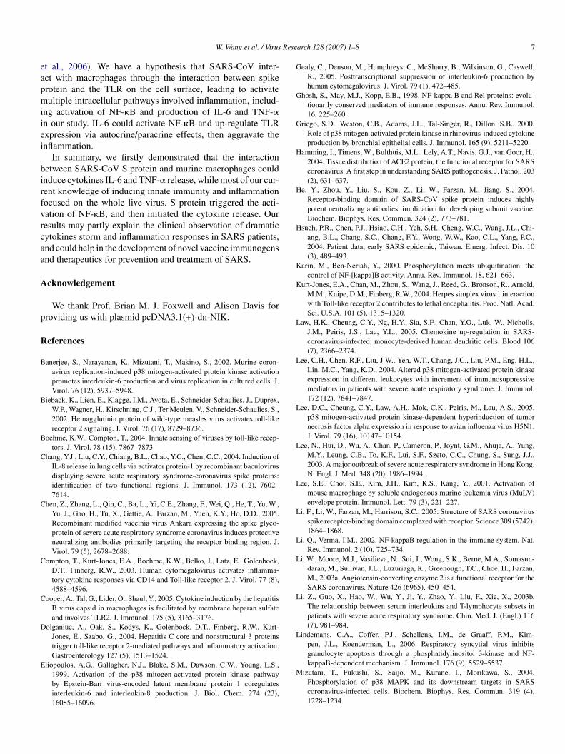
Fig. 4. IL-6 and TNF-� induction by purified S protein of SARS-CoV requiresNF-�B. RAW264.7 cells in a 24-well plate were transfected with eitherpcDNA3.1(+) (empty vector) or pcDNA3.1(+)-dn-NIK (expression plasmid fordominant negative mutant NIK). At 24 h posttransfection, the cells were treatedwsm
1lwli
3T
pNidNiia
4
rotein band was determined by using Bio-Rad Quantity One imaging software.he values in parentheses are density values of I-�B� relative to actin. Resultsf one of three independent experiments are shown.
.3. Spike protein induced NF-κB activation
To investigate the role of NF-�B in mediating S protein-nduced cytokines expression, we next analyzed the ability ofurified S protein to activate the NF-�B. RAW264.7 cells wereransfected with an NF-�B-luciferase reporter plasmid. At 24 hosttransfection, cells were stimulated with purified S protein10 �g/ml) or control medium for 12 h before a luciferase assayas performed. As shown in Fig. 3A, the purified S protein
ignificantly increased NF-�B activity to approximately 5-fold
ompared with the control.To further confirm that S protein can activate NF-�B, wenalyzed I-�B� by Western blotting. I-�B� was found to beecreased by 30% after incubation with purified S protein for
wac
ith S protein (10 �g/ml) for 12 h, IL-6 and TNF-� (A and B) production in theupernatant were determined by ELISA. The means and standard error of theeans are shown from three independent experiments.
.5 h, compared with the control situation (Fig. 3B, top). Equaloading of protein samples was shown by reprobing the blotith an anti-actin antibody (Fig. 3B, bottom). Together with the
uciferase assay, our results clearly demonstrated that S proteinnduced NF-�B activation and I-�B� degradation.
.4. NF-κB was essential for up-regulation of IL-6 andNF-α in purified spike protein-stimulated RAW264.7 cells
To determine whether S protein-induced IL-6 and TNF-�roduction is dependent on NF-�B, the effect of inhibition ofF-�B activation on IL-6 and TNF-� release in RAW264.7
ncubated with S protein was investigated. Transfection withominant-negative NIK (pcDNA3.1(+)-dn-NIK), which inhibitF-�B activation, led to a strong inhibition of purified S protein-
nduced release of IL-6 and TNF-� in RAW264.7 cells (Fig. 4),ndicating that NF-�B is required for effective induction of IL-6nd TNF-� by S protein.
. Discussion
Patients with severe acute respiratory syndrome (SARS)ere reported to have elevated levels of cytokines in blood
nd cytokine gene expression in peripheral blood mononuclearells (PBMC) (Sheng et al., 2005; Yu et al., 2005; Zhang et

6 Rese
aiDD2ttgusSwtofacteawtbBas
bwifCospaim
Nncpaid�LgpdCmaTRt
tEelSipeIviaa
tdaS(pctrdpgitvCaeAdTtptsicbrifaws2afp
W. Wang et al. / Virus
l., 2004). Previous reports have shown that live SARS-CoVnduced a production of IL-6 and TNF-� in human M� andC, importantly, the ability of SARS-CoV modulating M� andC functions was independent of viral replication (Law et al.,005; Tseng et al., 2005). The mechanism of SARS-CoV infec-ion inducing the release of cytokines remains obscure. Sincehe S protein can bind to the receptor, we sought to investi-ate whether the S protein can interact with macrophages thenp-regulate the release of cytokines/chemokines. In the currenttudy, we found that incubation of RAW264.7 with truncatedARS-CoV S protein led to production of IL-6 and TNF-�hich were depended on NF-�B activation. We expressed the
runcated S protein incorporating amino acids 262–606 insteadf the full-length spike protein in our study. As we all known, theull-length spike protein consists of S1 domain and S2 domain,nd S1 domain is responsible for the attachment to the targetells (Li et al., 2005; Wong et al., 2004b). So we tried to expresshe full-length S1 protein at the beginning, but it could not bexpressed in BL21(DE3). The probably reason is that there islarge hydrophobic region in N-terminal peptidase domain,hich is not necessary for the binding with receptor. Then we
ried to express the truncated S1 protein reserving the receptor-inding domain (RBD) and lacking the hydrophobic region inL21(DE3). Fortunately, the truncated protein was expressed atvery high level as inclusion bodies and it was sufficient for our
tudy.In our study, we found massive release of IL-6 and TNF-�,
ut no induction of IL-8 in RAW264.7 stimulated with S protein,hile the over-expression of IL-8 is one of the major phenomena
n SARS patients and human macrophages. First, our study justocused on the response of macrophages to S protein of SARS-oV in vitro, while over-expression of IL-8 in patients was onef quite complicated responses of the whole human’s immuneystem to the virus. We cannot explain all the symptoms in theatients by our relative simple study. Second, the RAW264.7 ismurine macrophage cell line. It may be different in S protein
nducing cytokines in murine macrophages from that in humanacrophages.It has been demonstrated that viral infection triggered the
F-�B pathway, resulting in transcription activation of a largeumber of proinflammatory genes, including cytokine andhemokines (Zhu et al., 1996). To delineate the mechanisms of Srotein inducing cytokine production, we first measured NF-�Bctivation by luciferase assay in S protein-induced RAW264.7ncreased 5-fold compare to the control. Activation of NF-�Bepends on degradation of I-�B, which normally sequesters NF-B in the cytoplasm, inhibiting its function (Ghosh et al., 1998;i and Verma, 2002; Verma et al., 1995). To further investi-ate the involvement of the NF-�B signaling pathways in Srotein-induced cytokine regulation, we examined I-�B� degra-ation in S protein-incubated cells by western blotting analysis.onsistent with our luciferase results, the level of I-�B� inacrophages incubated with S protein was reduced at 1.5 h
fter incubation. We also found S protein induced IL-6 andNF-� production through an NF-�B-dependent mechanism inAW264.7, because inhibition of NF-�B activity caused inhibi-
ion of IL-6 and TNF-� production. Mizutani et al. (2004) found
oaI6
arch 128 (2007) 1–8
hat p38 MAPK was activated in SARS-CoV-infected cells (Vero6), and SARS-CoV infection induced apoptotic cell death. Leet al. (2004) showed that SARS patients had a higher intracellu-ar phosphor-p38 level in total leukocytes than normal controls.ome studies have demonstrated p38 MAPK activation resulted
n accumulation of IL-6 and TNF-� mRNAs and an increase inroduction of IL-6 and TNF-� (Banerjee et al., 2002; Eliopoulost al., 1999; He et al., 2004; Lee et al., 2005; Wang et al., 2005).n our study, we did not examine the effects of S protein on acti-ation of the p38 MAPK, but according to the above studies, its possible that incubation RAW264.7 with S protein can alsoctivate p38 MAPK pathway leading to the production of IL-6nd TNF-�, future studies will address this issue.
Angiotensin-converting enzyme 2 (ACE2) has been iden-ified as a functional receptor for SARS-CoV, and the tissueistribution of ACE2 has been studied extensively (Hamming etl., 2004; To and Lo, 2004). Although immune cells lack ACE2,ARS-CoV can infect human macrophages and dedritic cellsLaw et al., 2005; Tseng et al., 2005; Yilla et al., 2005) withoutroducing infectious virus particles, suggesting that SARS-CoVan entry the immune cells via other receptors. Toll-like recep-ors (TLRs), a major class of molecular pattern recognitioneceptors, are activated by direct interaction of the extracellularomain of the receptor with a pathogen-associated molecularattern, resulting in activation of NF-�B, IRF3, and MAPK sin-aling pathways. TLR family is responsible for activation ofnnate immunity in response to various pathogens, including cer-ain viruses (Boehme and Compton, 2004). TLR2 interacts withiruses or their components, including measles virus, hepatitisvirus, and several herpesviruses, including cytomegalovirus
nd herpes simplex virus (HSV) (Bieback et al., 2002; Comptont al., 2003; Dolganiuc et al., 2004; Kurt-Jones et al., 2004).ctivation of TLR2 by these viruses is followed by the pro-uction of inflammatory cytokines, including IL-6, IL-8, andNF-�. In future studies, we will examine whether TLRs par-
icipate in the induction of IL-6 and TNF-� by SARS-CoV Srotein. When S protein pre-treated with anti-S antibody andhen incubated RAW264.7 cells, the cytokine induction reducedtrongly. The result showed that anti-S antibody could block thenteraction between S protein and the receptor in the immuneells resulting in reduced production of IL-6 and TNF-�. Pre-oiled S protein cannot induce IL-6 and TNF-� efficiently. Thisesult ruled out the possibility that contamination of endotoxins responsible for cytokine induction. Because heat treatmentor 1 h can destroy the functional structure of protein, therebybolish the protein-induced activation effect on macrophages,hile the similar heat treatment do not reduce the LPS-mediated
timulation (Cooper et al., 2005; Lee et al., 2001; Pathak et al.,006). And S protein-induced IL-6 and TNF-� release were notffected by Detoxi-GelTM Endotoxin Removing Gel treatment,urther conforming the cytokines production were induced by Srotein instead of endotoxin contamination.
IL-6, a pleiotropic factor, is a downstream target protein
f NF-�B, on the other hand, IL-6 can also influence NF-�Bctivation. Neutrophils were incubated with IL-6 for 30 min,-�B degradation was observed (Lindemans et al., 2006). IL-up-regulated TLR2 expression in vitro in monocytes (Pons
Rese
eapmiiei
birfvrcaa
A
p
R
B
B
B
C
C
C
C
D
E
G
G
G
H
H
H
K
K
L
L
L
L
L
L
L
L
L
L
W. Wang et al. / Virus
t al., 2006). We have a hypothesis that SARS-CoV inter-ct with macrophages through the interaction between spikerotein and the TLR on the cell surface, leading to activateultiple intracellular pathways involved inflammation, includ-
ng activation of NF-�B and production of IL-6 and TNF-�n our study. IL-6 could activate NF-�B and up-regulate TLRxpression via autocrine/paracrine effects, then aggravate thenflammation.
In summary, we firstly demonstrated that the interactionetween SARS-CoV S protein and murine macrophages couldnduce cytokines IL-6 and TNF-� release, while most of our cur-ent knowledge of inducing innate immunity and inflammationocused on the whole live virus. S protein triggered the acti-ation of NF-�B, and then initiated the cytokine release. Ouresults may partly explain the clinical observation of dramaticytokines storm and inflammation responses in SARS patients,nd could help in the development of novel vaccine immunogensnd therapeutics for prevention and treatment of SARS.
cknowledgement
We thank Prof. Brian M. J. Foxwell and Alison Davis forroviding us with plasmid pcDNA3.1(+)-dn-NIK.
eferences
anerjee, S., Narayanan, K., Mizutani, T., Makino, S., 2002. Murine coron-avirus replication-induced p38 mitogen-activated protein kinase activationpromotes interleukin-6 production and virus replication in cultured cells. J.Virol. 76 (12), 5937–5948.
ieback, K., Lien, E., Klagge, I.M., Avota, E., Schneider-Schaulies, J., Duprex,W.P., Wagner, H., Kirschning, C.J., Ter Meulen, V., Schneider-Schaulies, S.,2002. Hemagglutinin protein of wild-type measles virus activates toll-likereceptor 2 signaling. J. Virol. 76 (17), 8729–8736.
oehme, K.W., Compton, T., 2004. Innate sensing of viruses by toll-like recep-tors. J. Virol. 78 (15), 7867–7873.
hang, Y.J., Liu, C.Y., Chiang, B.L., Chao, Y.C., Chen, C.C., 2004. Induction ofIL-8 release in lung cells via activator protein-1 by recombinant baculovirusdisplaying severe acute respiratory syndrome-coronavirus spike proteins:identification of two functional regions. J. Immunol. 173 (12), 7602–7614.
hen, Z., Zhang, L., Qin, C., Ba, L., Yi, C.E., Zhang, F., Wei, Q., He, T., Yu, W.,Yu, J., Gao, H., Tu, X., Gettie, A., Farzan, M., Yuen, K.Y., Ho, D.D., 2005.Recombinant modified vaccinia virus Ankara expressing the spike glyco-protein of severe acute respiratory syndrome coronavirus induces protectiveneutralizing antibodies primarily targeting the receptor binding region. J.Virol. 79 (5), 2678–2688.
ompton, T., Kurt-Jones, E.A., Boehme, K.W., Belko, J., Latz, E., Golenbock,D.T., Finberg, R.W., 2003. Human cytomegalovirus activates inflamma-tory cytokine responses via CD14 and Toll-like receptor 2. J. Virol. 77 (8),4588–4596.
ooper, A., Tal, G., Lider, O., Shaul, Y., 2005. Cytokine induction by the hepatitisB virus capsid in macrophages is facilitated by membrane heparan sulfateand involves TLR2. J. Immunol. 175 (5), 3165–3176.
olganiuc, A., Oak, S., Kodys, K., Golenbock, D.T., Finberg, R.W., Kurt-Jones, E., Szabo, G., 2004. Hepatitis C core and nonstructural 3 proteinstrigger toll-like receptor 2-mediated pathways and inflammatory activation.Gastroenterology 127 (5), 1513–1524.
liopoulos, A.G., Gallagher, N.J., Blake, S.M., Dawson, C.W., Young, L.S.,1999. Activation of the p38 mitogen-activated protein kinase pathwayby Epstein-Barr virus-encoded latent membrane protein 1 coregulatesinterleukin-6 and interleukin-8 production. J. Biol. Chem. 274 (23),16085–16096.
M
arch 128 (2007) 1–8 7
ealy, C., Denson, M., Humphreys, C., McSharry, B., Wilkinson, G., Caswell,R., 2005. Posttranscriptional suppression of interleukin-6 production byhuman cytomegalovirus. J. Virol. 79 (1), 472–485.
hosh, S., May, M.J., Kopp, E.B., 1998. NF-kappa B and Rel proteins: evolu-tionarily conserved mediators of immune responses. Annu. Rev. Immunol.16, 225–260.
riego, S.D., Weston, C.B., Adams, J.L., Tal-Singer, R., Dillon, S.B., 2000.Role of p38 mitogen-activated protein kinase in rhinovirus-induced cytokineproduction by bronchial epithelial cells. J. Immunol. 165 (9), 5211–5220.
amming, I., Timens, W., Bulthuis, M.L., Lely, A.T., Navis, G.J., van Goor, H.,2004. Tissue distribution of ACE2 protein, the functional receptor for SARScoronavirus. A first step in understanding SARS pathogenesis. J. Pathol. 203(2), 631–637.
e, Y., Zhou, Y., Liu, S., Kou, Z., Li, W., Farzan, M., Jiang, S., 2004.Receptor-binding domain of SARS-CoV spike protein induces highlypotent neutralizing antibodies: implication for developing subunit vaccine.Biochem. Biophys. Res. Commun. 324 (2), 773–781.
sueh, P.R., Chen, P.J., Hsiao, C.H., Yeh, S.H., Cheng, W.C., Wang, J.L., Chi-ang, B.L., Chang, S.C., Chang, F.Y., Wong, W.W., Kao, C.L., Yang, P.C.,2004. Patient data, early SARS epidemic, Taiwan. Emerg. Infect. Dis. 10(3), 489–493.
arin, M., Ben-Neriah, Y., 2000. Phosphorylation meets ubiquitination: thecontrol of NF-[kappa]B activity. Annu. Rev. Immunol. 18, 621–663.
urt-Jones, E.A., Chan, M., Zhou, S., Wang, J., Reed, G., Bronson, R., Arnold,M.M., Knipe, D.M., Finberg, R.W., 2004. Herpes simplex virus 1 interactionwith Toll-like receptor 2 contributes to lethal encephalitis. Proc. Natl. Acad.Sci. U.S.A. 101 (5), 1315–1320.
aw, H.K., Cheung, C.Y., Ng, H.Y., Sia, S.F., Chan, Y.O., Luk, W., Nicholls,J.M., Peiris, J.S., Lau, Y.L., 2005. Chemokine up-regulation in SARS-coronavirus-infected, monocyte-derived human dendritic cells. Blood 106(7), 2366–2374.
ee, C.H., Chen, R.F., Liu, J.W., Yeh, W.T., Chang, J.C., Liu, P.M., Eng, H.L.,Lin, M.C., Yang, K.D., 2004. Altered p38 mitogen-activated protein kinaseexpression in different leukocytes with increment of immunosuppressivemediators in patients with severe acute respiratory syndrome. J. Immunol.172 (12), 7841–7847.
ee, D.C., Cheung, C.Y., Law, A.H., Mok, C.K., Peiris, M., Lau, A.S., 2005.p38 mitogen-activated protein kinase-dependent hyperinduction of tumornecrosis factor alpha expression in response to avian influenza virus H5N1.J. Virol. 79 (16), 10147–10154.
ee, N., Hui, D., Wu, A., Chan, P., Cameron, P., Joynt, G.M., Ahuja, A., Yung,M.Y., Leung, C.B., To, K.F., Lui, S.F., Szeto, C.C., Chung, S., Sung, J.J.,2003. A major outbreak of severe acute respiratory syndrome in Hong Kong.N. Engl. J. Med. 348 (20), 1986–1994.
ee, S.E., Choi, S.E., Kim, J.H., Kim, K.S., Kang, Y., 2001. Activation ofmouse macrophage by soluble endogenous murine leukemia virus (MuLV)envelope protein. Immunol. Lett. 79 (3), 221–227.
i, F., Li, W., Farzan, M., Harrison, S.C., 2005. Structure of SARS coronavirusspike receptor-binding domain complexed with receptor. Science 309 (5742),1864–1868.
i, Q., Verma, I.M., 2002. NF-kappaB regulation in the immune system. Nat.Rev. Immunol. 2 (10), 725–734.
i, W., Moore, M.J., Vasilieva, N., Sui, J., Wong, S.K., Berne, M.A., Somasun-daran, M., Sullivan, J.L., Luzuriaga, K., Greenough, T.C., Choe, H., Farzan,M., 2003a. Angiotensin-converting enzyme 2 is a functional receptor for theSARS coronavirus. Nature 426 (6965), 450–454.
i, Z., Guo, X., Hao, W., Wu, Y., Ji, Y., Zhao, Y., Liu, F., Xie, X., 2003b.The relationship between serum interleukins and T-lymphocyte subsets inpatients with severe acute respiratory syndrome. Chin. Med. J. (Engl.) 116(7), 981–984.
indemans, C.A., Coffer, P.J., Schellens, I.M., de Graaff, P.M., Kim-pen, J.L., Koenderman, L., 2006. Respiratory syncytial virus inhibitsgranulocyte apoptosis through a phosphatidylinositol 3-kinase and NF-
kappaB-dependent mechanism. J. Immunol. 176 (9), 5529–5537.izutani, T., Fukushi, S., Saijo, M., Kurane, I., Morikawa, S., 2004.Phosphorylation of p38 MAPK and its downstream targets in SARScoronavirus-infected cells. Biochem. Biophys. Res. Commun. 319 (4),1228–1234.

8 Rese
N
P
P
P
S
S
T
T
V
W
W
W
X
Y
Y
Z
W. Wang et al. / Virus
icholls, J.M., Poon, L.L., Lee, K.C., Ng, W.F., Lai, S.T., Leung, C.Y., Chu,C.M., Hui, P.K., Mak, K.L., Lim, W., Yan, K.W., Chan, K.H., Tsang, N.C.,Guan, Y., Yuen, K.Y., Peiris, J.S., 2003. Lung pathology of fatal severe acuterespiratory syndrome. Lancet 361 (9371), 1773–1778.
athak, S.K., Basu, S., Bhattacharyya, A., Pathak, S., Banerjee, A., Basu, J.,Kundu, M., 2006. TLR4-dependent NF-kappaB activation and mitogen- andstress-activated protein kinase 1-triggered phosphorylation events are centralto Helicobacter pylori peptidyl prolyl cis-, trans-isomerase (HP0175)-mediated induction of IL-6 release from macrophages. J. Immunol. 177(11), 7950–7958.
eiris, J.S., Lai, S.T., Poon, L.L., Guan, Y., Yam, L.Y., Lim, W., Nicholls, J.,Yee, W.K., Yan, W.W., Cheung, M.T., Cheng, V.C., Chan, K.H., Tsang, D.N.,Yung, R.W., Ng, T.K., Yuen, K.Y., 2003. Coronavirus as a possible cause ofsevere acute respiratory syndrome. Lancet 361 (9366), 1319–1325.
ons, J., Sauleda, J., Regueiro, V., Santos, C., Lopez, M., Ferrer, J., Agusti, A.G.,Bengoechea, J.A., 2006. Expression of Toll-like receptor 2 is up-regulatedin monocytes from patients with chronic obstructive pulmonary disease.Respir. Res. 7, 64.
heng, W.H., Chiang, B.L., Chang, S.C., Ho, H.N., Wang, J.T., Chen, Y.C.,Hsiao, C.H., Hseuh, P.R., Chie, W.C., Yang, P.C., 2005. Clinical manifes-tations and inflammatory cytokine responses in patients with severe acuterespiratory syndrome. J. Formos. Med. Assoc. 104 (10), 715–723.
mith, C., Andreakos, E., Crawley, J.B., Brennan, F.M., Feldmann, M., Foxwell,B.M., 2001. NF-kappaB-inducing kinase is dispensable for activation of NF-kappaB in inflammatory settings but essential for lymphotoxin beta receptoractivation of NF-kappaB in primary human fibroblasts. J. Immunol. 167(10), 5895–5903.
o, K.F., Lo, A.W., 2004. Exploring the pathogenesis of severe acute respiratory
syndrome (SARS): the tissue distribution of the coronavirus (SARS-CoV)and its putative receptor, angiotensin-converting enzyme 2 (ACE2). J. Pathol.203 (3), 740–743.seng, C.T., Perrone, L.A., Zhu, H., Makino, S., Peters, C.J., 2005. Severeacute respiratory syndrome and the innate immune responses: modulation
Z
arch 128 (2007) 1–8
of effector cell function without productive infection. J. Immunol. 174 (12),7977–7985.
erma, I.M., Stevenson, J.K., Schwarz, E.M., Van Antwerp, D., Miyamoto, S.,1995. Rel/NF-kappa B/I kappa B family: intimate tales of association anddissociation. Genes Dev. 9 (22), 2723–2735.
ang, J.P., Kurt-Jones, E.A., Shin, O.S., Manchak, M.D., Levin, M.J., Fin-berg, R.W., 2005. Varicella-zoster virus activates inflammatory cytokinesin human monocytes and macrophages via Toll-like receptor 2. J. Virol. 79(20), 12658–12666.
ong, C.K., Lam, C.W., Wu, A.K., Ip, W.K., Lee, N.L., Chan, I.H., Lit, L.C.,Hui, D.S., Chan, M.H., Chung, S.S., Sung, J.J., 2004a. Plasma inflammatorycytokines and chemokines in severe acute respiratory syndrome. Clin. Exp.Immunol. 136 (1), 95–103.
ong, S.K., Li, W., Moore, M.J., Choe, H., Farzan, M., 2004b. A 193-amino acidfragment of the SARS coronavirus S protein efficiently binds angiotensin-converting enzyme 2. J. Biol. Chem. 279 (5), 3197–3201.
ie, J., Han, Y., Li, T.S., Qiu, Z.F., Ma, X.J., Fan, H.W., Lu, W., Liu, Z.Y., Wang,Z., Wang, H.L., Deng, G.H., 2003. Dynamic changes of plasma cytokinelevels in patients with severe acute respiratory syndrome. Zhonghua Nei KeZa Zhi 42 (9), 643–645.
illa, M., Harcourt, B.H., Hickman, C.J., McGrew, M., Tamin, A., Goldsmith,C.S., Bellini, W.J., Anderson, L.J., 2005. SARS-coronavirus replication inhuman peripheral monocytes/macrophages. Virus Res. 107 (1), 93–101.
u, S.Y., Hu, Y.W., Liu, X.Y., Xiong, W., Zhou, Z.T., Yuan, Z.H., 2005. Geneexpression profiles in peripheral blood mononuclear cells of SARS patients.World J. Gastroenterol. 11 (32), 5037–5043.
hang, Y., Li, J., Zhan, Y., Wu, L., Yu, X., Zhang, W., Ye, L., Xu, S., Sun, R.,Wang, Y., Lou, J., 2004. Analysis of serum cytokines in patients with severe
acute respiratory syndrome. Infect. Immun. 72 (8), 4410–4415.hu, Z., Tang, W., Ray, A., Wu, Y., Einarsson, O., Landry, M.L., Gwaltney Jr., J.,Elias, J.A., 1996. Rhinovirus stimulation of interleukin-6 in vivo and in vitro.Evidence for nuclear factor kappa B-dependent transcriptional activation. J.Clin. Invest. 97 (2), 421–430.