University of Huddersfield Repository · 2.2.3 Determination of the Acid-Catalysed Rate of...
157
University of Huddersfield Repository Sutcliffe, Victoria Synthesis and Reactivity of -Sultams with the Potential to Act as Metallo-Enzyme Inhibitors β Original Citation Sutcliffe, Victoria (2013) Synthesis and Reactivity of -Sultams with the Potential to Act as β Metallo-Enzyme Inhibitors. Masters thesis, University of Huddersfield. This version is available at http://eprints.hud.ac.uk/19279/ The University Repository is a digital collection of the research output of the University, available on Open Access. Copyright and Moral Rights for the items on this site are retained by the individual author and/or other copyright owners. Users may access full items free of charge; copies of full text items generally can be reproduced, displayed or performed and given to third parties in any format or medium for personal research or study, educational or not-for-profit purposes without prior permission or charge, provided: • The authors, title and full bibliographic details is credited in any copy; • A hyperlink and/or URL is included for the original metadata page; and • The content is not changed in any way. For more information, including our policy and submission procedure, please contact the Repository Team at: [email protected]. http://eprints.hud.ac.uk/
Transcript of University of Huddersfield Repository · 2.2.3 Determination of the Acid-Catalysed Rate of...
University of Huddersfield Repository
Sutcliffe, Victoria
Synthesis and Reactivity of -Sultams with the Potential to Act as Metallo-Enzyme Inhibitorsβ
Original Citation
Sutcliffe, Victoria (2013) Synthesis and Reactivity of -Sultams with the Potential to Act as β
Metallo-Enzyme Inhibitors. Masters thesis, University of Huddersfield.
This version is available at http://eprints.hud.ac.uk/19279/
The University Repository is a digital collection of the research output of the
University, available on Open Access. Copyright and Moral Rights for the items
on this site are retained by the individual author and/or other copyright owners.
Users may access full items free of charge; copies of full text items generally
can be reproduced, displayed or performed and given to third parties in any
format or medium for personal research or study, educational or not-for-profit
purposes without prior permission or charge, provided:
• The authors, title and full bibliographic details is credited in any copy;
• A hyperlink and/or URL is included for the original metadata page; and
• The content is not changed in any way.
For more information, including our policy and submission procedure, please
contact the Repository Team at: [email protected].
http://eprints.hud.ac.uk/
SYNTHESIS AND REACTIVITY OF
β-SULTAMS WITH THE POTENTIAL TO ACT AS METALLO-ENZYME
INHIBITORS
VICTORIA LOUISE SUTCLIFFE
A thesis submitted to the University of Huddersfield in partial fulfilment of the
requirements for the degree of Master of Philosophy
The University of Huddersfield
Submission date: August 2013
2
Copyright statement
i. The author of this thesis (including any appendices and/or schedules to this thesis) owns any copyright in it (the “Copyright”) and s/he has given The University of Huddersfield the right to use such copyright for any administrative, promotional, educational and/or teaching purposes.
ii. Copies of this thesis, either in full or in extracts, may be made only in accordance with the regulations of the University Library. Details of these regulations may be obtained from the Librarian. This page must form part of any such copies made.
iii. The ownership of any patents, designs, trademarks and any and all other intellectual property rights except for the Copyright (the “Intellectual Property Rights”) and any reproductions of copyright works, for example graphs and tables (“Reproductions”), which may be described in this thesis, may not be owned by the author and may be owned by third parties. Such Intellectual Property Rights and Reproductions cannot and must not be made available for use without the prior written permission of the owner(s) of the relevant Intellectual Property Rights and/or Reproductions
3
Abstract
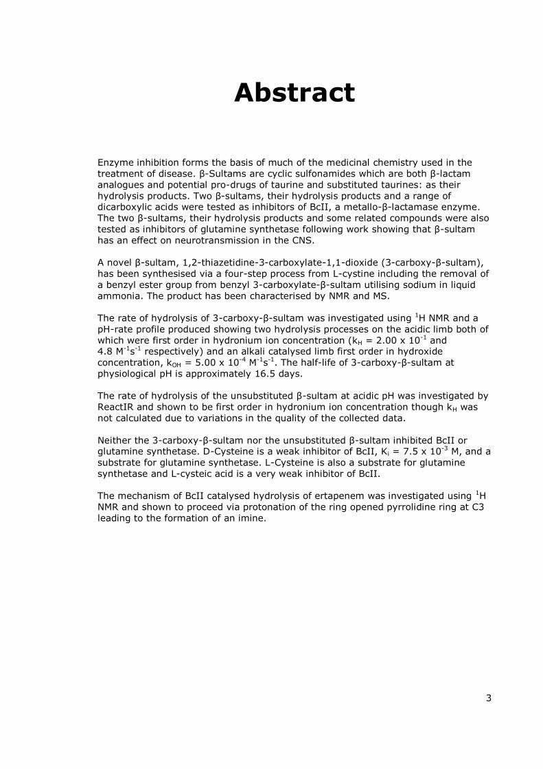
Enzyme inhibition forms the basis of much of the medicinal chemistry used in the treatment of disease. -Sultams are cyclic sulfonamides which are both -lactam analogues and potential pro-drugs of taurine and substituted taurines: as their hydrolysis products. Two -sultams, their hydrolysis products and a range of dicarboxylic acids were tested as inhibitors of BcII, a metallo- -lactamase enzyme. The two -sultams, their hydrolysis products and some related compounds were also tested as inhibitors of glutamine synthetase following work showing that -sultam has an effect on neurotransmission in the CNS. A novel -sultam, 1,2-thiazetidine-3-carboxylate-1,1-dioxide (3-carboxy- -sultam), has been synthesised via a four-step process from L-cystine including the removal of a benzyl ester group from benzyl 3-carboxylate- -sultam utilising sodium in liquid ammonia. The product has been characterised by NMR and MS. The rate of hydrolysis of 3-carboxy- -sultam was investigated using 1H NMR and a pH-rate profile produced showing two hydrolysis processes on the acidic limb both of which were first order in hydronium ion concentration (kH = 2.00 x 10⁻¹ and 4.8 M-1s-1 respectively) and an alkali catalysed limb first order in hydroxide concentration, kOH = 5.00 x 10⁻⁴ M-1s-1. The half-life of 3-carboxy- -sultam at physiological pH is approximately 16.5 days. The rate of hydrolysis of the unsubstituted -sultam at acidic pH was investigated by ReactIR and shown to be first order in hydronium ion concentration though kH was not calculated due to variations in the quality of the collected data. Neither the 3-carboxy- -sultam nor the unsubstituted -sultam inhibited BcII or glutamine synthetase. D-Cysteine is a weak inhibitor of BcII, Ki = 7.5 x 10⁻³ M, and a substrate for glutamine synthetase. L-Cysteine is also a substrate for glutamine synthetase and L-cysteic acid is a very weak inhibitor of BcII. The mechanism of BcII catalysed hydrolysis of ertapenem was investigated using 1H NMR and shown to proceed via protonation of the ring opened pyrrolidine ring at C3 leading to the formation of an imine.

4
Table of Contents
Abstract ........................................................................................................ 3
Table of Contents ......................................................................................... 4
Acknowledgements ...................................................................................... 7
List of abbreviations ..................................................................................... 8
Introduction ............................................................................................... 11
1.1 Background and Aims of Research ...................................................................................................................12
1.2 Bacteria, Antibiotics and Resistance .................................................................................................................14
1.2.1 Bacteria ......................................................................................................................................................14
1.2.2 Antibiotics ..................................................................................................................................................17
1.2.3 Antibiotic Resistance ..................................................................................................................................23
1.3 β-Lactamase Enzymes .......................................................................................................................................28
1.4 Active Site Serine β-Lactamases; Classes A, C and D.........................................................................................32
1.5 Metallo-β-Lactamases; Class B .........................................................................................................................38
1.5.1 Mechanisms of Metallo-β-Lactamases ........................................................................................................40
1.5.2 Metallo-β-Lactamase from Bacillus cereus; BcII ..........................................................................................43
1.6 Inhibition of Class B Metallo-β-Lactamases ......................................................................................................46
1.7 β-Sultams..........................................................................................................................................................51
1.7.1 Structure ....................................................................................................................................................51
1.7.2 Synthesis ....................................................................................................................................................52
1.7.3 Reactivity....................................................................................................................................................60
1.7.4 Sulfonyl Compounds as Enzyme Inhibitors ..................................................................................................65
. . β-Sultams as Enzyme Inhibitors ..................................................................................................................67
1.8 Glutamine Synthetase ......................................................................................................................................71
1.8.1 The Roles and Regulation of Glutamic Acid in The Human Body .................................................................71
5
1.8.2 The Role of Glutamic Acid in Neurodegeneration .......................................................................................72
1.8.3 Glutamic Acid Analogues (and Associated Compounds) as Enzyme Inhibitors and CNS Drugs .....................73
. . β-Sultams as Pro-Drugs for Treating Neurodegenerative Diseases ..............................................................75
1.9 Mechanism of Ertapenem Hydrolysis by the Metallo-β-Lactamase enzyme BcII .............................................77
1.10 Instrumental Techniques – React-IR ..............................................................................................................79
Experimental .............................................................................................. 81
2.1 Synthesis ..........................................................................................................................................................82
2.1.1 General ......................................................................................................................................................82
2.1.2 Synthesis of 1,2-thiazetidine-1,1-dio ide (β-sultam) ...................................................................................83
2.1.3 Synthesis of 1,2-thiazetidine-3-carboxylate-1,1-dioxide (3-carboxy-β-sultam) ...........................................85
2.1.4 Attempted syntheses of 1,2-thiazetidine-3-carboxylate-1,1-dioxide (3-carboxy-β-sultam) [8] ....................89
2.1.5 Debenzylation of proline benzyl ester hydrochloride ..................................................................................91
2.1.6 Attempted cyclisation of 2-amino-3-chlorosulfonyl-propanoic acid ............................................................92
2.2 Kinetic studies ..................................................................................................................................................94
2.2.1 Solutions and Buffers..................................................................................................................................94
2.2.2 pH Measurements ......................................................................................................................................94
2.2.3 Determination of the Acid-Catalysed Rate of Hydrolysis of 1,2-thiazetidine-1,1-dio ide (β-sultam) using
ReactIR ................................................................................................................................................................95
2.2.4 Determination of the Rate Constants for the Hydrolysis of 1,2-thiazetidine-3-carboxylate-1,1-dioxide (3-
carboxy-β-sultam) using 1H NMR ........................................................................................................................95
2.3 Enzyme Studies .................................................................................................................................................96
2.3.1 Inhibition of BcII .........................................................................................................................................96
2.3.2 Inhibition of Glutamine Synthetase ............................................................................................................98
2.4 Mechanistic Study of BcII Catalysed Hydrolysis of Ertapenem ....................................................................... 101
2.4.1 Solutions and Buffers................................................................................................................................ 101
2.4.2 Mechanistic studies .................................................................................................................................. 101
Results and Discussion ............................................................................. 102
3.1 Synthesis ........................................................................................................................................................ 103
3.2 Kinetics ........................................................................................................................................................... 110
6
3.2.1 Overview of Hydrolysis ............................................................................................................................. 110
. . H drol sis of the Unsubstituted β-Sultam [3] ........................................................................................... 112
3.2.3 Hydrolysis of 3-Carboxy-β-Sultam ............................................................................................................. 118
3.3 Inhibition Studies ........................................................................................................................................... 125
3.3.1 Inhibition of BcII ....................................................................................................................................... 125
3.3.2 Inhibition of Glutamine Synthetase .......................................................................................................... 134
3.4 Mechanism of Ertapenem Hydrolysis by BcII.................................................................................................. 139
Conclusion ................................................................................................ 145
Bibliography ............................................................................................. 147
Total word count = 30036

7
Acknowledgements
I would like to offer my sincere thanks to Prof. Mike Page for all his expertise and
advice throughout my project and for the opportunity to learn so much about his
field of research and also about myself. I would also like to thank Prof. Andy Laws for
all his encouragement over the last three years and for giving up so much time to
support me during the tough times. I would never have got to this point without
them.
My thanks also go to everyone in IPOS for their assistance and cheerful
conversations in the lab. I would particularly like to thank Dr Marcus Chadha for all
his help with mass specs and HPLC and also Dharmit Mistry for providing BcII and for
sharing his knowledge of the biological aspects of my work.
For his help with all things NMR I would like to thank Dr Neil McLay, and for all her
help and support over the past five years I would also like to thank Dr Fiona Ordway.
I owe my gratitude also to all the members of my research office (past and present)
who have been fabulous colleagues; particularly Heidi Joao, Amy Monnington,
Dharmit Mistry (again) and a special mention for Sam Bullock without whom I
guarantee I would now be insane.
Away from the university I would like to express my thanks to my family; my Mom,
my Pops and my sister Phil who have all listened to me rant at one time or another. I
have never been the easiest person to live with, but over the term of this degree I
have at times been impossible. My two beautiful daughters, Becki and Natasha, have
put up with my many moods and absences, more or less graciously and without fuss,
throughout this and my undergraduate degree. So for all the really hard times: Bex,
Taz; I am truly sorry.
And so finally to my amazing husband; Sut, I don’t know where I would be without
you! Thank you for all of your support, for being there for me through all of my highs
and extreme lows, for always knowing the right thing to say and when sometimes
saying nothing at all was a much safer option. We did it babes; here’s to the future!
8
List of abbreviations
6-APA 6-Aminopenicillanic acid
7-ACA 7-Aminocephalosporanic acid
Ac Acetyl
ADP Adenosine diphosphate
Ala Alanine
ALS Amyotrophic lateral sclerosis
ATP Adenosine triphosphate
ATR Attenuated total reflectance
Bn Benzyl
CNS Central nervous system
COSY Correlation spectroscopy
d.e. Diastereomeric excess
dec. Decomposed
DEPT Distortionless enhancement by polarization transfer
DMSO Dimethylsulfoxide
DNA Deoxyribonucleic acid
e.e. Enantiomeric excess
EDTA Ethylenediaminetetracetic acid
ESI-MS Electrospray ionization mass spectrometry
Et Ethyl
FTIR Fourier transform infrared
GABA -Amino butyric acid
Gln Glutamine
Glu Glutamic acid (or glutamate)
GS Glutamine synthetase

9
HMBC Heteronuclear multiple bond correlation
HSQC Heteronuclear single quantum coherence
HPLC High performance liquid chromatography
i-Bu Isobutyl
IC50 Half maximal inhibitory concentration (the concentration of inhibitor
required to give half the miximal rate)
i-Pr Isopropyl
IR Infrared
J Coupling constant
k0 Uncatalysed rate constant (spontaneous hydrolysis)
kA- Rate constant for hydrolysis catalysed by basic form of buffer
kbuf Rate constant for hydrolysis catalysed by both forms of buffer
kcat First order rate constant for the breakdown of ES
kcat/KM Second order rate constant for enzyme activity
kHA rate constant for hydrolysis catalysed by acidic form of buffer
Ki Rate constant for enzyme inactivation (the concentration of inhibitor
required to produce half maximum inhibition)
kint First order rate constant at zero buffer concentration
KM The Michaelis constant (the substrate concentration at which the
reaction rate is half of Vmax)
kobs Observed first order rate constant
LDA Lithium diisopropylamide
LDH Lactate dehydrogenase
Me Methyl
MES 2-(N-morpholino)ethanesulfonic acid
MRSA Methicillin resistant Staphylococcus aureus
NAD+ Nicotinamide adenine dinucleotide
NADH Nicotinamide adenine dinucleotide, reduced form
NAG N-Acetylglucosamine
10
NAM N-Acetylmuramic acid
n-Bu Butyl
NMR Nuclear magnetic resonance
n-Pr Propyl
pD Negative logarithm of deuterium ion concentration
PEP Phospho(enol)pyruvate
Ph Phenyl
Pi Inorganic phosphate
PK Pyruvate kinase
pKa Logarithm of acid dissociation constant
ppm Parts per million
PTSA Para-toluenesulfonic acid
SG Specific gravity
ssDNA Single stranded DNA
TBDMS tert-butyldimethylsilyl
t-Bu Tertiary-butyl
THF Tetrahydrofuran
TS Transition state
UV-Vis Ultra violet and visible
VISA Vancomycin intermediate Staphylococcus aureus
Vmax Maximal rate of the enzyme catalysed reaction
VRE Vancomycin resistant Enterococcus
VRSA Vancomycin resistant Staphylococcus aureus

11
Introduction

Introduction
12
1.1 Background and Aims of Research
Since antibiotics went into general use in the mid-twentieth century bacteria have
been developing strategies to fight against them so that today antibiotic resistance is
a major global health concern.1 Whilst antibiotics are still useful in the fight against
the majority of bacterial infections some strains of bacteria have evolved resistance
strategies so that they can no longer be controlled or killed by drugs which were
once effective against them. The rate of development of new antibiotics has now
been overtaken by the increase in bacterial resistance so that infections such as
pneumonia, tuberculosis and post-operative wound infections are becoming
increasingly resistant to current therapies and causing an increase in previously
preventable fatalities.2
One of the most commonly used classes of drugs for treating bacterial infections is
the -lactams which includes penicillins (core structures shown in Scheme 3, page
18). Following years of misuse of antibiotics and natural evolution some bacteria
have been able to develop innovative strategies for surviving against these drugs.
One of the most common forms of resistance to -lactam antibiotics is the
production of an enzyme which inactivates them: -lactamase.3 There are two main
types of -lactamase enzyme; the most common type are those with a serine-
residue at the active site with the rest being metallo-enzymes with one or two zinc
ions at the active site. One way of dealing with resistance to -lactam antibiotics is
to administer the drug with a second compound or co-drug which acts as a
-lactamase inhibitor. There are a number of clinically available inhibitors for serine
enzymes; the metallo-enzymes however are less well-studied and the current
clinically available serine-enzyme inhibitors are inactive against them.
The development of a compound which will inhibit metallo- -lactamase enzymes was
the inspiration for this project. Work carried out by previous members of the
research group has shown that substituted -sultams act as inhibitors of a range of
serine- -lactamase enzymes.4 In order for a compound to be of use as a drug an
understanding of its stability and reactivity at physiological pH is required. The aim
of this work was firstly to synthesise a new -sultam and to determine its inhibition
parameters. Secondly, work was carried out to investigate the reactivity of this new
compound and the parent molecule. The final part of this project was to look at the
inhibition of a metallo- -lactamase enzyme by a range of dicarboxylic acids.
In a second project the -sultams synthesised as metallo- -lactamase inhibitors
were tested as inhibitors of glutamine synthetase. Glutamine synthetase is an
Introduction
13
enzyme dependent on magnesium or manganese ions and found in the central
nervous system. Glutamine synthetase has an essential role in the regulation of
glutamate concentration within the brain and since glutamate is an excitatory
neurotransmitter the enzyme also has a vital role in regulating neurotransmission.
The unsubstituted -sultam, 1,2-thiazetidine-1,1-dioxide, has been shown to
influence the effect alcohol has on the brain and so its effect on enzymes involved in
neurotransmission was of interest.
The final project in this thesis is an investigation into the mechanism of hydrolysis of
a -lactam antibiotic, ertapenem, catalysed by the metallo- -lactamase enzyme,
BcII. There are ambiguities in the literature as to the site of protonation of the
pyrrolidine ring following -lactam ring opening. This investigation aimed to confirm
which of the proposed mechanisms occurs.
Introduction
14
1.2 Bacteria, Antibiotics and Resistance
1.2.1 Bacteria
There are two main types of cell; bacteria are prokaryotic cells whereas humans,
animals, plants and fungi are made up of eukaryotic cells. Prokaryotes are simple
cells with DNA, proteins and other water soluble components all held within one
membrane. In eukaryotes the separate components are held within separate
membranes within the outer cell membrane. Prokaryotes, plant cells and fungal cells
also have a cell wall, whereas animal and human cells do not. Plant cell walls are
made of cellulose, hemicellulose and pectin; fungus cell walls are made of chitin and
bacterial cell walls are made of a layer of peptidoglycan. This peptidoglycan cell wall
is the structural difference exploited by -lactam antibiotics; for this reason it will be
looked at in more detail.
The cell wall in all bacteria consists of layers of polysaccharide and protein/peptide
which varies in thickness between Gram positive and Gram negative strains. The
initial designation of bacteria as Gram positive or Gram negative was based on the
observations of Christian Gram in 1884.5 In studying the tissues of lungs taken from
people who had died of pneumonia he wanted to be able to study the bacterial cells
microsopically and so developed a new staining technique which selectively stained
only certain strains of bacteria. Those which were stained were deemed Gram
positive whereas those which resisted staining were Gram negative.
After the discovery of this considerable difference in bacterial cell biology much work
was done to establish exactly what caused the differential staining of the various
strains of bacteria. It wasn’t until the 1950s though that the composition of cell walls
in Gram positive and Gram negative bacteria was studied in any detail. One paper of
particular interest, published in the Journal of General Microbiology in 1956, studied
more than 60 strains of Gram positive bacteria and showed that, in most cases, the
cell wall was made up predominantly of four sugars: glucose, galactose, glucosamine
and galactosamine; and four amino acids: alanine, glutamic acid, lysine and aspartic
acid.6 By the 1960s the structure of both Gram positive and Gram negative bacteria
had been studied in much more detail, in particular by electron microscopy, and the
similarities and specific differences between the two types of bacteria was well
established.7

Introduction
15
In Gram positive bacteria the cell wall is made of multiple peptidoglycan layers up to
25 nm thick; in Gram negative bacteria there is only a single peptidoglycan layer, of
around 3 nm, sandwiched between an inner and outer lipid bilayer (Figure 1).
Figure 1 Cell wall structures in Gram positive and Gram negative bacteria
In both cases the polysaccharide is a NAG-NAM (N-acetylglucosamine –
N-acetylmuramic acid) linear chain of the two alternating amino sugars.8 These
glycan chains are then linked together by peptide chains. Each glycan strand is
initially synthesised with short amino acid chains attached to each N-acetylmuramic
acid. These chains are then joined by a DD-transpeptidase enzyme which cleaves
one amino acid from a chain ending with D-Ala-D-Ala residues and attaches this to
either a lysine residue in Gram negative bacteria or a pentaglycine bridge on a lysine
residue in Gram positive bacteria (see Scheme 1).

Introduction
16
D-ala-D-ala of first peptide
D-ala
acylated enzyme
second peptide
cross-linked peptides
Scheme 1 DD-transpeptidase enzyme action in cross-linking peptidoglycan
chains
This cross-linking between amino acids in different peptide chains results in a 3-
dimensional structure that is strong and rigid as shown diagramatically in Figure 2.9
Since bacteria reproduce by self-replication, in order to create a new bacterium it is
necessary to replicate all the features of the cell, including the cell wall.
Figure 2 Structure of peptidoglycan cell wall. Shows the cross-linking of oligosaccharide chains ( -linked NAG-NAM repeats) by tetrapeptides attached to the lactic acid group of NAM via an amide bond
Introduction
17
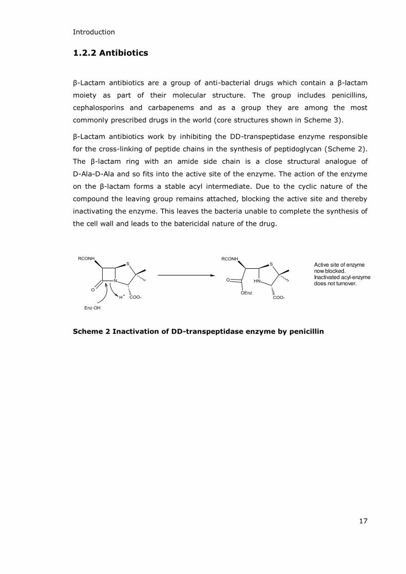
1.2.2 Antibiotics
-Lactam antibiotics are a group of anti-bacterial drugs which contain a -lactam
moiety as part of their molecular structure. The group includes penicillins,
cephalosporins and carbapenems and as a group they are among the most
commonly prescribed drugs in the world (core structures shown in Scheme 3).
-Lactam antibiotics work by inhibiting the DD-transpeptidase enzyme responsible
for the cross-linking of peptide chains in the synthesis of peptidoglycan (Scheme 2).
The -lactam ring with an amide side chain is a close structural analogue of
D-Ala-D-Ala and so fits into the active site of the enzyme. The action of the enzyme
on the -lactam forms a stable acyl intermediate. Due to the cyclic nature of the
compound the leaving group remains attached, blocking the active site and thereby
inactivating the enzyme. This leaves the bacteria unable to complete the synthesis of
the cell wall and leads to the batericidal nature of the drug.
Enz-OH
Active site of enzyme now blocked.Inactivated acyl-enzyme does not turnover.
Scheme 2 Inactivation of DD-transpeptidase enzyme by penicillin

Introduction
18
Penicillin
Carbapenem PenemOxapenem
Monobactam Nocardicin
Carbacephem
Cephalosporin
Oxacephem
Scheme 3 Structures of the main classes of β-lactam antibiotics
The antibiotic action of Penicillium mould was first noticed by Joseph Lister in 187110
and the presence of an antimicrobial agent was correctly identified by Alexander
Unfused -
lactam rings
-lactam rings
fused to six-
membered
rings
-lactam rings
fused to five-
membered
rings
Introduction
19
Fleming in 192911; despite these early observations the pure compound was not
extracted until 1940.12 Fermentation methods were utilised to produce useful
amounts of the compound and it went into clinical usage in the mid-1940s; since
then it has saved countless lives. In order to produce penicillin on a scale large
enough for the antibiotic to go into wide scale use a large amount of research was
conducted to optimise the process. One of the first improvements was the movement
from Penicillium notatum (the strain from which the first penicillin compound was
isolated) to Penicillium chrysogenum, which gave a much higher yield of bio-active
compound.13 Further improvements in the fermentation procedure have seen the
introduction of phenylacetic acid to the broth medium which gives a significant
increase in the yield of penicillin.14 Precise fermentation conditions have gradually
been optimised so that fermenters with a capacity of 100 000 to 300 000 gallons can
now be employed to produce 40-50 grams of penicillin per litre of broth every 5-8
days. The maintenance of pH and temperature and the rates of aeration and
agitation are all factors which influence the potential yield; the ideal conditions
generally employed are a pH of 6.0 with the temperature maintained at 25 oC.15
These improvements in manufacturing technology have increased the efficiency of
penicillin production from 70 % to more than 90 % in 50 years and have decreased
production costs from ~$350 per kilogram in 1950 to ~$15 per kilogram in 2000.16
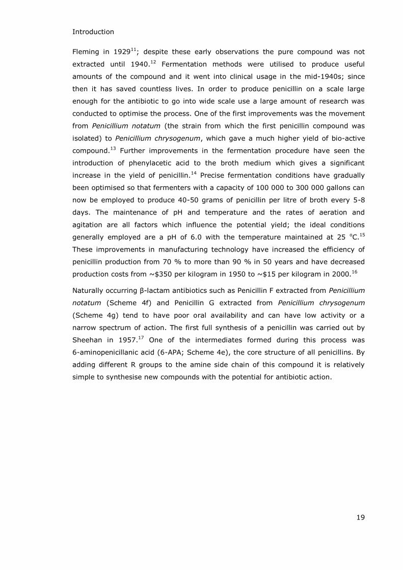
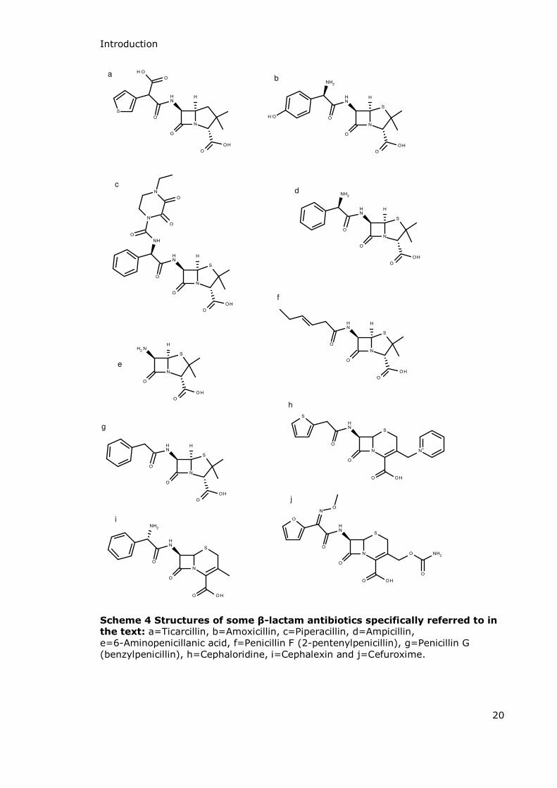
Naturally occurring -lactam antibiotics such as Penicillin F extracted from Penicillium
notatum (Scheme 4f) and Penicillin G extracted from Penicillium chrysogenum
(Scheme 4g) tend to have poor oral availability and can have low activity or a
narrow spectrum of action. The first full synthesis of a penicillin was carried out by
Sheehan in 1957.17 One of the intermediates formed during this process was
6-aminopenicillanic acid (6-APA; Scheme 4e), the core structure of all penicillins. By
adding different R groups to the amine side chain of this compound it is relatively
simple to synthesise new compounds with the potential for antibiotic action.
Introduction
20
a
h
f
i
d
j
g
b
c
e
Scheme 4 Structures of some β-lactam antibiotics specifically referred to in the text: a=Ticarcillin, b=Amoxicillin, c=Piperacillin, d=Ampicillin, e=6-Aminopenicillanic acid, f=Penicillin F (2-pentenylpenicillin), g=Penicillin G (benzylpenicillin), h=Cephaloridine, i=Cephalexin and j=Cefuroxime.

Introduction
21
One of the first major breakthroughs in this area came in 1961 when Doyle, Nayler
and Smith successfully used this technique to synthesise ampicillin, (6[D(-)-α-
aminophenylacetamido]penicillanic acid, Scheme 4d) a compound which was later
shown to have broad spectrum activity against both Gram positive and Gram
negative bacteria.18 One of the main advantages of this compound over previous
penicillins was its oral bioavailability, a property many penicillins lack due to the acid
catalysed hydrolysis of the -lactam ring in the stomach.
Since then numerous new compounds have been synthesised and tested for
antibacterial action. Some notable successes are the development of carbenicillin in
1967, amoxicillin in 1972 (Scheme 4b), piperacillin in 1978 (Scheme 4c) and
ticarcillin in 1973 (Scheme 4a).19-22 Changing the side chains on the penicillin ring
can give a number of advantages: increased stability to hydrolysis, a broader
spectrum of activity, increased bioavailability, increased activity and increased
resistance to inactivation by -lactamase enzymes.
The second family of naturally occurring -lactam antibiotics is the cephalosporins.
The first of these to be reported was Cephalosporin C from Cephalosporium
acremonium, a fungus which also produces Penicillin N.23 From this compound the
cephalosporin nucleus, 7-aminocephalosporanic acid (7-ACA), was isolated and
shown to be analogous to the penicillin nucleus 6-APA (Scheme 5). The main
differences between the two are the replacement of the 6-APA thiazolidine ring with
a modified dihydrothiazine ring in 7-ACA which also has a substituted methyl acetate
group at the 3 position on the thiazine ring. As with 6-APA, modification of the 7-ACA
amine side chain and methyl acetate group has resulted in the production of a
number of useful antibiotic agents; the first was cephalothin in 196424 and other
notable compounds are cephaloridine (1964)25, cephalexin (1967)26 and cefuroxime
(1976)27 (Scheme 4h, i and j respectively).
6-APA 7-ACA
Scheme 5 Core structures of penicillins and cephalosporins

Introduction
22
Carbapenems are yet another group of -lactam antibiotics based on natural
products. The first carbapenem, thienamycin (Scheme 6a), was isolated from
Streptomyces cattleya in 1978.28,29 Other compounds based on the structure of
thienamycin have since been synthesised and shown to have excellent antibacterial
activity, particularly against a number of bacteria resistant to penicillins and
cephalosporins. Imipenem (Scheme 6b) was the first synthetic carbapenem and was
initially synthesised by Kropp et al and reported in 1985.30 In the 1990s both
meropenem31 and panipenem32 were developed and these were followed by the
introduction of ertapenem (Scheme 6c),33 biapenem34 and doripenem35 during the
first part of the 21st Century.
a
b
c
Scheme 6 Structures of some carbapenems mentioned in the text
(a = Thienamycin, b = Imipenem, c = Ertapenem)
Introduction
23
1.2.3 Antibiotic Resistance
Staphylococcus aureus are bacteria found naturally on the skin and in the respiratory
tract and are usually harmless. In certain circumstances however, Staphylococci can
cause skin infections such as impetigo, respiratory infections, pneumonia and
meningitis. The development of penicillin had initially meant that these infections
could be treated effectively, but in 1944 a sample of Staphylococcus aureus was
discovered which was not killed by penicillin.36 This was the first instance of clinically
relevant antibiotic resistance and occurred only months after penicillin went into
general use. The first methicillin resistant Staphylococcus aureus (MRSA) was
detected in Britain in the 1960s and the strain’s resistance to drugs has continued to
evolve; it now shows resistance to a range of penicillins and cephalosporins as well
as non- -lactam antibiotics such as glycopeptides. Various different classes of
antibiotics have been introduced over the past 100 years and for most of these some
bacterial resistance mechanisms have now evolved (Figure 3).
There are four main mechanisms of antibiotic resistance present in bacteria. The best
known is antibiotic modification whereby the target enzyme remains sensitive to the
antibiotic but the bacterium produces another enzyme which can inactivate it prior to
reaching the target. Growing numbers of bacteria demonstrate this ability,
particularly via the production of -lactamase enzymes.37
A second mechanism conferring resistance is the modification of the target site.
Structural changes in the target molecule prevent the drug from interacting with it
whilst it still maintains the ability to carry out its primary function. Resistance to
cephalosporins by some species has been demonstrated by this mechanism via
alterations to the structure of their transpeptidase enzymes (often referred to as
penicillin binding proteins).38
Some bacteria have demonstrated resistance via alteration of the metabolic
pathway. Sulfonamides are drugs which work by inhibiting dihydropteroate synthase,
an enzyme involved in the synthesis of folic acid. Resistance to these drugs has been
developed by some bacteria which have evolved the ability to take up folic acid from
the environment, by-passing the need to synthesise it themselves.39
The last of the four main methods of antibiotic resistance is the ability of the bacteria
to reduce accumulation of the drug within the cell. This may occur by the reduction

24
Discovery references: Sulfonamides,40 penicillins,12 nitrofuran,41 chloramphenicol,42 tetracyclines,43 streptogramins,44 glycopeptides,45 ansamycins,46 nitroimidazoles,47 quinolones,48 trimethoprim,49 oxazolidinones,50 lipopeptides51. Resistance references: Penicillin,52 sulfonamides,53 streptomycin,54 macrolide,55 methicillin,56 quinolones,57 tetracycline,58 aminoglycoside,59 vancomycin,60 fluoroquinolones,61 linezolid.62
Figure 3
Colour coded diagram
of drug discovery above
arrow and drug resistance below arrow

Introduction
25
of cytoplasmic membrane permeability to the drug, preventing it entering the cell, or
by the enhanced expression of efflux pumps which actively remove drug molecules
from the cytoplasm, a method of resistance demonstrated in bacteria resistant to
fluoroquinolones.63
The main mode of -lactam antibiotic resistance in bacteria is via the production of a
-lactamase enzyme, a modified transpeptidase enzyme which interacts reversibly
with the drug. The hydrolysis of the -lactam ring renders the antibiotic ineffective.
Some bacteria are resistant to only selected penicillins but recently strains with
resistance to multiple drugs have been on the rise. The primary reason for the
spread of bacterial antibiotic resistance is horizontal gene transfer. There are three
main methods of horizontal gene transfer (see Table 1) all allowing antibiotic
resistance genes (along with other genetic material) to be transferred from one
bacterial species to another. Whilst vertical gene transfer (the passing of genetic
information from parents to offspring during traditional reproduction) is the focus of
the majority of genetics work regarding genetic mutations in mammals and plants,
the transfer of genes by horizontal transfer methods is an extremely common form
of genetic transfer in bacteria.64
Mode of Transfer
Type of resistance Description
Vertical Pre-existing A small number of cells contain mutant DNA which confers resistance. Antibiotics kill sensitive bacteria within the colony leaving the resistant cells to multiply unhindered.
Vertical Mutational The introduction of a drug to a bacterial colony forces bacteria to mutate and adapt leading to drug tolerance or dependence.
Horizontal Transmission A bacterium can acquire genetic material which confers resistance in three ways. Transformation – the genetic information moves between cellular DNA. Transduction – the genetic information is transferred by a virus. Conjugation – a “tunnel” is formed between cells through which genetic information may pass (see Figure 4).
Table 1 Details of the modes of antibiotic resistance in bacteria
Introduction
26
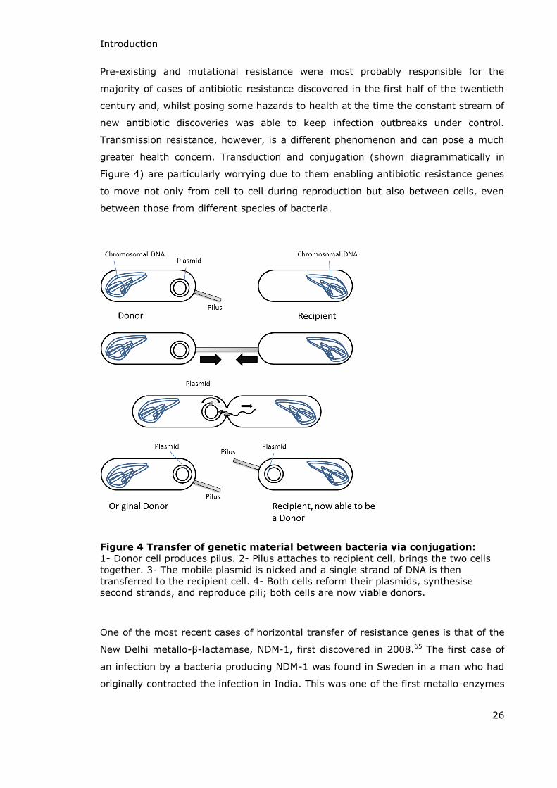
Pre-existing and mutational resistance were most probably responsible for the
majority of cases of antibiotic resistance discovered in the first half of the twentieth
century and, whilst posing some hazards to health at the time the constant stream of
new antibiotic discoveries was able to keep infection outbreaks under control.
Transmission resistance, however, is a different phenomenon and can pose a much
greater health concern. Transduction and conjugation (shown diagrammatically in
Figure 4) are particularly worrying due to them enabling antibiotic resistance genes
to move not only from cell to cell during reproduction but also between cells, even
between those from different species of bacteria.
Figure 4 Transfer of genetic material between bacteria via conjugation:
1- Donor cell produces pilus. 2- Pilus attaches to recipient cell, brings the two cells together. 3- The mobile plasmid is nicked and a single strand of DNA is then transferred to the recipient cell. 4- Both cells reform their plasmids, synthesise second strands, and reproduce pili; both cells are now viable donors.
One of the most recent cases of horizontal transfer of resistance genes is that of the
New Delhi metallo- -lactamase, NDM-1, first discovered in 2008.65 The first case of
an infection by a bacteria producing NDM-1 was found in Sweden in a man who had
originally contracted the infection in India. This was one of the first metallo-enzymes

Introduction
27
shown to confer resistance to carbapenems, though some serine-carbapenemases
had previously been described. Since that first discovery the enzyme has been
detected in over fifteen countries around the world, from India and Sweden to the
USA, UK and Japan among others.66
The first bacteria found to produce NDM-1 was Klebsiella pneumoniae but since then
horizontal transfer has led to the enzyme being isolated from various other bacteria
including some Escherichia coli, Enterobacter cloacae and Salmonella enterica
strains. This rapid spread of antibiotic resistance across countries and bacterial
strains is of real worldwide concern.

Introduction
28
1.3 β-Lactamase Enzymes
The first “penicillinase” enzyme was reported in a paper in Nature by Abraham and
Chain in 1940, three years before penicillin itself was in clinical use.52 At the time it
was not seen as a medical problem since it was discovered in Bacillus (Escherichia)
coli whereas penicillin was at the time used exclusively for Staphylococcus infections.
Kirby isolated the first -lactamase enzyme (or penicillin inactivator) from a colony of
Staphylococcus aureus in 1944 and there are now more than 850 distinct
-lactamase enzymes known to be produced by a wide range of both pathogenic and
non-pathogenic bacteria.36,67
In order to more easily understand and study the ever-growing number of
-lactamase enzymes they are classified according to either their specific amino acid
sequence or their affinities regarding substrates and inhibitors. The Ambler classes,
A to D, classify the enzymes according to their protein structure and the amino acid
sequence at the active site.68 Classes A, C and D all have serine amino acid residues
at the active site whereas class B are metallo-enzymes with one or two zinc-ions at
the active site. A more recent classification (Bush-Jacoby-Medeiros classes) was
established in 1995 as a way of classifying the enzymes according to their
functionality.3 This classification scheme again separates the enzymes into four main
classes, 1 to 4, but this time according to their preferred substrates (penicillin,
cephalosporin, carbapenem etc.) and inhibitors. A simplified description of the two
classification schemes and the correlations between them can be found in Table 2.
Whilst the two classification schemes may differ in their distribution of many of the
serine- -lactamase enzymes, Ambler Class B and Bush-Jacobi-Medeiros Class 3 both
contain all of the metallo- -lactamases. For reasons of clarity and simplicity only the
Ambler Classes will be referred to for the remainder of this thesis.
When Ambler originally proposed the first classification scheme for the -lactamase
enzymes they were separated into two main groups, A and B, dependent on their
mechanism of action.
Introduction
29
Ambler Class Bush-Jacoby-Medeiros Class
Typical Substrates
Known Inhibitors
Representative Enzymes
A 2a Penicillins Penicillinases from Gram positive bacteria
2b Penicillins, Cephalosporins
Clavulanic acid
TEM-1, TEM-2, SHV-1
2be Penicillins, Cephalosporins, Monobactams
Clavulanic acid
TEM-3 to TEM-26, SHV-2 to SHV-6, K1 from Klebsiella oxytoca
2br Penicillins Clavulanic acid
TEM-30 to TEM-36, TRC-1
2c Penicillins, Carbenicillin
PSE-1, PSE-3, PSE-4
2e Cephalosporins Clavulanic acid
Inducible Cephalosporinases from Proteus vulgaris
2f Penicillins, Cephalosporins, Carbapenems
Clavulanic acid
NMC-A from Enterobacter cloacae, Sme-1 from Seratia marcescens
B 3 εost -lactams including carbapenems
EDTA L1 from Xanthomonal maltophilia, CcrA from Bacteroides fragilis, BcII from Bacillus cereus
C 1 Cephalosporins AmpC from Gram negative bacteria, P99 from Enterobacter cloacae, MIR-1
D 2d Penicillins, Cloxacillin
OXA-1 to OXA-11, PSE-2
(not assigned)
4 Penicillins Penicillinase from Pseudomonas cepacia
Table 2 Comparison of the two classification systems for β-lactamase
enzymes including typical substrates and representative enzymes (adapted from Bush, Jacoby and Medeiros 3)
Class A -lactamases have a serine amino acid residue at the active site. In general
they all hydrolyse penicillins, though a few are capable of hydrolysing
cephalosporins, carbapenems and monobactams as well. The majority of enzymes in
this class are inhibited by clavulanic acid, sulbactam and tazobactam (structures

Introduction
30
shown in Scheme 12, page 36), three clinically active -lactam based inhibitors. The
Class A -lactamases are the most common source of resistance to -lactam
antibiotics and can be secreted by pathogenic bacteria such as Klebsiella
pneumoniae, Escherichia coli and Staphylococcus aureus as well as non-pathogenic
bacteria such as Bacillus cereus.
Class B -lactamases are dependent on divalent metal ions, usually zinc, for their
activity. They have a broader spectrum of activity than the serine enzymes and
effectively hydrolyse most -lactams including penicillins, cephalosporins,
monobactams and carbapenems. They are not inhibited by any of the -lactam
based inhibitors of Class A enzymes and the most effective inhibitors of Class B
enzymes (chelating agents such as EDTA) are not suitable for use in vivo.
The classification scheme has now been extended; Class C -lactamases (sometimes
referred to as cephalosporinases) were added to the Ambler classification system in
1981.69 Although they are serine enzymes like Class A and are structurally similar to
them they have noticeably different amino acid sequences and are more efficient at
hydrolysing cephalosporins than penicillins. The best known enzyme in this class is
AmpC produced by Gram negative bacteria from the Legionella species and
Escherichia coli and Gram positive Mycobacteria. The production of this enzyme is
encoded on a gene which occurs on transmissible plasmids, transferred to other
bacteria by conjugation, and so there is a possibility of the enzyme being produced
by any species of bacteria.68
The final group of enzymes in the Ambler classification system are the Class D
enzymes, with this category being first suggested for the enzyme OXA-1 in 1987.70
Jacoby et al. also suggested the addition of a fourth class to the Ambler system in
1988 when they sequenced a new -lactamase, PSE-2 (a carbenicillin hydrolysing
enzyme first isolated from Pseudomonas aeruginosa).71 Based on the lack of
structural similarity between this enzyme and enzymes from the established classes
A, B and C they suggested that PSE-2 had a distinct evolutionary origin along with
OXA-1 and that a new group of -lactamases, Ambler Class D, should be established
for them and similar enzymes.71 In contrast to the other classes of -lactamase, and
in particular Class A enzymes which have been known for many years, Class D is a
much newer, smaller and slightly overlooked class of -lactamases. Like Class A and
C they are active site serine enzymes but their primary structures are significantly
different to those in the other groups. Whilst Class D enzymes do hydrolyse standard
penicillins and cephalosporins, they are much more active against substrates from
the oxacillin class of -lactam antibiotics (Scheme 7) and are often given the
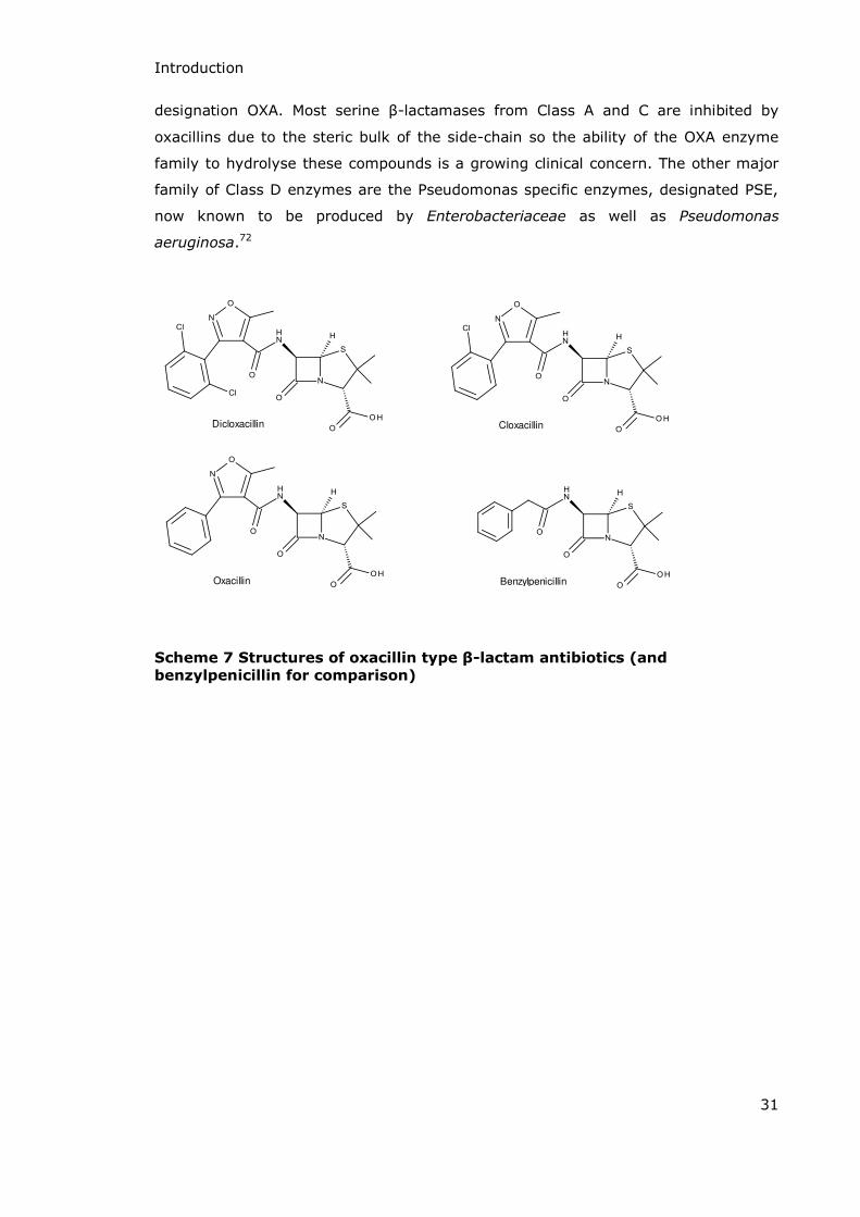
Introduction
31
designation OXA. εost serine -lactamases from Class A and C are inhibited by
oxacillins due to the steric bulk of the side-chain so the ability of the OXA enzyme
family to hydrolyse these compounds is a growing clinical concern. The other major
family of Class D enzymes are the Pseudomonas specific enzymes, designated PSE,
now known to be produced by Enterobacteriaceae as well as Pseudomonas
aeruginosa.72
Oxacillin
CloxacillinDicloxacillin
Benzylpenicillin
Scheme 7 Structures of oxacillin type β-lactam antibiotics (and
benzylpenicillin for comparison)
Introduction
32
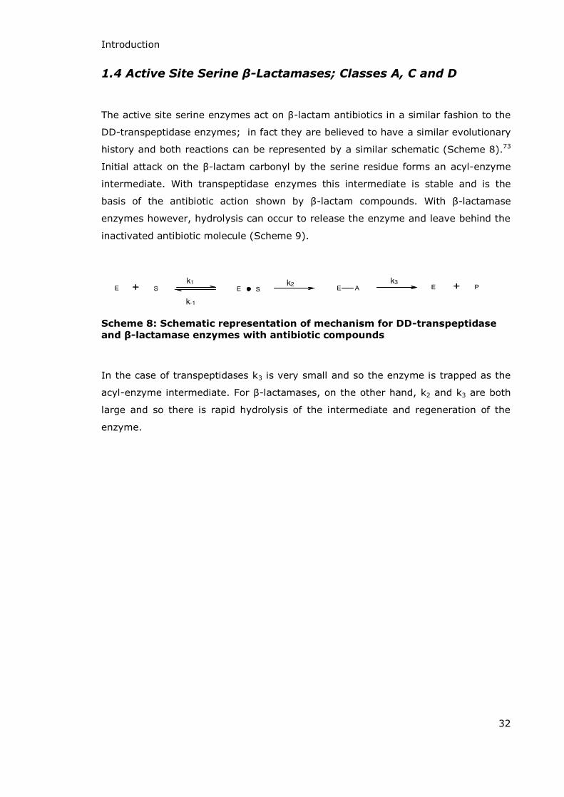
1.4 Active Site Serine β-Lactamases; Classes A, C and D
The active site serine enzymes act on -lactam antibiotics in a similar fashion to the
DD-transpeptidase enzymes; in fact they are believed to have a similar evolutionary
history and both reactions can be represented by a similar schematic (Scheme 8).73
Initial attack on the -lactam carbonyl by the serine residue forms an acyl-enzyme
intermediate. With transpeptidase enzymes this intermediate is stable and is the
basis of the antibiotic action shown by -lactam compounds. With -lactamase
enzymes however, hydrolysis can occur to release the enzyme and leave behind the
inactivated antibiotic molecule (Scheme 9).
k1
k-1
k2 k3
Scheme 8: Schematic representation of mechanism for DD-transpeptidase
and β-lactamase enzymes with antibiotic compounds
In the case of transpeptidases k3 is very small and so the enzyme is trapped as the
acyl-enzyme intermediate. For -lactamases, on the other hand, k2 and k3 are both
large and so there is rapid hydrolysis of the intermediate and regeneration of the
enzyme.

Introduction
33
..
k1
k2
k3
Scheme 9 Mechanism of benzylpenicillin hydrolysis by serine
β-lactamases 74-76
There are two main mechanisms for inhibition of serine- -lactamases: mechanism
based and transition state analogues. Mechanism based or suicide inhibition occurs
when a substrate analogue binds to the enzyme active site but, due to modifications
to the structure, an irreversible complex is formed often via reactions between the
enzyme and inhibitor involving carbonyl groups or imines. Transition state analogue
inhibitors work by mimicking the transition state of the normal substrate. Since
enzymes work by stabilising high energy transition state intermediates, transition
state mimics which do not undergo the normal reaction bind to the active site and so
block it. Mechanism based inhibitors for serine- -lactamases include clavulanic acid
(administered with amoxicillin, marketed as Augmentin), sulbactam (administered
with ampicillin, marketed as Unasyn) and tazobactam (administered with piperacillin,
marketed as Zozyn) (see Scheme 4, page 20 for structural details of the penicillins
and Scheme 12, page 36 for the structures of the inhibitor compounds).
The best known and probably most widely used mechanism based inhibitor is
clavulanic acid (Scheme 10).77 Clavulanic acid was first isolated from Streptomyces
clavuligerus by a research group at Beecham Pharmaceuticals in the mid-1970s.78 At
the time its fused -lactam structure was different to all known -lactams in that it
has an O-containing oxazolidine ring fused to the -lactam ring instead of the
S-containing thiazolidine ring and it does not possess the acyl-amino side chain
..

Introduction
34
found in penicillins and cephalosporins.77 The group carried out extensive
investigations on the compound and showed that, whilst it had little antibacterial
activity on its own (only weak inhibition of DD-transpeptidase enzymes is shown in
most cases), it was a potent inhibitor of the majority of -lactamases tested from
both Gram positive and Gram negative bacteria. Interestingly cephalosporinase type
enzymes were poorly inhibited as were the Bacillus cereus enzymes BcI and BcII.79
Whilst clavulanic acid shows very little antibacterial action on its own its ability to
inhibit -lactamase enzymes from Class A and C increases the activity of penicillin
drugs (usually amoxicillin or ticarcillin, structures shown in Scheme 4, page 20)
against -lactam resistant bacterial infections when they are prescribed in a
formulation together.
Scheme 10 Clavulanic acid (with numbered atoms)
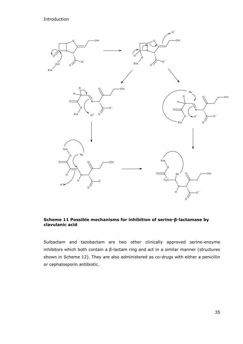
Numerous mechanisms have been put forward for the inactivation of -lactamase
enzymes by clavulanic acid80-82 with the two most credible being illustrated in
Scheme 11.83,84 Initially both mechanisms follow the same pathway. As a mechanism
based inhibitor clavulanic acid is recognised by the enzyme and initially turned over
like any other substrate. A serine residue forms an acyl-enzyme intermediate at C7
and the carbon-nitrogen bond is cleaved. The oxazolidine ring is then opened to form
a keto-imine. At this point there are two main proposals for the mechanism. The first
suggests protonation of the nitrogen and loss of hydrogen at C6 to form an enamine
which is then attacked by a second nucleophilic serine residue to form the
irreversibly inactivated complex. In the second proposal attack by a nucleophilic
amino acid residue at C5 leads to the alkylation at the active site which again
prevents further activity by the enzyme.81 The inactivation of the -lactamase
enzyme by clavulanic acid allows the coadministered amoxicillin to bind to the DD-
transpeptidase enzyme in the normal way, inactivating it and leading to cell death.
Introduction
35
Scheme 11 Possible mechanisms for inhibition of serine-β-lactamase by
clavulanic acid
Sulbactam and tazobactam are two other clinically approved serine-enzyme
inhibitors which both contain a -lactam ring and act in a similar manner (structures
shown in Scheme 12). They are also administered as co-drugs with either a penicillin
or cephalosporin antibiotic.

Introduction
36
Clavulanic acid
TazobactamSulbactam
Scheme 12 Selected serine-β-lactamase inhibitor structures
The second group of serine-enzyme inhibitors are the transition state analogues. A
wide range of structurally diverse boronic acids have been shown to act as reversible
inhibitors of serine- -lactamases.85-88 The reaction (overview in Scheme 13) involves
two separate binding steps with the slower of the two leading to inhibition. This slow
step involves a change in the protein conformation represented mechanistically as
Enz*.82
slow
Scheme 13 Mechanism of β-lactamase inhibition by boronic acids
Phosphonic acid derivatives are another family of compounds which have proven to
be useful as serine- -lactamase inhibitors.89 This time the reaction involves
phosphonylation of the enzyme and this phosphonylated enzyme has a tetrahedral
conformation. Although the mechanisms vary in detail between Class A and Class C
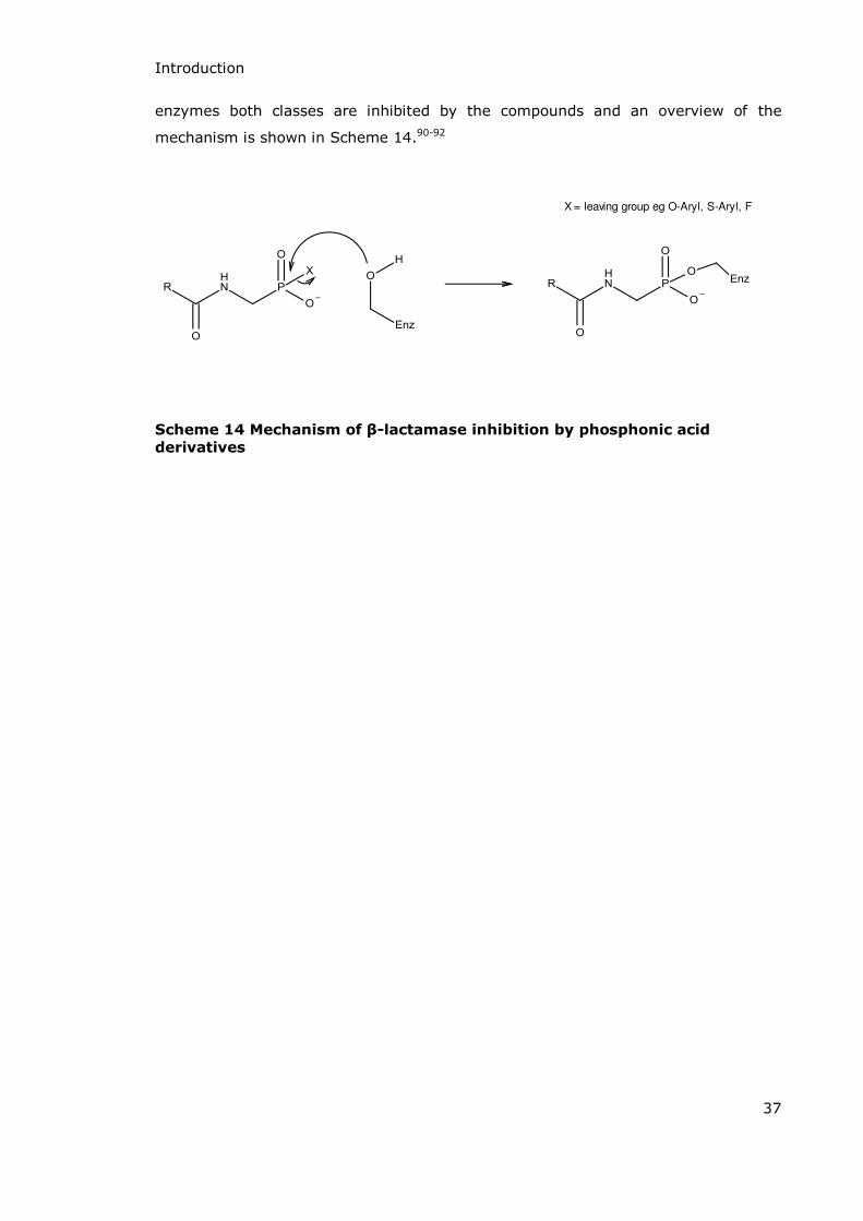
Introduction
37
enzymes both classes are inhibited by the compounds and an overview of the
mechanism is shown in Scheme 14.90-92
X = leaving group eg O-Aryl, S-Aryl, F
Scheme 14 Mechanism of β-lactamase inhibition by phosphonic acid
derivatives

Introduction
38
1.5 Metallo-β-Lactamases; Class B
Class B -lactamases are metallo-enzymes dependent on the coordination of bivalent
metal ions to demonstrate activity. In general cases the preferred metal is zinc but
there have been examples of cobalt, cadmium and manganese also conferring
activity in a variety of enzymes.93 The metallo- -lactamases can be further
separated into three subgroups, B1, B2 and B3; first suggested by Rasmussen and
Bush in 1997 the enzymes are grouped according to amino acid sequence and
substrate profile.94
Subclass B1 -lactamases are all approximately 28 kDa and have three histidine
residues participating in zinc and water binding at the active site. The subclass
includes BcII from Bacillus cereus (Figure 6a); CcrA from Bacteroides fragilis;95
IMP-1 from Pseudomonas aeruginosa, Klebsiella pneumoniae and Serratia
marscescens96 and BlaB from Chryseobacterium meningsepticum.97 This is the
largest of the three subgroups and is the best studied with crystal structures and
peptide sequences being available for a large number of the enzymes in the group as
well as kinetic data, substrate profiles and inhibition studies. All members of subclass
B1 require two zinc ions at the active site and they efficiently catalyse the hydrolysis
of almost all -lactam compounds including those which inhibit the serine enzymes
e.g. clavulanic acid and tazobactam. In Figure 5 are two images representing the
CcrA metallo- -lactamase enzyme from Bacteroides fragilis.98 The first image shows
the ribbon structure of the enzyme with α-helices shown in blue and -sheets in
yellow. The red spheres represent the zinc ions at the active site. In the second
image the active site of the enzyme is shown as the molecular surface. A model of a
benzylpenicillin molecule is shown in red, bound at the active site. The zinc ions are
represented by purple spheres and the water molecules by blue spheres. This image
shows the -lactam ring clearly positioned above the di-zinc bound water molecule.

Introduction
39
Figure 5 Representations of the metallo-β-lactamase enzyme produced by Bacteroides fragilis (as published by Concha et al)98
Subclass B2 -lactamases are similar to those from subclass B1 in size and amino
acid sequence but unlike the previous group they have a small substrate profile, only
being truly active against carbapenems. They require only one zinc ion for activity
and in many cases are actually inhibited by the binding of a second zinc.
Representative enzymes for this subclass are CphA from Aeromonas hydrophila99 and
Bacteroides fragilis (Figure 6b) and Sph-I from Serratia fonticola.100
Subclass B1 Subclass B2 Subclass B3
Figure 6 Representative enzymes from the three subclasses (a) BcII from Bacillus cereus, (b) CphA from Bacteroides fragilis and (c) FEZ-1 from Fluoribacter gormanii.101
Introduction
40
Subclass B3 is the most distinctive but least studied of the three classes. Whilst it
has a similarly broad substrate profile to Class B1 its structure and amino acid
sequence vary greatly from both subclasses B1 and B2. Enzymes currently assigned
to subclass B3 are FEZ-1 from Fluoribacter gormanii (Figure 6c),102 CAU-1 from
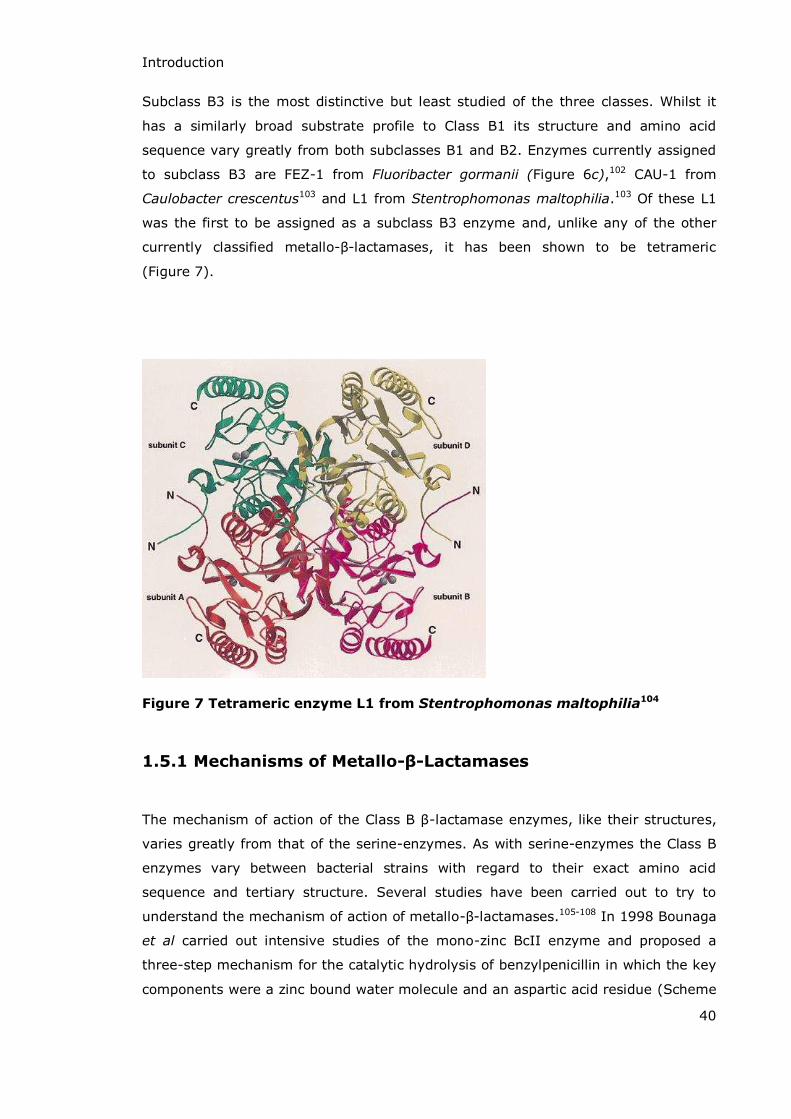
Caulobacter crescentus103 and L1 from Stentrophomonas maltophilia.103 Of these L1
was the first to be assigned as a subclass B3 enzyme and, unlike any of the other
currently classified metallo- -lactamases, it has been shown to be tetrameric
(Figure 7).
Figure 7 Tetrameric enzyme L1 from Stentrophomonas maltophilia104
1.5.1 Mechanisms of Metallo-β-Lactamases
The mechanism of action of the Class B -lactamase enzymes, like their structures,
varies greatly from that of the serine-enzymes. As with serine-enzymes the Class B
enzymes vary between bacterial strains with regard to their exact amino acid
sequence and tertiary structure. Several studies have been carried out to try to
understand the mechanism of action of metallo- -lactamases.105-108 In 1998 Bounaga
et al carried out intensive studies of the mono-zinc BcII enzyme and proposed a
three-step mechanism for the catalytic hydrolysis of benzylpenicillin in which the key
components were a zinc bound water molecule and an aspartic acid residue (Scheme

Introduction
41
15).109 In this proposal the zinc bound water molecule is deprotonated due to the
action of the zinc ion acting as a Lewis acid decreasing the pKa of the bound water.
The metal bound hydroxide ion acts as a nucleophile to attack the carbon of the -
lactam carbonyl group to form a tetrahedral intermediate and the zinc acts to
stabilise the negative charge formed on the carbonyl oxygen. An aspartic acid
carboxylate anion then deprotonates what was the zinc bound hydroxyl to form a
dianionic tetrahedral intermediate, again stabilised by the zinc ion. Protonation of the
amine leaving group by the aspartic acid as the C-N bond of the -lactam is broken
completes the hydrolysis.110 Whilst this mono-zinc BcII enzyme did show activity
against benzylpenicillin further studies have now shown that the di-zinc enzyme is
the one which shows most activity against -lactam antibiotics.
Asp 90Asp 90
Asp 90Asp 90
Scheme 15 Mechanism of benzylpenicillin hydrolysis by mono-zinc BcII109
All the metallo- -lactamases currently reported do have a striking number of
similarities, particularly the di-zinc enzymes. In almost all cases Zn1 is coordinated
to three histidine residues and a bridging water molecule in a tetrahedral
conformation. Zn2 is coordinated to the bridging water molecule and three other
amino acid residues with another molecule of water completing the trigonal

Introduction
42
bipyrimidal arrangement. These arrangements are shown in Figure 8 where the
4-coordinated Zn1 and 5-coordinated Zn2 in Bacillus cereus are clearly visible.
Figure 8 Dinuclear centre of Bacillus cereus111
More recently studies have shown that although some mono-zinc -lactamases are
catalytically active, for BcII the di-zinc form of the enzyme (as shown in Figure 8) is
the only one relevant to -lactam hydrolysis in vivo.112 In fact of the three subclasses
of metallo- -lactamase Class B1 (including BcII, BlaB, IMP and VIM) and B3 (L1,
FEZ-1, GOB-1 and THIN-B) both show maximum efficiency as di-zinc species but
Class B2 (CphA and Sfh-I) enzymes show a reduction in activity upon binding of a
second zinc ion.
The mechanism for the di-zinc metallo- -lactamase begins with nucleophilic attack of
the bridging hydroxide ion (Wat 1) on the carbonyl carbon resulting in a negatively
charged tetrahedral intermediate. The zinc bound water molecule (Wat 2) can then
donate a proton to the leaving nitrogen forming a hydroxide ion which replaces
Wat 1 in the now vacant site. The product (hydrolysed -lactam) can now dissociate
from the enzyme active site and, once Wat 2 is replaced from the bulk solution, the
enzyme is ready to catalyse hydrolysis of the next substrate molecule (Scheme 16).

Introduction
43
Scheme 16 Mechanism of benzylpenicillin hydrolysis by di-zinc BcII113
1.5.2 Metallo-β-Lactamase from Bacillus cereus; BcII
The first -lactamase enzyme which was reported to require metal ions for activity
was the BcII enzyme from the bacteria Bacillus cereus.114 There are two strains of
the bacteria; Bacillus cereus 569/H/9 and Bacillus cereus 5/B/6 both of which
produce a metallo- -lactamase enzyme (BcII) and a serine- -lactamase enzyme
(BcI). The two metallo-enzymes are almost identical; differing only by 17 amino acid
substitutions, none of which are involved in the enzyme active site.112 Although the
majority of work on BcII has been carried out on enzyme from Bacillus cereus
569/H/9 work done on the alternative enzyme and differences in some experimental
conditions have meant that characterisation of the structure, the mechanism of
action and physical measurements such as kcat, KM and zinc ion binding constants are
still under debate.
BcII is a Class B1 metallo- -lactamase and was first reported in 1966 by Sabath and
Abraham.114 Although they did not isolate the enzyme they recognised that it was a

Introduction
44
separate entity to the previously known BcI serine-enzyme. They showed that the
mixture of the two enzymes efficiently catalysed the hydrolysis of penicillin and
cephalosporin substrates in the presence of ZnSO4 but in the absence of Zn2+ ions
(after treatment with EDTA) the “penicillinase” activity was retained whilst the
“cephalosporinase” activity was lost.
In 1974 Abraham succeeded in isolating BcI and BcII from Bacillus cereus 569/H/9
using chromatography.115 Along with Davies and Melling he managed to show that
the two enzymes were fundamentally different; the two enzymes have different
molecular weights (28000 and 22000 Da respectively), very different substrate
profiles (Table 3), and different amino acid sequences (and of particular note is the
presence of cysteic acid in BcII which is completely absent from BcI).
Enzyme Substrate Rate of hydrolysis (moles of substrate/ minute/mole of enzyme)
BcI Benzylpenicillin 2.100 x 10⁵
BcII Benzylpenicillin 0.800 x 10⁵
BcII Cephalosporin C 0.506 x 10⁵
Table 3 Enzymatic activities of BcI and BcII isolated by Davies, Abraham
and Melling115
This data was very important as an indication of bacterial ability to evolve new
resistance strategies with Bacillus cereus now able to resist both penicillin and
cephalosporin type antibiotics via the production of different enzymes; BcI: a serine-
enzyme and BcII: a metallo-enzyme. Although BcII shows activity against both
penicillins and cephalosporins it is interesting to note that its penicillin hydrolysis
rate, whilst being less than that of the BcI rate, is still greater than the BcII
catalysed rate of cephalosporin hydrolysis. Figure 9 shows a representation of BcII
with a cephalosporin bound at the active site.

Introduction
45
Figure 9 Representation of the BcII enzyme with cephalosporin substrate superimposed116
On the left of the image is a simulated representation of the BcII metallo- -
lactamase. The two purple spheres represent the two zinc ions coordinated to
various amino acid residues, represented by stick models. The space filled molecule
is a representative -lactam antibiotic.
On the right is an enlarged view of the active site of the enzyme. Again the purple
spheres represent the zinc ions and in this view it is possible to see the Wat 1
hydroxide ion bound between them (red oxygen and white hydrogen) and the Wat 2
water molecule (red V with white tips) which is held between the metal and the
substrate. Comparison of this model structure with the mechanism in Scheme 16
shows how the arrangements of the water molecule and hydroxide ion are essential
to the function of the enzyme. The -lactam antibiotic (a cephalosporin in this case)
is situated above the zinc ions with the -lactam ring almost directly above the zinc
bound hydroxide ion which initiates the mechanism. The Wat 2 water molecule is
also held in an ideal position to allow protonation of the nitrogen in the second step.
The amino acids involved in interactions with the zinc ions, the water molecules and
the antibiotic are shown as stick representations.116

Introduction
46
1.6 Inhibition of Class B Metallo-β-Lactamases
Various different classes of compounds have been shown to have an inhibitory effect
on the rate of –lactam hydrolysis by a range of metallo- -lactamase enzymes.
Despite the basic mechanism of catalysis being the same for most of the known
metallo- -lactamases the inhibitors which have been reported show massively
different inhibitory abilities against the different enzymes.
A high percentage of the reported inhibitor compounds contain sulfur, as a thiol117 or
thioester118 for example or mimic the structure of the natural -lactam substrate
e.g. cyclobutanones.119
Many zinc dependent enzymes coordinate the metal ion via cysteine residues
(though histidine and aspartic acid residues are also commonly found to be zinc
binding amino acids) and compounds containing sulfur are known to be effective as
zinc dependent enzyme inhibitors due to the mutual affinity of zinc and sulfur.120 It is
therefore a logical step to assume that sulfur containing compounds may well act as
effective inhibitors of Class B -lactamases.
One of the first reports of sulfur containing compounds being used as inhibitors of
metallo- -lactamases was made in 1999 by Nagano et al. They synthesised
carbapenem analogues with sulfur containing side-chains most of which gave IC50
values of less than 10 με against the IεP-1 enzyme from Pseudomonas aeruginosa
using nitrocefin as the substrate.121 The same group published further examples of
this family of sulfur compounds the following year122 when a second family of sulfur
containing inhibitors, mercaptocarboxylates, were also reported by a collaborative
group from SmithKline Beecham and the University of Liege.123 Although these
compounds (unlike the previous work in this area) did not contain a -lactam ring
they did show structural similarities to benzylpenicillin (Scheme 17). Whilst the
kinetics of the inhibition were not specifically explored in this paper it was reported
as an inhibitor and the mode of inhibitor binding was investigated using X-ray
diffraction.

Introduction
47
Scheme 17 Structural similarities between mercaptoacetic acids and benzylpenicillin (in bold)
Another family of sulfur containing inhibitors, the cysteinyl peptides, were first
reported by a research group from the University of Huddersfield in 2001. A range of
peptides containing a cysteine residue and a number of control compounds were
investigated as inhibitors of BcII. The best inhibitors of all the compounds tested
were those with a thiol group and a hydrophobic side chain α to the carboxyl
terminus of the peptide. The peptide isomers with D-D configuration were the most
efficient, with N-carbobenzoxy-D-cysteinyl-D-phenylalanine (Scheme 18c) giving the
best Ki of approximately 3.0 με. Captopril (Scheme 18d), a thiol containing inhibitor
of the zinc dependent angiotensin converting enzyme (ACE) was shown to have
moderate inhibitory activity (Ki ~ 42 με) whilst the non-sulfur containing N-
phenylacetylglycine (Scheme 18e) showed very poor activity (Ki ~1000 + 150 με).
Another interesting result was that for N-carbobenzoxy-D-cysteinyl-D-penicillamine
(Scheme 18f) which initially showed inhibitory action; however, this was discovered
to be due to the dithiol nature of the compound chelating the zinc. The mono-thiol
compounds (Scheme 18a-c) demonstrated their activity by displacing the zinc bound
hydroxide ion (Wat 1) from the enzyme active site and therefore showed potential
for clinical usage.124

Introduction
48
a) b)
c) d)
e) f)
Scheme 18 Structures of relevant cysteinyl peptides and analogues
Following on from the work of the SmithKline Beecham / University of Liege
collaboration reported in 2000 the Frère group investigated some simple
mercaptocarboxylic acids (thiomandelic acids) in 2001 and showed that they
inhibited a broad spectrum of metallo- -lactamases from across the three subclasses
(Table 4).
Enzyme BcII CfiA
(CcrA)
L1 IMP-1 IMP-2 VIM-1 BlaB FEZ-1 CphA
Subclass
B1 B1 B3 B1 B1 B1 B1 B3 B2
Ki (με)
0.34 0.80 0.051 0.029 0.059 0.23 0.56 0.27 144
Table 4 Inhibition of a range of metallo-β-lactamases by thiomandelic acid
(structure shown)
The data they collected showed that the mode of inhibition was via the thiol group
binding to the zinc atoms at the active site whilst the carboxylate group was bound
to an arginine residue found in most of these enzymes.125

Introduction
49
Cyclobutanone mimics of -lactam antibiotics were reported as modest inhibitors of
both serine- and metallo- -lactamases in 2010 following on from some less
successful work in the 1980s.126,127 Enzymes from all four -lactamase classes were
chosen for testing and the two best compounds (Table 5) showed IC50 values of less
than 400 µM for each of the tested enzymes. The best inhibition was shown against
the Class C enzyme GC1 which had IC50 values of less than 10 με but the activity of
the inhibitors against Class B enzymes was also satisfactory with activity against the
representative Class B enzyme (IMP-1, a subclass B1 enzyme) having IC50 values of
~100-200 με.119
Inhibitor structure Class A KPC-2, με
Class B IMP-1, με
Class C GC1, με
Class D OXA-10, με
26 + 2 213 + 21 4.5 + 0.3 370 + 15
58 + 2 122 + 5 6.5 + 1.4 156 + 6
Table 5 IC50 values for inhibition of β-lactamase enzymes by (1S,5S)-6,6-dichloro-7-oxo-4-thiabicyclo[3.2.0]hept-2-ene-2-carboxylic acid (top) and (2S,3S,5S)-6,6-dichloro-3-methoxy-7-oxo-4-thiabicyclo[3.2.0]heptane-2-carboxylic acid (bottom) 119
Structural data for the binding of the cyclobutanones at the enzyme active site was
obtained for the Class D OXA-1 enzyme. This data enabled the authors to show that
the conformation of the enzyme-substrate complex was similar enough to that of -
lactam structures to confirm the binding modes shown in Scheme 19.

Introduction
50
a) b)
Enzyme
Enzyme
Scheme 19 Interactions between cyclobutanones and a) serine β-
lactamases and b) metallo-β-lactamases
The use of single-stranded DNA (ssDNA) is another interesting idea for metallo- -
lactamase inhibition and it has been shown to reversibly inhibit BcII from Bacillus
cereus 5/B/6. The initial paper on this was published in 2009 and to date no further
work in this area has been reported. However, the initial data looks promising with Ki
values for a 30 residue ssDNA and a 10 residue section of ssDNA being 0.92 nM and
0.31 nM respectively. Investigations with a second metallo-enzyme showed no
inhibitory activity suggesting the ssDNA is not simply a chelating agent but actually
interacts with the active site. Interestingly the same sequence of ssDNA also showed
no activity against the serine- -lactamase, BcI. All of this evidence suggests that the
ssDNA binds the metallo- -lactamase by interfering with the coordination of either
one or both of the zinc ions at the active site. The ssDNA molecules were also shown
to suppress the growth of both Gram positive and Gram negative bacteria when
added to cultures in combination with the antibiotic cephalexin.128
Another major group of metallo- -lactamase inhibitors are dicarboxylates, with some
showing better activity than the sulfur containing compounds.129 Various different
dicarboxylates have been investigated starting with succinic acids in 2001130 and
including phthalic acid derivatives,131 furans132 and most recently diethyl maleates133
all of which seem to show similar binding interactions with one carboxylate displacing
the di-zinc bound water molecule (Wat 2) and the second forming a bridge between
Zn2 and a lysine residue at the active site.129 The investigations carried out on
succinic acids and phthalic acids both looked at the enzyme IMP-1 from
Pseudomonas aeruginosa, the furan investigations were computational and focussed
on the BcII and L1 enzymes and for the study with diethyl maleates isolates from
Enterobateriaceae and Acinetobacter with IMP, VIM or NDM enzymes were tested.

Introduction
51
1.7 β-Sultams
1.7.1 Structure
-Sultams are the sulfonyl analogues of -lactams. The simplest -sultam is the
unsubstituted 1,2-thiazetidine-1,1-dioxide (Scheme 21a, page 53), but many
-sultams with substitutions have been reported. The ring may be planar or
nonplanar and this depends on the nature of any substituents and their positions on
the ring.134 In either case the ring is highly strained and contains three heteroatomic
bonds; C-S, S-N and N-C.135 A crystal structure of the substituted -sultam
N-methyl-3,4-diphenyl-1,2-thiazetidine-1,1-dioxide was published in 1985.136 This
showed that the -sultam ring was distorted relative to the -lactam ring with a
buckling of 14.7o and a C-S-N bond angle of 82o. The S-N bond in this -sultam was
measured at 1.643 Å which is substantially more than the 1.35 Å carbonyl C-N bond
length observed in analogous -lactams. These geometrical variations are likely to
influence the chemical and enzymatic reactivity with respect to -lactams. An
interesting comparison of reactivity in -sultams and -lactams has been
reviewed.137 The paper commences with an overview of the chemical and biological
activity and reactivity of -lactams which includes a description of their effectiveness
as mechanism based inhibitors of serine enzymes. The reactivity of -sultams (and
-phospholactams) is also discussed with reference to the variations in rate
enhancements seen between the four-membered rings and their respective acyclic
counterparts. Specific reactions involving acid and base catalysed hydrolysis of -
sultams are then discussed as explained in greater detail in section 1.7.3.

Introduction
52
1.7.2 Synthesis
Four main routes to -sultam synthesis have been reported:
Cyclisation by S-N bond formation Cyclisation by C-N bond formation [2+2] cycloadditions of sulfonimines and alkenes (C-N and C-S bond formation) [2+2] cycloadditions of sulfenes and imines (C-C and N-S bond formation).
1.7.2.1 Cyclisation by S-N Bond Formation
By far the most common method of -sultam synthesis is cyclisation, and of the two
possible methods for this the formation of the S-N bond is the most popular (Scheme
20).
-HCl -HCl
..
Scheme 20 Cyclisation via S-N bond formation
The first reported -sultam was synthesised in this way by Baganz and Dransch in
1960 (Scheme 21d).138 They took the modified amino acid cystine diethyl ester and
reacted it with chlorine and ethanol to give the sulfonyl chloride. Neutralisation then
gave the cyclised product, ethyl 1,1-dioxothiazetidine-3-carboxylate. The first
reported synthesis of the unsubstituted -sultam (Scheme 21a) was made by
Le Berre in 1972 and this was carried out following a similar method.139 Since then
the same process has been used to synthesise a range of N-substituted and
3-substituted -sultams with hypochlorous acid or phosphorus pentachloride being
used to convert the taurine derivative to the sulfonyl chloride prior to neutralisation
and cyclisation, usually with either sodium carbonate suspended in ethyl acetate or a
solution of ammonia in chloroform.
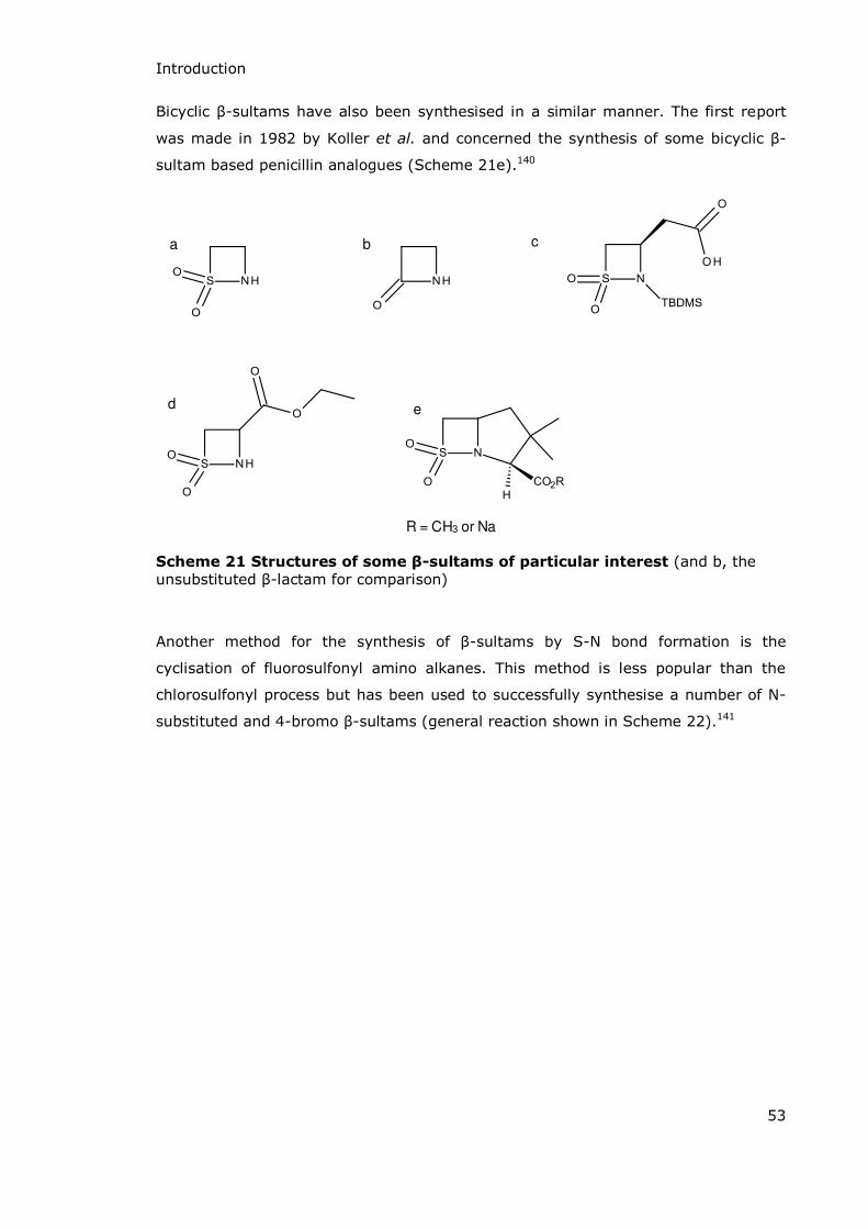
Introduction
53
Bicyclic -sultams have also been synthesised in a similar manner. The first report
was made in 1982 by Koller et al. and concerned the synthesis of some bicyclic -
sultam based penicillin analogues (Scheme 21e).140
e
R = CH3 or Na
d
a b c
Scheme 21 Structures of some β-sultams of particular interest (and b, the unsubstituted -lactam for comparison)
Another method for the synthesis of -sultams by S-N bond formation is the
cyclisation of fluorosulfonyl amino alkanes. This method is less popular than the
chlorosulfonyl process but has been used to successfully synthesise a number of N-
substituted and 4-bromo -sultams (general reaction shown in Scheme 22).141

Introduction
54
Na2CO3
EtOAc
Br2 RNH2
RNH2
R = Et, i-Pr, t-Bu, Bn. Ph
Scheme 22 N-substituted and 4-bromo β-sultams as synthesised by
fluorosulfonyl amino alkane cyclisation
1.7.2.2 Cyclisation by C-N Bond Formation
The second method of cyclisation is via C-N bond formation. The first report of this
method was made in 1984 and involved the cyclisation of -hydroxysulfonamide
mesylates. This is a relatively simple reaction requiring only exposure to potassium
carbonate in DMSO at 80 oC to form the substituted -sultam. In all reported cases
this method always yields an N-substituted -sultam but is useful as a method of
synthesising highly substituted -sultams (examples shown in Scheme 23).142

Introduction
55
t-But-Bu
K2CO3
DMSO, 80oC
Scheme 23 β-Sultam synthesis by C-N bond formation during cyclisation
(scheme for synthesis of 2-tert-butyl-3-(o-nitrophenyl)-1,2-thiazetidine-1,1-dioxide)
1.7.2.3 [2 + 2] Cycloaddition (C-N and C-S Bond Formation)
[2+2] cycloaddition reactions have also been successfully utilised in the preparation
of -sultams. N-Sulfonylamines react with nucleophilic olefins to give 1,2-thiazetidine
1,1-dioxides. The first report of this reaction (an example of C-N and C-S bond
formation) was made by Burgess and Atkins in 1967 during their research into new
preparation methods for sulfonylamines. During the attempted preparation of one
such compound in the presence of 2-(dichloromethylene)-1,3-dioxolane they
collected and characterised the cycloaddition product; 4,4-dichloro-3,3-
ethylenedioxy-2-ethyl-1,2-thiazetidine 1,1-dioxide (Scheme 24).143
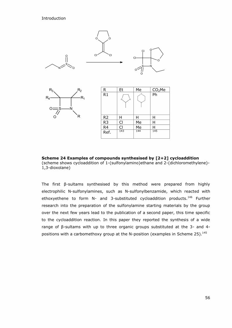
Introduction
56
Scheme 24 Examples of compounds synthesised by [2+2] cycloaddition
(scheme shows cycloaddition of 1-(sulfonylamino)ethane and 2-(dichloromethylene)-1,3-dioxolane)
The first -sultams synthesised by this method were prepared from highly
electrophilic N-sulfonylamines, such as N-sulfonylbenzamide, which reacted with
ethoxyethene to form N- and 3-substituted cycloaddition products.146 Further
research into the preparation of the sulfonylamine starting materials by the group
over the next few years lead to the publication of a second paper, this time specific
to the cycloaddition reaction. In this paper they reported the synthesis of a wide
range of -sultams with up to three organic groups substituted at the 3- and 4-
positions with a carbomethoxy group at the N-position (examples in Scheme 25).145
R Et Me CO2Me R1
Ph
R2 H H H R3 Cl Me H R4 Cl Me H Ref. 143 144 145
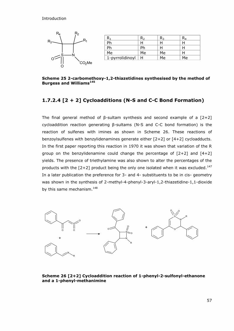
Introduction
57
Scheme 25 2-carbomethoxy-1,2-thiazetidines synthesised by the method of
Burgess and Williams145
1.7.2.4 [2 + 2] Cycloadditions (N-S and C-C Bond Formation)
The final general method of -sultam synthesis and second example of a [2+2]
cycloaddition reaction generating -sultams (N-S and C-C bond formation) is the
reaction of sulfenes with imines as shown in Scheme 26. These reactions of
benzoylsulfenes with benzylidenamines generate either [2+2] or [4+2] cycloadducts.
In the first paper reporting this reaction in 1970 it was shown that variation of the R
group on the benzylidenamine could change the percentage of [2+2] and [4+2]
yields. The presence of triethylamine was also shown to alter the percentages of the
products with the [2+2] product being the only one isolated when it was excluded.147
In a later publication the preference for 3- and 4- substituents to be in cis- geometry
was shown in the synthesis of 2-methyl-4-phenyl-3-aryl-1,2-thiazetidine-1,1-dioxide
by this same mechanism.148
Scheme 26 [2+2] Cycloaddition reaction of 1-phenyl-2-sulfonyl-ethanone
and a 1-phenyl-methanimine
R1 R2 R3 R4 Ph H H H Ph Ph H H Me Me Me H 1-pyrrolidinoyl H Me Me

Introduction
58
1.7.2.5 Advances in β-Sultam Synthesis
Since the early investigations of -sultams numerous advances in the synthesis of
1,2-thiazetidine-1,1-dioxides have been made. According to a review of sultam
synthesis published in 2013 there has been a dramatic increase in the amount of
research being carried out in this area over the past 20 years.149 It is proposed that
there may be two contributing factors for the increase. Firstly, an academic interest
in methods of sultam synthesis due to a lack of efficient general reaction schemes
applicable to sultams of varying ring sizes and substitution patterns. Secondly there
is a growing interest in the biological activity of sultams and their potential use in
both the pharmaceutical and agricultural industries.149
Whilst -sultam synthesis still mainly relies on the four processes initially highlighted
above, new methods do show improvements in the asymmetry and potential
complexity of the structures. Asymmetry may be present within the ring or with
respect to substituents on C-3 or C-4.
One of the early reports regarding the synthesis of -sultams with internal
asymmetry was made in 2002 by Enders and Wallert and used S-N bond formation
for the cyclisation of an asymmetric starting material to give 3-substituted -sultams
in excellent enantiomeric purity (examples shown in Scheme 27).135
a) HBr, HOAc, CH2Cl2, rt, 3h
b) NEt3, 2h
R group Yield (%) over 2 steps ee (%) Et 29 >96 n-Pr 68 >96 i-Pr 47 >96 n-Bu 78 >96 (CH2)2Ph 55 >96
Scheme 27 Examples of high enantio-purity 3-substituted β-sultams as
prepared by Enders and Wallert135
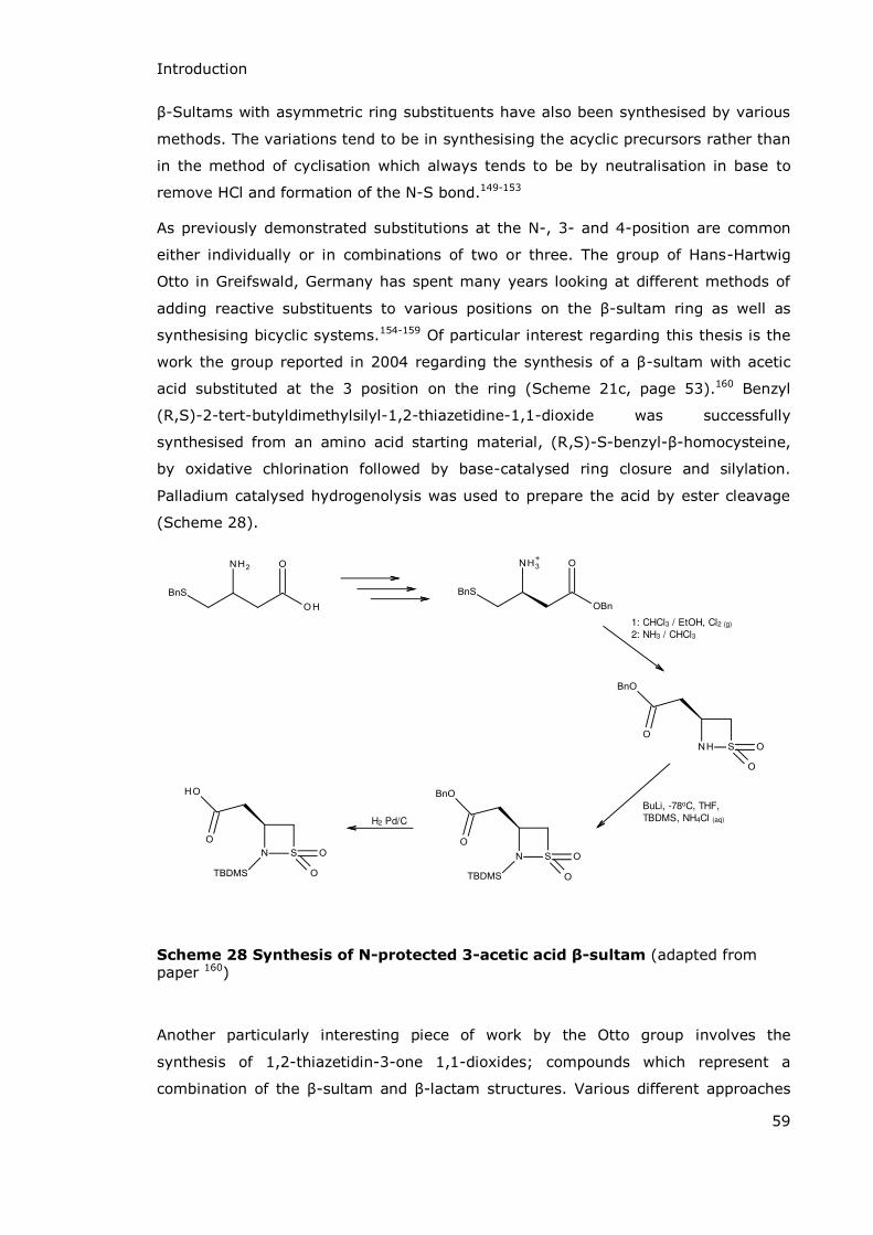
Introduction
59
-Sultams with asymmetric ring substituents have also been synthesised by various
methods. The variations tend to be in synthesising the acyclic precursors rather than
in the method of cyclisation which always tends to be by neutralisation in base to
remove HCl and formation of the N-S bond.149-153
As previously demonstrated substitutions at the N-, 3- and 4-position are common
either individually or in combinations of two or three. The group of Hans-Hartwig
Otto in Greifswald, Germany has spent many years looking at different methods of
adding reactive substituents to various positions on the -sultam ring as well as
synthesising bicyclic systems.154-159 Of particular interest regarding this thesis is the
work the group reported in 2004 regarding the synthesis of a -sultam with acetic
acid substituted at the 3 position on the ring (Scheme 21c, page 53).160 Benzyl
(R,S)-2-tert-butyldimethylsilyl-1,2-thiazetidine-1,1-dioxide was successfully
synthesised from an amino acid starting material, (R,S)-S-benzyl- -homocysteine,
by oxidative chlorination followed by base-catalysed ring closure and silylation.
Palladium catalysed hydrogenolysis was used to prepare the acid by ester cleavage
(Scheme 28).
1: CHCl3 / EtOH, Cl2 (g)
2: NH3 / CHCl3
BuLi, -78oC, THF,
TBDMS, NH4Cl (aq)H2 Pd/C
Scheme 28 Synthesis of N-protected 3-acetic acid β-sultam (adapted from paper 160)
Another particularly interesting piece of work by the Otto group involves the
synthesis of 1,2-thiazetidin-3-one 1,1-dioxides; compounds which represent a
combination of the -sultam and -lactam structures. Various different approaches

Introduction
60
were used to synthesise the 4,4-disubstituted 3-oxo- -sultams. The first was direct
cyclisation of 2-chlorosulfonyl-2-methyl-propanoyl chloride with an amine, though
this only proved successful in the synthesis of the N-unsubstituted compound and
the N-benzyl derivative. Other derivatives were synthesised by N-alkylation of the
N-unsubstituted compound via reaction with a bromoalkane and sodium hydride in
DMF. N-acylated derivatives were also synthesised via reaction with either an acid
halide or an anhydride with triethylamine in THF. These were particularly unstable
compounds but interesting due to the presence of a diacylated and sulfonated N-
atom (see Scheme 29).154
NH3
BnNH2
R-Br
NaH
Et3N
Acyl-halideR = PhCH2, EtOCOCH2, MeOCH2
Acyl = BrCH2CO, MeOCH2CO, NH2COCH2
Scheme 29 Synthesis of 3-oxo-β-sultams
1.7.3 Reactivity
Except under highly specific conditions -sultams undergo hydrolysis by S-N bond
fission.161 The acid and base catalysed hydrolysis is particularly rapid in comparison
to both the analogous acyclic sulphonamides (which are especially stable to
hydrolysis under alkaline conditions) and the analogous -lactam compounds. As
sulfonyl transfer reactions are usually much slower (~103 fold) than acyl transfer
reactions the difference in reactivity between -sultams and -lactams is particularly

Introduction
61
striking.162 Both acid and base catalysed hydrolysis of -sultams yield the same final
product but the mechanisms do vary, and this is often reflected in the relative rates
of the processes when electron donating or withdrawing substituents are present.163
A huge amount of work has been done by previous members of this group regarding
the mechanisms and kinetics of -sultam hydrolysis. In 2000 Baxter et al published a
thorough report on “Reactivity and εechanism in the Hydrolysis of -Sultams”.163
Within this paper they reported the second order rate constants for the alkaline
hydrolysis of structurally comparable -sultams and -lactams showing the
significant rate enhancement due to the presence of a sulfonyl group rather than an
acyl group (compared to the converse situation with the acyclic analogues) (data
summarised in Table 6).
The difference in rate enhancements can be explained in terms of thermodynamics
and transition state stabilisations. Four-membered ring systems are such strained
systems that ring opening is always thermodynamically favourable. Because the
opening of the ring reduces the bond strain, reactions which result in the cleavage of
a ring bond in cyclic systems normally have a lower activation energy due to the
increased stability of the transition state. This leads to these reactions almost always
being faster than in the analogous acyclic systems. In -lactams this rate
enhancement in the hydrolysis of cyclic over acyclic compounds is only minimal and
this is because the strain in the ring is not released on formation of the transition
state (TS). In -sultams the formation of the TS is accompanied by a large relief in
strain which explains the significant rate enhancements seen over acyclic
sulfonamides.
In -lactams the initial state and transition state are both destabilised by ring strain
since the ideal C-C-N bond angle in the initial state is ~120o and in the TS it is ~109o
based on the preferred trigonal and tetrahedral arrangements respectively. Since the
ring strain is not significantly reduced by movement to the TS there is little energetic
advantage and so only minimal rate enhancement is seen.
Conversely in -sultams the ring strain relief shown by the five-coordinate
intermediate significantly enhances rates compared to acyclic systems. As with -
lactams there is significant ring strain in the initial state with an ideal C-S-N bond
angle of 109o in the tetrahedral arrangement. The transition state, however, shows a
trigonal bipyramidal arrangement so that the new C-S-N bond angle of 90o now
shows the preferred geometry. In acyclic sulfonamides the formation of the TBPI
does not introduce a more favoured bond angle. The difference in free energy
between initial and transition states for the cyclic system is therefore reduced in
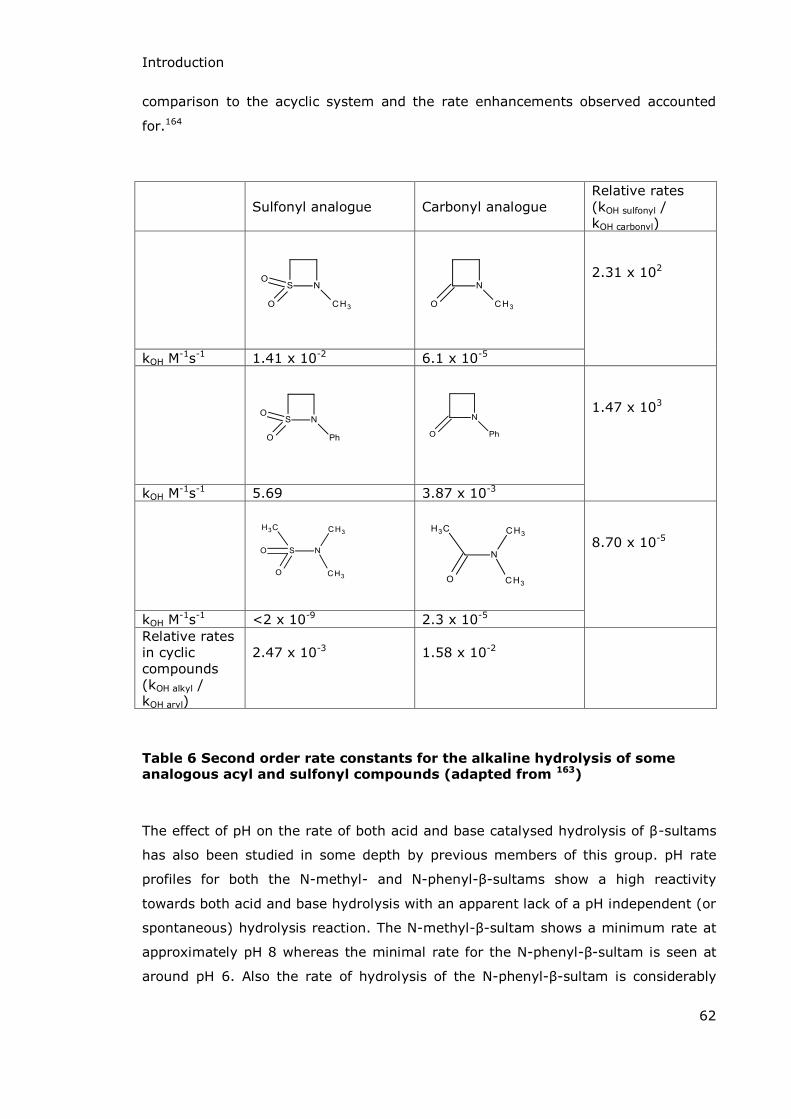
Introduction
62
comparison to the acyclic system and the rate enhancements observed accounted
for.164
Sulfonyl analogue
Carbonyl analogue
Relative rates (kOH sulfonyl / kOH carbonyl)
2.31 x 10²
kOH M-1s-1 1.41 x 10⁻² 6.1 x 10⁻⁵
1.47 x 10³
kOH M-1s-1 5.69 3.87 x 10⁻³
8.70 x 10⁻⁵
kOH M-1s-1 <2 x 10⁻⁹ 2.3 x 10⁻⁵
Relative rates in cyclic compounds (kOH alkyl / kOH aryl)
2.47 x 10⁻³
1.58 x 10⁻²
Table 6 Second order rate constants for the alkaline hydrolysis of some analogous acyl and sulfonyl compounds (adapted from 163)
The effect of pH on the rate of both acid and base catalysed hydrolysis of -sultams
has also been studied in some depth by previous members of this group. pH rate
profiles for both the N-methyl- and N-phenyl- -sultams show a high reactivity
towards both acid and base hydrolysis with an apparent lack of a pH independent (or
spontaneous) hydrolysis reaction. The N-methyl- -sultam shows a minimum rate at
approximately pH 8 whereas the minimal rate for the N-phenyl- -sultam is seen at
around pH 6. Also the rate of hydrolysis of the N-phenyl- -sultam is considerably

Introduction
63
more rapid at pH 12 than that of the N-methyl- -sultam (kobs = 6.67 x 10⁻² s-1 and
1.41 x 10⁻⁴ s-1 respectively).165 Through their studies on the effect of N-substituents
on the rate of hydrolysis the group has shown that electron withdrawing substituents
on nitrogen increase the rate of the base catalysed reaction and, along with various
other pieces of data, they were able to convincingly prove the mechanism of alkaline
hydrolysis and show that it proceeds via the trigonal bipyrimidal intermediate
previously mentioned.134,163 Of interest here though are the two possible mechanisms
of bond cleavage after the formation of the trigonal bipyramidal anionic intermediate.
One route shows a unimolecular ring-opening mechanism via proton transfer and the
second shows a bimolecular process involving a water molecule (Scheme 30). Both
methods are kinetically viable and the actual route taken may be influenced by the
nature and number of substituents on the ring.
Proton transfer
Scheme 30 Mechanism of the alkaline catalysed hydrolysis of β-sultams
Acid catalysed hydrolysis is a much more facile process showing only one viable
mechanism (Scheme 31). Initial protonation of the -sultam nitrogen is followed by
unimolecular S-N bond fission. The hydrolysis reaction then proceeds via an unstable
sulfonylium ion which is trapped by water to give the zwitterionic product. In
contrast to the base catalysed process with acid catalysed hydrolysis N-alkyl-
-sultams are more reactive than N-aryl- -sultams (Table 7) but the differences in
rate are not so dramatic.134,163
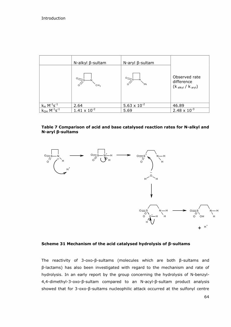
Introduction
64
N-alkyl -sultam
N-aryl -sultam
Observed rate difference (k alkyl / k aryl)
kH M-1s-1 2.64 5.63 x 10⁻² 46.89 kOH M
-1s-1 1.41 x 10⁻² 5.69 2.48 x 10⁻³
Table 7 Comparison of acid and base catalysed reaction rates for N-alkyl and
N-aryl β-sultams
..
Scheme 31 Mechanism of the acid catalysed hydrolysis of β-sultams
The reactivity of 3-oxo- -sultams (molecules which are both -sultams and
-lactams) has also been investigated with regard to the mechanism and rate of
hydrolysis. In an early report by the group concerning the hydrolysis of N-benzyl-
4,4-dimethyl-3-oxo- -sultam compared to an N-acyl- -sultam product analysis
showed that for 3-oxo- -sultams nucleophilic attack occurred at the sulfonyl centre
Introduction
65
and hydrolysis proceeded via expulsion of a carboxamide leaving group.161 In a later
paper the second order rate constants for alkaline hydrolysis for a range of N- and 4-
substituted 3-oxo- -sultams were shown to be ~103 – 105 M-1s-1 with the fastest
reaction being the hydrolysis of the previously reported N-benzyl-4,4-dimethyl-3-
oxo- -sultam with a kOH of 1.83 x 10⁵ M-1s-1. In all cases the hydrolysis proceeded
via attack at the sulfonyl centre leading to S-N not C-N bond fission which is
consistent with previous observations that -sultams are 102 to 103-fold more
reactive than -lactams towards alkaline hydrolysis.166
1.7.4 Sulfonyl Compounds as Enzyme Inhibitors
As previously mentioned -sultams are cyclic compounds which can be formed by
the cyclisation of taurines: 2-carbon systems with terminal sulfonyl and amide
groups. The first report of sulfonyl compounds being used as enzyme inhibitors was
published in 1963, a paper which discussed a range of organic sulfonyl fluorides as
inhibitors of acetylcholinesterase, α-chymotrypsin and trypsin.167 This paper was
followed a year later by one which showed phenylmethanesulfonyl fluoride to be an
irreversible inhibitor of α-chymotrypsin via sulfonylation of an active site serine
residue.168
This work was continued over the next two decades with benzenesulfonyl fluorides
being developed as inhibitors of a range of enzymes (trypsin169, thrombin,170 and
kallikrein171 among others). The reactivity of aromatic sulfonyl fluorides was also
developed with the addition of a positively charged benzamidine or pyridinium group
to the benzene ring, increasing the reactivity. As part of this work
(p-amidinophenyl)methanesulfonyl fluoride was synthesised and shown to be an
irreversible inhibitor of serine proteases, particularly bovine trypsin and human
thrombin.172 Structures of some of the sulfonyl compounds investigated are shown in
Scheme 32.
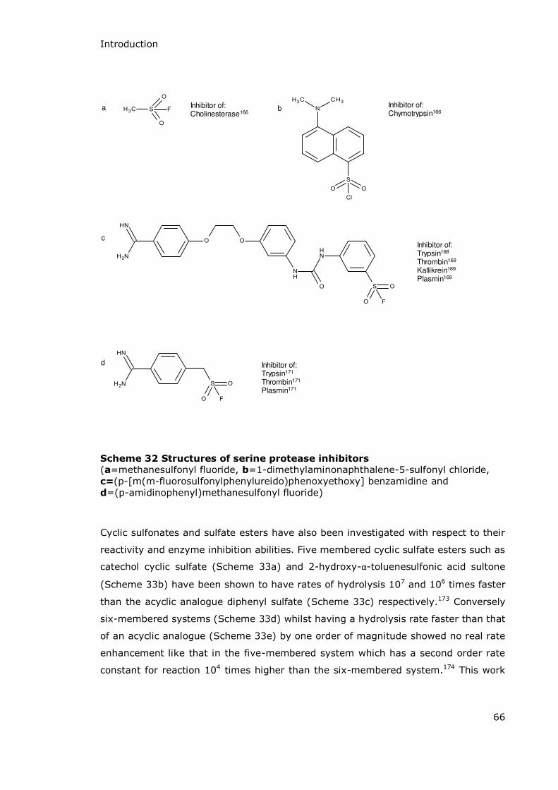
Introduction
66
d
c
Inhibitor of:Chymotrypsin166
a Inhibitor of:Cholinesterase166
b
Inhibitor of:Trypsin168
Thrombin169
Kallikrein169
Plasmin169
Inhibitor of:Trypsin171
Thrombin171
Plasmin171
Scheme 32 Structures of serine protease inhibitors (a=methanesulfonyl fluoride, b=1-dimethylaminonaphthalene-5-sulfonyl chloride, c=(p-[m(m-fluorosulfonylphenylureido)phenoxyethoxy] benzamidine and d=(p-amidinophenyl)methanesulfonyl fluoride)
Cyclic sulfonates and sulfate esters have also been investigated with respect to their
reactivity and enzyme inhibition abilities. Five membered cyclic sulfate esters such as
catechol cyclic sulfate (Scheme 33a) and 2-hydroxy-α-toluenesulfonic acid sultone
(Scheme 33b) have been shown to have rates of hydrolysis 107 and 106 times faster
than the acyclic analogue diphenyl sulfate (Scheme 33c) respectively.173 Conversely
six-membered systems (Scheme 33d) whilst having a hydrolysis rate faster than that
of an acyclic analogue (Scheme 33e) by one order of magnitude showed no real rate
enhancement like that in the five-membered system which has a second order rate
constant for reaction 104 times higher than the six-membered system.174 This work
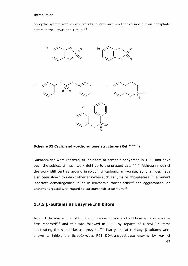
Introduction
67
on cyclic system rate enhancements follows on from that carried out on phosphate
esters in the 1950s and 1960s.175
e)
a) b)
c) d)
Scheme 33 Cyclic and acyclic sultone structures (Ref 173,176)
Sulfonamides were reported as inhibitors of carbonic anhydrase in 1940 and have
been the subject of much work right up to the present day.177-180 Although much of
the work still centres around inhibition of carbonic anhydrase, sulfonamides have
also been shown to inhibit other enzymes such as tyrosine phosphatase,181 a mutant
isocitrate dehydrogenase found in leukaemia cancer cells182 and aggrecanase, an
enzyme targeted with regard to osteoarthritis treatment.183
1.7.5 β-Sultams as Enzyme Inhibitors
In 2001 the inactivation of the serine protease enzymes by N-benzoyl- -sultam was
first reported184 and this was followed in 2003 by reports of N-acyl- -sultams
inactivating the same elastase enzyme.185 Two years later N-acyl- -sultams were
shown to inhibit the Streptomyces R61 DD-transpeptidase enzyme by way of

Introduction
68
sulfonylating the active site serine residue.186 In another paper in the same year
these same N-acyl- -sultams were shown to inactivate the Class C -lactamase
P99 from Enterobacter cloacae.187 This paper also looked at the action of analogous
-lactams on the same enzyme. The -lactams were shown to be substrates for the
enzyme with hydrolysis taking place via an acylation-deacylation process. Conversely
the -sultams inactivated the enzyme by sulfonylation of the active site serine
residue forming a sulfonate ester which then underwent C-O bond fission. ESI-MS
investigations showed that elimination of the sulfonate anion led to the formation of
a dehydroalanine residue.
-Sultams have the potential to act as inhibitors of -lactamases via either the
mechanism based or transition state analogue route. Mechanism based inhibition of
serine- -lactamases is possible with -sultams which have a leaving group at the 3-
position or where there is a leaving group on an N-acyl substituent (Scheme 34).
Scheme 34 Representative structures of β-sultams with the potential to
inhibit serine β-lactamase enzymes
In a similar manner to -lactam type inhibitors (clavulanic acid or sulbactam for
example) the attack of the enzyme at the sulfonyl centre (the carbonyl centre in the
-lactams) and loss of the leaving group results in formation of an electron-deficient
imine or iminium ion which could be attacked by a second nucleophilic amino acid
residue at the active site. This would trap the enzyme and cause irreversible
inhibition (see Scheme 35).

Introduction
69
Scheme 35 Mechanism based inhibition of serine β-lactamase enzymes by β-
sultams
Transition state analogue inhibition could be achieved with metallo- -lactamases due
to the tetrahedral sulfonyl group which imitates the sp3 intermediate formed by
enzyme attack at the carbonyl centre of -lactam antibiotics during normal enzyme
action. This could be especially true upon ionisation of the -sultam to generate an
anion suitable for complexing to Zn2+. Non-covalent interactions between the zinc ion
of metallo-enzymes and the sulfonyl group can potentially lead to transition state
analogue inhibition. A similar result is also possible via interaction of the sulfonyl
centre with the oxyanion hole of serine enzymes.
Scheme 36 Zinc interactions with a representative β-sultam and β-lactam transition state demonstrating potential for β-sultams to act as transition
state analogues
It is proposed that 1,2-thiazetidine-3-carboxylate-1,1-dioxide (3-carboxy- -sultam)
has the potential to act as a transition state analogue of the Class B -lactamase
BcII via interactions of the zinc ion (Zn1) with the sulfonyl group as shown above.
Further interactions should occur between the amine and zinc bound hydroxide ion
(Wat 1) and also between the second zinc ion (Zn2) and the carboxylate group as
demonstrated in Scheme 37.
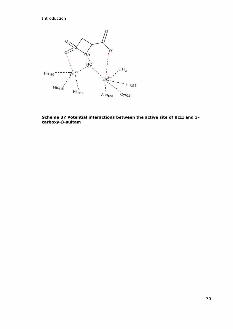
Introduction
70
Scheme 37 Potential interactions between the active site of BcII and 3-
carboxy-β-sultam
Introduction
71
1.8 Glutamine Synthetase
1.8.1 The Roles and Regulation of Glutamic Acid in The Human Body
Glutamic acid, or glutamate, is a non-essential, proteinogenic amino acid which also
has a role in the human body as an excitatory neurotransmitter. It is non-essential
as it can be synthesised within the body via the synthesis and degradation pathway
detailed in Scheme 38. The inhibition of any of the enzymes along this pathway will
necessarily cause a change in the concentration of glutamic acid.
glucose Krebs cycle -ketoglutarate
GABA Transaminase
Glu
Gln
GABA
Glutamate decarboxylase
GlutaminaseGlutaminesynthetase
Scheme 38 Synthesis and degradation of glutamic acid (Glu)
Glutamine synthetase (GS) is an important enzyme in this pathway and plays a vital
role in the brain, kidneys and liver. Within the brain this enzyme is involved in the
regulation of glutamate and the metabolism of nitrogen by catalysing the synthesis
of glutamine. This is an ATP-dependent pathway which involves the phosphorylation
of the side chain carboxylate group to yield ADP and an acyl-phosphate intermediate.

Introduction
72
This intermediate then reacts with ammonia to form glutamine and inorganic
phosphate (as shown in Scheme 39).
Glu
Gln
ATP ADP
NH3
Pi
Scheme 39 Overview of glutamine synthetase catalysed formation of
glutamine from glutamic acid
1.8.2 The Role of Glutamic Acid in Neurodegeneration
Glutamate is the major excitatory neurotransmitter within the central nervous
system (CNS).188 The concentrations of glutamate within the CNS are approximately
1000-fold higher than those of the neurotransmitters dopamine, noradrenaline and
serotonin.189 Glutamate has been shown to be an excitotoxic agent under certain
conditions; high concentrations of glutamate can cause excessive stimulation of
glutamate receptors causing the death of cells which express these receptors.188 The
concentration of glutamate in the extracellular space can be controlled by glutamate
transporters which transport glutamate across cellular membranes. This is important
as the removal of glutamate from the extracellular space is the major method of
terminating an excitatory signal. Alterations in the expression of glutamate

Introduction
73
transporters and the accompanying changes to extracellular glutamate
concentrations have been implicated in the following diseases (among others):
Amyotrophic lateral sclerosis (ALS)190-192
Alzheimer’s disease193,194
Huntington’s disease195
Parkinson’s disease189
Epilepsy.195
1.8.3 Glutamic Acid Analogues (and Associated Compounds) as
Enzyme Inhibitors and CNS Drugs
Numerous analogues of glutamate and other compounds in the glutamic acid
synthesis/degradation pathway have been investigated as potential inhibitors of key
enzymes in the CNS and as potential drugs for the treatment of a range of diseases,
particularly those of a neurological nature (some structures are shown in Scheme
40). Numerous studies have also been carried out on glutamine synthetase from
sources other than the human CNS; enzymes from rat brain and liver, sheep brain
and even pea plants have all been investigated.
L-Methionine sulfoximine is structurally very similar to L-glutamic acid and has been
shown to act as a convulsant. It irreversibly inhibits glutamine synthetase via tight
binding of the phosphorylated methionine sulfoximine and ADP at the active site.196
Another structural analogue of glutamic acid which inhibits glutamine synthetase
(from pea plants) is tabtoxinine- -lactam. This is a particularly interesting structure
which both mimics the structure of glutamate and contains a -lactam ring, yet it
does not inhibit DD-transpeptidase enzymes or -lactamases.197 Taurine, the
hydrolysis product of -sultam, is a glutamic acid analogue which has been
implicated as a potential therapeutic agent. It has been shown to have an effect in
the treatment of neurodegenerative diseases such as Parkinson’s disease and
alcohol-induced brain damage possibly due to its anti-inflammatory actions.198 Some
compounds structurally related to taurine have also been reported to be useful as
drugs which can reduce the craving for alcohol in recovering post-detoxification
alcoholics199-201 Acamprosate (N-acetyl homotaurine or 3-acetamidopropane-1-
sulfonic acid) is one such compound; marketed under the trade name Campral it is
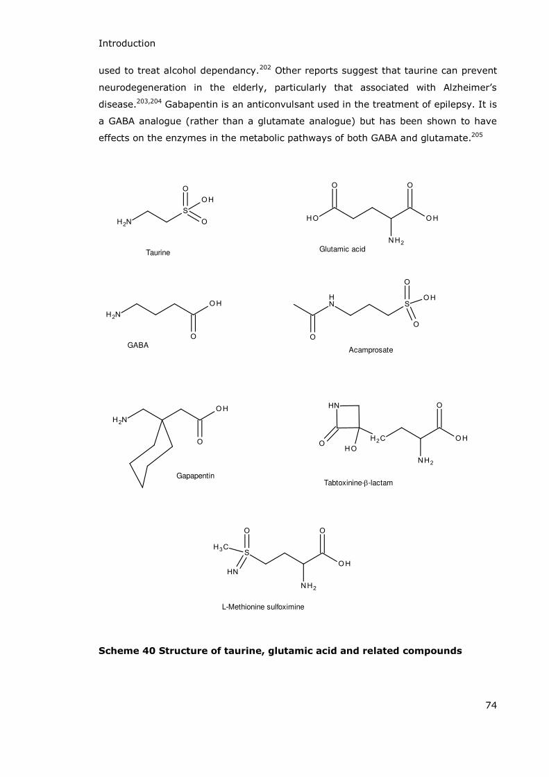
Introduction
74
used to treat alcohol dependancy.202 Other reports suggest that taurine can prevent
neurodegeneration in the elderly, particularly that associated with Alzheimer’s
disease.203,204 Gabapentin is an anticonvulsant used in the treatment of epilepsy. It is
a GABA analogue (rather than a glutamate analogue) but has been shown to have
effects on the enzymes in the metabolic pathways of both GABA and glutamate.205
Tabtoxinine--lactam
L-Methionine sulfoximine
Gapapentin
Taurine Glutamic acid
GABAAcamprosate
Scheme 40 Structure of taurine, glutamic acid and related compounds

Introduction
75
1.8.4 β-Sultams as Pro-Drugs for Treating Neurodegenerative
Diseases
As has been previously discussed in section 1.7.3 the hydrolysis of the unsubstituted
-sultam (1,2-thiazetidine-1,1-dioxide) yields taurine, a compound which is
zwitterionic in aqueous solution (Scheme 41).
Scheme 41 Hydrolysis of β-sultam to yield taurine
The administration of taurine as a drug however, is not a facile process. The
zwitterionic nature of the compound makes it highly lipophobic and hydrophilic and
this decreases the ability of taurine to diffuse across cell membranes so that it is
poorly absorbed from the gut.206 The charged nature of the molecule also reduces
the amount of taurine which can be taken across the blood-brain barrier. In order for
taurine to be a useful drug in the treatment or prevention of neurodegeneration it is
necessary to find a method of getting it into the brain and to the site of action. Two
possible ways of facilitating this are:
Producing taurine analogues with lower hydrophilicity
Producing a pro-drug which in some way releases taurine into cells once it has crossed into the brain.
-Sultams could be ideal pro-drugs for taurine analogues if their half-lives at
physiological pH are of an order which allows delivery of the pro-drug to the site of
action prior to hydrolysis. Previous work by Ward et al. has studied the effects of -
sultam and taurine on the inflammatory response and rate of glutamate release both
in cells and in rats.198 Their work showed that -sultam had an enhanced ability to
reduce the inflammatory response over taurine (its hydrolysis product) both in vitro
and in vivo. It also had an effect on the amount of glutamate release, and again this
was a greater effect than with taurine. It is suggested that the increased lipophilicity
of the -sultam over taurine increases the uptake across cell membranes and that

Introduction
76
the use of -sultams as pro-drugs for taurine analogues may be of significant benefit
in the treatment of neurodegenerative conditions. Enzymes within the CNS are
potentially targets for these compounds and their activity as inhibitors of glutamine
synthetase is therefore of interest.
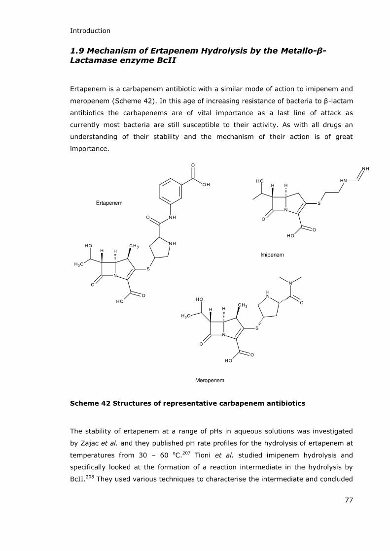
Introduction
77
1.9 Mechanism of Ertapenem Hydrolysis by the Metallo-β-
Lactamase enzyme BcII
Ertapenem is a carbapenem antibiotic with a similar mode of action to imipenem and
meropenem (Scheme 42). In this age of increasing resistance of bacteria to -lactam
antibiotics the carbapenems are of vital importance as a last line of attack as
currently most bacteria are still susceptible to their activity. As with all drugs an
understanding of their stability and the mechanism of their action is of great
importance.
Ertapenem
Imipenem
Meropenem
Scheme 42 Structures of representative carbapenem antibiotics
The stability of ertapenem at a range of pHs in aqueous solutions was investigated
by Zajac et al. and they published pH rate profiles for the hydrolysis of ertapenem at
temperatures from 30 – 60 oC.207 Tioni et al. studied imipenem hydrolysis and
specifically looked at the formation of a reaction intermediate in the hydrolysis by
BcII.208 They used various techniques to characterise the intermediate and concluded

Introduction
78
that protonation during hydrolysis at the active site of the enzyme occurred initially
on nitrogen (Scheme 43B), but that tautomerisation may lead to the formation of the
imine product (Scheme 43C).
A B C
Scheme 43 Tautomerisation of carbapenem (imipenem core structure) after hydrolysis
Our studies of ertapenem aim to confirm that protonation actually occurs at C3 of
the pyrrolidine ring to form the product shown in Scheme 43C.

Introduction
79
1.10 Instrumental Techniques – React-IR
React-IR is a relatively new technique first introduced in the early 1990’s. It can be
used to monitor a reaction in situ and in real time without the need for removing
samples or for further sample preparation. Computer software is available to allow
the monitoring of specific characteristic wavenumbers producing graphs which show
the change in absorption due to disappearance of starting materials, appearance and
disappearance of intermediates or the production of the final products.
The individual spectra produced during a React-IR experiment are the same as those
produced in a standard FT-IR experiment. The molecules in the solution absorb the
IR radiation at frequencies which correspond to the vibrational frequencies of the
bonds within them. The use of a Fourier transformation allows these absorbances to
be displayed as a frequency spectrum specific to the solution at that moment in
time.
In a React-IR instrument (Figure 10) the IR source, interferometer, beam splitter
and detector are all housed within the main unit. The optical path of the IR radiation
is from the source along the probe, through an optical crystal and back up the probe
to the detector in the main unit.
Figure 10 React-IR spectrometer
The infrared light passes through the crystal via total internal reflection with multiple
reflections. This total internal reflection causes an evanescent wave which penetrates
0.5 to 2 μm into the sample. This is known as attenuated total reflectance (ATR) and
allows extremely small samples (for example, one drop of < 0.1 ml) to be analysed
by IR spectroscopy. In order for an evanescent wave to form, the crystal must be
made of a material with a higher refractive index than the sample solution. Materials
Introduction
80
often used as ATR crystals are germanium, zinc selenide or, as in the case of our
instrument, diamond.
As with other types of spectroscopy the Beer-Lambert Law ( ) can be applied to
infrared spectroscopy.209 The law states that the absorbance (A) of a solution is
equal to the concentration (c) of the solution multiplied by the path-length (l) of the
sample and its molar absorptivity ( ). In the case of solutions containing multiple
components it is necessary to find a wavelength (or in this case wavenumber) where
only one component shows an absorbance. In this way it is possible to monitor the
concentration of a species in solution using ReactIR if a unique absorption band for
that species can be found. In the case of a reaction where there is a change in
functional group this can be quite easy using ReactIR, and it is an advantage of
ReactIR over UV-Vis spectroscopy where no chromophores are created or removed.
ReactIR was chosen as the technique to follow the hydrolysis of -sultam to yield
taurine. Sulfur-oxygen double bonds tend to absorb IR radiation between 1050 and
1450 cm-1. The exact wavenumber of the absorption depends on the functionality of
the double bond (part of a sulfone, sulfonic acid, sulphate etc.) and on the presence
of other functional groups on the molecule. In this case preliminary work showed
that the -sultam S=O bond absorbed at 1301 cm-1 and the taurine S=O peak was
found at 1200 cm-1 when in an aqueous solution. A plot of the absorbances at these
wavenumbers with respect to time could be fitted to a first order rate law and
exponential plots would give the pseudo-first-order rate constants for hydrolysis at
low pH.
81
Experimental

Experimental
82
2.1 Synthesis
2.1.1 General
Solvents and reagents were obtained from commercial sources (Sigma Aldrich or
Fisher Scientific) and used as obtained unless otherwise stated.
NMR spectra were recorded on a 400MHz Bruker Ascend spectrometer (unless
stated) and chemical shifts ( H or C) are quoted in ppm. The abbreviations used are
br: broad, s: singlet, d: doublet, t: triplet, m: multiplet, dd: doublet of doublets,
dt: doublet of triplets. Coupling constants (J) are quoted in Hertz. Infrared spectra
were obtained on a Thermo Nicolet 380 FT-IR spectrometer as neat samples and the
absorption peaks are quoted as wavenumbers (cm-1). Mass spectra were recorded on
an Agilent 6530 Accurate Mass Q-TOF LC-MS fitted with an Agilent 1290 Infinity
Autosampler. Melting points were recorded on a Gallenkamp melting point
apparatus.

Experimental
83
2.1.2 Synthesis of 1,2-thiazetidine-1,1-dioxide (β-sultam)
Synthesis of 2-aminoethanesulfonyl chloride hydrochloride (taurine sulfonyl chloride) [2]
EtOH, Cl2CHCl3
-10 oC
[1] [2]
2-(2-Aminoethyldisulfanyl)ethanamine dihydrochloride (cystamine dihydrochloride)
[1] (17.38g, 77.0mmol) was suspended in chloroform (400ml) and ethanol (200ml).
Chlorine gas was passed into the suspension at -10 oC for 2 hours until saturation
(noted by a permanent green colouration). The system was purged with nitrogen,
dry diethyl ether (100ml) was added and the solution stirred at room temperature
for 2 hours. The white precipitate was recovered by vacuum filtration and washed
with dry diethyl ether (2 x 25ml) to yield a white crystalline solid (24.9g, 138mmol,
89%). This was then used directly in the next step without further analysis.

Experimental
84
Synthesis of 1,2-thiazetidine-1,1-dioxide (β-sultam) [3]
Na2CO3
EtOAc
rt, 60 hours
[3][2]
2-Aminoethanesulfonyl chloride hydrochloride (taurine sulfonyl chloride) [2] (8.500g,
47.0mmol) was added to anhydrous sodium carbonate (20.93g, 194mmol) in ethyl
acetate (250 ml) and stirred at room temperature for 60 hours. The reaction mixture
was filtered through celite and the solvent removed by reduced pressure rotary
evaporation at 30 oC to afford a white crystalline solid (1.11g, 10.4mmol, 22%).
m.p. 52-54 oC (literature value 53 oC).139
IR υmax (cm-1) (Neat): 3308, 2958, 2021, 1298, 1260, 1150 (S=O), 799.
1H NεR (CDCl3): 5.58 (1H, br s, NH), 4.32 (2H, dt, J1.78 & 6.96, CH2-SO2), 3.39
(2H, dt, J3.87 & 6.96, CH2-NH). (literature values 3.39 (2H, dt, J4 & 7, CH2NH),
4.32 (2H, dt, J2 & 7, SO2CH2), 5.53 (1H, bs, NH) ).210
13C NεR (CDCl3): 60.91 (CH2-SO2), 28.08 (CH2-NH). (literature values 26.8,
60.6).210
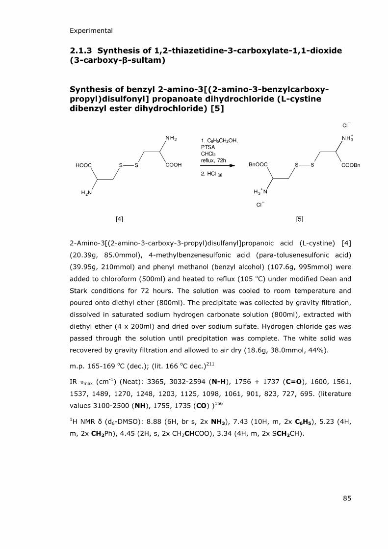
Experimental
85
2.1.3 Synthesis of 1,2-thiazetidine-3-carboxylate-1,1-dioxide
(3-carboxy-β-sultam)
Synthesis of benzyl 2-amino-3[(2-amino-3-benzylcarboxy-
propyl)disulfonyl] propanoate dihydrochloride (L-cystine
dibenzyl ester dihydrochloride) [5]
1. C6H5CH2OH,
PTSA
CHCl3reflux, 72h
2. HCl (g)
[4] [5]
2-Amino-3[(2-amino-3-carboxy-3-propyl)disulfanyl]propanoic acid (L-cystine) [4]
(20.39g, 85.0mmol), 4-methylbenzenesulfonic acid (para-tolusenesulfonic acid)
(39.95g, 210mmol) and phenyl methanol (benzyl alcohol) (107.6g, 995mmol) were
added to chloroform (500ml) and heated to reflux (105 oC) under modified Dean and
Stark conditions for 72 hours. The solution was cooled to room temperature and
poured onto diethyl ether (800ml). The precipitate was collected by gravity filtration,
dissolved in saturated sodium hydrogen carbonate solution (800ml), extracted with
diethyl ether (4 x 200ml) and dried over sodium sulfate. Hydrogen chloride gas was
passed through the solution until precipitation was complete. The white solid was
recovered by gravity filtration and allowed to air dry (18.6g, 38.0mmol, 44%).
m.p. 165-169 oC (dec.); (lit. 166 oC dec.)211
IR υmax (cm-1) (Neat): 3365, 3032-2594 (N-H), 1756 + 1737 (C=O), 1600, 1561,
1537, 1489, 1270, 1248, 1203, 1125, 1098, 1061, 901, 823, 727, 695. (literature
values 3100-2500 (NH), 1755, 1735 (CO) )156
1H NεR (d6-DMSO): 8.88 (6H, br s, 2x NH3), 7.43 (10H, m, 2x C6H5), 5.23 (4H,
m, 2x CH2Ph), 4.45 (2H, s, 2x CH2CHCOO), 3.34 (4H, m, 2x SCH2CH).
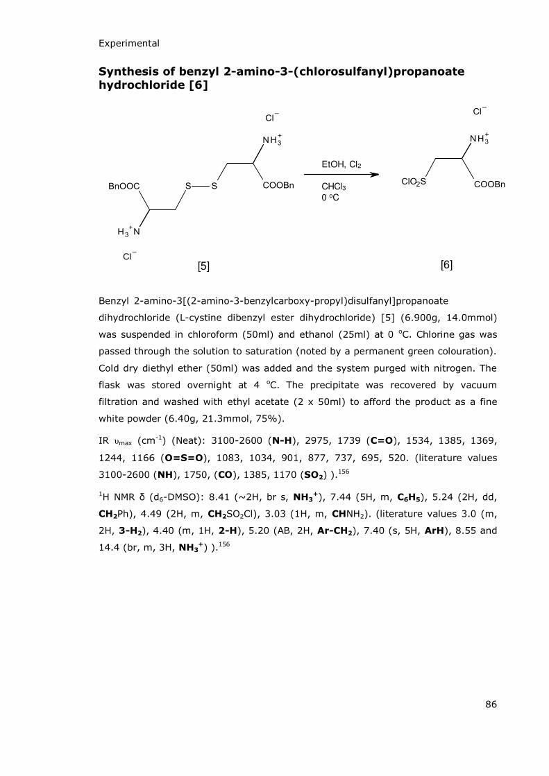
Experimental
86
Synthesis of benzyl 2-amino-3-(chlorosulfanyl)propanoate
hydrochloride [6]
EtOH, Cl2
CHCl30 oC
[5] [6]
Benzyl 2-amino-3[(2-amino-3-benzylcarboxy-propyl)disulfanyl]propanoate .
dihydrochloride (L-cystine dibenzyl ester dihydrochloride) [5] (6.900g, 14.0mmol)
was suspended in chloroform (50ml) and ethanol (25ml) at 0 oC. Chlorine gas was
passed through the solution to saturation (noted by a permanent green colouration).
Cold dry diethyl ether (50ml) was added and the system purged with nitrogen. The
flask was stored overnight at 4 oC. The precipitate was recovered by vacuum
filtration and washed with ethyl acetate (2 x 50ml) to afford the product as a fine
white powder (6.40g, 21.3mmol, 75%).
IR υmax (cm-1) (Neat): 3100-2600 (N-H), 2975, 1739 (C=O), 1534, 1385, 1369,
1244, 1166 (O=S=O), 1083, 1034, 901, 877, 737, 695, 520. (literature values
3100-2600 (NH), 1750, (CO), 1385, 1170 (SO2) ).156
1H NεR (d6-DMSO): 8.41 (~2H, br s, NH3+), 7.44 (5H, m, C6H5), 5.24 (2H, dd,
CH2Ph), 4.49 (2H, m, CH2SO2Cl), 3.03 (1H, m, CHNH2). (literature values 3.0 (m,
2H, 3-H2), 4.40 (m, 1H, 2-H), 5.20 (AB, 2H, Ar-CH2), 7.40 (s, 5H, ArH), 8.55 and
14.4 (br, m, 3H, NH3+) ).156

Experimental
87
Synthesis of benzyl 1,2-thiazetidine-3-carboxylate-1,1-dioxide
(3-benzyl carboxylate β-sultam) [7]
NH3, CHCl3
0 oC, 1h
[6] [7]
Benzyl 2-amino-3-(chlorosulfanyl)propanoate hydrochloride [6] (2.340g, 7.80mmol)
was suspended in chloroform (40ml) and stirred vigorously at 0 oC. A solution of
saturated ammonia in chloroform (40ml) was added slowly and the resulting solution
stirred for 1 hour. The solution was concentrated by reduced pressure rotary
evaporation, ethyl acetate (20ml) added and washed with distilled water (2 x 125ml)
until the organics were clear. The solvent was removed by reduced pressure rotary
evaporation to leave a yellow oil which solidified on cooling to yield a waxy pale
yellow solid (0.98g, 4.07mmol, 52%).
IR υmax (cm-1) (Neat): 3283 (N-H), 1745 (C=O), 1306, 1288, 1204, 1145 (S=O),
1074, 739, 732, 696, 674, 464. (literature values 3320 (NH), 1740, (CO), 1320,
1300, 1155 (SO2) ).156
1H NεR (CDCl3): 7.45 (5H, m, C6H5), 6.12 (0.75H, br s, NH), 5.33 (2H, s, CH2Ph),
4.63 (1H, dd, J = 8.92 & 13.06, CHAHBSO2), 4.44 (1H, dd, J = 4.27 & 13.06,
CHAHBSO2), 4.27 (1H, dd, J = 4.27 & 8.92, CHNH). (literature values 4.15 (dd, J =
10.2 Hz, 2 Hz 1H, 3-H), 4.32 (dd, J = 13 Hz, 2 Hz, 1H, 4-H), 4.57 (dd, J = 13 Hz,
10.2 Hz, 1H, 4-H’), 5.22 (s, 2H, Ar-CH2), 7.37 (m, 5H, ArH) ).156
13C NεR (CDCl3): 183 (COO), 141 (Ar-C), 129 (Ar-CH), 128 (Ar-CH), 127 (Ar-
CH), 64 (CH2-SO2), 52 (CH-NH).

Experimental
88
Synthesis of 1,2-thiazetidine-3-carboxylate-1,1-dioxide (3-
carboxy-β-sultam) [8]
orNH3 (l), Na
-78 oC, 30 min
[7] [8]
Benzyl-1,2-thiazetidine-3-carboxylate-1,1-dioxide (363.1mg, 1.50mmol) was
dissolved in liquid ammonia (35ml) at -78 oC under a nitrogen atmosphere. Sodium
metal (50.09mg, 2.18mmol) was added until the solution just remained deep blue.
Ammonium chloride (37.34mg, 0.70mmol) was added to dissipate the blue colour
and the ammonia was allowed to evaporate under a stream of nitrogen to yield a
white solid (450.12mg, includes NH4Cl salt).
1H NεR (D2O): 4.57 (1H, dd, J8.78 & 13.96, CHAHBSO2), 4.23 (1H, dd, J5.34 &
13.96, CHAHBSO2), 3.98 (1H, dd, J5.34 & 8.78, CHNH).
13C NεR (D2O): 181 (COOH), 64 (CH2-SO2), 43 (CH-NH).
m/z (LC-MS-QTOF) 149.97 (M-H)-, 105.98 (M-COOH)-.
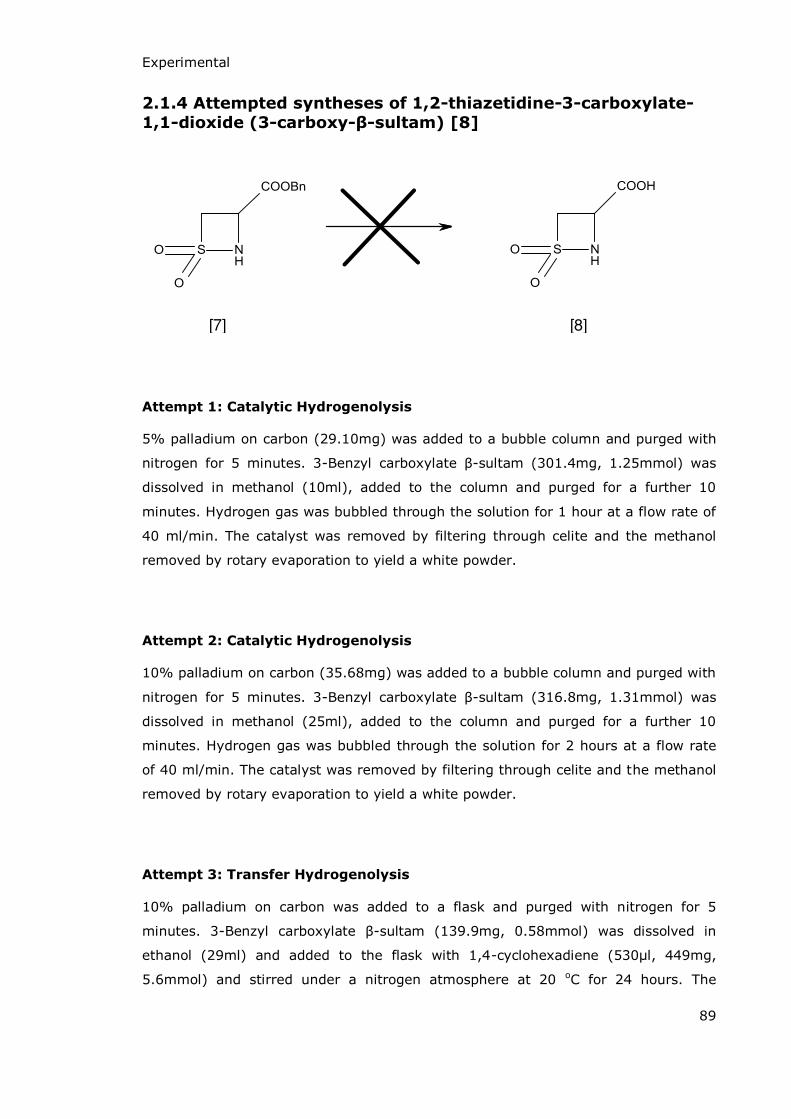
Experimental
89
2.1.4 Attempted syntheses of 1,2-thiazetidine-3-carboxylate-
1,1-dioxide (3-carboxy-β-sultam) [8]
[7] [8]
Attempt 1: Catalytic Hydrogenolysis
5% palladium on carbon (29.10mg) was added to a bubble column and purged with
nitrogen for 5 minutes. 3-Benzyl carboxylate -sultam (301.4mg, 1.25mmol) was
dissolved in methanol (10ml), added to the column and purged for a further 10
minutes. Hydrogen gas was bubbled through the solution for 1 hour at a flow rate of
40 ml/min. The catalyst was removed by filtering through celite and the methanol
removed by rotary evaporation to yield a white powder.
Attempt 2: Catalytic Hydrogenolysis
10% palladium on carbon (35.68mg) was added to a bubble column and purged with
nitrogen for 5 minutes. 3-Benzyl carboxylate -sultam (316.8mg, 1.31mmol) was
dissolved in methanol (25ml), added to the column and purged for a further 10
minutes. Hydrogen gas was bubbled through the solution for 2 hours at a flow rate
of 40 ml/min. The catalyst was removed by filtering through celite and the methanol
removed by rotary evaporation to yield a white powder.
Attempt 3: Transfer Hydrogenolysis
10% palladium on carbon was added to a flask and purged with nitrogen for 5
minutes. 3-Benzyl carboxylate -sultam (139.9mg, 0.58mmol) was dissolved in
ethanol (29ml) and added to the flask with 1,4-cyclohexadiene (530μl, 449mg,
5.6mmol) and stirred under a nitrogen atmosphere at 20 oC for 24 hours. The

Experimental
90
catalyst was removed by filtering through celite and the solvents removed by rotary
evaporation to yield a white powder.
Attempt 4: Base Catalysed Hydrolysis
3-Benzyl carboxylate -sultam (116.5mg, 0.483mmol) was dissolved in 1M NaOH
(5ml) and stirred for 10 minutes. Half the solution was removed and the remainder
allowed to stir for a further 30 minutes. Upon removal from the reaction flask both
samples were treated as follows: the solution was extracted with DCM (2 x 1.5ml),
the aqueous portion was acidified with 2M HCl (1ml), extracted into diethyl ether (2
x 1ml) and the solvent removed by rotary evaporation.
Attempt 5: Base Catalysed Hydrolysis
3-Benzyl carboxylate -sultam (931mg, 3.86mmol) was dissolved in 1.0M NaOH
(9.5ml) and stirred for 2 minutes at 0 oC. The benzyl alcohol formed was extracted
with DCM (3 x 6ml). 25ml DCM was added to the aqueous portion which was
acidified with dropwise addition of 2.0M HCl until the solution reached pH 1. The
organic layer was separated and a further extraction of the aqueous portion carried
out with DCM (8ml). These two organic solutions were combined and the solvent
removed by reduced pressure rotary evaporation.
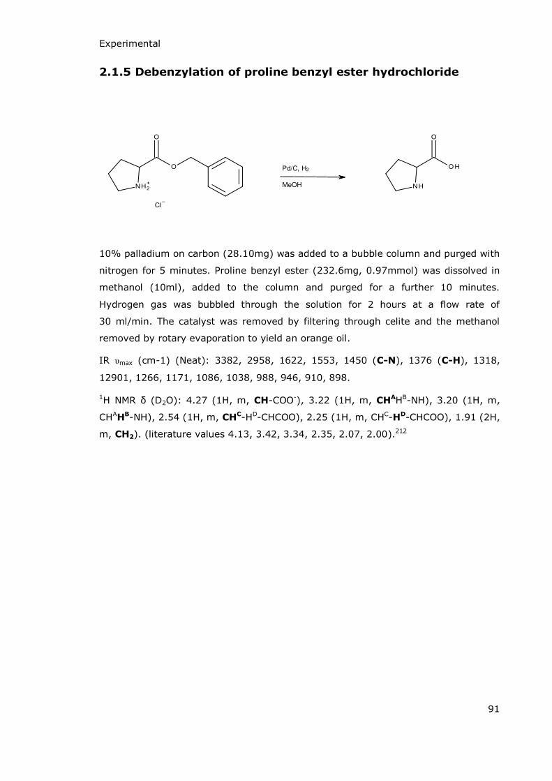
Experimental
91
2.1.5 Debenzylation of proline benzyl ester hydrochloride
Pd/C, H2
MeOH
10% palladium on carbon (28.10mg) was added to a bubble column and purged with
nitrogen for 5 minutes. Proline benzyl ester (232.6mg, 0.97mmol) was dissolved in
methanol (10ml), added to the column and purged for a further 10 minutes.
Hydrogen gas was bubbled through the solution for 2 hours at a flow rate of
30 ml/min. The catalyst was removed by filtering through celite and the methanol
removed by rotary evaporation to yield an orange oil.
IR υmax (cm-1) (Neat): 3382, 2958, 1622, 1553, 1450 (C-N), 1376 (C-H), 1318,
12901, 1266, 1171, 1086, 1038, 988, 946, 910, 898.
1H NεR (D2O): 4.27 (1H, m, CH-COO-), 3.22 (1H, m, CHAHB-NH), 3.20 (1H, m,
CHAHB-NH), 2.54 (1H, m, CHC-HD-CHCOO), 2.25 (1H, m, CHC-HD-CHCOO), 1.91 (2H,
m, CH2). (literature values 4.13, 3.42, 3.34, 2.35, 2.07, 2.00).212

Experimental
92
2.1.6 Attempted cyclisation of 2-amino-3-chlorosulfonyl-
propanoic acid
Synthesis of 2-amino-3-chlorosulfonyl-propanoate
hydrochloride (L-cysteine sulfonyl chloride)
EtOH, Cl2CHCl3
-10 oC
2-Amino-3[(2-amino-3-carboxy-3-propyl)disulfanyl]propanoic acid (L-cystine)
(4.75g, 19.8mmol) was suspended in ethanol (18ml) and chloroform (30ml).
Chlorine gas was passed into the suspension at -10 oC for 2 hours until saturation
(noted by a permanent green colouration). The system was purged with nitrogen and
the solution stirred at room temperature for 1 hours. Dry diethyl ether (30ml) was
added and the solution stored at 4 oC overnight. The pale yellow precipitate was
recovered by gravity filtration and washed with dry diethyl ether (2 x 25ml) (4.14g,
30.6mmol, 77%).
1H NεR (D2O): 4.44 (1H, dd, J4.5 & 8.3, CH-NH3+), 3.60 (1H, dd, J4.5 & 15.2,
CHAHB-SO2Cl), 3.28 (1H, dd, J8.3 & 15.2, CHAHB-SO2Cl).

Experimental
93
Synthesis of 1,2-thiazetidine-3-carboxylate-1,1-dioxide (3-
carboxy-β-sultam) [8]
2-amino-3-(chlorosulfanyl)propanoate hydrochloride (L-cysteine sulfonyl chloride)
(2.12g, 9.51mmol) and sodium carbonate (3.44g, 32.4mmol) were ground together
and suspended in ethyl acetate (80ml). The suspension was stirred at room
temperature for 24 hours. The solid residue was recovered by gravity filtration as an
off white solid (531mg, 3.52mmol, 37%).
Analysis of the solid product by 1H NMR indicated that the required compound had
not been prepared as there were no doublet of doublet peaks in the region from 5 to
3 ppm. Further analysis to identify what had been made was not carried out.
Experimental
94
2.2 Kinetic studies
2.2.1 Solutions and Buffers
Hydrochloric acid solutions were prepared from AnalaR grade hydrochloric acid
1.18 SG ~37 %. Solutions of deuterium chloride and sodium deuteroxide were
prepared by diluting DCl (20 wt % in D2O, 99.5 % D) and NaOD (30 wt %,
99+ % D) with D2O (99.9 % D). AnalaR grade reagents were used where available in
the preparation of buffers and Ultra-Pure Deionised water at 18.2 Ω was used
throughout. The ionic strength of buffers was maintained at 1.0 M using AnalaR
grade potassium chloride in all cases. Where buffers were required for use in NMR
studies the appropriate masses of buffer salts and potassium chloride were dissolved
in a minimum quantity of D2O, freeze-dried and re-dissolved in the appropriate
volume of D2O just prior to use.
The buffers used were chloroacetic acid/chloroacetate (pKa = 2.88), acetic
acid/acetate (pKa = 4.76), phosphate (pKa = 2.15, 7.2, 12.33) and carbonate (pKa =
10.25).
2.2.2 pH Measurements
The pH of aqueous buffers was measured at the start and end of each reaction to
ensure no significant change in pH had occurred. The pH of buffer solutions was
measured using a Metrohm 751 GPD Titrino instrument fitted with an Ag/AgX
electrode. Calibration was carried out using standard buffers of pH 9.2, 7.0 and 4.0
at 25 oC prior to each use.
For deuterated buffers the pH was measured upon preparation as above and at the
end of the reaction using a Sigma-Aldrich Micro pH Combination Electrode calibrated
as previously stated. For deuterated solutions pD is quoted as pH meter reading
+0.4.

Experimental
95
2.2.3 Determination of the Acid-Catalysed Rate of Hydrolysis of
1,2-thiazetidine-1,1-dioxide (β-sultam) using ReactIR
The kinetics of the acid-catalysed hydrolysis of -sultam were followed by monitoring
the decrease in reactant concentration at 1301 cm-1 (S=O stretch) and the increase
in product concentration, monitored at 1200 cm-1 (S=O stretch). IR spectra were
recorded at regular intervals on a Mettler Toledo ReactIR 4000 fitted with a 6.3mm
AgX Di-Comp Probe in the range 3000 – 650 cm-1 and the reaction followed until no
further change in absorbance at either wavenumber was observed.
Reaction concentrations of -sultam were between 0.05 mM and 0.20 mM.
Reactions were started by addition of acid or buffer solution (1.5 ml) to a stirring
solution of -sultam (10 mg – 40 mg) in deionised water (0.25 ml).
Pseudo-first order rate constants were calculated using Microsoft Excel from
exponential plots of corrected absorbance versus time. pH rate profiles and buffer
catalysis rates were all calculated by curve plotting in Excel software.
2.2.4 Determination of the Rate Constants for the Hydrolysis of
1,2-thiazetidine-3-carboxylate-1,1-dioxide (3-carboxy-β-
sultam) using 1H NMR
The rates of hydrolysis of 3-carboxy- -sultam were followed using 1H NMR spectra
recorded on a Bruker Ascend 400 spectrometer at 20 oC. Hydrolysis of the S-N bond
is accompanied by a decrease in the chemical shift of the three ring protons. The
change in integral for each of the protons was plotted against time to give a rate
related to that proton and the average of these gave the overall rate for that
reaction. The concentration of 3-carboxy- -sultam was between 0.210 and 0.248 M.
Pseudo-first order rate constants were calculated using Microsoft Excel from
exponential plots of integration value versus time. pH rate profiles and buffer
catalysis rates were all calculated by curve plotting in Excel software.

Experimental
96
2.3 Enzyme Studies
2.3.1 Inhibition of BcII
2.3.1.1 Solutions and Equipment
The enzyme used was the Class B -lactamase enzyme, BcII, from Bacillus cereus
569/H which was kindly supplied by Dharmit Mistry (University of Huddersfield) as
solutions in MES buffer at pH 6.5 and at known concentrations between 2 and 6 mM.
It was diluted prior to use with pH 7.1 HEPES buffer containing 0.1 μM ZnSO4.
Buffers were prepared daily using AnalaR reagents where available and Ultra-Pure
Deionised water at 18.2 Ω was used throughout. The ionic strength of buffers was
maintained at 1.0 M using AnalaR potassium chloride or sodium chloride.
All UV-Vis experiments were carried out on a Cary 4000 UV-Vis spectrometer using
quartz cells at 30 oC with a water circulator and peltier system to maintain the
temperature.
2.3.1.2 Reactions
25 μl stock penicillin solution was added to a quartz cell containing 2.5 ml pH 7.1
HEPES buffer solution (0.1 M, I= 1.0 M, [Zn2+] = 1x10⁻⁶ M) and equilibrated at
30 oC. Inhibitor solution was added to the cell with an appropriate volume of ultra-
pure water to maintain a constant volume across all experiments. The experiment
was initiated by addition of enzyme solution (20 μl of known concentration between
1.3 x 10⁻⁶ and 7.4 x 10⁻⁶ M). The final concentrations within the cell were:
Penicillin 0.12 - 1.10 mM
Inhibitor 1.0 με – 25.0 mM
Enzyme 1.0 x 10⁻⁸ – 5.5 x 10⁻⁸ M
Hydrolysis of benzylpenicillin was followed by measuring the decrease in absorbance
at 230 nm as a function of time.
Experimental
97
2.3.1.3 Michaelis-Menten Kinetics
Previous publications have shown that the action of BcII on benzylpenicillin follows
standard Michaelis-Menten kinetics (Equation 1) and the kinetic constants, kcat, KM
and kcat/KM were determined using this equation and its derivations as described
below.
(Equation 1)
Below saturation, when [S] << KM, the curves were fitted to a simple first order rate
law to obtain the pseudo-first-order rate constant kobs, which were shown to be first
order in enzyme concentration and independent of substrate concentration. Above
saturation, when [S] >> KM, the curves were fitted to a zero order rate law to give
the Vmax for the reaction. The second order rate constant was obtained by
dividing kobs by enzyme concentration; kcat was obtained by dividing Vmax by enzyme
concentration.
In order to best analyse the kinetics the data was also fitted to the following linear
forms of the Michaelis-Menten equation:
Lineweaver-Burk: Hanes-Woolf: Eadie-Hofstee:
A Lineweaver-Burk plot gives (and hence ) as the y intercept and as
the x intercept. Because it is a double reciprocal plot, of versus , any
errors in measurement are increased and so the parameters calculated in this way
are not very reliable.
Hanes-Woolf plots can again be used to determine values of KM and Vmax. A plot of against [S] gives the x intercept as –KM and the slope as .

Experimental
98
The Eadie-Hofstee equation is represented graphically as a plot of rate versus which yields Vmax as the y intercept and –KM as the slope.
When plotting data in Microsoft Excel software for initial analysis the Hanes-Woolf
version of the Michaelis-Menten equation gives easiest access to the parameters kcat
and KM and is most reliable so was used most often when looking at BcII inhibition.
2.3.1.4 Inhibition studies
The kinetic parameters were determined from the second-order rate constant, .
Inhibition constants, Ki, were calculated using the equation for competitive inhibition
(Equation 2) which can be rearranged to give Equation 3.
(Equation 2)
(Equation 3)
By plotting against [I] the value of –Ki is given by the intercept on the inhibitor
concentration axis.
2.3.2 Inhibition of Glutamine Synthetase
2.3.2.1 Solutions and Equipment
L-Glutamine synthetase from Escherichia coli W was obtained from Sigma Aldrich as
a lyophilised powder containing >30% protein and having an activity of 100 - 400
units/mg protein. The enzymes required for the coupled assay (details in section
2.3.2.2) were pyruvate kinase and L-lactic dehydrogenase from rabbit muscle
obtained from Sigma Aldrich as a buffered aqueous glycerol solution containing 900-
1400 units/ml LDH and 600 - 1000 units/ml PK. All other reagents were obtained
from commercial sources as AnalaR grade products where available and used as
Experimental
99
supplied. Buffer solutions and solutions of ATP, PEP and NADH were prepared fresh
in all cases. Ultra-Pure Deionised water at 18.2 Ω was used throughout.
2.3.2.2 Reactions
The rate of conversion of glutamate to glutamine catalysed by glutamic acid was
monitored by means of the coupled assay detailed in Scheme 44.
Three cells were prepared simultaneously according to the following procedure to act
as a blank cell, control cell and an inhibition cell. Phospho(enol)pyruvate (0.1 ml)
and -NADH (0.06 ml) were added to a quartz cell containing 2.6 ml reaction cocktail
(imidazole buffer at pH 7.1, glutamate, ATP, magnesium chloride, potassium chloride
and ammonium chloride) and incubated at 37 oC until the absorbance at 340 nm was
constant. Pyruvate kinase/L-lactic dehydrogenase solution (0.04 ml) was then added
and the absorbance at 340 nm again monitored until it was constant at 37 oC. The
following reagents were then added to the three cells:
Blank: 0.2 ml ultra-pure water
Control: 0.1 ml ultra-pure water and 0.1 ml glutamine synthetase solution
Inhibition: 0.1 ml inhibitor solution and 0.1 ml glutamine synthetase solution.
The final concentrations within each cell were:
Imidazole 34.1 mM
Sodium glutamate 102 mM
ATP 8.50 mM
PEP 1.10 mM
Magnesium chloride 60.0 mM
Potassium chloride 18.9 mM
Ammonium chloride 45.0 mM
-NADH 0.250 mM
PK 28.0 units
LDH 40.0 units

Experimental
100
Inhibitor 1.00 x 10⁻⁴ – 1.00 x 10⁻¹ M
GS 0.400 – 0.800 units
Scheme 44 Glutamine synthetase coupled assay
The concentrations of PK and LDH were kept high so that the rate of conversion of
glutamate to glutamine was the overall rate limiting reaction. The oxidation of -
NADH was followed by measuring the decrease in absorbance at 340 nm as a
function of time. This was taken to be equivalent to the rate of glutamate conversion
to glutamine.
2.3.2.3 Calculations
The glutamine synthetase catalysed conversion of glutamate to glutamine is a
reaction which follows Michaelis-Menten kinetics, as described in section 2.3.1.3.
The effects of any inhibitor on the rate of this reaction were determined by the
comparison of initial rates (where [S] >> KM) so that the initial rate was equal to
Vmax when the curves were fitted to a zero order rate law. The kinetic parameters kcat
and Ki were not specifically calculated. KM values were calculated from Hanes-Woolf
plots so that comparisons could be made between control rates and inhibitor reaction
rates, and between rates of reaction with different inhibitor concentrations.
Experimental
101
2.4 Mechanistic Study of BcII Catalysed Hydrolysis of
Ertapenem
2.4.1 Solutions and Buffers
Ertapenem was kindly provided by Merck & Co. BcII was provided by Dharmit Mistry
as a solution in MES buffer at pH 6.5 at a fixed concentration between 2 and 6 mM
and was used as supplied. Phosphate buffer (0.1 M, pH 7.0, I = 1.0 M) was prepared
in deuterium oxide, freeze dried and redissolved in D2O just prior to use.
2.4.2 Mechanistic studies
The mechanism of BcII catalysed hydrolysis of ertapenem was investigated using
NMR spectroscopy with spectra which were recorded on a Bruker Ascend 400 NMR
spectrometer. Ertapenem (13.7 mg, 0.288 μmol) was dissolved in deuterated
phosphate buffer (0.7 ml) and the solution monitored by NMR for 48 hours to ensure
no uncatalysed hydrolysis took place. BcII (5.0 μl) was added to the solution and this
solution was monitored for a further 12 hours. 1H, 13C, DEPT, COSY, HSQC and HMBC
spectra of the compound before and after hydrolysis by BcII were compared to
propose the mechanism of ring cleavage for the -lactam ring.

102
Results and Discussion

Results and Discussion
103
3.1 Synthesis
[9]
[7]
[3]
[8]
[2]
[6]
[5]
Scheme 45 Compounds synthesised and/or discussed in this section
Compounds 2, 3, 5, 6 and 7 have been previously reported and were synthesised
following the procedures suggested either by former members of the group or, in the
case of compound 7, by Otto et al.156,210 In all cases analytical data for the products
synthesised corresponded to that published. 13C NMR data for compound [7] has not
previously been reported; the spectrum showed that the carboxyl carbon gave a
peak at 181.6 ppm, there were also peaks at 140.7, 129.1, 127.6 and 127.3 ppm for
the aromatic carbons, the benzyl CH2 carbon appeared at 67.3 ppm and those at
63.9 and 51.9 ppm were peaks for the -sultam ring carbons (CH2 and CH
respectively).
Results and Discussion
104
Compound 8 is a novel compound; previous work only reported isolation of the
N-substituted version.156,210 Following the methods for the de-esterification of the
N-substituted compound [9] suggested by both Rigoreau210 and Schwenkkraus156
initial attempts to form the 3-carboxy- -sultam were made using hydrogenolysis.
When preliminary reactions were not successful further attempts were made using
different conditions to those originally suggested (following the general procedures
set out in the Experimental section, pages 89-90). The alternative solvents used
were methanol, ethanol, benzyl alcohol and ethyl acetate; alternative catalysts (5%
or 10% palladium on carbon) at different catalyst loadings were utilised; the time
period of the reaction was increased (1 hour to 2 days) and the flow rate of
hydrogen was monitored and increased (10 ml/min to 40 ml/min) but in all cases the
product isolated was either the starting material (under mild conditions, short
reaction time etc.) or the ring opened -sultam. Ester cleavage was seen in a few
attempts, but only when accompanied by S-N bond cleavage.
In order to validate these methods of de-esterification and to confirm the activity of
the catalysts a sample of proline benzyl ester hydrochloride was submitted to some
of the reaction conditions described above (as detailed in the Experimental section,
page 91). 1H NMR of the isolated product from this reaction confirmed that the
benzyl ester group had been removed and that the techniques used were viable. It is
therefore concluded that 3-benzyl carboxylate -sultam [7] is not susceptible to de-
esterification by hydrogenolysis without simultaneous S-N cleavage and ring-
opening, and so an alternative method was sought.
As a means of trying to overcome the problems encountered with the previous
reactions it was decided to try a transfer hydrogenation route to remove the benzyl
ester group (details in the Experimental section, page 89). Transfer hydrogenation
utilising 1,4-cyclohexadiene as the hydrogen source is less susceptible to catalyst
poisoning and benzyl esters can be selectively and efficiently deprotected by this
method.213,214 Catalyst poisoning was suspected as being one reason why catalytic
hydrogenolysis had been unsuccessful and so this method initially presented as a
viable alternative. Unfortunately, as with the previous reactions, no debenzylated -
sultam was recovered after several attempts. Analysis of the recovered solid in each
case showed that whilst the -sultam ring remained intact the benzyl ester group
was still present (determined by the appearance of a benzyl CH2 peak at 5.2 ppm on
the 1H NMR spectrum). Previous work on the removal of benzyl ester groups by
transfer hydrogenation concerning amino acids and peptides has also encountered
problems with sulfur containing compounds, particularly S-benzylcysteine.214

Results and Discussion
105
The next method implemented in the attempt to synthesise compound [8] was base
catalysed hydrolysis. The rationale to this method was the expectation that
hydrolysis of the ester would be faster than that of the -sultam because the latter
becomes pH independent at high pH as the NH ionises. Compound [7] was dissolved
in 0.1 M sodium deuteroxide (NaOD) and the reaction was monitored by 1H NMR.
Cleavage of the benzyl ester was too fast to observe; the benzyl CH2 protons had
shifted from 5.2 ppm, characteristic of an ester, to 4.6 ppm indicating the formation
of benzyl alcohol during the time taken to collect the spectrum. Initial studies
showed that the -sultam ring remained intact with S-N bond cleavage not
noticeable until after about 20 minutes. Attempts were therefore made to recover
the 3-carboxy- -sultam from the aqueous reaction mixture by initially removing the
benzyl alcohol. The aqueous solution was extracted with deuterated chloroform
(CDCl3) and both phases were subjected to NMR analysis. The organic phase showed
the presence of the benzyl alcohol which had been extracted into the CDCl3 along
with a relatively small amount of the -sultam [8] or starting ester [7]. The aqueous
solution showed the presence of a residual amount of benzyl alcohol with a higher
proportion of [8], some of which had undergone hydrolysis and S-N bond fission.
In order to improve the chance of recovering the -sultam [8] intact benzyl-3-
carboxylate -sultam [7] was hydrolysed in 0.1 M NaOD, this time at low
temperature (< 0 oC). The extraction into deuterated chloroform was carried out
rapidly and NMRs of both solutions run immediately (aqueous before organic). Again
the spectra suggested that hydrolytic removal of the benzyl group had been achieved
and that the benzyl alcohol produced had been taken into the organic phase whilst
the majority of the -sultam remained in the aqueous solution. Under these
conditions more of the -sultam ring remained intact and only a small amount of
hydrolysis had occurred. Having established that the 3-carboxy- -sultam could be
prepared in this manner the next step in the synthesis was to isolate the product
from solution. Attempts were made to acidify the aqueous solution in order to
protonate the acid group and facilitate its extraction into an organic solvent;
however, 1H NMR showed that the hydrolysis of the S-N bond occurred on
acidification. This discovery prompted studies of the pH dependence of hydrolysis of
the 3-carboxy- -sultam as discussed in section 3.2.3.
Since the benzyl ester protecting group was proving difficult to remove and the 3-
carboxy- -sultam was proving difficult to isolate from aqueous solution it was
decided to attempt the direct cyclisation of 2-amino-3-chlorosulfonyl-propanoic acid.
The 2-amino-3-chlorosulfonyl-propanoic acid was synthesised by reaction of L-
cystine with hypochlorous acid (produced in situ by the reaction of chlorine and
Results and Discussion
106
ethanol in chloroform). The product was characterised by 1H NMR; although no
literature reference for this was found, comparison to the NMR of L-cystine and
similarities with the 1H NMR of the benzyl ester analogue and L-cysteic acid
suggested that the reaction had proceeded as expected and the product was used in
the second step. The cyclisation of 2-amino-3-chlorosulfonyl-propanoic acid was
attempted initially using the same conditions as for the benzyl protected compound
(neutralisation with a saturated solution of ammonia in chloroform) and secondly
using the milder conditions utilised in the synthesis of the unsubstituted -sultam
(sodium carbonate suspension in ethyl acetate). In the first reaction the product was
a brown solid which showed no 1H NMR peaks characteristic of either the starting
material or the desired -sultam. The second reaction yielded a white solid. 1H NMR
of this compound showed numerous peaks between 1 and 5 ppm, some of which
were due to contamination by residual solvent. There were three peaks between 2.7
and 4.2 ppm which showed as doublet of doublets as expected for the ABX system in
the substituted -sultam. The A and B proton peaks at 2.8 and 3.0 ppm however
were at much lower chemical shifts than expected for the -sultam and so it was
concluded that cyclisation had not occurred. The presence of the ABX splitting can
still be accounted for by looking at a Newman projection of the ring-open molecule
(as discussed in section 3.2.1).
The final and only successful method attempted for the de-esterification of benzyl-3-
carboxylate -sultam [7] and isolation of 3-carboxy- -sultam [8] was the use of
sodium in liquid ammonia; reagents best known for their use in the Birch reduction
of aromatic rings.
There are precedents for the use of sodium and liquid ammonia to remove benzyl
esters and other functional groups present as N-, S- or carboxyl protecting groups,
with the first reports involving amino acids and peptides dating back to the
1930’s.215-217 However, there appear to be very few specific examples of this method
of ester removal yielding an acid, the normal products of the reaction being alcohols,
diketones and aldehydes. There was no evidence for the presence of any of these
functional groups in our studies. Presumably this reductive method involves electron
transfer from Na and proceeds via a radical mechanism (Scheme 46).
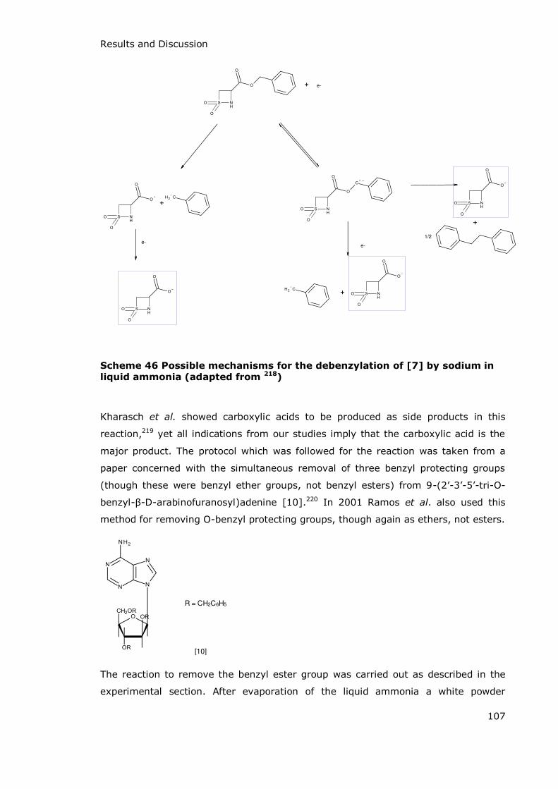
Results and Discussion
107
e-
1/2
e-e-
Scheme 46 Possible mechanisms for the debenzylation of [7] by sodium in liquid ammonia (adapted from 218)
Kharasch et al. showed carboxylic acids to be produced as side products in this
reaction,219 yet all indications from our studies imply that the carboxylic acid is the
major product. The protocol which was followed for the reaction was taken from a
paper concerned with the simultaneous removal of three benzyl protecting groups
(though these were benzyl ether groups, not benzyl esters) from 9-(2’-3’-5’-tri-O-
benzyl- -D-arabinofuranosyl)adenine [10].220 In 2001 Ramos et al. also used this
method for removing O-benzyl protecting groups, though again as ethers, not esters.
R = CH2C6H5
[10]
The reaction to remove the benzyl ester group was carried out as described in the
experimental section. After evaporation of the liquid ammonia a white powder

Results and Discussion
108
remained of which a sample was dissolved in D2O and analysed by NMR. The 1H NMR
spectrum was studied for evidence of the benzyl ester, bibenzyl (1,2-
diphenylethane) or toluene. In fact, as can be seen in the spectrum in Figure 11,
there were no peaks in the aromatic region of the spectrum. There are, however,
three doublet of doublet peaks in the region between 4 and 5 ppm, characteristic of
the 3-substituted -sultam ring protons. A small amount of the hydrolysis product
was also indicated and there were various other unidentified peaks in the region
below 4 ppm.
Figure 11 1H NMR of 3-carboxy-β-sultam with expansion of the three
doublet of doublets characteristic of the β-sultam ABX system
A sample of the crude solid was subjected to mass spectrometry and this showed
two major peaks; one at m/z 150 and the second at m/z 106 (Figure 12). The peak
at m/z 150 relates to the desired product (accurate mass 149.97033, calculated
mass 149.986652) and the one at m/z 106 relates to the decarboxylated product
(accurate mass 105.98151, calculated mass 105.996276) as shown in Scheme 47.
The isotope patterns for the molecular anion were in-keeping with those predicted.
Although the sample masses were not exactly the same as the calculated masses
9.0 8.5 8.0 7.5 7.0 6.5 6.0 5.5 5.0 4.5 4.0 3.5 3.0 2.5 2.0 1.5 1.0 0.5 0.0 ppm
0.9325
0.9494
0.9668
1.0340
1.0488
1.0641
1.0814
1.0993
1.1224
1.1383
1.1601
1.1773
1.2170
1.2345
1.2710
1.2879
1.3210
1.3626
1.3717
1.3810
1.4459
1.8151
1.9138
1.9296
1.9494
1.9686
1.9845
2.0525
2.1328
2.2462
2.2725
2.5528
2.5718
2.7157
2.7356
2.7544
2.9196
3.0603
3.0873
3.0978
3.1234
3.2057
3.2191
3.2255
3.2449
3.3548
3.3756
3.3853
3.4579
3.5271
3.5447
3.5624
3.5804
3.6120
3.6279
3.6435
3.7145
3.7685
3.7899
3.8528
3.8773
3.9629
3.9768
3.9850
3.9989
4.0212
4.1519
4.1660
4.1747
4.1889
4.2061
4.2200
4.2395
4.2534
4.2785
4.3470
4.3612
4.3811
4.3952
4.5386
4.5607
4.5720
4.5941
4.6997
7.1886
7.2237
7.3274
7.5878
0.23
0.82
0.39
1.00
0.63
1.90
1.92
4.04.14.24.34.44.54.6 ppm

Results and Discussion
109
they are close enough to confirm the structure and any deviance from the actual
mass may be due to the low mass of the molecule.
- CO2
Scheme 47 Structures of the two ions suggested by mass spectrometry and
the mechanism of decarboxylation
Figure 12 Mass spectrum for 3-carboxy-β-sultam

Results and Discussion, Kinetics
110
3.2 Kinetics
3.2.1 Overview of Hydrolysis
It is well established that -sultam hydrolysis occurs by exclusive S-N bond fission
(Scheme 48).163
H2O
Taurine
Cysteic acid
H2O
Scheme 48 Hydrolysis of β-sultams (unsubstituted and the 3-carboxy
derivative)
The unsubstituted -sultam undergoes acid catalysed hydrolysis to yield taurine
(characterised by comparison to a standard IR spectrum of taurine) and the
3-carboxy- -sultam undergoes both acid and base catalysed hydrolysis to yield
cysteic acid (characterised by comparison to an NMR spectrum of a standard sample
of cysteic acid; see Figure 13).
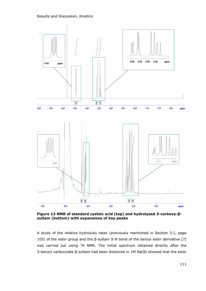
Results and Discussion, Kinetics
111
Figure 13 NMR of standard cysteic acid (top) and hydrolysed 3-carboxy-β-
sultam (bottom) with expansions of key peaks
A study of the relative hydrolysis rates (previously mentioned in Section 3.1, page
105) of the ester group and the -sultam S-N bond of the benzyl ester derivative [7]
was carried out using 1H NMR. The initial spectrum obtained directly after the
3-benzyl carboxylate -sultam had been dissolved in 1M NaOD showed that the ester
6.0 5.5 5.0 4.5 4.0 3.5 3.0 2.5 2.0 1.5 1.0 0.5 ppm
1.07
1.06
1.00
3.453.503.553.60 ppm4.50 ppm
3.03.54.04.55.05.5 ppm
1.00
1.03
0.99
0.49
3.6 ppmppm
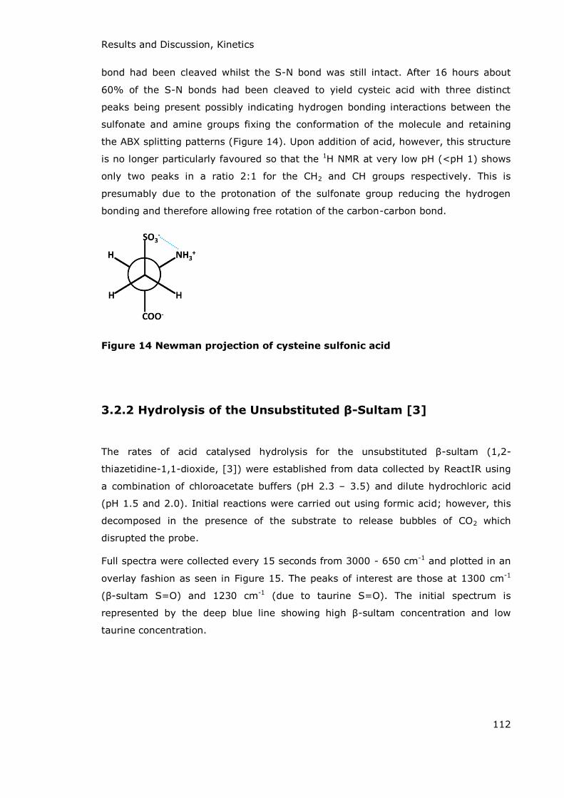
Results and Discussion, Kinetics
112
bond had been cleaved whilst the S-N bond was still intact. After 16 hours about
60% of the S-N bonds had been cleaved to yield cysteic acid with three distinct
peaks being present possibly indicating hydrogen bonding interactions between the
sulfonate and amine groups fixing the conformation of the molecule and retaining
the ABX splitting patterns (Figure 14). Upon addition of acid, however, this structure
is no longer particularly favoured so that the 1H NMR at very low pH (<pH 1) shows
only two peaks in a ratio 2:1 for the CH2 and CH groups respectively. This is
presumably due to the protonation of the sulfonate group reducing the hydrogen
bonding and therefore allowing free rotation of the carbon-carbon bond.
Figure 14 Newman projection of cysteine sulfonic acid
3.2.2 Hydrolysis of the Unsubstituted β-Sultam [3]
The rates of acid catalysed hydrolysis for the unsubstituted -sultam (1,2-
thiazetidine-1,1-dioxide, [3]) were established from data collected by ReactIR using
a combination of chloroacetate buffers (pH 2.3 – 3.5) and dilute hydrochloric acid
(pH 1.5 and 2.0). Initial reactions were carried out using formic acid; however, this
decomposed in the presence of the substrate to release bubbles of CO2 which
disrupted the probe.
Full spectra were collected every 15 seconds from 3000 - 650 cm-1 and plotted in an
overlay fashion as seen in Figure 15. The peaks of interest are those at 1300 cm-1
( -sultam S=O) and 1230 cm-1 (due to taurine S=O). The initial spectrum is
represented by the deep blue line showing high -sultam concentration and low
taurine concentration.
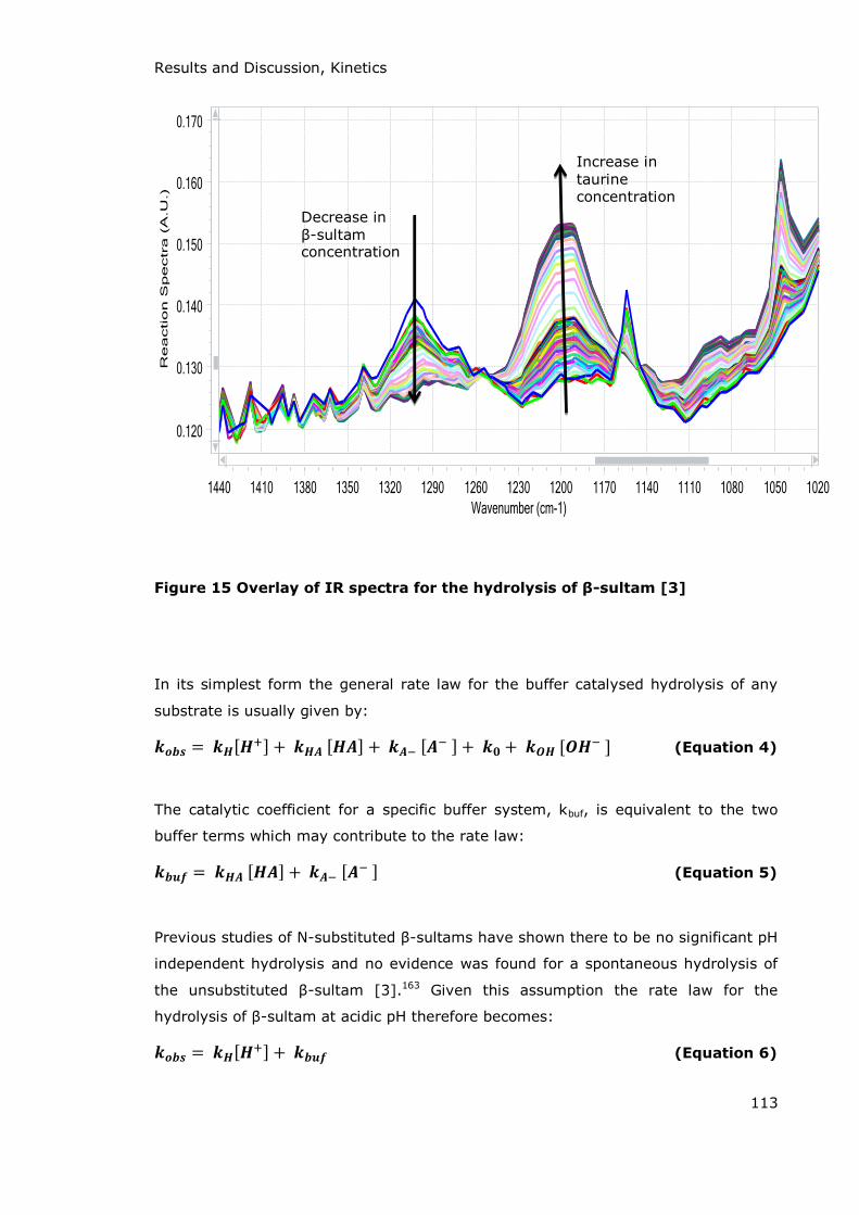
Results and Discussion, Kinetics
113
Figure 15 Overlay of IR spectra for the hydrolysis of β-sultam [3]
In its simplest form the general rate law for the buffer catalysed hydrolysis of any
substrate is usually given by: (Equation 4)
The catalytic coefficient for a specific buffer system, kbuf, is equivalent to the two
buffer terms which may contribute to the rate law: (Equation 5)
Previous studies of N-substituted -sultams have shown there to be no significant pH
independent hydrolysis and no evidence was found for a spontaneous hydrolysis of
the unsubstituted -sultam [3].163 Given this assumption the rate law for the
hydrolysis of -sultam at acidic pH therefore becomes: (Equation 6)
Decrease in -sultam
concentration
Increase in taurine concentration

Results and Discussion, Kinetics
114
In order to determine the individual constants for the rate law, kH and kbuf, the rate
of hydrolysis of -sultam was measured at a range of pHs in chloroacetate buffer at
constant pH and ionic strength and varying concentrations of buffer under pseudo
first-order conditions. The observed rate constants, kobs, for hydrolysis of
1,2-thiazetidine-1,1-dioxide ( -sultam [3]) increased linearly with increasing total
buffer concentration. This indicates the buffer is also contributing to the overall rate
of the reaction (as suggested by Equation 4). By plotting the observed pseudo-first-
order rate constants for hydrolysis against the total buffer concentration two
important pieces of information can be determined (Figure 16). Extrapolation of each
line gives the intercept on the y axis, kint, which corresponds to the rate constant at
zero buffer concentration. This can be used to calculate kH; the rate constant for
specific acid catalysis. The slopes of the lines in Figure 16 give kbuf at each pH which
is the total contribution to the rate law by both the undissociated chloroacetic acid
and the chloroacetate anions in solution (Table 8).
Figure 16 Plot of the observed pseudo-first order rate constant for the hydrolysis of β-sultam [3] as a function of total chloroacetate buffer
concentration at the pH indicated by the key
0
0.005
0.01
0.015
0.02
0.025
0.03
0.035
-0.05 0 0.05 0.1 0.15 0.2 0.25 0.3 0.35
ko
bs
/ s-1
Buffer concentration / M
pH2.3
pH2.55
pH2.8
pH3.3
pH3.5

Results and Discussion, Kinetics
115
pH 2.3 2.55 2.8 3.3 3.5 kint (s
-1) 6.9 x 10⁻³ + 3.45 x 10⁻⁴
6.1 x 10⁻³ + 3.05 x 10⁻⁴
4.1 x 10⁻³ + 2.05 x 10⁻⁴
2.5 x 10⁻³ + 1.25 x 10⁻⁴
1.7 x 10⁻³ + 8.50 x 10⁻⁵
kbuf (M-1s-1) 7.59 x 10⁻² +
3.80 x 10⁻³ 5.99 x 10⁻² + 3.00 x 10⁻³
4.99 x 10⁻² + 2.50 x 10⁻³
2.49 x 10⁻² + 1.25 x 10⁻⁴
1.49 x 10⁻² + 7.45 x 10⁻⁴
Table 8 Values of parameters derived from Figure 15
A graph of kbuf as a function of the fraction of free base, α, at each pH studied gives
the individual second order rate constants for catalysis by the acidic and basic
components of the buffer, kHA and kA-, at α = 0.0 and α = 1.0 respectively (Equation
7) (Figure 17).
(Equation 7)
Figure 17 Plot of kbuf vs α (fraction of free base) for the chloroacetate buffer
catalysed hydrolysis of the unsubstituted β-sultam
The intercept at α = 1.0 is indistinguishable from zero, so no rate constant for
catalysis of -sultam hydrolysis by chloroacetate anions, kA-, can be determined. The
second order rate constant for catalysis by chloroacetic acid, kHA, is given by the
intercept at α = 0.0 as 9.51 x 10⁻² M-1s-1.
There is only one previous mention of the investigation of the rate of -sultam
hydrolysis in chloroacetate buffers to be found and this relates to a study of the
effect of carboxylic acid catalysed hydrolysis of N-benzyl- -sultam.137 The value of
0
0.02
0.04
0.06
0.08
0.1
0
0.02
0.04
0.06
0.08
0.1
0 0.2 0.4 0.6 0.8 1
kb
uf M
-1s-1
kb
uf M
-1s-1
Fraction of free base, α

Results and Discussion, Kinetics
116
kHA for this reaction is quoted as 7.61 x 10⁻² M-1s-1, which is of the same order of
magnitude as that of the unsubstituted -sultam. The proposed mechanism for
buffer catalysis in this paper was specific acid-nucleophilic catalysis and this is one
possibility for the mechanism for the hydrolysis of the unsubstituted -sultam also.
The general acid catalysed hydrolysis of [3] could be due to several possible
mechanisms as seen in Scheme 49. Both mechanisms, the concerted general acid
catalysis and stepwise specific acid catalysis followed by nucleophilic or general base
catalysis, show equivalent rate laws (Equation 8).
Nucleophilic General
Base
General Acid
Scheme 49 Possible mechanisms of catalysis by chloroacetic acid in the
hydrolysis of β-sultam [3]
(Equation 8)
The intercepts of the rate-buffer plots (Figure 16), kint, increase with decreasing pH
indicative of an acid catalysed pathway. Pseudo first order rate constants for
hydrolysis of [3] were also determined in solutions of hydrochloric acid. By
combining the data from buffer catalysis experiments (kH values) with these values a

Results and Discussion, Kinetics
117
pH rate profile for the hydrolysis of the unsubstituted -sultam [3] at low pH is
produced (Figure 18).
Figure 18 pH rate profile for acid catalysed hydrolysis of unsubstituted beta-
sultam
Unusually the slope of the plot of log kobs / log kint against pH is 0.6, whereas a
simple acid catalysed reaction would be expected to generate a slope of 1.0. This
non-first order dependence on [H+] is also seen by the variation in the values of
kobs/[H+] and kint/[H
+] (Table 9). It is difficult to explain this and may be due to error
in the novel experimental method for determining the rates using the React-IR
probe. It seems unlikely that this is due to partial formation of the conjugate acid of
[3] or to the partial occurrence of a pH-independent pathway.
pH
1.6 2.0 2.3 2.6 2.8 3.3 3.5
kobs /[H+]
1.67 1.32 1.42 2.23 2.72 5.39 6.01
Table 9 Values of kobs/[H+] over pH range of study
y = -0.5742x - 0.7554
-4
-3.5
-3
-2.5
-2
-1.5
1 1.5 2 2.5 3 3.5 4 4.5 5
log
(ra
te c
on
sta
nt)
pH
Results and Discussion, Kinetics
118
3.2.3 Hydrolysis of 3-Carboxy-β-Sultam
The rates of hydrolysis of 3-carboxy- -sultam [8] at various pH were established
using data collected by pre-saturation 1H NMR spectra taken at appropriate time
intervals. Low pH/D experiments were carried out in deuterium chloride (DCl)
solutions and higher pH/D experiments were carried out in sodium deuteroxide
(NaOD) solutions. For mid-range pH experiments various deuterated buffers were
used (as detailed in Table 10) at a range of concentrations in order to establish and
eliminate any contribution from buffer catalysis.
Buffer pH range (measured)
(pD range) (calculated)
Concentrations utilised (M)
Phosphate 2.99 - 3.55
(3.39 – 3.95)
0.50, 0.25, 0.10
Acetate 4.78 - 5.32
(5.18 – 5.72)
0.50, 0.25, 0.10
Phosphate 6.59 - 8.01
(6.99 – 8.41)
0.50, 0.25, 0.10
Carbonate 9.69
(10.09)
0.50, 0.25, 0.10
Table 10 Details of deuterated buffers employed in the study of 3-carboxy-
β-sultam hydrolysis rates
As with previous work on the hydrolysis of the unsubstituted -sultam, plots of kobs
versus buffer concentration gave kint (the rate constant for hydrolysis at zero buffer
concentration) and kbuf (the contribution to the rate law by all components of the
buffer solution; kHA and kA-) (Equation 5). These plots can be seen for a range of
buffers in Figure 19 showing the increase in observed pseudo-first order rate

Results and Discussion, Kinetics
119
constant with increasing buffer concentration indicative of buffer catalysis, either by
the acidic or basic component of the buffer. The values of kint are shown in Table 11
where it can be seen that the value of the rate constant increases as the pH
decreases. There is a greater amount of scatter in the data than would be preferred.
Figure 19 Plot of the observed pseudo-first order rate constants for the
hydrolysis of 3-carboxy-β-sultam [8] as a function of total buffer
concentration (measured pH indicated by the key)
pD 3.39 5.20 5.70 7.00 7.70 8.00 10.09
kint (s-1) 3.02
x 10-2 6.3 x 10⁻³
1.43 x 10⁻²
2.3 x 10⁻³
3.7 x 10⁻³
2.1 x 10⁻³
5.00 x 10⁻⁴
Table 11 Values of rate constants (kint) for buffers, determined from Figure 19
In order to establish which form of the buffer is responsible for catalysis it is
necessary to carry out experiments at a range of pHs in the same buffer system.
Experiments were carried out in phosphate buffer (at pKa = 7.2) at three pHs. Plots
of kobs against the total buffer concentration for these experiments gave two
important pieces of information about the rates of catalysis. The intercept on the y
axis, kint, corresponds to the rate constant at zero buffer concentration and is the
0.00E+00
1.00E-02
2.00E-02
3.00E-02
4.00E-02
5.00E-02
6.00E-02
7.00E-02
8.00E-02
0 0.1 0.2 0.3 0.4 0.5 0.6
ko
bs
/ s-1
Buffer concentration / M
pH6.6
pH7.3
pH7.6
pH4.8
pH5.3
pH9.69
pH2.99

Results and Discussion, Kinetics
120
observed first-order rate constant for buffer independent hydrolysis. The slope of the
graph, kbuf, is equivalent to the contribution to the rate law by both buffer species;
the dihydrogen phosphate monoanion and the monohydrogen phosphate dianion. A
plot of kbuf against α, the fraction of free base, gave intercepts of kHA when α = 0.0
and kA- when α = 1.0. These are the second order rate constants for catalysis by the
acidic (H2PO4-) and basic (HPO4
2-) components of the buffer respectively. As with the
chloroacetate buffer catalysed hydrolysis of the unsubstituted -sultam this plot
(seen in Figure 20) shows that the kinetically relevant species for the phosphate
buffer catalysis (pKa = 7.2) of 3-carboxy- -sultam is the acidic phosphate
monoanion, H2PO4- and that there is no significant catalysis by the dianion HPO4
2-.
Although there is only limited data (three points) it is still possible to determine the
value of the rate constant kHA from the graph which is equal to 2.243 x 10⁻² M-1s-1.
Figure 20 Plot of kbuf vs α (fraction of free base) for the phosphate buffer
catalysed hydrolysis of the 3-carboxy-β-sultam
In carbonate buffer systems (pKa = 10.25) data is only available for one pH, pH
9.69, and so the contribution of the individual buffer species to the catalytic
coefficient kbuf can only be estimated via Equation 9 where α is equal to the fraction
of the buffer present in the basic form (CO32-). This assumes that kA- is zero so that it
0.00E+00
5.00E-03
1.00E-02
1.50E-02
2.00E-02
2.50E-02
0.00E+00
5.00E-03
1.00E-02
1.50E-02
2.00E-02
2.50E-02
0 0.2 0.4 0.6 0.8 1
kb
uf,
M-1
s-1
kb
uf,
M-1
s-1
fraction of free base, α
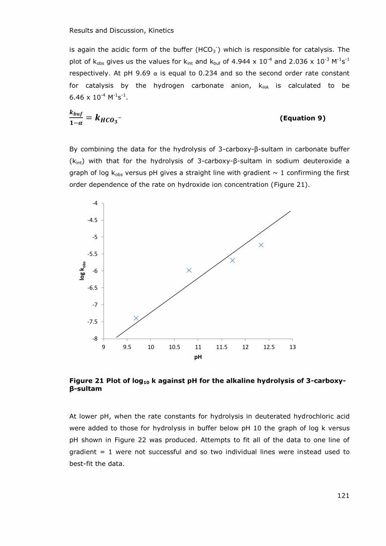
Results and Discussion, Kinetics
121
is again the acidic form of the buffer (HCO3-) which is responsible for catalysis. The
plot of kobs gives us the values for kint and kbuf of 4.944 x 10⁻⁴ and 2.036 x 10⁻³ M-1s-1
respectively. At pH 9.69 α is equal to 0.234 and so the second order rate constant
for catalysis by the hydrogen carbonate anion, kHA is calculated to be
6.46 x 10⁻⁴ M-1s-1. (Equation 9)
By combining the data for the hydrolysis of 3-carboxy- -sultam in carbonate buffer
(kint) with that for the hydrolysis of 3-carboxy- -sultam in sodium deuteroxide a
graph of log kobs versus pH gives a straight line with gradient ~ 1 confirming the first
order dependence of the rate on hydroxide ion concentration (Figure 21).
Figure 21 Plot of log10 k against pH for the alkaline hydrolysis of 3-carboxy-
β-sultam
At lower pH, when the rate constants for hydrolysis in deuterated hydrochloric acid
were added to those for hydrolysis in buffer below pH 10 the graph of log k versus
pH shown in Figure 22 was produced. Attempts to fit all of the data to one line of
gradient = 1 were not successful and so two individual lines were instead used to
best-fit the data.
-8
-7.5
-7
-6.5
-6
-5.5
-5
-4.5
-4
9 9.5 10 10.5 11 11.5 12 12.5 13
log
ko
bs
pH
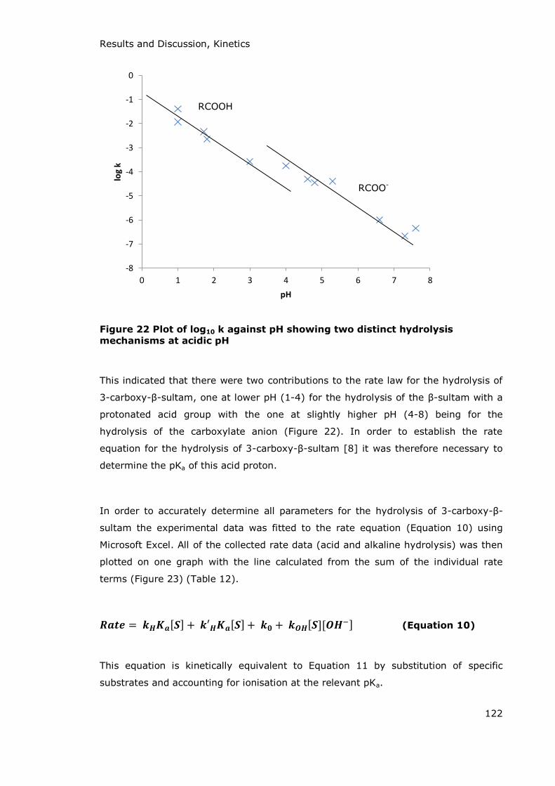
Results and Discussion, Kinetics
122
Figure 22 Plot of log10 k against pH showing two distinct hydrolysis
mechanisms at acidic pH
This indicated that there were two contributions to the rate law for the hydrolysis of
3-carboxy- -sultam, one at lower pH (1-4) for the hydrolysis of the -sultam with a
protonated acid group with the one at slightly higher pH (4-8) being for the
hydrolysis of the carboxylate anion (Figure 22). In order to establish the rate
equation for the hydrolysis of 3-carboxy- -sultam [8] it was therefore necessary to
determine the pKa of this acid proton.
In order to accurately determine all parameters for the hydrolysis of 3-carboxy- -
sultam the experimental data was fitted to the rate equation (Equation 10) using
Microsoft Excel. All of the collected rate data (acid and alkaline hydrolysis) was then
plotted on one graph with the line calculated from the sum of the individual rate
terms (Figure 23) (Table 12).
(Equation 10)
This equation is kinetically equivalent to Equation 11 by substitution of specific
substrates and accounting for ionisation at the relevant pKa.
-8
-7
-6
-5
-4
-3
-2
-1
0
0 1 2 3 4 5 6 7 8
log
k
pH
RCOOH
RCOO-

Results and Discussion, Kinetics
123
(Equation 11)
The second term in Equation 11 is kinetically ambiguous and could be due to
hydrolysis of [RCOOH]:
Ka
The second order rate constant for the acid catalysed hydrolysis of [8] with
dissociated carboxylic acid group [RCOO-] is 4.80 M-1s-1 which is 24 fold greater than
that for [8] with an undissociated carboxylic acid [RCOOH]. This rate enhancement is
potentially due to the enhanced stability of the conjugate acid (Scheme 50A). The
rate enhancement seen is not as large as would be expected for the potential
increase in stability shown and this could be due to intramolecular general acid
catalysis with the protonated form of the compound (Scheme 50B).
A B
Scheme 50 Potential mechanisms of rate enhancement with dissociated 3-
carboxy-β-sultam [8]
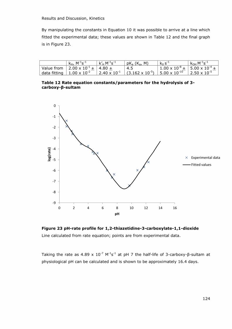
Results and Discussion, Kinetics
124
By manipulating the constants in Equation 10 it was possible to arrive at a line which
fitted the experimental data; these values are shown in Table 12 and the final graph
is in Figure 23.
kH, M-1s-1 k’H M-1s-1 pKa (Ka, M) k0 s
-1 kOH M-1s-1
Value from data fitting
2.00 x 10⁻¹ + 1.00 x 10⁻²
4.80 + 2.40 x 10⁻¹
4.5 (3.162 x 10⁻⁵)
1.00 x 10⁻⁸ + 5.00 x 10⁻¹⁰
5.00 x 10⁻⁴ + 2.50 x 10⁻⁵
Table 12 Rate equation constants/parameters for the hydrolysis of 3-carboxy-β-sultam
Figure 23 pH-rate profile for 1,2-thiazetidine-3-carboxylate-1,1-dioxide
Line calculated from rate equation; points are from experimental data.
Taking the rate as 4.89 x 10⁻⁷ M-1s-1 at pH 7 the half-life of 3-carboxy- -sultam at
physiological pH can be calculated and is shown to be approximately 16.4 days.
-9
-8
-7
-6
-5
-4
-3
-2
-1
0
0 2 4 6 8 10 12 14 16
log
(ra
te)
pH
Experimental data
Fitted values

Results and Discussion, Inhibition Studies
125
3.3 Inhibition Studies
3.3.1 Inhibition of BcII
BcII is the di-zinc Class B1 metallo- -lactamase enzyme produced by Bacillus cereus
569/H. Few compounds have been discovered to be inhibitors of the enzyme,
particularly inhibitors which are specific only to BcII and do not interact with other
metallo-enzymes such as carbonic anhydrase. BcII is an efficient catalyst of -lactam
ring hydrolysis with a kcat/KM value of 4.5 x 10⁵ M-1s-1 for benzylpenicillin
(kcat = 680 s-1, KM = 1.5 x 10⁻³ M).110
3.3.1.1 β-Sultams and their Analogues as BcII Inhibitors
All -lactamase enzymes, including BcII are efficient catalysts for the hydrolysis of -
lactam antibiotics, all of which have a carboxylic acid group at the three position of
the fused ring. This structure is important for recognition by the enzyme active site
as it is an analogue of the D-Ala-D-Ala backbone recognised by the DD-
transpeptidase enzymes which -lactam antibiotics inhibit.
3-Carboxy- -sultam [8] was chosen as a potential inhibitor of BcII as possibly the
sulfonyl centre could interact with Zn1 and the carboxylate with Zn2. Scheme 51
compares the structures of the 3-carboxy- -sultam and a generic penicillin. This
shows the position of the carboxylic acid group relative to the sulfonyl and carbonyl
groups respectively in each compound. Although both structures are named with the
carboxylate functionality at the 3-position this is not exactly the same position on
both molecules, though they are both two bond lengths from the carbonyl/sulfonyl
group.

Results and Discussion, Inhibition Studies
126
Scheme 51 Numbering of 3-carboxy-β-sultam and penicillin to show
carboxylate functionality at 3-position
Due to the similar geometry shown between the two structures (Scheme 51) it was
hoped that the -sultam would interact with the active site zinc ions and zinc bound
water molecules as shown in Scheme 52.
Scheme 52 Possible mode of interaction between 3-carboxy β-sultam and BcII
Experimental data showed that 3-carboxy- -sultam is not a substrate for BcII -
lactamase, and neither is the unsubstituted -sultam. Equally, neither compound
showed inhibitory action against BcII despite the structural similarity between the
two compounds and the -lactam moeity of the normal substrate. It is possible that
the lack of steric bulk and functional groups on the test compounds reduced the
recognition by the active site. There are comparable studies in the literature for work
done on the inhibition of BcII by analogous -lactam compounds.221 Although BcII

Results and Discussion, Inhibition Studies
127
has a broad substrate profile, including penicillins, cephalosporins and carbapenems,
it has been shown that it does not efficiently catalyse the hydrolysis of
monobactams. Aztreonam (Scheme 53) is a representative monobactam which binds
to BcII but is not hydrolysed by it. The azetidinone shown in Scheme 53B is a -
lactam with an N-acetoxy substituent showing similar functionality to 3-carboxy- -
sultam which does not bind to BcII at all. The suggestion in this paper was that a
lack of other substituents may contribute to the lack of binding. It would be
interesting to investigate 3-carboxy- -sultams with substituents at the 4-position to
see if the addition of extra fucnctionality affected the inhibitory action.
A B
Scheme 53 Structures of monobactams, aztreonam (A) and 2-oxoazetidinyl- acetate sodium salt (B)
1,2-Thiazetidine-1,1-dioxide has been shown to be a potentially useful pro-drug,
hydrolysing in situ to yield taurine.198 Following on from this it seemed logical to
study the hydrolysis products of the two -sultams and their analogues as potential
inhibitors of BcII. The compounds chosen were taurine, L-cysteic acid, L-cysteine and
D-cysteine (based on their structural relevance and commercial availability) (Scheme
54). Taurine (the hydrolysis product of -sultam) and L-cysteine (the thiol analogue
of the hydrolysis product of 3-carboxy- -sultam) showed no effect on the activity of
BcII in the hydrolysis of benzylpenicillin (results for taurine shown in Figure 24).

Results and Discussion, Inhibition Studies
128
L-Cysteine D-Cysteine
L-Cysteic acid Taurine
Scheme 54 Hydrolysis products of β-sultams and their analogues
Figure 24 Effect on the rate of the BcII catalysed hydrolysis of
benzylpenicillin by taurine
0
0.0005
0.001
0.0015
0.002
0.0025
0.003
0.0035
0.004
0.0045
0.005
0 10 20 30 40 50 60 70
Pe
nG
co
nce
ntr
ati
on
, M
Time, s
[I]=0
[I]=0.00379
[I]=0.0148
[I]=0.0355
[I]=0.0514
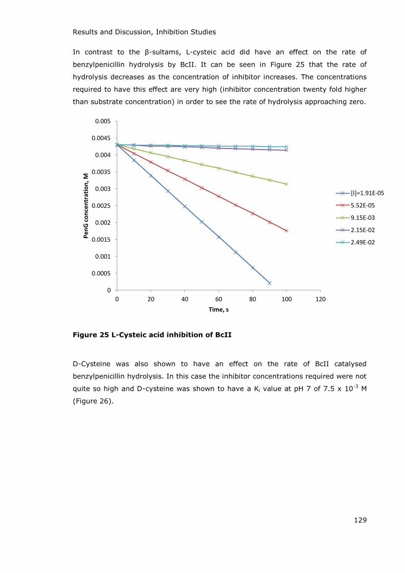
Results and Discussion, Inhibition Studies
129
In contrast to the -sultams, L-cysteic acid did have an effect on the rate of
benzylpenicillin hydrolysis by BcII. It can be seen in Figure 25 that the rate of
hydrolysis decreases as the concentration of inhibitor increases. The concentrations
required to have this effect are very high (inhibitor concentration twenty fold higher
than substrate concentration) in order to see the rate of hydrolysis approaching zero.
Figure 25 L-Cysteic acid inhibition of BcII
D-Cysteine was also shown to have an effect on the rate of BcII catalysed
benzylpenicillin hydrolysis. In this case the inhibitor concentrations required were not
quite so high and D-cysteine was shown to have a Ki value at pH 7 of 7.5 x 10⁻³ M
(Figure 26).
0
0.0005
0.001
0.0015
0.002
0.0025
0.003
0.0035
0.004
0.0045
0.005
0 20 40 60 80 100 120
Pe
nG
co
nce
ntr
ati
on
, M
Time, s
[I]=1.91E-05
5.52E-05
9.15E-03
2.15E-02
2.49E-02

Results and Discussion, Inhibition Studies
130
Figure 26 Inhibition of BcII by D-Cysteine
Compound Effect on activity of BcII
-Sultam No effect 3-Carboxy- -Sultam No effect Taurine No effect L-Cysteic Acid Very weak inhibition L-Cysteine No effect D-Cysteine Ki = 7.5 x 10⁻³ M
Table 13 Summary of results for the inhibition of BcII by β-sultams and their analogues
R² = 0.9859
-2.E-06
-1.E-06
0.E+00
1.E-06
2.E-06
3.E-06
4.E-06
5.E-06
6.E-06
7.E-06
8.E-06
-1.5E-02 -1.0E-02 -5.0E-03 0.0E+00 5.0E-03 1.0E-02 1.5E-02 2.0E-02 2.5E-02
KM
/kca
t (M
s)
Inhibitor concentration (M)

Results and Discussion, Inhibition Studies
131
3.3.1.2 Dicarboxylic Acids as BcII Inhibitors
The second group of compounds chosen as potential inhibitors of BcII were
dicarboxylic acids. One possibility of the interactions between the two dicarboxylates
and the two active site zinc ions is demonstrated in Scheme 55A. Derivatives of
succinic and phthalic acids have been shown to inhibit another Class B1 enzyme,
IMP-1, via the displacement of the di-zinc bound water molecule, Wat1, by one
carboxylate and the forming of a bridge by the second carboxylate between Zn2 and
a lysine residue at the active site as seen in Scheme 55B.129
A B
EnzymeEnzyme
Scheme 55 Potential interactions between dicarboxylic acids and BcII
The following dicarboxylic acids were tested as potential inhibitors of the BcII
catalysed hydrolysis of benzylpenicillin:
Pimelic acid [10], malonic acid [11], succinic acid [12], oxalic acid [13],
cyclopentane 1,2-dicarboxylic acid [14] and 2,3-norbornanedicarboxylic acid [15]
(structures in Scheme 56).

Results and Discussion, Inhibition Studies
132
oxalic acid [13]
malonic acid [11]
succinic acid [12]
pimelic acid [10]
cyclopentane-1,2-dicarboxylic acid [14] 2,3-norbornanedicarboxylic acid [15]
Scheme 56 Structures of dicarboxylic acids tested as inhibitors of BcII
In all cases the ability of the compound to inhibit the hydrolysis of benzylpenicillin by
BcII was determined by comparison of the initial rate of substrate hydrolysis in the
presence of inhibitor to the control rate (with no inhibitor present). In all cases the
initial rates of the reactions were of the same order with the average rate in the
presence of a dicarboxylic acid being 11.2 x 10⁻³ + 1.3 x 10⁻³ s-1 compared to the
control rate of 9.2 x 10⁻³ s-1. No change in pH was noted between the start and end
of any experiment. This is in sharp contrast to the results seen for L-cysteic acid
which, other than the amino group and the substitution of a carboxylic for a sulfonic
acid group is structurally similar to the dicarboxylic acids, particularly succinic acid
(Scheme 57). Previous work has shown that IMP-1 is inhibited by disubstituted
succinic acids (dibenzyl-succinic acid shown in Scheme 57) via interactions between
the two carboxylate groups with Wat1 and Zn2 at the active site. It appears that
whilst the substituents are not involved in the enzyme-inhibitor interactions they
may be involved in recognition for the active site. This suggestion is strengthened
when looking at the difference in IC50 values for the S,S, R,R and R,S stereoisomers
of dibenzylsuccinic acid which are 0.0027, 0.21 and 200 με respectively.

Results and Discussion, Inhibition Studies
133
Succinic acid L-Cysteic acid
Dibenzyl succinic acid
Scheme 57 Structures of L-cysteic acid and succinic acids
The difference in activity seen between L-cysteic acid and succinic acid with BcII may
also be a function of better substrate recognition for L-cysteic acid due to the
presence of the amine group. It is not clear why this may be, but it may be due to
hydrogen bonding or electrostatic interactions between the molecule and amino acid
residues at the active site. Another possibility is that it may be due to the reduction
in free rotation of the carbon backbone due to intramolecular hydrogen bonding
interactions fixing the L-cysteic acid in a preferential conformation.

Results and Discussion, Inhibition Studies
134
3.3.2 Inhibition of Glutamine Synthetase
Glutamine synthetase is a polymeric enzyme requiring two metal ions (either Mg2+ or
Mn2+) at each active site. It effectively catalyses the formation of glutamine from
glutamate and ammonia; ATP is required to activate the glutamate carboxylate
group via phosphorylation.
Initial attempts to monitor the rate of glutamine production were carried out using
HPLC. Calibration was carried out for glutamate and glutamine in the presence of
ATP. The reaction was set up with glutamate, ammonium salt, magnesium salt, ATP
and enzyme in imidazole buffer solution at pH 7.1 and 1.0 M ionic strength
maintained by addition of potassium chloride. Samples of the solution were removed
at set intervals and quenched in 1.0 M sodium hydroxide solution to denature the
enzyme and stop the reaction. The production of ADP by the reaction was an issue as
it acted as an inhibitor of the enzyme and so only a limited number of enzyme
turnovers could be monitored. Despite multiple attempts to modify the procedure no
progress was made and this lead to the use of a coupled assay.
The coupled assay utilised for the glutamine synthetase (GS) reaction was that
detailed in the experimental (and shown again in Scheme 58). This procedure has
been widely used for investigations involving glutamine synthetase.222-224
Kinetic measurements of the biosynthetic activity of the enzyme were performed at
37 oC in a coupled assay with pyruvate kinase (PK) and lactate dehydrogenase
(LDH). For every molecule of glutamate which is converted to glutamine, one
molecule of ATP is converted to ADP. The production of ADP can be measured by the
following procedure: ADP is converted into ATP by the reaction with
phospho(enol)pyruvate, catalysed by PK; the pyruvate produced by this reaction is
then reduced by NADH (catalysed by LDH) to produce lactate and NAD+. Thus the
rate of reduction in concentration of NADH measured spectrophotometrically at
340 nm is representative of the rate of conversion of glutamate to glutamine. In
each experiment a blank (with no GS enzyme present) was run to account for the
background rate of NADH oxidation. A second cell was also set up as a control to
measure the rate of GS activity under “normal” conditions. The third cell contained
all other reactants and enzymes along with the potential inhibitor.

Results and Discussion, Inhibition Studies
135
Scheme 58 Coupled assay utilised in the UV-Vis monitoring of the glutamine
synthetase reaction
The compounds tested as inhibitors of glutamine synthetase were selected either due
to their structural similarity to the normal substrate (glutamate) or because they
were potential taurine (or taurine analogue) pro-drugs following work carried out by
Ward et al. showing taurine had a potential effect on neurotransmission in the
brain.198 The structures of these potential inhibitors are shown in Scheme 59 and a
summary of the results is shown in Table 14.
L-Cysteine D-Cysteine L-Cysteic acid
-SultamTaurineL-Glutamate
Scheme 59 Structures of compounds investigated as potential inhibitors of
glutamine synthetase (and glutamate for comparison)

Results and Discussion, Inhibition Studies
136
As can be seen from the sample data shown for -sultam inhibition of glutamine
synthetase in Figure 27 there is no significant difference in the rate of NADH
oxidation in the presence of increasing concentrations of inhibitor. Similar results
were also obtained for reactions involving taurine.
Figure 27 Effect of β-sultam on the rate of glutamine production catalysed
by GS (stock inhibitor concentration = 1.5 M)
The rate enhancements seen in the case of L- and D-cysteine are possibly due to
these compounds acting as substrates for the enzyme, GS. The increased
concentration of substrate leads to an increase in the rate of ADP production and
hence an increase in the rate of NADH oxidation. An assay was run with cysteine in
the absence of glutamate and this confirmed the substrate action by following the
decrease in the concentration of NADH.
L-Cysteic acid initially showed a similar rate enhancement to that seen with D- and
L-cysteine (Figure 28). This was not however due to the compound being a substrate
but rather the action of the compound catalysing the oxidation of NADH. This is
assumed to be via an acid-catalysis mechanism but further investigations into this
were not carried out.
98.95
99
99.05
99.1
99.15
99.2
0 200 400 600 800 1000 1200
Glu
tam
ic a
cid
co
nce
ntr
ati
on
, m
M
Time, s
blank
control
20ul I
50ul I
100ul I
200ul I
blank
control
20μl
50μl
100μl
200μl

Results and Discussion, Inhibition Studies
137
Figure 28 Apparent rate enhancements due to L-cysteic acid (stock inhibitor concentration = 0.5M)
Compound Effect on activity of glutamine synthetase
-Sultam No effect 3-Benzyl Carboxylate -Sultam No effect 3-Carboxy- -Sultam No effect Taurine No effect L-Cysteic Acid No effect L-Cysteine Rate enhancement D-Cysteine Rate enhancement
Table 14 Summary of results regarding glutamine synthetase inhibition
Further work in this area is required to fully explain many of the observations.
Testing of L- and D-cysteine as substrates of each of the three assay enzymes and
product analysis would show why the rate enhancements seen occurred. They may
be due to phosphorylation by GS producing ADP, or reduction by LDH and NADH
going to NAD+. Another avenue of interest is to test the potential inhibitors against
other neurotransmitter enzymes such as glutamate decarboxylate or GABA
transaminase.
0.85
0.9
0.95
1
1.05
1.1
1.15
1.2
1.25
0 200 400 600 800 1000 1200
Ab
sorb
an
ce (
du
e t
o N
AD
H a
t 3
40
nm
)
Time, s
Blank
Control
20ul I
50ul I
100ul I
bla
nk
con
trol
μl
blank
control
20μl
50μl
100μl

Results and Discussion, Inhibition Studies
138
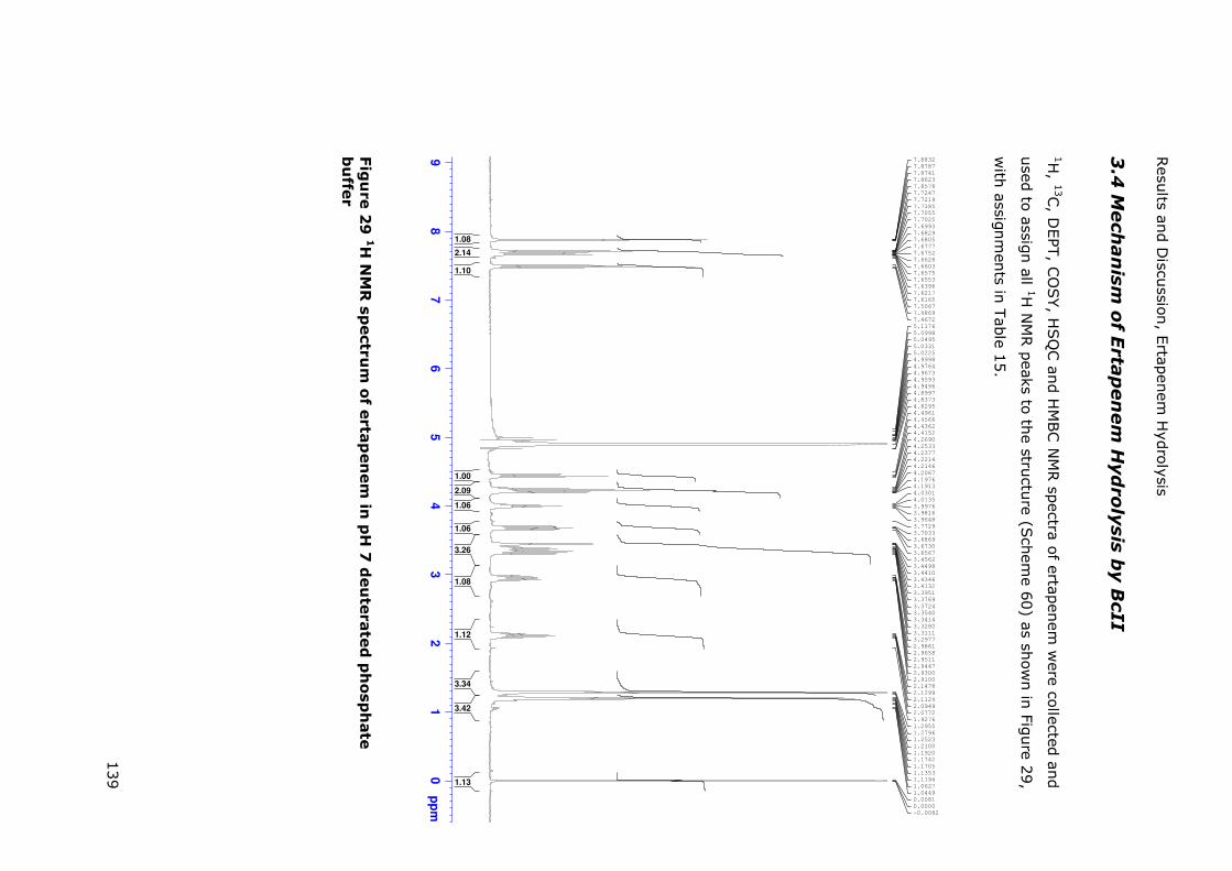
Resu
lts and D
iscussion
, Ertap
enem
Hyd
rolysis
139
3.4
Mech
an
ism
of E
rta
pen
em
Hyd
ro
lysis
by B
cII
1H, 1
3C, D
EPT
, CO
SY, H
SQ
C an
d H
MBC
NM
R sp
ectra of ertapen
em w
ere collected an
d
used
to assign all 1H
NM
R p
eaks to the stru
cture
(Sch
eme 6
0) as sh
own in
Figure 2
9,
with
assignm
ents in
Table 1
5.
Fig
ure 2
9 1H
NM
R s
pectr
um
of e
rta
pen
em
in p
H 7
deu
terate
d p
hosp
hate
bu
ffer
98
76
54
32
10
ppm
-0.0082
0.0000
0.0081
1.0449
1.0627
1.1194
1.1353
1.1705
1.1742
1.1920
1.2100
1.2523
1.2796
1.2955
1.9276
2.0772
2.0949
2.1124
2.1299
2.1478
2.9100
2.9300
2.9447
2.9511
2.9658
2.9861
3.2977
3.3111
3.3280
3.3414
3.3540
3.3724
3.3769
3.3951
3.4132
3.4346
3.4410
3.4498
3.4562
3.6567
3.6730
3.6869
3.7033
3.7729
3.9648
3.9816
3.9976
4.0135
4.0301
4.1913
4.1976
4.2067
4.2146
4.2214
4.2377
4.2533
4.2690
4.4152
4.4362
4.4566
4.4961
4.8295
4.8373
4.8997
4.9496
4.9593
4.9673
4.9764
4.9998
5.0225
5.0331
5.0495
5.0998
5.1176
7.4672
7.4869
7.5067
7.6165
7.6217
7.6396
7.6553
7.6575
7.6603
7.6628
7.6752
7.6777
7.6805
7.6829
7.6993
7.7025
7.7055
7.7185
7.7219
7.7247
7.8578
7.8623
7.8741
7.8787
7.8832
1.13
3.42
3.34
1.12
1.08
3.26
1.06
1.06
2.09
1.00
1.10
2.14
1.08

Results and Discussion, Ertapenem Hydrolysis
140
22
21
1918
17
16
13
14
15
1011
12
9
8
4
3
2
1
7
6
5
20
Scheme 60 Ertapenem with carbons numbered for assignment to NMR
spectra
Peak shift (ppm)
Splitting Number of hydrogens
Coupling constants (Hz)
Position
7.88 Singlet 1 3 7.71 Doublet 1 7.6 5 7.67 Doublet 1 7.6 7 7.49 Triplet 1 7.8 6 4.44 Triplet 1 8.0 9 4.25 Doublet 1 5.98 18 4.21 Doublet of doublets 1 2.6 + 9.1 21 4.00 Quintet 1 6.5 11 3.68 Doublet of doublets 1 6.5 + 11.9 12 3.45 Doublet of doublets 1 2.5 + 6.2 19 3.37 Triplet 1 8.2 16 3.32 Doublet of doublets 1 5.2 + 11.9 12 2.95 Doublet of triplets 1 7.9 + 14.3 10 2.21 Doublet of triplets 1 7.1 10 1.29 Doublet 3 6.2 22 1.20 Doublet 3 7.3 17
Table 15 Assignment of 1H NMR peaks

Results and Discussion, Ertapenem Hydrolysis
141
Initial attack of the BcII enzyme on the -lactam carbonyl (carbon-20) of ertapenem
leads to the cleavage of the C-N bond of the -lactam ring. The aim of this
investigation was to prove whether protonation then occurred on the -lactam
nitrogen (via enamine formation) or on carbon-13 (via imine formation) (Scheme
61).
Enamineformation
Imineformation
Scheme 61 BcII hydrolysis of ertapenem could lead to formation of an imine or enamine
Comparison of the 1H NMR spectrum from after ertapenem hydrolysis to that in
Figure 29 shows several differences in key peaks. There are two new sets of doublets
at 1.19 and 0.72 ppm which relate to small shifts of the methyl protons (carbons 22
and 17). There are, however, more significant changes in the peaks related to the
protons attached to the -lactam and dihydropyrrole rings. There are significant

Results and Discussion, Ertapenem Hydrolysis
142
changes in the chemical shift of the protons on carbons 16, 18 and 19 associated
with the changes in functionality in this area of the molecule (Table 16).
Chemical shift after BcII hydrolysis (ppm)
Chemical shift before hydrolysis (ppm)
Splitting Carbon number
Change in ppm (from pre-hydrolysis)
2.62 4.25 Doublet 18 -1.63 4.32 3.45 Doublet of
doublets 19 +0.87
2.55 3.37 Triplet 16 -0.82
Table 16 Chemical shift changes for ring protons during hydrolysis
The decrease in chemical shift for the C-16 proton suggests a movement from the
enamine to imine structure. Further evidence of this mechanism was derived from
studying the 2D NMR spectra.
From the HSQC spectrum (Figure 30) there is a correlation between the protons at
1.05 ppm and the carbon at 14 ppm. These are assigned to the methyl group
(carbon-17) and correlations between these protons and other carbons can be
established by the use of an HMBC spectrum. The protons at 1.05 ppm show three
cross-peaks by HMBC (Figure 31) which can relate to carbons 16 (2 bond lengths),
18 and 13 (both three bond lengths away). The cross-peak observed at 43 ppm is
due to interaction of H-17 and C-16. The second cross-peak at 73 ppm is between H-
17 and C-18. The third cross-peak has to be between H-17 and C-13 and is seen at
57 ppm. There is no peak in the 1H NMR spectrum for this position (nor can a cross-
peak be found on the HSQC spectrum) due to deuterium incorporation. This is
further evidence for the formation of the imine rather than protonation of the
nitrogen. If the enamine had formed then C-13 would be expected to give a peak at
around 139 ppm (as in the spectrum from before hydrolysis). The drop to 57 ppm
and lack of signal on the HSQC spectrum indicates deuterium incorporation at this
position during hydrolysis.
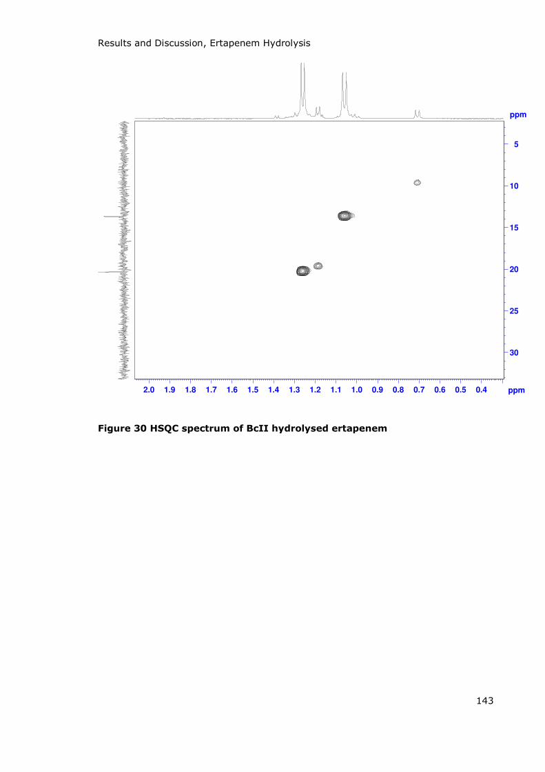
Results and Discussion, Ertapenem Hydrolysis
143
Figure 30 HSQC spectrum of BcII hydrolysed ertapenem
ppm
0.40.50.60.70.80.91.01.11.21.31.41.51.61.71.81.92.0 ppm
5
10
15
20
25
30

Results and Discussion, Ertapenem Hydrolysis
144
Figure 31 HMBC spectrum of BcII hydrolysed ertapenem
ppm
0.900.951.001.051.101.151.20 ppm
90
80
70
60
50
40
30
20
10
0

145
Conclusion
146
A new -sultam, 3-carboxylate-1,2-thiazetidine-1,1-dioxide was synthesised by the
debenzylation of 3-benzyl-1,2-thiazetidine-1,1-dioxide using sodium metal in liquid
ammonia.
Chloroacetate buffer catalyses the hydrolysis of the unsubstituted -sultam via the
acidic form of the buffer with a rate constant of 9.66 x 10⁻² M-1s-1.
3-carboxy- -sultam undergoes acid and base catalysed hydrolysis and the acid group
has a pKa of 4.5 (Ka = 3.162 x 10⁻⁵).
D-Cysteine is a weak inhibitor of BcII with a Ki value of 7.5 x 10⁻³ M.
None of the -sultams tested (the unsubstituted -sultam, 3-benzyl- -sultam or 3-
carboxy- -sultam) acted as inhibitors of BcII or GS.
Dicarboxylic acids did not show inhibition of BcII at millimolar concentrations. Both
L- and D-Cysteine were shown to be substrates of GS.
Ertapenem is hydrolysed by BcII to yield an imine functionality in the dihydropyrrole
ring.

147
Bibliography
(1) Kieny, M.-P.; Kieny, M.-P., Ed.; World Health Organization: 2012.
(2) Piddock, L. J. V. The Lancet Infectious Diseases 2012, 12, 249.
(3) Bush, K.; Jacoby, G. A.; Medeiros, A. A. Antimicrobial Agents and Chemotherapy 1995, 39, 1211.
(4) Page, M. I.; Hinchliffe, P. S.; Wood, J. M.; Harding, L. P.; Laws, A. P. Bioorganic & Medicinal Chemistry Letters 2003, 13, 4489.
(5) Gram, C. Fortschritte der Medicin 1884, 2, 185.
(6) Cummins, C. S.; Harris, H. Journal of General Microbiology 1956, 14, 583.
(7) Ghuysen, J.-M. Bacteriological Reviews 1968, 32, 425.
(8) Perkins, H. Biochemical Journal 1960, 74, 182.
(9) Meroueh, S. O.; Bencze, K. Z.; Hesek, D.; Lee, M.; Fisher, J. F.; Stemmler, T. L.; Mobashery, S. P Natl Acad Sci USA 2006, 103, 4404.
(10) Selwyn, S. Journal of Antimicrobial Chemotherapy 1979, 5, 249.
(11) Fleming, A. British Journal of Experimental Patholog 1929, 10, 226.
(12) Chain, E.; Florey, H. W.; Hardner, A.; Heatley, N. G.; Jennings, M. A.; Orr-Ewing, J.; Sanders, A. G. Lancet 1940, 239, 226.
(13) Backus, M. P.; Stauffer, J. F.; Johnson, M. J. Journal of the American Chemical Society 1946, 68, 152.
(14) Moyer, A. J.; Coghill, R. D. Journal of Bacteriology 1947, 53, 329.
(15) Ding, M.-Z.; Lu, H.; Cheng, J.-S.; Chen, Y.; Jiang, J.; Qiao, B.; Li, B.-Z.; Yuan, Y.-J. Appl Biochem Biotechnol 2012, 168, 1223.
(16) Elander, R. Applied Microbiology and Biotechnology 2003, 61, 385.
(17) Sheehan, J. C.; Henery-Logan, K. R. Journal of the American Chemical Society 1957, 79, 1262.
(18) Acred, P.; Brown, D.; Turner, D.; Wilson, M. British Journal of Pharmacology and chemotherapy 1962, 18, 356.
(19) Sutherland, R.; Croydon, E.; Rolinson, G. British Medical Journal 1972, 3, 13.
(20) Fu, K. P.; Neu, H. C. Antimicrobial Agents and Chemotherapy 1978, 13, 358.
(21) Brumfitt, W.; Percival, A.; Leigh, D. The Lancet 1967, 289, 1289.
(22) Antibiotic Discovery and Development; Dougherty, T. J.; Pucci, M. J., Eds.; Springer: New York and London, 2012.
(23) Newton, G. G.; Abraham, E. Biochem. J 1955, 62, 651.
(24) Griffith, R. S.; Black, H. R. JAMA: the Journal of the American Medical Association 1964, 189, 823.
(25) Murdoch, J. M.; Speirs, C.; Geddes, A.; Wallace, E. British Medical Journal 1964, 2, 1238.
(26) Wick, W. E. Applied Microbiology 1967, 15, 765.
148
(27) O'Callaghan, C. H.; Sykes, R.; Ryan, D.; Foord, R.; Muggleton, P. The Journal of Antibiotics 1976, 29, 29.
(28) Kahan, J.; Kahan, F.; Goegelman, R.; Currie, S.; Jackson, M.; Stapley, E.; Miller, T.; Miller, A.; Hendlin, D.; Mochales, S. The Journal of Antibiotics 1979, 32, 1.
(29) Kropp, H.; Sundelof, J.; Kahan, J.; Kahan, F. M.; Birnbaum, J. Antimicrobial Agents and Chemotherapy 1980, 17, 993.
(30) Kropp, H.; Gerckens, L.; Sundelof, J. G.; Kahan, F. M. Review of Infectious Diseases 1985, 7, S389.
(31) Pryka, R. D.; Haig, G. M. The Annals of Pharmacotherapy 1994, 28, 1045.
(32) Kurihara, A.; Naganuma, H.; Hisaoka, M.; Tokiwa, H.; Kawahara, Y. Antimicrobial Agents and Chemotherapy 1992, 36, 1810.
(33) Xuan, D.; Banevicius, M.; Capitano, B.; Kim, M.-K.; Nightingale, C.; Nicolau, D. Antimicrobial Agents and Chemotherapy 2002, 46, 2990.
(34) Papp-Wallace, K. M.; Endimiani, A.; Taracila, M. A.; Bonomo, R. A. Antimicrobial Agents and Chemotherapy 2011, 55, 4943.
(35) Jones, R. N.; Sader, H. S.; Fritsche, T. R. Diagnostic Microbiology and Infectious Disease 2005, 52, 71.
(36) Kirby, W. M. Journal of Clinical Investigation 1945, 24, 170.
(37) Hawkey, P. M. BMJ: British Medical Journal 1998, 317, 657.
(38) Malouin, F.; Bryan, L. Antimicrobial Agents and Chemotherapy 1986, 30, 1.
(39) Parija, S. C. Textbook of Microbiology and Immunology; 1 ed.; Elsevier: Haryana, 2009.
(40) Gibberd, G. British Medical Journal 1937, 2, 695.
(41) Dodd, M. C.; Stillman, W. B.; Roys, M.; Crosby, C. Journal of Pharmacology and Experimental Therapeutics 1944, 82, 11.
(42) Gottlieb, D.; Bhattacharyya, P.; Anderson, H.; Carter, H. Journal of Bacteriology 1948, 55, 409.
(43) Duggar, B. M. Annals of the New York Academy of Sciences 1948, 51, 177.
(44) Vazquez, D. Journal of General Microbiology 1966, 42, 93.
(45) Pfeiffer, R. R. Review of Infectious Diseases 1981, 3, S205.
(46) Gallo, G.; Chiesa, L.; Sensi, P. Analytical Chemistry 1962, 34, 423.
(47) Watt, L.; Jennison, R. British Medical Journal 1960, 2, 902.
(48) Lesher, G. Y.; Froelich, E. J.; Gruett, M. D.; Bailey, J. H.; Brundage, R. P. Journal of Medicinal Chemistry 1962, 5, 1063.
(49) Noall, E.; Sewards, H.; Waterworth, P. M. British Medical Journal 1962, 2, 1101.
(50) Diekema, D. J.; Jones, R. N. The Lancet 2001, 358, 1975.
(51) Tally, F. P.; DeBruin, M. F. Journal of Antimicrobial Chemotherapy 2000, 46, 523.
(52) Abraham, E. P.; Chain, E. Nature 1940, 146, 837.
(53) Hamburger, M.; Schmidt, L.; Sesler, C. L.; Ruegsegger, J.; Grupen, E. S. Journal of Infectious Diseases 1943, 73, 12.

149
(54) Klein, M. Journal of Bacteriology 1947, 53, 463.
(55) Hobson, D. The British Medical Journal 1954, 1, 236.
(56) Barber, M. Journal of Clinical Pathology 1961, 14, 385.
(57) Hane, M. W.; Wood, T. H. Journal of Bacteriology 1969, 99, 238.
(58) Franklin, T. Biochemical Journal 1967, 105, 371.
(59) Moellering, R. C.; Wennersten, C.; Weinberg, A. N. Journal of Infectious Diseases 1971, 124, S207.
(60) Orberg, P. K.; Sandine, W. E. Applied and Environmental Microbiology 1984, 48, 1129.
(61) Neu, H. C. Review of Infectious Diseases 1988, 10, S57.
(62) Marshall, S. H.; Donskey, C. J.; Hutton-Thomas, R.; Salata, R. A.; Rice, L. B. Antimicrobial Agents and Chemotherapy 2002, 46, 3334.
(63) Hooper, D. C. Emerging Infectious Diseases 2001, 7, 337.
(64) McDaniel, L. D.; Young, E.; Delaney, J.; Ruhnau, F.; Ritchie, K. B.; Paul, J. H. Science 2010, 330, 50.
(65) Yong, D.; Toleman, M. A.; Giske, C. G.; Cho, H. S.; Sundman, K.; Lee, K.; Walsh, T. R. Antimicrobial Agents and Chemotherapy 2009, 53, 5046.
(66) Bushnell, G.; Mitrani-Gold, F.; Mundy, L. M. International Journal of Infectious Diseases 2013, 17, e325.
(67) Drawz, S. M.; Bonomo, R. A. Clinical Microbiology Reviews 2010, 23, 160.
(68) Hall, B. G.; Barlow, M. Journal of Antimicrobial Chemotherapy 2005, 55, 1050.
(69) Jaurin, B.; Grundström, T. Proceedings of the National Academy of Sciences 1981, 78, 4897.
(70) Ouellette, M.; Bissonnette, L.; Roy, P. H. Proceedings of the National Academy of Sciences 1987, 84, 7378.
(71) Huovinen, P.; Huovinen, S.; Jacoby, G. A. Antimicrobial Agents and Chemotherapy 1988, 32, 134.
(72) Medeiros, A. A.; Hedges, R. W.; Jacoby, G. A. Journal of Bacteriology 1982, 149, 700.
(73) Tipper, D. J.; Strominger, J. L. P Natl Acad Sci USA 1965, 54, 1133.
(74) Matagne, A.; Dubus, A.; Galleni, M.; Frère, J. M. Natural Product Reports 1999, 16, 1.
(75) Strynadka, N. C.; Adachi, H.; Jensen, S. E.; Johns, K.; Sielecki, A.; Betzel, C.; Sutoh, K.; James, M. N. Nature 1992, 359.
(76) Lamotte-Brasseur, J.; Dive, G.; Dideberg, O.; Charlier, P.; Frère, J.-M.; Ghuysen, J.-M. Biochemical Journal 1991, 279, 213.
(77) Howarth, T. T.; Brown, A. G.; King, T. J. Journal of the Chemical Society, Chemical Communications 1976, 266b.
(78) Brown, A.; Butterworth, D.; Cole, M.; Hanscomb, G.; Hood, J.; Reading, C.; Rolinson, G. The Journal of Antibiotics 1976, 29, 668.
(79) Reading, C.; Cole, M. Antimicrobial Agents and Chemotherapy 1977, 11, 852.
(80) Knowles, J. R. Accounts of Chemical Research 1985, 18, 97.
150
(81) Imtiaz, U.; Billings, E.; Knox, J. R.; Manavathu, E. K.; Lerner, S. A.; Mobashery, S. Journal of the American Chemical Society 1993, 115, 4435.
(82) Page, M. G. P. Drug Resistance Updates 2000, 3, 109.
(83) Padayatti, P. S.; Helfand, M. S.; Totir, M. A.; Carey, M. P.; Carey, P. R.; Bonomo, R. A.; van den Akker, F. Journal of Biological Chemistry 2005, 280, 34900.
(84) Buynak, J. D. Biochemical Pharmacology 2006, 71, 930.
(85) Beesley, T.; Gascoyne, N.; Knott-Hunziker, V.; Petursson, S.; Waley, S.; Jaurin, B.; Grundström, T. Biochemical Journal 1983, 209, 229.
(86) Weston, G. S.; Blázquez, J.; Baquero, F.; Shoichet, B. K. Journal of Medicinal Chemistry 1998, 41, 4577.
(87) Powers, R. A.; Shoichet, B. K. Journal of Medicinal Chemistry 2002, 45, 3222.
(88) Eidam, O.; Romagnoli, C.; Caselli, E.; Babaoglu, K.; Pohlhaus, D. T.; Karpiak, J.; Bonnet, R.; Shoichet, B. K.; Prati, F. Journal of Medicinal Chemistry 2010, 53, 7852.
(89) Pratt, R. Science 1989, 246, 917.
(90) Chen, C. C. H.; Rahil, J.; Pratt, R. F.; Herzberg, O. Journal of Molecular Biology 1993, 234, 165.
(91) Lobkovsky, E.; Billings, E. M.; Moews, P. C.; Rahil, J.; Pratt, R. F.; Knox, J. R. Biochemistry 1994, 33, 6762.
(92) Maveyraud, L.; Pratt, R. F.; Samama, J.-P. Biochemistry 1998, 37, 2622.
(93) Badarau, A.; Damblon, C.; Page, M. I. Biochemical Journal 2007, 401, 197.
(94) Rasmussen, B. A.; Bush, K. Antimicrobial Agents and Chemotherapy 1997, 41, 223.
(95) Yang, Y.; Rasmussen, B. A.; Bush, K. Antimicrobial Agents and Chemotherapy 1992, 36, 1155.
(96) Galleni, M.; Lamotte-Brasseur, J.; Rossolini, G. M.; Spencer, J.; Dideberg, O.; Frère, J.-M. Antimicrobial Agents and Chemotherapy 2001, 45, 660.
(97) Rossolini, G. M.; Franceschini, N.; Riccio, M. L.; Mercuri, P. S.; Perilli, M.; Galleni, M.; Frère, J.-M.; Amicosante, G. Biochemical Journal 1998, 332, 145.
(98) Concha, N. O.; Rasmussen, B. A.; Bush, K.; Herzberg, O. Structure 1996, 4, 823.
(99) Bebrone, C.; Delbrück, H.; Kupper, M. B.; Schlömer, P.; Willmann, C.; Frère, J.-M.; Fischer, R.; Galleni, M.; Hoffmann, K. M. Antimicrobial Agents and Chemotherapy 2009, 53, 4464.
(100) Saavedra, M. J.; Peixe, L.; Sousa, J. C.; Henriques, I.; Alves, A.; Correia, A. Antimicrobial Agents and Chemotherapy 2003, 47, 2330.
(101) Garau, G.; Bebrone, C.; Anne, C.; Galleni, M.; Frère, J.-M.; Dideberg, O. Journal of Molecular Biology 2005, 345, 785.
(102) García-Sáez, I.; Mercuri, P. S.; Papamicael, C.; Kahn, R.; Frère, J. M.; Galleni, M.; Rossolini, G. M.; Dideberg, O. Journal of Molecular Biology 2003, 325, 651.
(103) Docquier, J.-D.; Pantanella, F.; Giuliani, F.; Thaller, M. C.; Amicosante, G.; Galleni, M.; Frère, J.-M.; Bush, K.; Rossolini, G. M. Antimicrobial Agents and Chemotherapy 2002, 46, 1823.

151
(104) Ullah, J. H.; Walsh, T. R.; Taylor, I. A.; Emery, D. C.; Verma, C. S.; Gamblin, S. J.; Spencer, J. Journal of Molecular Biology 1998, 284, 125.
(105) Wang, Z.; Fast, W.; Valentine, A. M.; Benkovic, S. J. Current Opinion in Chemical Biology 1999, 3, 614.
(106) Zhang, H.; Hao, Q. The FASEB Journal 2011, 25, 2574.
(107) Rasia, R. M.; Vila, A. J. Biochemistry 2002, 41, 1853.
(108) Paul-Soto, R.; Bauer, R.; Frère, J. M.; Galleni, M.; Meyer-Klaucke, W.; Nolting, H.; Rossolini, G. M.; de Seny, D.; Hernandez-Valladares, M.; Zeppezauer, M.; Adolph, H. W. Journal of Biological Chemistry 1999, 274, 13242.
(109) Bounaga, S.; Laws, A. P.; Galleni, M.; Page, M. I. Biochemical Journal 1998, 331, 703.
(110) Bebrone, C. Biochemical Pharmacology 2007, 74, 1686.
(111) Breece, R. M.; Llarrull, L. I.; Tioni, M. F.; Vila, A. J.; Tierney, D. L. Journal of Inorganic Biochemistry 2012, 111, 182.
(112) Jacquin, O.; Balbeur, D.; Damblon, C.; Marchot, P.; De Pauw, E.; Roberts, G. C. K.; Frère, J.-M.; Matagne, A. Journal of Molecular Biology 2009, 392, 1278.
(113) Page, M. I.; Badarau, A. Bioinorganic Chemistry and Applications 2008, 1.
(114) Sabath, L. D.; Abraham, E. P. Biochem J 1966, 98, 11C.
(115) Davies, R. B.; Abraham, E.; Melling, J. Biochemical Journal 1974, 143, 115.
(116) http://www.psc.edu/science/2006/enzyme/ Schneider, M. Pennsylvania, 2006; Accessed 2013.
(117) Lienard, B. M. R.; Hueting, R.; Lassaux, P.; Galleni, M.; Frère, J. M.; Schofield, C. J. Journal of Medicinal Chemistry 2008, 51, 684.
(118) Hammond, G. G.; Huber, J. L.; Greenlee, M. L.; Laub, J. B.; Young, K.; Silver, L. L.; Balkovec, J. M.; Pryor, K. D.; Wu, J. K.; Leiting, B.; Pompliano, D. L.; Toney, J. H. Fems Microbiology Letters 1999, 179, 289.
(119) Johnson, J. W.; Gretes, M.; Goodfellow, V. J.; Marrone, L.; Heynen, M. L.; Strynadka, N. C. J.; Dmitrienko, G. I. Journal of the American Chemical Society 2010, 132, 2558.
(120) Maret, W. Biochem. J. 2004, 43, 3301.
(121) Nagano, R.; Adachi, Y.; Imamura, H.; Yamada, K.; Hashizume, T.; Morishima, H. Antimicrobial Agents and Chemotherapy 1999, 43, 2497.
(122) Nagano, R.; Adachi, Y.; Hashizume, T.; Morishima, H. Journal of Antimicrobial Chemotherapy 2000, 45, 271.
(123) Concha, N. O.; Janson, C. A.; Rowling, P.; Pearson, S.; Cheever, C. A.; Clarke, B. P.; Lewis, C.; Galleni, M.; Frère, J. M.; Payne, D. J.; Bateson, J. H.; Abdel-Meguid, S. S. Biochemistry 2000, 39, 4288.
(124) Bounaga, S.; Galleni, M.; Laws, A. P.; Page, M. I. Bioorganic & Medicinal Chemistry 2001, 9, 503.
(125) Mollard, C.; Moali, C.; Papamicael, C.; Damblon, C.; Vessilier, S.; Amicosante, G.; Schofield, C. J.; Galleni, M.; Frère, J. M.; Roberts, G. C. K. Journal of Biological Chemistry 2001, 276, 45015.
(126) Lowe, G.; Swain, S. Journal of the Chemical Society, Perkin Transactions 1 1985, 0, 391.
152
(127) Meth-Cohn, O.; Reason, A. J.; Roberts, S. M. Journal of the Chemical Society, Chemical Communications 1982, 0, 90.
(128) Kim, S. K.; Sims, C. L.; Wozniak, S. E.; Drude, S. H.; Whitson, D.; Shaw, R. W. Chemical Biology & Drug Design 2009, 74, 343.
(129) Fast, W.; Sutton, L. D. Biochimica et Biophysica Acta (BBA) - Proteins and Proteomics 2013, 1834, 1648.
(130) Toney, J. H.; Hammond, G. G.; Fitzgerald, P. M. D.; Sharma, N.; Balkovec, J. M.; Rouen, G. P.; Olson, S. H.; Hammond, M. L.; Greenlee, M. L.; Gao, Y. D. Journal of Biological Chemistry 2001, 276, 31913.
(131) Hiraiwa, Y.; Morinaka, A.; Fukushima, T.; Kudo, T. Bioorganic & Medicinal Chemistry Letters 2009, 19, 5162.
(132) Olsen, L.; Jost, S.; Adolph, H.-W.; Pettersson, I.; Hemmingsen, L.; Jørgensen, F. S. Bioorganic & Medicinal Chemistry 2006, 14, 2627.
(133) Livermore, D. M.; Mushtaq, S.; Morinaka, A.; Ida, T.; Maebashi, K.; Hope, R. Journal of Antimicrobial Chemotherapy 2013, 68, 153.
(134) Page, M. I. Accounts of Chemical Research 2004, 37, 297.
(135) Enders, D.; Wallert, S. Tetrahedron Letters 2002, 43, 5109.
(136) Chiaroni, A.; Riche, C.; Loiseau, P.; Bonnafous, M.; Adam, Y. Acta Crystallographica Section C: Crystal Structure Communications 1985, 41, 1265.
(137) Page, M. I.; Laws, A. P. Tetrahedron 2000, 56, 5631.
(138) Baganz, H.; Dransch, G. Chemische Berichte 1960, 93, 784.
(139) Le Berre, A.; Petit, J. Tetrahedron Letters 1972, 13, 213.
(140) Cavagna, F.; Koller, W.; Linkies, A.; Rehling, H.; Reuschling, D. Angewandte Chemie International Edition in English 1982, 21, 548.
(141) Champseix, A.; Chanet, J.; Etienne, A.; Leberre, A.; Masson, J. C.; Napierala, C.; Vessiere, R. Bulletin De La Société Chimique De France 1985, 463.
(142) Thompson, M. E. The Journal of Organic Chemistry 1984, 49, 1700.
(143) Atkins, G. M.; Burgess, E. M. Journal of the American Chemical Society 1967, 89, 2502.
(144) Nagai, T.; Shingaki, T.; Inagaki, M.; Ohshima, T. Bulletin of the Chemical Society of Japan 1979, 52, 1102.
(145) Burgess, E. M.; Williams, W. M. Journal of the American Chemical Society 1972, 94, 4386.
(146) Atkins, G. M.; Burgess, E. M. Journal of the American Chemical Society 1968, 90, 4744.
(147) Tsuge, O.; Iwanami, S. Bulletin of the Chemical Society of Japan 1970, 43, 3543.
(148) Hiraoka, T.; Kobayashi, T. Bulletin of the Chemical Society of Japan 1975, 48, 480.
(149) Rassadin, V.; Grosheva, D.; Tomashevskii, A.; Sokolov, V. Chemistry of Heterocyclic Compounds 2013, 49, 39.
(150) Enders, D.; Moll, A.; Schaadt, A.; Raabe, G.; Runsink, J. European Journal of Organic Chemistry 2003, 2003, 3923.
(151) Enders, D.; Moll, A. Synthesis 2005, 2005, 1807.
153
(152) Wani, M. C.; Campbell, H. F.; Brine, G. A.; Kepler, J. A.; Wall, M. E.; Levine, S. G. Journal of the American Chemical Society 1972, 94, 3631.
(153) Bernard, A. M.; Cerioni, G.; Piras, P. P.; Seu, G. Synthesis 1990, 1990, 871.
(154) Glasl, D.; Otto, H.-H.; Rihs, G. Helvetica Chimica Acta 1997, 80, 671.
(155) Otto, H.-H.; Schwenkkraus, P. Tetrahedron Letters 1982, 23, 5389.
(156) Schwenkkraus, P.; Merkle, S.; Otto, H.-H. Liebigs Annalen-Recueil 1997, 1261.
(157) Schwenkkraus, P.; Otto, H.-H. Archiv der Pharmazie 1993, 326, 437.
(158) Schwenkkraus, P.; Otto, H.-H. Liebigs Annalen der Chemie 1994, 1994, 251.
(159) Meinzer, A.; Breckel, A.; Thaher, B. A.; Manicone, N.; Otto, H.-H. Helvetica Chimica Acta 2004, 87, 90.
(160) Röhrich, T.; Thaher, B. A.; Manicone, N.; Otto, H.-H. Monatshefte für Chemie/Chemical Monthly 2004, 135, 979.
(161) Ahmed, N.; Tsang, W. Y.; Page, M. I. Organic Letters 2003, 6, 201.
(162) Baxter, N. J.; Laws, A. P.; Rigoreau, L.; Page, M. I. Journal of the Chemical Society: Perkin Transactions 1996, 2, 2245.
(163) Baxter, N. J.; Rigoreau, L. J. M.; Andrew, P.; Page, M. I. Journal of the American Chemical Society 2000, 122, 3375.
(164) Wood, J. M., PhD Thesis, University of Huddersfield, 2001.
(165) Baxter, N. J., PhD Thesis, University of Huddersfield, 1998.
(166) Tsang, W.-Y.; Ahmed, N.; Hemming, K.; Page, M. I. Organic & Biomolecular Chemistry 2007, 5, 3993.
(167) Fahrney, D. E.; Gold, A. M. Journal of the American Chemical Society 1963, 85, 997.
(168) Gold, A. M.; Fahrney, D. Biochemistry 1964, 3, 783.
(169) Baker, B. R.; Erickson, E. H. Journal of Medicinal Chemistry 1968, 11, 245.
(170) Geratz, J. D. FEBS Letters 1972, 20, 294.
(171) Markwardt, F.; Drawert, J.; Walsmann, P. Biochemical Pharmacology 1974, 23, 2247.
(172) Laura, R.; Robison, D. J.; Bing, D. H. Biochemistry 1980, 19, 4859.
(173) Zaborsky, O. R.; Kaiser, E. T. Journal of the American Chemical Society 1966, 88, 3084.
(174) Kaiser, E. T.; Kudo, K.; Zaborsky, O. R. Journal of the American Chemical Society 1967, 89, 1393.
(175) Westheimer, F. H. Accounts of Chemical Research 1968, 1, 70.
(176) Zaborsky, O. R.; Kaiser, E. T. Journal of the American Chemical Society 1970, 92, 860.
(177) Mann, T.; Keilin, D. Nature 1940, 146, 164.
(178) Vullo, D.; Leewattanapasuk, W.; Mühlschlegel, F. A.; Mastrolorenzo, A.; Capasso, C.; Supuran, C. T. Bioorganic & Medicinal Chemistry Letters 2013.
154
(179) Pan, P.; Vermelho, A. B.; Capaci Rodrigues, G.; Scozzafava, A.; Tolvanen, M. E.; Parkkila, S.; Capasso, C.; Supuran, C. T. Journal of Medicinal Chemistry 2013, 56, 1761.
(180) Akbaba, Y.; Akincioglu, A.; Göçer, H.; Göksu, S.; Gülçin, İ.; Supuran, C. T. Journal of Enzyme Inhibition and Medicinal Chemistry 2013, 1.
(181) Martins, P. G. A.; Menegatti, A. C. O.; Chiaradia-Delatorre, L. D.; de Oliveira, K. N.; Guido, R. V. C.; Andricopulo, A. D.; Vernal, J.; Yunes, R. A.; Nunes, R. J.; Terenzi, H. European Journal of Medicinal Chemistry 2013, 64, 35.
(182) Wang, F.; Travins, J.; DeLaBarre, B.; Penard-Lacronique, V.; Schalm, S.; Hansen, E.; Straley, K.; Kernytsky, A.; Liu, W.; Gliser, C.; Yang, H.; Gross, S.; Artin, E.; Saada, V.; Mylonas, E.; Quivoron, C.; Popovici-Muller, J.; Saunders, J. O.; Salituro, F. G.; Yan, S.; Murray, S.; Wei, W.; Gao, Y.; Dang, L.; Dorsch, M.; Agresta, S.; Schenkein, D. P.; Biller, S. A.; Su, S. M.; de Botton, S.; Yen, K. E. Science 2013, 340, 622.
(183) Nuti, E.; Santamaria, S.; Casalini, F.; Yamamoto, K.; Marinelli, L.; La Pietra, V.; Novellino, E.; Orlandini, E.; Nencetti, S.; Marini, A. M. European Journal of Medicinal Chemistry 2013, 62, 379.
(184) Beardsell, M.; Hinchliffe, P. S.; Wood, J. M.; Wilmouth, R. C.; Schofield, C. J.; Page, M. I. Chemical Communications 2001, 0, 497.
(185) Hinchliffe, P. S.; Wood, J. M.; Davis, A. M.; Austin, R. P.; Beckett, R. P.; Page, M. I. Organic & Biomolecular Chemistry 2003, 1, 67.
(186) Llinas, A.; Ahmed, N.; Cordaro, M.; Laws, A. P.; Frere, J. M.; Delmarcelle, M.; Silvaggi, N. R.; Kelly, J. A.; Page, M. I. Biochemistry 2005, 44, 7738.
(187) Tsang, W. Y.; Ahmed, N.; Hinchliffe, P. S.; Wood, J. M.; Harding, L. P.; Laws, A. P.; Page, M. I. Journal of the American Chemical Society 2005, 127, 17556.
(188) Garattini, S. The Journal of Nutrition 2000, 130, 901.
(189) Sheldon, A. L.; Robinson, M. B. Neurochemistry International 2007, 51, 333.
(190) Howland, D. S.; Liu, J.; She, Y.; Goad, B.; Maragakis, N. J.; Kim, B.; Erickson, J.; Kulik, J.; DeVito, L.; Psaltis, G.; DeGennaro, L. J.; Cleveland, D. W.; Rothstein, J. D. Proceedings of the National Academy of Sciences 2002, 99, 1604.
(191) Lin, C.-L. G.; Bristol, L. A.; Jin, L.; Dykes-Hoberg, M.; Crawford, T.; Clawson, L.; Rothstein, J. D. Neuron 1998, 20, 589.
(192) Shaw, P. J.; Ince, P. G. J Neurol 1997, 244, S3.
(193) Hynd, M. R.; Scott, H. L.; Dodd, P. R. Neurochemistry International 2004, 45, 583.
(194) Lauderback, C. M.; Hackett, J. M.; Huang, F. F.; Keller, J. N.; Szweda, L. I.; Markesbery, W. R.; Butterfield, D. A. J Neurochem 2001, 78, 413.
(195) Mattson, M. P. Annals of the New York Academy of Sciences 2008, 1144, 97.
(196) Ronzio, R. A.; Rowe, W. B.; Meister, A. Biochemistry 1969, 8, 1066.
(197) Thomas, M. D.; Langston-Unkefer, P. J.; Uchytil, T. F.; Durbin, R. D. Plant Physiology 1983, 71, 912.
(198) Ward, R. J.; Lallemand, F.; de Witte, P.; Crichton, R. R.; Piette, J.; Tipton, K.; Hemming, K.; Pitard, A.; Page, M.; Della Corte, L.; Taylor, D.; Dexter, D. Biochemical Pharmacology 2011, 81, 743.
(199) Wilde, M. I.; Wagstaff, A. J. Drugs 1997, 53, 1038.
(200) Mason, B. J. Journal of Clinical Psychiatry 2001, 62, 42.

155
(201) Zornoza, T.; Cano, M. J.; Polache, A.; Granero, L. CNS Drug Reviews 2003, 9, 359.
(202) Lhuintre, J. P.; Moore, N.; Tran, G.; Steru, L.; Langrenon, S.; Daoust, S.; Parot, P.; Ladure, P.; Libert, C.; Boismare, F.; Hillemand, B. Alcohol and Alcoholism 1990, 25, 613.
(203) Wallace, D. R.; Dawson Jr, R. Gerontology 1990, 36, 19.
(204) DawsonJr, R.; Liu, S.; Eppler, B.; Patterson, T. Mechanisms of Ageing and Development 1999, 107, 73.
(205) Goldlust, A.; Su, T.-Z.; Welty, D. F.; Taylor, C. P.; Oxender, D. L. Epilepsy Research 1995, 22, 1.
(206) Della Corte, L.; Crichton, R. R.; Duburs, G.; Nolan, K.; Tipton, K. F.; Tirzitis, G.; Ward, R. J. Amino Acids 2002, 23, 367.
(207) Zając, ε.; Cielecka-Piontek, J.; Jelińska, A. Journal of Pharmaceutical and Biomedical Analysis 2007, 43, 445.
(208) Tioni, M. F.; Llarrull, L. I.; Poeylaut-Palena, A. s. A.; εart , M. A.; Saggu, M.; Periyannan, G. R.; Mata, E. G.; Bennett, B.; Murgida, D. H.; Vila, A. J. Journal of the American Chemical Society 2008, 130, 15852.
(209) Stuart, B. H. Infrared Spectroscopy: Fundamentals and Applications; John Wiley and Sons Ltd: Chichester, 2004.
(210) Rigoreau, L. J. M., PhD Thesis, University of Huddersfield, 1999.
(211) Erlanger, B. F.; Hall, R. M. Journal of the American Chemical Society 1954, 76, 5781.
(212) http://sdbs.riodb.aist.go.jp/sdbs/cgi-bin/direct_frame_top.cgi, A.; "National Institute of Advanced Industrial Science and Technology": 2013; Accessed 2013.
(213) Kocienski, P. J. Protecting Groups; 3rd ed.; Thieme Marketing: Bad Langensalza, 2005.
(214) Felix, A. M.; Heimer, E. P.; Lambros, T. J.; Tzougraki, C.; Meienhofer, J. The Journal of Organic Chemistry 1978, 43, 4194.
(215) Sifferd, R. H.; du Vigneaud, V. Journal of Biological Chemistry 1935, 108, 753.
(216) du Vigneaud, V.; Behrens, O. K. Journal of Biological Chemistry 1937, 117, 27.
(217) Harington, C. R.; Mead, T. H. Biochem. J. 1936, 30, 1598.
(218) Dewald, R. R. The Journal of Physical Chemistry 1975, 79, 3044.
(219) Kharasch, M. S.; Sternfeld, E.; Mayo, F. R. The Journal of Organic Chemistry 1940, 05, 362.
(220) Reist, E. J.; Bartuska, V. J.; Goodman, L. The Journal of Organic Chemistry 1964, 29, 3725.
(221) Poeylaut-Palena, A. A.; Tomatis, P. E.; Karsisiotis, A. I.; Damblon, C.; Mata, E. G.; Vila, A. J. Bioorganic & Medicinal Chemistry Letters 2007, 17, 5171.
(222) Kingdon, H. S.; Hubbard, J. S.; Stadtman, E. R. Biochemistry 1968, 7, 2136.
(223) Ginsburg, A.; Yeh, J.; Hennig, S. B.; Denton, M. D. Biochemistry 1970, 9, 633.
(224) Maurizi, M. R.; Pinkofsky, H. B.; Ginsburg, A. Biochemistry 1987, 26, 5023.

156