UNIVERSIDADE FEDERAL DO RIO GRANDE DO SUL FACULDADE …€¦ · Rebbeca Angell, Melena Mcclure,...
168
UNIVERSIDADE FEDERAL DO RIO GRANDE DO SUL FACULDADE DE AGRONOMIA PROGRAMA DE PÓS-GRADUAÇÃO EM ZOOTECNIA METABOLISMO DE LIPÍDEOS EM GATOS: Estudo da aceitação de ácidos graxos de cadeia média e dos efeitos da inclusão de ácido γ-linolênico na formação de ácido araquidônico Luciano Trevizan Médico Veterinário (UFRGS) Mestre em Zootecnia (UFRGS) Tese apresentada como um dos requisitos a obtenção do grau de Doutor em Zootecnia Porto Alegre (RS), Brasil Fevereiro de 2009
Transcript of UNIVERSIDADE FEDERAL DO RIO GRANDE DO SUL FACULDADE …€¦ · Rebbeca Angell, Melena Mcclure,...
UNIVERSIDADE FEDERAL DO RIO GRANDE DO SUL
FACULDADE DE AGRONOMIA
PROGRAMA DE PÓS-GRADUAÇÃO EM ZOOTECNIA
METABOLISMO DE LIPÍDEOS EM GATOS:
Estudo da aceitação de ácidos graxos de cadeia média e dos efeitos da inclusão de ácido γ-linolênico na formação de ácido araquidônico
Luciano Trevizan Médico Veterinário (UFRGS)
Mestre em Zootecnia (UFRGS)
Tese apresentada como um dos requisitos a obtenção do grau de Doutor em Zootecnia
Porto Alegre (RS), Brasil Fevereiro de 2009
ii
Agradecimentos
À Universidade Federal do Rio Grande do Sul.
Ao CNPq pelo suporte através das bolsas de iniciação científica, de doutorado e de doutorado
sanduíche que me possibilitaram adquirir parte de minha formação no exterior, experiência
marcante na minha vida.
À toda minha família, em especial minha Mãe Maria, que foram a base forte de minha
formação como ser humano, pelo estímulo e suporte nos momentos difíceis, pelo entendimento
das minhas ausências e pelo crédito incondicional. À Adri, minha namorada, pela cumplicidade
e por suportar minha ausência contínua durante um ano.
Ao Professor Alexandre de Mello Kessler, pela orientação, amizade e confiança. Por ter a
percepção dos fatos diferente daquela que afeta a maior parte das pessoas, capaz de
proporcionar novo ponto de vista, característica fundamental para um pesquisador. Meu
sincero sentimento de gratidão por estes 6 anos de trabalho, aprendi e contínuo aprendendo
muito contigo.
Ao professor John E. Bauer e sua equipe de laboratório: Yuka Mitsahashi, Daisuke Nakaoka,
Rebbeca Angell, Melena Mcclure, Karen Bigley, Amy Chamberly, por me receberem no
laboratório da Texas A&M University para conduzir meu experimento. Obrigado pela amizade
e pela compreensão durante todo o período.
À Professora Andréa Machado Leal Ribeiro pela amizade, respeito, dedicação, exemplo de
profissionalismo, compartilhados no dia a dia do Lezo.
Ao Professor Antônio Mario Penz Júnior por representar o exemplo de um profissional com
conhecimento, profissionalismo moderno e dedicação que lembro a cada momento quando
penso como conduzir minha carreira profissional.
Aos meus amigos que conheci no Lezo e que hoje se tornaram parte da minha vida: Alemão,
Laurício, Isabel, Marco, João Dionísio, Tomas, Felipe, Vicente, Mariana, Rodrigo, Maitê,
Raquel, Dóris, Manuela pela amizade e por dividirem parte de suas vidas comigo no dia a dia
deste laboratório que eu acho fantástico.
A Secretária do Departamento de Zootecnia, Ione Borcelli, pela amizade, bom humor e pela
disponibilidade em atender os estudantes e professores.
Muito obrigado.
iii
Metabolismo de lipídeos em gatos: estudo da aceitação de ácidos graxos de cadeia média e dos efeitos da inclusão de ácido γ-linolênico na
formação de ácido araquidônico1
Autor: Luciano Trevizan Orientador: Alexandre de Mello Kessler Co-orientador: John E. Bauer RESUMO
Os lipídeos representam uma porção importante da dieta dos carnívoros. São responsáveis pelo fornecimento de energia e ácidos graxos essenciais. Ácidos graxos de cadeia média são conhecidos por causar recusa alimentar em gatos. Gatos são incapazes de dessaturar ácido linoléico (AL) para formar ácido araquidônico (AA) devido à baixa atividade enzima Δ6 desaturase. O objetivo deste trabalho foi determinar se haveria aversão à dieta, alterações nos lipídios e lipoproteínas plasmáticas em gatos alimentados com dietas contendo triglicerídeos de cadeia média (TCM). O segundo trabalho teve como objetivo determinar se aumentando a concentração de AL seria possível induzir Δ6 desaturase a produzir ácido γ-linolênico (GLA) e, em seguida, a síntese de AA, ou se outra via alternativa existiria para produzir AA independentemente da enzima Δ6 desaturase. Vinte e nove gatos adultos, fêmeas, clinicamente normais foram divididos aleatoriamente em três grupos alimentados por 8 semanas com dietas diferindo apenas no perfil lipídico (baixo AL com alto TCM (LL ou HMCT) n = 10; alto AL (HL ou LMCT) n = 9; e outros com dieta GLA (GLA) n = 10. Os gatos foram alimentados de acordo com o seu peso metabólico (100 kcalEM*kg0.67dia-1). O consumo diário, peso corporal semanal (PC) e escore de condição corporal (ECC, 1-9, ideal=5) foram utilizados para ajustar o consumo diário e calcular a energia de mantença para cada gato, visando ECC ideal. Amostras de sangue foram obtidas após jejum noturno no dia 0, 14, 28 e 56, sendo avaliados triglicerídeos plasmáticos (TG), colesterol total (CT) e suas frações (LP-C). No segundo experimento, estudou-se o perfil dos ácidos graxos dos fosfolipídeos plasmáticos e das membranas plasmáticas das hemácias. No primeiro e segundo estudo medidas repetidas no tempo - ANOVA e teste de Tukey (α=0,05) para comparação múltipla não revelaram diferenças entre as dietas com relação ao consumo alimentar, PC, ECC e exigência basal de energia quando a primeira semana deixou de ser considerada. No primeiro estudo a dieta HMCT aumentou significativamente TG, porém os valores ficaram dentro da normalidade para a espécie. Não foram observados efeitos sobre CT ou LP-C entre dietas, somente efeito de tempo. O segundo estudo não demonstrou uma via alternativa para formar AA. A enzima Δ6 dessaturase mostrou-se inativa, mesmo na dieta com alta concentração de substrato (AL). Porém, quando GLA foi adicionado a concentração de AA nos tecidos foi mantida. Os resultados destes estudos demonstram que gatos consomem TCM sem recusa alimentar e que não existe uma via alternativa funcional para a formação de AA, mas gatos são capazes de produzi-lo quando GLA é incluído na dieta.
1 Tese de Doutorado em Zootecnia – Produção Animal, Faculdade de Agronomia, Universidade Federal do Rio Grande do Sul, Porto Alegre, RS. (159p.) Fevereiro de 2009.
iv
Lipids metabolism in cats: study of the acceptance of medium-chain fatty acids and effects of the inclusion of γ-linolenic acid in the formation of
arachidonic acid2
Author: Luciano Trevizan Advisor: Alexandre de Mello Kessler Co-Advisor: John E. Bauer ABSTRACT
Lipids represent an important portion in carnivorous diets. It can provide energy and essencial fatty acids. Medium-chain fatty acids are known to cause aversion in cats when it is including in the diet. Cats are incompetent to desaturate linoleic acid (LA) to form arachidonic acid (AA) because of Δ6 desaturase seem like to be very low activity. The first objective was to determine possible diet aversion, lipid and lipoprotein alterations in cats fed diets containing medium chain triglycerides (MCT). In the second work the objective was to determine if including high amount of LA acid could induce Δ6 desaturase to produce γ-linolenic acid (GLA) and then AA, or if other pathway could be possible to produce AA without Δ6 desaturase. Both trials were conducted together. Twenty nine clinically normal, adult female cats were randomly assigned into three groups fed diets differing only in the lipids profile (Low LA with high MCT (HMCT or LL diet) n=10; high LA (LMCT or HL diet) n=9; GLA (GLA diet) n=10) fed for 9 weeks. Cats were fed according to their metabolic body weights (100 kcalME*Wkg0.67day-1). Daily consumption records, weekly body weights (BW), and body condition scores (BCS, 1 to 9 scale where 5 is ideal) were used to adjust amounts fed and to calculate daily metabolic energy factors for each cat to maintain an ideal BCS. Blood samples were obtained after overnight fasting at day 0, 14, 28 and 56 for plasma triglyceride (TG), total cholesterol (TC), and lipoprotein cholesterol distribution (LP-C). In the second study red blood cells and plasma phospholipids fatty acids profile were performed. In the first and second study repeated measures ANOVA and Tukey (α=0.05) multiple comparison tests revealed no differences between diets with respect to food consumption, BW, BCS, and maintenance energy requirement (MER) if first week could be removed from de analyses. In the first study a statistically significant diet effect on plasma TG was seen with the HMCT diet; however values were within the normal feline range. No diet effects were seen on TC or LP-C. The second study showed no alternative pathway from LA to form AA. The Δ6 desaturase was inactive even though when high amount of LA was provided. When Δ6 desaturase step was bypassed the concentration of AA acid in the tissues was maintained, showing the possible way to provide efficient precursor for AA synthesis. Cats consumed the diet normally and no alterations in plasma parameters were observed between groups. Time effect was observed increasing all parameters until week 4 and decreasing to the same levels week 2 at week 8. Results of these studies demonstrate that it is feasible to include MCT in normal feline diets without refusal and with minimal effect on lipid metabolism and that there is no functional alternative pathway to AA, but cats are able to produce it when GLA is included in the diet.
2 Doctoral thesis in Animal Science, Faculdade de Agronomia, Universidade Federal do Rio Grande do Sul, Porto Alegre, RS, Brazil (159p.) February, 2009.
v
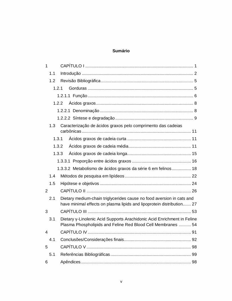
Sumário
1 CAPÍTULO I .......................................................................................... 1
1.1 Introdução ............................................................................................. 2
1.2 Revisão Bibliográfica ............................................................................. 5
1.2.1 Gorduras ........................................................................................ 5
1.2.1.1 Função ........................................................................................ 6
1.2.2 Ácidos graxos ................................................................................. 8
1.2.2.1 Denominação .............................................................................. 8
1.2.2.2 Síntese e degradação ................................................................. 9
1.3 Caracterização de ácidos graxos pelo comprimento das cadeias carbônicas ........................................................................................... 11
1.3.1 Ácidos graxos de cadeia curta ..................................................... 11
1.3.2 Ácidos graxos de cadeia média .................................................... 11
1.3.3 Ácidos graxos de cadeia longa ..................................................... 15
1.3.3.1 Proporção entre ácidos graxos ................................................. 16
1.3.3.2 Metabolismo de ácidos graxos da série 6 em felinos ................ 18
1.4 Métodos de pesquisa em lipídeos ....................................................... 22
1.5 Hipótese e objetivos ............................................................................ 24
2 CAPÍTULO II ....................................................................................... 26
2.1 Dietary medium-chain triglycerides cause no food aversion in cats and have minimal effects on plasma lipids and lipoprotein distribution....... 27
3 CAPÍTULO III ...................................................................................... 53
3.1 Dietary γ-Linolenic Acid Supports Arachidonic Acid Enrichment in Feline Plasma Phospholipids and Feline Red Blood Cell Membranes .......... 54
4 CAPÍTULO IV ...................................................................................... 91
4.1 Conclusões/Considerações finais........................................................ 92
5 CAPÍTULO V ....................................................................................... 98
5.1 Referências Bibliográficas ................................................................... 99
6 Apêndices ............................................................................................ 98
vi
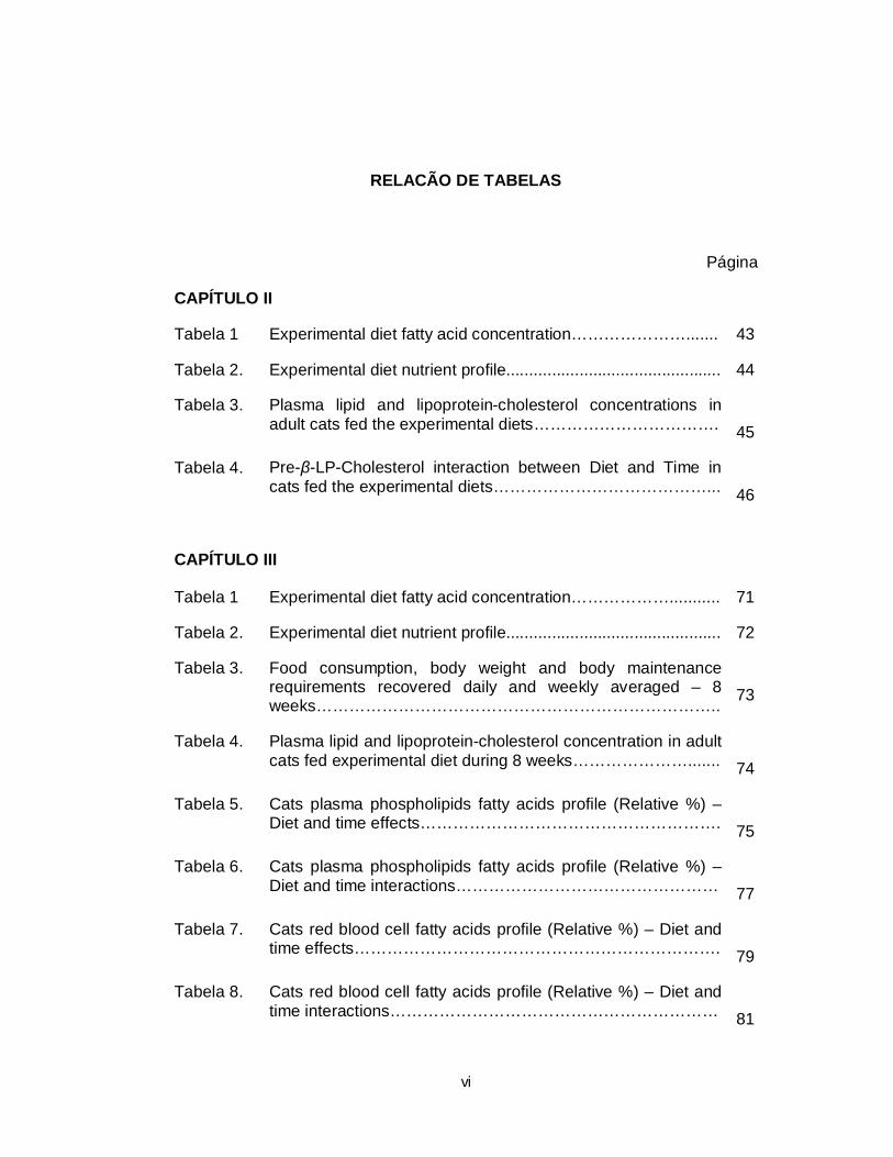
RELACÃO DE TABELAS
Página
CAPÍTULO II
Tabela 1 Experimental diet fatty acid concentration…………………....... 43
Tabela 2. Experimental diet nutrient profile............................................... 44
Tabela 3. Plasma lipid and lipoprotein-cholesterol concentrations in adult cats fed the experimental diets…………………………….
45
Tabela 4. Pre-β-LP-Cholesterol interaction between Diet and Time in cats fed the experimental diets…………………………………...
46
CAPÍTULO III
Tabela 1 Experimental diet fatty acid concentration………………........... 71
Tabela 2. Experimental diet nutrient profile............................................... 72
Tabela 3. Food consumption, body weight and body maintenance requirements recovered daily and weekly averaged – 8 weeks………………………………………………………………..
73
Tabela 4. Plasma lipid and lipoprotein-cholesterol concentration in adult cats fed experimental diet during 8 weeks………………….......
74
Tabela 5. Cats plasma phospholipids fatty acids profile (Relative %) – Diet and time effects……………………………………………….
75
Tabela 6. Cats plasma phospholipids fatty acids profile (Relative %) – Diet and time interactions…………………………………………
77
Tabela 7. Cats red blood cell fatty acids profile (Relative %) – Diet and time effects………………………………………………………….
79
Tabela 8. Cats red blood cell fatty acids profile (Relative %) – Diet and time interactions……………………………………………………
81
vii
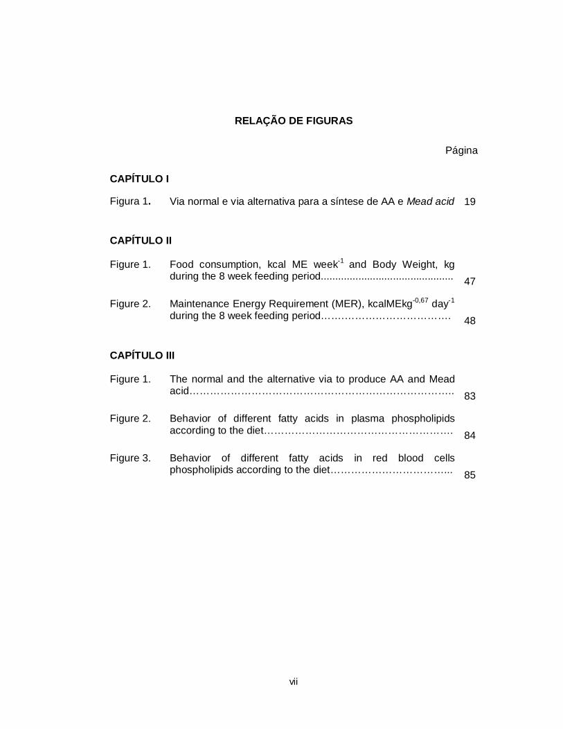
RELAÇÃO DE FIGURAS
Página
CAPÍTULO I
Figura 1. Via normal e via alternativa para a síntese de AA e Mead acid 19
CAPÍTULO II
Figure 1. Food consumption, kcal ME week-1 and Body Weight, kg during the 8 week feeding period..............................................
47
Figure 2. Maintenance Energy Requirement (MER), kcalMEkg-0,67 day-1 during the 8 week feeding period…….………………………….
48
CAPÍTULO III
Figure 1. The normal and the alternative via to produce AA and Mead acid…………………………………………………………………..
83
Figure 2. Behavior of different fatty acids in plasma phospholipids according to the diet……………………………………………….
84
Figure 3. Behavior of different fatty acids in red blood cells phospholipids according to the diet……………………………...
85
viii
RELAÇÃO DE ABREVIATURAS E DE SÍMBOLOS
AA Ácido araquidônico / Arachidonic acid
AGCL Ácido graxo de cadeia longa
AGCM Ácidos graxos de cadeia média
AGE / EFA Ácidos graxos essenciais / Essential fatty acid
AL / LA Ácido linoleico / Linoleic acid
ALA Ácido α-linolênico / α-linolenic acid
BCS Body condition score
BW Body weight
DE Digestible energy
DGLA Ácido dihomo-γ- linolênico
DHA Ácido decosaexaenóico
DM Dry matter
EPA Ácido eicosapentaenóico
FA Fatty acids
GLA Ácido γ-linolênico / γ-linolenic acid
HL High linoleic
HMCT High medium-chain triglycerides
LL Low linoleic
LMCT Low medium-chain triglycerides
LP-C Lipoprotein cholesterol
ix
LTB4 Leukotrienes serie 4
MCFA Medium-chain fatty acids
ME Metabolic energy
MER Maintenance energy requirement
PED Pre-experimental diet
PG Prostaglandines
PL Phospholipids
PLFA Plasma phospholipids fatty acids profile
Pre-β-LP-C / VLDL Pre-beta lipoprotein fraction / Very low density lipoprotein
RBC Red blood cells
RBCPL Red blood cell membranes phospholipids
TC Total cholesterol
TCL / LCT Triglicerídeos de cadeia longa / Long-chain triglycerides
TCM / MCT Triglicerídeos de cadeia média / Medium-chain triglycerides
TG Triglicerídeos plasmáticos / Triglycerides
α-LP-C / HDL Alpha lipoprotein fraction / High density lipoprotein
β-LP-C / LDL Beta lipoporotein fraction / Low density lipoprotein
Δ6 Dessaturase Delta 6 dessaturase

1 CAPÍTULO I
2
1.1 Introdução
Cães e gatos representam a maior população de animais de
companhia do mundo. O número de animais agregados às famílias é uma
tendência em evolução. Gatos, por seu comportamento peculiar, têm se
mostrado uma espécie bastante interessante para o sistema de vida atual, no
qual há uma limitação no tempo de convívio entre homem e animal. No Brasil,
seguindo a tendência Norte Americana, o número de animais de estimação tem
crescido. O Brasil possui o segundo maior número de animais de companhia
do mundo (31 milhões de cães, 14 milhões de gatos) (ANFAL PET, 2009)
ficando somente atrás dos Estados Unidos (EUA - 74,8 milhões de cães, 88,3
milhões de gatos) (McLeod, 2009). Como nos EUA a população de gatos
ultrapassa a de cães pressupõe-se que esta será a realidade brasileira a médio
prazo.
A evolução zoológica dos felídeos como carnívoros estritos gerou
espécies bastante exigentes quanto à alimentação e à nutrição. As diversas
particularidades metabólicas dos felídeos tem sido um desafio para
nutricionistas e pesquisadores, exigindo atenção no processo de escolha de
ingredientes e nutrientes para a confecção de dietas secas que atendam todas
as suas exigências nutricionais. Além das elevadas necessidades protéicas
exigidas pelos felídeos, devido a sua incapacidade de reduzir a atividade de
suas aminotransferases (Schimke, 1962), como fazem outras espécies, gatos
exigem outros nutrientes como a arginina, vitamina D e A, niacina e ácido
araquidônico (AA) pré-formados (Rogers & Morris, 2007).
3
Junto às necessidades nutricionais e suas particularidades, gatos
são sensíveis ao paladar, textura e cheiro dos alimentos, devendo-se ter
atenção na produção de dietas específicas para gatos, do contrário os gatos
apresentam um alto grau de rejeição ao alimento (Kvamme, 2003).
O atual conhecimento a respeito da nutrição de gatos foi gerado a
partir de pesquisas iniciadas na década de 70. Na época, a grande dificuldade
estava em fazer com que os gatos ingerissem as dietas purificadas, pouco
palatáveis a estes animais, fato que foi contornado com a descoberta de
substâncias palatabilizantes. Vencida esta barreira, a manutenção das colônias
e as doenças infecto-contagiosas tornaram os biotérios de felinos difíceis de
serem mantidos. Mas o metabolismo intrigante deste carnívoro e a afeição do
homem para com o animal foram fundamentais para a intensificação nas
pesquisas e o desenvolvimento de dietas cada vez mais balanceadas (Rogers
& Morris, 2007).
No entanto, certas rotas metabólicas continuam sendo investigadas.
O metabolismo de lipídeos em felinos permanece em discussão. Pesquisas em
outras espécies revelaram uma série de ingredientes lipídicos com
propriedades interessantes para a utilização em dietas convencionais e
terapêuticas para gatos. Neste contexto, os ácidos graxos de cadeia média
(AGCM) apresentam uma série de propriedades funcionais devido à forma
como são absorvidos e metabolizados, podendo ter potencial na nutrição de
felídeos. Entretanto, muitos estudos revelaram à baixa palatabilidade destes
4
ácidos graxos quando compondo dietas para diferentes espécies, inclusive um
relato em gatos (Wanten & Naber, 2004).
Outro aspecto do metabolismo de lipídeos de grande importância em
gatos são as rotas de síntese do AA. O AA e seus precursores são usualmente
encontrados em baixas concentrações nos alimentos industrializados para
gatos. A engenharia genética, associada a novas espécies vegetais
desenvolvidas e cultivadas (Huang et al., 2001; Liu et al., 2001), assim como os
métodos de extração e separação de lipídeos a partir dos compostos vegetais
e fermentados fúngicos tem ajudado na produção e concentração de alguns
ácidos graxos que antes não eram encontrados facilmente nos alimentos
(Carvalho et al., 2003; Sagilata et al., 2008). Este é o caso do óleo de
borragem, que pode ser concentrado e originar um óleo com 70% de ácido
graxo γ-linolênico (GLA, 18:3 n6) (Sanmark). Este ácido graxo em particular,
pode ter função importante na nutrição de felinos. Para a maior parte das
espécies a síntese orgânica deste composto ocorre pela ação de uma enzima
Δ6-desaturase sobre o ácido linoléico (AL, 18:2 n6). No entanto, felinos
demonstram nenhuma ou muito baixa eficiência nesta enzima, de forma que a
síntese de AA (20:4 n6) fica comprometida. Além disso, a inclusão deste ácido
graxo na dieta pode ser ferramenta para o melhor entendimento das vias de
produção de AA, bem como atividade da cascata enzimática pertinente à
síntese do AA.
5
1.2 Revisão Bibliográfica
1.2.1 Gorduras
Mediante a classificação generalista, todos os compostos que se
mostram solúveis em solventes orgânicos e insolúveis em água são
classificados como lipídeos. No entanto, resultante do processo de extração
pelos solventes orgânicos nem todas as substâncias apresentam a mesma
proximidade molecular. Exemplo clássico são os pigmentos vegetais e as
vitaminas A, D, E e K, que são extraídos juntamente com os lipídeos (Gurr et
al., 2002). Os lipídeos de interesse podem ser classificados como: lipídeos
simples, que compreendem os triglicerídeos (ácidos graxos unidos a uma
molécula de glicerol) e as ceras (contém um número maior de ácidos graxos
unidos a uma molécula de álcool de cadeia longa); lipídeos compostos, que
constam de um ácido graxo, unido a uma molécula não lipídica, que pode ser
uma proteína, um carboidrato ou outros compostos como fósforo e nitrogênio
(exemplo: lipoproteínas, glicoproteínas e fosfolipídeos). Ainda existem lipídeos
derivados de compostos de esterol, como o colesterol (Case et al., 2000).
O triglicerídeo é o tipo de gordura mais importante na dieta e pode
ser diferenciado nos alimentos, dependendo do tipo de cada ácido graxo
contido em cada triglicerídeo e da posição que este ácido graxo ocupa no
glicerol (Gurr et al., 2002). A maioria dos triglicerídeos contidos nos alimentos
contém ácidos graxos de cadeia longa (AGCL - variando entre 14 e 24 átomos
6
de carbono). No entanto, dois alimentos fogem à regra, os derivados lácteos e
o óleo de coco, ambos contendo apreciável quantidade de AGCM (NRC, 2006).
1.2.1.1 Função
De acordo com Bauer (2006) os lipídeos dietéticos podem ser
divididos em dois grupos por suas funções: os lipídeos que atuam como fonte
de energia, melhorando a textura do alimento e aumentando a palatabilidade, e
os lipídeos funcionais, do qual fariam parte os lipídeos ditos essenciais, como o
AL e o ácido α linolênico (ALA) e alguns de seus derivados, dependendo da
espécie e da faixa etária que se deseja atender (NRC, 2006).
As gorduras possuem numerosas funções no organismo. Os
triglicerídeos constituem a maior forma de armazenamento de energia.
Enquanto as reservas de carboidratos são extremamente limitadas nos
animais, os triglicerídeos podem ser depositados de forma quase ilimitada. A
maior parte dos depósitos de gordura localizam-se sob a pele, na forma de
gordura subcutânea, ao redor dos órgãos vitais e nas membranas que rodeiam
os intestinos. Alguns destes depósitos podem ser observados com facilidade
em animais obesos. Este tecido possui mobilidade estática, a síntese e a
degradação ocorrem para armazenamento e produção de energia,
respectivamente. Ainda assim, possui a propriedade de isolante térmico e
confere proteção física a regiões vitais do organismo (Case et al., 2000).
Além da propriedade energética as gorduras têm funções
metabólicas e estruturais. Uma camada isolante de gordura rodeia as fibras
nervosas e contribui para a transmissão de impulsos nervosos. Os fosfolipídeos
7
e os glicolipídeos são compostos esterificados de ácidos graxos que atuam
como componentes estruturais das membranas celulares e participam no
transporte de nutrientes e metabólitos através da membrana, conferem fluidez,
compressibilidade, permeabilidade e capacidade de fusão (Bauer, 2006). As
lipoproteínas permitem o transporte de gorduras via corrente sanguínea. O
colesterol é precursor de hormônios esteróides e está envolvido na formação
dos sais biliares, necessário para correta absorção e digestão dos lipídeos.
Juntamente com outros lipídeos, o colesterol forma uma camada protetora na
pele que evita a dessecação excessiva e a invasão de substâncias estranhas
(Case et al., 2000).
Na dieta a gordura é a forma mais concentrada de energia, mais
concentrada do que qualquer outro nutriente. Ainda que a energia fornecida
pelos carboidratos e pelas proteínas estejam ao redor de 4 kcal/g a energia da
gordura fornece mais que o dobro disto, cerca de 9 kcal/g (NRC, 2006).
Quando são fornecidas misturas de gordura animal e vegetal aos cães o
coeficiente de digestibilidade da gordura fica em torno de 90%, podendo chegar
a 95%, de forma que sua digestibilidade supera a da proteína e dos
carboidratos. Assim, o aumento de gordura na dieta, além de aumentar a
densidade energética, promove maior digestibilidade, importante para dietas de
animais de companhia. Além disso, a inclusão de gordura na dieta de cães e
gatos torna o alimento mais palatável, pois interfere com aroma, sabor e textura
no produto acabado (Kendall, 1984). De fato, concentrações mais elevadas de
gordura (25 a 40%) são preferidas por cães e gatos. Ao mesmo tempo, este
conteúdo elevado em gordura pode ser um problema para animais que não
8
fazem bem o controle da ingestão pelo valor calórico do alimento, podendo
ocorrer casos de excesso de peso e obesidade em cães e gatos (Case et al.,
2000).
1.2.2 Ácidos graxos
Estruturalmente são ácidos carboxílicos com cadeia variando entre 2
e 26 carbonos, conectados uns aos outros por ligação simples ou dupla.
Apresentam um grupamento carboxílico (COOH) em uma extremidade e um
grupo metil (CH3) na outra (Gurr et al., 2002).
1.2.2.1 Denominação
A posição em relação ao grupo metil indica o carbono ômega (n) e a
posição em relação ao grupamento carboxila indica a posição Delta (Δ), dessa
forma pode-se reconhecer estruturas que são isômeras (Reinhart, 1996a). Por
exemplo, quando se trabalha com o ácido dihomo-γ-linolênico (DGLA - 20:3
n6), este possui um isômero de posição, ambos n6. A posição Δ é o diferencial
entre os dois ácidos graxos: 20:3 n6 Δ5,11,14 e o outro 20:3 n6 Δ8,11,14, (Gurr
et al., 2002). De forma geral, a ligação Δ está ligada ao nome da enzima que
aplica a insaturação (NRC, 2006).
Uma das mais clássicas classificações é a dos ácidos graxos
essênciais, são divididos fisicamente ou quimicamente em dois grupos de
acordo com a ligação com o grupo metil, mais próximo ou mais final. Na
nutrição de mamíferos em geral destacam-se os grupamentos Ômega 3 e
Ômega 6. Felídeos possuem particularidades na metabolização destes
compostos devido a alterações específicas nas vias enzimáticas (Bauer, 1997)
9
1.2.2.2 Síntese e degradação
Felinos, assim como todos os mamíferos podem sintetizar ácidos
graxos saturados e com uma insaturação, (independentemente do tamanho da
cadeia carbônica, a partir de Acetil-CoA (Salati & Goodridge, 1996). As dietas
ricas em gordura, com grande concentração de ácidos graxos monoinsaturados
(16:1 n7 e 18:1 n9), são inibidoras das enzimas que fazem a insaturação de
ácidos graxos saturados (NRC, 2006). Dessa forma, quando dietas com altas
concentrações de carboidratos e proteínas são fornecidas aos animais as
atividades destas desaturases se elevam. No entanto, insaturações duplas a
partir de compostos de 18 carbonos na posição n3 e n6 são impossíveis em
felídeos e estes ácidos graxos se tornam essenciais, devendo, portanto ser
fornecidos via dieta. Plantas possuem as enzimas necessárias para a síntese
destes compostos, sendo o AL e o ALA abundantes em ingredientes de origem
vegetal. Esta capacidade de síntese está ligada à expressão de duas enzimas
indispensáveis no processo, a Δ12 e a Δ15 desaturases (Cook, 1996).
Curiosamente, após a síntese de AL e ALA a maior parte das plantas não
adicionam demais insaturações nestes compostos, não sendo encontrados
outros ácidos graxos importantes como o AA, ácido decosaexaenóico (DHA) e
ácido eicosapentaenóico (EPA) em óleos vegetais (Gurr et al., 2002).
Plantas marinhas, zooplânctrons e fitoplâctons são capazes de
adicionar insaturações especialmente na série n3, podendo elongá-los até a
produção de EPA e DHA (Cook, 1996). Mamíferos herbívoros também o fazem,
mas especialmente os carnívoros apresentam certa debilidade neste processo.
10
O processo evolutivo e o consumo concomitante de ingredientes de origem
animal provavelmente tenham contribuído para a redução da expressão das
enzimas responsáveis pela iniciação das desaturações nos ácidos graxos de
cadeia longa (Rogers & Morris, 2007). Felídeos, como carnívoros estritos,
talvez sejam uma das espécies mais afetadas. É sabido que gatos são
incapazes de insaturar o AL para formar o GLA, devido a ausência completa ou
parcial da enzima Δ6 desaturase. Consequentemente, não são capazes de
produzir AA, um componente lipídico essencial para felinos. Dessa forma, a
dieta deve fornecer AA para estes animais (Rivers et al., 1975; Pawlosky et al.,
1994).
Sabe-se que ambas as séries, n3 e n6, não são interconversíveis e
dependem das mesmas enzimas para dessaturar e elongar ácidos graxos.
Assim, felídeos privados de dietas animais poderiam sofrer deficiência em
ambas as séries. Embora sejam reconhecidos os sinais da deficiência de AL
em gatos, com sinais como pele seca, descamativa, pelo sem brilho,
infertilidade e lipidose hepática (Hassan et al., 1977; Rivers, 1976 ab; Frankel &
Rivers, 1978; Rivers, 1982), nenhum sinal de deficiência específica foi provada
para o ALA, muito embora a indicação de uma concentração mínima já estar
descrita para gatos em crescimento (NRC, 2006).
11
1.3 Caracterização de ácidos graxos pelo comprimento das cadeias
carbônicas
1.3.1 Ácidos graxos de cadeia curta
Os ácidos graxos de cadeia curta são compostos por cadeias de 2, 3
e 4 carbonos sem insaturações: ácido acético, ácido propiônico e ácido
butírico, respectivamente. Não são encontrados normalmente compondo
triglicerídeos e de forma geral são produto do metabolismo microbiano, cuja
importância é significativa para animais que fazem fermentação. Nos
monogástricos estes ácidos graxos têm função importante no metabolismo
absortivo intestinal, já que são as principais fontes de energia dos colonócitos
(Roediger, 1990). Em gatos seu papel estimulante sobre a contratilidade da
musculatura longitudinal e circular do cólon tem sido estudada para estimar a
necessidade de fibra alimentar (Rondeau, 2003). Apesar do curto cólon dos
gatos a fermentação da fibra alimentar seria uma das formas de fornecer
ácidos graxos de cadeia curta, já que via dieta provavelmente não chegariam
intactos ao cólon.
1.3.2 Ácidos graxos de cadeia média
Ácidos graxos de cadeia média apresentam cadeias de 6 a 12
carbonos sem insaturações, ou seja, compostos de cadeias retilíneas. São
encontrados facilmente nos produtos derivados do leite, especialmente o leite
de cabra, cuja espécie prestou seu sufixo para a denominação da maior parte
dos AGCM (ácido capróico (6C), ácido caprílico (8C), ácido cáprico (10C),
12
ácido láurico (12C). A gordura de coco também é fonte destes ácidos graxos,
embora cerca de 50% da gordura seja de ácido láurico.
Triglicerídeos de cadeia longa (TCL) se diferem dos triglicerídeos de
cadeia média (TCM) por serem compostos por ácidos graxos com mais de 14
carbonos. O baixo peso molecular dos AGCM quando comparado ao dos
AGCL confere a eles maior hidrossolubilidade, facilitando o processo digestivo,
sua absorção e transporte ao fígado, tornando a digestão e a absorção mais
rápidas e fáceis (Back & Babayan, 1982). Sob a ação dos sais biliares e da
lipase pancreática, os triglicerídeos contendo AGCM são transformados nas
unidades absorvíveis: ácidos graxos livres e monoacilglicerol, que prontamente
são absorvidos (Bach et al., 1996). No enterócito, os AGCM não são re-
esterificados como ocorre com os AGCL, pois a enzima Acil-CoA sintetase
possui mais afinidade pelo AGCL do que pelo AGCM Assim, a maior parte dos
produtos da digestão de TCM vão diretamente à via portal, seguindo em
direção ao fígado ligados à albumina (Bach & Babayan, 1982; Bach et al.,
1996; Papamandjaris et al., 1998). Os AGCL são geralmente esterificados e
incorporados nos quilomicrons e então entram nos dutos linfáticos em direção
ao ducto torácico. Dessa forma atingem primeiramente a circulação periférica e
não diretamente a hepática como fazem os AGCM (Papamandjaris et al.,
1998). No fígado, AGCM podem seguir várias vias catabólicas incluindo beta,
ômega e a oxidação peroxisomal ou podem ser elongados para formar outros
ácidos graxos (Jones et al., 2006).
13
O metabolismo celular dos AGCM é também bastante específico.
Nos tecidos, são normalmente independentes de carnitina para entrar nas
mitocôndrias (Friedman et al., 1990), embora alguns estudos com o ácido
láurico (C12:0) demostram que uma pequena porção deste ácido graxo pode
estar associado à carnitina para acessar a matriz mitocondrial (Christensen et
al., 1989; Rossle et al., 1990). Dentro da mitocôndria a maioria dos lipídeos é
catabolizada pela beta-oxidação. Os AGCM que não são metabolizados pelo
fígado normalmente não são incorporados nos triglicerídeos, fosfolipídeos ou
frações de ésteres de colesterol, uma vez que a enzima Acil-CoA sintetase é
mais ávida por ácidos graxos com mais de 14 carbonos, havendo menor
preferência pela esterificação dos AGCM,como resultado pouco AGCM é
recuperado nos triglicerídeos, nos fosfolipídeos, assim como em vários tecidos
(Papamandjaris et al., 1998).
Em função deste metabolismo particular, os TCM podem ser
ferramenta para melhorar a nutrição em casos específicos. Os TCM podem ser
utilizados em síndromes de malabsorção, má digestão, assim como em
insuficiência pancreática exócrina, linfangectasia ou quilotórax (Nelson &
Couto, 1992). Alguns efeitos dos TCM sobre a obesidade tem sido
investigados: sabe-se que o valor energético dos TCM é mais baixo do que o
dos TCL e esses parecem levar ao aumento da taxa metabólica pós-prandial
(Papamandjaris et al., 1998). Neste caso a inclusão destes ácidos graxos
poderia ser útil para formular dietas destinadas a redução de peso ou para
manter saudáveis os animais que já foram submetidos a programa de
emagrecimento. Uma revisão sobre TCM relatou os efeitos benéficos da
14
infusão parenteral de AGCM sobre o sistema imune (Wanten & Naber, 2004).
Neste caso a inclusão de AGCM foi proposta em substituição a uma porção do
AL, reduzindo desta forma a proporção entre AL e ALA.
Recentemente, a utilização de TCM em pacientes portadores de
doenças cardíacas demonstrou melhorar o status de energia do coração e a
assim sua função contrátil (Labarthe et al., 2008). Nenhum efeito tóxico foi
observado em diversos estudos em seres humanos ou animais, mesmo
quando administrado oral ou parenteralmente ou quando consumido como
suplemento de dietas equilibradas, em concentrações de até 15% da energia
dietética (Traul et al., 2000). No entanto, o potencial dos TCM pode ser
contraposto pelo fato de que sua inclusão pode causar aversão alimentar.
Alguns autores demonstraram que cães e gatos não consomem dietas com
TCM. Gatos prontamente recusam o alimento quando o ácido caprílico (8:0) é
incluído na composição da dieta (MacDonald et al., 1985). Também foi
observado aumento nos lipídeos plasmáticos em cães alimentados com AGCM
(Van Dongen et al., 2000). A menor palatabilidade dos óleos contendo TCM
tem sido observada por muitos autores em diferentes espécies (Lewis et al.,
1987; Hill, 1994; Hand et al., 2000). Os gatos recusaram prontamente o
alimento demostrando alta sensibilidade à inclusão de 0,1% de ácido caprílico
e 5,0% de inclusão de TCM contendo ácido caprílico purificado (MacDonald et
al., 1985). Em cães alimentados com dietas purificadas contendo 22% da EM
na forma de TCM o consumo foi afetado negativamente e a concentração dos
lipídeos plasmásticos aumentou (Van Dongen et al., 2000). Entretanto, quando
os cães foram alimentados com 11% da EM na forma de TCM, nenhuma
15
recusa foi vista e um pequeno aumento no coeficiente de digestibilidade da
gordura foi observado. Os triglicerídeos plasmáticos (TG) foram aumentados
em 23% nos animais que receberam 11% da EM na forma de TCM em
comparação ao grupo de controle (Beynen et al., 2002).
Dietas utilizando fontes naturais de triglicerídeos podem agir
diferentemente na aceitação de ácidos graxos pelos animais. Existem 3 formas
de se encontrar ácidos graxos de cadeia média: forma pura, como ácidos
graxos livres, ácido caprílico purificado (C8); forma de triglicerídeos puros, na
qual há uma esterificação artificial dos ácidos graxos de mesmo tamanho nas
posições sn-1, sn-2 e sn-3 do glicerol formando o tricaproin, tricaprilin, tricaprin,
trilaurin (Ulrich et al., 1996); triglicerídeos naturais originários de fontes que
naturalmente contém AGCM, como o óleo de coco e a gordura do leite. Nestes
triglicerídeos, no entanto, os ácidos graxos que o compõem não são
exclusivamente de cadeia média, podendo haver em algumas posições do
glicerol AGCL (Wanten & Naber, 2004).
Os trabalhos analisados foram conduzidos através da utilização de
dietas purificadas para alimentar cães e gatos. Na maior parte dos
experimentos os pesquisadores utilizaram triglicerídeos purificados e ácidos
graxos livres, não fontes naturais de gordura.
1.3.3 Ácidos graxos de cadeia longa
Ácidos graxos de cadeia longa podem ser saturados,
monoinsaturados ou poli-insaturados. Os ácidos graxos ditos essenciais são
poli-insaturados, com mais de 18 carbonos e com duas ou mais ligas duplas.
16
Dentre estes ácidos graxos essenciais duas séries se destacam: ômega 3 e
ômega 6 (n-3 e n-6) (Gurr et al., 2002).
1.3.3.1 Proporção entre ácidos graxos
De acordo o NRC (2006), nos últimos anos houve intensa discussão
a respeito das relações entre ácidos graxos poli-insaturados de cadeia longa,
ômega 3 e 6. No entanto, não há consenso entre os trabalhos e esses são
difíceis de serem comparados, já que há confundimento dentro dos desenhos
experimentais. Vegetais, de uma forma geral, fornecem ácidos graxos
essenciais que iniciam as séries, o AL e o ALA, em diferentes proporções.
Ingredientes de origem animal são mais completos e fornecem a maior parte
dos ácidos graxos de ambas as séries: AL, ALA, AA, EPA, DHA. No entanto,
quando se fala de gorduras derivadas de fontes marinhas, a concentração de
derivados da série 3 é bastante expressiva, especialmente o DHA. Sabe-se
que a adição de fontes marinhas às dietas não pode ser comparada ao
aumento de fontes vegetais, porque as fontes marinhas já trazem ácidos
graxos mais longos que não necessitam metabolização para causar efeito, pois
já estão prontos. Assim, as relações entre ácidos graxos são estabelecidas das
mais variadas formas nos diferentes experimentos: somente entre AL e ALA;
entre o somatório de todos os componentes de cada série; entre AA o
somatório entre EPA e DHA. Dessa forma, comparações entre proporções de
ácidos graxos se tornam difíceis. Existem, ainda, particularidades entre os
ácidos graxos das séries que precisam ser revistas: a série 6 é considerada
geradora de eicosanóides, prostaglandinas e tromboxanos pró-inflamatórios; a
17
série 3 é geradora de mediadores opostos destas vias, sendo considerados
anti-inflamatórios. No entanto, o GLA e o ácido dihomo-γ-linolênico (DGLA) da
série 6 são mediadores da produção de prostaglandinas da série 1 e
tromboxanos da série 3, com características anti–inflamatórias, mas também
são precursores do AA, conhecido como precursor da prostaglandinas e
tromboxanos da série 2 e leucotrienos da série 4, altamente inflamatórios. O
fornecimento, via dieta, de diferentes ácidos graxos da mesma série, como AL,
GLA ou AA, apresentam potencialidades diferentes entre si. Isso significa que a
mesma inclusão dietética de diferentes ácidos graxos resulta em respostas
diferentes pelo animal (NRC, 2006).
Na nutrição de animais de companhia conhecer detalhadamente
estas relações seria importante para atuar na modulação do sistema imune, no
entanto as relações entre as duas séries deixam de ser claras à medida que o
confundimento entre experimentos ocorre. Atualmente, no estudo dos ácidos
graxos, como ocorreu no estudo das proteínas e dos carboidratos, testar a
unidade funcional deve ser o objetivo principal. Dessa forma, lipídeos passam a
ser estudados na forma de ácidos graxos individualizados. Nem mesmo as
divisões entre ácidos graxos de acordo com o seu carbono ômega parecem ser
adequadas em muitas circunstâncias. No entanto, é sempre importante lembrar
que ambas as séries compartilham das mesmas enzimas e a proporção entre
ácidos graxos pode ter efeito indutivo de síntese através do substrato (Gurr et
al., 2002).
18
1.3.3.2 Metabolismo de ácidos graxos da série 6 em felinos
O metabolismo ácidos graxos poli-insaturados em felídeos é
bastante curioso e desperta a atenção de pesquisadores desde a década de
70, quando Rivers et al. (1975), a partir da análise de amostras de fígado,
descobriram que gatos não possuíam a enzima Δ6 desaturase. Em mamíferos
a conversão do AL para AA, assim como a conversão de ALA para EPA
necessitam da ação da Δ6 desaturase seguida por uma elongase e então uma
Δ5 desaturase (Sinclair, 1979), metabolismo este que estaria prejudicado em
gatos mediante a descoberta da deficiência enzimática. Ainda no início das
pesquisas, foi notado que gatos privados de ácidos graxos essenciais
apresentavam a formação de um ácido graxo identificado como 20:3 n9
(Holman, 1970). O acúmulo deste ácido graxo também foi observado quando
animais foram alimentados com gordura animal hidrogenada (Sinclair, 1981),
embora o grupo de Rivers et al. (1976ab) não tenha relatado a presença deste
ácido graxo em gatos recebendo dietas deficientes em ácidos graxos
essenciais (Rivers et al., 1976ab).
O 20:3 n9, por sua vez, poderia ser formado pela ação da mesma
cascata enzimática responsável pela conversão do AL em AA agindo sobre o
18:1 n9, que por sua vez seria gerado a partir do 18:0 pela ação da Δ9
desaturase (Figura 1). Dessa forma, alguns autores levantaram a hipótese de
que Δ6-desaturase deveria estar presente para a formação de 20:3 n9 e que
alguma produção de AA poderia ser possível (Sinclair et al., 1981; Rivers et al.,
1981). Sinclair et al., (1979; 1981) sugeriram uma via alternativa para a
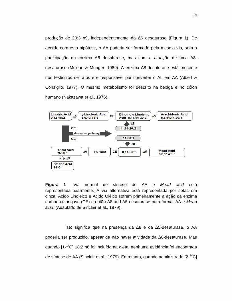
19
produção de 20:3 n9, independentemente da Δ6 desaturase (Figura 1). De
acordo com esta hipótese, o AA poderia ser formado pela mesma via, sem a
participação da enzima Δ6 desaturase, mas com a atuação de uma Δ8-
desaturase (Mclean & Monger, 1989). A enzima Δ8-desaturase está presente
nos testículos de ratos e é responsável por converter o AL em AA (Albert &
Consiglio, 1977). O mesmo metabolismo foi descrito na bexiga e no cólon
humano (Nakazawa et al., 1976).
Figura 1– Via normal de síntese de AA e Mead acid está representadalinearmente. A via alternativa está representada por setas em cinza. Ácido Linoleico e Ácido Oléico sofrem primeiramente a ação da enzima carbono elongase (CE) e então Δ8 and Δ5 desaturase para formar AA e Mead acid. (Adaptado de Sinclair et al., 1979).
Isto significa que na presença da Δ8 e da Δ5-desaturase, o AA
poderia ser produzido, apesar de não haver atividade da Δ6-desaturase. Mas
quando [1-14C] 18:2 n6 foi incluído na dieta, nenhuma evidência foi encontrada
de síntese de AA (Sinclair et al., 1979). Entretanto, quando administrado [2-14C]
20
20:3 n6, houve significativa produção de AA, sugerindo que gatos possuem Δ5-
desaturase ativa (Sinclair et al., 1979).
A sintomatologia de pele seca, perda de pelo, infertilidade e
infiltração de lipídeos no fígado foram conhecidas quando gatos foram privados
de ácidos graxos essenciais na dieta (Hassam et al., 1977; Rivers et al.,
1976a,b; Frankel & Rivers, 1978; Rivers, 1982). Foi demonstrado que devido à
deficiência enzimática, os sintomas de infertilidade não são revertidos mesmo
com a adição de AL e ALA (MacDonald et al., 1983, 1984). Os mesmos autores
notaram que gatos recebendo AL possuíam maior concentração de AA nos
testículos e que fêmeas foram incapazes de conceber quando alimentadas
somente com AL.
Recentemente foram testadas dietas isentas de AA e suplementadas
com óleo de milho a 1%, 3% e 1% acrescido de 0,02% de AA (Pawlosky &
Salem, 1996). Fêmeas foram alimentadas antes de serem cruzadas com os
machos e durante toda a gestação. Todas as fêmeas ciclaram normalmente e
emprenharam, no entanto, as fêmeas que receberam dieta com apenas 1% de
óleo de milho tiveram ninhadas com alta incidência de defeitos congênitos e
baixa viabilidade quando comparadas aos demais grupos. Mas as fêmeas
suplementadas com 3% de óleo de milho tiveram ninhadas normais. Concluiu-
se que fêmeas são incapazes de reproduzir normalmente quando mantidas em
dietas muito baixas em AGE, mas um pequeno acréscimo de AA é capaz de
reverter essa situação. A dieta com 3% de óleo de milho e sem AA atendeu
normalmente às necessidades das fêmeas sendo concluído que outros fatores
21
dietéticos poderiam estar envolvidos. Num estudo adicional, Pawlosky et al.
(1997), com as fêmeas filhas das mães utilizadas no experimento anterior,
demonstraram acúmulo de carbono marcado no AA a partir de AL.
Morris et al. (2004), descreveram que gatos mantidos em dietas com
AL e livres de AA são férteis, concluindo que AA não é essencial para machos
em reprodução. As fêmeas alimentadas no mesmo sistema tiveram todo o
processo reprodutivo normal, no entanto, após o parto houve uma alta
incidência de canibalismo sobre a ninhada, logo após o nascimento. Após este
experimento as fêmeas foram separadas em dois grupos e receberam uma
dosagem diária de AA de 0,5 ou 1 ml de AA concentrado a 40,7% e após 10
semanas foram cruzadas com machos novamente. Nenhuma das fêmeas
concebeu, sendo concluído que algum outro ácido graxo pode estar envolvido
no processo reprodutivo sozinho ou associado com o AA. Quando o óleo
contendo GLA (18:3n-6) foi adicionado à dieta, os problemas reprodutivos
melhoraram. O GLA pode ter melhorado o nível de AA já que é o produto pré
formado da ação da Δ6 desaturase (Rivers & Frankel, 1980). Estes estudos
têm servido de suporte até hoje para o conceito de que gatos necessitam de
AA preformado, pois a enzima Δ6 desaturase possui baixa ou inexistente
atividade em gatos.
No entanto, ainda há a possibilidade de que o AA possa ser
produzido a partir das enzimas ∆8 and ∆5 desaturase (Sinclair et al., 1981).
Dessa forma, apesar da não atividade da Δ6 desaturase, alimentando gatos
com dietas ricas em AL poderia induzir uma vai alternativa para a produção de
22
AA. Da mesma forma, o GLA seria uma forma fácil de fornecer precursores
para a síntese de AA.
1.4 Métodos de pesquisa em lipídeos
O estudo da composição dos lipídeos nos tecidos dos animais pode
ser uma forma de se evidenciar alterações dietéticas e patológicas em animais.
Os fosfolipídeos, por sua vez, com suas diferentes classes podem revelar ainda
mais a respeito do metabolismo celular envolvendo ácidos graxos e os
produtos de sua degradação. Dessa forma, o estudo dos fosfolipídeos
mediante sua composição de ácidos graxos pode ser uma forma de se estimar
como os componentes dietéticos alteram a formação destes compostos
(Ivanova et al., 2004).
Os métodos para análise de lipídeos estão baseados na solubilidade
das substâncias. A metodologia segue uma série de passos para a extração e
separação das porções lipídicas desejadas (Christie, 2001). A extração dos
lipídeos a partir de tecidos, animal ou vegetal, é feita de acordo com o método
de Folch (1957), no qual a matéria a ser analisada é submetida a extração de
lipídeos totais pelo contato do tecido a ser analisado com uma mistura de
álcool e clorofórmio. Os lipídeos extraídos podem ser analisados na forma total
ou submetidos à separação das diferentes porções: tecidos animais como
membranas de células possuem basicamente fosfolipídeos e colesterol. O
plasma, além destes dois componentes ainda apresenta ácidos graxos livres e
triacilgliceróis.
23
Os métodos de cromatografia em camada delgada são indicados
para a separação de componentes dos lipídeos totais. Devido à polaridade,
misturas de clorofórmio, álcool e ácido acético separam as porções nos
diversos componentes através do arraste das substâncias mais apolares ao
longo de uma camada de sílica (Touchstone, 1995).
Os próprios fosfolipídeos totais podem ser separados nas suas
porções componentes pelo mesmo método somente se alterando a
composição e a proporção entre os solventes. Os fosfolipídeos, por sua vez,
podem ser derivatizados para a análise de composição de ácidos graxos por
cromatografia gasosa (Wang & Gustafson, 1992; Homan & Anderson, 1998;
Lesnefsky et al., 2000; Kim et al., 2000). De uma forma geral, estes métodos
são bastante onerosos, consomem muito tempo e são capazes de detectar um
limitado número de componentes.
A utilização de técnicas mais modernas como espectrometria de
massa confere maior sensibilidade, especificidade e rapidez à análise de
lipídeos. No início o fast atom bombardment (FAB-MS) permitiu gerar um
abundante número de íons não fragmentados juntamente com alguns
fragmentados (Matsubara & Hayashi, 1991; Murphy & Harrison, 1994) os quais
permitiram encontrar informações adicionais e revelaram um grande potencial
para o Tanden MS (MS/MS – análise de moléculas e fragmentados ionizados a
partir de compostos lipídicos). Lipídeos termolábeis e não voláteis podem ser
analisados através de Soft Ionization, por um método denominado matrix-
assisted laser desorption/ionization (MALDI) (Schiller et al., 1999) e ionização
24
por eletrospray (ESI) (Fenn et al., 1989; Weintraub et al., 1991), os quais não
causam extensiva fragmentação dos compostos. Esta pode vir a ser uma
ferramenta disponível para a análise direta de fosfolipídeos e seus ácidos
graxos que dentro de uma mistura complexa podem ser correlacionados com
as condições experimentais ou com doenças.
1.5 Hipótese e objetivos
O presente trabalho foi dividido em dois estudos. O primeiro trabalho
visou esclarecer se os gatos aceitam dietas contendo óleo de coco rico em
ácidos graxos de cadeia média em substituição a dieta com óleo de açafroa
(Carthamus tinctorius) rico em AL, e se ocorreriam efeitos negativos sobre o
metabolismo lipídico. O segundo estudo foi baseado na adição de óleo de
borragem (Borago officinalis) na dieta, com vistas a elucidar os seguintes
pontos: se os gatos são capazes de produzir AA a partir de GLA consumido; se
a enzima Δ6 desaturase está realmente inativa, mediante a adição de ácido
linoléico em alta concentração; se existe uma rota alternativa para produção de
AA a partir do AL.
As hipóteses estabelecidas foram as seguintes:
Gatos consomem AGCM quando estes são provenientes do óleo de
coco e incluídos na concentração de 11% da EM da dieta.
25
Gatos são capazes de produzir AA quando o produto da Δ6
desaturase sobre o ácido linoléico, o GLA, é oferecido pré-formado via dieta,
pois a enzima Δ5 desaturase está presente e é ativa.
A síntese de AA pode ser possível através de uma via alternativa,
não utilizando a Δ6 desaturase, mediante o acréscimo de ácido linoléico à
dieta em altas concentrações.

2 CAPÍTULO II3
3 Artigo formatado conforme normas do American Journal of Veterinary Research.
27
2.1 Dietary medium-chain triglycerides cause no food aversion in cats and have minimal
effects on plasma lipids and lipoprotein distribution.1
Luciano Trevizan, DVM, MSc, DSc, 2,3 Alexandre de Mello Kessler, MSc, DSc, 2
Karen E. Bigley, BS, 3 Wendy Anderson, DVM, PhD4 Mark K. Waldron, MS, RD,
PhD 4 John E. Bauer, DVM, PhD, DACVN 3*.
2 Department of Animal Science, Universidade Federal do Rio Grande do Sul, Porto
Alegre, RS 91540-000, Brazil.
3 Department of Small Animal Clinical Science, Comparative Nutrition Laboratory,
College of Veterinary Medicine and Biomedical Sciences, Texas A & M University,
College Station, TX 77843.
4 Nestlé Purina Pet Care Research, St Louis, MO.
1 Funded by Nestlé-Purina Pet Care, St. Louis, MO, USA and the Mark L. Morris
Professorship in Clinical Nutrition, College of Veterinary Medicine and Biomedical
Sciences, Texas A & M University, College Station, TX, USA.
6 Abbreviations used: MCT, medium-chain triglycerides; ME, metabolic energy; LA,
linoleic acid; BW, body weight; BCS, body condition score; TG, plasma
triacylglycerol; TC, total cholesterol; LP-C, lipoprotein cholesterol distributions.
* To whom correspondence should be addressed. E-mail: [email protected].
Phone number: 979 845 2321.
28
Objective – To determine possible diet aversion and lipid and lipoprotein alterations
in cats fed diets containing medium chain triglycerides.
Animals - Nineteen clinically normal, adult female cats were randomly assigned into
two groups (low medium chain triglyceride (MCT) diet, n=10; and high MCT diet (at
11% ME MCT, n=9) and fed for 9 weeks.
Procedures - Cats were fed according to their metabolic body weights (100 kcal
ME*Wkg0.67 day-1). Daily consumption records, weekly body weights (BW), and body
condition scores (BCS, 1 to 9 scale where 5 is ideal) were used to adjust amounts fed
and to calculate daily metabolic energy factors for each cat to maintain an ideal BCS.
Blood samples were obtained after withholding food at d 0, 14, 28 and 56 for plasma
triglyceride (TG), total cholesterol (TC), and lipoprotein cholesterol distribution (LP-
C).
Results - Repeated measures ANOVA and Tukey (α=0.05) multiple comparison tests
revealed no differences between diets with respect to food consumption, BW, BCS,
and metabolic energy factors. A statistically significant increase in plasma TG was
seen with the HMCT diet; however values were within the normal feline range. No
diet effects were seen on TC or LP-C although increases over time were observed.
Conclusions and Clinical Relevance – Results of this study demonstrate that it is
feasible to include MCT in normal feline diets without refusal in the amounts fed with
minimal effect on lipid metabolism. Such diets appear to be useful for not only
clinically normal cats and may have utility in metabolic disorders. The MCT oils are
thus an example of a bioactive dietary lipid that may benefit feline metabolism and
can serve as a useful functional food ingredient for feline species.
29
Introduction
Medium chain triglycerides (MCT) are readily found in products such as coconut oil
and milk by-products. They consist of 8 to 12 carbon atoms with no double bonds. By
contrast long chain triglycerides (LCT) typically contain >14 carbons and may be
either saturated or unsaturated. Because of their low molecular weight and higher
water solubility compared with LCT, MCT are more readily digested and absorbed
resulting in their more rapid transport to the liver via portal circulation.1 During
digestion, the smaller molecular weight of MCT compared to LCT facilitates the
action of pancreatic lipase enabling the ready release and absorption of free fatty acids
and monoacylglycerol.2 After absorption, triacylglycerol are re-esterified by Acyl-
CoA synthetase but this enzyme has greater affinity for LCT than MCT.
Consequently, a major portion of absorbed MCT are directly enter the portal venous
circulation, are bound to albumin, and transported directly to the liver.1-3 By
comparison, LCT fatty acids are typically resynthesized into triacylglycerols,
incorporated into chylomicrons, and transported via the lymphatic system to the
thoracic duct; initially bypassing the liver.3 Once in the liver, medium-chain fatty
acids (MCFA) may follow various catabolic pathways including beta-, omega-, and
peroxisomal oxidation or elongation to other fatty acids.4 In tissues, transfer of MCFA
into mitochondria for beta-oxidation is normally carnitine independent.5 However,
some studies using 12 carbon fatty acids (C12:0) showed that carnitine transport may
provide at least a minor pathway for C12:0 metabolism.6,7 Although the bulk of fatty
acid (FA) catabolism occurs via mitochondrial beta-oxidation, triacylglycerol re-
esterification may also occur via FA synthetase. However, this latter pathway is more
effective with FA of ≥14 carbons than with MCFA. Consequently, few dietary MCFA
30
are recovered per se in triacylglycerol, phospholipids, or cholesterol ester fractions of
plasma or tissues.3
Because of their unique metabolism, dietary MCT supplementation may be beneficial
in several respects. For example, MCT may be used in malabsorptive and
maldigestion disorders as well as exocrine pancreatic insufficiency, lymphangiectasia,
or chylothorax.8 Some effects of MCT have also been explored in obesity
management because the energy content of MCT is less than LCT and postprandial
energy expenditure increases when MCT are used.3 Thus, diets containing MCT may
help during weight reduction and for health maintanence thereafter. Other possible
benefits of MCT oils include effects of parenteral infusion on the immune system and
improvement of cardiac energetics and contractile function in certain cardiac
disorders.9,10 In addition no toxicologic effects of MCT oils have been reported in
several studies of both humans and animals whether administered orally or
parenterally or if consumed as a supplement to a balanced diet at levels up to 15% of
energy.11 In spite of these potential benefits, some authors have reported food
aversion with MCT diet inclusion in dogs and cats. This effect was especially
pronounced when caprylic acid (C8:0) was fed.12 Also, poor palatability of various
MCT oils have been reported by several authors.13-15 In cats, aversion was seen at
0.1% inclusion of C8:0 fatty acid and 5.0% MCT containing purified C8:0.12 In dogs,
purified diets containing 22% ME MCT depressed consumption and increased plasma
lipid concentrations.16 However, when dogs were fed 11% ME as MCTs, no refusals
were seen and a slight increase in crude fat digestibility was observed. Also, plasma
triacylglycerol concentrations increased by 23% in the groups which received MCT
compared to control.17 It is important to note, however, that the previous studies all
used purified diets. The present study was conducted using natural source ingredients

31
by replacing safflower oil with coconut oil to achieve an 11% ME MCT inclusion in a
complete and balanced diet. Acceptance of this diet as well as its possible effects on
feline lipid metabolism was investigated. The objective was to feed the diets to
achieve and maintain normal body weights of cats and equalize their metabolism at
steady state while achieving body condition scores of 5 out of 9.
32
Materials and Methods
Animals and diets. Nineteen adult, sexually intact female cats ranging in age between
1.5 to 2 years with body weights between 2.4 and 6.0 kg were used. Expect for three
cats, the animals had body conditions score (BCS) of 5-6 out of 9, with 5 considered
ideal. These initial scores were determined during a 4 week pre-experimental diet
period and prior to starting the feeding study. One of the animals scored a BCS of 8
while the other two were BCS of 7 each and all appeared more resistant to BCS
lowering than the other cats. Scoring of cats was performed by the same individual
each time (LT) to minimize variation and observer bias during the course of the study.
Cats were individually maintained in kennels according to the American Physiological
Society Guidelines for Animal Research, and the protocols were approved by Texas A
& M University Animal Care and Use Committee. The cats were assessed to be
clinically normal prior to entering the study as indicated by complete blood counts,
serum biochemistry profiles. T3 and TSH tests, and physical examination by one of
the authors (LT) and all findings were within normal limits.
The cats consumed a pre-experimental diet (PED) for 4 weeks prior to the start of the
trial and were fed the experimental diets for 9 weeks. The PED was a commercially
available extruded dry cat food that was complete and balanced. It was purchased
locally and contained 30% (minimum) crude protein, 8% (minimum) crude fat, 4,5%
(maximum) crude fiber, and 12% (minimum) moisture.a During the PED period, cat
body weights and BCS were determined weekly and food consumption was
determined daily. Cats were fed in the morning and all animals consumed their ration
quickly, usually within the first hours after feeding. All cats fed according to their
individual metabolic weights (100 kcal * Wkg-0,67/day). 18 Modifications of amounts
fed were made, where necessary to maintain each animal’s weight at a BCS of 5/9.
33
They were stimulated every week to play with paper bags to have some physical
activity. The measured amounts of diet fed and physical activity helped control the
cat’s body weights for the most part. As noted above, three of the animals appeared
resistant to BCS lowering. Nonetheless, all cats were randomly assigned to two
groups according to diet fed: High MCT (HMCT, n=9) and Low MCT (LMCT,
n=10). The cats were fed these diets again using their individual weekly metabolic
weights (100 kcal ME * Wkg-0,67/day) to maintain the BCS of 5 out of 9. 18
The experimental diets were formulated to be complete and balanced to meet or
exceed minimal nutrient requirements for adult cats. 19,20 They were manufactured as
dry, extruded products by Nestlé Purina Product Technology Center (St Louis, MO,
USA). These two diets were similar in all respects except for fatty acid type vary only
in relative amounts of coconut oil, rich in MCT, and safflower oil, rich in linoleic acid
(Table 1). The fatty acid profile of the PED is also shown (Table 1).
The expected nutrient compositions of the experimental diets were: 35% protein, 18%
fat (acid hydrolysed), 7,5% ash, 8% moisture, and 2% crude fiber. After manufacture,
the diets were analyzed by Nestlé Purina Analytical Laboratories and found to be
within expected analytical variance of these targets (Table 2).
During the experimental period, the animals were fed once a daily. Food consumption
and Body Maintenance Energy Requirement ((BMR) in kcal ME kg-0,67/day) were
recorded after 24 hours and averaged by week for statistical analyses. Based on
weekly weight evaluations, it was necessary to reduce caloric intake to 80 kcal ME *
W kg0,67 in the three cats that began the study at BCS of 7-8 because appeared
resistent to BCS lowering when fed at the recommended amounts fed the other cats.
34
Sample collection and analysis. Blood samples were obtained from each cat by vena
puncture at weeks 0, 2, 4 and 8 during the experimental periods. Food had been
withheld for 12 hours and 7 ml of blood were drawn from a saphenous vein into
EDTA-containing tubes. At week 0, complete blood counts and plasma biochemistry
profiles were evaluated. Blood samples were centrifuged at 2800 rpm for 15 minutes
and plasma was separated in small aliquots and frozen at -80oC. Triglycerides and
total cholesterol (TC) concentration determinations were performed using enzymatic
methods.21 Lipoprotein cholesterol distributions (LP-C) were determined using fresh
plasma by electrophoresis on 1% agarose, stained and quantified by scanning
densitometry, and results presented as β, pre-β, and α LP-C fractions.21
Digestibility assay. All cats were submitted to a digestibility study during week 6.
The cats’ feces were individually collected twice a day for 5 consecutive days and
frozen at -20 C. All samples, including feces and diets, were sent to the Nestlé Purina
Petcare Laboratory for analysis. Parameters evaluated were coefficient of digestibility
from crude protein, total fat, crude fiber, ash, and gross energy measurements. The
coefficient for digestible energy (DE) was calculated and metabolizable energy was
calculated based on DE and urinary loss.20
Statistical methods. Values presented are means + SEM. All data except for the
normative BCS data were found to be normally distributed by Shapiro-Wilks test. The
BCS were analyzed by the nonparametric test, Kruskal-Wallis one-way ANOVA
(SAS 9). Digestibility coefficients were compared between diet, time, and diet x time
interactions and tested using Proc GLM ANOVA by SAS (P<0.05). For the other
data, statistical significance was determined using repeated measures ANOVA for
diet, time, and diet x time interactions for plasma parameters (TG, TC, LP-C
fractions), food consumption, body weight, and metabolic factor using Proc Mixed by

35
SAS ( P < 0.05). For all parameters Tukey’s multiple comparison of means was
performed where appropriate.
36
Results
Energy Consumption. For the most part, all cats readily consumed all diets at the
time they were offered and all diets were consumed within 4 hours. No diet refusals
were observed with either the HMCT or LMCT diets, although two cats consumed a
smaller number of calories than was fed compared to the other animals. No diet
effects were observed during the study and total energy consumption or amounts of
food consumed per week were also not different (P=0.9444). It should be noted,
however, that a significant time effect on weekly energy consumption was seen
between week 0 and weeks 5 and 7 (Figure 1). Although the energy densities of the
experimental diets were approximately equal, the PED had less energy per kg than the
experimental diets (3.200 kcal kg-1 calculated).
Body weight and Body Condition Scores. The cats showed a modest weight loss
(time effect) between week 0 to week 8, (P=0.0257), but this difference was not
statistically significant between the diets (P=0.5953) (Figure 1). Although some of the
cats began the study at BCS of 7-8, all but one had reached the desired BCS (5-6/9) by
the end of the experimental period and this animal had a BCS of 7/9. No significant
differences between diets (P=0.2696) and no time effects were observed (P=0.0572)
during the study. All animals were again assessed by physical exam and laboratory
blood profile analyses found to be clinically normal at the end of the study (data not
shown).
Maintenance Energy Requirement. No difference was observed between the diets
with respect to energy needed to meet the animals’ maintenance energy requirements
(P=0.5751). Furthermore, energy requirements were stable throughout the study even

37
though a statistically significant difference was observed when week 0 was compared
to the subsequent periods (P<0.0001) (Figure 2).
Plasma Lipid Parameters. A significant time effect was observed on plasma
cholesterol concentrations for all cats beginning at week 2 with a maximal elevation
seen at week 4. However, no diet differences were observed (P=0.7813). The α-LP-C
fraction was similarly elevated over time and was increased 67% on week 4 vs week
0. This fraction was responsible for most of the total plasma cholesterol elevation
although the other LP fractions also contributed to a lesser extent to this increase over
time (Table 3). The Pre-β-LP-C followed a similar pattern with a maximal value
achieved at week 2 but decreasing until week 8 to the same concentrations found at
week 0 (P=0.0003). However, a diet x time interaction was seen in this fraction
(P=0.0348, Table 4). In this case, cats fed the HMCT diet showed increased pre-β-LP-
C vs the LMCT diet at week 4, returning to basal levels at week 8. The β-LP-C also
increased but remained elevated at week 8 unlike the other LP-C fractions.
Both time (P=0.0156) and diet (P=0.0234) effects on plasma TG concentrations were
observed (Table 3). Feeding the HMCT diet resulted in higher TG concentrations vs.
the LMCT diet. Time effects included a maximal increase of plasma TG at week 4
which decreased to its initial concentration at week 8.
Digestibility assay. No significant effects were found for any parameters tested (data
not shown) although apparent total fat digestibility was numerically higher for HMCT
diet (HMCT=93.22% and LMCT=92.12%) (P=0.3067).
38
Discussion
This work is the first to report that cats will readily consume MCT containing diets
without refusal. This finding is in contrast to several earlier studies in cats and other
species that observed feed refusal when MCT was included in the diets.13-15 It should
be noted that dietary oils containing MCT have no unpleasant odors or taste, per se.
By contrast, MCFA in their non-esterified form may have an objectionable flavor or
odor often associated with goats.22 It should also be noted that most of the earlier
reports used higher inclusion amounts of either MCT or nonesterified MCFA,
typically ≥ than 22% ME. Another important factor for consideration is the type of
MCTs fed. In some studies, purified triglycerides like tricaproin (6:0 C), tricaprylin
(8:0 C), tricaprin (10:0 C) and trilaurin (12:0 C) were investigated9 while in the
present study, mixed MCTs from coconut oil were used.
One important objective of the present work was to verify acceptance of practical
diets containing MCT oils by cats. For this reason coconut oil was used as a source of
MCT. Coconut oil contains approximately 50% of total fat as MCT while LCT
comprises the other fatty acid portion. This blend of fatty acids is most likely the
reason for acceptance by cats in this study compared to purified MCT oils fed
exclusively. In dogs, diets containing 11% MCT were also readily consumed and
with increased crude fat digestibility.17
It is unknown why feed refusal occured in cats fed purified MCT. However, some
properties of these dietary triglycerides may affect palatability. Diets containing free
MCFA (0.1% caprylic acid (C8:0)) were refused by cats presumably due to an
objectionable taste as reported by McDonald et al.12 Because MCT are more water
soluble than LCT, they may be more readily released from the triglyceride molecule
39
in aqueous solution in spite of the absence of lingual lipase in the feline species.1,3
Thus, the possibility exists that some partial hydrolysis may have occurred in the
mouths of cats fed purified MCT leading to MCFA release thereby affecting taste. By
contrast, food intake was not affected after intragastric administration of LCT vs.
MCT 23 which would eliminate a systemic effect on satiety and food intake.
Consequently, the feed refusal seen may be related to physical events occurring in the
mouth, oral cavity, or taste receptors.22 Additionally, triglycerides containing both
MCFA and LCFA may have different olfactory properties or hydrolysis patterns
which, in turn, may also have some impact on palatability.
Some of cats entered the study at a greater BCS than ideal. In these cases the amount
of energy fed was reduced so that they would achieve the desired BCS. Because
animal energy requirements are inversely proportional to fat mass, calories needed to
maintain fat tissue is much lower than that of lean tissues.20 The small loss of body
weight during the initial week of the study occurred only during adaptation to the
experimental diets because the animals received fewer total calories than in
subsequent weeks.
The metabolic factor recommended in the National Research Council report on
nutrient requirements for adult cats (NRC) was used to initially determine how much
to feed the cats in this study (i.e. 100 kcal ME * Wkg-0,67 day-1).20 However, because
actual consumption amounts needed to maintain ideal body condition were recorded
each day, the actual average metabolic constant could be calculated and compared
with the NRC value. As a result, this calculated constant was similar to that reported
in the NRC t which helps substantiate current energy recommendations for the feline
species.20 This factor is calculated as the ratio between food consumption and
metabolic BW and can used to compare the energy metabolism between diets. It was
40
helpful in this work because it was used to estimate the amount of daily energy
necessary to maintain a BCS of 5/9 and is most useful when it is applied to animals
with the same BCS. If not, comparisons are difficult because maintenance energy
varies according to the relative amounts of lean and fat mass in each individual.24
Regarding lipid metabolic alterations, several studies in humans have reported
hypercholesterolemia, especially in low density lipoprotein (LDL) fractions, when
MCT are fed compared to LCT.25,26 Other studies reported an increase in plasma TG
concentrations. For example, in dogs, plasma TG concentrations were increased 80%
when 22% ME MCT was included in the diet.16 Dogs fed 11% ME MCT also tended
toward TG concentration elevations of 23% but with no effect on the LDL fraction.
One study in cats, found a similar effect with a diet containing 11% ME MCT.
Time effects showing plasma TG and TC elevations in the present study are likely
related to increase fat contents of the experimental diets compared to the PED.
Increased dietary MCT did not appear to modify this effect. While PED contained
12% crude fat on dry matter basis (DM), the LCMT and HMCT diets had 19,9% and
18,2% crude fat (DM), respectively. It should be noted, however, that the TG and TC
never exceeded a normal upper limit seen in cats at any time point. It is noteworthy,
however that the increase in TG observed with the HMCT diet coincided with an
increase pre-β-LP-C (VLDL) fraction. High MCT diets may promote an increase in β-
oxidation generating more acetyl-CoA1 and, during positive energy balance,
subsequent stimulation of triacylglycerol synthesis and increased VLDL production.27
Because there was a slightly higher total fat content in the LMCT diet compared to
HMCT, had both diets been equivalent in total fat, it is possible that the TG elevation
seen with HMCT may have been even somewhat higher. However, this remains to be
determined.
41
In the present work, TC elevations were specifically associated with α-LP-C
corresponding to a high density lipoprotein (HDL) fraction. It should be noted that
lipoprotein metabolism is unique in both dogs and cats. In humans the fraction that is
often associated with hypercholesterolemia is β-LP-C corresponding to LDL.
However, in dogs and cats, α-LP-C is most frequently increased with
hypercholesterolemia and it is this phenomenon which helps protect these species
against atherogenesis, coronary artery diseases, and their complications.27
Time effects seen on plasma TG and TC are likely related to an increased fat content
in the experimental diets compared to the PED. While PED contained 12% crude fat
on a dry matter basis (DM), the experimental diets had more than 18% crude fat DM.
It should be noted, however, that the TG and TC never exceeded a normal upper limit
seen in cats at any time. The increase in TG observed with the HMCT diet appeared to
coincide with an increased pre-β-LP-C (VLDL) fraction. High MCT diets may
promote an increase in β-oxidation generating more acetyl-CoA1 and, during positive
energy balance, subsequent stimulation of triacylglycerol synthesis and increased
VLDL production.28
In summary, cats readily consumed an MCT containing diet with no refusal and all
animals achieved the ideal BCS at the end of the study. The lesser amount of energy
provided by MCT (11% ME as MCT) and natural source of fat (coconut oil - which
kind, specie? I don’t know, maybe just Wendy can inform us about it) may be reasons
for normal consumption of the HMCT diet compared to earlier studies. Coconut oil
may thus be considered as an ingredient for inclusion in cat diets. Diets incorporating
MCT may also have potential to help manage several disorders of cats associated with
lipid metabolism (e.g. malabsorption syndrome) because of its unique digestion and
metabolism. While some increases in plasma TG and TC were observed in this study,

42
none of these changes were found to be in excess of normal limits. MCT oils are thus
an example of a bioactive lipid important to help manage several types of metabolic
diseases29 and can serve as a useful functional food ingredient30 for feline species.
Acknowledgement
The authors wish to acknowledge the Conselho Nacional de Desenvolvimento
Científico e Tecnológico (CNPq) from the Brazilian Goverment for their sponsorship
of a student to conduct this study.
43
Literature Cited
1. Bach AC, Babayan VK. Medium-chain triglycerides: an update. Am J Clin Nutr
1982;36:950-962.
2. Bach AC, Ingenbleek Y, Frey A. The usefulness of dietary medium-chain
triglycerides in body weight control: fact or fancy? J Lipid Res 1996;37:708-726.
3. Papamandjaris AA, MacDougall DE, Jones PJH. Medium-chain fatty acid
metabolism and energy expenditure: obesity treatment implications. Life Sciences
1998;62:1203-1215.
4. Jones PM, Butt Y, Messmer B, et al. Medium-chain fatty acids undergo elongation
before β-oxidation in fibroblasts. Biochem Biophys Res Commun 2006;346:193-
197.
5. Friedman MI, Ramirez I, Bowden CR, et al. Fuel partitioning and food intake: role
for mitochondrial fatty acid transport. Am J Physiol Regul Integ Comp Physiol
1990;258:R216–R221
6. Christensen E, Hagve TA, Gronn M, et al. Biochim Biophys Acta 1989;1004:187-
195.
7. Rossle C, Carpentier YA, Richelle M, et al. Medium-chain triglycerides induce
alterations in carnitine metabolism. Am J Physiol Endocrinol Metab
1990;258:E944-E947.
8. Nelson RW, Couto CG. The exocrine pancreas. In: Essentials of small animal
internal medicine. 3th ed. St. Louis: Mosby Year Book, 2003; 552-567.
9. Wanten, GJ, Naber, AH. Cellular and physiological effects of medium-chain
triglycerides. Mini Rev Med Chem 2004;4:847-857.
44
10. Labarthe F, Gélinas R, Rosiers CD. Medium-chain fatty acids as metabolic
therapy in cardiac disease. Cardiovasc Drugs Ther 2008;22:97-106.
11. Traul KA, Driedger A, Ingle DL, et al. Rewiew of the toxicologic properties of
medium-chain triglycerides. Food Chem Toxicol 2000;38:79-98.
12. MacDonald M, Rogers, QR, Morris, JG. Aversion of the cat to dietary medium
chain triglycerydes and caprilic acid. Physiol Behav 1985;35:371-375.
13. Lewis LD, Morris Jr ML, Hand MS. Small Animal Clinical Nutrition, Vol. III,
Topeka: Mark Morris Associates, 1987.
14. Hill C. Clinical care nutrition. In: Wills JM, Simpson KW, eds. The Waltham Book
of Clinical Nutrition of Dog and Cat. Oxford: Elsevier Science Ltd, 1994; 39-61.
15. Hand MS, Thatcher CD, Remillard RL, et al. Small Animal Clinical Nutrition. 4th
ed. Marceline, MO: Walsworth Publishing Co; 2000.
16. Van Dongen AM, Stokhof AA, Geelen MJH, et al. An observation: the high intake
of medium-chain triglycerides elevates plasma cholesterol in dogs. Folia Vet
2000;44:173-174.
17. Beynen AC, Kappert HJ, Lemmens AG, Dongen AM. Plasma lipid
concentrations, macronutrient digestibility and animal absorption in dogs fed a dry
food containing medium-chain triglycerides. J Anim Physiol Anim Nutr
2002;86:306-312.
18. Laflamme DP. Development and validation of a body condition score system for
cats: a clinical tool. Fel Prac 1997;5-6:13-18.
19. AAFCO. Official Publication. Oxford (IN): Association of American Feed Control
Office; 2008.
20. National Research Council. Nutrient requirements of dogs and cats. Washington,
DC: National Academies Press; 2006.
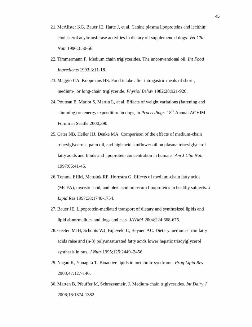
45
21. McAlister KG, Bauer JE, Harte J, et al. Canine plasma lipoproteins and lecithin:
cholesterol acyltransferase activities in dietary oil supplemented dogs. Vet Clin
Nutr 1996;3:50-56.
22. Timmermann F. Medium chain triglycerides. The unconventional oil. Int Food
Ingredients 1993;3:11-18.
23. Maggio CA, Koopmans HS. Food intake after intragastric meals of short-,
medium-, or long-chain triglyceride. Physiol Behav 1982;28:921-926.
24. Pouteau E, Mariot S, Martin L, et al. Effects of weight variations (fattening and
slimming) on energy expenditure in dogs, in Proceedings. 18th Annual ACVIM
Forum in Seattle 2000;390.
25. Cater NB, Heller HJ, Denke MA. Comparison of the effects of medium-chain
triacylglycerols, palm oil, and high acid sunflower oil on plasma triacylglycerol
fatty acids and lipids and lipoprotein concentration in humans. Am J Clin Nutr
1997;65:41-45.
26. Temme EHM, Mensink RP, Hornstra G, Effects of medium-chain fatty acids
(MCFA), myristic acid, and oleic acid on serum lipoproteins in healthy subjects. J
Lipid Res 1997;38:1746-1754.
27. Bauer JE. Lipoprotein-mediated transport of dietary and synthesized lipids and
lipid abnormalities and dogs and cats. JAVMA 2004;224:668-675.
28. Geelen MJH, Schoots WJ, Bijleveld C, Beynen AC. Dietary medium-chain fatty
acids raise and (n-3) polyunsaturated fatty acids lower hepatic triacylglycerol
synthesis in rats. J Nutr 1995;125:2449–2456.
29. Nagao K, Yanagita T. Bioactive lipids in metabolic syndrome. Prog Lipid Res
2008;47:127-146.
30. Marten B, Pfeuffer M, Schrezenmeir, J. Medium-chain triglycerides. Int Dairy J
2006;16:1374-1382.
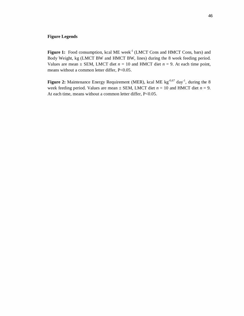
46
Figure Legends Figure 1: Food consumption, kcal ME week-1 (LMCT Cons and HMCT Cons, bars) and Body Weight, kg (LMCT BW and HMCT BW, lines) during the 8 week feeding period. Values are mean ± SEM, LMCT diet n = 10 and HMCT diet n = 9. At each time point, means without a common letter differ, P<0.05. Figure 2: Maintenance Energy Requirement (MER), kcal ME kg-0,67 day-1, during the 8 week feeding period. Values are mean ± SEM, LMCT diet n = 10 and HMCT diet n = 9. At each time, means without a common letter differ, P<0.05.
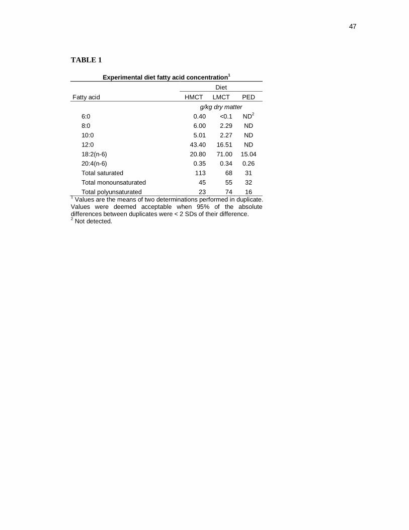
47
TABLE 1
Experimental diet fatty acid concentration1 Diet
Fatty acid HMCT LMCT PED g/kg dry matter 6:0 0.40 <0.1 ND2 8:0 6.00 2.29 ND 10:0 5.01 2.27 ND 12:0 43.40 16.51 ND 18:2(n-6) 20.80 71.00 15.04 20:4(n-6) 0.35 0.34 0.26 Total saturated 113 68 31 Total monounsaturated 45 55 32 Total polyunsaturated 23 74 16
1 Values are the means of two determinations performed in duplicate. Values were deemed acceptable when 95% of the absolute differences between duplicates were < 2 SDs of their difference. 2 Not detected.
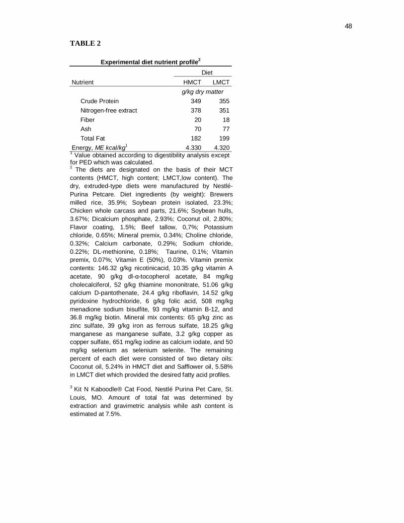
48
TABLE 2
Experimental diet nutrient profile2 Diet
Nutrient HMCT LMCT g/kg dry matter Crude Protein 349 355 Nitrogen-free extract 378 351 Fiber 20 18 Ash 70 77 Total Fat 182 199 Energy, ME kcal/kg1 4.330 4.320
1 Value obtained according to digestibility analysis except for PED which was calculated. 2 The diets are designated on the basis of their MCT contents (HMCT, high content; LMCT,low content). The dry, extruded-type diets were manufactured by Nestlé-Purina Petcare. Diet ingredients (by weight): Brewers milled rice, 35.9%; Soybean protein isolated, 23.3%; Chicken whole carcass and parts, 21.6%; Soybean hulls, 3.67%; Dicalcium phosphate, 2.93%; Coconut oil, 2.80%; Flavor coating, 1.5%; Beef tallow, 0,7%; Potassium chloride, 0.65%; Mineral premix, 0.34%; Choline chloride, 0.32%; Calcium carbonate, 0.29%; Sodium chloride, 0.22%; DL-methionine, 0.18%; Taurine, 0.1%; Vitamin premix, 0.07%; Vitamin E (50%), 0.03%. Vitamin premix contents: 146.32 g/kg nicotinicacid, 10.35 g/kg vitamin A acetate, 90 g/kg dl-α-tocopherol acetate, 84 mg/kg cholecalciferol, 52 g/kg thiamine mononitrate, 51.06 g/kg calcium D-pantothenate, 24.4 g/kg riboflavin, 14.52 g/kg pyridoxine hydrochloride, 6 g/kg folic acid, 508 mg/kg menadione sodium bisulfite, 93 mg/kg vitamin B-12, and 36.8 mg/kg biotin. Mineral mix contents: 65 g/kg zinc as zinc sulfate, 39 g/kg iron as ferrous sulfate, 18.25 g/kg manganese as manganese sulfate, 3.2 g/kg copper as copper sulfate, 651 mg/kg iodine as calcium iodate, and 50 mg/kg selenium as selenium selenite. The remaining percent of each diet were consisted of two dietary oils: Coconut oil, 5.24% in HMCT diet and Safflower oil, 5.58% in LMCT diet which provided the desired fatty acid profiles.
3 Kit N Kaboodle® Cat Food, Nestlé Purina Pet Care, St. Louis, MO. Amount of total fat was determined by extraction and gravimetric analysis while ash content is estimated at 7.5%.
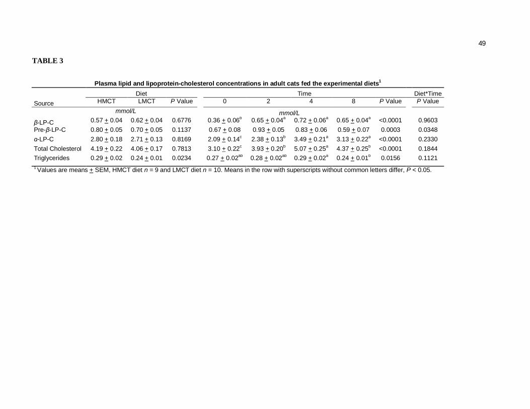
49
TABLE 3
Plasma lipid and lipoprotein-cholesterol concentrations in adult cats fed the experimental diets1 Diet Time Diet*Time
Source HMCT LMCT P Value 0 2 4 8 P Value P Value
mmol/L mmol/L β-LP-C 0.57 + 0.04 0.62 + 0.04 0.6776 0.36 + 0.06b 0.65 + 0.04a 0.72 + 0.06a 0.65 + 0.04a <0.0001 0.9603 Pre-β-LP-C 0.80 + 0.05 0.70 + 0.05 0.1137 0.67 + 0.08 0.93 + 0.05 0.83 + 0.06 0.59 + 0.07 0.0003 0.0348 α-LP-C 2.80 + 0.18 2.71 + 0.13 0.8169 2.09 + 0.14c 2.38 + 0.13b 3.49 + 0.21a 3.13 + 0.22a <0.0001 0.2330 Total Cholesterol 4.19 + 0.22 4.06 + 0.17 0.7813 3.10 + 0.22c 3.93 + 0.20b 5.07 + 0.25a 4.37 + 0.25b <0.0001 0.1844 Triglycerides 0.29 + 0.02 0.24 + 0.01 0.0234 0.27 + 0.02ab 0.28 + 0.02ab 0.29 + 0.02a 0.24 + 0.01b 0.0156 0.1121 1 Values are means + SEM, HMCT diet n = 9 and LMCT diet n = 10. Means in the row with superscripts without common letters differ, P < 0.05.
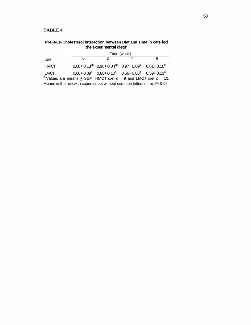
50
TABLE 4 Pre-β-LP-Cholesterol interaction between Diet and Time in cats fed
the experimental diets2
Time (week) Diet 0 2 4 8
HMCT 0.68+ 0.13ab 0.96+ 0.04ab 0.97+ 0.09a 0.61+ 0.10b
LMCT 0.66+ 0.09a 0.88+ 0.10a 0.66+ 0.06a 0.59+ 0.11a 2 Values are means + SEM, HMCT diet n = 9 and LMCT diet n = 10. Means in the row with superscripts without common letters differ, P<0.05.
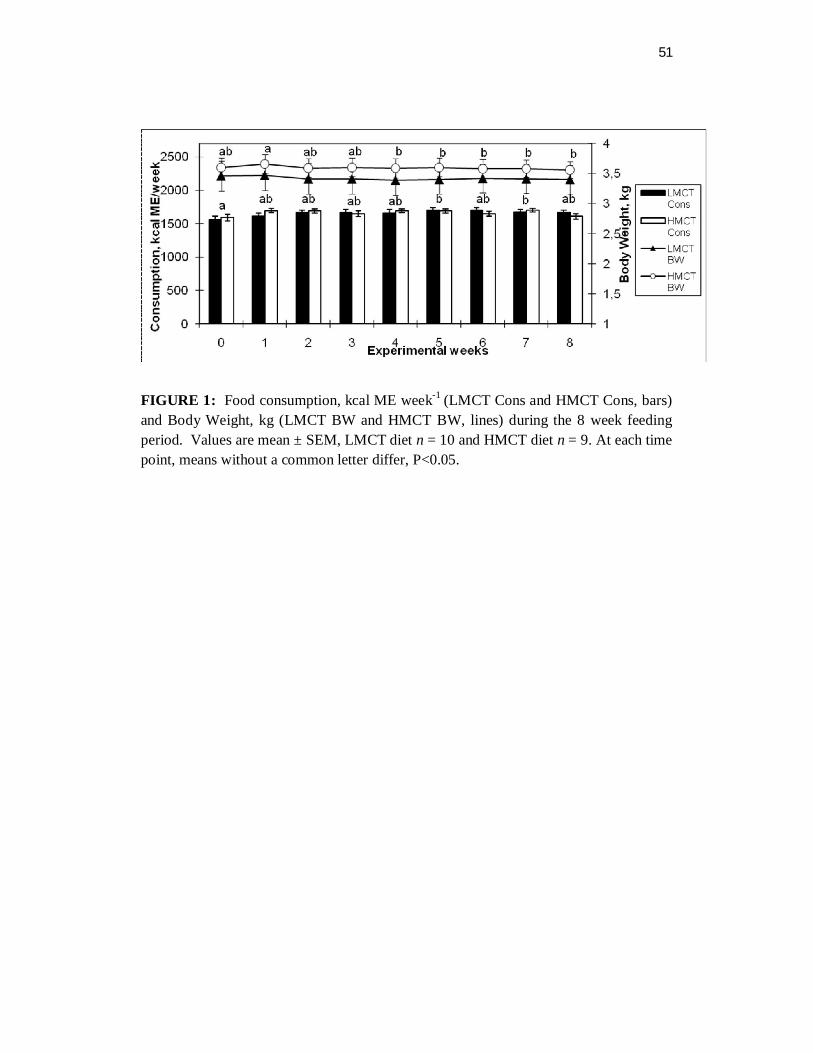
51
FIGURE 1: Food consumption, kcal ME week-1 (LMCT Cons and HMCT Cons, bars) and Body Weight, kg (LMCT BW and HMCT BW, lines) during the 8 week feeding period. Values are mean ± SEM, LMCT diet n = 10 and HMCT diet n = 9. At each time point, means without a common letter differ, P<0.05.
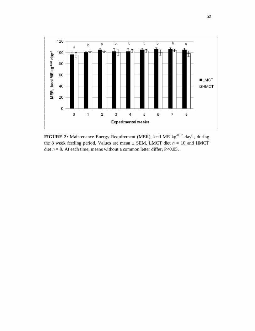
52
FIGURE 2: Maintenance Energy Requirement (MER), kcal ME kg-0,67 day-1, during the 8 week feeding period. Values are mean ± SEM, LMCT diet n = 10 and HMCT diet n = 9. At each time, means without a common letter differ, P<0.05.

53
3 CAPÍTULO III4
4 Artigo formatado conforme normas do Journal of Nutrition.
54
3.1 Dietary γ-Linolenic Acid Supports Arachidonic Acid Enrichment in Feline
Plasma Phospholipids and Feline Red Blood Cell Membranes 1
Luciano Trevizan, 2,3 Alexandre de Mello Kessler, 2 Karen E. Bigley, 3 Wendy
Anderson, 4 Mark K. Waldron, 4 John E. Bauer, 3*.
2 Department of Animal Science, Universidade Federal do Rio Grande do Sul, Porto
Alegre, RS 91540-000, Brazil.
3 Department of Small Animal Clinical Science, Comparative Nutrition Laboratory,
College of Veterinary Medicine and Biomedical Sciences, Texas A & M University,
College Station, TX 77843.
4 Nestlé Purina Pet Care, St Louis, MO.
1 Funded by Nestlé-Purina Pet Care, Saint Louis, MO, USA and the Mark L. Morris
Professorship of Clinical Nutrition, Texas A & M University, TX USA.
6 Abbreviations used: LA, linoleic acid; AA, arachidonic acid; GLA, γ-linolenic
acid; BCS, body conditions score; BW, body weight; TG, plasma triglycerides; TC,
total cholesterol; LP, lipoprotein fractions; DGLA, dihomo-γ-linolenic acid.
* To whom correspondence should be addressed. E-mail: [email protected].
Phone number: 979 845 2321
55
Abstract
Conversion of linoleic acid (LA) to arachidonic acid (AA) in cats is limited due to low Δ6-
desaturase. Thus, it may be possible to induce ∆6 desaturase directly by providing large dietary
amounts of LA precursor. In addition, an alternative pathway for AA may also exist involving
LA chain elongation, followed by ∆8- and ∆5-desaturation. However, should an active ∆5-
desaturase exist in feline tissues, it is more likely that dietary γ-linolenic acid (GLA) may
bypass ∆6-desaturation and result in AA synthesis. Consequently, we hypothesized that GLA
feeding bypasses ∆6-desaturation for AA synthesis. We further hypothesized that cats fed high
LA results in AA accumulation either directly, by inducing ∆6-desaturase, or perhaps by the
alternate route. To test these hypotheses, fatty acid profiles were determined after feeding high
LA (HL), high GLA (GLA), or adequate LA (LL, control) diets. Adult female cats (n=29) were
separated into three groups and fed for 8 wks according to their metabolic body weights to
maintain body condition scores (BCS) of 5/9 with water ad libitum. Daily food consumption
and weekly body weights (BW), maintenance energy requirements (MER) and BCS were
recorded. Blood samples were collected after withholding food overnight at wks 0, 2, 4 and 8 to
measure plasma triglyceride (TG) and total cholesterol (TC) concentrations as well as
lipoprotein distributions (LP) and plasma and red blood cell phospholipid fatty acids. Repeated
measures analysis of variance and Tukey (α=0.05) multiple comparisons were performed. No
significant time effects were observed on food consumption, BW, BCS, and MER after Wk 1.
No diet effects were observed for TG, TC, and lipoprotein fractions. However, time effects were
observed at Wk2 and Wk4 with significant increases in these parameters but all were within
normal limits and most likely due to the increased dietary fat content of the test diets. No
evidence of ∆8-desaturase enzyme activity was found with the HL diet. However, a GLA diet
effect was observed as early as wk2 with the formation of dihomo-γ-linolenic acid (DGLA
(20:3Δ8,11,14)) and AA as early as wk2, suggesting the presence of ∆5-desaturase activity. It is
thus possible to use dietary GLA to induce AA synthesis.
56
Introduction
In most mammalian species the conversion of linoleic acid (LA) to arachidonic acid
(AA) involves specific enzyme steps including ∆6 desaturation followed by chain
elongation and ∆5 desaturation (1). By contrast, evidence that cats lacked sufficient
Δ6 desaturase activity suggested that a dietary source of AA was necessary in this
species, (2-5). Further speculation at that time about the existence of a feline Δ6
desaturase developed when cats were fed essential fatty acid (EFA) deficient diet
using hydrogenated beef fat (6). In this case the accumulation of 20:3n-9 was found
even though earlier studies did not detect this fatty acid (4-5). Because 20:3n-9 is
produced by chain elongation and Δ6 desaturation of 18:1n-9 (7), it was
hypothesized that Δ6-desaturase should be present in cats (8) (Figure 1).
In addition to this speculation, other authors suggested an alternative pathway for
20:3n9, synthesis in tissues independent of Δ6 desaturase. It is known that a Δ8-
desaturase occur in rat testis is responsible for conversion of LA to AA (9). The Δ8-
desaturase was also, described in human bladder and colon, (10). Thus, it may be
possible that such a pathway might exist in feline species (1) (Figure 1).
When [1-14C]-LA was included in the diet, no evidence was found for AA
production. However, when [2-14C] 20:3n-6 was given, there was significant
production of AA, suggesting that cats do possess Δ5-desaturase activity (1).
Cats fed diets adequate in LA but deprived of other essential fatty acids (EFA),
showed dry, lusterless hair coats, dandruff, behavioral infertility, and hepatic lipid
infiltration (2). A series of studies (3-4; 12-13) ultimately demonstrated EFA
deficiency, even though cats had been fed the requisite LA and ALA (14). Thus,
came out the probability of other FA to be involved besides C18 precursors. When
57
γ-linolenic acid (GLA - 18:3n-6) containing oils were added to the diets,
reproductive status improved (12). The GLA may have improved AA status to some
extent by virtue of bypassing the Δ6 desaturase step. These studies have provided
the basis for the present day concept that cats require a preformed source of AA and
that Δ6 desaturase activity is either low or non-existent in feline species. However,
the possibility remains that sufficient AA acid may be synthesized from LA under
some condition using ∆5 and ∆8desaturases (6). In view of these possibilities, we
hypothesized, that feeding cats dietary oil enriched in GLA could be a practical way
to provide precursors for AA synthesis and that high dietary amounts of LA would
induce an alternative AA synthetic pathway using ∆5 and ∆8desaturase enzymes.
58
Material and Methods
Animals and diets. Twenty nine adult, sexually intact female cats ranging in age
between 1.5 to 2 years with body weights between 2.4 and 6.0 kg were used. Cats
were individually maintained in kennels according to the American Physiological
Society Guidelines for Animal Research, and the protocols were approved by Texas
A & M University Animal Care and Use Committee. The cats were assessed to be
clinically normal. The cats were assessed to be clinically normal prior to entering
the study as indicated by complete blood counts, serum biochemistry profiles, and
physical examination by one of the authors all of which were within normal limits.
The animals were then randomly assigned into three groups according diet fed: High
Linoleic Acid (HL, n=9), formulated with LA-rich safflower oil; Low Linoleic Acid
(LL, n=10), in which safflower oil was replaced by coconut oil reduce LA to
adequate levels, and γ-Linolenic Acid (GLA, n=10), formulated to contain adequate
LA plus added borage oil with 70% GLA. The diets were similar in all respects
except for FA type (Table 1). The expected nutrient composition of the diet was:
35% protein, 18% fat, 7,5% ash, 8% moisture, 2% crude fiber. After manufacture,
analyses of the diets were performed analysis by Nestlé Purina Laboratories and
results were within expected analytical variance for these targets (Table 2).
Cats were fed according to their individual metabolic weights (0,418 MJ ME *
Wkg-0,67 day-1) and to maintain a body condition score (BCS) 5 out of 9 (18). The
cats consumed a pre-experimental diet (PED) for 4 weeks prior to the start of the
trial and were fed the experimental diets for 8 weeks. The nutrient composition of
PED was: 30% protein, 10% fat, 12% moisture, 4,5% crude fiber and AA
represented 0,02% of total diet. During the PED feeding period, daily food
consumption was determined and used to maintain the animal’s body weight at a
59
BCS 5. They were allowed to play with paper bags to provide physical activity. The
controlled diet amounts fed and the stimuli to play helped them control their body
weights. According to weekly weight evaluations the cats with BCS higher than 5
had were provided a reduced calorie allowance to 0.334*Wkg0,67 day-1, because
these cats were resistant to weight loss. Cats were maintained in this fashion until
reaching the desired BCS 5. The experimental diets were formulated to be complete
and balanced in order to meet or exceed minimal nutrient requirements established
for adult cats (15-16) and manufactured by Nestlé Purina PetCare, Saint Louis.
Cat body weights (BW) and BCS were determined weekly during the PED period.
The animals were fed once daily and food consumption and BMR (Body
Maintenance Energy Requirement – MJ ME kg0,67 day-1) were recorded daily after
24 hours and averaged weekly for statistical analysis.
Blood sample collection and analyses. Blood samples were obtained from each cat
by venipuncture at weeks 0, 2, 4 and 8 during the experimental periods. Food had
been withheld for 12 hours and 7 ml of blood were drawn from a saphenous vein
into EDTA-containing tubes. At week 0, complete blood counts and plasma
biochemistry profiles were evaluated. Blood samples were centrifuged at 2800 rpm
for 15 minutes and plasma was separated from the red blood cells (RBC) and
plasma was frozen at -80oC in small aliquots. Packed RBC’s were kept on ice for
immediate RBC membrane ghost preparation. RBC membranes were prepared using
an isotonic saline wash, lysis with deionized water, followed by slow speed
centrifugation and storage at -80oC (17). Total lipids were then extracted from the
both the plasma and RBC membranes (18). Total phospholipids fractions were
separated by thin layer chromatography (TLC) scraped from the plates and
60
methylated to their fatty acid methyl esters (FAMEs) for analysis via gas
chromatography (19).
Plasma triglycerides (TG) and total cholesterol (TC) concentrations were
determined using enzymatic methods (20). Lipoprotein cholesterol distributions
(LP-C) were determined using freshly isolated plasma by eletrophoresis (agarose
gel at 1%) and quantified by scanning densitometry (20).
Digestibility assays. All cats were submitted to a digestibility study during week
6of the feeding period. Fecal samples were individually collected twice a day from
for 5 consecutive days, pooled for each cat and frozen at -40 C. All samples,
including the pooled samples and diets, were sent to the Nestlé Purina Petcare
Laboratory, Saint Louis, for analysis. The parameters evaluated were coefficient of
digestibility from crude protein, total fat, crude fiber, ash and gross energy. The
coefficient for digestible energy (DE) was calculated and the metabolizable energy
was calculated based on DE and urinary loss (15).
Statistical methods. Values presented are means + SEM. All data except for the
normative BCS data were found to be normally distributed by the Shapiro-Wilks.
The BCS were thus analyzed by the nonparametric test Kruskal-Wallis one-way
ANOVA (SAS 9). Digestibility coefficients were analyzed for main diet and time
effects, and diet x time interactions using Proc GLM ANOVA by SAS (P<0.05). For
the other data, statistical significance was determined using repeated measures
ANOVA for diet, time, and diet x time interactions for the following plasma
parameters (TG, TC, LP-C, plasma phospholipid fatty acid profile), RBC membrane
fatty acid profile, food consumption, body weight, and metabolic factor using Proc
61
Mixed by SAS ( P < 0.05). For all parameters Tukey’s multiple comparison of
means was performed where appropriate.
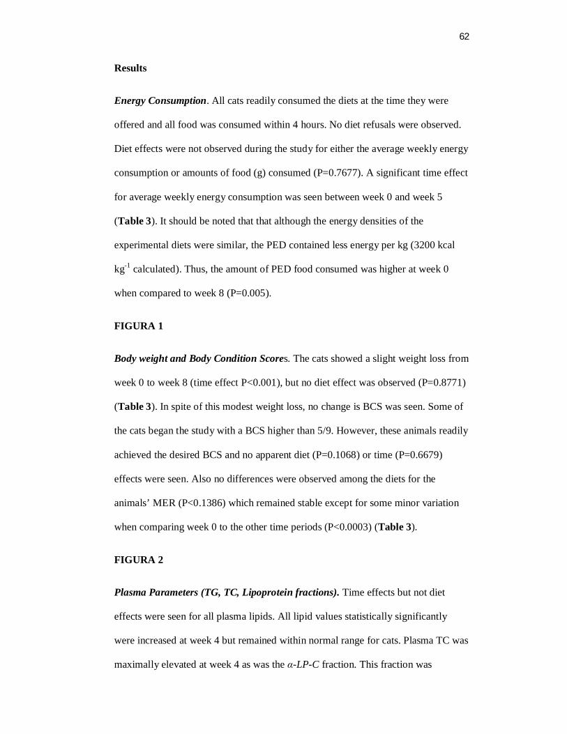
62
Results
Energy Consumption. All cats readily consumed the diets at the time they were
offered and all food was consumed within 4 hours. No diet refusals were observed.
Diet effects were not observed during the study for either the average weekly energy
consumption or amounts of food (g) consumed (P=0.7677). A significant time effect
for average weekly energy consumption was seen between week 0 and week 5
(Table 3). It should be noted that that although the energy densities of the
experimental diets were similar, the PED contained less energy per kg (3200 kcal
kg-1 calculated). Thus, the amount of PED food consumed was higher at week 0
when compared to week 8 (P=0.005).
FIGURA 1
Body weight and Body Condition Scores. The cats showed a slight weight loss from
week 0 to week 8 (time effect P<0.001), but no diet effect was observed (P=0.8771)
(Table 3). In spite of this modest weight loss, no change is BCS was seen. Some of
the cats began the study with a BCS higher than 5/9. However, these animals readily
achieved the desired BCS and no apparent diet (P=0.1068) or time (P=0.6679)
effects were seen. Also no differences were observed among the diets for the
animals’ MER (P<0.1386) which remained stable except for some minor variation
when comparing week 0 to the other time periods (P<0.0003) (Table 3).
FIGURA 2
Plasma Parameters (TG, TC, Lipoprotein fractions). Time effects but not diet
effects were seen for all plasma lipids. All lipid values statistically significantly
were increased at week 4 but remained within normal range for cats. Plasma TC was
maximally elevated at week 4 as was the α-LP-C fraction. This fraction was
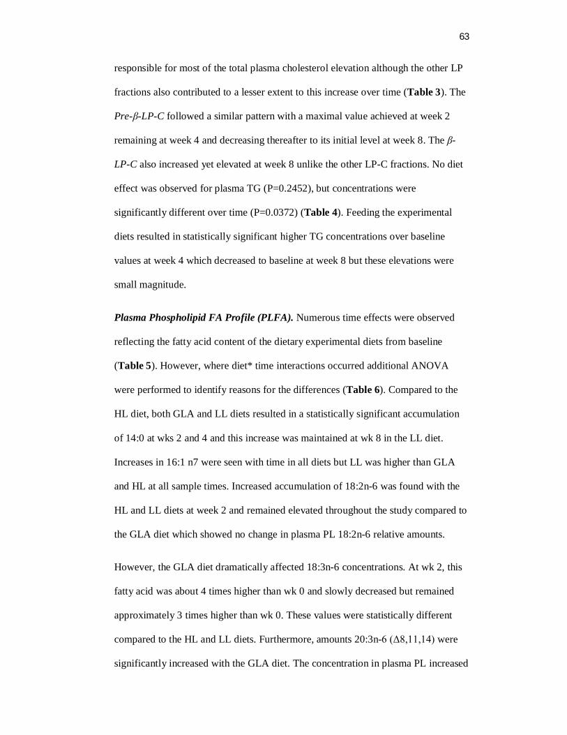
63
responsible for most of the total plasma cholesterol elevation although the other LP
fractions also contributed to a lesser extent to this increase over time (Table 3). The
Pre-β-LP-C followed a similar pattern with a maximal value achieved at week 2
remaining at week 4 and decreasing thereafter to its initial level at week 8. The β-
LP-C also increased yet elevated at week 8 unlike the other LP-C fractions. No diet
effect was observed for plasma TG (P=0.2452), but concentrations were
significantly different over time (P=0.0372) (Table 4). Feeding the experimental
diets resulted in statistically significant higher TG concentrations over baseline
values at week 4 which decreased to baseline at week 8 but these elevations were
small magnitude.
Plasma Phospholipid FA Profile (PLFA). Numerous time effects were observed
reflecting the fatty acid content of the dietary experimental diets from baseline
(Table 5). However, where diet* time interactions occurred additional ANOVA
were performed to identify reasons for the differences (Table 6). Compared to the
HL diet, both GLA and LL diets resulted in a statistically significant accumulation
of 14:0 at wks 2 and 4 and this increase was maintained at wk 8 in the LL diet.
Increases in 16:1 n7 were seen with time in all diets but LL was higher than GLA
and HL at all sample times. Increased accumulation of 18:2n-6 was found with the
HL and LL diets at week 2 and remained elevated throughout the study compared to
the GLA diet which showed no change in plasma PL 18:2n-6 relative amounts.
However, the GLA diet dramatically affected 18:3n-6 concentrations. At wk 2, this
fatty acid was about 4 times higher than wk 0 and slowly decreased but remained
approximately 3 times higher than wk 0. These values were statistically different
compared to the HL and LL diets. Furthermore, amounts 20:3n-6 (Δ8,11,14) were
significantly increased with the GLA diet. The concentration in plasma PL increased
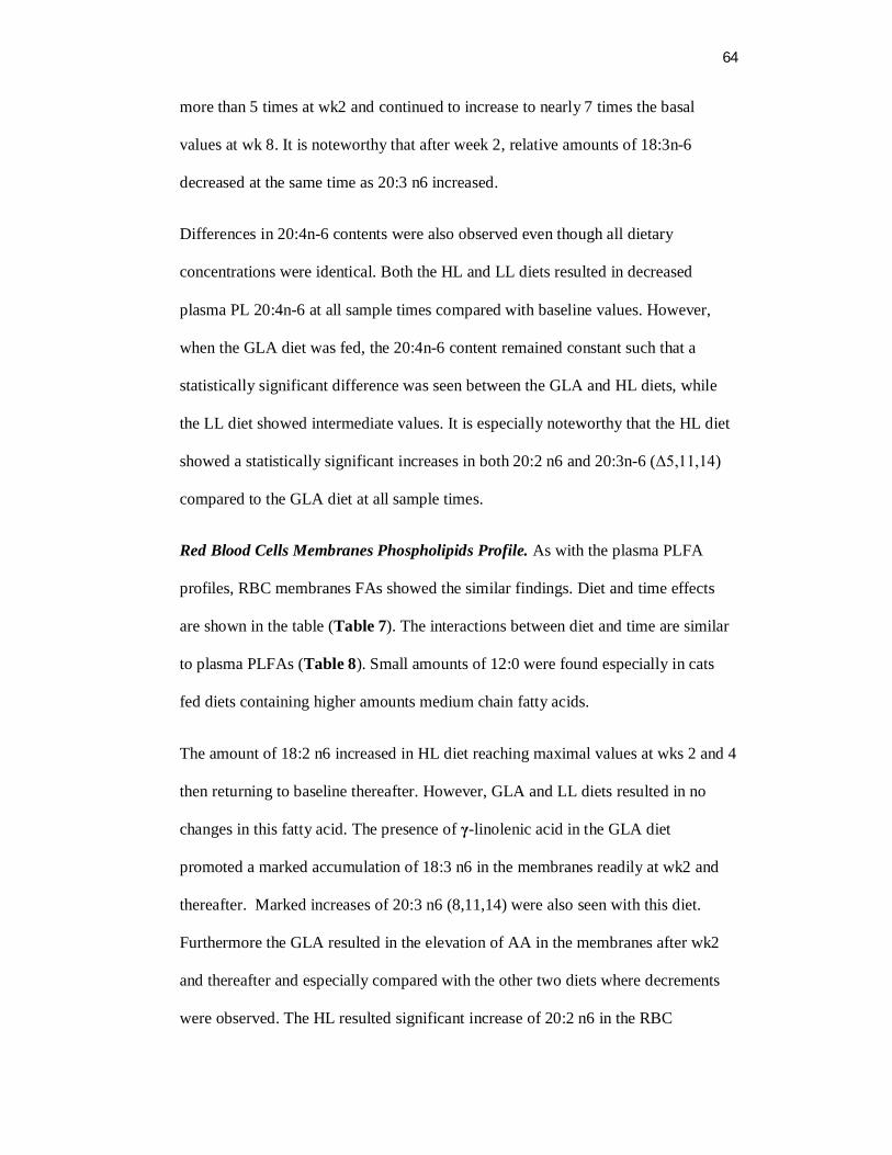
64
more than 5 times at wk2 and continued to increase to nearly 7 times the basal
values at wk 8. It is noteworthy that after week 2, relative amounts of 18:3n-6
decreased at the same time as 20:3 n6 increased.
Differences in 20:4n-6 contents were also observed even though all dietary
concentrations were identical. Both the HL and LL diets resulted in decreased
plasma PL 20:4n-6 at all sample times compared with baseline values. However,
when the GLA diet was fed, the 20:4n-6 content remained constant such that a
statistically significant difference was seen between the GLA and HL diets, while
the LL diet showed intermediate values. It is especially noteworthy that the HL diet
showed a statistically significant increases in both 20:2 n6 and 20:3n-6 (Δ5,11,14)
compared to the GLA diet at all sample times.
Red Blood Cells Membranes Phospholipids Profile. As with the plasma PLFA
profiles, RBC membranes FAs showed the similar findings. Diet and time effects
are shown in the table (Table 7). The interactions between diet and time are similar
to plasma PLFAs (Table 8). Small amounts of 12:0 were found especially in cats
fed diets containing higher amounts medium chain fatty acids.
The amount of 18:2 n6 increased in HL diet reaching maximal values at wks 2 and 4
then returning to baseline thereafter. However, GLA and LL diets resulted in no
changes in this fatty acid. The presence of γ-linolenic acid in the GLA diet
promoted a marked accumulation of 18:3 n6 in the membranes readily at wk2 and
thereafter. Marked increases of 20:3 n6 (8,11,14) were also seen with this diet.
Furthermore the GLA resulted in the elevation of AA in the membranes after wk2
and thereafter and especially compared with the other two diets where decrements
were observed. The HL resulted significant increase of 20:2 n6 in the RBC
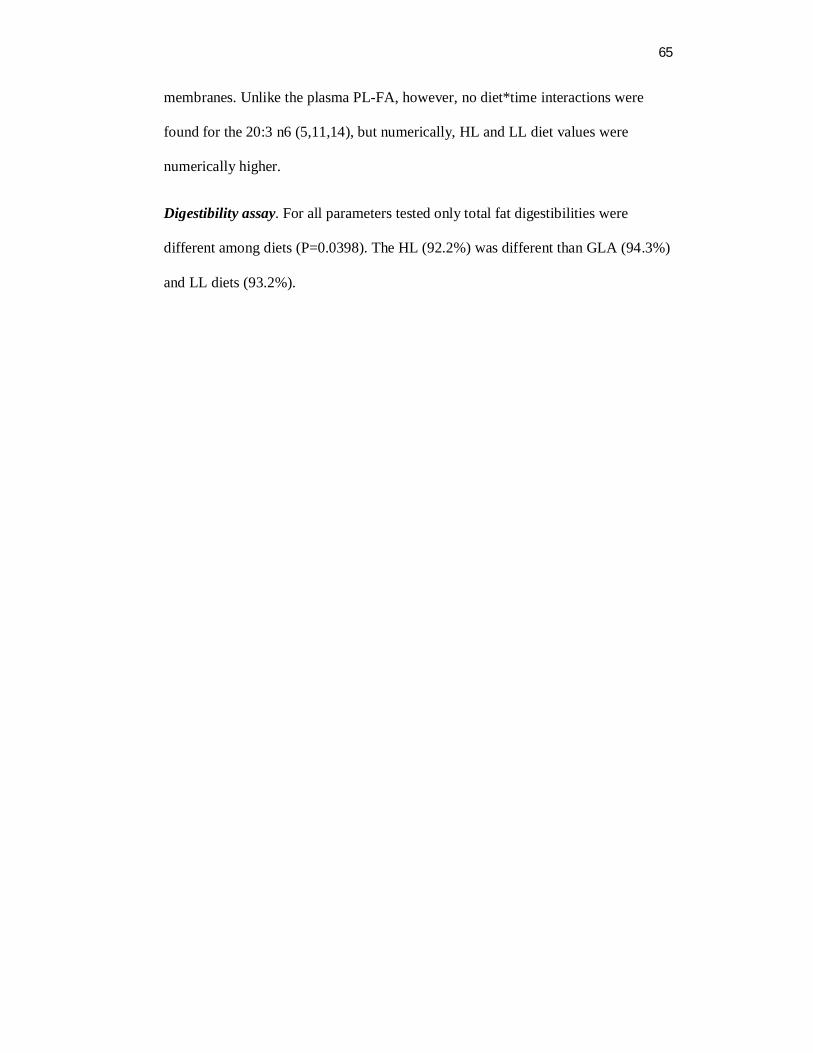
65
membranes. Unlike the plasma PL-FA, however, no diet*time interactions were
found for the 20:3 n6 (5,11,14), but numerically, HL and LL diet values were
numerically higher.
Digestibility assay. For all parameters tested only total fat digestibilities were
different among diets (P=0.0398). The HL (92.2%) was different than GLA (94.3%)
and LL diets (93.2%).
66
Discussion
None of the diets had any differential effects on amounts consumed, BW, BMR and
BCS. Comparative values at wk 0 were somewhat different from the other values
because the first week of feeding was used for adaptation to the experimental diets
which, for cats generally requires 4-5 days (16). Eliminating the wk 0 values from
the analysis revealed neither time nor diet effects except for the decrease in body
weight which was planned. In the other weeks some variation can be observed and
at the end of this trial it is possible see reduced values for BW and consumption
parameters. Cats were fed to maintain the BCS 5 out 9 and for that reason they
reached such values at the end of this trial. Cats reduced slightly the BW being
difficult to detect it by BCS evaluation. The BMR found for cats in maintenance
was approximately the same indicated for this category (15).
Plasma lipids, as TG and TC and their fractions showed similar behavior without
any diet effect, so γ-linolenic diet did not produce any isolated effect. Along of the
weeks the increase and decrease in plasma parameters are directly associated to the
content of crude fat present in the experimental diets more than in their qualities.
Cats showed to be versatile in their metabolism, adapting easily to the new diets
after wk 4. The elevation of lipids in the plasma is an expected effect since the diet
they were eating had around 190 g kg-1 DM crude fat, while the PED had around
120 g kg-1 DM. In addition, the high digestibility of fat is other factor to the
enrichment of fat in the plasma. Unlike in humans the cat reaction against a high fat
diet seems like to be protective (21). The increase in lipoproteins fraction rises up
the α-LP-C fraction (HDL) as a response to the over uptake of lipids from the diet,
working as a protective metabolism against cardiac diseases (22-23).
67
The effects of the diets were seem more clearly when the fatty acids accumulation
or in plasma PL (Figure 2) either in red blood cells PL (Figure 3) were evaluated.
When fatty acids in the diets overpass the animal needs is common see the
accumulation in the tissues. Usually, LCFA are more readily incorporated due to the
Fatty Acid Synthetase enzyme which is more effective in esterify triglycerides with
LCT (24). It was notorious the incorporation of 18:2 n6 in the membranes
associated to HL diet, but it did not show any alteration in 18:3 n6 concentration in
the plasma PL or in the RBC PL, demonstrating Δ6 desaturase absence or very low
activity as already was known (2-5). However, the effect of elongase enzyme was
expressed over the high concentration of 18:2 n6. In this case 20:2 n6 was increased
continually in the plasma and RBC PL initiating the alternative pathway forward to
AA synthesis. This route was viable until the next step which one depended on Δ5
desaturase to produce 20:3 n6 (Δ5,11,14) from 20:2 n6, and the concentration was
really increased.
The knowledge that some species showed to present Δ8-desaturase (9-10) was not
applicable to the cats in this trial, since the AA concentration decreased in the
plasma and in the membranes in the time. The high concentration in the wk 0
probably was consequence of the diet that cats had been fed before PED. It was a
diet for adult cats with higher concentration of AA, compounding around 0,07% of
total diet. Probably that is the cause of AA plasma levels decreased in the time so
fast.
The LL diet had the LA 3.5 times smaller than HL diet but, the effects over the
normal pathway and the alternative pathway seems to be the same for most part of
the fatty acids when compared to HL diet. However, the HL diet induced the
elongase enzyme more efficiently that LL diet, probably through the concentration
68
resulting in a higher increase in 20:2n6. The concentration of AA in plasma PL and
RBC PL did not changed between diets, but curiously the LL had higher numeric
values when compared to HL diet. It probably happened in HL diet because of Δ5
desaturase was more demanded in the alternative pathway than in the normal
pathway since 20:2 n6 was increased. By the values it is possible see in plasma and
in RBC that the concentration of 20:3 n6 (8,11,14) is always higher in LL diet and
that FA is a product of elogase enzyme and a substrate for Δ5 desaturase enzyme
which ones are involved in the alternative pathway in HL diet by substrate
induction. It reflects the AA concentration that followed the same way being
numerically higher in LL, since alternative pathway is not functional probably due
to Δ8-desaturase enzyme inexists.
The GLA diet bypassed the Δ6 enzyme efficiently. The incorporation in the
membranes was faster end efficient. The γ-linolenic acid in plasma PL was
decreasing after wk2 showing an effect of elongase enzyme induction. It can be
better noticed when DGLA is observed, its concentration is progressively increased
in the time, which supports AA concentration. The same happened in the RBC, as
prove of that in the tissues AA can be maintained by supplementing γ-linolenic acid
in the diet. Interestingly, if observed the concentration of γ-linolenic acid in the
plasma PL or RBC it is not more than 0,9% at anytime, but the concentration of its
product (DGLA) is increasing continuously and that values are always higher than
2,5% showing the high and progressive increase in elongase enzyme activity as
described previously (25). It is the guaranty that providing GLA, DGLA will be
formed in cats. The DGLA belongs to a group of FA proved to have anti-
inflamatory effects and immunomodulating properties on the skin. It modulates the
eicosanoid production by competing with AA for ciclo-oxigenase and lipo-
69
oxigenase in the AA cascade. This competition results in a shift towards production
of LT with anti-inflamatory properties (prostaglandins and tromboxanos, serie 1 and
leukotrienes serie 3) instead of proinflammatory LT (LTB4 and PG2). Atopic
dermatitis is marked by pruritus and had been treated by association between GLA
and EPA as anti-inflammatory FA. Some authors still have doubts about this type of
treatment due to high variation and difficult way to evaluate the animals based in
their symptoms. A study in dogs concluded that early stages cases seem to be more
responsive to the anti-inflamatory FA supplementation (26). Other study in guinea
pig induced epidermal hyperproliferation was reverted adding different sources of
GLA containing oils (27). The supplementation of DGLA in NC/Nga atopic mice
resulted in incorporation in the PL and reduction of symptoms which ones were
induced again through discontinuation of supplementation (28).
Several sources of γ-linolenic acid are available as a borage oil, evening primrose
oil, canola oil genetically modified (29), Spirulina platensis (30) and it is possible
find DGLA oil extract from the fungus Mortierella alpine which one contain
approximately 40% DGLA (31). According to this can be possible include in the
diets its components.
In summary, diets containing GLA can be consumed by cats normally with no
alteration in BW if the requirements are attended. Cats showed do not have Δ6 and
Δ8 desaturase active. The elongase enzyme is highly active and its activity is
induced by substrate as was demonstrated in HL diet, 18:2 n6 to 20:2 n6, and GLA
diet (18:3 n6 to 20:3 n6). In the same way Δ5 desaturase showed to be active in cats.
In HL diet it promoted the desaturation in 20:2 n6 to 20:3 n6 (Δ5,11,14) and in the
GLA diet promoted the desaturation over DGLA to form AA. Additionally, GLA
diet promoted the accumulation of γ-linolenic acid and dihomo-γ-linoleic acid in the

70
plasma and tissues which ones can be effective anti-inflammatory eicosanoids and
prostaglandins being a key point to modulate the immunological system. In the
same time GLA diet supported AA concentration in the membranes in cats fed
minimal requirements of AA.
71
Literature Cited
1. Sinclair AJ, McLean JG, Monger EA. Metabolism of linoleic acid in the cat. Lipids 1979;14:932-936.
2. Rivers JPW, Sinclair AJ, Crawford MA. Inability of the cat to desaturate essential fatty acids. Nature 1975;258:171-173.
3. Rivers JPW, Hassam AG, Crawford MA, Brambell MR. The absence of Δ6-desaturase activity in cats. Proceedings of the nutrition society 1976a;35, 69A.
4. Rivers JPW, Sinclair AJ, Moore DP, Crawford MA. The abnormal metabolism of essencial fatty acids in cat. Proceedings of nutrition Society 1976b;35, 68A.
5. Rivers JPW, Frankel TL. Fat in the diet of dogs and cats. In: Anderson RS (ed), Nutrition of the dog and cat. Pergamon press, Oxford, UK, pp.67-99, 1980.
6. Sinclair AJ, Slattery W, M cLean JG, Monger EA. Essential fatty acid deficiency and evidence for arachidonate synthesis in the cat. Br J Nutr 1981;46:93-96.
7. Holman RT. Biological activities of polyunsaturated fatty acids. Prog chem. Fats lipids 1970;9:607-682.
8. McLean JG, Monger EA. Factors determining the essencials fatty acid requirements of the cat. In Nutrition of the Dog and Cat. Waltham Symposium no. 7, pp. 329-342 [I H Burger and JPW Rivers, editors]. Cambridge: Cambridge University Press.1989
9. Albert DH, Coniglio JG. Metabolism of eicosan-11,14 dienoic acid in rat testes. Evidence for delta 8-desaturase activity. Biochimica et Biophysica Acta 1977;489:390-396.
10. Nakazawa I, Mead JF, Yonemoto RH. In vitro activity of the fatty acyl desaturase of human cancerous and noncancerous tissues. Lipids 1976;11:79-81.
11. Hassam AG, Rivers JPW, Crawford MA () The failure of the cat to desaturate linoleic acid: its nutritional implications. Nutr Metab 1977;21:321-328.
12. Frankel T, Rivers JPW. The nutritional and metabolic impact of γ-linolenic acid (18:3 n6) on cats deprived of animal lipid. Br J Nutr 1978;39:227-231.
72
13. Rivers JPW. Essencial fatty acids in cats. Journal of small animal practice 1982;23:563-576.
14. MacDonald ML, Rogers QR, Morris JG. Nutrition of the domestic cat, a mammalian carnivore. Ann Rev of Nutr 1984;4:521-562.
15. National Research Council. Nutrient requirements of dogs and cats. Washington, DC: National Academies Press; 2006.
16. AAFCO. Official Publication. Oxford (IN): Association of American Feed Control Office; 2008.
17. Burton GW, Ingold KU, Thompson KE. An improved procedure for the isolation of ghost membranes from human red blood cells. Lipids 1981;16:946.
18. Folch J, Lees M, Sloane-Stanley GH. A simple method for the isolation and purification of total lipids from animal tissues. J Biol Chem. 1957;226:497-506.
19. Angell R, Mitsuhashi Y, Bigley K, Bauer JE. Plasma LCAT activity and lipid subfraction composition in obese beagles undergoing weight loss. Lipids (in press, 2009).
20. McAlister KG, Bauer JE, Harte J, et al. Canine plasma lipoproteins and lecithin: cholesterol acyltransferase activities in dietary oil supplemented dogs. Vet Clin Nutr. 1996;3:50-56.
21. Bauer, J E. Facilitative and functional fats in canine and feline diets. JAVMA 2006;229:680-684.
22. Demacker PNM, Van Heijst PJ, Hak-Lemmers HLM et al. A study of the lipid transport system in the cat, Felix domesticus. Atherosclerosis 66:113-123, 1987.
23. Bauer JE. Lipoprotein-mediated transport of dietary and synthesized lipids and lipid abnormalities and dogs and cats. JAVMA. 2004;224,5:668-75.
24. Papamandjaris AA, MacDougall DE, Jones PJH. Medium-chain fatty acid metabolism and energy expenditure: obesity treatment implications. Life Sciences 1998;62:1203-1215.
25. Van Rooyen J, Swanevelder S, Morgenthal, JC, Spinnler Benadé AJ. Diet can manipulate the metabolism of EPA and GLA in erythrocyte membrane and plasma. Prostaglandins, Leukotrienes and Essencial Fatty Acids 1998;59:27-38.
26. Abba C, Mussa PP, Vercelli A, Raviri G. Essencial fatty acids supplementation in different-stage atopic dogs fedo n a controlled diet. J Anim Phys Anim Nutr. 2005;89:203-207.
73
27. Chung S, Kong S, Seong K, Cho Y. Gamma-linolenic acid in borage oil recerses epidermal hypeproliferation in guinea pigs. American Society for Nutritional Sciences 2002;3090-3097.
28. Kawashima H, Tateishi N, Shiraishi A, Teraoka N, Tanaka T, Tanaka A, Matsuda H, Kiso Y. Oral administration of dihomo-γ-linolenic acid prevents development of atopic dermatitis in NC/Nga mice. Lipids 2008;43:37-43.
29. Liu JW, DeMichele SJ, Palombo J, Chuang LT, Hastilow C, Bobik Jr E, Huang YS. J Agric Food Chem. 2004;52:3960-3966.
30. Sagilata MG, Singhal RS, Kamat MY. Fractionation of lipids and purification of γ-linolenic acid (GLA) from Spirulina platensis. Food Chemistry. 2008;109:580-586.
31. Kawashima H, Akimoto K, Higashiyama K, Fugikawa S, Shimizu S. Industrial production of dihomo-γ-linolenic acid by a Δ5 desaturase-defective mutant of Mortierella alpina 1S-4 fungus. J Am Oil Chem Soc. 2000;77:1135-1138.
74
Acknowledgement
The authors wish to acknowledge the Conselho Nacional de Desenvolvimento
Científico e Tecnológico (CNPq) from the Brazilian Goverment for their
sponsorship of a student to conduct this study.
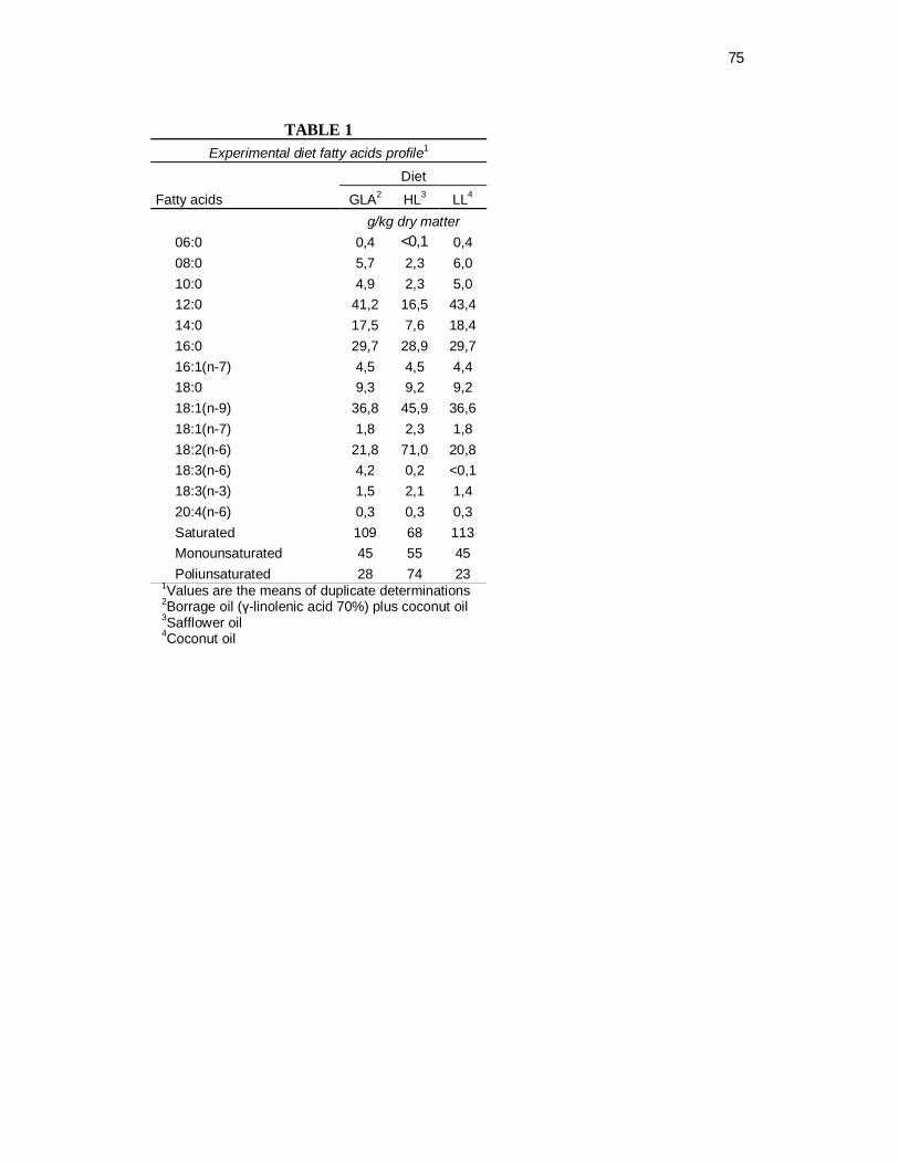
75
TABLE 1
Experimental diet fatty acids profile1 Diet
Fatty acids GLA2 HL3 LL4 g/kg dry matter 06:0 0,4 <0,1 0,4 08:0 5,7 2,3 6,0 10:0 4,9 2,3 5,0 12:0 41,2 16,5 43,4 14:0 17,5 7,6 18,4 16:0 29,7 28,9 29,7 16:1(n-7) 4,5 4,5 4,4 18:0 9,3 9,2 9,2 18:1(n-9) 36,8 45,9 36,6 18:1(n-7) 1,8 2,3 1,8 18:2(n-6) 21,8 71,0 20,8 18:3(n-6) 4,2 0,2 <0,1 18:3(n-3) 1,5 2,1 1,4 20:4(n-6) 0,3 0,3 0,3 Saturated 109 68 113 Monounsaturated 45 55 45 Poliunsaturated 28 74 23
1Values are the means of duplicate determinations
2Borrage oil (γ-linolenic acid 70%) plus coconut oil 3Safflower oil 4Coconut oil
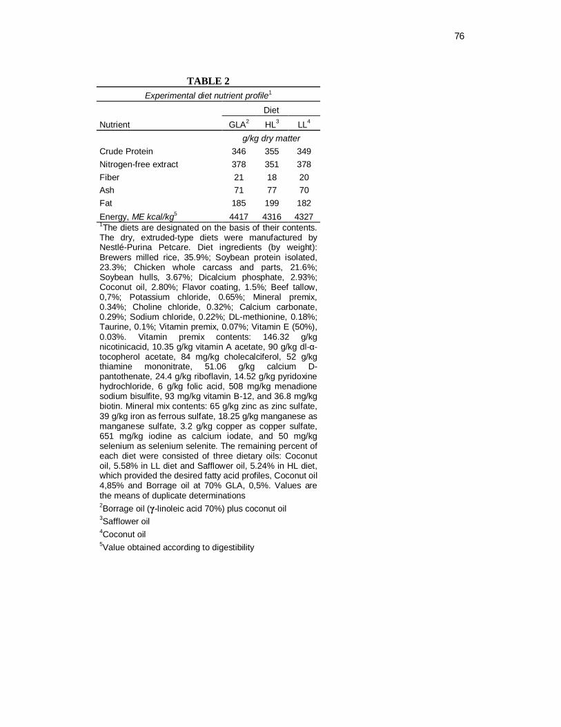
76
TABLE 2
Experimental diet nutrient profile1 Diet
Nutrient GLA2 HL3 LL4 g/kg dry matter Crude Protein 346 355 349 Nitrogen-free extract 378 351 378 Fiber 21 18 20 Ash 71 77 70 Fat 185 199 182 Energy, ME kcal/kg5 4417 4316 4327 1The diets are designated on the basis of their contents. The dry, extruded-type diets were manufactured by Nestlé-Purina Petcare. Diet ingredients (by weight): Brewers milled rice, 35.9%; Soybean protein isolated, 23.3%; Chicken whole carcass and parts, 21.6%; Soybean hulls, 3.67%; Dicalcium phosphate, 2.93%; Coconut oil, 2.80%; Flavor coating, 1.5%; Beef tallow, 0,7%; Potassium chloride, 0.65%; Mineral premix, 0.34%; Choline chloride, 0.32%; Calcium carbonate, 0.29%; Sodium chloride, 0.22%; DL-methionine, 0.18%; Taurine, 0.1%; Vitamin premix, 0.07%; Vitamin E (50%), 0.03%. Vitamin premix contents: 146.32 g/kg nicotinicacid, 10.35 g/kg vitamin A acetate, 90 g/kg dl-α-tocopherol acetate, 84 mg/kg cholecalciferol, 52 g/kg thiamine mononitrate, 51.06 g/kg calcium D-pantothenate, 24.4 g/kg riboflavin, 14.52 g/kg pyridoxine hydrochloride, 6 g/kg folic acid, 508 mg/kg menadione sodium bisulfite, 93 mg/kg vitamin B-12, and 36.8 mg/kg biotin. Mineral mix contents: 65 g/kg zinc as zinc sulfate, 39 g/kg iron as ferrous sulfate, 18.25 g/kg manganese as manganese sulfate, 3.2 g/kg copper as copper sulfate, 651 mg/kg iodine as calcium iodate, and 50 mg/kg selenium as selenium selenite. The remaining percent of each diet were consisted of three dietary oils: Coconut oil, 5.58% in LL diet and Safflower oil, 5.24% in HL diet, which provided the desired fatty acid profiles, Coconut oil 4,85% and Borrage oil at 70% GLA, 0,5%. Values are the means of duplicate determinations 2Borrage oil (γ-linoleic acid 70%) plus coconut oil 3Safflower oil 4Coconut oil 5Value obtained according to digestibility

77
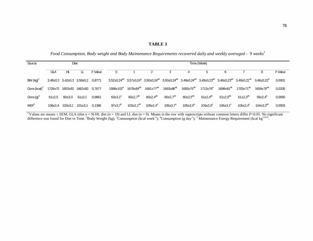
78
TABLE 3
Food Consumption, Body weight and Body Maintenance Requirements recovered daily and weekly averaged - 9 weeks1
Source Diet Time (Week)
GLA HL LL P Value 0 1 2 3 4 5 6 7 8 P Value
BW (kg)2 3,48±0,3 3,42±0,3 3,59±0,2 0,8771 3,52±0,24ab 3,57±0,24a 3,50±0,24ab 3,50±0,24ab 3,49±0,24ab 3,49±0,23ab 3,48±0,23ab 3,49±0,22ab 3,46±0,22b 0,0001
Cons (kcal)3 1726±70 1653±92 1662±83 0,7677 1598±102b 1678±84ab 1691±77ab 1693±88ab 1693±79ab 1713±76a 1698±81ab 1703±71ab 1659±78ab 0,0335
Cons (g)4 61±2,5 60±3,3 61±2,0 0,9861 63±3,1a 60±2,7ab 60±2,4ab 60±2,7ab 60±2,5ab 61±2,4ab 61±2,5ab 61±2,3ab 59±2,4b 0,0050
MER5 108±2,4 103±3,2 101±3,1 0,1386 97±3,7b 103±2,1ab 105±2,4a 105±3,7a 105±2,9a 106±2,6a 106±3,1a 106±2,4a 104±3,3ab 0,0003
1 Values are means + SEM, GLA (diet n = 9) HL diet (n = 10) and LL diet (n = 9). Means in the row with superscripts without common letters differ P<0.05. No significant difference was found for Diet vs Time. 2Body Weight (kg); 3Consumption (kcal week-1); 4Consumption (g day-1); 5 Maintenance Energy Requirement (kcal kg-0,67).
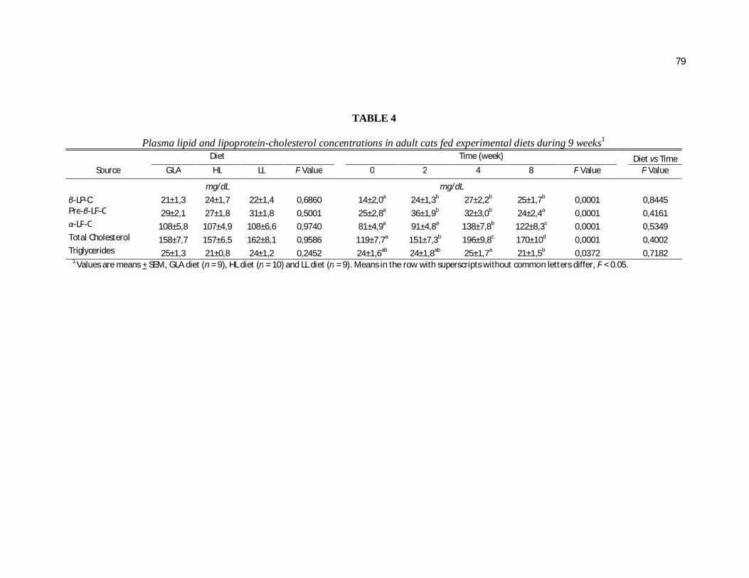
79
TABLE 4
Plasma lipid and lipoprotein-cholesterol concentrations in adult cats fed experimental diets during 9 weeks1 Diet Time (week) Diet vs Time
Source GLA HL LL P Value 0 2 4 8 P Value P Value
mg/dL mg/dL
β-LP-C 21±1,3 24±1,7 22±1,4 0,6860 14±2,0a 24±1,3b 27±2,2b 25±1,7b 0,0001 0,8445 Pre-β-LP-C 29±2,1 27±1,8 31±1,8 0,5001 25±2,8a 36±1,9b 32±3,0b 24±2,4a 0,0001 0,4161 α-LP-C 108±5,8 107±4,9 108±6,6 0,9740 81±4,9a 91±4,8a 138±7,8b 122±8,3c 0,0001 0,5349 Total Cholesterol 158±7,7 157±6,5 162±8,1 0,9586 119±7,7a 151±7,3b 196±9,8c 170±10d 0,0001 0,4002 Triglycerides 25±1,3 21±0,8 24±1,2 0,2452 24±1,6ab 24±1,8ab 25±1,7a 21±1,5b 0,0372 0,7182 1 Values are means + SEM, GLA diet (n = 9), HL diet (n = 10) and LL diet (n = 9). Means in the row with superscripts without common letters differ, P < 0.05.
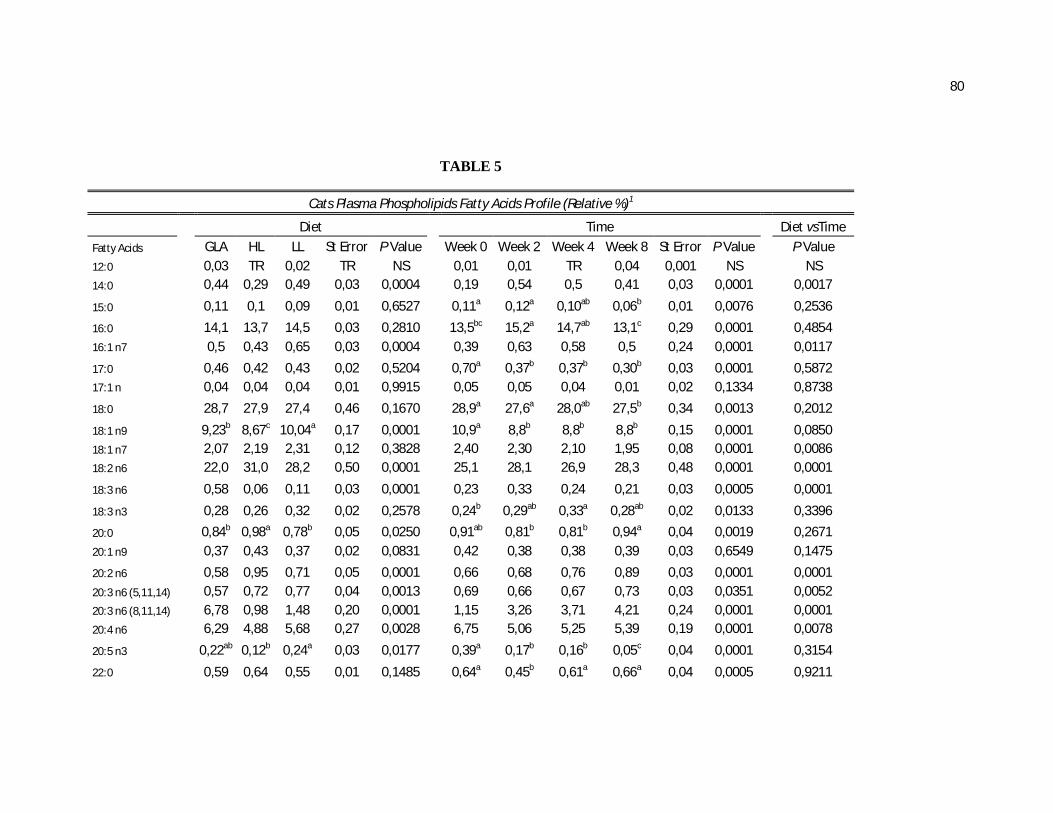
80
TABLE 5
Cats Plasma Phospholipids Fatty Acids Profile (Relative %)1
Diet Time Diet vsTime
Fatty Acids GLA HL LL St Error P Value Week 0 Week 2 Week 4 Week 8 St Error P Value P Value 12:0 0,03 TR 0,02 TR NS 0,01 0,01 TR 0,04 0,001 NS NS 14:0 0,44 0,29 0,49 0,03 0,0004 0,19 0,54 0,5 0,41 0,03 0,0001 0,0017
15:0 0,11 0,1 0,09 0,01 0,6527 0,11a 0,12a 0,10ab 0,06b 0,01 0,0076 0,2536
16:0 14,1 13,7 14,5 0,03 0,2810 13,5bc 15,2a 14,7ab 13,1c 0,29 0,0001 0,4854 16:1 n7 0,5 0,43 0,65 0,03 0,0004 0,39 0,63 0,58 0,5 0,24 0,0001 0,0117
17:0 0,46 0,42 0,43 0,02 0,5204 0,70a 0,37b 0,37b 0,30b 0,03 0,0001 0,5872 17:1 n 0,04 0,04 0,04 0,01 0,9915 0,05 0,05 0,04 0,01 0,02 0,1334 0,8738
18:0 28,7 27,9 27,4 0,46 0,1670 28,9a 27,6a 28,0ab 27,5b 0,34 0,0013 0,2012
18:1 n9 9,23b 8,67c 10,04a 0,17 0,0001 10,9a 8,8b 8,8b 8,8b 0,15 0,0001 0,0850 18:1 n7 2,07 2,19 2,31 0,12 0,3828 2,40 2,30 2,10 1,95 0,08 0,0001 0,0086 18:2 n6 22,0 31,0 28,2 0,50 0,0001 25,1 28,1 26,9 28,3 0,48 0,0001 0,0001
18:3 n6 0,58 0,06 0,11 0,03 0,0001 0,23 0,33 0,24 0,21 0,03 0,0005 0,0001
18:3 n3 0,28 0,26 0,32 0,02 0,2578 0,24b 0,29ab 0,33a 0,28ab 0,02 0,0133 0,3396
20:0 0,84b 0,98a 0,78b 0,05 0,0250 0,91ab 0,81b 0,81b 0,94a 0,04 0,0019 0,2671 20:1 n9 0,37 0,43 0,37 0,02 0,0831 0,42 0,38 0,38 0,39 0,03 0,6549 0,1475
20:2 n6 0,58 0,95 0,71 0,05 0,0001 0,66 0,68 0,76 0,89 0,03 0,0001 0,0001 20:3 n6 (5,11,14) 0,57 0,72 0,77 0,04 0,0013 0,69 0,66 0,67 0,73 0,03 0,0351 0,0052 20:3 n6 (8,11,14) 6,78 0,98 1,48 0,20 0,0001 1,15 3,26 3,71 4,21 0,24 0,0001 0,0001 20:4 n6 6,29 4,88 5,68 0,27 0,0028 6,75 5,06 5,25 5,39 0,19 0,0001 0,0078
20:5 n3 0,22ab 0,12b 0,24a 0,03 0,0177 0,39a 0,17b 0,16b 0,05c 0,04 0,0001 0,3154
22:0 0,59 0,64 0,55 0,01 0,1485 0,64a 0,45b 0,61a 0,66a 0,04 0,0005 0,9211
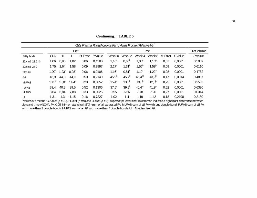
81
Continuing… TABLE 5
Cats Plasma Phospholipids Fatty Acids Profile (Relative %)1
Diet Time Diet vsTime
Fatty Acids GLA HL LL St Error P Value Week 0 Week 2 Week 4 Week 8 St Error P Value P Value
22:4 n6 22:5 n3 1,06 0,96 1,02 0,06 0,4580 1,16a 0,68b 1,06a 1,16a 0,07 0,0001 0,5909
22:6 n3 24:0 1,75 1,64 1,58 0,09 0,3897 2,17a 1,31b 1,56b 1,59b 0,09 0,0001 0,6110
24:1 n9 1,00b 1,23a 0,98b 0,06 0,0106 1,16a 0,81b 1,10a 1,22a 0,08 0,0001 0,4782
Sat 45,9 44,8 44,9 0,50 0,2140 45,9a 45,7a 45,4ab 43,9b 0,47 0,0014 0,4837
MUFAS 13,3b 13,0b 14,4a 0,28 0,0052 15,4a 13,0b 13,0b 12,8b 0,23 0,0001 0,2583
PUFAS 39,4 40,8 39,5 0,52 0,1306 37,6c 39,8b 40,4ab 41,9a 0,52 0,0001 0,6370 HUFAS 8,64 6,84 7,88 0,33 0,0026 9,55 6,56 7,78 7,26 0,27 0,0001 0,0314
UI 1,31 1,3 1,15 0,16 0,7227 1,02 1,4 1,19 1,42 0,18 0,2198 0,2180 1 Values are means, GLA diet (n = 10), HL diet (n = 9) and LL diet (n = 9); Superscript letters not in common indicate a significant difference between diets and time ANOVA, P < 0.05; NI=non statistical; SAT =sum of all saturated FA; MUFAS=sum of all FA with one double bond; PUFAS=sum of all FA with more than 2 double bonds; HUFAS=sum of all FA with more than 4 double bonds; UI = No identified FA.
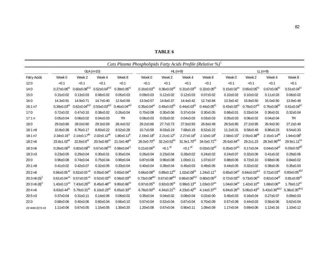
82
TABLE 6
Cats Plasma Phospholipids Fatty Acids Profile (Relative %)1
GLA (n=10) HL (n=9) LL (n=9)
Fatty Acids Week 0 Week 2 Week 4 Week 8 Week 0 Week 2 Week 4 Week 8 Week 0 Week 2 Week 4 Week 8
12:0 <0.1 <0.1 <0.1 <0.1 <0.1 <0.1 <0.1 <0.1 <0.1 <0.1 <0.1 <0.1
14:0 0.27±0.06a1 0.60±0.08b12 0.52±0.04b12 0.39±0.05a1 0.16±0.03a1 0.36±0.03a2 0.31±0.03a2 0.32±0.05a1 0.15±0.02a1 0.65±0.05b1 0.67±0.06b1 0.51±0.04b1
15:0 0.15±0.02 0.13±0.03 0.08±0.02 0.05±0.03 0.09±0.03 0.12±0.02 0.12±0.03 0.07±0.02 0.10±0.02 0.10±0.02 0.11±0.03 0.06±0.02
16:0 14.3±0.91 14.9±0.71 14.7±0.40 12.6±0.59 13.0±0.57 14.8±0.37 14.4±0.42 12.7±0.84 13.3±0.42 15.8±0.50 15.0±0.50 13.9±0.49
16:1 n7 0.39±0.03a1 0.62±0.04b12 0.53±0.03a12 0.46±0.04a12 0.35±0.04a1 0.49±0.03b1 0.44±0.03b1 0.44±0.05b1 0.43±0.03a1 0.78±0.07b2 0.76±0.08b2 0.61±0.04b2
17:0 0.72±0.02 0.47±0.10 0.38±0.02 0.26±0.04 0.70±0.08 0.30±0.06 0.37±0.04 0.30±0.05 0.68±0.01 0.33±0.04 0.36±0.01 0.32±0.04
17:1 n 0.05±0.04 0.06±0.02 0.04±0.03 TR 0.06±0.03 0.03±0.02 0.04±0.03 0.03±0.03 0.05±0.03 0.06±0.02 0.04±0.04 TR
18:0 29.0±0.66 28.0±0.60 29.3±0.59 28.4±0.52 29.2±0.66 27.7±0.73 27.9±0.93 26.8±0.49 28.5±0.85 27.2±0.95 26.9±0.50 27.2±0.49
18:1 n9 10.8±0.38 8.76±0.17 8.83±0.22 8.52±0.28 10.7±0.59 8.03±0.19 7.68±0.19 8.52±0.22 11.2±0.31 9.58±0.49 8.86±0.23 9.54±0.33
18:1 n7 2.34±0.19 a 2.14±0.17ab 2.02±0.12ab 1.80±0.12b 2.19±0.18a 2.21±0.12a 2.27±0.18a 2.10±0.18a 2.58±0.15a 2.55±0.38ab 2.15±0.14ab 1.94±0.08b
18:2 n6 23.8±1.02a1 22.8±0.6a1 20.5±0.65a1 21.0±0.49a1 26.0±0.70a1 32.2±0.52b2 31.9±1.70b2 34.0±0.71b2 25.6±0.64a1 29.2±1.23 28.3±0.96ab2 29.9±1.11b3
18:3 n6 0.26±0.06a1 0.82±0.08b1 0.67±0.06bc1 0.58±0.04c1 0.21±0.06a1 <0.1 a2 <0.1 a2 0.02±0.02a2 0.20±0.07a1 0.17±0.04 0.04±0.04a2 0.03±0.03a2
18:3 n3 0.23±0.05 0.29±0.04 0.30±0.01 0.30±0.04 0.26±0.04 0.23±0.04 0.28±0.02 0.24±0.02 0.24±0.07 0.32±0.06 0.41±0.02 0.29±0.06
20:0 0.96±0.08 0.74±0.04 0.75±0.04 0.90±0.04 0.87±0.08 0.96±0.08 1.00±0.11 1.07±0.07 0.88±0.06 0.72±0.10 0.68±0.06 0.84±0.02
20:1 n9 0.41±0.02 0.42±0.07 0.32±0.05 0.33±0.04 0.40±0.04 0.39±0.04 0.45±0.03 0.49±0.05 0.44±0.05 0.32±0.02 0.38±0.05 0.35±0.03
20:2 n6 0.66±0.05 a1 0.52±0.03 a1 0.55±0.04a1 0.60±0.04a1 0.66±0.08a1 0.89±0.12b2 1.02±0.09b2 1.24±0.11c1 0.65±0.04a1 0.64±0.03a12 0.72±0.03a1 0.83±0.05a12
20:3 n6 (5)2 0.61±0.04 a1 0.57±0.03 a1 0.52±0.03a1 0.56±0.03a1 0.73±0.08ab1 0.67±0.08ab1 0.66±0.06b12 0.80±0.05a2 0.72±0.03a1 0.73±0.06a1 0.82±0.04a2 0.81±0.05a2
20:3 n6 (8)3 1.43±0.13 a1 7.43±0.26b2 8.45±0.48c2 9.80±0.66d2 0.97±0.05a1 0.92±0.05a1 0.99±0.13a1 1.03±0.07a1 1.04±0.04a1 1.42±0.10a1 1.68±0.06a1 1.79±0.12a1
20:4 n6 6.63±0.44a1 5.79±0.31a1 6.10±0.23a1 6.65±0.33a1 6.78±0.50a1 4.34±0.21b1 4.23±0.42b2 4.14±0.37b2 6.84±0.26a1 5.06±0.43b1 5.43±0.30ab12 5.38±0.35ab12
20:5 n3 0.37±0.04 0.31±0.11 0.14±0.06 0.06±0.02 0.35±0.04 0.04±0.02 0.08±0.04 0.02±0.00 0.45±0.03 0.16±0.04 0.27±0.07 0.09±0.03
22:0 0.68±0.06 0.40±0.06 0.60±0.04 0.66±0.10 0.67±0.04 0.52±0.04 0.67±0.04 0.70±0.09 0.57±0.08 0.44±0.03 0.56±0.06 0.62±0.04
22:4n6+22:5 n3 1.11±0.06 0.67±0.05 1.15±0.05 1.30±0.20 1.20±0.08 0.67±0.04 0.90±0.11 1.09±0.09 1.17±0.04 0.69±0.06 1.12±0.16 1.10±0.12

83
Continuing… TABLE 6 Cats Plasma Phospholipids Fatty Acids Profile (Relative %)1
GLA (n=10) HL (n=9) LL (n=9)
22:6 n3+24:0 2.06±0.18 1.43±0.12 1.81±0.25 1.72±0.25 2.23±0.15 1.28±0.08 1.50±0.16 1.56±0.18 2.21±0.13 1.23±0.11 1.38±0.08 1.50±0.15
24:1 n9 1.09±0.10 0.75±0.06 1.00±0.06 1.16±0.18 1.21±0.15 0.93±0.15 1.36±0.13 1.41±0.19 1.19±0.08 0.73±0.08 0.93±0.07 1.08±0.11
SAT 47.0±1.17 45.9±0.62 46.5±0.79 44.3±0.69 45.6±0.97 45.5±0.82 45.3±1.09 43.0±0.86 45.1±1.00 45.8±1.46 44.4±0.58 44.4±0.67
MUFAS 15.3±0.51 12.8±0.29 12.7±0.38 12.3±0.41 15.0±0.92 12.1±0.37 12.3±0.31 12.7±0.46 15.9±0.45 14.0±0.44 14.1±0.35 13.5±0.44
PUFAS 36.3±1.30 39.7±0.71 40.0±0.96 41.9±0.80 38.4±1.04 40.6±0.64 41.1±1.44 43.1±1.13 38.2±0.71 39.1±1.62 39.9±0.73 40.9±0.71
HUFAS 9.30±0.53a1 7.49±0.30a1 9.05±0.41a1 8.73±0.61a1 9.64±0.66a1 5.63±0.24a1 6.28±0.59b2 5.82±0.52b2 9.72±0.35b2 6.56±0.57b12 8.01±0.46ab12 7.22±0.53ab12
UI 1.29±0.28 1.38±0.31 0.73±0.43 1.86±0.20 0.96±0.29 1.76±0.32 1.30±0.45 1.20±0.21 0.81±0.29 1.06±0.33 1.54±0.45 1.19±0.21 1 Values are means + SEM, GLA diet (n = 10), HL diet (n = 9) and LL diet (n = 9); Superscript letters not in common in a row within a diet group indicate a significant difference between weeks by ANOVA, p < 0.05 and superscript numbers not in common in a row within a week indicate a significant difference between diets by ANOVA, p < 0.05; Fatty Acid 20:3 n6 (Δ5,11,14), Fatty acid 20:3 n6 (Δ8,11,14). TR = Trace (<0.01); SAT =sum of all saturated FA; MUFAS=sum of all FA with one double bond; PUFAS=sum of all FA with more than 2 double bonds; HUFAS=sum of all FA with more than 4 double bonds; UI = non identified FA.
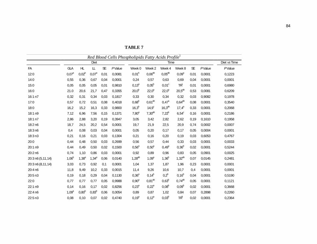
84
TABLE 7
Red Blood Cells Phospholipids Fatty Acids Profile1 Diet Time Diet vs Time
FA GLA HL LL SE P Value Week 0 Week 2 Week 4 Week 8 SE P Value P Value
12:0 0,07a 0,02b 0,07a 0,01 0,0081 0,01b 0,06ab 0,05ab 0,09a 0,01 0,0001 0,1223
14:0 0,55 0,36 0,67 0,04 0,0001 0,24 0,57 0,63 0,69 0,04 0,0001 0,0001
15:0 0,05 0,05 0,05 0,01 0,9810 0,13a 0,05b 0,01c TRc 0,01 0,0001 0,6980
16:0 21.0 20,6 21,7 0,47 0,3355 20,0b 22,0a 22,0a 20,5ab 0,53 0,0081 0,6209
16:1 n7 0,32 0,31 0,34 0,03 0,1817 0,33 0,30 0,34 0,32 0,03 0,9082 0,1978
17:0 0,57 0,72 0,51 0,08 0,4018 0,68b 0,61ab 0,47a 0,64ab 0,08 0,0001 0,3540
18:0 16,2 15,2 16,3 0,33 0,9800 16,3b 14,9c 16,3ab 17,4a 0,33 0,0001 0,2068
18:1 n9 7,12 6,96 7,56 0,15 0,1371 7,80a 7,30ab 7,22b 6,54c 0,16 0,0001 0,2186
18:1 n7 2,86 2,88 3,20 0,19 0,3947 3,05 3,42 2,82 2,62 0,19 0,1610 0,1958
18:2 n6 18,7 24,5 20,2 0,54 0,0001 19,7 21,9 22,5 20,9 0,74 0,0002 0,0007
18:3 n6 0,4 0,08 0,03 0,04 0,0001 0,05 0,20 0,17 0,17 0,05 0,0004 0,0001
18:3 n3 0,21 0,16 0,21 0,03 0,1304 0,21 0,16 0,20 0,19 0,03 0,6053 0,4767
20:0 0,44 0,48 0,50 0,03 0,2699 0,56 0,57 0,44 0,33 0,03 0,0001 0,0033
20:1 n9 0,44 0,49 0,50 0,02 0,1500 0,56a 0,50a 0,49a 0,36b 0,02 0,0001 0,5244
20:2 n6 0,74 1,10 0,86 0,03 0,0001 0,92 0,89 0,96 0,83 0,05 0,0901 0,0025
20:3 n6 (5,11,14) 1,06b 1,38a 1,34a 0,06 0,0140 1,28ab 1,09a 1,36b 1,32ab 0,07 0,0145 0,2481
20:3 n6 (8,11,14) 3,03 0,73 0,92 0,1 0,0001 1,04 1,37 1,87 1,96 0,23 0,0001 0,0001
20:4 n6 11,8 9,49 10,2 0,33 0,0015 11,4 9,26 10,6 10,7 0,4 0,0001 0,0001
20:5 n3 0,19 0,18 0,29 0,04 0,1130 0,36a 0,14b 0,2b 0,16b 0,04 0,0001 0,5190
22:0 0,77 0,77 0,77 0,05 0,9988 0,90a 0,81ab 0,63b 0,74ab 0,05 0,0001 0,1121
22:1 n9 0,14 0,16 0,17 0,02 0,8256 0,23a 0,22a 0,08b 0,09b 0,02 0,0001 0,3668
22:4 n6 1,09a 0,80b 0,83b 0,06 0,0054 0,89 0,87 1,02 0,84 0,07 0,2898 0,2260
22:5 n3 0,08 0,10 0,07 0,02 0,4740 0,19a 0,12a 0,03b TRb 0,02 0,0001 0,2364
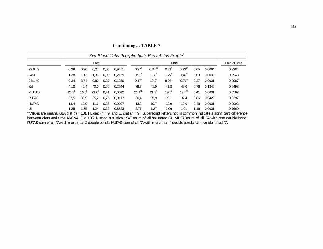
85
Continuing… TABLE 7
Red Blood Cells Phospholipids Fatty Acids Profile1 Diet Time Diet vs Time
22:6 n3 0,29 0,30 0,27 0,05 0,9401 0,37a 0,34ab 0,21b 0,23ab 0,05 0,0064 0,8284
24:0 1,28 1,13 1,36 0,09 0,2159 0,91b 1,38a 1,27a 1,47a 0,09 0,0009 0,8948
24:1 n9 9,34 8,74 9,80 0,37 0,1369 9,17a 10,2a 8,05b 9,76a 0,37 0,0001 0,3987
Sat 41,0 40,4 42,0 0,66 0,2544 39,7 41,0 41,8 42,0 0,76 0,1346 0,2493
MUFAS 20,2b 19,5b 21,6a 0,41 0,0012 21,1ab 21,9a 19,0c 19,7bc 0,41 0,0001 0,0582
PUFAS 37,5 38,9 35,2 0,75 0,0117 36,4 35,9 39,1 37,4 0,86 0,0422 0,0297
HUFAS 13,4 10,9 11,6 0,36 0,0007 13,2 10,7 12,0 12,0 0,48 0,0001 0,0003 UI 1,25 1,35 1,24 0,26 0,8863 2,77 1,27 0,06 1,01 1,16 0,0001 0,7660
1 Values are means, GLA diet (n = 10), HL diet (n = 9) and LL diet (n = 9); Superscript letters not in common indicate a significant difference between diets and time ANOVA, P < 0.05; NI=non statistical; SAT =sum of all saturated FA; MUFAS=sum of all FA with one double bond; PUFAS=sum of all FA with more than 2 double bonds; HUFAS=sum of all FA with more than 4 double bonds; UI = No identified FA.
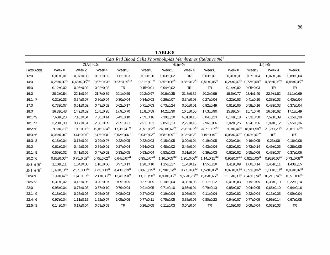
86
TABLE 8
Cats Red Blood Cells Phospholipids Membranes (Relative %)1 GLA (n=10) HL (n=9) LL (n=9)
Fatty Acids Week 0 Week 2 Week 4 Week 8 Week 0 Week 2 Week 4 Week 8 Week 0 Week 2 Week 4 Week 8
12:0 0,01±0,01 0,07±0,03 0,07±0,02 0,11±0,03 0,013±0,0 0,03±0,02 TR 0,03±0,01 0,01±0,0 0,07±0,04 0,07±0,04 0,88±0,04
14:0 0,25±0,02a1 0,63±0,05b12 0,67±0,03b2 0,67±0,09b12 0,21±0,01a1 0,35±0,06ab1 0,38±0,03b1 0,51±0,06b1 0,24±0,02a1 0,72±0,09b2 0,85±0,06b2 0,88±0,06b2
15:0 0,12±0,02 0,05±0,02 0,02±0,02 TR 0,15±0,01 0,04±0,02 TR TR 0,14±0,02 0,05±0,03 TR TR
16:0 20,2±0,84 22,1±0,94 21,7±0,39 20,1±0,59 20,2±0,97 20,6±0,35 21,3±0,82 20,2±0,99 19,5±0,77 23,4±1,40 22,9±1,62 21,1±0,69
16:1 n7 0,32±0,03 0,34±0,07 0,30±0,04 0,30±0,04 0,34±0,03 0,26±0,07 0,34±0,03 0,27±0,04 0,32±0,03 0,41±0,10 0,38±0,03 0,40±0,04
17:0 0,73±0,07 0,51±0,02 0,43±0,02 0,62±0,17 0,71±0,03 0,73±0,24 0,50±0,01 0,92±0,49 0,61±0,06 0,58±0,16 0,48±0,03 0,37±0,04
18:0 16,3±0,48 14,9±0,52 15,8±0,28 17,9±0,70 16,8±0,59 14,2±0,30 16,5±0,50 17,3±0,90 15,8±0,54 15,7±0,70 16,6±0,62 17,1±0,49
18:1 n9 7,60±0,23 7,18±0,34 7,30±0,14 6,43±0,18 7,59±0,16 7,39±0,18 6,81±0,13 6,04±0,23 8,14±0,18 7,33±0,50 7,57±0,39 7,15±0,39
18:1 n7 3,20±0,30 3,17±0,51 2,68±0,09 2,35±0,21 2,91±0,31 2,85±0,13 2,79±0,18 2,96±0,66 3,02±0,25 4,24±0,56 2,99±0,12 2,55±0,36
18:2 n6 18,6±0,78a1 19,0±0,98a1 19,8±0,34a1 17,3±0,41a1 20,5±0,62a1 26,3±0,62b2 26,6±0,37b2 24,7±1,87ab2 19,9±0,44a1 18,8±1,58a1 21,2±1,20a1 20,8±1,12a12
18:3 n6 0,06±0,04a1 0,44±0,06b2 0,47±0,08b2 0,62±0,06b2 0,03±0,02a1 0,08±0,09a12 0,03±0,03a1 0,19±0,19a12 0,06±0,02a1 0,07±0,07a1 TRa1 TRa1
18:3 n3 0,18±0,04 0,17±0,04 0,29±0,07 0,22±0,05 0,22±0,03 0,15±0,05 0,09±0,04 0,19±0,05 0,23±0,04 0,16±0,05 0,23±,06 0,16±0,06
20:0 0,61±0,04 0,49±0,05 0,39±0,01 0,27±0,04 0,54±0,03 0,48±0,02 0,45±0,04 0,43±0,04 0,52±0,02 0,73±0,14 0,49±0,05 0,28±0,05
20:1 n9 0,55±0,02 0,41±0,05 0,47±0,02 0,33±0,05 0,53±0,04 0,53±0,03 0,51±0,04 0,39±0,03 0,62±0,02 0,55±0,06 0,48±0,07 0,37±0,06
20:2 n6 0,85±0,05a1 0,75±0,02a1 0,75±0,02a1 0,64±0,07a1 0,95±0,07a1 1,10±0,05ab2 1,20±0,06b2 1,14±0,11ab2 0,96±0,04a1 0,82±0,05a1 0,93±0,06a1 0,73±0,08a12
20:3 n6 (5)2 1,10±0,11 1,04±0,08 1,10±0,06 0,97±0,13 1,28±0,10 1,15±0,17 1,54±0,12 1,55±0,18 1,41±0,09 1,08±0,14 1,45±0,11 1,43±0,15
20:3 n6 (8)3 1,39±0,11a1 2,57±0,17b1 3,73±0,13c1 4,43±0,19d1 0,86±0,15a1 0,78±0,12a2 0,77±0,08a2 0,52±0,06a2 0,87±0,05a1 0,77±0,06a2 1,11±0,10a2 0,93±0,07a2
20:4 n6 11,4±0,47a1 10,4±0,57b1 12,1±0,36ab1 13,4±0,50a1 11,1±0,58a1 8,90±0,35b1 9,56±0,78ab1 8,35±0,66b2 11,6±0,33a1 8,47±0,74b1 10,2±0,74ab1 10,5±0,60ab2
20:5 n3 0,31±0,02 0,15±0,05 0,20±0,07 0,09±0,05 0,37±0,05 0,10±0,04 0,08±0,03 0,17±0,12 0,41±0,03 0,19±0,05 0,33±0,10 0,22±0,14
22:0 0,95±0,04 0,77±0,08 0,57±0,10 0,79±0,04 0,91±0,05 0,71±0,10 0,66±0,04 0,79±0,13 0,85±0,07 0,94±0,05 0,65±0,10 0,64±0,16
22:1 n9 0,18±0,04 0,26±0,08 0,05±0,03 0,08±0,03 0,27±0,03 0,19±0,04 0,06±0,04 0,11±0,04 0,23±0,02 0,22±0,04 0,13±0,05 0,09±0,04
22:4 n6 0,97±0,04 1,11±0,15 1,22±0,07 1,05±0,06 0,77±0,11 0,75±0,05 0,88±0,05 0,80±0,23 0,94±0,07 0,77±0,09 0,95±0,14 0,67±0,08
22:5 n3 0,14±0,04 0,17±0,04 0,03±0,03 TR 0,26±0,05 0,11±0,03 0,04±0,04 TR 0,16±0,03 0,09±0,04 0,03±0,03 TR
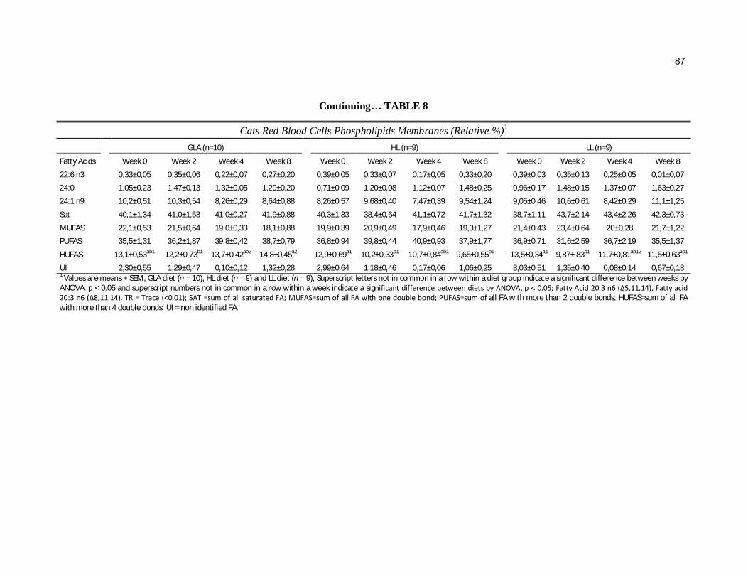
87
Continuing… TABLE 8
Cats Red Blood Cells Phospholipids Membranes (Relative %)1 GLA (n=10) HL (n=9) LL (n=9)
Fatty Acids Week 0 Week 2 Week 4 Week 8 Week 0 Week 2 Week 4 Week 8 Week 0 Week 2 Week 4 Week 8
22:6 n3 0,33±0,05 0,35±0,06 0,22±0,07 0,27±0,20 0,39±0,05 0,33±0,07 0,17±0,05 0,33±0,20 0,39±0,03 0,35±0,13 0,25±0,05 0,01±0,07
24:0 1,05±0,23 1,47±0,13 1,32±0,05 1,29±0,20 0,71±0,09 1,20±0,08 1,12±0,07 1,48±0,25 0,96±0,17 1,48±0,15 1,37±0,07 1,63±0,27
24:1 n9 10,2±0,51 10,3±0,54 8,26±0,29 8,64±0,88 8,26±0,57 9,68±0,40 7,47±0,39 9,54±1,24 9,05±0,46 10,6±0,61 8,42±0,29 11,1±1,25
Sat 40,1±1,34 41,0±1,53 41,0±0,27 41,9±0,88 40,3±1,33 38,4±0,64 41,1±0,72 41,7±1,32 38,7±1,11 43,7±2,14 43,4±2,26 42,3±0,73
MUFAS 22,1±0,53 21,5±0,64 19,0±0,33 18,1±0,88 19,9±0,39 20,9±0,49 17,9±0,46 19,3±1,27 21,4±0,43 23,4±0,64 20±0,28 21,7±1,22
PUFAS 35,5±1,31 36,2±1,87 39,8±0,42 38,7±0,79 36,8±0,94 39,8±0,44 40,9±0,93 37,9±1,77 36,9±0,71 31,6±2,59 36,7±2,19 35,5±1,37
HUFAS 13,1±0,53ab1 12,2±0,73b1 13,7±0,42ab2 14,8±0,45a2 12,9±0,69a1 10,2±0,33b1 10,7±0,84ab1 9,65±0,55b1 13,5±0,34a1 9,87±,83b1 11,7±0,81ab12 11,5±0,63ab1
UI 2,30±0,55 1,29±0,47 0,10±0,12 1,32±0,28 2,99±0,64 1,18±0,46 0,17±0,06 1,06±0,25 3,03±0,51 1,35±0,40 0,08±0,14 0,67±0,18 1 Values are means + SEM, GLA diet (n = 10), HL diet (n = 9) and LL diet (n = 9); Superscript letters not in common in a row within a diet group indicate a significant difference between weeks by ANOVA, p < 0.05 and superscript numbers not in common in a row within a week indicate a significant difference between diets by ANOVA, p < 0.05; Fatty Acid 20:3 n6 (Δ5,11,14), Fatty acid 20:3 n6 (Δ8,11,14). TR = Trace (<0.01); SAT =sum of all saturated FA; MUFAS=sum of all FA with one double bond; PUFAS=sum of all FA with more than 2 double bonds; HUFAS=sum of all FA with more than 4 double bonds; UI = non identified FA.
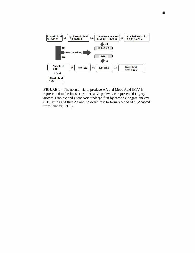
88
FIGURE 1 – The normal via to produce AA and Mead Acid (MA) is represented in the lines. The alternative pathway is represented in gray arrows. Linoleic and Oleic Acid undergo first by carbon elongase enzyme (CE) action and then Δ8 and Δ5 desaturase to form AA and MA (Adapted from Sinclair, 1979).
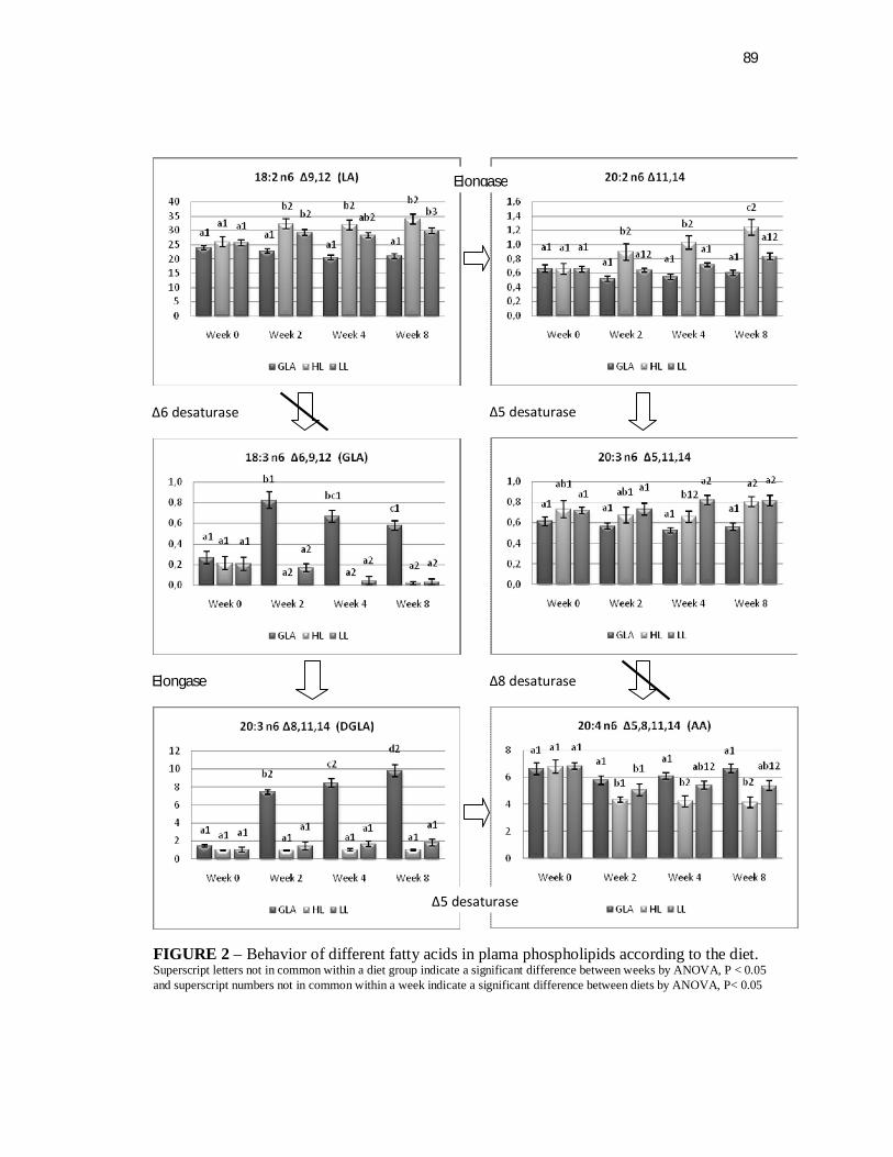
89
Δ5 desaturase
Δ8 desaturase
Δ6 desaturase
Elongase
Elongase
Δ5 desaturase
FIGURE 2 – Behavior of different fatty acids in plama phospholipids according to the diet. Superscript letters not in common within a diet group indicate a significant difference between weeks by ANOVA, P < 0.05 and superscript numbers not in common within a week indicate a significant difference between diets by ANOVA, P< 0.05
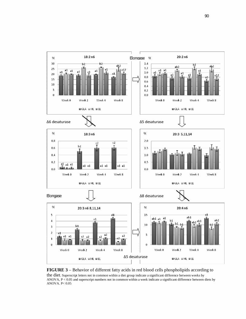
90
Δ5 desaturase
Δ8 desaturase
Δ6 desaturase
Elongase
Elongase
Δ5 desaturase
% %
% %
% %
FIGURE 3 – Behavior of different fatty acids in red blood cells phospholipids according to the diet. Superscript letters not in common within a diet group indicate a significant difference between weeks by ANOVA, P < 0.05 and superscript numbers not in common within a week indicate a significant difference between diets by ANOVA, P< 0.05

4 CAPÍTULO IV
92
4.1 Conclusões/Considerações finais
A pesquisa em felinos tem sido um desafio para os pesquisadores
desde a década de 70. Felinos, conhecidos pelo comportamento agressivo,
apresentam restrições à pesquisa em ambientes laboratoriais. Cães, ratos e
camundongos têm sido preferencialmente utilizados nas pesquisas, como
modelos experimentais, escolhidos principalmente pela docilidade de manejo
e suas características metabólicas, que serviram de modelo para estudos em
diferentes espécies. Os gatos, por sua vez, não se ajustaram como modelos
experimentais. Suas particularidades nutricionais e a dificuldade de
aceitação às dietas purificadas foram um empecilho para que isso
acontecesse.
Atualmente, a pesquisa em felinos foi facilitada pela criação de
animais SPF (Specific Patogen Free). Por outro lado, os aspectos
comportamentais indesejados referentes à espécie ainda representam uma
barreira para experimentos em nutrição, quando coletas de amostras de
sangue são inevitáveis. Pela experiência adquirida durante os experimentos
conduzidos, pode-se dizer que o contato no dia a dia com os animais
melhora a socialização e facilita o manejo. Métodos de coleta de sangue e
sedativos podem facilitar este processo. Gatos apresentam coagulação
sanguínea rápida, e é necessária habilidade por parte dos coletores para a
obtenção de amostras significativas e íntegras. Este procedimento somente
é possível se houver uma equipe treinada. A contenção do animal é parte do
sucesso do procedimento. O animal deve sentir-se confortável, em um
ambiente sem ruído. O coletor deve ter experiência na manipulação de
animais, caso contrário podem acontecer acidentes, como mordidas,
93
arranhaduras e a consequente obtenção de amostras de baixa qualidade, o
que impossibilita sua utilização. Em experimentos em que o sangue é
dividido em alíquotas, plasma e células vermelhas, não pode haver a
contaminação. A ruptura das hemácias ou células brancas do sangue
liberam o conteúdo interno e contaminam as amostras de plasma, afetando a
concentração de lipídeos plasmáticos.
Felídeos, por se tratarem de um grupo de animais carnívoros
estritos, apresentam significativas particularidades metabólicas,
especialmente no metabolismo lipídico. Os gatos revelam versatilidade em
metabolizar estes componentes, podendo os mesmos tornarem-se elemento
chave no estudo dos transtornos do colesterol e doenças ateromatosas em
humanos. A porção “α” das lipoproteínas plasmáticas, que corresponde ao
HDL, é naturalmente elevada nos carnívoros. O aumento do HDL é
proporcional à ingestão de lipídeos, forma de proteção natural contra doenças
cardiovasculares, inerente aos felídeos, contrário ao que normalmente ocorre
em humanos.
Muitas questões permanecem sem resposta sobre o metabolismo
desta espécie. Os resultados observados no Capítulo II, atestam que gatos
podem consumir TCM sem recusa alimentar e sem alterações significativas
no metabolismo lipídico, contrariando resultados conhecidos até então, a
respeito da aceitação de AGCM por gatos. Em uma breve revisão sobre os
produtos encontrados no Brasil e nos EUA, destinados a alimentação de
gatos, não foi possível encontrar a inclusão de ingredientes como óleo de
coco compondo dietas para gatos, provavelmente em função da publicação
de McDonald et al. feita há 24 anos. Além disso, diversos trabalhos relatam
94
alterações lipídicas no metabolismo, como hipertrigliceridemia e
hipercolesterolemia em animais alimentados com inclusões elevadas de TCM
(>22% da EM). Neste estudo, houve um aumento imediato da concentração
de triglicerídeos e lipoproteínas, logo após o início da fase experimental,
devido à composição elevada de gordura dietética, quando comparada à dieta
consumida no período pré-experimental. No entanto, após 4 semanas, os
parâmetros plasmáticos começaram a decrescer, retornando aos níveis
basais, demonstrando a versatilidade do sistema enzimático dos carnívoros
em adaptar-se ao consumo constante de uma dieta. Este experimento suscita
a hipótese de que os efeitos observados na literatura, quando dietas
purificadas foram confeccionadas e testadas, não são necessariamente
reproduzidos com dietas práticas. A sensibilidade felina pelo sabor é
conhecida e este pode ter sido o fator determinante afetando o consumo das
dietas purificadas. Possivelmente pelo sabor repugnante proporcionado pelos
ácidos graxos livres, ainda quando o alimento se encontrava na boca dos
animais. O uso de dietas práticas para a experimentação, em que há
interação entre os ingredientes das fórmulas, é de fundamental importância
no estudo de ingredientes específicos em dietas comerciais. Os processos
térmicos, de uma forma geral, tendem a modificar a palatabilidade dos
alimentos. No processo de extrusão a temperatura e umidade, associados à
pressão, podem ter impacto maior ou menor sobre os ingredientes. As
reações de tipo Maillard e outras modificam a palatabilidade do alimento,
assim como a estrutura dos nutrientes. Dessa forma, diferentes ingredientes
afetam a aceitabilidade e a digestibilidade das dietas nas diferentes espécies
e categorias e devem ser testados antes da inclusão definitiva.

95
No Capítulo III, foram pesquisadas as interações entre a
suplementação de ácido γ-linolênico (GLA) e a atividade da enzima Δ6
dessaturase no metabolismo lipídico dos gatos. De acordo com uma série de
experimentos prévios, gatos não apresentam significante atividade dessa
enzima. Neste trabalho, a análise do perfil de lipídeos plasmáticos e do perfil
de lipídeos das membranas plasmáticas das células vermelhas revelou a
proximidade entre a concentração das dietas e o perfil de ácidos graxos
encontrados nos tecidos, fato consolidado na literatura. A suplementação de
ácido linolêico (AL), mesmo em altas concentrações, não foi eficiente para
induzir a ação da Δ6 desaturase ou induzir uma via alternativa para a
produção de ácido araquidônico (AA). O óleo de borragem como fonte de
GLA, no entanto, foi capaz de permitir a manutenção dos níveis elevados de
AA nos tecidos. Dessa forma a suplementação de AL em dietas para gatos
somente é necessária para o atendimento das necessidades do próprio ácido.
No entanto, não é uma fonte para a síntese de AA, como ocorre com o GLA.
O AA é essencial para gatos, especialmente em categorias de
maior demanda como animais jovens, fêmeas em lactação e reprodução,
observação esta descrita no NRC (2006). As fêmeas respondem mais
rapidamente à deficiência, com problemas reprodutivos, mas machos adultos
privados de AA não apresentam prejuízos na taxa reprodutiva. O AA, junto
com o ácido esteárico, são usualmente os únicos componentes da fosfatidil
inositol (Berg et al., 2004), uma provável razão para sua alta concentração
nos fosfolipideos plasmáticos (em torno dos 6%) e nas membranas dos
eritrócitos (em torno de 10%). Uma questão a ser melhor explicada é a origem
destas altas concentrações, uma vez conhecida a deficiência da atividade da
96
enzima Δ6 dessaturase em gatos e a baixa concentração dietética usual de
AA (<0,03% para dietas de 4000 kcal EM). Neste contexto mais estudos são
necessários para estabelecer se há alguma via alternativa ou se o AA possui
outra forma de recuperação para que tais concentrações sejam mantidas.
Este estudo também demonstra que a inclusão de um ingrediente
alternativo, o óleo de borragem, foi capaz de manter as concentrações de AA
nos tecidos. As fontes tradicionais de AA são as gorduras presentes nos
ingredientes de origem animal. Dietas para gatos, livres desses ingredientes,
seriam certamente deficientes em AA, entre outros nutrientes. O óleo de
borragem, como suplemento de origem vegetal, teria potencial para atender
as necessidades de AA dos gatos.
O GLA, além de servir como substrato para a síntese de AA, ainda
promove a pronta acumulação nas membranas do seu derivado DGLA, em
menos de 14 dias após o início da suplementação. Devido às propriedades
que os ácidos graxos poli-insaturados de 20 carbonos possuem de gerar
eicosanóides, prostaglandianas e tromboxanos, o GLA pode ter importância
significativa na modulação da resposta imune, já que ele rapidamente é
elongado ao DGLA, fator este muito pouco estudado em felídeos.
Eicosanóides provenientes do DGLA possuem características menos
inflamatórias que aqueles normalmente gerados pelo AA. Dessa forma, uma
das ações complementares deste estudo será submeter as amostras de
plasma a novas metodologias, como o laser e espectrometria de massas,
para detectar componentes plasmáticos, que serão correlacionados às dietas
experimentais. Estas novas metodologias estão sendo desenvolvidas nos
laboratórios da Nestlé Purina em Saint Louis, Estados Unidos (Luminex -
97
Perfil imune por tecnologia a laser) e no outro laboratório da Nestlé Purina
em Lausanne, Suíça (Metabolomic profile testing) para os quais as amostras
serão enviadas e submetidas à analise.
Os dois trabalhos que nesta tese foram apresentados são de
caráter prático e aplicável a dietas específicas para gatos. Pelas
características dos ingredientes, óleos de coco e de borragem, abre-se a
possibilidade da utilização dos mesmos em dietas normais ou dietas
terapêuticas para gatos. A inclusão de 11% EM na forma de TCM não afeta o
consumo e não causa recusa alimentar. Para animais com síndrome de
malabsorção, ou problemas pancreáticos em que a digestão de lipídeos
esteja prejudicada, a inclusão de TCM pode representar uma fonte de energia
disponível, prática e barata na dietas de felinos. O GLA por sua vez, acrescido
na dieta como componente do óleo de borragem, pode ser uma fonte para a
síntese do AA, proporcionando acumulação de DGLA nas membranas, fato
que pode ter implicações positivas no sistema imunológico.

98
5 CAPÍTULO V
99
5.1 Referências Bibliográficas
ALBERT, D. H.; CONIGLIO, J. G. Metabolism of eicosan-11,14 dienoic acid in rat testes. Evidence for delta 8-desaturase activity. Biochimica et Biophysica Acta, St. Louis, MO, v. 489, p.390-396, 1977.
ANFAL PET. Associação dos fabricantes de alimentos. Disponível em: http://anfalpet.org.br/Site/principal.php?id_menu=6. Acesso em: 24/02/2009.
BACH, A. C.; INGENBLEEK, Y.; FREY, A. The usefulness of dietary medium-chain triglycerides in body weight control: fact or fancy? Journal of Lipid Research, Bethesda, MD, v. 37, p.708-26, 1996.
BACH, A.C.; BABAYAN, V.K. Medium-chain triglycerides: an update. American Journal of Clinical Nutrition, Bethesda, MD, v. 36, p.950-962, 1982.
BAUER, J. E. Facilitative and functional fats in canine and feline diets. Journal of the American Veterinary Medical Association, Washington, v. 229, n. 5, p.680-684, 2006.
BAUER, J. E. Fatty acid metabolism in domestic cats (Felis catus) and cheetahs (Acinonyx jubatas). Proceedings of the Nutrition Society, Aberystwyth, UK, v.56, p.1013-1024, 1997.
BERG, J. M.; TYMOCZKO, J. L.; STRYER, L. Bioquímica. 5.ed. Rio de Janeiro : Guanabara Koogan, 2004. 1059p.
CARVALHO, P. O.; CAMPOS, P. R. B.; NOFFS, M. D’A.; OLIVEIRA, J. G.; SHIMIZU, M. T.; SILVA, D. M. Aplicação de lípases microbianas na obtenção de concentrados de ácidos graxos poliinsaturados. Quimica Nova, São Paulo, n. 26, p. 75-80, 2003.
CASE. L. P.; CAREY, D.; HIRAKAWA, D.; DARISTOTLE, L. Canine and feline nutrition. A resouce for companion animal professionals. Philadelphia : Mosby, 2000. p.592.
CHRISTENSEN E.; HAGUE, T. A.; GRONN, M.; CHRISTOPHERSEN, B. O. β-oxidation of medium chain (C8-C14) fatty acids studied in isolated liver cells. Biochimica et Biophysica Acta, St. Louis, MO, n.1004, p. 187-195, 1989.
CHRISTIE, W. W. Lipid analyses. 2.ed. Oxford, England: Pergamon Press, 1982. p.52-56.
COOK, H. W. Fatty acids desaturation and chain elongation in eukaryotes. In: VANCE, D.E.; VANCE, J.E. (Eds) Biochemistry of lipids, lipoproteins and membranes. Amsterdam: Elsevier, 1996. p.129-152.
100
FENN, J. B.; MANN, M.; MENG, C. K.; WONG, S. F.; WHITEHOUSE, C. M. Electrospray ionization for mass spectrometry of large biomolecules. Science, Washington, n. 246, p.64–71, 1989.
FOLCH, J.; LEES, M.; SLOANE, G. H. A simple method for the isolation and purification of total lipids from animal tissues. Journal of Biological Chemistry, Bethesda, MD, n. 226, p. 497-509, 1957.
FRANKEL, T.; RIVERS, J. P. W. The nutritional and metabolic impact of γ-linolenic acid (18:3 n6) on cats deprived of animal lipid. British Journal of Nutrition, Cambridge, UK, n. 39, p. 227-231, 1978.
FRIEDMAN, M. I.; RAMIREZ, I.; BOWDEN, C. R.; TORDOFF, M. G. Fuel partitioning and food intake: role for mitochondrial fatty acid transport. American Journal of Physiology, Bethesda, MD, n.258, R216–R221, 1990. (Regulatory Integrative Comp. Physiol. 27).
GURR, M. I.; HARWOOD, J. L.; FRAYN, K. N. Lipid Biochemistry:An introduction. 5.ed. Oxford, UK : Blackwell Science, 2002.
HAND, M. S.; THATCHER, C. D.; REMILLARD, R. L.; ROUDEBUSH, P. Small Animal Clinical Nutrition. 4th ed. Marceline, MO: Walsworth Publishing, 2000.
HASSAM, A.G.; RIVERS, J.P.W.; CRAWFORD, M.A. The failure of the cat to desaturate linoleic acid: its nutritional implications. Annals of Nutrition & Metabolism, Basel, Switzerland, v.21, p.321-328, 1977.
HILL C. Clinical care nutrition. In: WILLS, J. M.; SIMPSON, K. W. (eds). The Waltham Book of Clinical Nutrition of Dog and Cat. Oxford: Elsevier Science, 1994. p. 39-61.
HOLMAN, R. T. Biological activities of polyunsaturated fatty acids. Progress in the Chemistry of Fats and other Lipids, Oxford, n.9, p. 607-682, 1970.
HOMAN, R.; ANDERSON, M. K. Rapid separation and quantitation of combined neutral and polar lipid classes by high-performance liquid chromatography and evaporative light-scattering mass detection. Journal of Chromatography B: Biomedical Sciences and Applications, St. Louis, MO, n. 708, p. 21–26, 1998.
HUANG, Y –S.; MUKERJI, P; KNUTZON, D. S. Transgenic production of long-chain polyunsaturated Fatty acid. World Review of Nutrition and Dietetics, Basel, Switzerland, n. 88, p. 243-248, 2001.
JOHNSON, M. M.; SWAN, D. D.; SURETTE, M. E.; STEGNER, J.; CHILTON, T.; FONTECH, A. N.; CHILTON, F. H. Dietary supplementation with γ-linolenic acid alters fatty acid content and eicosanoid production in healthy humans. Journal of Nutrition, Bethesda, MD, n.127, p.1435-1444, 1997.
JONES, P. M.; BUTT, Y.; MESSMER, B.; BORIAK, R.; BENNETT, M. J. Medium-chain fatty acids undergo elongation before β-oxidation in fibroblasts.
101
Biochemical and Biophysical Research Communications, St. Louis, MO, n. 346, p.193-97, 2006.
KENDALL, P. T. The use of fat in dog and cat diets. Fats in animal nutrition. London: Butterworths, 1984. p. 383-404.
KIM, H. Y.; WANG, T. C. L.; MA, Y. C. Liquid chromatography/mass spectrometry of phospholipids using electrospray ionization. Analytical Chemistry, Washington, n. 66, p. 3977–3982, 1994.
KVAMME, J. L. What is palatability? In: PETFOOD technology. Illinois: Watt, 2003. Section IV, cap 1 – Palatability, p. 176-177.
LABARTHE F.; GÉLINAS, R.; ROSIERS, C. D. Medium-chain fatty acids as metabolic therapy in cardiac disease. Cardiovascular Drugs and Therapy, Netherlands, n. 22, p. 97-106, 2008.
LESNEFSKY, E. J.; STOLL, M. S. K.; MINKLER, P.E.; HOPPEL, C. L. Separation and quantitation of phospholipids and lysophospholipids by high-performance liquid chromatography. Analytical Biochemistry, St. Louis, MO, n. 285, p. 246–254, 2000.
LEWIS, L. D.; MORRIS Jr., M. L.; HAND, M. S. Small Animal Clinical Nutrition. Topeka : Mark Morris Associates,1987. v.3, Chapter 7.
LIU, J. –W.; DEMICHELE, S.; BERGANA, M.; BOBIK, E. Jr.; HASTILOW, C.; CHUANG, L. –T.; MUKERJI, P.; HUANG, Y. -S. Characterization of oil exhibiting high γ-linolenic acid from a genetically transformed canola strain. Journal of the American Oil Chemistis’ Society, Champaign, IL, n. 78, p. 489-493, 2001.
MacDONALD M.; ROGERS, Q. R.; MORRIS, J. G. Aversion of the cat to dietary medium chain triglycerydes and caprilic acid. Physiology & Behavior, St. Louis, MO, n. 35, p. 371-75, 1985.
MacDONALD, M. L.; ROGERS, Q. R.; MORRIS, J. G. Role of linoleate as an essential fatty acid for the cat, independent of arachidonate synthesis. Journal of Nutrition, Bethesda, MD, n. 113, p. 1422-1433, 1983.
MacDONALD, M. L.; ROGERS, Q. R.; MORRIS, J. G. Nutrition of the domestic cat, a mammalian carnivore. Annual Review of Nutrition, Palo Alto, CA, n. 4, p. 521-562, 1984.
MATSUBARA, T.; HAYASHI, A. FAB/mass spectrometry of lipids. Progress of Lipid Research, Oxford, n. 3013, p. 301–322, 1991.
MCLEAN, J.G.; MONGER, E.A. Factors determining the essencials fatty acid requirements of the cat. In: WALTHAM SYMPOSIUM ON RECENT ADVANCES IN DOG AND CAT NUTRITION, 7., Cambridge, UK, 1985. [Proceedings…] Cambridge: Cambridge University Press, 1985. p.329-342.

102
MCLEOD, L. American Pet Ownership Statistics. Disponível em: http://exoticpets.about.com/cs/resourcesgeneral/a/petstates.htm. Acesso em: 24/02/2009.
MORRIS, J.G. Do cats need arachidonic acid in the diet for reproduction? Journal of Animal Physiology and Animal Nutrition, Berlin, v.88, p.131-137, 2004.
MURPHY, R.C.; HARRISON, K.A. Fast atom bombardment mass spec-trometry of phospholipids. Mass Spectrometry Reviews, Maiden, MA, v.13, p.57–75, 1994.
NAKAZAWA, I; MEAD, J.F.; YONEMOTO, R.H. In vitro activity of the fatty acyl desaturase of human cancerous and noncancerous tissues. Lipids, Champaign, v.11, p.79-81, 1976.
NATIONAL RESEARCH COUNCIL. Nutrient requirements of dogs and cats. Washington, DC: National Academies Press, 2006.
NELSON, R.W.; COUTO, C.G. Essential of small animal internal medicine. St. Louis: Mosby, 1992.
PAPAMANDJARIS, A.A.; MACDOUGALL, D.E.; JONES, P.J.H. Medium-chain fatty acid metabolism and energy expenditure: obesity treatment implications. Life Sciences, St. Louis, MO, v.62, n.14, p.1203-15, 1998.
PAWLOSKY, R.; BARNES, A.; SALEM, N. Essential fatty acid metabolism in the feline:Relationship between liver and brain production of long-chain polyunsaturated fatty acids. Journal of Lipid Research, Bethesda, MD, v.35, p.2032-2040, 1994.
PAWLOSKY, R.; SALEM JR., N. Is dietary arachidonic acid necessary for feline reproduction? Journal of Nutrition, Bethesda, MD, v.126, p.1081S-1085S, 1996.
REINHART, G. A. Review of omega-3 fatty acids and dietary influences on tissue concentrations. In: IAMS INTERNATIONAL NUTRITION SYMPOSIUM, 30., Wilmington, OH, 1996. Proceedings…: Recent advances in canine and feline nutritional research. Wilmington, Ohio: Orange Frazer, c1996. p. 235-242.
RIVERS, J.P.W.; FRANKEL, T.L. (1981) The production of 5,8,11-eicosatrienoic acid (20:3n-9) in the essential fatty acid deficient cat. Proceedings of the Nutrition Society, Cambridge, UK, v.40, p.117A, 1981.
RIVERS. J.P.W.; FRANKEL, T.L. Fat in the diet of dogs and cats. In: ANDERSON, R.S. (Ed). Nutrition of the dog and cat. Oxford, UK : Pergamon, 1980. p.67-99.
RIVERS, J.P.W.; HASSAM, A.G.; CRAWFORD, M.A.; BRAMBELL, M.R. The absence of Δ6-desaturase activity in cats. Proceedings of the Nutrition Society, Cambridge, UK, v.35, p.69A, 1976a.
103
RIVERS, J.P.W.; SINCLAIR, A.J.; CRAWFORD, M.A. Inability of the cat to desaturate essential fatty acids. Nature, Washington, v.258, p.171-173, 1975.
RIVERS, J.P.W.; SINCLAIR, A.J.; MOORE, D.P.; CRAWFORD, M,A. The abnormal metabolism of essencial fatty acids in cat. Proceedings of Nutrition Society, Cambridge, UK, v.35, p.68A, 1976b.
RIVERS, J.P.W. Essencial fatty acids in cats. Journal of small animal practice, Quedgeley, Gloucester, v.23, p.563-576, 1982.
ROEDIGER, W.E. The starved colon--diminished mucosal nutrition, diminished absorption, and colitis. Diseases of the Colon & Rectum, New York, v.33, n.10, 858-62. 1990.
ROGERS, R. R.; MORRIS, J. G. Some highlights in elucidating the peculiar nutritional needs of cats. In: NESTLÉ PURINA NUTRITION FORUM. FOCUS ON FELINES, Davis, CA, 2007. Proceedings...: Some highlights in elucidating the peculiar nutritional needs of cats. Davis, CA, [2007].
RONDEAU, M.P. Short chain fatty acids stimulate feline colonic smooth muscle contraction. Journal of Feline Medicine and Surgery, St. Louis, MO, v.5, n.3, p.167-173, 2003.
ROSSLE, C.; CARPENTIER, Y.A.; RICHELLE, M.; DAHLAN, W.; D’ATTELIS, N.P.; FURST, P.; ELWYN, D.H. Medium-chain triglycerides induce alterations in carnitine metabolism. American Journal of Physiology, Bethesda, MD, v.258, p.E944-E947, 1990.
SAGILATA, M.G.; SINGHAL, R.S.; KAMAT, M.Y. Fractionation of lipids and purification of γ-linolenic acid (GLA) from Spirulina platensis. Food Chemistry, St. Louis, MO, v.109, p.580-586, 2008.
SALATI, L.M.; GOODRIDGE, A.G. Fatty acids synthesis in eukaryotes. In: BIOCHEMISTRY of lipids, lipoproteins and membranes Amsterdam: Elsevier, 1996.. p. 101-128.
SCHILLER, J.; ARNHOLD, J.; BENARD, S.; MÜLLER, M.; REICHL, S.; ARNOLD, K. Lipid analysis by matrix-assisted laser desorption and ionization mass spectrometry: A methodological approach. Analytical Biochemistry, St. Louis, MO, v.267, p.46–56, 1999.
SCHIMKE, R.T. Adaptive caracteristics of urea cycle enzymes in rat. Journal of Biological Chemistry, Bethesda, MD, v.237, p.459-463, 1962.
SINCLAIR, A.J.; MCLEAN, J.G.; MONGER, E.A. Metabolism of linoleic acid in the cat. Lipids, Champaign, v.14, p.932-936, 1979.
SINCLAIR, A.J.; SLATTERY, W.; MCLEAN, J.G.; MONGER, E.A. Essential fatty acid deficiency and evidence for arachidonate synthesis in the cat. British Journal of Nutrition, Cambridge, UK, v.46, p.93-96, 1981.
104
TOUCHSTONE, J.C. Thin-layer chromatographic procedures for lipid separation. Journal of Chromatography B: Biomedical Sciences and Applications, St.Louis, MO, n.67, p. 169-195,1995.
TRAUL, K.A.; DRIEDGER, A.; INGLE, D.L.; NAKHASI, D. Rewiew of the toxicologic properties of medium-chain triglycerides. Food Chemical Toxicology, St.Louis, MO, v.38, p.79-98, 2000.
ULRICH, H.; MCCARTHY PASTORES, S.; KATZ, D. P.; KVETAN, V. Parenteral use of medium-chain triglycerides: a reappraisal. Nutrition, Bethesda, MD, v.12, p.231-238, 1996.
VAN DONGEN, A.M.; STOKHOF, A.A.; GEELEN, M.J.H.; BEYNEN, A.C. An observation: the high intake of medium-chain triglycerides elevates plasma cholesterol in dogs. Folia Veterinaria, Kosice Slovak Republic, v.44, p.173-74, 2000.
WANG, W.Q.; GUSTAFSON, E.J. One-dimensional thin-layer chromato-graphic separation of phospholipids and lysophospholipids from tissue lipid extracts. Journal of Chromatography, St. Louis, MO, v.581, p.139–142, 1992.
WANTEN, G.J.; NABER, A.H. Cellular and physiological effects of medium-chain triglycerides. Mini Reviews in Medicinal Chemistry, Oak Park, IL, v.4, p.847-857, 2004.
WEINTRAUB, S.T.; PINCKARD, R.N.; HAIL, M. Electrospray ionization for analysis of platelet-activating factor. Rapid Communications in Mass Spectrometry, Malden, MA, v.5, p.309–311, 1991.

105
6 APENDICES
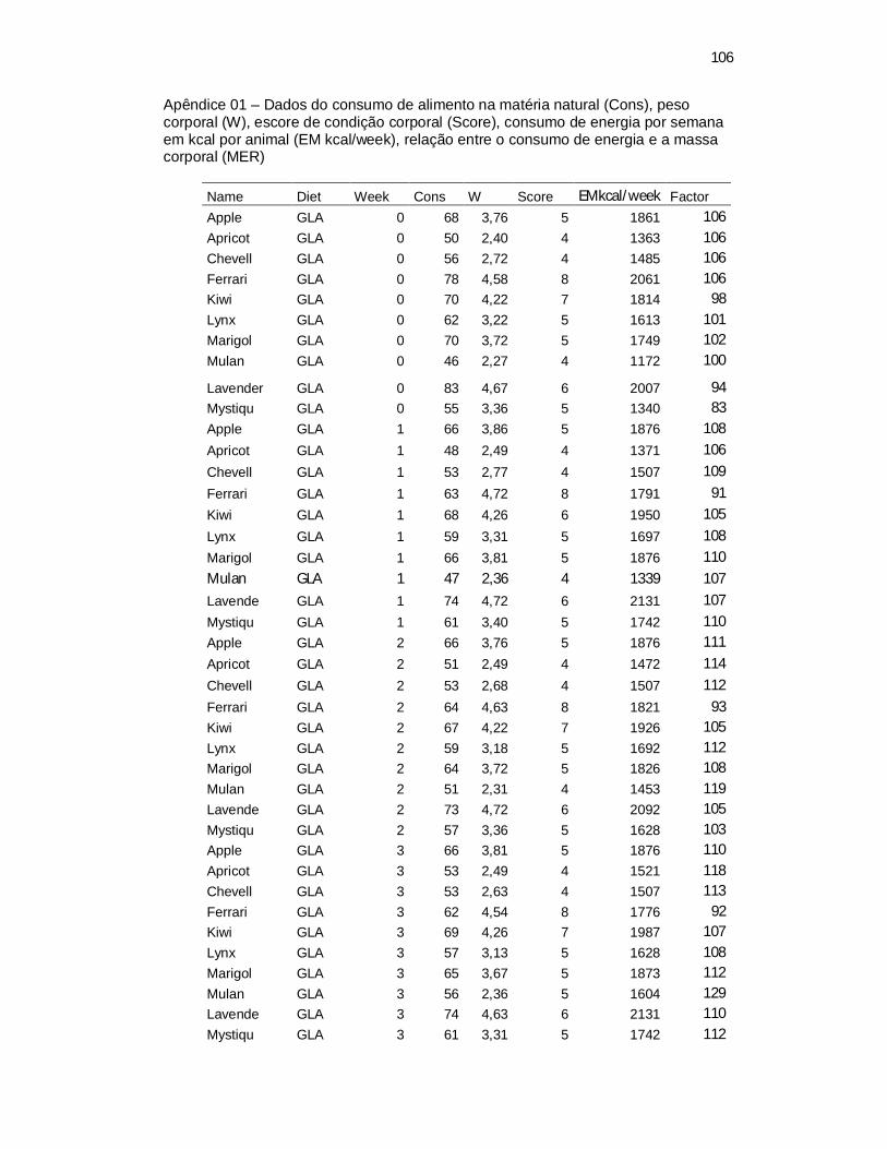
106
Apêndice 01 – Dados do consumo de alimento na matéria natural (Cons), peso corporal (W), escore de condição corporal (Score), consumo de energia por semana em kcal por animal (EM kcal/week), relação entre o consumo de energia e a massa corporal (MER)
Name Diet Week Cons W Score EMkcal/week Factor Apple GLA 0 68 3,76 5 1861 106 Apricot GLA 0 50 2,40 4 1363 106 Chevell GLA 0 56 2,72 4 1485 106 Ferrari GLA 0 78 4,58 8 2061 106 Kiwi GLA 0 70 4,22 7 1814 98 Lynx GLA 0 62 3,22 5 1613 101 Marigol GLA 0 70 3,72 5 1749 102 Mulan GLA 0 46 2,27 4 1172 100
Lavender GLA 0 83 4,67 6 2007 94 Mystiqu GLA 0 55 3,36 5 1340 83 Apple GLA 1 66 3,86 5 1876 108
Apricot GLA 1 48 2,49 4 1371 106
Chevell GLA 1 53 2,77 4 1507 109
Ferrari GLA 1 63 4,72 8 1791 91
Kiwi GLA 1 68 4,26 6 1950 105
Lynx GLA 1 59 3,31 5 1697 108
Marigol GLA 1 66 3,81 5 1876 110
Mulan GLA 1 47 2,36 4 1339 107
Lavende GLA 1 74 4,72 6 2131 107
Mystiqu GLA 1 61 3,40 5 1742 110 Apple GLA 2 66 3,76 5 1876 111
Apricot GLA 2 51 2,49 4 1472 114
Chevell GLA 2 53 2,68 4 1507 112
Ferrari GLA 2 64 4,63 8 1821 93 Kiwi GLA 2 67 4,22 7 1926 105 Lynx GLA 2 59 3,18 5 1692 112 Marigol GLA 2 64 3,72 5 1826 108 Mulan GLA 2 51 2,31 4 1453 119 Lavende GLA 2 73 4,72 6 2092 105 Mystiqu GLA 2 57 3,36 5 1628 103 Apple GLA 3 66 3,81 5 1876 110 Apricot GLA 3 53 2,49 4 1521 118 Chevell GLA 3 53 2,63 4 1507 113 Ferrari GLA 3 62 4,54 8 1776 92 Kiwi GLA 3 69 4,26 7 1987 107 Lynx GLA 3 57 3,13 5 1628 108 Marigol GLA 3 65 3,67 5 1873 112 Mulan GLA 3 56 2,36 5 1604 129 Lavende GLA 3 74 4,63 6 2131 110 Mystiqu GLA 3 61 3,31 5 1742 112
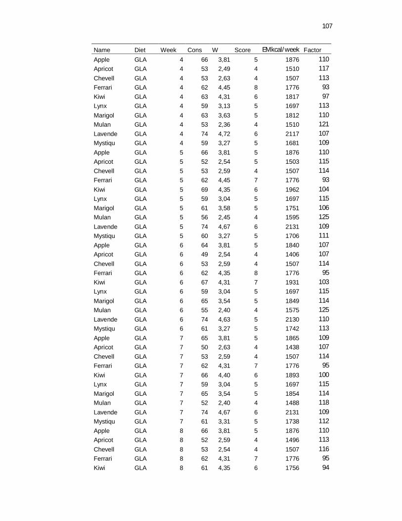
107
Name Diet Week Cons W Score EMkcal/week Factor Apple GLA 4 66 3,81 5 1876 110 Apricot GLA 4 53 2,49 4 1510 117
Chevell GLA 4 53 2,63 4 1507 113 Ferrari GLA 4 62 4,45 8 1776 93 Kiwi GLA 4 63 4,31 6 1817 97 Lynx GLA 4 59 3,13 5 1697 113
Marigol GLA 4 63 3,63 5 1812 110 Mulan GLA 4 53 2,36 4 1510 121 Lavende GLA 4 74 4,72 6 2117 107 Mystiqu GLA 4 59 3,27 5 1681 109
Apple GLA 5 66 3,81 5 1876 110 Apricot GLA 5 52 2,54 5 1503 115 Chevell GLA 5 53 2,59 4 1507 114 Ferrari GLA 5 62 4,45 7 1776 93
Kiwi GLA 5 69 4,35 6 1962 104 Lynx GLA 5 59 3,04 5 1697 115 Marigol GLA 5 61 3,58 5 1751 106 Mulan GLA 5 56 2,45 4 1595 125
Lavende GLA 5 74 4,67 6 2131 109 Mystiqu GLA 5 60 3,27 5 1706 111 Apple GLA 6 64 3,81 5 1840 107 Apricot GLA 6 49 2,54 4 1406 107
Chevell GLA 6 53 2,59 4 1507 114 Ferrari GLA 6 62 4,35 8 1776 95 Kiwi GLA 6 67 4,31 7 1931 103 Lynx GLA 6 59 3,04 5 1697 115
Marigol GLA 6 65 3,54 5 1849 114 Mulan GLA 6 55 2,40 4 1575 125 Lavende GLA 6 74 4,63 5 2130 110 Mystiqu GLA 6 61 3,27 5 1742 113
Apple GLA 7 65 3,81 5 1865 109 Apricot GLA 7 50 2,63 4 1438 107 Chevell GLA 7 53 2,59 4 1507 114 Ferrari GLA 7 62 4,31 7 1776 95
Kiwi GLA 7 66 4,40 6 1893 100 Lynx GLA 7 59 3,04 5 1697 115 Marigol GLA 7 65 3,54 5 1854 114 Mulan GLA 7 52 2,40 4 1488 118
Lavende GLA 7 74 4,67 6 2131 109 Mystiqu GLA 7 61 3,31 5 1738 112 Apple GLA 8 66 3,81 5 1876 110 Apricot GLA 8 52 2,59 4 1496 113
Chevell GLA 8 53 2,54 4 1507 116 Ferrari GLA 8 62 4,31 7 1776 95 Kiwi GLA 8 61 4,35 6 1756 94
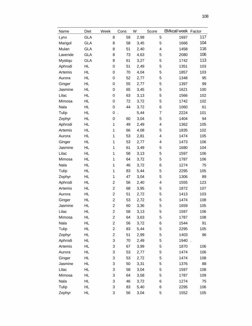
108
Name Diet Week Cons W Score EMkcal/week Factor Lynx GLA 8 59 2,99 5 1697 117 Marigol GLA 8 58 3,45 5 1666 104
Mulan GLA 8 51 2,40 4 1458 116 Lavende GLA 8 73 4,63 5 2080 106 Mystiqu GLA 8 61 3,27 5 1742 113 Aphrodi HL 0 51 2,49 5 1351 103 Artemis HL 0 70 4,04 5 1857 103 Aurora HL 0 52 2,77 5 1348 95 Ginger HL 0 55 2,77 5 1397 99 Jasmine HL 0 65 3,45 5 1621 100 Lilac HL 0 63 3,13 5 1566 102 Mimosa HL 0 72 3,72 5 1742 102 Nala HL 0 44 3,72 6 1060 61 Tulip HL 0 . 5,44 7 2224 101 Zephyr HL 0 60 3,04 5 1404 94 Aphrodi HL 1 49 2,49 4 1362 105 Artemis HL 1 66 4,08 5 1835 102 Aurora HL 1 53 2,81 4 1474 105 Ginger HL 1 53 2,77 4 1473 106 Jasmine HL 1 61 3,49 5 1680 104 Lilac HL 1 58 3,13 5 1597 106 Mimosa HL 1 64 3,72 5 1787 106 Nala HL 1 46 3,72 6 1274 75 Tulip HL 1 83 5,44 5 2295 105 Zephyr HL 1 47 3,04 5 1306 89 Aphrodi HL 2 56 2,40 4 1555 123 Artemis HL 2 68 3,95 5 1872 107 Aurora HL 2 51 2,72 5 1413 103 Ginger HL 2 53 2,72 5 1474 108 Jasmine HL 2 60 3,36 5 1659 105 Lilac HL 2 58 3,13 5 1597 106 Mimosa HL 2 64 3,63 5 1787 108 Nala HL 2 56 3,72 6 1544 91 Tulip HL 2 83 5,44 5 2295 105 Zephyr HL 2 51 2,99 5 1403 96 Aphrodi HL 3 70 2,49 5 1940 . Artemis HL 3 67 3,99 5 1870 106 Aurora HL 3 53 2,77 5 1474 106 Ginger HL 3 53 2,72 5 1474 108 Jasmine HL 3 50 3,31 5 1376 88 Lilac HL 3 58 3,04 5 1597 108 Mimosa HL 3 64 3,58 5 1787 109 Nala HL 3 46 3,72 6 1274 75 Tulip HL 3 83 5,40 6 2295 106 Zephyr HL 3 56 3,04 5 1552 105
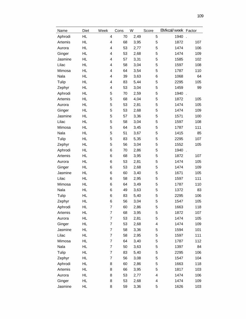
109
Name Diet Week Cons W Score EMkcal/week Factor Aphrodi HL 4 70 2,49 5 1940 . Artemis HL 4 68 3,95 5 1872 107 Aurora HL 4 53 2,77 5 1474 106 Ginger HL 4 53 2,68 5 1474 109 Jasmine HL 4 57 3,31 5 1585 102 Lilac HL 4 58 3,04 5 1597 108 Mimosa HL 4 64 3,54 5 1787 110 Nala HL 4 39 3,63 6 1068 64 Tulip HL 4 83 5,44 5 2295 105 Zephyr HL 4 53 3,04 5 1459 99 Aphrodi HL 5 70 2,59 5 1940 . Artemis HL 5 68 4,04 5 1872 105 Aurora HL 5 53 2,81 5 1474 105 Ginger HL 5 53 2,68 5 1474 109 Jasmine HL 5 57 3,36 5 1571 100 Lilac HL 5 58 3,04 5 1597 108 Mimosa HL 5 64 3,45 5 1787 111 Nala HL 5 51 3,67 5 1415 85 Tulip HL 5 83 5,35 5 2295 107 Zephyr HL 5 56 3,04 5 1552 105 Aphrodi HL 6 70 2,86 5 1940 . Artemis HL 6 68 3,95 5 1872 107 Aurora HL 6 53 2,81 5 1474 105 Ginger HL 6 53 2,68 5 1474 109 Jasmine HL 6 60 3,40 5 1671 105 Lilac HL 6 58 2,95 5 1597 111 Mimosa HL 6 64 3,49 5 1787 110 Nala HL 6 49 3,63 5 1372 83 Tulip HL 6 83 5,40 5 2295 106 Zephyr HL 6 56 3,04 5 1547 105 Aphrodi HL 7 60 2,86 5 1663 118 Artemis HL 7 68 3,95 5 1872 107 Aurora HL 7 53 2,81 5 1474 105 Ginger HL 7 53 2,68 4 1474 109 Jasmine HL 7 58 3,36 5 1594 101 Lilac HL 7 58 2,95 5 1597 111 Mimosa HL 7 64 3,40 5 1787 112 Nala HL 7 50 3,63 5 1397 84 Tulip HL 7 83 5,40 5 2295 106 Zephyr HL 7 56 3,08 5 1547 104 Aphrodi HL 8 60 2,86 5 1663 118 Artemis HL 8 66 3,95 5 1817 103 Aurora HL 8 53 2,77 4 1474 106 Ginger HL 8 53 2,68 4 1474 109 Jasmine HL 8 59 3,36 5 1626 103
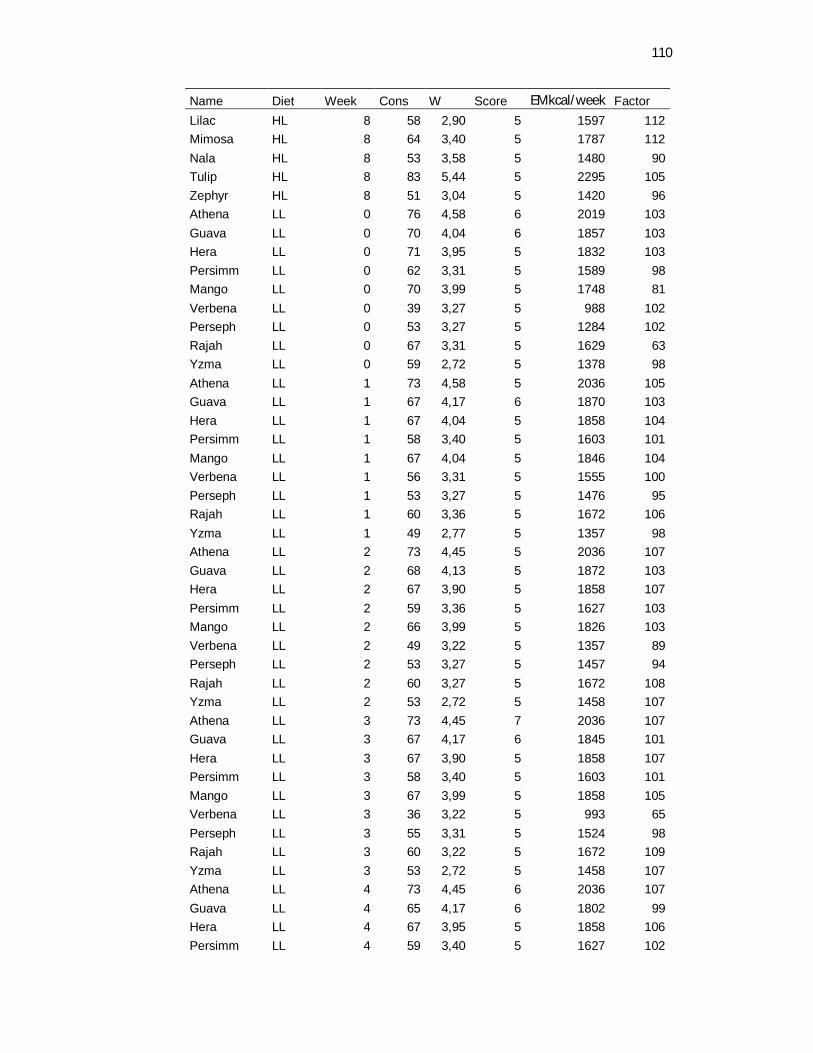
110
Name Diet Week Cons W Score EMkcal/week Factor Lilac HL 8 58 2,90 5 1597 112 Mimosa HL 8 64 3,40 5 1787 112 Nala HL 8 53 3,58 5 1480 90 Tulip HL 8 83 5,44 5 2295 105 Zephyr HL 8 51 3,04 5 1420 96 Athena LL 0 76 4,58 6 2019 103 Guava LL 0 70 4,04 6 1857 103 Hera LL 0 71 3,95 5 1832 103 Persimm LL 0 62 3,31 5 1589 98 Mango LL 0 70 3,99 5 1748 81 Verbena LL 0 39 3,27 5 988 102 Perseph LL 0 53 3,27 5 1284 102 Rajah LL 0 67 3,31 5 1629 63 Yzma LL 0 59 2,72 5 1378 98 Athena LL 1 73 4,58 5 2036 105 Guava LL 1 67 4,17 6 1870 103 Hera LL 1 67 4,04 5 1858 104 Persimm LL 1 58 3,40 5 1603 101 Mango LL 1 67 4,04 5 1846 104 Verbena LL 1 56 3,31 5 1555 100 Perseph LL 1 53 3,27 5 1476 95 Rajah LL 1 60 3,36 5 1672 106 Yzma LL 1 49 2,77 5 1357 98 Athena LL 2 73 4,45 5 2036 107 Guava LL 2 68 4,13 5 1872 103 Hera LL 2 67 3,90 5 1858 107 Persimm LL 2 59 3,36 5 1627 103 Mango LL 2 66 3,99 5 1826 103 Verbena LL 2 49 3,22 5 1357 89 Perseph LL 2 53 3,27 5 1457 94 Rajah LL 2 60 3,27 5 1672 108 Yzma LL 2 53 2,72 5 1458 107 Athena LL 3 73 4,45 7 2036 107 Guava LL 3 67 4,17 6 1845 101 Hera LL 3 67 3,90 5 1858 107 Persimm LL 3 58 3,40 5 1603 101 Mango LL 3 67 3,99 5 1858 105 Verbena LL 3 36 3,22 5 993 65 Perseph LL 3 55 3,31 5 1524 98 Rajah LL 3 60 3,22 5 1672 109 Yzma LL 3 53 2,72 5 1458 107 Athena LL 4 73 4,45 6 2036 107 Guava LL 4 65 4,17 6 1802 99 Hera LL 4 67 3,95 5 1858 106 Persimm LL 4 59 3,40 5 1627 102
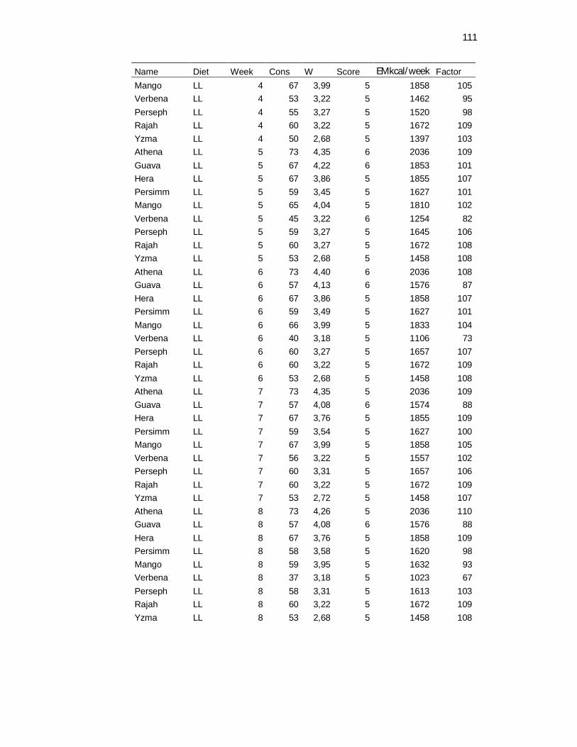
111
Name Diet Week Cons W Score EMkcal/week Factor Mango LL 4 67 3,99 5 1858 105 Verbena LL 4 53 3,22 5 1462 95 Perseph LL 4 55 3,27 5 1520 98 Rajah LL 4 60 3,22 5 1672 109 Yzma LL 4 50 2,68 5 1397 103 Athena LL 5 73 4,35 6 2036 109 Guava LL 5 67 4,22 6 1853 101 Hera LL 5 67 3,86 5 1855 107 Persimm LL 5 59 3,45 5 1627 101 Mango LL 5 65 4,04 5 1810 102 Verbena LL 5 45 3,22 6 1254 82 Perseph LL 5 59 3,27 5 1645 106 Rajah LL 5 60 3,27 5 1672 108 Yzma LL 5 53 2,68 5 1458 108 Athena LL 6 73 4,40 6 2036 108 Guava LL 6 57 4,13 6 1576 87 Hera LL 6 67 3,86 5 1858 107 Persimm LL 6 59 3,49 5 1627 101 Mango LL 6 66 3,99 5 1833 104 Verbena LL 6 40 3,18 5 1106 73 Perseph LL 6 60 3,27 5 1657 107 Rajah LL 6 60 3,22 5 1672 109 Yzma LL 6 53 2,68 5 1458 108 Athena LL 7 73 4,35 5 2036 109 Guava LL 7 57 4,08 6 1574 88 Hera LL 7 67 3,76 5 1855 109 Persimm LL 7 59 3,54 5 1627 100 Mango LL 7 67 3,99 5 1858 105 Verbena LL 7 56 3,22 5 1557 102 Perseph LL 7 60 3,31 5 1657 106 Rajah LL 7 60 3,22 5 1672 109 Yzma LL 7 53 2,72 5 1458 107 Athena LL 8 73 4,26 5 2036 110 Guava LL 8 57 4,08 6 1576 88 Hera LL 8 67 3,76 5 1858 109 Persimm LL 8 58 3,58 5 1620 98 Mango LL 8 59 3,95 5 1632 93 Verbena LL 8 37 3,18 5 1023 67 Perseph LL 8 58 3,31 5 1613 103 Rajah LL 8 60 3,22 5 1672 109 Yzma LL 8 53 2,68 5 1458 108

112
Apêndice 02 – Peso corporal, consumo de alimento em gramas e em Kcal consumida por gato por semana e necessidade de energia de mantença considerando as duas dietas (HL ou LMCT e LL ou HMCT)
Body weight
Type 3 Tests of Fixed Effects Num Den Effect DF DF F Value Pr > F Diet 1 17 0.29 0.5953 Time 8 136 2.28 0.0257 Diet*Time 8 136 0.34 0.9498 Maintenance energy requirement
Type 3 Tests of Fixed Effects Num Den Effect DF DF F Value Pr > F Diet 1 17 0.48 0.4990 Time 8 17 7.37 0.0003 Diet*Time 8 17 0.76 0.6433 Consumption g/cat/day
Type 3 Tests of Fixed Effects Num Den Effect DF DF F Value Pr > F Diet 1 17 0.01 0.9398 Time 8 17 5.40 0.0017 Diet*Time 8 17 0.70 0.6901
Apêndice 03 – Peso corporal, consumo de alimento em gramas e em Kcal consumida por gato por semana e necessidade de energia de mantença considerando as três dietas utilizadas no experimento.
Body Weight
Type 3 Tests of Fixed Effects Num Den Effect DF DF F Value Pr > F Diet 2 26 0.13 0.8771 Time 8 208 4.40 <.0001 Diet*Time 16 208 0.44 0.9715 Consumption Kcal/week/cat
Type 3 Tests of Fixed Effects Num Den Effect DF DF F Value Pr > F Diet 2 26 0.27 0.7677 Time 8 26 2.55 0.0335 Diet*Time 16 26 0.74 0.7307
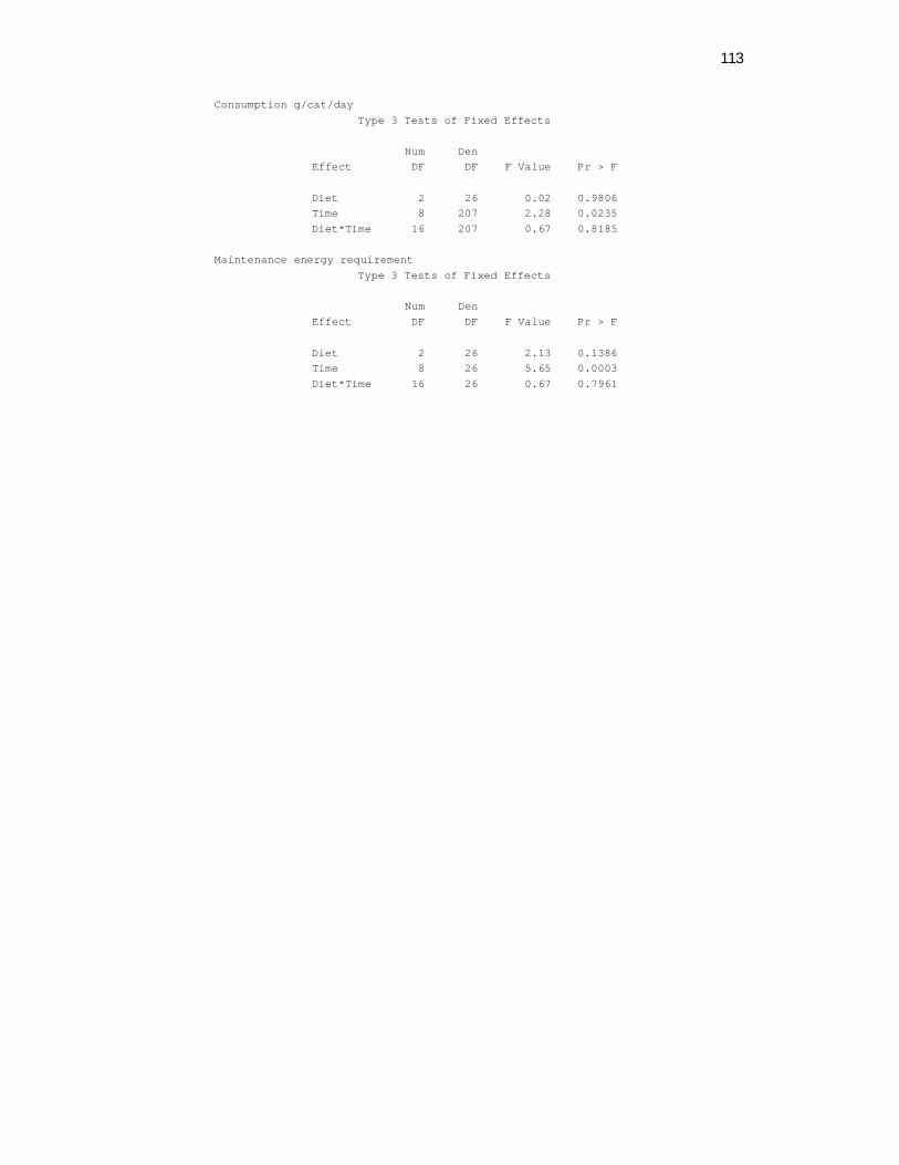
113
Consumption g/cat/day Type 3 Tests of Fixed Effects
Num Den Effect DF DF F Value Pr > F Diet 2 26 0.02 0.9806 Time 8 207 2.28 0.0235 Diet*Time 16 207 0.67 0.8185 Maintenance energy requirement
Type 3 Tests of Fixed Effects Num Den Effect DF DF F Value Pr > F Diet 2 26 2.13 0.1386 Time 8 26 5.65 0.0003 Diet*Time 16 26 0.67 0.7961

114
Apêndice 04 - Dados referentes ao experimento de digestibilidade Obs Cat Diet Drymater Protein Fat Fiber CHO Energy DE ME 1 Apple GLA 87.91 90.85 95.30 17.34 91.68 90.92 4409.50 4159.50 2 Chevel GLA 84.29 88.35 93.71 -1.43 87.83 87.73 4254.90 4011.80 3 Lynx GLA 102.14 101.38 100.64 111.08 102.45 101.71 4933.20 4654.20 4 Mulan GLA 97.18 98.26 98.97 77.70 97.99 97.98 4751.90 4481.50 5 Lavend GLA 82.81 86.81 94.16 -9.38 85.44 86.52 4196.10 3957.20 6 Mystiq GLA 84.18 87.53 94.15 -9.58 88.19 87.61 4248.90 4008.00 7 Chevel GLA 92.39 94.78 97.35 46.70 93.43 94.10 4564.10 4303.20 8 Lynx GLA 79.67 87.42 94.15 7.26 75.76 84.19 4083.20 3842.70 9 Mulan GLA 99.64 99.77 99.86 96.94 99.76 99.73 4837.10 4562.60 10 Lavend GLA 88.31 91.60 96.11 33.97 89.15 90.95 4410.90 4158.80 11 Mystiq GLA 83.72 88.44 94.57 -9.32 86.30 87.62 4249.50 4006.10 12 Kiwi GLA 76.95 84.75 90.67 -70.54 84.20 83.15 4032.80 3799.60 13 Marigol GLA 77.03 84.94 88.58 -68.39 83.99 82.52 4002.30 3768.50 14 Apple GLA 77.23 84.69 91.31 -61.42 83.36 83.34 4041.80 3808.70 15 Chevel GLA 85.45 89.92 94.59 9.67 87.52 88.89 4311.00 4063.50 16 Lynx GLA 81.85 88.50 94.44 11.01 79.15 85.68 4155.40 3911.90 17 Mulan GLA 94.19 96.27 97.83 54.56 96.07 95.92 4652.10 4387.20 18 Mystiq GLA 93.76 95.38 97.69 59.12 95.15 95.25 4619.50 4357.10 19 Marigol GLA 79.75 86.08 92.47 -36.80 84.54 84.96 4120.40 3883.50 20 Ferrari GLA 79.75 86.46 91.40 -55.59 86.91 85.08 4126.50 3888.50 21 Apricot GLA 71.64 81.71 88.44 -119.16 79.85 79.15 3838.80 3613.90 22 Apple GLA 81.21 87.13 93.25 -40.01 86.39 86.13 4177.20 3937.40 23 Chevel GLA 84.21 87.96 93.18 -6.30 88.87 87.83 4259.60 4017.50 24 Lynx GLA 86.10 90.53 95.85 28.28 85.51 89.23 4327.40 4078.30 25 Mulan GLA 104.47 102.86 101.60 136.90 103.21 103.22 5006.20 4723.10 26 Lavend GLA 76.41 79.91 91.51 -62.32 84.25 82.31 3992.00 3772.10 27 Kiwi GLA 77.95 85.61 92.17 -83.14 85.72 84.34 4090.70 3855.10 28 Marigol GLA 92.74 95.04 97.42 52.37 94.07 94.64 4590.00 4328.50 29 Lynx GLA 89.11 91.64 95.54 26.45 91.56 91.42 4433.90 4181.70 30 Mulan GLA 94.16 96.02 97.85 52.11 96.06 95.76 4644.20 4380.00 31 Lavend GLA 88.72 91.78 96.17 30.67 90.67 91.68 4446.30 4193.80 32 Mystiq GLA 81.81 86.24 93.73 -33.88 87.44 86.72 4205.80 3968.50 33 Kiwi GLA 84.59 89.63 94.77 -17.22 88.53 88.84 4308.90 4062.20 34 Marigol GLA 93.10 95.08 97.59 52.49 94.30 94.81 4598.20 4336.50 35 Ferrari GLA 74.81 83.00 89.29 -92.67 83.30 81.58 3956.60 3728.20 36 Apricot GLA 86.64 91.54 93.95 -7.33 91.48 90.23 4376.00 4124.10 37 Aphrod HL 80.15 83.55 85.88 -20.10 86.33 82.55 3970.69 3736.44 38 Artemis HL 81.22 86.83 91.38 -25.69 84.76 85.23 4099.71 3856.29 39 Aurora HL 91.40 93.90 96.13 37.14 93.50 93.36 4490.42 4227.17 40 Ginger HL 81.60 86.33 91.08 -35.03 86.36 85.39 4107.21 3865.17 41 Jasmine HL 86.47 89.98 93.88 17.58 87.30 88.86 4274.00 4021.73 42 Tulip HL 87.91 91.18 94.38 16.14 90.30 90.36 4346.41 4090.76 43 Zepher HL 97.69 98.45 98.77 82.52 98.24 97.68 4698.29 4422.29 44 Aphrod HL 78.96 83.59 84.82 -15.20 83.80 81.61 3925.43 3691.07 45 Artemis HL 86.80 90.86 94.43 0.39 89.79 90.07 4332.36 4077.62 46 Aurora HL 86.67 91.77 95.22 0.58 88.86 90.42 4349.13 4091.84 47 Ginger HL 83.15 87.47 92.41 -27.80 87.85 86.71 4170.75 3925.52 48 Jasmine HL 74.72 78.00 86.73 -29.34 79.01 77.97 3750.48 3531.81 49 Tulip HL 73.58 80.40 85.69 -93.94 69.65 82.56 4796.97 4571.57 50 Zepher HL 76.73 85.42 87.44 -99.22 77.24 82.70 3978.00 3738.53 51 Lilac HL 72.61 82.76 88.58 -110.28 77.70 79.70 3833.66 3601.64 52 Mimosa HL 62.23 75.80 85.61 -144.63 64.45 71.36 3432.44 3219.91 53 Nala HL 63.92 74.69 82.93 -167.11 74.20 72.66 3494.84 3285.44
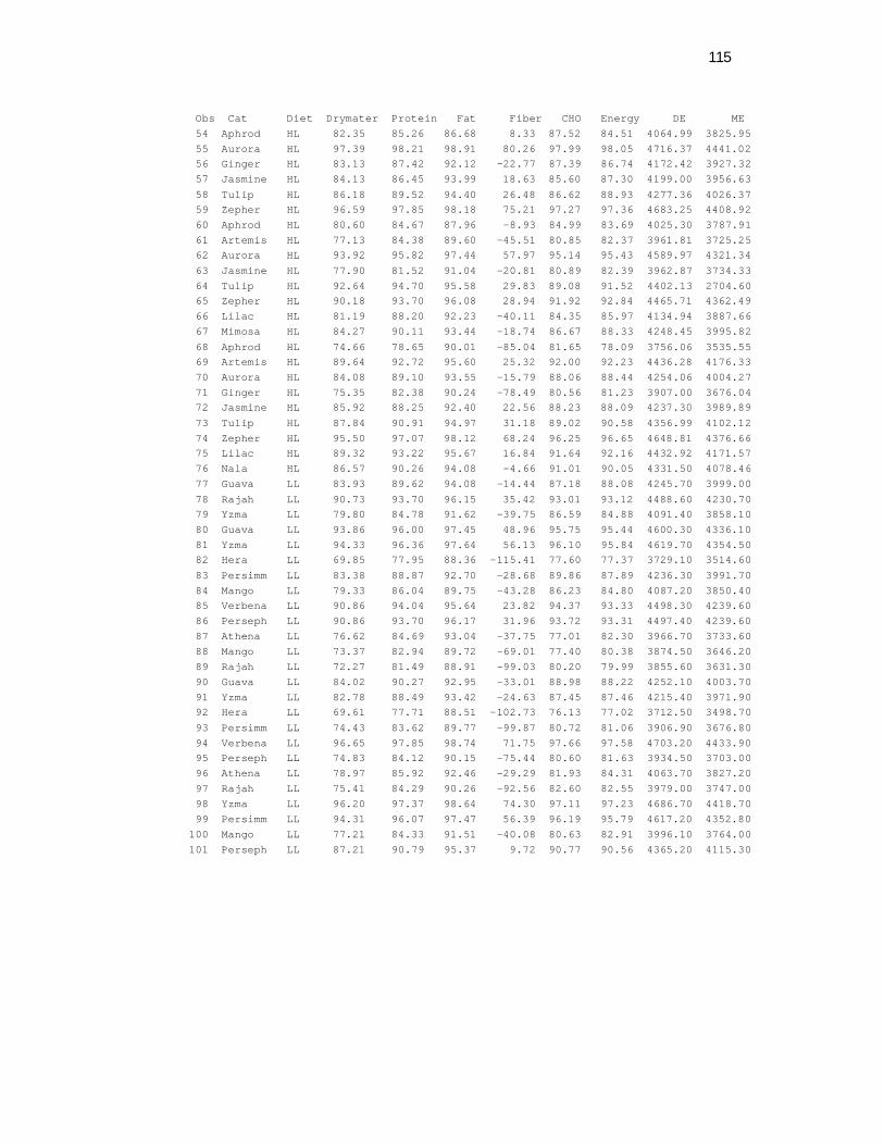
115
Obs Cat Diet Drymater Protein Fat Fiber CHO Energy DE ME 54 Aphrod HL 82.35 85.26 86.68 8.33 87.52 84.51 4064.99 3825.95 55 Aurora HL 97.39 98.21 98.91 80.26 97.99 98.05 4716.37 4441.02 56 Ginger HL 83.13 87.42 92.12 -22.77 87.39 86.74 4172.42 3927.32 57 Jasmine HL 84.13 86.45 93.99 18.63 85.60 87.30 4199.00 3956.63 58 Tulip HL 86.18 89.52 94.40 26.48 86.62 88.93 4277.36 4026.37 59 Zepher HL 96.59 97.85 98.18 75.21 97.27 97.36 4683.25 4408.92 60 Aphrod HL 80.60 84.67 87.96 -8.93 84.99 83.69 4025.30 3787.91 61 Artemis HL 77.13 84.38 89.60 -45.51 80.85 82.37 3961.81 3725.25 62 Aurora HL 93.92 95.82 97.44 57.97 95.14 95.43 4589.97 4321.34 63 Jasmine HL 77.90 81.52 91.04 -20.81 80.89 82.39 3962.87 3734.33 64 Tulip HL 92.64 94.70 95.58 29.83 89.08 91.52 4402.13 2704.60 65 Zepher HL 90.18 93.70 96.08 28.94 91.92 92.84 4465.71 4362.49 66 Lilac HL 81.19 88.20 92.23 -40.11 84.35 85.97 4134.94 3887.66 67 Mimosa HL 84.27 90.11 93.44 -18.74 86.67 88.33 4248.45 3995.82 68 Aphrod HL 74.66 78.65 90.01 -85.04 81.65 78.09 3756.06 3535.55 69 Artemis HL 89.64 92.72 95.60 25.32 92.00 92.23 4436.28 4176.33 70 Aurora HL 84.08 89.10 93.55 -15.79 88.06 88.44 4254.06 4004.27 71 Ginger HL 75.35 82.38 90.24 -78.49 80.56 81.23 3907.00 3676.04 72 Jasmine HL 85.92 88.25 92.40 22.56 88.23 88.09 4237.30 3989.89 73 Tulip HL 87.84 90.91 94.97 31.18 89.02 90.58 4356.99 4102.12 74 Zepher HL 95.50 97.07 98.12 68.24 96.25 96.65 4648.81 4376.66 75 Lilac HL 89.32 93.22 95.67 16.84 91.64 92.16 4432.92 4171.57 76 Nala HL 86.57 90.26 94.08 -4.66 91.01 90.05 4331.50 4078.46 77 Guava LL 83.93 89.62 94.08 -14.44 87.18 88.08 4245.70 3999.00 78 Rajah LL 90.73 93.70 96.15 35.42 93.01 93.12 4488.60 4230.70 79 Yzma LL 79.80 84.78 91.62 -39.75 86.59 84.88 4091.40 3858.10 80 Guava LL 93.86 96.00 97.45 48.96 95.75 95.44 4600.30 4336.10 81 Yzma LL 94.33 96.36 97.64 56.13 96.10 95.84 4619.70 4354.50 82 Hera LL 69.85 77.95 88.36 -115.41 77.60 77.37 3729.10 3514.60 83 Persimm LL 83.38 88.87 92.70 -28.68 89.86 87.89 4236.30 3991.70 84 Mango LL 79.33 86.04 89.75 -43.28 86.23 84.80 4087.20 3850.40 85 Verbena LL 90.86 94.04 95.64 23.82 94.37 93.33 4498.30 4239.60 86 Perseph LL 90.86 93.70 96.17 31.96 93.72 93.31 4497.40 4239.60 87 Athena LL 76.62 84.69 93.04 -37.75 77.01 82.30 3966.70 3733.60 88 Mango LL 73.37 82.94 89.72 -69.01 77.40 80.38 3874.50 3646.20 89 Rajah LL 72.27 81.49 88.91 -99.03 80.20 79.99 3855.60 3631.30 90 Guava LL 84.02 90.27 92.95 -33.01 88.98 88.22 4252.10 4003.70 91 Yzma LL 82.78 88.49 93.42 -24.63 87.45 87.46 4215.40 3971.90 92 Hera LL 69.61 77.71 88.51 -102.73 76.13 77.02 3712.50 3498.70 93 Persimm LL 74.43 83.62 89.77 -99.87 80.72 81.06 3906.90 3676.80 94 Verbena LL 96.65 97.85 98.74 71.75 97.66 97.58 4703.20 4433.90 95 Perseph LL 74.83 84.12 90.15 -75.44 80.60 81.63 3934.50 3703.00 96 Athena LL 78.97 85.92 92.46 -29.29 81.93 84.31 4063.70 3827.20 97 Rajah LL 75.41 84.29 90.26 -92.56 82.60 82.55 3979.00 3747.00 98 Yzma LL 96.20 97.37 98.64 74.30 97.11 97.23 4686.70 4418.70 99 Persimm LL 94.31 96.07 97.47 56.39 96.19 95.79 4617.20 4352.80 100 Mango LL 77.21 84.33 91.51 -40.08 80.63 82.91 3996.10 3764.00 101 Perseph LL 87.21 90.79 95.37 9.72 90.77 90.56 4365.20 4115.30
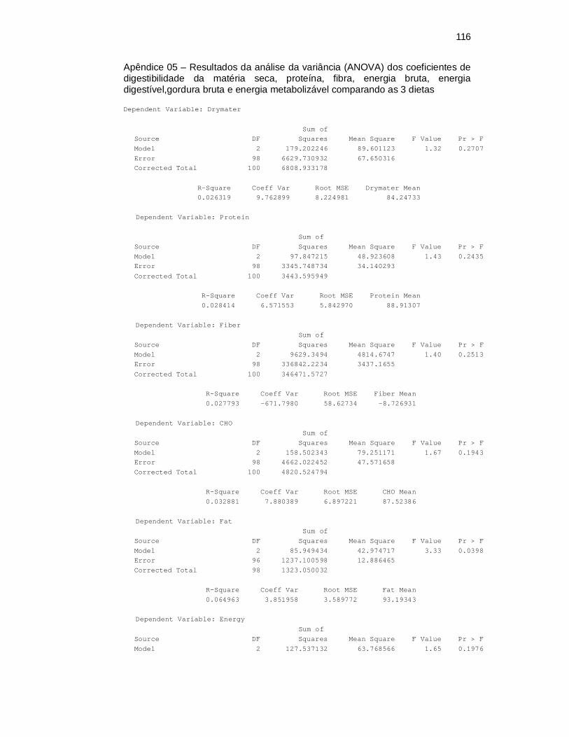
116
Apêndice 05 – Resultados da análise da variância (ANOVA) dos coeficientes de digestibilidade da matéria seca, proteína, fibra, energia bruta, energia digestível,gordura bruta e energia metabolizável comparando as 3 dietas
Dependent Variable: Drymater
Sum of Source DF Squares Mean Square F Value Pr > F Model 2 179.202246 89.601123 1.32 0.2707 Error 98 6629.730932 67.650316 Corrected Total 100 6808.933178 R-Square Coeff Var Root MSE Drymater Mean 0.026319 9.762899 8.224981 84.24733
Dependent Variable: Protein Sum of Source DF Squares Mean Square F Value Pr > F Model 2 97.847215 48.923608 1.43 0.2435 Error 98 3345.748734 34.140293 Corrected Total 100 3443.595949 R-Square Coeff Var Root MSE Protein Mean 0.028414 6.571553 5.842970 88.91307 Dependent Variable: Fiber Sum of Source DF Squares Mean Square F Value Pr > F Model 2 9629.3494 4814.6747 1.40 0.2513 Error 98 336842.2234 3437.1655 Corrected Total 100 346471.5727 R-Square Coeff Var Root MSE Fiber Mean 0.027793 -671.7980 58.62734 -8.726931 Dependent Variable: CHO Sum of Source DF Squares Mean Square F Value Pr > F Model 2 158.502343 79.251171 1.67 0.1943 Error 98 4662.022452 47.571658 Corrected Total 100 4820.524794 R-Square Coeff Var Root MSE CHO Mean 0.032881 7.880389 6.897221 87.52386 Dependent Variable: Fat Sum of Source DF Squares Mean Square F Value Pr > F Model 2 85.949434 42.974717 3.33 0.0398 Error 96 1237.100598 12.886465 Corrected Total 98 1323.050032 R-Square Coeff Var Root MSE Fat Mean 0.064963 3.851958 3.589772 93.19343 Dependent Variable: Energy Sum of Source DF Squares Mean Square F Value Pr > F Model 2 127.537132 63.768566 1.65 0.1976
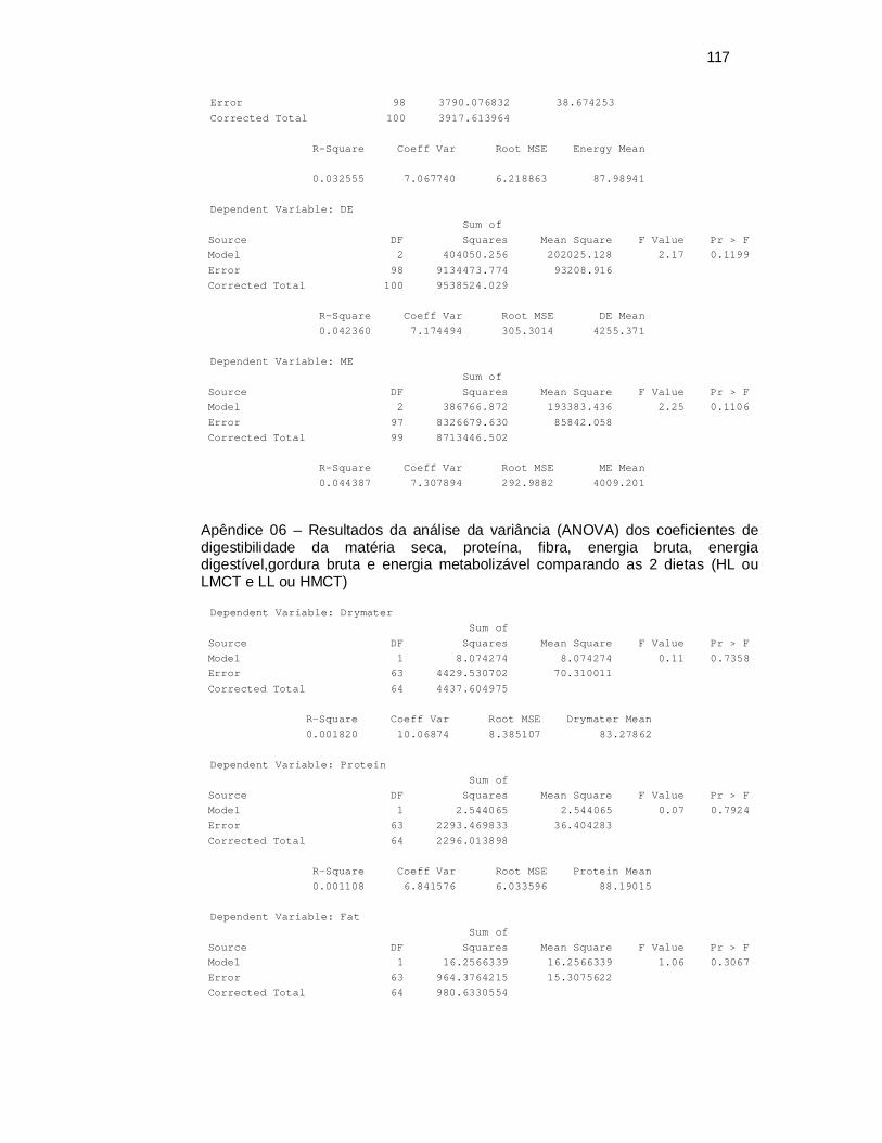
117
Error 98 3790.076832 38.674253 Corrected Total 100 3917.613964 R-Square Coeff Var Root MSE Energy Mean 0.032555 7.067740 6.218863 87.98941 Dependent Variable: DE Sum of Source DF Squares Mean Square F Value Pr > F Model 2 404050.256 202025.128 2.17 0.1199 Error 98 9134473.774 93208.916 Corrected Total 100 9538524.029 R-Square Coeff Var Root MSE DE Mean 0.042360 7.174494 305.3014 4255.371 Dependent Variable: ME Sum of Source DF Squares Mean Square F Value Pr > F Model 2 386766.872 193383.436 2.25 0.1106 Error 97 8326679.630 85842.058 Corrected Total 99 8713446.502 R-Square Coeff Var Root MSE ME Mean 0.044387 7.307894 292.9882 4009.201
Apêndice 06 – Resultados da análise da variância (ANOVA) dos coeficientes de digestibilidade da matéria seca, proteína, fibra, energia bruta, energia digestível,gordura bruta e energia metabolizável comparando as 2 dietas (HL ou LMCT e LL ou HMCT)
Dependent Variable: Drymater Sum of Source DF Squares Mean Square F Value Pr > F Model 1 8.074274 8.074274 0.11 0.7358 Error 63 4429.530702 70.310011 Corrected Total 64 4437.604975 R-Square Coeff Var Root MSE Drymater Mean 0.001820 10.06874 8.385107 83.27862 Dependent Variable: Protein Sum of Source DF Squares Mean Square F Value Pr > F Model 1 2.544065 2.544065 0.07 0.7924 Error 63 2293.469833 36.404283 Corrected Total 64 2296.013898 R-Square Coeff Var Root MSE Protein Mean 0.001108 6.841576 6.033596 88.19015 Dependent Variable: Fat Sum of Source DF Squares Mean Square F Value Pr > F Model 1 16.2566339 16.2566339 1.06 0.3067 Error 63 964.3764215 15.3075622 Corrected Total 64 980.6330554
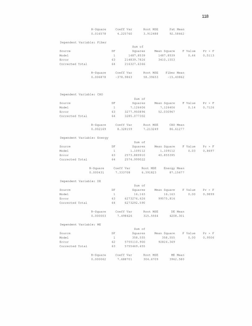
118
R-Square Coeff Var Root MSE Fat Mean 0.016578 4.225760 3.912488 92.58662 Dependent Variable: Fiber Sum of Source DF Squares Mean Square F Value Pr > F Model 1 1487.8539 1487.8539 0.44 0.5113 Error 63 214839.7826 3410.1553 Corrected Total 64 216327.6366 R-Square Coeff Var Root MSE Fiber Mean 0.006878 -378.9863 58.39653 -15.40862 Dependent Variable: CHO Sum of Source DF Squares Mean Square F Value Pr > F Model 1 7.126406 7.126406 0.14 0.7126 Error 63 3277.950896 52.030967 Corrected Total 64 3285.077302 R-Square Coeff Var Root MSE CHO Mean 0.002169 8.328159 7.213249 86.61277 Dependent Variable: Energy Sum of Source DF Squares Mean Square F Value Pr > F Model 1 1.109112 1.109112 0.03 0.8697 Error 63 2573.889910 40.855395 Corrected Total 64 2574.999022 R-Square Coeff Var Root MSE Energy Mean 0.000431 7.333708 6.391823 87.15677 Dependent Variable: DE Sum of Source DF Squares Mean Square F Value Pr > F Model 1 16.163 16.163 0.00 0.9899 Error 63 6273276.426 99575.816 Corrected Total 64 6273292.590 R-Square Coeff Var Root MSE DE Mean 0.000003 7.498426 315.5564 4208.301 Dependent Variable: ME Sum of Source DF Squares Mean Square F Value Pr > F Model 1 358.555 358.555 0.00 0.9506 Error 62 5755110.900 92824.369 Corrected Total 63 5755469.455 R-Square Coeff Var Root MSE ME Mean 0.000062 7.688701 304.6709 3962.580
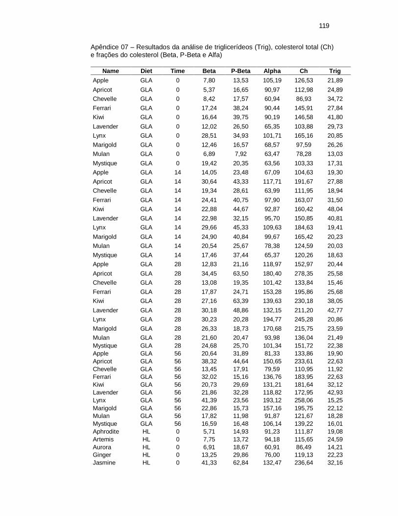
119
Apêndice 07 – Resultados da análise de triglicerídeos (Trig), colesterol total (Ch) e frações do colesterol (Beta, P-Beta e Alfa)
Name Diet Time Beta P-Beta Alpha Ch Trig
Apple GLA 0 7,80 13,53 105,19 126,53 21,89 Apricot GLA 0 5,37 16,65 90,97 112,98 24,89 Chevelle GLA 0 8,42 17,57 60,94 86,93 34,72 Ferrari GLA 0 17,24 38,24 90,44 145,91 27,84 Kiwi GLA 0 16,64 39,75 90,19 146,58 41,80 Lavender GLA 0 12,02 26,50 65,35 103,88 29,73 Lynx GLA 0 28,51 34,93 101,71 165,16 20,85 Marigold GLA 0 12,46 16,57 68,57 97,59 26,26 Mulan GLA 0 6,89 7,92 63,47 78,28 13,03 Mystique GLA 0 19,42 20,35 63,56 103,33 17,31 Apple GLA 14 14,05 23,48 67,09 104,63 19,30 Apricot GLA 14 30,64 43,33 117,71 191,67 27,88 Chevelle GLA 14 19,34 28,61 63,99 111,95 18,94 Ferrari GLA 14 24,41 40,75 97,90 163,07 31,50 Kiwi GLA 14 22,88 44,67 92,87 160,42 48,04 Lavender GLA 14 22,98 32,15 95,70 150,85 40,81 Lynx GLA 14 29,66 45,33 109,63 184,63 19,41 Marigold GLA 14 24,90 40,84 99,67 165,42 20,23 Mulan GLA 14 20,54 25,67 78,38 124,59 20,03 Mystique GLA 14 17,46 37,44 65,37 120,26 18,63 Apple GLA 28 12,83 21,16 118,97 152,97 20,44 Apricot GLA 28 34,45 63,50 180,40 278,35 25,58 Chevelle GLA 28 13,08 19,35 101,42 133,84 15,46 Ferrari GLA 28 17,87 24,71 153,28 195,86 25,68 Kiwi GLA 28 27,16 63,39 139,63 230,18 38,05 Lavender GLA 28 30,18 48,86 132,15 211,20 42,77 Lynx GLA 28 30,23 20,28 194,77 245,28 20,86 Marigold GLA 28 26,33 18,73 170,68 215,75 23,59 Mulan GLA 28 21,60 20,47 93,98 136,04 21,49 Mystique GLA 28 24,68 25,70 101,34 151,72 22,38 Apple GLA 56 20,64 31,89 81,33 133,86 19,90 Apricot GLA 56 38,32 44,64 150,65 233,61 22,63 Chevelle GLA 56 13,45 17,91 79,59 110,95 11,92 Ferrari GLA 56 32,02 15,16 136,76 183,95 22,63 Kiwi GLA 56 20,73 29,69 131,21 181,64 32,12 Lavender GLA 56 21,86 32,28 118,82 172,95 42,93 Lynx GLA 56 41,39 23,56 193,12 258,06 15,25 Marigold GLA 56 22,86 15,73 157,16 195,75 22,12 Mulan GLA 56 17,82 11,98 91,87 121,67 18,28 Mystique GLA 56 16,59 16,48 106,14 139,22 16,01 Aphrodite HL 0 5,71 14,93 91,23 111,87 19,08 Artemis HL 0 7,75 13,72 94,18 115,65 24,59 Aurora HL 0 6,91 18,67 60,91 86,49 14,21 Ginger HL 0 13,25 29,86 76,00 119,13 22,23 Jasmine HL 0 41,33 62,84 132,47 236,64 32,16

120
Name Diet Time Beta P-Beta Alpha Ch Trig Lilac HL 0 14,45 40,09 56,81 111,35 29,11 Mimosa HL 0 12,87 18,57 72,74 104,18 25,08 Nala HL 0 6,06 13,56 91,82 111,43 21,74 Tulip HL 0 15,35 19,29 61,76 96,41 24,79 Zephyr HL 0 16,79 24,02 53,85 94,67 19,23 Aphrodite HL 14 18,35 27,64 97,86 143,83 15,53 Artemis HL 14 19,61 29,54 81,95 131,10 13,67 Aurora HL 14 23,01 30,21 78,67 131,88 18,01 Ginger HL 14 26,66 33,12 92,64 152,41 18,48 Jasmine HL 14 43,51 42,11 155,99 241,71 32,43 Lilac HL 14 24,85 29,45 82,83 137,12 15,89 Mimosa HL 14 27,33 32,11 92,91 152,35 18,48 Nala HL 14 26,83 35,06 98,40 160,30 24,37 Tulip HL 14 17,01 39,03 63,19 119,23 28,19 Zephyr HL 14 30,91 40,67 96,56 168,13 16,61 Aphrodite HL 28 34,10 36,24 125,75 196,09 31,13 Artemis HL 28 29,24 41,13 103,71 174,08 22,12 Aurora HL 28 23,64 17,99 138,75 180,38 16,35 Ginger HL 28 23,24 22,40 152,97 198,60 19,71 Jasmine HL 28 44,98 85,68 199,59 330,23 27,25 Lilac HL 28 11,53 22,43 133,22 167,17 20,76 Mimosa HL 28 32,91 18,43 122,80 174,15 18,97 Nala HL 28 40,85 36,16 158,03 235,03 19,18 Tulip HL 28 15,11 18,89 88,67 122,68 26,21 Zephyr HL 28 37,51 22,84 108,79 169,14 21,59 Aphrodite HL 56 31,22 42,11 101,72 175,05 23,74 Artemis HL 56 27,32 47,35 92,47 167,13 23,43 Aurora HL 56 19,09 14,84 116,43 150,36 15,56 Ginger HL 56 30,97 22,46 126,79 180,23 17,58 Jasmine HL 56 31,70 18,85 119,67 170,22 21,41 Lilac HL 56 19,39 15,38 117,79 152,56 15,71 Mimosa HL 56 23,53 14,28 122,00 159,81 16,36 Nala HL 56 25,41 16,44 174,89 216,73 17,27 Tulip HL 56 12,36 16,55 78,41 107,31 24,44 Zephyr HL 56 32,23 21,95 156,29 210,46 17,22 Zephyr HL 140 34,75 11,78 138,54 185,06 11,075 Athena LL 0 5,74 20,09 100,03 125,86 40,23 Guava LL 0 6,07 13,43 75,13 94,63 21,59 Hera LL 0 10,09 20,64 59,28 90,00 22,13 Mango LL 0 18,43 33,23 58,37 110,02 29,31 Persephone LL 0 12,74 25,65 100,71 139,11 18,35 Persimmon LL 0 11,39 22,77 56,11 90,26 15,74 Rajah LL 0 7,29 17,51 112,38 137,18 23,80 Verbena LL 0 20,59 44,57 62,33 127,49 21,64 Yzma LL 0 33,10 38,42 116,05 187,58 24,39 Athena LL 14 24,12 32,43 88,53 143,65 35,43 Guava LL 14 19,50 30,92 68,93 119,35 24,06 Hera LL 14 21,34 35,40 79,95 136,70 20,34 Mango LL 14 23,18 38,88 84,80 146,87 14,75 Persephone LL 14 27,08 31,13 105,10 163,31 23,44 Persimmon LL 14 19,17 20,05 58,81 98,04 19,82 Rajah LL 14 31,38 51,19 110,18 192,75 37,08 Verbena LL 14 25,17 39,93 82,99 148,08 48,25
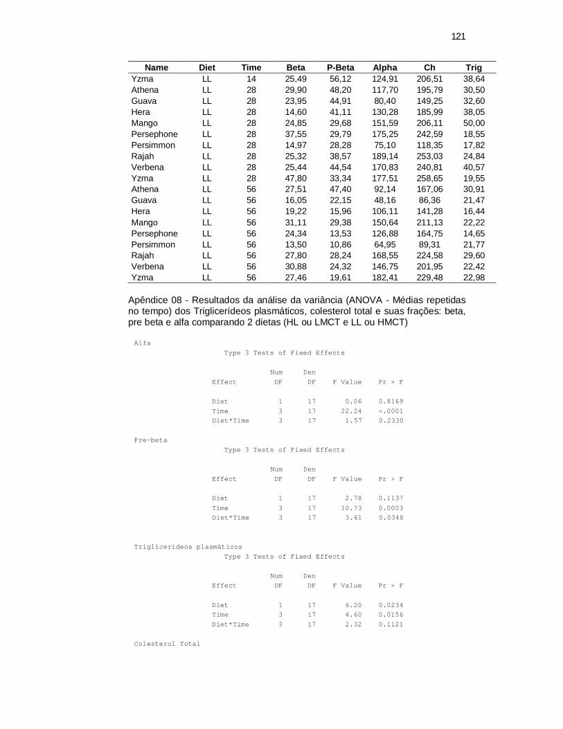
121
Name Diet Time Beta P-Beta Alpha Ch Trig Yzma LL 14 25,49 56,12 124,91 206,51 38,64 Athena LL 28 29,90 48,20 117,70 195,79 30,50 Guava LL 28 23,95 44,91 80,40 149,25 32,60 Hera LL 28 14,60 41,11 130,28 185,99 38,05 Mango LL 28 24,85 29,68 151,59 206,11 50,00 Persephone LL 28 37,55 29,79 175,25 242,59 18,55 Persimmon LL 28 14,97 28,28 75,10 118,35 17,82 Rajah LL 28 25,32 38,57 189,14 253,03 24,84 Verbena LL 28 25,44 44,54 170,83 240,81 40,57 Yzma LL 28 47,80 33,34 177,51 258,65 19,55 Athena LL 56 27,51 47,40 92,14 167,06 30,91 Guava LL 56 16,05 22,15 48,16 86,36 21,47 Hera LL 56 19,22 15,96 106,11 141,28 16,44 Mango LL 56 31,11 29,38 150,64 211,13 22,22 Persephone LL 56 24,34 13,53 126,88 164,75 14,65 Persimmon LL 56 13,50 10,86 64,95 89,31 21,77 Rajah LL 56 27,80 28,24 168,55 224,58 29,60 Verbena LL 56 30,88 24,32 146,75 201,95 22,42 Yzma LL 56 27,46 19,61 182,41 229,48 22,98
Apêndice 08 - Resultados da análise da variância (ANOVA - Médias repetidas no tempo) dos Triglicerídeos plasmáticos, colesterol total e suas frações: beta, pre beta e alfa comparando 2 dietas (HL ou LMCT e LL ou HMCT)
Alfa
Type 3 Tests of Fixed Effects Num Den Effect DF DF F Value Pr > F Diet 1 17 0.06 0.8169 Time 3 17 22.24 <.0001 Diet*Time 3 17 1.57 0.2330 Pre-beta
Type 3 Tests of Fixed Effects Num Den Effect DF DF F Value Pr > F Diet 1 17 2.78 0.1137 Time 3 17 10.73 0.0003 Diet*Time 3 17 3.61 0.0348 Triglicerideos plasmáticos
Type 3 Tests of Fixed Effects Num Den Effect DF DF F Value Pr > F Diet 1 17 6.20 0.0234 Time 3 17 4.60 0.0156 Diet*Time 3 17 2.32 0.1121 Colesterol Total
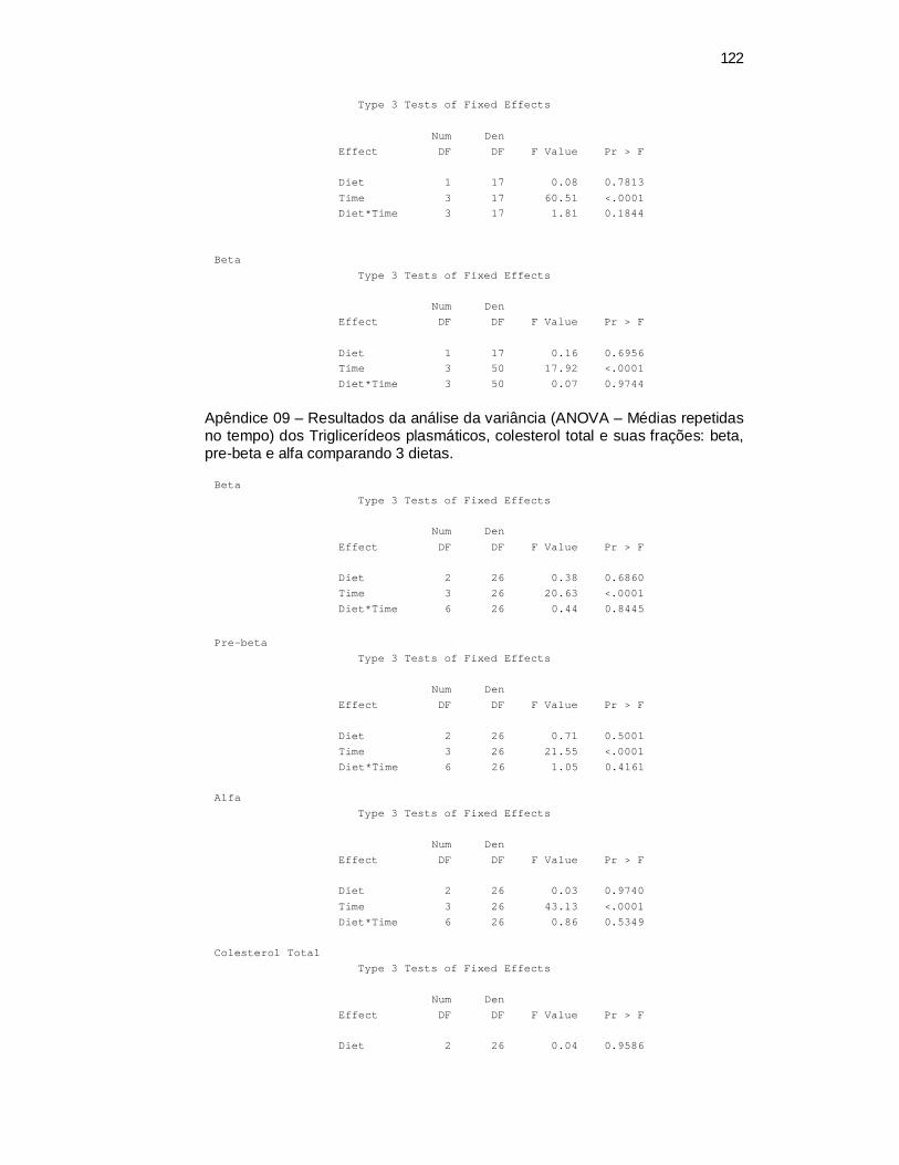
122
Type 3 Tests of Fixed Effects Num Den Effect DF DF F Value Pr > F Diet 1 17 0.08 0.7813 Time 3 17 60.51 <.0001 Diet*Time 3 17 1.81 0.1844 Beta
Type 3 Tests of Fixed Effects Num Den Effect DF DF F Value Pr > F Diet 1 17 0.16 0.6956 Time 3 50 17.92 <.0001 Diet*Time 3 50 0.07 0.9744
Apêndice 09 – Resultados da análise da variância (ANOVA – Médias repetidas no tempo) dos Triglicerídeos plasmáticos, colesterol total e suas frações: beta, pre-beta e alfa comparando 3 dietas.
Beta
Type 3 Tests of Fixed Effects Num Den Effect DF DF F Value Pr > F Diet 2 26 0.38 0.6860 Time 3 26 20.63 <.0001 Diet*Time 6 26 0.44 0.8445 Pre-beta
Type 3 Tests of Fixed Effects Num Den Effect DF DF F Value Pr > F Diet 2 26 0.71 0.5001 Time 3 26 21.55 <.0001 Diet*Time 6 26 1.05 0.4161 Alfa
Type 3 Tests of Fixed Effects Num Den Effect DF DF F Value Pr > F Diet 2 26 0.03 0.9740 Time 3 26 43.13 <.0001 Diet*Time 6 26 0.86 0.5349 Colesterol Total
Type 3 Tests of Fixed Effects Num Den Effect DF DF F Value Pr > F Diet 2 26 0.04 0.9586
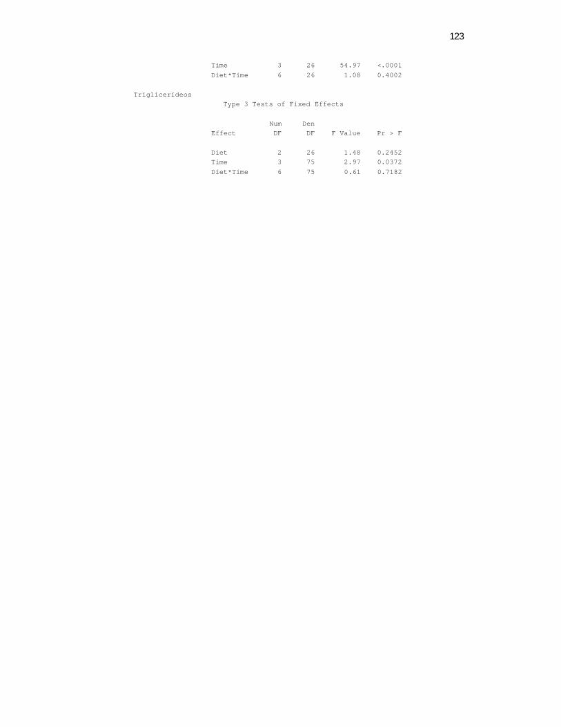
123
Time 3 26 54.97 <.0001 Diet*Time 6 26 1.08 0.4002 Triglicerídeos
Type 3 Tests of Fixed Effects Num Den Effect DF DF F Value Pr > F Diet 2 26 1.48 0.2452 Time 3 75 2.97 0.0372 Diet*Time 6 75 0.61 0.7182
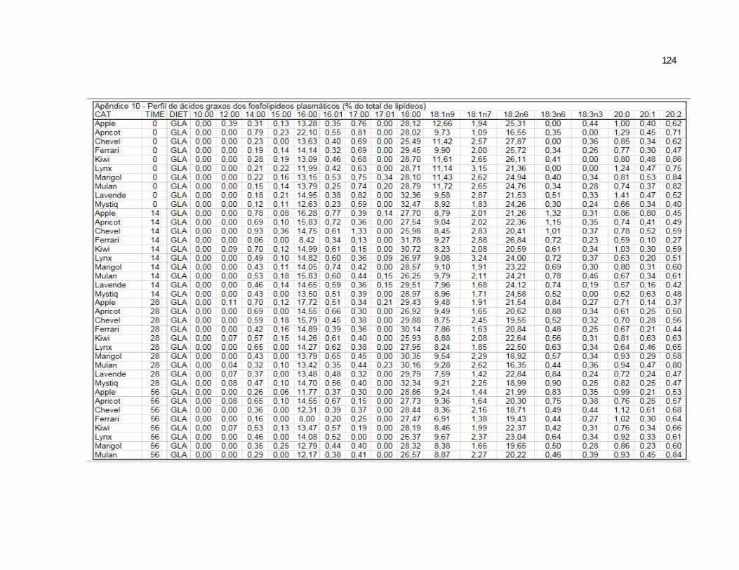
124
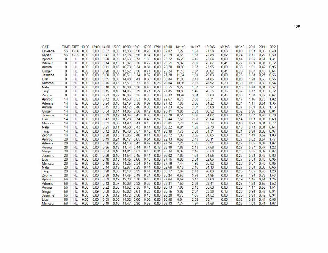
125
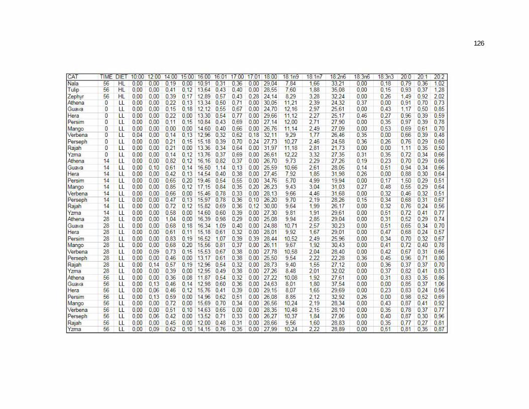
126

127
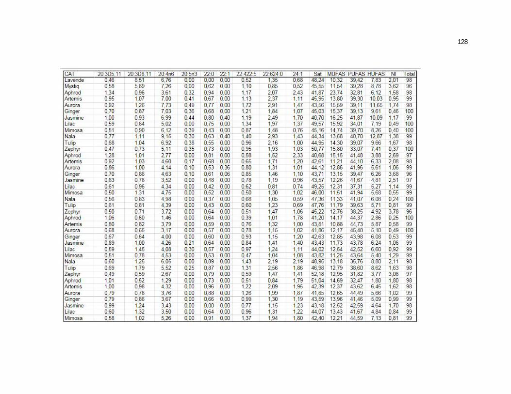
128
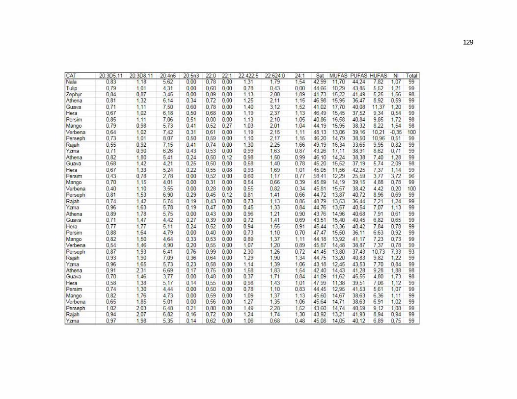
129
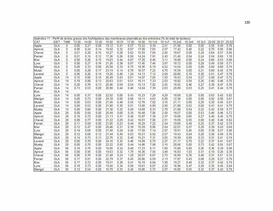
130
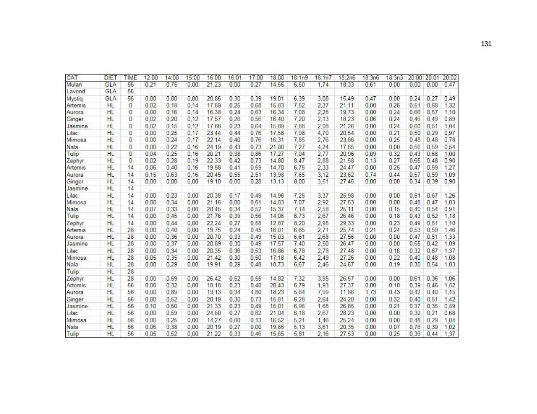
131
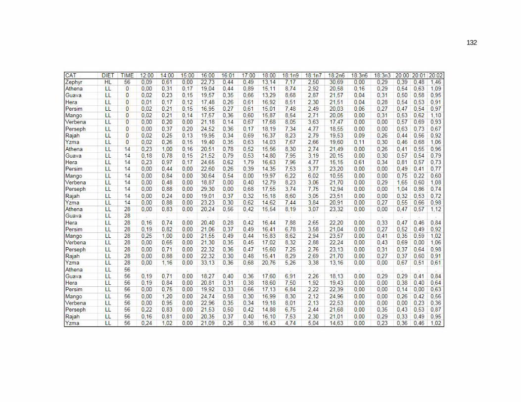
132
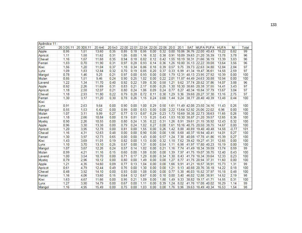
133
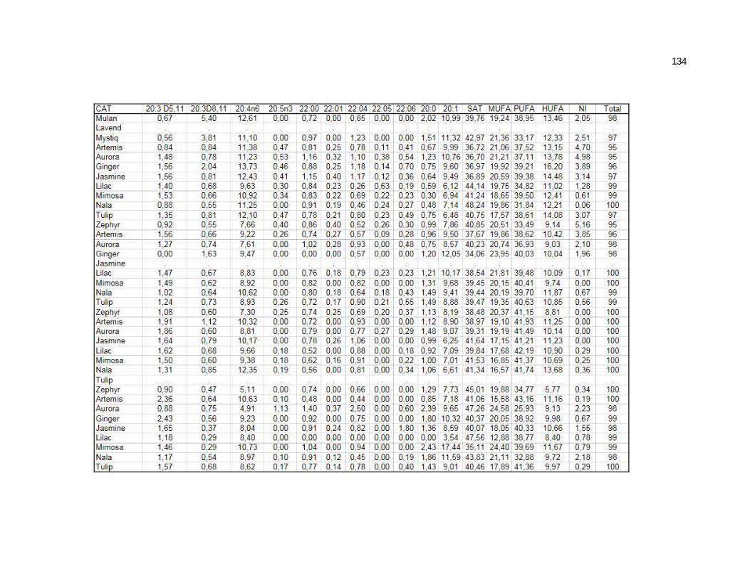
134
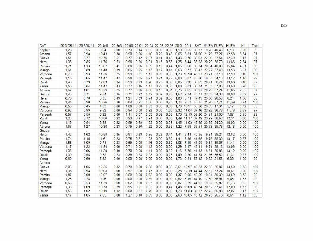
135
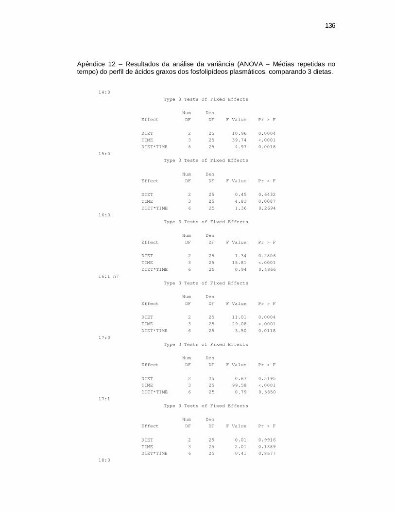
136
Apêndice 12 – Resultados da análise da variância (ANOVA – Médias repetidas no tempo) do perfil de ácidos graxos dos fosfolipídeos plasmáticos, comparando 3 dietas.
14:0 Type 3 Tests of Fixed Effects
Num Den Effect DF DF F Value Pr > F DIET 2 25 10.96 0.0004 TIME 3 25 39.74 <.0001 DIET*TIME 6 25 4.97 0.0018 15:0
Type 3 Tests of Fixed Effects Num Den Effect DF DF F Value Pr > F DIET 2 25 0.45 0.6432 TIME 3 25 4.83 0.0087 DIET*TIME 6 25 1.36 0.2694 16:0
Type 3 Tests of Fixed Effects Num Den Effect DF DF F Value Pr > F DIET 2 25 1.34 0.2806 TIME 3 25 15.81 <.0001 DIET*TIME 6 25 0.94 0.4866 16:1 n7
Type 3 Tests of Fixed Effects Num Den Effect DF DF F Value Pr > F DIET 2 25 11.01 0.0004 TIME 3 25 29.08 <.0001 DIET*TIME 6 25 3.50 0.0118 17:0
Type 3 Tests of Fixed Effects Num Den Effect DF DF F Value Pr > F DIET 2 25 0.67 0.5195 TIME 3 25 99.58 <.0001 DIET*TIME 6 25 0.79 0.5850 17:1
Type 3 Tests of Fixed Effects
Num Den Effect DF DF F Value Pr > F DIET 2 25 0.01 0.9916 TIME 3 25 2.01 0.1389 DIET*TIME 6 25 0.41 0.8677 18:0
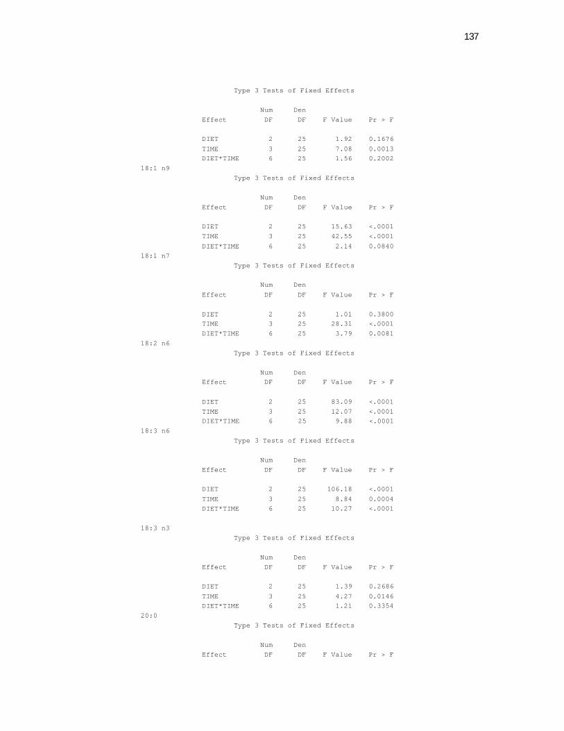
137
Type 3 Tests of Fixed Effects Num Den Effect DF DF F Value Pr > F DIET 2 25 1.92 0.1676 TIME 3 25 7.08 0.0013 DIET*TIME 6 25 1.56 0.2002 18:1 n9
Type 3 Tests of Fixed Effects Num Den Effect DF DF F Value Pr > F DIET 2 25 15.63 <.0001 TIME 3 25 42.55 <.0001 DIET*TIME 6 25 2.14 0.0840 18:1 n7
Type 3 Tests of Fixed Effects Num Den Effect DF DF F Value Pr > F DIET 2 25 1.01 0.3800 TIME 3 25 28.31 <.0001 DIET*TIME 6 25 3.79 0.0081 18:2 n6
Type 3 Tests of Fixed Effects Num Den Effect DF DF F Value Pr > F DIET 2 25 83.09 <.0001 TIME 3 25 12.07 <.0001 DIET*TIME 6 25 9.88 <.0001 18:3 n6
Type 3 Tests of Fixed Effects Num Den Effect DF DF F Value Pr > F DIET 2 25 106.18 <.0001 TIME 3 25 8.84 0.0004 DIET*TIME 6 25 10.27 <.0001 18:3 n3
Type 3 Tests of Fixed Effects Num Den Effect DF DF F Value Pr > F DIET 2 25 1.39 0.2686 TIME 3 25 4.27 0.0146 DIET*TIME 6 25 1.21 0.3354 20:0
Type 3 Tests of Fixed Effects Num Den Effect DF DF F Value Pr > F
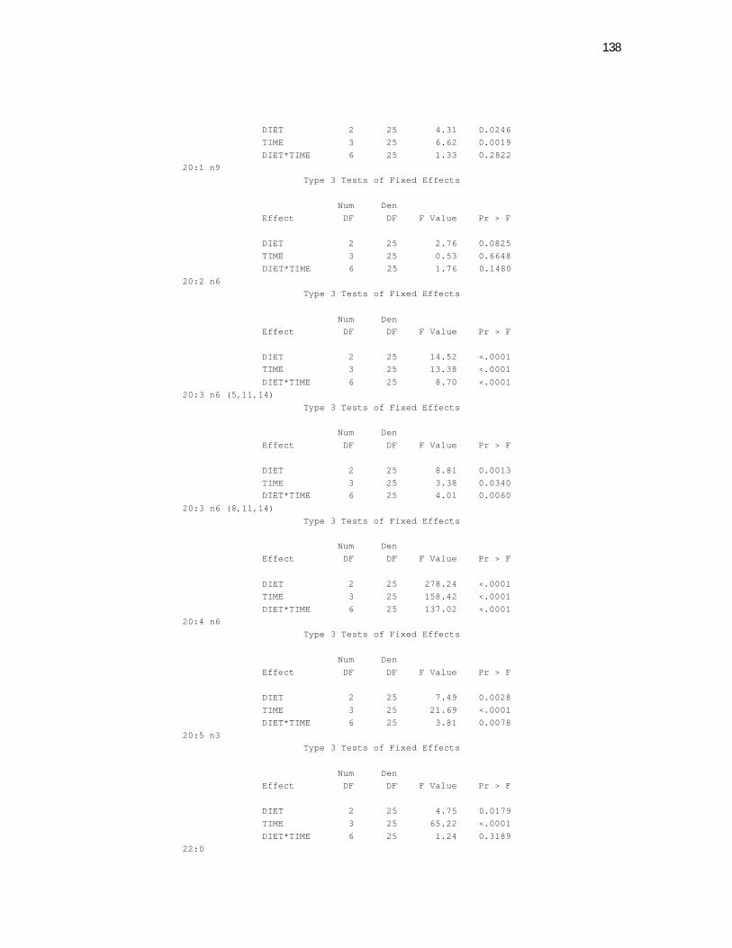
138
DIET 2 25 4.31 0.0246 TIME 3 25 6.62 0.0019 DIET*TIME 6 25 1.33 0.2822 20:1 n9
Type 3 Tests of Fixed Effects Num Den Effect DF DF F Value Pr > F DIET 2 25 2.76 0.0825 TIME 3 25 0.53 0.6648 DIET*TIME 6 25 1.76 0.1480 20:2 n6
Type 3 Tests of Fixed Effects Num Den Effect DF DF F Value Pr > F DIET 2 25 14.52 <.0001 TIME 3 25 13.38 <.0001 DIET*TIME 6 25 8.70 <.0001 20:3 n6 (5,11,14)
Type 3 Tests of Fixed Effects Num Den Effect DF DF F Value Pr > F DIET 2 25 8.81 0.0013 TIME 3 25 3.38 0.0340 DIET*TIME 6 25 4.01 0.0060 20:3 n6 (8,11,14)
Type 3 Tests of Fixed Effects Num Den Effect DF DF F Value Pr > F DIET 2 25 278.24 <.0001 TIME 3 25 158.42 <.0001 DIET*TIME 6 25 137.02 <.0001 20:4 n6
Type 3 Tests of Fixed Effects Num Den Effect DF DF F Value Pr > F DIET 2 25 7.49 0.0028 TIME 3 25 21.69 <.0001 DIET*TIME 6 25 3.81 0.0078 20:5 n3
Type 3 Tests of Fixed Effects Num Den Effect DF DF F Value Pr > F DIET 2 25 4.75 0.0179 TIME 3 25 65.22 <.0001 DIET*TIME 6 25 1.24 0.3189 22:0

139
Type 3 Tests of Fixed Effects Num Den Effect DF DF F Value Pr > F DIET 2 25 2.07 0.1477 TIME 3 73 6.70 0.0005 DIET*TIME 6 73 0.32 0.9239 22:4 n6 + 22:5 n3
Type 3 Tests of Fixed Effects Num Den Effect DF DF F Value Pr > F DIET 2 25 0.81 0.4562 TIME 3 25 59.05 <.0001 DIET*TIME 6 25 0.79 0.5880 22:6 n3 + 24:0
Type 3 Tests of Fixed Effects Num Den Effect DF DF F Value Pr > F DIET 2 25 0.97 0.3927 TIME 3 25 21.31 <.0001 DIET*TIME 6 25 0.76 0.6069 24:1 n9
Type 3 Tests of Fixed Effects Num Den Effect DF DF F Value Pr > F DIET 2 25 5.51 0.0104 TIME 3 25 16.12 <.0001 DIET*TIME 6 25 0.94 0.4819 Sat
Type 3 Tests of Fixed Effects Num Den Effect DF DF F Value Pr > F DIET 2 25 1.64 0.2140 TIME 3 25 7.05 0.0014 DIET*TIME 6 25 0.94 0.4837 MUFAS
Type 3 Tests of Fixed Effects Num Den Effect DF DF F Value Pr > F DIET 2 25 6.53 0.0052 TIME 3 25 32.36 <.0001 DIET*TIME 6 25 1.39 0.2583 PUFAS
Type 3 Tests of Fixed Effects Num Den Effect DF DF F Value Pr > F
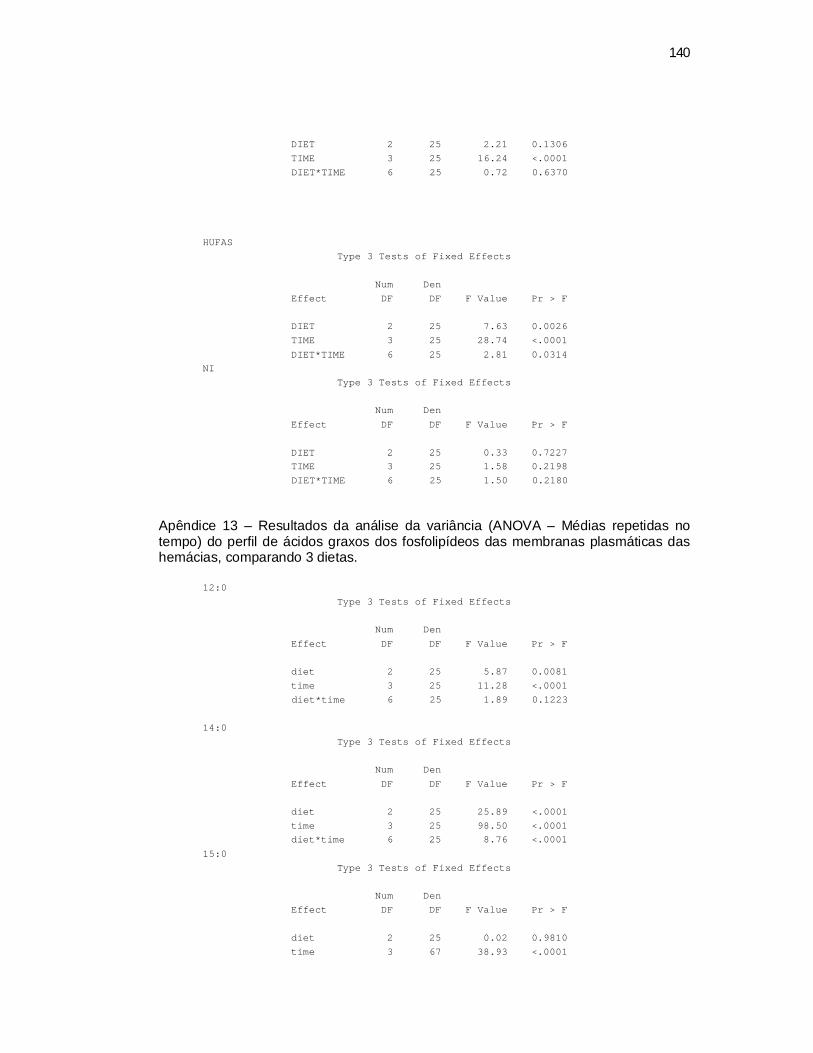
140
DIET 2 25 2.21 0.1306 TIME 3 25 16.24 <.0001 DIET*TIME 6 25 0.72 0.6370 HUFAS
Type 3 Tests of Fixed Effects Num Den Effect DF DF F Value Pr > F DIET 2 25 7.63 0.0026 TIME 3 25 28.74 <.0001 DIET*TIME 6 25 2.81 0.0314 NI
Type 3 Tests of Fixed Effects Num Den Effect DF DF F Value Pr > F DIET 2 25 0.33 0.7227 TIME 3 25 1.58 0.2198 DIET*TIME 6 25 1.50 0.2180
Apêndice 13 – Resultados da análise da variância (ANOVA – Médias repetidas no tempo) do perfil de ácidos graxos dos fosfolipídeos das membranas plasmáticas das hemácias, comparando 3 dietas.
12:0
Type 3 Tests of Fixed Effects Num Den Effect DF DF F Value Pr > F diet 2 25 5.87 0.0081 time 3 25 11.28 <.0001 diet*time 6 25 1.89 0.1223 14:0
Type 3 Tests of Fixed Effects Num Den Effect DF DF F Value Pr > F diet 2 25 25.89 <.0001 time 3 25 98.50 <.0001 diet*time 6 25 8.76 <.0001 15:0
Type 3 Tests of Fixed Effects Num Den Effect DF DF F Value Pr > F diet 2 25 0.02 0.9810 time 3 67 38.93 <.0001
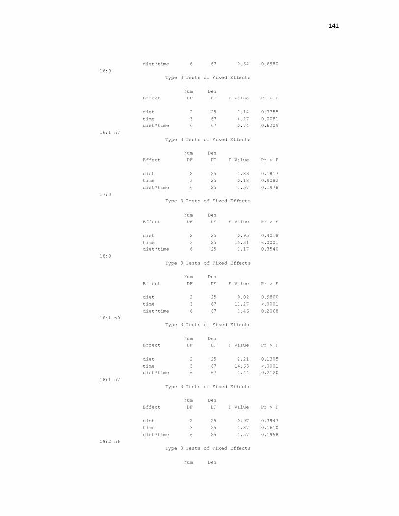
141
diet*time 6 67 0.64 0.6980 16:0
Type 3 Tests of Fixed Effects Num Den Effect DF DF F Value Pr > F diet 2 25 1.14 0.3355 time 3 67 4.27 0.0081 diet*time 6 67 0.74 0.6209 16:1 n7
Type 3 Tests of Fixed Effects Num Den Effect DF DF F Value Pr > F diet 2 25 1.83 0.1817 time 3 25 0.18 0.9082 diet*time 6 25 1.57 0.1978 17:0
Type 3 Tests of Fixed Effects Num Den Effect DF DF F Value Pr > F diet 2 25 0.95 0.4018 time 3 25 15.31 <.0001 diet*time 6 25 1.17 0.3540 18:0
Type 3 Tests of Fixed Effects Num Den Effect DF DF F Value Pr > F diet 2 25 0.02 0.9800 time 3 67 11.27 <.0001 diet*time 6 67 1.46 0.2068 18:1 n9
Type 3 Tests of Fixed Effects Num Den Effect DF DF F Value Pr > F diet 2 25 2.21 0.1305 time 3 67 16.63 <.0001 diet*time 6 67 1.44 0.2120 18:1 n7
Type 3 Tests of Fixed Effects Num Den Effect DF DF F Value Pr > F diet 2 25 0.97 0.3947 time 3 25 1.87 0.1610 diet*time 6 25 1.57 0.1958 18:2 n6
Type 3 Tests of Fixed Effects Num Den
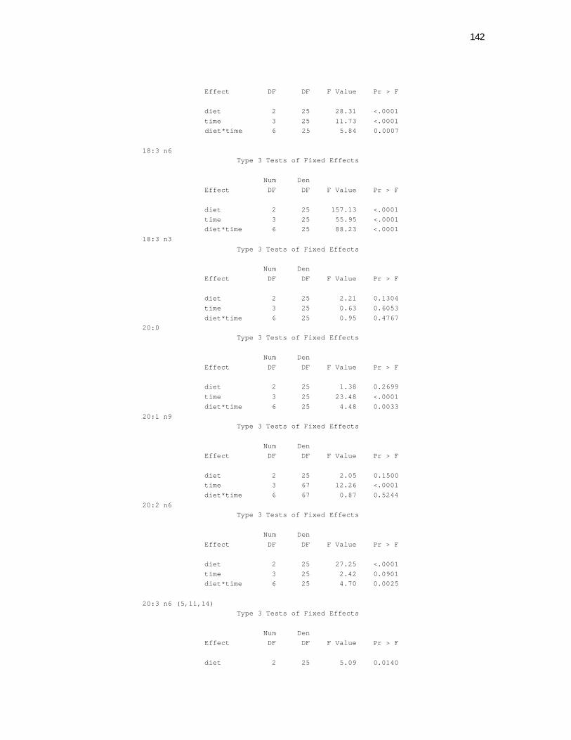
142
Effect DF DF F Value Pr > F diet 2 25 28.31 <.0001 time 3 25 11.73 <.0001 diet*time 6 25 5.84 0.0007 18:3 n6
Type 3 Tests of Fixed Effects Num Den Effect DF DF F Value Pr > F diet 2 25 157.13 <.0001 time 3 25 55.95 <.0001 diet*time 6 25 88.23 <.0001 18:3 n3
Type 3 Tests of Fixed Effects Num Den Effect DF DF F Value Pr > F diet 2 25 2.21 0.1304 time 3 25 0.63 0.6053 diet*time 6 25 0.95 0.4767 20:0
Type 3 Tests of Fixed Effects Num Den Effect DF DF F Value Pr > F diet 2 25 1.38 0.2699 time 3 25 23.48 <.0001 diet*time 6 25 4.48 0.0033 20:1 n9
Type 3 Tests of Fixed Effects Num Den Effect DF DF F Value Pr > F diet 2 25 2.05 0.1500 time 3 67 12.26 <.0001 diet*time 6 67 0.87 0.5244 20:2 n6
Type 3 Tests of Fixed Effects Num Den Effect DF DF F Value Pr > F diet 2 25 27.25 <.0001 time 3 25 2.42 0.0901 diet*time 6 25 4.70 0.0025 20:3 n6 (5,11,14)
Type 3 Tests of Fixed Effects Num Den Effect DF DF F Value Pr > F diet 2 25 5.09 0.0140
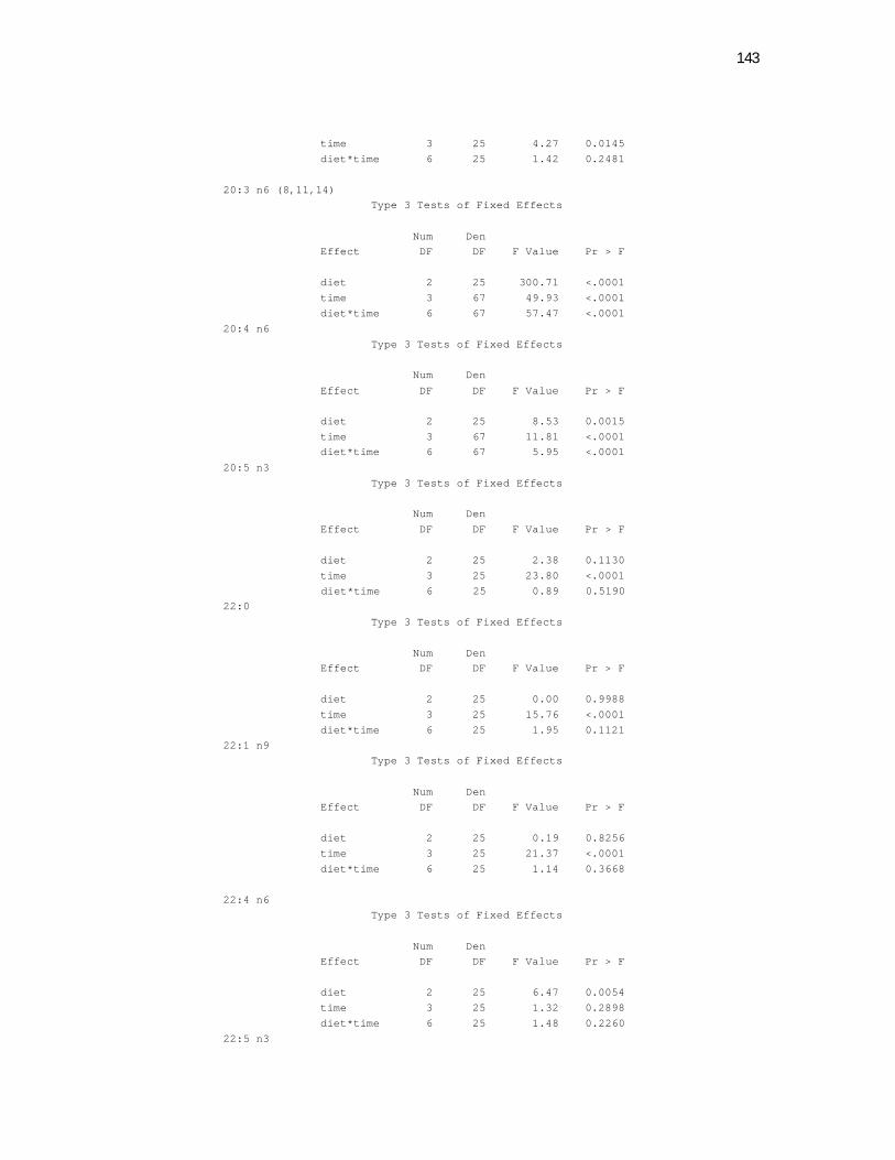
143
time 3 25 4.27 0.0145 diet*time 6 25 1.42 0.2481 20:3 n6 (8,11,14)
Type 3 Tests of Fixed Effects Num Den Effect DF DF F Value Pr > F diet 2 25 300.71 <.0001 time 3 67 49.93 <.0001 diet*time 6 67 57.47 <.0001 20:4 n6
Type 3 Tests of Fixed Effects Num Den Effect DF DF F Value Pr > F diet 2 25 8.53 0.0015 time 3 67 11.81 <.0001 diet*time 6 67 5.95 <.0001 20:5 n3
Type 3 Tests of Fixed Effects Num Den Effect DF DF F Value Pr > F diet 2 25 2.38 0.1130 time 3 25 23.80 <.0001 diet*time 6 25 0.89 0.5190 22:0
Type 3 Tests of Fixed Effects Num Den Effect DF DF F Value Pr > F diet 2 25 0.00 0.9988 time 3 25 15.76 <.0001 diet*time 6 25 1.95 0.1121 22:1 n9
Type 3 Tests of Fixed Effects Num Den Effect DF DF F Value Pr > F diet 2 25 0.19 0.8256 time 3 25 21.37 <.0001 diet*time 6 25 1.14 0.3668 22:4 n6
Type 3 Tests of Fixed Effects Num Den Effect DF DF F Value Pr > F diet 2 25 6.47 0.0054 time 3 25 1.32 0.2898 diet*time 6 25 1.48 0.2260 22:5 n3
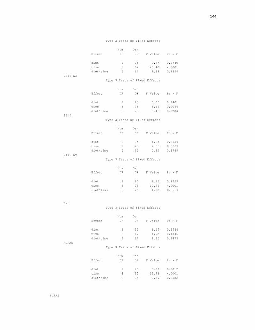
144
Type 3 Tests of Fixed Effects Num Den Effect DF DF F Value Pr > F diet 2 25 0.77 0.4740 time 3 67 20.48 <.0001 diet*time 6 67 1.38 0.2364 22:6 n3
Type 3 Tests of Fixed Effects Num Den Effect DF DF F Value Pr > F diet 2 25 0.06 0.9401 time 3 25 5.19 0.0064 diet*time 6 25 0.46 0.8284 24:0
Type 3 Tests of Fixed Effects Num Den Effect DF DF F Value Pr > F diet 2 25 1.63 0.2159 time 3 25 7.66 0.0009 diet*time 6 25 0.36 0.8948 24:1 n9
Type 3 Tests of Fixed Effects Num Den Effect DF DF F Value Pr > F diet 2 25 2.16 0.1369 time 3 25 12.76 <.0001 diet*time 6 25 1.08 0.3987 Sat
Type 3 Tests of Fixed Effects Num Den Effect DF DF F Value Pr > F diet 2 25 1.45 0.2544 time 3 67 1.92 0.1346 diet*time 6 67 1.35 0.2493 MUFAS
Type 3 Tests of Fixed Effects Num Den Effect DF DF F Value Pr > F diet 2 25 8.89 0.0012 time 3 25 22.94 <.0001 diet*time 6 25 2.39 0.0582 PUFAS
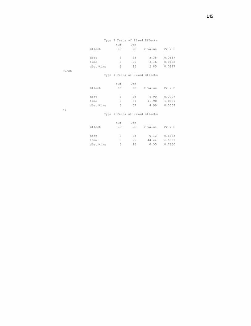
145
Type 3 Tests of Fixed Effects Num Den Effect DF DF F Value Pr > F diet 2 25 5.35 0.0117 time 3 25 3.16 0.0422 diet*time 6 25 2.85 0.0297 HUFAS
Type 3 Tests of Fixed Effects Num Den Effect DF DF F Value Pr > F diet 2 25 9.90 0.0007 time 3 67 11.90 <.0001 diet*time 6 67 4.99 0.0003 NI
Type 3 Tests of Fixed Effects Num Den Effect DF DF F Value Pr > F diet 2 25 0.12 0.8863 time 3 25 44.44 <.0001 diet*time 6 25 0.55 0.7660
146
Apêndice 14 – Normas para redação de artigos científicos para a submissão ao Journal of Nutrition JANUARY 2009 - NEW SAMPLE FILES ADDED TO ASSIST AUTHORS WITH MANUSCRIPT PREPARATION See links below under "Manuscript Preparation" to view sample files for a submitted manuscript, tables, figures, and an editorial checklist.
GUIDELINES, INCLUDING MANUSCRIPT WORD LIMIT - PLEASE READ CAREFULLY Manuscripts longer than 7500 words will be returned without review. Word count includes the abstract, all text and the references (but excludes tables.)The Journal is limited in the number of pages that can be published each year and article length is a consideration in the editorial process. Manuscript Preparation: Prepare your manuscript in Word 6.0 or later, savng the file in the .doc format (please note: the Word 2007 .docx file format is not accepted). Please consult the "Help" feature in Word for assistance with fonts, line numbering, etc. Times, Times Roman, Courier, Helvetica and Arial are the recommended text fonts. Please see section on Tables and Figures for information on figure fonts. For best quality conversions of special characters and symbols, use the Symbol font. Papers must be completely double-spaced. Papers must have consecutively numbered lines from the first line, first manuscript page throughout the last line, last manuscript page. Do not number the Literature Cited section. If you are uncertain about how to do this, please consult the ‘Help’ feature in Word. Figures and tables should be clearly labeled (Fig, 1, Fig 2, etc. or Table 1, Table 2,etc). Please refer to "Manuscript Digital Files" for information on electronic file requirements. Use only standard units of measure (SI - le Systeme Internationale d'Unites). Use only standard abbreviations. Use standard chemical and biochemical terms and follow ASN nomenclature policy. Include Conflict of Interest and Funding Disclosure footnotes. Manuscript submissions which are not formatted correctly are returned to authors. For a list of the most frequent reasons manuscripts are returned to authors, please see Returns to Authors Sample Files are available to assist you in the preparation of the paper: (1) The editorial checklist that is completed for manuscripts being returned to authors for revision or resubmission. (2) A JN manuscript with comments, and (3) Several JN tables and figures with comments. Your Manuscript should include:
A) Title Page
B) Abstract Page
C) Introduction
D) Materials and Methods

147
E) Results and Discussion
F) Literature Cited
G) Acknowledgement (if applicable)
A) TITLE PAGE: The title page must include: 1. The title should be composed as a single declarative statement. The title should be
focused on the results presented in the manuscript. Please do not use a colon or semicolon in the title. Please keep the title as generally applicable as possible. It usually is not necessary to include the exact study location or a specific study name in the title, as this information can be included in the abstract.
2. The names of all authors (first name, middle initial, last name) including their departmental and institutional addresses. Indicate which authors are associated with which institutions by numbered footnotes. Identify a corresponding author and provide a complete mailing address, telephone number, fax number, and email address. Please note that all authors’ names should appear on the manuscript exactly as they should appear in PubMed if the paper is published. ASN will not replace files to correct author names once published.
3. The word count for the entire manuscript (title through references). See word limit above.
4. The number of figures. 5. The number of tables. 6. Whether supplementary online material has been submitted. 7. A running title of 48 characters or less. 8. Footnotes to the title disclosing: (a) all sources of financial support; (b) all potential
conflicts of interest; (c) the existence of online supporting material, if appropriate (see section on Online Supporting Material). Conflict of Interest and Funding Disclosure: Any existing financial arrangements between an author and a company whose product figures prominently in the submitted manuscript or between the author and any company or organization sponsoring the research reported in the submitted manuscript should be brought to the attention of the Editor in the cover letter that accompanies the manuscript submission. In addition, all authors must declare all sources of funding for research reported in their manuscript and report all potential conflicts of interest in separate footnotes on the manuscript title page. If an author has no conflicts of interest, the footnote should list the author’s name, followed by “no conflicts of interest”. A conflict of interest includes, but is not limited to:
o Any existing financial or personal interests with a company whose product figures prominently in the submitted manuscript
o Any financial or personal interests with any company or organization sponsoring the research reported in the submitted manuscript
o Financial or personal interests include: a current grant, contract or subcontract, or consulting agreement with a company; employment with the company/organization; acting as an expert witness on behalf of a company/organization; holding stocks or shares in a company
o Coordinators of supplement publications should also report any financial and personal interests, as defined above, with sponsors of supplement publications. In addition, supplement coordinators should disclose receipt of compensation from sponsor for editorial services on manuscripts published in the supplement publication and/or for attending, speaking or organizing a meeting or symposium Individuals who are asked to review a manuscript should decline the solicitation if they have: (1) served as an adviser or advisee to an author on the manuscript (2) collaborated or served as a coauthor with an author of the manuscript during the past
148
3 years; (3) are currently affiliated with, were previously employed within the past 12 months by, or are being considered for employment at the institution of an author; (4) participated in a consulting/financial arrangement with an author in the past 3 years; or (5) are the spouse, child, sibling, parent, partner, or close friend, or otherwise have a relationship that might affect judgment, or could be seen as doing so by a reasonable person familiar with the relationship. B) ABSTRACT PAGE: The abstract must be a single paragraph of no more than 250 words summarizing the relevant problem addressed by the study and the theory or hypothesis that guided the research. The abstract should include the study design/methodology and clear statements of the results, conclusions and importance of the findings. C) INTRODUCTION: Background to the research conducted and specific objectives should be clearly indicated. This should not be a comprehensive review of the literature, however. D) MATERIALS AND METHODS: Documentation of methods and materials used should be sufficient to permit replication of the research. State the source of specialized materials, diets, chemicals, and instruments and other equipment, with model or catalog numbers, where appropriate. Specify kits, analyzers, and commercial laboratories used. Cite references for methods whenever possible and briefly explain any modifications made. HUMAN AND ANIMAL RESEARCH. Reports of human studies must include a statement that the protocol was approved by the appropriate institutional committee or that it complied with the Helsinki Declaration as revised in 1983. When preparing reports of randomized, clinical trials, authors should refer to the checklist published in the CONSORT Statement and should include a trial profile summarizing participant flow (2). Research on animals should include a statement that the protocol was approved by the appropriate committee or complied with the Guide for the Care and Use of Laboratory Animals (3). Describe how animals were killed. Describe control and experimental subjects giving age, weight, sex, race, and for animals, breed or strain. Include the supplier of experimental animals. DIETS. Composition of control and experimental diets must be presented. When a diet composition is published for the first time in The Journal of Nutrition, utilize a table or a footnote to provide complete information on all components. If previously described in The Journal of Nutrition or The American Journal of Clinical Nutrition, a literature citation may be used. State specifically any modifications made to the published diet compositions. The proximate composition of closed formula diets should be given as amounts of protein, energy, fat, and fiber. Components should be expressed as g/kg diet. Vitamin and mineral mixture compositions should be included using Journal of Nutrition units and nomenclature. For a discussion of the formulation of purified animal diets, refer to Baker (4) and to a series of ASN publications (5-8). STATISTICAL METHODS. Describe all statistical tests utilized and indicate the probability level (P) at which differences were considered significant. If data are presented in the text, state what they represent (e.g. means ± SEM). Indicate whether data were transformed before analysis. Specify any statistical computer programs used. Present the results of the statistical analysis of data in the body of each and on figures per se. Use letters or symbols to indicate significant differences; define these in a table footnote or the figure legend. Provide the appropriate statistics of variability. An estimate of the error variance (SD or SEM) of group means should be displayed in figures. Standard ANOVA methodology assumes a homogeneous variance. If error variance is
149
tested and found to be heterogeneous, data should be transformed before ANOVA, or nonparametric tests should be used. For a discussion of variability calculations and curve-fitting procedures, see Baker (4). E) RESULTS AND DISCUSSION Report the results of the study. Discuss the significance of the findings, interpret the results and conclusions. F) LITERATURE CITED The Journal of Nutrition reference format will be modified to be consistent with the International Committee of Medical Journal Editors (ICMJE) recommended format for bibliographic citations with the following exception: references should include the names of all authors, unless there are more than ten, in which case list the first nine plus "et al." There is no limit on the number of citations allowed; recent literature should be comprehensively cited. The list of references must begin on a new page and should include the heading "Literature Cited." Abbreviate journal names according to the National Library of Medicine (NLM) journal abbreviations list . References should be numbered consecutively in the order in which they are first mentioned in the text. References should be formatted according to the International Committee of Medical Journal Editors (ICMJE) recommended format for bibliographic citations with the following exception: references should include the names of all authors, unless there are more than ten, in which case list the first nine plus "et al." Personal communications, submitted manuscripts and unpublished data cannot be included in the Literature Cited section but should appear parenthetically in the text. Personal communications must be written and the affiliation of the person providing the communication indicated in the text. Articles accepted for publication but not published when final revisions are completed on the current article may be cited as "in press." References in tables and figures: References cited for the first time in tables or figure legends should be numbered in order, based on the placement of the table or figure in text. Identify references in text, tables, and legends for illustrations by arabic numbers in parentheses. See current print issues of The Journal of Nutrition for style. Make sure your Literature Cited section includes a recognized heading and that the heading is not set in all caps (use upper and lower case letters, as shown below). Recognized headings include the following: References Reference List Literature Cited References and further reading Bibliography Literature G) ACKNOWLEDGMENTS. Technical assistance and advice may be acknowledged in a section at the end of the text. Only named individuals should be included in this section. Authors are responsible for obtaining written permission from everyone acknowledged by name and for providing copies of signed permission statements to The Journal of Nutrition. These statements should be submitted to The Journal along with the manuscript Authors' Statement/Copyright Release Form.
UNITS OF MEASURE. Most measurements must conform to le Systeme Internationale d'Unites (SI) (9). The metric system and the Celsius scale (º C) must be used. Concentrations should be expressed on a molar basis. Except for diet composition, convert to substance concentration, e.g., mol/L. The denominator should be L. Do not use M, mM, N, etc. Use one of three acceptable options to express measurements. (a) Use SI units exclusively. (b) Use SI units and, if appropriate, provide conventional units parenthetically in the text and give conversion factors in table footnotes and figure legends. (c) Use conventional units, if appropriate, and provide SI units parenthetically in
150
the text and give conversion factors in table footnotes and figure legends. Units should not be pluralized. Useful websites are: SI conversion website: http://www.ex.ac.uk/cimt/dictunit/dictunit.htm Clinical SI conversions: http://www.unc.edu/~rowlett/units/scales/clinical_data.html Clinical SI conversions: http://dwjay.tripod.com/conversion.html ABBREVIATIONS. Use only standard abbreviations. Table 2 is an abridged list of abbreviations that may be used without definition in Journal of Nutrition articles. Other standard abbreviations are listed in Scientific Style and Format (1). If there are three or more abbreviations defined in the text, define each the first time it is used in the text and prepare an abbreviation footnote. The footnote should be associated with the first abbreviated term in the text and should be an alphabetized listing of all author-defined abbreviations and their definitions. Abbreviations should not be followed by a period and should not be pluralized (e.g. AA should represent both "amino acid" and "amino acids"). Use the verb (e.g."is" or "are") that is consistent with the context in which the abbreviation is used in the sentence. Units and statistical terms also should not be followed by a period or pluralized. Use the standard abbreviations for SI prefixes found in Young (9) and in Table 3 and those for units of measure in Table 4. Abbreviations used only in tables and figures must be separately defined in the footnotes or legend for each table or figure. Abbreviations that are in the abbreviation footnote should not be redefined in table footnotes or figure legends. NOMENCLATURE. Chemical and biochemical terms and abbreviations and identification of enzymes must conform to the recommended usage of the International Union of Biochemistry and Molecular Biology (10). Names for vitamins, related compounds, and abbreviations for amino acids should follow the ASN nomenclature policy (11,12). Apêndice 15 – Normas para redação de artigos científicos para a submissão ao American Journal of veterinary research
Journals > I nst ruct ions for JAVMA aut hors
Instructions for Journal of the American Veterinary Medical Association authors
The mission of the Journal of the American Veterinary Medical Association is to promote the science and art of veterinary medicine and to provide a forum for discussion and dissemination of ideas important to the profession.
Keywords for Authors and Reviewers Copyright Transfer Agreement Statement on Prior Publication, Concurrent Submission, Authorship, and
Funding (PDF) Guidelines for Preparation of Scientific Abstracts AVMA Manuscript Central
151
Editorial policies The Journal of the American Veterinary Medical Association is a peer-reviewed general veterinary medical journal that publishes manuscripts dealing with any subject germane to the practice of veterinary medicine. For scientific manuscripts, preference will be accorded to those that have clinical or practical value. Authors who submit manuscripts to the journal should carefully read these Instructions for Authors when preparing their manuscripts, because compliance with these instructions will help reduce delays in manuscript processing. Authors submitting manuscripts for publication in specific features of the journal should also read recent issues of the journal for examples of how such reports and features are typically organized. Authors who have additional questions are encouraged to consult with an AVMA editor prior to manuscript submission. A manuscript is received with the understanding that it and all revisions have been approved by all authors and that neither the manuscript nor any of its parts has been published, except as an abstract less than 250 words long, or is under concurrent consideration by any other publication. The corresponding author must provide a signed statement to this effect. A manuscript containing information published in any compiled printed (eg, journals, symposia, proceedings, newsletters, books) or electronic (eg, Web sites, CD-ROMs, DVDs) format will be rejected on the grounds of prior publication. Publication of abstracts less than 250 words long does not constitute prior publication; however, publication of longer abstracts may. At the time of manuscript submission, the corresponding author must include copies of any abstracts of the manuscript that have been published or submitted for publication or that are expected to be submitted for publication. Authors preparing abstracts for publication in a proceedings are encouraged to review the Guidelines for Preparation of Scientific Abstracts. Readers who submit letters to the editor must limit them to 500 words (longer letters will be condensed as needed) and 6 references. All letters are subject to editing. The Journal of the American Veterinary Medical Association is covered by copyright. All authors will be required to sign a written statement transferring copyright to the AVMA prior to publication of any manuscript or letter. Requests to copy, reprint, or use portions of published material (including information in figures and tables) should be addressed to the editor-in-chief. Manuscript submission Manuscripts should be submitted online at http://mc.manuscriptcentral.com/avma. Manuscripts may also be submitted by conventional mail, but online submission is recommended to expedite processing of manuscripts. Online manuscript submission — For online submission, manuscripts must be in Microsoft Word format (.doc) or rich text format (.rtf). The manuscript (including footnotes, references, figure legends, and tables) must be double-space typed, using 12-point Times New Roman font, 1-inch margins, and left justification. Manuscripts should be arranged as follows: title page, structured abstract (when applicable), text, footnotes, references, figure legends, and tables. The title page must include the title and the first name, middle initial, and last name of each author, along with each author's professional degree, highest earned academic degree, and diplomate status (for authors who are diplomates of AVMA-recognized specialty organizations). Professional affiliations of the authors at the time of the study should be indicated. If an author's affiliation has changed since the study was performed, the author's new affiliation should be identified. If information in the text has been presented at a scientific meeting, this should be indicated on the title page. Acknowledgments, sources of funding, and the name of the corresponding author should also be included on the title page. Software programs that automatically create endnotes, footnotes, and references should not be used.
152
Each line and page of the manuscript must be numbered. Tables should be included at the end of the manuscript in the same electronic file; however, if necessary, they can be saved as separate files. All figures should be saved as separate electronic files; figures should not be embedded in the manuscript. Simple figures such as line drawings, bar graphs, and line graphs prepared in Excel should be saved as Excel files (.xls). Line drawings and graphs that were not prepared in Excel should be submitted as .TIF files; however, .JPG, .GIF, .EPS, and .BMP files are also acceptable. Figures created with software programs that use proprietary graphic formats (eg, SigmaPlot, Statistix) cannot be used; most such software programs have the capability to save figures in one of the aforementioned formats. Minimum resolution for line drawings and charts is 1,000 dots per inch. Images (eg, photographs, photomicrographs, and radiographs) that are not available in a digital format should be scanned on a flatbed scanner at a resolution of at least 300 dots per inch. Files should be saved as .TIF files; however, .JPG, .GIF, .EPS, and .BMP files are also acceptable. Color figures should be submitted in CMYK, rather than RGB, format to prevent color shift during production. Once electronic files of the manuscript and all of its parts have been prepared, log on to AVMA Manuscript Central at http://mc.manuscriptcentral.com/avma. If you already have an account with the system, login with your user id and password, click on "Author Center," and select "Submit First Draft of a New Manuscript." Follow the instructions for submitting your manuscript. After submitting your manuscript, please check that your User Information (including mailing address, telephone and fax numbers, and e-mail address) is current. If you do not have an account with the system, click on "create a new account." Fill in all fields carefully; all fields in bold are required. The corresponding author is also responsible for submitting required supplementary materials, including a completed Copyright Transfer Agreement signed by all authors; a copy of the Statement on Prior Publication, Concurrent Submission, Authorship, and Funding (PDF) signed by the corresponding author; copies of any references listed as "in press" or "submitted"; copies of any abstracts containing information from the manuscript that have been published or submitted for publication; and a copy of the signed permission form from the copyright holder if the manuscript contains any tables or illustrations that have been published previously. This supplementary material may be submitted electronically (eg, by scanning and uploading with the manuscript or by uploading the electronic file) or by fax. Manuscripts will not be considered for publication until both the Copyright Transfer Agreement and Statement on Prior Publication have been received. During submission of your manuscript, you will be requested to supply up to 5 keywords for the manuscript. Lists of the keywords that can be used are available on the AVMA Web site. Conventional paper submission — Manuscripts should be prepared as described for online submission and mailed to JAVMA, American Veterinary Medical Association, 1931 N Meacham Rd, Suite 100, Schaumburg, IL 60173-4360. Three hard copies of the manuscript and three high-quality hard copies of each figure must be submitted, along with electronic copies of the manuscript and figures on a 3.5-in PC-formatted disk. Electronic files should be saved as Microsoft Word documents; electronic figures should be prepared as described for online manuscript submission. Manuscripts must be accompanied by a cover letter from the corresponding author that includes his or her mailing address, telephone and fax numbers, and e-mail address. Corresponding authors are also responsible for submitting required supplementary material, including a completed Copyright Transfer Agreement signed by all authors; a copy of the Statement on Prior Publication, Concurrent Submission, Authorship, and Funding signed by the corresponding author; three hard copies of any reference listed as "in press" or "submitted"; copies of any abstracts containing information from the manuscript that have been
153
published or submitted for publication; and written permission from the copyright holder if the manuscript contains any tables or illustrations that have been published previously. Manuscripts will not be considered for publication until both the Copyright Transfer Agreement and Statement on Prior Publication have been received. Authorship Individuals should be listed as authors only if they 1) made a substantial contribution to the conception and design of the study, the acquisition of the data used in the study, or the analysis and interpretation of that data; 2) were involved in drafting or revising the manuscript critically for important intellectual content; and 3) will have an opportunity to approve subsequent revisions of the manuscript, including the version to be published. All three conditions must be met. Each individual listed as an author must have participated sufficiently to take public responsibility for the work. Acquisition of funding, collection of data, or general supervision of the research team does not, alone, justify authorship. At least one author of a manuscript dealing with clinical interpretations or treatments must be a veterinarian. For multi-institutional studies, the individual who headed the study should be listed as an author, along with individuals who provided assistance with pathologic studies (eg, review of gross and histologic specimens) and statistical analyses and any other individual who had a substantial impact on the study design or made a unique contribution to the study. Individuals who submitted case material should be listed as authors only if they contributed at least 10% of the cases included in the study; individuals who contributed less than 10% of the cases should be listed in the acknowledgments. Requests to list a working group or study group in the byline will be handled on a case-by-case basis. Acknowledgments Acknowledgments can be used to identify important specific contributions from individuals who do not qualify for authorship. In particular, individuals who have contributed intellectually to the study or report but whose contributions do not justify authorship may be named and their function or contribution described. In general, this includes individuals who provided technical assistance (eg, individuals who performed special tests or research) and individuals who provided assistance with statistical analyses. Acknowledgments should not include individuals whose only contribution to the study or report involved the routine performance of their normal job duties and who did not offer any unusual intellectual contribution or technical expertise. The acknowledgments should not be used simply as a method of expressing gratitude to individuals who had a minor role in the study. Acknowledgments of nonspecific groups (eg, the intensive care unit technicians) and unidentifiable groups (eg, the anonymous contributors) are not allowed. Individuals named in the acknowledgments must have given their permission to the authors to be listed, because readers may infer their endorsement of the data and conclusions. Funding Authors are expected to acknowledge all sources of funding or financial support and to disclose to the editor any financial interests (including ownership, employment, consultancy arrangements, and service as an officer or board member) they have with companies that manufacture products that are the subject of their research or with companies that manufacture competing products. Humane animal care and use All research studies involving animals must have been performed in compliance with guidelines outlined in the Animal Welfare Act, US Public Health Service Policy on the Humane Care and Use of Laboratory Animals, NRC Guide for the Care and Use of Laboratory Animals, or Guide for the Care and Use of Agricultural Animals in Agricultural Research and Teaching (Fass 1999) or with equivalent guidelines. A manuscript containing information that suggests that animals were subjected to adverse, stressful, or harsh conditions or treatments will not be considered for publication unless the authors demonstrate convincingly that the knowledge
154
gained was of sufficient value to justify these conditions or treatments. Manuscripts describing studies that involved the use of animals, including studies that involved the use of privately owned animals (eg, animals owned by clients, staff members, students, or private entities), must include a statement that the study protocol was reviewed and approved by an appropriate oversight entity (eg, an animal care and use committee or institutional review board) or was performed in compliance with institutional guidelines for research on animals. Manuscripts describing prospective studies that involved privately owned animals must include a statement indicating that owner consent was obtained. If animals were euthanatized, the method of euthanasia must be indicated. Methods of euthanasia must comply with the AVMA Guidelines on Euthanasia (PDF). Style For questions of style, refer to the latest edition of the American Medical Association Manual of Style. For anatomic terms, use anglicized versions of official terms listed in the Nomina Anatomica Veterinaria. Refer to the latest editions of the American Drug Index and USP Dictionary of USAN and International Drug Names for proper spelling of chemical and drug names and to the latest edition of Dorland's Illustrated Medical Dictionary for proper spelling and use of medical terms. Refer to Bergey's Manual of Determinative Microbiology for spelling and correct taxonomic classifications of microorganisms. Use of abbreviations should be kept to a minimum. Except for standard abbreviations and units of measure, all abbreviations used 3 or more times in the text, figures, and tables of a manuscript should be listed at the beginning of the manuscript, along with their definitions. These abbreviations should then be used without expansion. Abbreviations that appear only in the figures or tables should be defined in the table or figure legend. Abbreviations should not be used to begin a sentence. Except for the abbreviations ELISA, ACTH, EDTA, DNA, and RNA, abbreviations should not be used in titles. In addition, abbreviations in the structured abstract must be expanded at first mention, with the abbreviation given in parentheses after first mention of the expanded term. Products, equipment, and drugs should be identified by chemical or generic names or descriptions. A trade name may be included in a lettered footnote if that specific product, equipment, or drug was essential for the outcome. A manuscript reporting results of a study that involved evaluation of the efficacy or safety of a pharmaceutical, biologic, or other product or in which such products were relevant to the diagnosis, treatment, or outcome will be considered only if the product is commercially available in the United States and can legally be used in the species of interest. Body weights and temperatures must be reported in metric and traditional US (lb, oF) units. Dosages must be given on a mg/kg and mg/lb of body weight basis. All dosages must include route of administration and interval (eg, 10 mg/kg [4.5 mg/lb], IV, q 12 h). Categories of manuscripts Authors may submit manuscripts for publication in the Views, Veterinary Medicine Today, and Scientific Reports sections of the journal. The Views section is a forum for exchange of ideas and includes the Letters to the Editor and Commentaries. Letters to the Editor may not exceed 500 words and 6 references. Not all letters are published; all letters accepted for publication are subject to editing. Those pertaining to anything published in the JAVMA should be received within one month of the date of publication. Submission via e-mail ([email protected]) or fax (847-925-9329) is encouraged; authors should give their full contact information including address, daytime telephone number, fax number, and e-mail address. Commentaries can relate to any aspect of the veterinary medical profession and should be submitted online at http://mc.manuscriptcentral.com/avma.
155
The Veterinary Medicine Today section promotes continuing education through didactic exercises, case discussions, and updates on clinical topics. Not every feature is published in every issue. Authors who wish to contribute a manuscript to the following features should consult the instructions for those features. Instructions for Authors of Veterinary Medicine Today feature articles:
Anesthesia Case of the Month Animal Behavior Case of the Month Diagnostic Imaging in Veterinary Dental Practice ECG of the Month Pathology in Practice Theriogenology Question of the Month Timely Topics in Nutrition Topics in Drug Therapy Veterinary Medicine and the Law What Is Your Diagnosis? What Is Your Neurologic Diagnosis?
Authors who wish to contribute a manuscript to another feature in this section should refer to recent issues of the JAVMA that contain that feature for general format. The Scientific Reports section contains reports on important original research, clinical case reports, and concise reviews. A manuscript based on original research that involved animals with a naturally developing or experimentally induced disease or condition will be considered for publication as an Original Study. This includes a manuscript based on evaluation of case records accumulated during a specific period (ie, retrospective case series). A manuscript that describes features of 1 or more clinical cases will be considered as a Clinical Report. Reference Point articles are concise reviews concerning subject areas in which important advances have been made during the past 5 years and contain information that has, or will have, clinical application. Manuscript preparation With the exception of review articles, all manuscripts submitted to the Scientific Reports section must include a Structured Abstract of 250 or fewer words. For Original Studies, the structure abstract must be include the following headings: Objective, Design, Animals (or Sample Population), Procedures, Results, and Conclusions and Clinical Relevance. For Clinical Reports, the structured abstract must include the following headings: Case Description, Clinical Findings, Treatment and Outcome, and Clinical Relevance. The text for an Original Study is organized under the following headings: Introduction, Materials and Methods, Results, and Discussion. The Introduction should supply sufficient pertinent background information to allow readers to understand and interpret results. It must include the rationale for the study, the investigators' hypothesis, and a clear statement of the purpose of the study. The Materials and Methods section should describe the experimental design in sufficient detail to allow others to reproduce the study. A subsection detailing statistical methods used to summarize data and test hypotheses and the level of significance used for hypothesis testing should be provided. When citing software products, use a footnote to cite software (eg, PROC GLM, SAS Institute, Cary, NC) and a reference to cite a User's Guide (eg, SAS user's guide: statistics, version 5 edition. Cary, NC: SAS Institute Inc, 1985;page number). The Results section should provide data that are clearly and simply stated without discussion or conclusions. Tables and figures should be cited parenthetically. Authors of manuscripts reporting gene sequences should submit those sequences to an appropriate data bank. The Discussion section should focus on findings in the manuscript and should be brief, containing only discussion that is necessary for interpretation of findings. The Discussion should concentrate on what is known in animals, not what is known in humans. A retrospective case series must include a meaningful statement of purpose, clinically relevant
156
data, and clinically useful conclusions or interpretations derived directly from evaluation of the cases described. Except for rare conditions, retrospective case series should contain information on at least 10 animals and include appropriate statistical analyses. A Clinical Report begins with the signalment of the animal or animals, followed by a chronologic description of pertinent aspects of the diagnostic examination, treatment, and outcome, and ends with a brief discussion. When more than one animal is involved, a representative of the group should be described in detail; important differences among animals can be addressed separately. For reports in which there are 3 or fewer animals, pertinent abnormal findings should be summarized in the text. For 4 or more animals, one table that provides a summary of pertinent abnormal findings may be accommodated, provided that such findings are not repeated in the text. Footnotes Cite footnotes by superscript, lowercase letters in the order in which they appear in the text. List footnotes alphabetically just before the references. For products and equipment, provide complete information in the footnote, including manufacturer's name and location (ie, city, state, and country [if other than the United States]). Abstracts, personal communications, and theses should be cited as footnotes. References Authors bear primary responsibility for accuracy of all references. References must be limited to those that are necessary and must be cited in the text by superscript numbers in order of citation. Journal titles in the Reference section should be abbreviated in accordance with the National Library of Medicine and Index Medicus. For references with more than 3 authors, only the first 3 authors should be listed, followed by "et al." The following is the style used for common types of references: Article in journal 1. Lamont LA, Bulmer BJ, Sisson DD, et al. Doppler echocardiographic effects of medetomidine on dynamic left ventricular outflow tract obstruction in cats. J Am Vet Med Assoc 2002;221:1276-1281. Book chapter 2. Muir P, Johnson KA, Manley PA. Fractures of the pelvis. In: Birchard SJ, Sherding RG, eds. Saunders manual of small animal practice. 2nd ed. Philadelphia: WB Saunders Co, 2000;1126-1132. Proceedings 3. Moore MP, Bagley RS, Harrington ML, et al. Intracranial tumors, in Proceedings. 14th Annu Meet Vet Med Forum 1996;331-334. Electronic material 4. Animal and Plant Health Inspection Service Web site. Bovine spongiform encephalopathy (BSE). Available at: www.aphis.usda.gov/lpa/issues/bse/bse.html. Accessed Feb 18, 2003. Figures Limit figures to those that reduce or clarify the text. Text and symbols should be large enough that they will still be legible when the figure is reduced to one column width during publication. To ensure high-quality reproduction, symbols used in graphs should be limited to open and closed circles, triangles, and squares; axes should be labeled in Helvetica or Arial font. Keys to symbols may be placed in a small box inserted into the unused portion of graphs. Photomicrographs and electron micrographs must have an internal scale marker. To express magnification with an internal scale marker, divide the length of the marker by the original magnification. For figures that consist of multiple parts, individual parts of the figure should be identified by capital letters embedded in the figure, rather than by describing the location of the part in the legend (eg, top right).
157
For preparation of electronic copies of figures, please see the section on online manuscript submission. Hard copies of figures that are submitted must be identified on the back along the top margin with the first author's name, the figure number, and an arrow (or "top") indicating the top of the figure, taking care not to write in the area of interest. Radiographs and transparency slides will not be accepted for review or publication. Figure legends must be given at the end of the manuscript. Sufficient information should be included to allow the figure to be understood without reference to the text. When applicable, stains used for histologic sections should be indicated in the legend. Authors wishing to use any previously published figures must submit written permission from the copyright holder. Tables Submission of excessive tabular data is discouraged, and tables should be limited to those containing data important to understanding and interpreting results of the study. Authors will be asked to delete tables containing data that could be given more succinctly in the text. Do not use tables that focus on findings in individual animals. Authors wishing to use any previously published tables must submit written permission from the copyright holder. Peer-review process The JAVMA reserves the right to reject any manuscript. Manuscripts submitted to the Scientific Reports section are subject to peer review, as are didactic exercises, case discussions, reviews of clinical topics, and features sponsored by specialty colleges or academies submitted to the Veterinary Medicine Today section. Manuscripts are reviewed initially by an AVMA scientific editor. Those with insufficient priority for publication are rejected promptly. Manuscripts considered for publication are sent to a minimum of 2 experts for external peer review. Instructions provided for external reviewers are available on the AVMA Web site for authors' perusal. Identity of peer reviewers is kept confidential; identity of authors is not. Authors are expected to respond to reviewer comments and make appropriate revisions within 30 days. Revised manuscripts may be rereviewed. Manuscripts that pass peer review are accepted for publication provided that authors respond meaningfully to questions and concerns raised by an AVMA scientific editor. For manuscripts that are rejected, hard copies of the text and accompanying materials will not be returned to the authors. Sequence of publication The JAVMA is published twice a month. Manuscripts are processed for publication in the order that they pass peer review, except that manuscripts dealing with emerging or zoonotic diseases or biodefense are prepared for publication as soon as they pass peer review. Adherence to these instructions and expedient revision and return of manuscripts will minimize time from submission to publication. Version: October 26, 2007
AVMA Home | Privacy Notice | Terms of Use | About the AVMA | RSS feeds
AVMA Journals | JAVMA News | Discussion Groups | Professional Issues | Contact Us
American Veterinary Medical Association Copyright © 2009
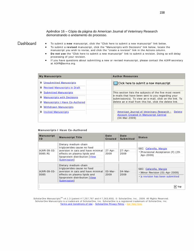
158
Apêndice 16 – Cópia da página do American Journal of Veterinary Research demonstrando o andamento do processo.
To submit a new manuscript, click the "Click here to submit a new manuscript" link below. To submit a revised manuscript, click the "Manuscripts with Decisions" link below, locate the
manuscript you wish to revise, and click the "create a revision" link in the Actions column. Do not use the "Click here to submit a new manuscript" link to submit a revision. Doing so will delay
processing of your revision. If you have questions about submitting a new or revised manuscript, please contact the AJVR secretary
My Manuscripts Author Resources
0 Unsubmitted Manuscripts
0 Revised Manuscripts in Draft
0 Submitted Manuscripts
0 Manuscripts with Decisions
2 Manuscripts I Have Co-Authored
0 Withdrawn Manuscripts
0 Invited Manuscripts
This section lists the subjects of the five most recent e-mails that have been sent to you regarding your submission(s). To view an e-mail, click on the link. To delete an e-mail from this list, click the delete link.
American Journal of Veterinary Research - Account Created in Manuscript Central (04-Mar-2009)
Delete
Manuscripts I Have Co-Authored
Manuscript ID Manuscript Title Date
Created Date Submitted Status
AJVR-09-03-0085.R1
Dietary medium-chain triglycerides cause no food aversion in cats and have minimal effects on plasma lipids and lipoprotein distribution [View Submission]
27-Apr-2009
27-Apr-2009
SEC: Cafarella, Margie Provisional Acceptance (F) (29-Apr-2009)
AJVR-09-03-0085
Dietary medium-chain triglycerides cause no food aversion in cats and have minimal effects on plasma lipids and lipoprotein distribution [View Submission]
03-Mar-2009
04-Mar-2009
SEC: Cafarella, Margie Minor Revision (01-Apr-2009) a revision has been submitted
ScholarOne ManuscriptsTM v4.1.3 (patent #7,257,767 and #7,263,655). © ScholarOne, Inc., 2009. All Rights Reserved.
ScholarOne Manuscripts is a trademark of ScholarOne, Inc. ScholarOne is a registered trademark of ScholarOne, Inc. Terms and Conditions of Use - ScholarOne Privacy Policy - Get Help Now
VITA
Luciano Trevizan, filho de Odorico Trevizan (in memorian) e de Maria Tittoni
Trevizan, nasceu em 02 de junho de 1976, Capão da Canoa, RS.
Estudou na Escola Estadual “Lourenço Leon von Langendonk”, em
Maquiné, na qual completou seu estudo de primeiro grau. Na Escola Estadual
Ilderfonso Simões Lopes, em Osório/RS, cursou o segundo grau. Em agosto de 1997,
ingressou no Curso de Medicina Veterinária da Universidade Federal do Rio Grande
do Sul (UFRGS), RS, graduando-se como Médico Veterinário em janeiro de 2003.
Em março de 2003, iniciou seu Curso de Mestrado no Programa de Pós-
Graduação em Zootecnia, da Faculdade de Agronomia, da UFRGS, na área de
concentração de nutrição de não-ruminantes, tendo trabalhado com nutrição de cães.
Em março de 2005, iniciou o curso de Doutorado, no mesmo Programa de
Pós-Graduação. Durante o período de doutorado foi aprovado no Programa de Bolsas
Sanduiche do CNPq. Em março de 2007, realizou parte do doutorado na Texas A&M
University, College Station, Texas, USA, encerrando este período em março de 2008.
Em fevereiro de 2009, defendeu a tese de doutorado referente ao metabolismo de
lipídeos em gatos e foi aprovado no concurso para Professor Substituto do
Departamento de Zootecnia da UFRGS.