UNIVERSIDADE DA BEIRA INTERIOR · controlada, a partir de modelos criados por desenho assistido por...
75
Transcript of UNIVERSIDADE DA BEIRA INTERIOR · controlada, a partir de modelos criados por desenho assistido por...

UNIVERSIDADE DA BEIRA INTERIOR Ciências da Saúde
Produção de Scaffolds de β-TCP/Alginato para uso
na Regeneração Óssea por Prototipagem Rápida
Gabriela Soares Diogo Carlos
Dissertação para obtenção do grau de mestre em
Ciências Biomédicas (2º ciclo de estudos)
Orientador: Professor Doutor Ilídio Joaquim Sobreira Correia.
Covilhã, junho 2013

iii

iv
List of publications
Torres, A.L. , Gaspar, V.M. , Serra, I.R. , Diogo, G.S. , Silva, A.P. and Correia, I.J. , Bioactive
Hybrid Polymeric-Ceramic 3D Scaffold for Improved Bone Tissue Regeneration. 2013,
submitted to Materials Science and Engineering: C.

v

vi
“Que os vossos esforços desafiem as impossibilidades, lembrai-vos de que as grandes coisas do
homem foram conquistadas do que parecia impossível”
Charles Chaplin

vii

viii
I would like to dedicate my master thesis to my parents…

ix

x
Acknowledgments
First, I would like to thank to my supervisor professor Ilídio Correia for the opportunity to
develop the theme of my master's thesis with him. For all the dedication and all the time
spent in carrying out the whole project.
To Eng. Ana Paula from the Optics center of Universidade da Beira Interior for helping in the
acquisition of the scanning electron microscopy images.
To Professor Abílio Silva for all availability to perform the mechanical characterization of the
scaffolds.
To all my fellow group colleges. One way or another all contributed to the development of my
work. Especially thank to Inês, one of the people who spent more time with me both inside
and outside the laboratory. For all the help and understanding throughout this year.
To a very special person who were always by my side, both at personal and professional level,
Paulo Machado.
Thank to all my special friends for all its contribution. To Ana Luisa Torres for all the help and
support.
A special thank to the most important people in my life, my parents and my siblings for all
the support, love, affection and attention. To them I owe my life and my happiness. Special
thanks to my best friend, my mother. What I am today, I owe you.

xi
xii
Abstract
The rise of bone defects in the last decades has become a worldwide problem. They can arise
from several causes such as tumors, trauma, infection, nutrition and bone diseases. This may
compromise the mechanical and biological functions of bone tissue. Autografts, allografts and
xenografts are some attractive alternatives used for bone tissue regeneration, however,
several factors such as high risk of infection, immunogenic response and lack of donor has
limited their use. In this context, Tissue Engineering appears as a promising solution. Tissue
Engineering is an interdisciplinary area that combines biomaterials and bioactive molecules to
promote the repair and regeneration of bone. Scaffolds are 3D matrices that act as temporary
templates, allowing cell adhesion and proliferation and providing mechanical support until
new bone tissue formation. A 3D scaffold success depends on their chemical, mechanical and
biological properties. Rapid prototyping technologies allow the production of 3D structures
with controlled architecture from models created by computer-aided design, through a layer-
by-layer process. The present study describes the characterization of the chemical,
mechanical and biological properties of βeta-Tricalcium phosphate/Alginate 3D scaffolds. The
scaffolds were characterized by Scanning Electron Microscopy, Fourier Transform Infrared
Spectroscopy, X-Ray Diffraction and Water Contact Angle. Porosity and Mechanical properties
(Compressive Strength and Young´s Modulus) were also analyzed. The cytotoxic profile was
evaluated by in vitro MTS assays, using human osteoblastic cells. Confocal Laser Scanning
Microscopy was also performed. The results obtained showed that the scaffolds produced by
the Rapid Prototyping technique have good chemical and biological properties, which is
fundamental for its application on bone tissue regeneration. Furthermore, it is concluded that
the scaffolds showed better mechanical and biological properties with the increase of the
percentage of βeta-Tricalcium phosphate within scaffolds.
Keywords
βeta-Tricalcium Phosphate/Alginate Scaffolds, Bone Regeneration, Computer-Aided Design,
Rapid Prototyping, Tissue Engineering.
xiii
Resumo
O aumento de defeitos ósseos, nas últimas décadas tem-se tornado um problema de saúde a
nível mundial. Estes defeitos têm causas, tais como tumores, traumatismos, infeções,
nutrição e doenças ósseas, e podem comprometer as funções mecânicas e biológicas do tecido
ósseo. Os autoenxertos, aloenxertos e xenoenxertos são algumas das alternativas utilizadas
para a regeneração dos defeitos do tecido ósseo. No entanto, vários factores, tais como, alto
risco de infeção, resposta imunogénica no hospedeiro e falta de doadores têm limitado o seu
uso. Neste contexto, apareceu a Engenharia de Tecidos como uma área interdisciplinar que
combina biomateriais com moléculas bioativas para promover a reparação e regeneração do
osso. Nesta área de investigação têm sido produzidas matrizes 3D (scaffolds) que funcionam
como modelos temporários, permitindo a adesão e proliferação celular, fornecendo suporte
mecânico até á formação de novo tecido ósseo. O sucesso de um scaffold depende das suas
propriedades químicas, mecânicas e biológicas. As tecnologias de prototipagem rápida
permitem a produção de estruturas 3D com arquitetura controlada, a partir de modelos
criados por desenho assistido por computador, através de um processo de camada por
camada. O presente estudo descreve a caracterização das propriedades químicas, mecânicas
e biológicas dos scaffolds 3D produzidos com βeta-Tricálcio fosfato/Alginato onde foram
usados vários rácios de concentrações de βeta-Tricálcio fosfato e alginato. Os scaffolds foram
caracterizados por Microscopia Eletrónica de Varrimento, Espectroscopia de Infravermelho por
Transformada de Fourier, Difração de Raios-X e Angulo de Contato com a Água. A Porosidade
e Propriedades Mecânicas foram também estudadas. O perfil citotóxico foi avaliado in vitro,
através do ensaio de MTS utilizando osteoblastos humanos. Microscopia Confocal Laser foi
também realizada de modo a observar a distribuição das células nos scaffolds. Os resultados
obtidos mostraram que os scaffolds 3D produzidos pela técnica de Prototipagem Rápida têm
boas propriedades químicas e biológicas, o que é fundamental para a sua aplicação na
regeneração do tecido ósseo. Além disso, concluiu-se que estas estruturas tridimensionais
apresentam melhores propriedades mecânicas e biológicas com o aumento de βeta-Tricálcio
fosfato.
Palavras-Chave
βeta - Tricálcio fosfato/Alginato Scaffolds, Desenho Assistido por Computador, Engenharia de
Tecidos, Prototipagem Rápida, Regeneração Óssea.
xiv
Resumo alargado
O osso é um tecido dinâmico e altamente vascularizado com capacidade de auto-regeneração,
responsável por muitas das funções do corpo humano. É constituído pela matriz orgânica
(maioritariamente colagénio), matriz inorgânica (hidroxiapatite), células (osteoblastos,
osteócitos e osteoclastos) e água. No entanto, vários factores, tais como, infeções, doenças e
nutrição conduzem á perda das suas funções, nomeadamente perda da capacidade natural de
auto-regeneração. Assim, o aumento de defeitos ósseos, nas últimas décadas tem-se tornado
um problema de saúde a nível mundial.
Os autoenxertos, aloenxertos e xenoenxertos são algumas das alternativas utilizadas para a
regeneração dos defeitos do tecido ósseo, contudo o elevado risco de resposta imunológica
por parte do paciente, risco de infeção e rejeição tem limitado o seu uso.
Neste contexto, apareceu a Engenharia de Tecidos como uma área promissora para a
resolução deste tipo de problemas. A Engenharia de Tecidos é uma área interdisciplinar que
combina biomateriais com moléculas bioativas para promover a reparação e regeneração do
osso. O primeiro objetivo da Engenharia de Tecidos passa pela alteração do conceito de
substituição pelo conceito de regeneração. Em que matrizes 3D (scaffolds) funcionam como
moldes temporários que se degradam á medida que nova matriz óssea é formada.
O sucesso de um scaffold depende de diversos parâmetros, tais como, biocompatibilidade,
biodegradabilidade, propriedades de superfície, porosidade e propriedades mecânicas, com o
objetivo de promover osteoindução, osteocondução e neovascularização.
Diversos materiais, como metais, polímeros e cerâmicas têm sido utilizados nesta área de
investigação. Além disso várias técnicas têm sido utilizadas para produção de estruturas 3D.
As tecnologias de prototipagem rápida permitem a produção de estruturas 3D com arquitetura
controlada, a partir de modelos criados por desenho assistido por computador, através de um
processo de camada por camada.
O presente estudo descreve a caraterização das propriedades químicas, mecânicas e
biológicas de scaffolds 3D produzidos com β-TCP/Alginato. Foram produzidos três tipos de
scaffolds diferentes, variando os rácios entre as concentrações de β-TCP e alginato, scaffolds
50/50 % (w/w), 30/70 % (w/w) e 20/80 % (w/w), respetivamente. Os scaffolds foram
produzidos pelo método de prototipagem rápida, recorrendo ao uso de uma impressora 3D
chamada Fab@home. O equipamento escolhido para a produção dos scaffolds aplica a técnica
de plotting, em que um material simples ou um compósito é extrudido formando os scaffolds.
Os scaffolds foram caraterizados por Microscopia Eletrónica de Varrimento, Espectroscopia de
Infravermelho por Transformada de Fourier, Difração de Raios-X e Angulo de Contato com a
água. Os resultados obtidos relativamente à caraterização físico-química dos scaffolds
permitem concluir que após todo o processo de produção estes não sofrem alterações na sua
estrutura cristalina. Além disso, estes apresentam um carater hidrofílico promovendo adesão
e proliferação celular. Um dos objetivos na produção de scaffolds é atingir um equilíbrio
xv
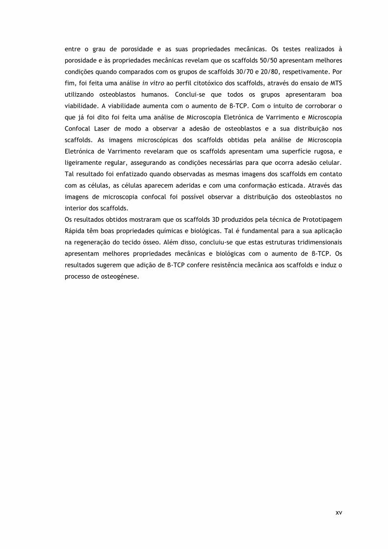
entre o grau de porosidade e as suas propriedades mecânicas. Os testes realizados à
porosidade e às propriedades mecânicas revelam que os scaffolds 50/50 apresentam melhores
condições quando comparados com os grupos de scaffolds 30/70 e 20/80, respetivamente. Por
fim, foi feita uma análise in vitro ao perfil citotóxico dos scaffolds, através do ensaio de MTS
utilizando osteoblastos humanos. Conclui-se que todos os grupos apresentaram boa
viabilidade. A viabilidade aumenta com o aumento de β-TCP. Com o intuito de corroborar o
que já foi dito foi feita uma análise de Microscopia Eletrónica de Varrimento e Microscopia
Confocal Laser de modo a observar a adesão de osteoblastos e a sua distribuição nos
scaffolds. As imagens microscópicas dos scaffolds obtidas pela análise de Microscopia
Eletrónica de Varrimento revelaram que os scaffolds apresentam uma superfície rugosa, e
ligeiramente regular, assegurando as condições necessárias para que ocorra adesão celular.
Tal resultado foi enfatizado quando observadas as mesmas imagens dos scaffolds em contato
com as células, as células aparecem aderidas e com uma conformação esticada. Através das
imagens de microscopia confocal foi possível observar a distribuição dos osteoblastos no
interior dos scaffolds.
Os resultados obtidos mostraram que os scaffolds 3D produzidos pela técnica de Prototipagem
Rápida têm boas propriedades químicas e biológicas. Tal é fundamental para a sua aplicação
na regeneração do tecido ósseo. Além disso, concluiu-se que estas estruturas tridimensionais
apresentam melhores propriedades mecânicas e biológicas com o aumento de β-TCP. Os
resultados sugerem que adição de β-TCP confere resistência mecânica aos scaffolds e induz o
processo de osteogénese.

xvi
xvii
Table of contents
Chapter I - Introduction ..................................................................................... 1
1. Introduction ................................................................................................. 2
1.1. Anatomy and Physiology of Bone................................................................... 2
1.1.1. Types of Bone .................................................................................... 3
1.1.2. Bone Cells ........................................................................................ 4
1.1.3. Bone Remodeling ................................................................................ 5
1.2. Bone Disorders ........................................................................................ 7
1.3. Bone Grafts ............................................................................................ 8
1.4. Bone Tissue Engineering ............................................................................. 9
1.4.1. The importance of 3D scaffolds for bone regeneration .................................. 9
1.4.2. Materials used in scaffolds production .................................................... 11
1.4.3. Production of 3D scaffolds by Rapid Prototyping technique .......................... 12
1.4.3.1. Fab@Home model for scaffolds production ........................................ 14
1.5. Aims ................................................................................................... 17
Chapter II - Materials and Methods ...................................................................... 18
2.1. Materials ................................................................................................. 19
2.2. Methods .................................................................................................. 19
2.2.1. Preparation of β-TCP/Alginate composite scaffolds by Rapid Prototyping. ........... 19
2.3. Morphological and Physicochemical characterization of scaffolds ......................... 20
2.3.1. Scanning Electron Microscopy (SEM) analysis ............................................ 20
2.3.2. Fourier Transform Infrared Spectroscopy (FTIR) analysis .............................. 21
2.3.3. X-Ray Diffraction (XRD) analysis............................................................ 21
2.3.4. Energy Dispersive Spectroscopy (EDS) analysis. ......................................... 21
2.4. Mechanical characterization of the β-TCP/Alginate composite scaffolds: Resistance to
Compression and Young’s Modulus. ................................................................... 21
2.5. Contact Angle Measurements ..................................................................... 22
2.6. Porosity evaluation ................................................................................. 22
2.7. Biological characterization of the β-TCP/Alginate composite scaffolds .................. 23
2.7.1. Seeding cell culture in the presence of β-TCP/Alginate scaffolds. .................. 23
xviii
2.7.2. Evaluation of cell viability in the presence of the scaffolds. ......................... 23
2.7.3. Analysis of 3D scaffolds biologic properties .............................................. 24
2.8. Statistical analysis .................................................................................. 24
Chapter III - Results and Discussion ..................................................................... 25
3.1. Morphology and Macroscopic properties of the scaffolds. ...................................... 26
3.2. Physicochemical characterization................................................................... 29
3.2.1. Fourier Transform Infrared Spectroscopy (FTIR)............................................ 29
3.2.2. X-Ray Diffraction (XRD) ......................................................................... 30
3.2.3. Energy Dispersive Spectroscopy (EDS) ........................................................ 32
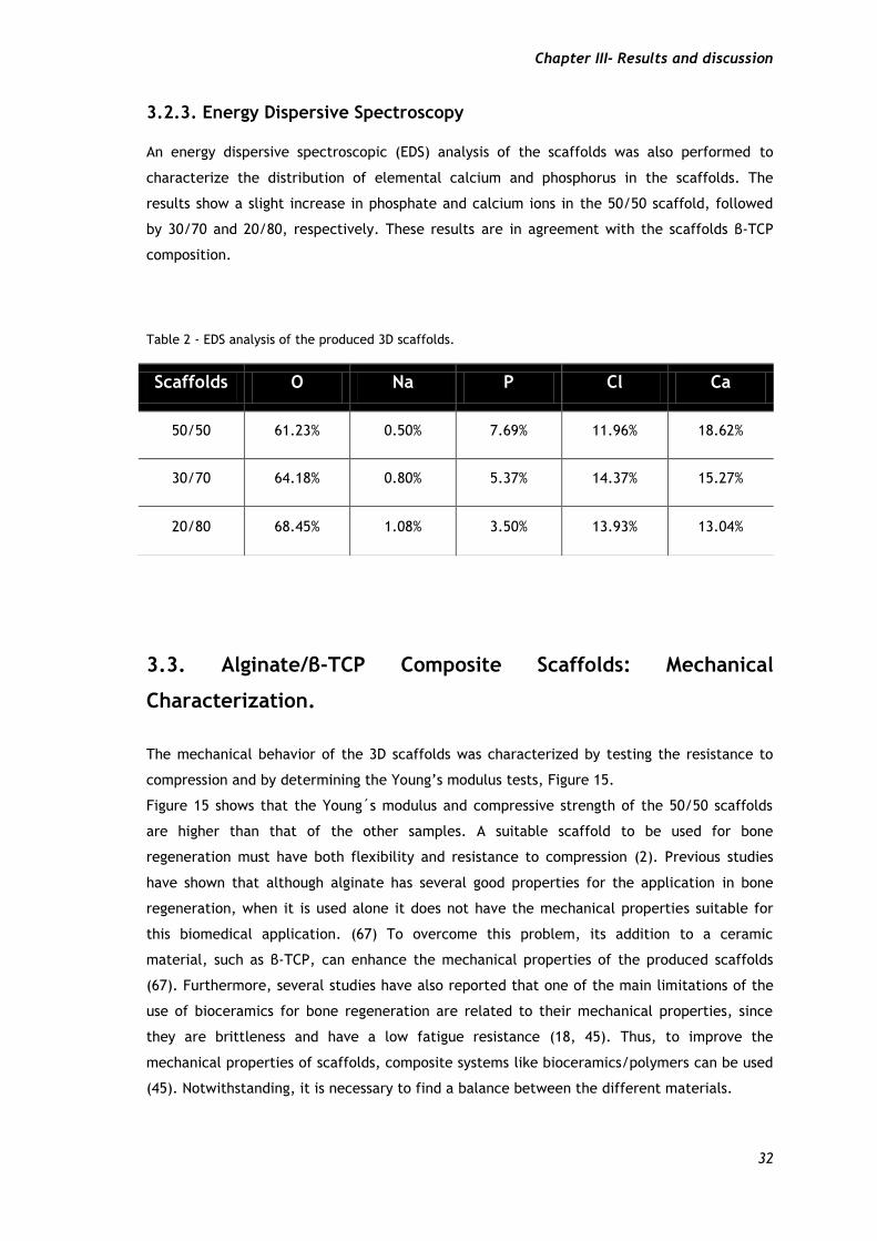
3.3. Alginate/β-TCP composite scaffolds: Mechanical characterization. .......................... 32
3.4. Determination of the Water Contact Angle ....................................................... 34
3.5. Determination of the Total Porosity of scaffolds ................................................. 34
3.6. Analysis of the Biological properties of the scaffolds ........................................... 36
3.6.1. Characterization of the Cytotoxic Profile of the scaffolds ............................... 36
Chapter IV – Conclusions and Future Perspectives .................................................. 40
4. Conclusions ................................................................................................ 41
Chapter V – Bibliography.................................................................................42
5. Bibliography ............................................................................................... 43

xix
xx
List of figures Figure 1 – Bone matrix components. ....................................................................... 2 Figure 2 – Representation of the types of bones. ........................................................ 3 Figure 3 - Representation of the internal structure and organization of bone ..................... 4 Figure 4 – Schematic representation of the bone. ....................................................... 5 Figure 5 - Schematic representation of the bone remodeling proces................................7 Figure 6 - Chemical structure of sodium alginate ...................................................... 12 Figure 7 - Rapid Prototyping technology applications ................................................. 13 Figure 8 - Representation of the various parameters used for the extrusion of the composite material. ....................................................................................................... 15 Figure 9 - Fab@home photograph used to produce 3D scaffolds for bone regeneration. ....... 15 Figure 10 – Images of the different scaffolds surface. ................................................. 27 Figure 11 – SEM images of the morphology of 50/50 scaffolds. ...................................... 28 Figure 12 - SEM images of pores sizes of 50/50 scaffolds and 20/80 scaffolds................... 28 Figure 13 – FTIR analysis of the powders and 3D scaffolds. ........................................... 29 Figure 14 - X-Ray spectra of the powders and 3D scaffolds........................................... 31 Figure 15 – Characterization of scaffolds mechanical properties........ ............................ 33 Figure 16 – Total porosity of scaffolds. ................................................................... 35 Figure 17- Macroscopic images of human osteoblasts cells in the presence of scaffolds ........ 36 Figure 18 – SEM micrographs of osteoblasts morphology in the presence of the scaffolds ...... 37 Figure 19 – Evaluation of human osteoblast cell viability . ........................................... 38 Figure 20 – CSLM images of osteoblasts in contact with the 50/50 scaffold.......................39

xxi

xxii
List of tables
Table 1- Composition of the β-TCP/ Aginate scaffolds produced...................................20
Table 2- EDS analysis of the produced 3D scaffolds...................................................32
Table 3- Contact Angles of the produced scaffolds...................................................34

xxiii
xxiv
Acronyms
3D Three-Dimensional
3DP Three-Dimensional Printing
β-TCP βeta-Tricalcium Phosphate
BMP Bone Morphogenetic Proteins
BSA Bovine Serum Albumin
BTE Bone Tissue Engineering
CaCl2 Calcium Chloride
CAD Computer-Aided Design
CAM Computer-Aided-Manufacturing
CLSM Confocal Laser Scanning Microscopy
CT Computerized Tomography
DMEM-F12 Dulbecco’s Modified Eagle’s Medium
ECM Extracellular Matrix
EDS Energy Dispersive Spectroscopy
EDTA Ethylenediaminetetraacetic Acid
EtOH Ethanol
FBS Fetal Bovine Serum
FDM Fused Deposition Modeling
FTIR Fourier-Transform Infrared Spectroscopy
H+ Hydrogen Ions
HA Hydroxyapatite
IL-1 Interleukin-1
MSC Mesenchymal Stem Cells
MTS 3-(4,5-dimethylthiazol-2-yl)-5-(3-carboxymethoxyphenyl)-2-(4-sulfophenyl)
-2H-tetrazolium
OB Osteoblasts
OC Osteoclasts
PBS Phosphate-Buffered Saline

xxv
PCL Poly (ε-caprolactone)
PLGA Poli-lactide-co-glico-lide
PLLA Poli(L-lactic-acid)
PI Propidium Iodide
PTH Paratiroid Hormone
PVA Poly(vinyl) Alcohol
RANKL NF-kappa B ligand
RT Room Temperature
SEM Scanning Electron Microscopy
SLA Stereolithography
SLS Selective Laser Sintering
STL Stereolithography
TE Tissue Engineering
TNF Tumor Necrosis Factor
Ts Resistance to Compression
XRD X-Ray Diffraction
YM Young’s Modulus

xxvi

Chapter I - Introduction
Chapter I- Introduction
2
1. Introduction
1. 1. Anatomy and Physiology of Bone
Bone tissue is responsible for the human body support and also by other different biological
functions (2-4). Mechanical functions include protection of vital organs and movement, since
bones are connected with muscles that when stimulated are responsible for moving the whole
body. Biologically, bone tissue is responsible for the production of blood cells, through a
process called hematopoiesis, and for the storage of ions, such as phosphorus and calcium (3).
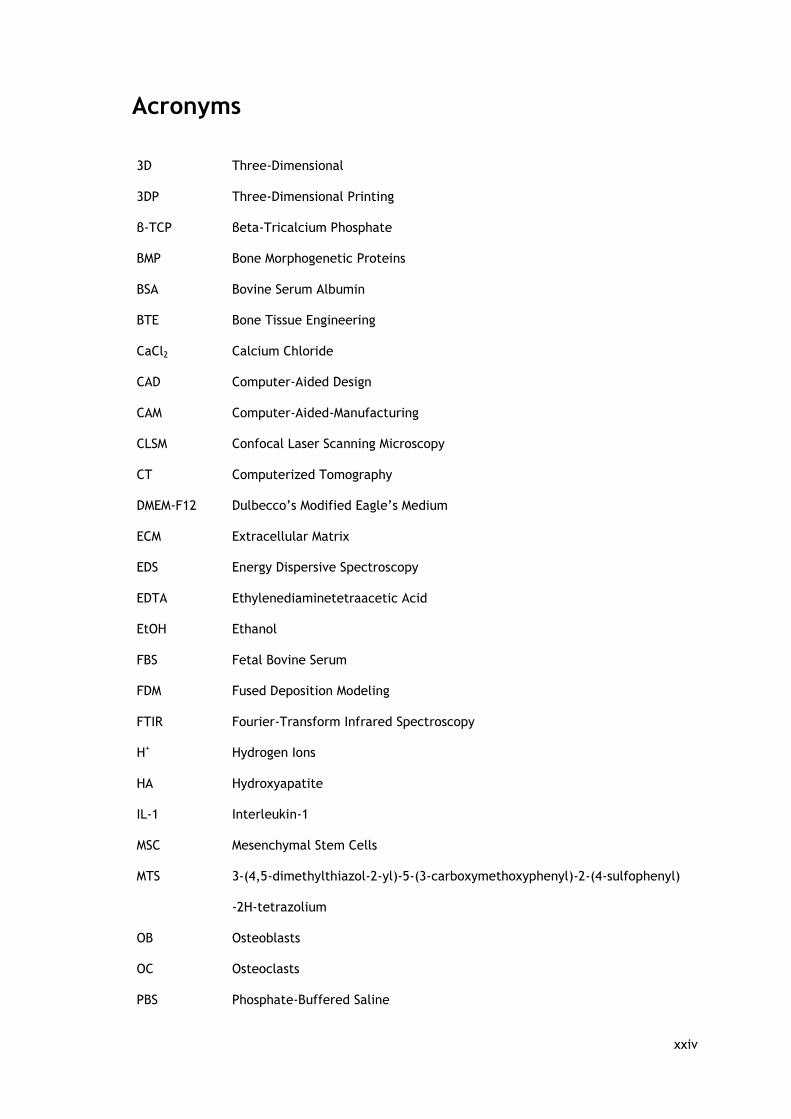
Bone is composed by cells, water and bone matrix. This matrix is formed by organic (35%) and
inorganic material (65%) (2, 3). The organic material is constituted by collagen and
proteoglycans, while crystals of calcium phosphate, concretely hydroxyapatite (HA) form the
inorganic part of this tissue. Both the organic and inorganic compounds of bone tissue are
important to maintain flexibility and resistance to compression, respectively (Figure 1a) (2). A
decrease in collagen composition contributes for increasing bone fragility and subsequently
this tissue can break easily, Figure 1c. On the contrary, if the quantity of bone mineral
decreases, bone will bend without breaking, as can be seen in Figure 1b (2).
Figure 1 - Bone matrix components. Normal bone a), demineralize bone, without hydroxyapatite b) and mineralized bone, without collagen c) (adapted from(2)).
There are different categories of bone cells, like osteoblasts (OB) (bone-forming cells),
osteocytes (bone maintaining cells) and osteoclasts (OC) (bone resorbing cells). Each cell type
has different origins and functions, but all of them are equally important to produce new
bone matrix (5). Nevertheless, the process of bone formation and regeneration depends on
the vascularization system for the transport of nutrients and oxygen, and also for the delivery
of circulating osteogenic factores and stem cells (6).
Chapter I- Introduction
3
1.1.1. Types of Bone
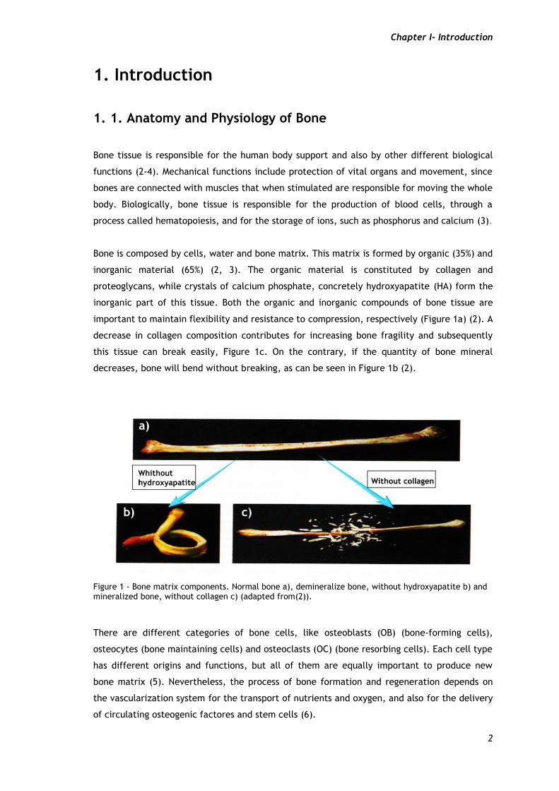
The human body has several types of bones that can be distinguished according to their
shape, surface features and bone matrix. Based on shape, bones can be grouped into four
categories: long bones, irregular bones, flat bones and short bones, as presented in Figure 2
(2, 3). Long bones, as their name suggests, have an elongated shape and represent the
majority of limb bones, except the patella, wrist and ankle, Figure 2a (3, 4). Irregular bones
are named due to their variety of shapes. They present many surface features for muscle or
articulation attachment. Examples of this type of bone are the vertebrae and hip bones,
Figure 2b (3, 4). Flat bones, have a broad surface that is important for muscle attachment or
organs protection, examples are the bones of the shoulder girdle, ribs and breastbone, Figure
2c (3, 4). Finally, Short bones have a roughly cube shape, for instance, the bones of the wrist
and ankle, Figure 2d (3, 4).
Figure 2 – Representation of the types of bones. According to the shape there are four types of bones: long bones (a), irregular bones (b), flat bones (c) and short bones (d) (adapted from (4)).
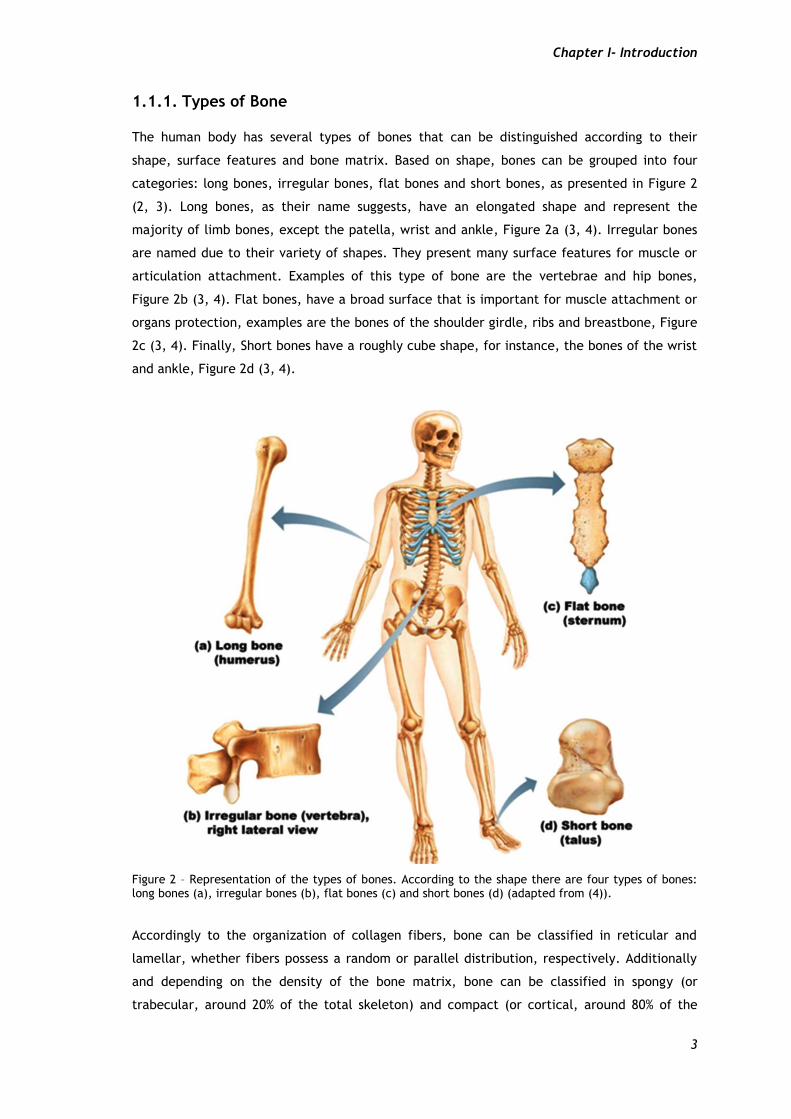
Accordingly to the organization of collagen fibers, bone can be classified in reticular and
lamellar, whether fibers possess a random or parallel distribution, respectively. Additionally
and depending on the density of the bone matrix, bone can be classified in spongy (or
trabecular, around 20% of the total skeleton) and compact (or cortical, around 80% of the
Chapter I- Introduction
4
total skeleton), as can be seen in Figure 3 (7). The spongy bone has a high porosity (around
50-90%), lower density and lower compressive strength, when compared with the compact
type (5, 7, 8).
Figure 3 - Representation of the internal structure and organization of bone (adapted from (4)).
1.1.2. Bone Cells
Bone cells, namely OB and OC, have different origins and are responsible for the production
and resorption of bone, respectively (9).
OB are mononuclear cells that derive from mesenchymal stem cells (MSC) and are present at
bone surfaces (10). These cells present a spherical nucleus, a basophilic cytoplasm, a very
developed endoplasmic reticulum and also a large number of ribosomes (11). Structurally they
have a cuboid shape or sometimes a slightly elongated appearance, as can be seen in Figure
4. Functionally, OB play an important role in the production of organic matrix, and on its
mineralization, being responsible for the production of collagen type I, osteocalcin,
osteopontin, osteonectin, proteoglycans and cytokines (8, 11). Furthermore, OB release high
levels of alkaline phosphatase, which is normally present on the OB cytoplasmatic membrane,
to promote bone mineralization (11).
Moreover, OB possess vesicles that contain phosphate, calcium ions and enzymes, that are
released by exocytose and are responsible by hydroxyapatite crystals formation (2). On the
other hand, alkaline phosphate and noncollagenous proteins, such as, osteocalcin,
osteopontin and bone sialoprotein are important for matrix maturation. It is believed that
these calcium and phosphate-binding proteins help in the deposition of mineral by regulation
of hydroxyapatite crystals (12). Furthermore, OB also plays an important role in bone

Chapter I- Introduction
5
resorption. They promote the contact between OC and the mineralized matrix, followed by
the process of re-mineralization (2).
While OB are producing new bone, they became surrounded by bone matrix, leading to their
differentiation into osteocytes (2).
Osteocytes are mature cells that have the ability to maintain the bone matrix integrity (2).
Osteocytes fill the gaps in the bone matrix and are responsible for intercellular
communication (13). They possess cytoplasmatic prolongations called filopodia located in
spaces or channels in the bone matrix (13). The arrangement of the canaliculi (Figure 4),
allows the passage of nutrients and oxygen between the blood vessels and distant osteocytes
(13). Osteocytes are also responsible for osteocytic osteolysis, breaking down the bone matrix
for the release of calcium for calcium homeostasis (13).
OC are multinucleated, polymorphic irregularly shaped giant cells, that derive from the
hematopoietic lineage, as can be seen in the Figure 4 (2). The OC are the primary cells
involved in bone resorption process and are rich in actin and myosin. This is crucial for OC
attachment to the bone surface and allows bone resorption to occur in a restricted area (12).
Figure 4 – Schematic representation of the bone. Osteoblasts are involved in bone formation, osteoclasts are involved in bone resorption and osteocytes cells maintain the integrity of bone tissue (adapted from (13)).
1.1.3. Bone Remodeling
Bone is a dynamic and metabolically active tissue that is constantly remodeled. Such process
allows the replacement of old bone tissue by new mineralized matrix (2)
Bone remodeling involves two main cell populations, OB and OC, as well as hormones,
cytokines and mechanical stimuli. This process occurs in three distinct stages: resorption,
reversal and formation stage (Figure 5) (2).
Resorption: Osteoclastic bone resorption involves mineral dissolution and degradation of the
organic phase (13). This process depends on the acidic environment and lysosomal enzyme
Chapter I- Introduction
6
secretion (13). The acidic environment is crucial for the enzymatic activity of lysosomes and
for the activation of metalloproteinases (matrix proteins), that are involved in
demineralization of the matrix, contributing for bone degradation (11).
To initiate bone resorption, pre-OC cells migrate towards the bone surface, and attach to the
bone matrix, through integrins, present in the cytoplasmic membrane of OC that recognize
specific receptors of the bone matrix proteins (14, 15). After OC adhesion to the substrate,
the αvβ3 integrin promotes the cytoskeletal reorganization, involving the formation of
dynamic structures called podosomes (11). Podosomes are important for this attachment and
trough their continual assembly and disassembly, they promote OC displacement during the
bone resorption process (14). Simultaneously, paratiroid hormone (PTH) stimulates OB to
produce proteins, such as collagenase. This enzyme is involved in the degradation of the
unmineralized bone matrix and contributes for OC adhesion to the matrix (2). Additionally, an
increase in the PTH levels, increases the expression of the receptor of NF-kappa B ligand
(RANKL) (1). RANKL will then bind to its receptor on the OC surface, promoting pre-OC
maturation, differentiation and activation into OC, leading to bone resorption (Figure 5a)
(11). However, when this increase is continuous a decrease in the osteoprogesterin (OPG)
protein levels is verified (2). OPG is released by OB and compete with OC by RANKL binding.
When PTH increases, OPG decrease and RANKL increase, resulting in an increase of the OC
levels (2). Bone resorption depends on the action of OC, namely, secretion of hydrogen ions
(H+) and cathepsin K enzyme (13). H+ are responsible for acidification and proteolysis of the
bone matrix, through enzymes degradation (lysosomal enzymes), whereas cathepsin K
degrade all the components of bone matrix, including collagen, at low pH (14, 15). After bone
being resorbed by OC, resorption pits are formed on the bone surface (13).
Reversal phase: The process of reversal phase begins and the mononuclear cells appear on
the bone surface, as shown in Figure 5b. However, the reversal phase is poorly understood,
but it is believed that this phase involves further degradation of collagen and release of
growth factores that are fundamental for bone formation initiation (16).
Bone formation: Following this, OB migrate into the areas where bone has been resorbed and
start to produce new mineralized matrix, in order to fill the resorption pits (14, 16). First, OB
produce osteoid matrix as illustrated in Figure 5b, trough deposition of collagen (16). This is
followed by the entrapment of OB within the newly produced matrix and their differentiation
into osteocytes (1). OB are also responsible for the production of a variety of growth factors,
namely bone morphogenetic proteins (BMP) that play important roles in bone formation
(Figure 5c) (1). Finally, in the last osteoid phase, bone formation stops and following bone
mineralization, bone lining cells remains in a quiescent state, as it is illustrated in Figure 5d
(14).
Chapter I- Introduction
7
Despite the ability of bone self-remodeling, most diseases that affect bone result from
abnormalities in bone remodeling (1). This can compromise the architecture, structure and
mechanical strength of bone tissue (1). As a result, clinical symptoms such as pain, deformity,
fracture and abnormalities of calcium and phosphate ions homeostasis occur. Biological,
mechanical and environmental factors can affect this process (11).
1.2. Bone disorders
As described above, bone is a highly vascularized and mineralized, dynamic and metabolically
active tissue, with self-remodeling and healing capacities (17). However, in the last decades
the bone defects have been increasing, due to several causes, such as, trauma, tumors,
infection or bone diseases (18). A wide-range of diseases and injuries can lead to the loss of
the innate capacity of bone to regenerate and lead to loss of function (19). Bony diseases
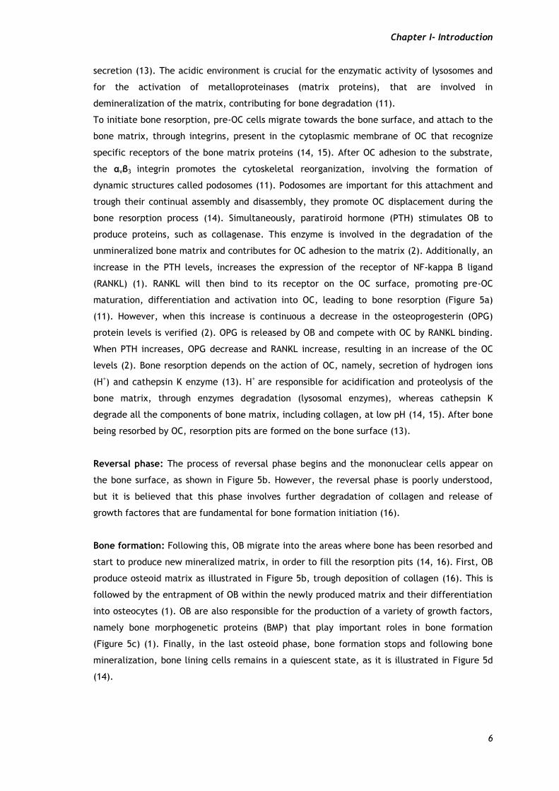
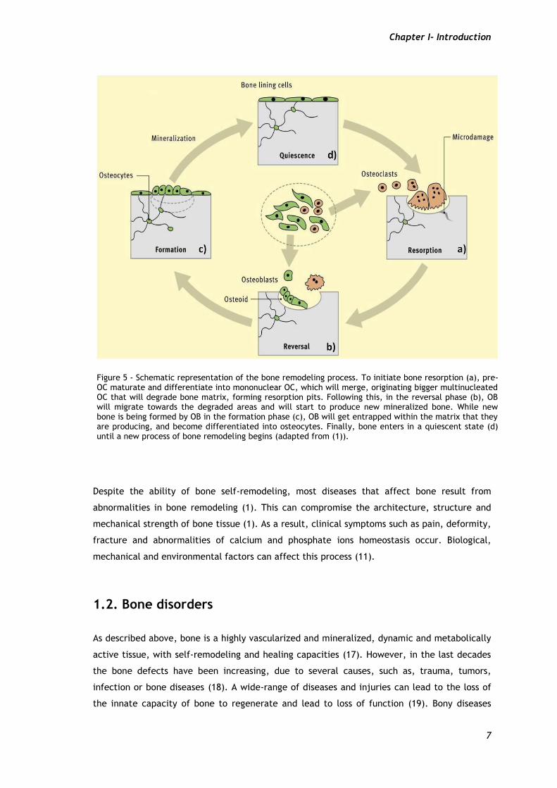
Figure 5 - Schematic representation of the bone remodeling process. To initiate bone resorption (a), pre-OC maturate and differentiate into mononuclear OC, which will merge, originating bigger multinucleated OC that will degrade bone matrix, forming resorption pits. Following this, in the reversal phase (b), OB will migrate towards the degraded areas and will start to produce new mineralized bone. While new bone is being formed by OB in the formation phase (c), OB will get entrapped within the matrix that they are producing, and become differentiated into osteocytes. Finally, bone enters in a quiescent state (d) until a new process of bone remodeling begins (adapted from (1)).
Chapter I- Introduction
8
have three main origins: bone loss in inflammatory diseases, disorders of bone remodeling and
monogenic bone diseases (20).
Bone loss in inflammatory diseases are caused by a deregulation of the immune system, i.e.,
the immune system does not respond properly (20).
Disorders of bone remodeling are related with disparities between the process of resorption
and bone formation (15, 20). Examples of disorders of bone remodeling are:
Osteoporosis that appears when the rate of bone resorption exceeds the rate of bone
formation. Osteoporosis increases with age, especially in the women, due to
hormonal changes (1).
Bone metastases occur due to increasing of osteoclastic bone resorption. Osteoclast-
activating is stimulated by factors released by tumors cells (e.g. interleukin-1, IL-1
and tumor necrosis factor, TNF) (21).
Monogenic bone diseases are associated with an imperfect process of osteogenesis, which
occurs when a lack of control between the levels of collagen products happens (20).
Bone diseases can cause defects in the process of bone remodeling (20).
1.3. Bone Grafts
Nowadays, different therapeutic strategies have been studied to overcome the bone
remodeling defects. Treatment options depends not only on the size and location of the
defect but also on the patient characteristics, namely, bone quality and age (22). To repair
large bone defects, attractive alternatives such as autografts, allografts and xenografts
therapies have been used so far in bone tissue reconstruction (18). Bone grafts are the most
commonly transplanted tissue after blood transplants (23).
Autografts are bone grafts, in which the bone is obtained from another part of the patient’s
body (7, 18). Nowadays, autografts are the most commonly used grafts for bone tissue
regeneration (22). They have osteoconductive, osteioconductive, and osteogenic properties
(18, 23). The major problem in the use of autografts is the limited supply by donor (18).
Frequently the size of the bone defect is larger than the area of the tissue obtained for
transplantation (18). Furthermore, the use of autografts is limited, by a considerable donor
site morbidity, which increases with the amount of harvested bone. Harvesting can induce
bleeding, hematoma, infection and chronic pain for patient (22).
To overcome the lack of donor tissue, allografts have also been applied. However, since the
grafts are from different donors (of the same species), there is always a risk of disease
transmission and of triggering of an immune response from the host (7, 18, 22).
Xenografts are grafts obtained from organisms of different species, which increases the
probability of graft rejection by the host (22).
Chapter I- Introduction
9
As an alternative to these bone substitutes, different biomaterials (ceramics, polymers and
composite systems) have been studied for scaffolds production. The purpose of using scaffolds
is not to replace the bone but create temporary matrices for bone growth.
1.4. Bone Tissue Engineering
As previously described, the aging of the population and bone disorders have as consequence
a decrease in the bone remodeling and regeneration capacity (1). In these cases, it is
indispensable the use of substitutes that replace the function of the damaged or degraded
bony tissues due to the limitations associated with autografts, allografts and xenografts in
bone tissue regeneration (18, 23). Tissue Engineering (TE) appears has a promising solution to
overcome these limitations. TE is “an interdisciplinary field of research that applies the
principles of engineering and life sciences, that have contributed for the development of
biological substitutes to restore, maintain, or improve tissue function”, i.e, TE is a new
multidisciplinary field that involves the use of scientific knowledge to solve practical and
clinical problems (7).
A primary objective of TE is changing practical practice, addressing the lack of donors and
organ rejection (24). In the last years, healthcare has been changing its replacement concepts
to a regeneration concept, trough TE and regenerative medicine (25).
The combination of TE and regenerative medicine promotes the development of techniques
for the treatment of bone defects (19).
TE approaches have been developed to create new different therapeutics for bone defects
regeneration. Which include drug delivery systems and scaffolds, among others, to promote
the regeneration of bone without causing adverse effects on the patient (26).
1.4.1. The importance of 3D Scaffolds for Bone Regeneration
Several types of biomaterials, are being used to produce injectable substances, hydrogels and
three-dimensional (3D) scaffolds to be applied in bone regeneration (27). Specifically, the
design of 3D porous scaffolds is being extensively explored in order to produce 3D structures
that allow tissue and organs regeneration (28).
Scaffolds are porous 3D matrices that act as temporary templates, allowing cell adhesion and
proliferation, and providing mechanical support until new bone tissue is formed at the defect
site (14).
The successful of a 3D scaffold depends on several parameters, such as biocompatibility,
biodegradability, surface properties, porosity and mechanical properties, in order to induce
osteoinductivity, osteoconductivity of bone-producing cells and neovascularization (6, 18).
Osteoinduction is the ability of a scaffold to induce non-differentiated stem cells, or
Chapter I- Introduction
10
osteoprogenitor cells from the surrounding tissue, to differentiate into bone-forming OB (18).
Considering the importance of OB for the production of new bone matrix, it is essential that
the scaffolds have good osteoinductive properties. Osteoconduction is characterized as the
ability of osteogenic cells to migrate into the surface of the scaffold and then promote the
formation of new blood vessels (18). Neovascularization is the formation of new blood vessels
from existing blood vessels to restore the blood supply, helping bone regeneration, during the
fracture healing process (6).
Biocompatibility: A biomaterial can be classified as biocompatible whether, when in contact
with any tissue it do not causes any immune reaction by the host (29). The biocompatibility
depends on the material variables (e.g. surface topography, surface charge), the location of
the injury in the body, age, genre, general health, lifestyle features and the presence or
absence of microorganisms at the site of implantation (29).
Biodegradability: Scaffolds must be composed by biodegradable materials (30, 31). The rate
of degradation of scaffolds must be accompanying the rate of regeneration of new bone (7,
32). As a result a scaffold must be completely degraded when the injury site is totally
regenerated (7). Moreover, the rate of scaffolds degradation depends on the extent and site
of the injury, as well as on the biomaterial and on the technique applied for its production
(7).
Surface properties: Also important are the materials surface properties (30). Among them
are the hydrophobic/hydrophilic character, charge, roughness, softness and chemical
composition of the biomaterials (30). Surface modifications of biomaterials allow a greater
understanding and control of the materials characteristics (33). These surface properties
should be controlled in order to induce osteoinductivity, osteocondutivity and
osteointegration (18). These properties have an essential role on cellular response, namely,
morphology, attachment, differentiation and proliferation of cells (30).
Porosity, pore size and mechanical properties: These properties are important
requirements to obtain optimized scaffolds for tissue regeneration (33). The use of ordered
structures is essential to achieve a better control of these properties that can influence cells
behavior (18). Scaffolds must possess pores with inter-connectivity among them, in order to
ensuring the nutrients and oxygen exchange within the scaffold to maintain cell viability and
bone regeneration (30, 31). The porosity of the native bone is approximately 10% for the
cortical bone and between 50 to 90% for the trabecular bone (7). It is also well accepted that
materials ideal pore size for bone tissue engineering should be in the range between 200 µm-
900 µm (7). The pores size must be adequate in order to not compromise both the biological
functions and the mechanical stability (7). Pores with adequate size allow cells to migrate
and attach to the surface of a material, but interconnected pores are essential for cell

Chapter I- Introduction
11
growth within the scaffold (30). Mechanical properties of human bones vary accordingly to
the type of bone. Cortical bone has a compressive strength of 100-230 MPa and cancellous
bone has a compressive strength of 2-12 MPa (7, 34). The Young´s modulus of cortical bone
varies between 7-30 GPa and for cancellous bone it varies between 0.5-0.05 GPa (34). Thus,
an ideal scaffold should combine good biological properties with good mechanical properties
(31). They must have sufficient mechanical strength to support the region to be repaired (7).
This is important to stand hydrostatic pressures and to maintain the spaces needed for cell
growth and new matrix formation (7).
1.4.2. Materials used for Scaffolds Production
Metals, ceramics and polymers are the most commonly used materials in TE for the
production of scaffolds aimed for bone tissue regeneration (18). Despite of its high
mechanical strength, the use of metals for bone regeneration has been decreasing due to
poor and incomplete osteointegration with surrounding bone (18). So, ceramics and polymers
have been studied for being applied in TE (18). Ceramics have been used due to their intrinsic
excellent biocompatibility and bioactivity, which means that they are able to promote cell
adhesion, proliferation and differentiation (35). Hydroxyapatite (HA) is the major inorganic
component of bone and it is usually the first option for scaffold production to be used in bone
regeneration (36). However, HA has the disadvantage of having low biodegradability (26).
Conversely, β-Tricalcium phosphate (β-TCP) has been used for the production of scaffolds,
since it revealed better bone formation and degradation rate than HA (36, 37). Furthermore,
β-TCP is an interesting ceramic material studied to be applied in bone tissue regeneration due
to their osteocondutivity and osteoinductivity, contributing for bone formation through
induction of OB differentiation (38). Due to high biocompatibility, calcium phosphate
ceramics are also applied as carrier materials for cell adhesion and growth (35). However, β-
TCP presents also some limitations, like brittleness and poor resistance to fatigue (18).
The combination of ceramics with polymers can provide 3D structures with good bioactivity,
adequate mechanical properties and stability (18). Nowadays, there are several types of
polymers that can be divided in two different groups: natural polymers (e.g collagen,
chitosan, fibrin, alginate) and synthetic polymers (e.g PCL Poly (ε-caprolactone), PLLA poli(L-
lactic-acid), PLGA (poli-lactide-co-glico-lide)). Thus, synthetic or natural polymers have been
used to improve the bioceramics mechanical properties (7, 18). Natural polymers have high
biocompatibility and lower immunogenic risk (7). They exhibit good osteocondutive properties
with a high capacity to interact with host´s tissues. However the use of these natural
polymers is limited due to their low mechanical stability. On the other hand, the mechanical
properties of synthetic polymers can be controlled (7).
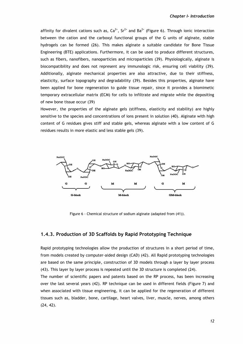
Alginate is a natural polysaccharide extracted from brown seaweeds (26, 39). This polymer is
composed by 1,4-linked β-D-mannuronic acid (M) and α-L-guluronic acid (G) residues and has
Chapter I- Introduction
12
affinity for divalent cations such as, Ca2+, Sr2+ and Ba2+ (Figure 6). Through ionic interaction
between the cation and the carboxyl functional groups of the G units of alginate, stable
hydrogels can be formed (26). This makes alginate a suitable candidate for Bone Tissue
Engineering (BTE) applications. Furthermore, it can be used to produce different structures,
such as fibers, nanofibers, nanoparticles and microparticles (39). Physiologically, alginate is
biocompatibility and does not represent any immunologic risk, ensuring cell viability (39).
Additionally, alginate mechanical properties are also attractive, due to their stiffness,
elasticity, surface topography and degradability (39). Besides this properties, alginate have
been applied for bone regeneration to guide tissue repair, since it provides a biomimetic
temporary extracellular matrix (ECM) for cells to infiltrate and migrate while the depositing
of new bone tissue occur (39)
However, the properties of the alginate gels (stiffness, elasticity and stability) are highly
sensitive to the species and concentrations of ions present in solution (40). Alginate with high
content of G residues gives stiff and stable gels, whereas alginate with a low content of G
residues results in more elastic and less stable gels (39).
Figure 6 - Chemical structure of sodium alginate (adapted from (41)).
1.4.3. Production of 3D Scaffolds by Rapid Prototyping Technique
Rapid prototyping technologies allow the production of structures in a short period of time,
from models created by computer-aided design (CAD) (42). All Rapid prototyping technologies
are based on the same principle, construction of 3D models through a layer by layer process
(43). This layer by layer process is repeated until the 3D structure is completed (24).
The number of scientific papers and patents based on the RP process, has been increasing
over the last several years (42). RP technique can be used in different fields (Figure 7) and
when associated with tissue engineering, it can be applied for the regeneration of different
tissues such as, bladder, bone, cartilage, heart valves, liver, muscle, nerves, among others
(24, 42).
Chapter I- Introduction
13
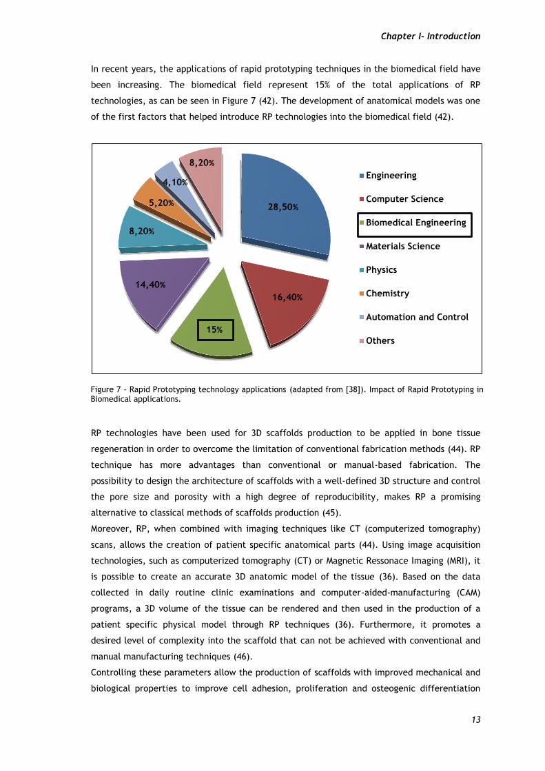
In recent years, the applications of rapid prototyping techniques in the biomedical field have
been increasing. The biomedical field represent 15% of the total applications of RP
technologies, as can be seen in Figure 7 (42). The development of anatomical models was one
of the first factors that helped introduce RP technologies into the biomedical field (42).
RP technologies have been used for 3D scaffolds production to be applied in bone tissue
regeneration in order to overcome the limitation of conventional fabrication methods (44). RP
technique has more advantages than conventional or manual-based fabrication. The
possibility to design the architecture of scaffolds with a well-defined 3D structure and control
the pore size and porosity with a high degree of reproducibility, makes RP a promising
alternative to classical methods of scaffolds production (45).
Moreover, RP, when combined with imaging techniques like CT (computerized tomography)
scans, allows the creation of patient specific anatomical parts (44). Using image acquisition
technologies, such as computerized tomography (CT) or Magnetic Ressonace Imaging (MRI), it
is possible to create an accurate 3D anatomic model of the tissue (36). Based on the data
collected in daily routine clinic examinations and computer-aided-manufacturing (CAM)
programs, a 3D volume of the tissue can be rendered and then used in the production of a
patient specific physical model through RP techniques (36). Furthermore, it promotes a
desired level of complexity into the scaffold that can not be achieved with conventional and
manual manufacturing techniques (46).
Controlling these parameters allow the production of scaffolds with improved mechanical and
biological properties to improve cell adhesion, proliferation and osteogenic differentiation
28,50%
16,40%
15%
14,40%
8,20%
5,20%
4,10%
8,20%
Engineering
Computer Science
Biomedical Engineering
Materials Science
Physics
Chemistry
Automation and Control
Others
Figure 7 - Rapid Prototyping technology applications (adapted from [38]). Impact of Rapid Prototyping in Biomedical applications.
Chapter I- Introduction
14
(44). As previously described in section 1.4.1, these properties of scaffolds play a key role in
the process of bone regeneration.
RP techniques: RP techniques involve for example stereolithography (SLA), selective laser
sintering (SLS), fused deposition modeling (FDM), three-dimensional printing (3DP) and 3D
plotting (47). SLA uses the capacity of some polymers to suffer photo polymerization (in
which liquid polymers solidify when exposed to a ultraviolet light) (21). In SLS, a laser beam is
focused on a polymer powder and raises the temperature of the powders, causing the fusion
of the particles (7). Another RP technique is FDM, which uses a small temperature-controlled
extruded material and deposit simultaneously polymer onto a platform and constructs a 3D
structure through a layer-by-layer process (46). 3D printing, employs inkjet for powders
materials and a printer head to print a liquid binder to form a layer by layer scaffold (7).
3D Plotting was described for the first time by Landers et al. (24). Briefly, 3D plotting is a
system based on a dispenser solution that is forced to pass through a syringe onto a platform
to generally form a hydrogel. For this, a dispenser solution must fall onto a crosslink agent
(7). One of the newer techniques in 3D plotting consists in the fabrication technology based
on the extrusion of continuous filaments (48).
In this work, a 3D printer called Fab@Home was used for scaffolds production. Fab@Home was
developed by Cohen and collaborators, and they applied the plotting technology for scaffold
production (48).
1.4.3.1. Fab@Home Model for Scaffolds Production
There are several machines that can be used for RP. However, the majority of the
commercially available machines are not suitable for biomedical purposes, like bone
regeneration. Fab@home is one of the available machines that can be applied in bone 3D
structures production (42, 49).
The Fab@home model has advantages over other equipments, since it allows the employment
of different samples, such as composite materials and viscous solutions, like hydrogels (49).
However, the printing accuracy and resolution of the extruded material depends on a set of
factors, such as material viscosity, dispensing pressure, pushout (specifies the initiate
material-flow before the cartridge moves along the print path), suckback (sets how long the
syringe plunger with draws at the end of the path to stop the extrusion), nozzle diameter,
deposition rate, printspeed, pathheight (distance between adjacent layers) and pathspace
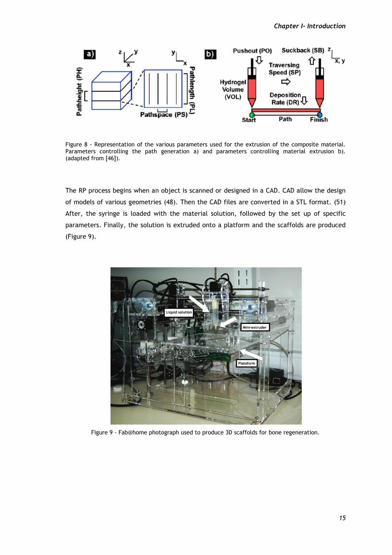
(distance between adjacent paths for each layer), (Figure 8) (50).
Chapter I- Introduction
15
Figure 8 - Representation of the various parameters used for the extrusion of the composite material. Parameters controlling the path generation a) and parameters controlling material extrusion b). (adapted from [46]).
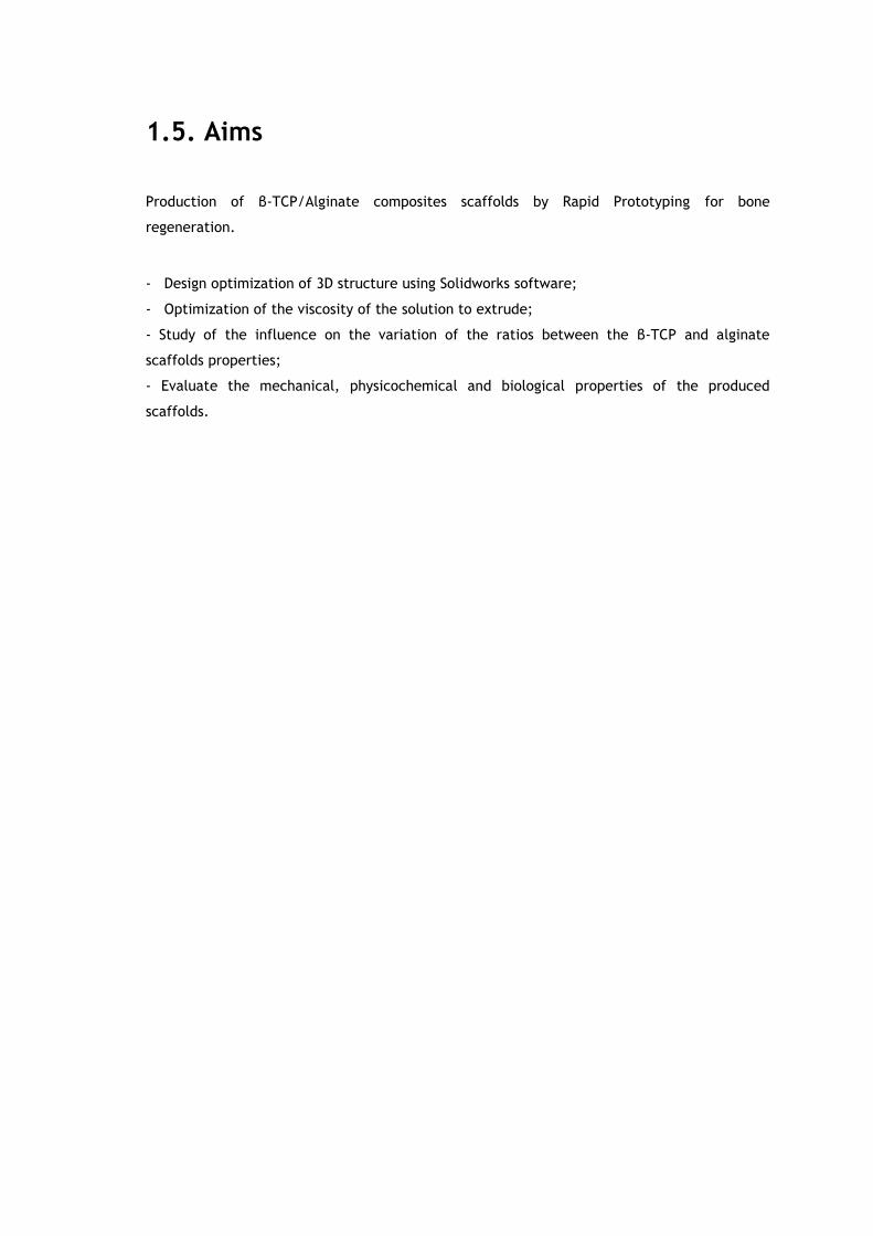
The RP process begins when an object is scanned or designed in a CAD. CAD allow the design
of models of various geometries (48). Then the CAD files are converted in a STL format. (51)
After, the syringe is loaded with the material solution, followed by the set up of specific
parameters. Finally, the solution is extruded onto a platform and the scaffolds are produced
(Figure 9).
Figure 9 - Fab@home photograph used to produce 3D scaffolds for bone regeneration.

Chapter I- Introduction
16
1.5. Aims
Production of β-TCP/Alginate composites scaffolds by Rapid Prototyping for bone
regeneration. - Design optimization of 3D structure using Solidworks software;
- Optimization of the viscosity of the solution to extrude;
- Study of the influence on the variation of the ratios between the β-TCP and alginate
scaffolds properties;
- Evaluate the mechanical, physicochemical and biological properties of the produced
scaffolds.

Chapter II - Materials and Methods
Chapter II-Materials and Methods
19
2.1 Materials
Amphotericin B, bovine serum albumin (BSA), cacodylate (Mw=214.03 g/mol) buffer, calcium
chloride (CaCl2) (Mw=110.98g/mol) solution, dulbecco’s modified eagle’s medium (DMEM-
F12), Ethanol (EtOH), ethylenediaminetetraacetic acid (EDTA), 2.5 % (v/v) glutharaldehyde,
L-glutamine, penicillin G, phosphate-buffered saline (PBS), poly(vinyl) alcohol (PVA) (Mw=31
000g/mol), sodium alginate solution (Mw=120.000 – 190.000 Da), streptomycin, trypan blue,
trypsin were purchased from Sigma-Aldrich (Sintra, Portugal). β-TCP powder, molecular
weight (Mw=310,20g/mol) was purchased from Panreac® (Barcelona,Spain). 3-(4,5-
dimethylthiazol-2-yl)-5-(3-carboxymethoxyphenyl)-2-(4-sulfophenyl)-2H-tetrazolium reagent,
inner salt (MTS) was obtained from Promega (Madison, USA). Fetal bovine serum (FBS) was
purchased from Biochrom AG (Berlin, Germany). Human osteoblast cells (CRL-11372) were
purchased from American Type Culture Collection (VA, USA). 96-well plates were acquired
from Orange Scientific (Braine LÁlleud, Belgium). µ-Slide 8 well Ibidi® chamber coverslips
(Ibidi® GmbH, Germany). Propidium Iodide was purchased from Invitrogen (Carlsbad, USA).
2.2 Methods
2.2.1. Preparation of β-TCP/Alginate Composite Scaffolds by
Rapid Prototyping.
The bioactive 3D scaffolds were produced by the RP technique. This technique allows the
production of scaffolds with controllable porosity, well-defined 3D microstructures and
greater reproducibility (47). A Fab@home printing model was used for scaffolds production,
since the scaffolds architecture produced with 3D plotters can be controlled trough a
computer-assisted designed. Such is fundamental for allowing the manipulation of 3D models
that are capable of sustaining cellular viability (45).
First, alginate and β-TCP solutions were prepared. Briefly, to prepare alginate solution,
alginate was dissolved in milli-Q water. Then, the solution was left overnight under magnetic
stirring, and then sonicated for 15 minutes to homogenize the mixture. Following, β-TCP
powder was weighed and added to the alginate solution.
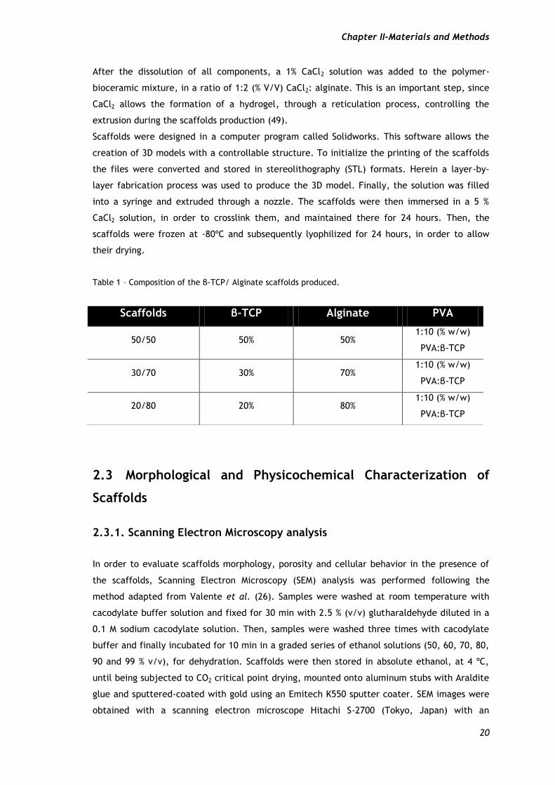
In this study, three different types of scaffolds were produced, varying the ratios between
the concentrations of β-TCP and alginate. β-TCP/Alginate scaffolds were produced using
solutions prepared with β-TCP and alginate in a proportion of 50/50 % (w/w), 30/70 % (w/w)
and 20/80 % (w/w), respectively. For that, PVA (β-TCP binder agent) was added to the
polymer-bioceramic mixture in a ratio of 1:10 (% w/w) PVA:β-TCP.
Chapter II-Materials and Methods
20
After the dissolution of all components, a 1% CaCl2 solution was added to the polymer-
bioceramic mixture, in a ratio of 1:2 (% V/V) CaCl2: alginate. This is an important step, since
CaCl2 allows the formation of a hydrogel, through a reticulation process, controlling the
extrusion during the scaffolds production (49).
Scaffolds were designed in a computer program called Solidworks. This software allows the
creation of 3D models with a controllable structure. To initialize the printing of the scaffolds
the files were converted and stored in stereolithography (STL) formats. Herein a layer-by-
layer fabrication process was used to produce the 3D model. Finally, the solution was filled
into a syringe and extruded through a nozzle. The scaffolds were then immersed in a 5 %
CaCl2 solution, in order to crosslink them, and maintained there for 24 hours. Then, the
scaffolds were frozen at -80ºC and subsequently lyophilized for 24 hours, in order to allow
their drying.
Table 1 – Composition of the β-TCP/ Alginate scaffolds produced.
2.3 Morphological and Physicochemical Characterization of
Scaffolds
2.3.1. Scanning Electron Microscopy analysis
In order to evaluate scaffolds morphology, porosity and cellular behavior in the presence of
the scaffolds, Scanning Electron Microscopy (SEM) analysis was performed following the
method adapted from Valente et al. (26). Samples were washed at room temperature with
cacodylate buffer solution and fixed for 30 min with 2.5 % (v/v) glutharaldehyde diluted in a
0.1 M sodium cacodylate solution. Then, samples were washed three times with cacodylate
buffer and finally incubated for 10 min in a graded series of ethanol solutions (50, 60, 70, 80,
90 and 99 % v/v), for dehydration. Scaffolds were then stored in absolute ethanol, at 4 ºC,
until being subjected to CO2 critical point drying, mounted onto aluminum stubs with Araldite
glue and sputtered-coated with gold using an Emitech K550 sputter coater. SEM images were
obtained with a scanning electron microscope Hitachi S-2700 (Tokyo, Japan) with an
Scaffolds β-TCP Alginate PVA
50/50 50% 50% 1:10 (% w/w)
PVA:β-TCP
30/70 30% 70% 1:10 (% w/w)
PVA:β-TCP
20/80 20% 80% 1:10 (% w/w)
PVA:β-TCP
Chapter II-Materials and Methods
21
acceleration voltage of 20 kV at suitable magnifications.
2.3.2. Fourier Transform Infrared Spectroscopy analysis
Fourier-transform infrared (FTIR) spectroscopy was used to measure the physicochemical
characteristics of the scaffolds and the chemical cross-linking of different compounds (26).
FTIR spectra represent the average of 128 scans between 400 and 4000 cm-1, at a resolution
of 4 cm-1. Briefly, all samples were crushed and the resulting powders were mounted on a
diamond window and recorded on a Fourier-transform infrared spectrophotometer (Nicolet
iS10), from Thermo Scientific, (Waltham, MA, USA). β-TCP and Alginate powders were also
analyzed to perform a comparative study with scaffolds samples (45).
2.3.3. X-Ray Diffraction analysis
To evaluate the characteristic phases and crystallinity of the scaffolds after being produced,
X-ray diffractometry measurements were performed with a diffractometer (Rigaku Americas
Corporation, USA) (52). XRD technique was used to examine material composition after the
process of freezing and lyophilization to confirm the presence of β-TCP and Alginate in the
scaffolds. Samples were mounted in appropriate silica supports and the data was recorded
over a range of 5 º to 90 º 2θ degrees, with continuous scans at a rate of 1º/min with a copper
ray tube operated at 30 kV and 20 mA (53).
2.3.4. Energy Dispersive Spectroscopy analysis
In order to perform the elementary characterization of the materials, in this specific case to
evaluate the distribution of elemental calcium and phosphorus in the scaffolds, an energy-
dispersive spectroscopy (EDS) (Rontec) analysis was carried out. For that, samples were
placed on an aluminum stub support, air-dried at room temperature (RT) and sputter-coated
with gold (53).
2.4. Mechanical Characterization of the β-TCP/Alginate
Composite Scaffolds: Resistance to Compression and Young’s
Modulus.
In order to study the mechanical behavior of the scaffolds, compression assays were
performed by following a method adapted from Santos et al. (36). First, the previously
Chapter II-Materials and Methods
22
lyophilized scaffolds were cut into similar sizes fragments and their dimensions determined.
The compression assays were performed using a Zwick® 1435 Material prüfung (Ulm, Germany)
with a crosshead speed of 0.2 mm /min and a load cell of 5 kN. Five specimens from each
sample were tested and their dimensions acquired. The calculation of the resistance to
compression (Ts) of each scaffold was determined through equation 1 (54).
(1)
Where F is the load at the time of the fracture and a and l represent the width and length of
the scaffold, respectively. Young’s Modulus (YM) was obtained from the stress-strain relations
calculated and applying the equation 2 (55).
(2)
Where Hd is the scaffold height deformation and Ts is the scaffold tensile strength. Average
values and standard deviations (s.d.) were determined for each sample (n=3).
2.5. Contact Angle Measurements
The contact angle measurements of the samples were performed using the sessile drop
technique using water as a reference fluid. The method used was adapted from Correia et al.
(56). Contact angle data was acquired in a Data Physics Contact Angle System OCAH 200
apparatus, operating in static mode at RT. For each sample, water drops were placed at
various locations of the analyzed surface [47].
2.6. Porosity Evaluation
The total porosity (P) of the different types of β-TCP/Alginate scaffolds was determined by
following a method adapted from Nie et al. (57). The total amount of absolute ethanol (EtOH)
that the scaffolds were able to absorb in 48 h, was determined by applying the equation 3,
adapted from Correia et al. (56).
( )
(3)
Chapter II-Materials and Methods
23
Where W1 and W2 is the weight of the dry and the wet scaffold, respectively, dethanol is the
density of the ethanol at RT and Vscaffold is the volume of the wet scaffold, directly
determined by immersion. For each scaffold, three replicates were analyzed and data
represents the average of each replicate.
2.7. Biological Characterization of the β-TCP/Alginate Composite
Scaffolds
2.7.1. Cell Culture and Seeding in the β-TCP/Alginate Scaffolds
Human Osteoblasts (CRL-11372), were cultured in Dulbecco’s modified Eagle medium (DMEM-
F12), containing 10% of heat inactivated fetal bovine serum (FBS), streptomycin (100 μg /mL)
and gentamicin (100 μg /mL) antibiotics, in a 75 cm2 T-flasks. Cultures were maintained in a
humidified environment at 37ºC, with 5% CO2, until confluence was attained. After that, cells
were trypsinized with 0.18 % trypsin (1:250) and 5 mM EDTA, centrifuged at 260rpm for 5
minutes and the resulting pellet resuspended into 5 mL of complete culture medium. After
that, the cellular density (number of cells per mL) was determined by using the trypan blue
method. Cells were counted in a Neubauer Chamber and the total number of cells determined
accordingly to equation 4:
× dilution factor × 104 (4)
Prior to cell being seeded in contact with scaffolds, they were cut into the desired size and
placed into 96-well plates for sterilization and disinfection. Briefly, scaffolds were disinfected
with 70% of EtOH solution for 30 minutes and then washed with DMEM-F12 medium twice,
followed by sterilization with ultraviolet (UV) light for another 30 minutes. The scaffolds were
previously equilibrated with complete culture medium at 37 ºC, for 24 hours. Finally, cells
were seeded at scaffolds surface with a density of 10x103 cells per well, to evaluated cell
viability and proliferation at days 1 and 7 respectively. The culture medium was changed
every two days until day 7.
2.7.2. Evaluation of Cell Viability in the Presence of the Scaffolds.
To evaluate cell viability in the presence of the materials, Human Osteoblasts (CRL-11372)
cells were seeded in the presence of the materials in 96-well plates at a density of 10x103,
Chapter II-Materials and Methods
24
cells per well. Cell viability was determined using a MTS assay at 1 and 7 days after seeding.
After an incubation of 24h, the metabolic activity of cells was evaluated by quantifying the
metabolic reduction of the MTS to formazan. Briefly, the medium of each well was removed
and the cells in contact with scaffolds were incubated with a mixture of 100μL of fresh
culture medium and 20μL of MTS/PMS reagent solution, for 4 hours, at 37 ºC. After 4 hours of
incubation, 80 L of the supernatant was transferred into a 96-well microplate and the
fluorescence intensity was measured at 492 nm using a microplate reader (Sanofi, Diagnostics
Pauster).
Five replicates of each sample were used for each incubation day. Cells cultured without
materials were used as negative controls (K-) and cells cultured with EtOH (96%) were used as
a positive control (K+).
2.7.3. Analysis of 3D Scaffolds Biologic Properties
The analysis of osteoblasts adhesion and morphology on the 3D scaffolds surfaces was
visualized by Confocal Laser Scanning Microscopy (CLSM). Briefly, 4 days after seeding cells,
the culture medium was removed and cells washed three times with cacodylate buffer at
room temperature (RT). The remaining cells were then fixed with cacodylate buffer for 30
min, at RT. Afterwards, the cell nucleus was labeled with Propidium Iodide (PI) solution,
during 15 min, at RT, followed by 6 additional washes with cacodylate buffer. The cell-seeded
scaffolds were then transferred into µ-Slide 8 well Ibidi® chamber coverslips and imaged in a
Zeiss LSM 710 confocal microscope (Carl Zeiss SMT Inc., USA) equipped with a Plan-Neofluar
10x/NA 0.3. All data was acquired in z-stack mode with a step of 4.67 µm. Z-stacks were then
rendered into 3D images in the Zeiss Zen software. Depth code rendering of z-stacks was also
performed in Zeiss software with the open GL and transparent rendering mode to provide
visualization of cell spatial distribution within the scaffold architecture.
2.8. Statistical Analysis
Comparison of the results obtained for the different groups of scaffolds at various conditions
was performed by using one-way analysis of variance (ANOVA), with the Newman-Keuls test. A
p value less than 0.05 (p<0.05) was considered statistically significant.

Chapter III - Results and Discussion
Chapter III- Results and discussion
26
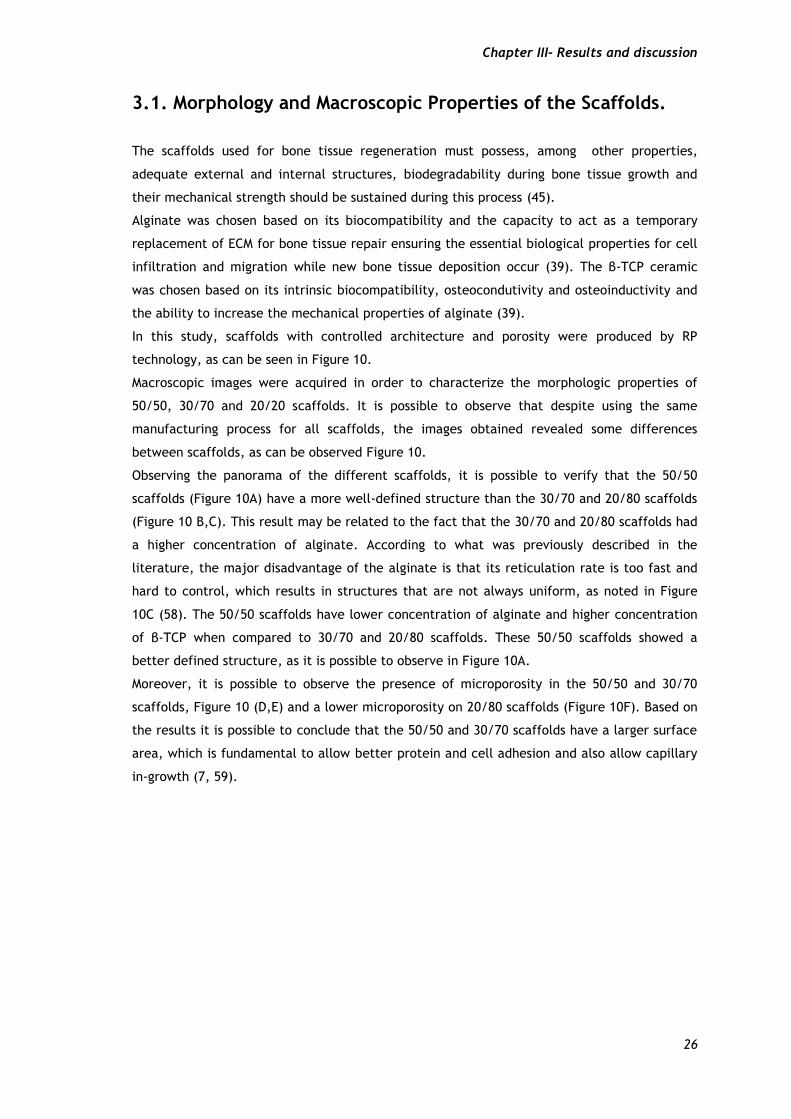
3.1. Morphology and Macroscopic Properties of the Scaffolds. The scaffolds used for bone tissue regeneration must possess, among other properties,
adequate external and internal structures, biodegradability during bone tissue growth and
their mechanical strength should be sustained during this process (45).
Alginate was chosen based on its biocompatibility and the capacity to act as a temporary
replacement of ECM for bone tissue repair ensuring the essential biological properties for cell
infiltration and migration while new bone tissue deposition occur (39). The β-TCP ceramic
was chosen based on its intrinsic biocompatibility, osteocondutivity and osteoinductivity and
the ability to increase the mechanical properties of alginate (39).
In this study, scaffolds with controlled architecture and porosity were produced by RP
technology, as can be seen in Figure 10.
Macroscopic images were acquired in order to characterize the morphologic properties of
50/50, 30/70 and 20/20 scaffolds. It is possible to observe that despite using the same
manufacturing process for all scaffolds, the images obtained revealed some differences
between scaffolds, as can be observed Figure 10.
Observing the panorama of the different scaffolds, it is possible to verify that the 50/50
scaffolds (Figure 10A) have a more well-defined structure than the 30/70 and 20/80 scaffolds
(Figure 10 B,C). This result may be related to the fact that the 30/70 and 20/80 scaffolds had
a higher concentration of alginate. According to what was previously described in the
literature, the major disadvantage of the alginate is that its reticulation rate is too fast and
hard to control, which results in structures that are not always uniform, as noted in Figure
10C (58). The 50/50 scaffolds have lower concentration of alginate and higher concentration
of β-TCP when compared to 30/70 and 20/80 scaffolds. These 50/50 scaffolds showed a
better defined structure, as it is possible to observe in Figure 10A.
Moreover, it is possible to observe the presence of microporosity in the 50/50 and 30/70
scaffolds, Figure 10 (D,E) and a lower microporosity on 20/80 scaffolds (Figure 10F). Based on
the results it is possible to conclude that the 50/50 and 30/70 scaffolds have a larger surface
area, which is fundamental to allow better protein and cell adhesion and also allow capillary
in-growth (7, 59).
Chapter III- Results and discussion
27
Figure 10 – Images of the different 50/50 (A, D), 30/70 (B, E) and 20/80 (C, F) scaffolds surface.
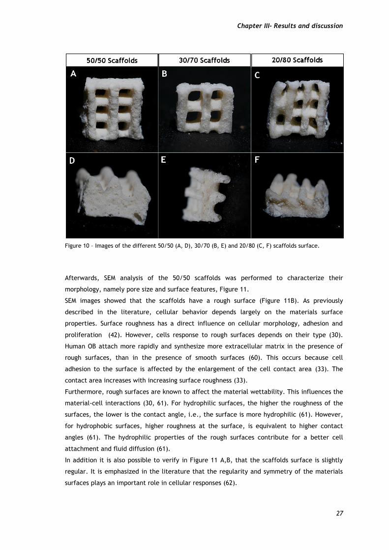
Afterwards, SEM analysis of the 50/50 scaffolds was performed to characterize their
morphology, namely pore size and surface features, Figure 11.
SEM images showed that the scaffolds have a rough surface (Figure 11B). As previously
described in the literature, cellular behavior depends largely on the materials surface
properties. Surface roughness has a direct influence on cellular morphology, adhesion and
proliferation (42). However, cells response to rough surfaces depends on their type (30).
Human OB attach more rapidly and synthesize more extracellular matrix in the presence of
rough surfaces, than in the presence of smooth surfaces (60). This occurs because cell
adhesion to the surface is affected by the enlargement of the cell contact area (33). The
contact area increases with increasing surface roughness (33).
Furthermore, rough surfaces are known to affect the material wettability. This influences the
material-cell interactions (30, 61). For hydrophilic surfaces, the higher the roughness of the
surfaces, the lower is the contact angle, i.e., the surface is more hydrophilic (61). However,
for hydrophobic surfaces, higher roughness at the surface, is equivalent to higher contact
angles (61). The hydrophilic properties of the rough surfaces contribute for a better cell
attachment and fluid diffusion (61).
In addition it is also possible to verify in Figure 11 A,B, that the scaffolds surface is slightly
regular. It is emphasized in the literature that the regularity and symmetry of the materials
surfaces plays an important role in cellular responses (62).
Chapter III- Results and discussion
28
Figure 11 – SEM images of the morphology of 50/50 Scaffolds. Representation of the 50/50 scaffolds at different magnification 50x (A) and 3000x (B).
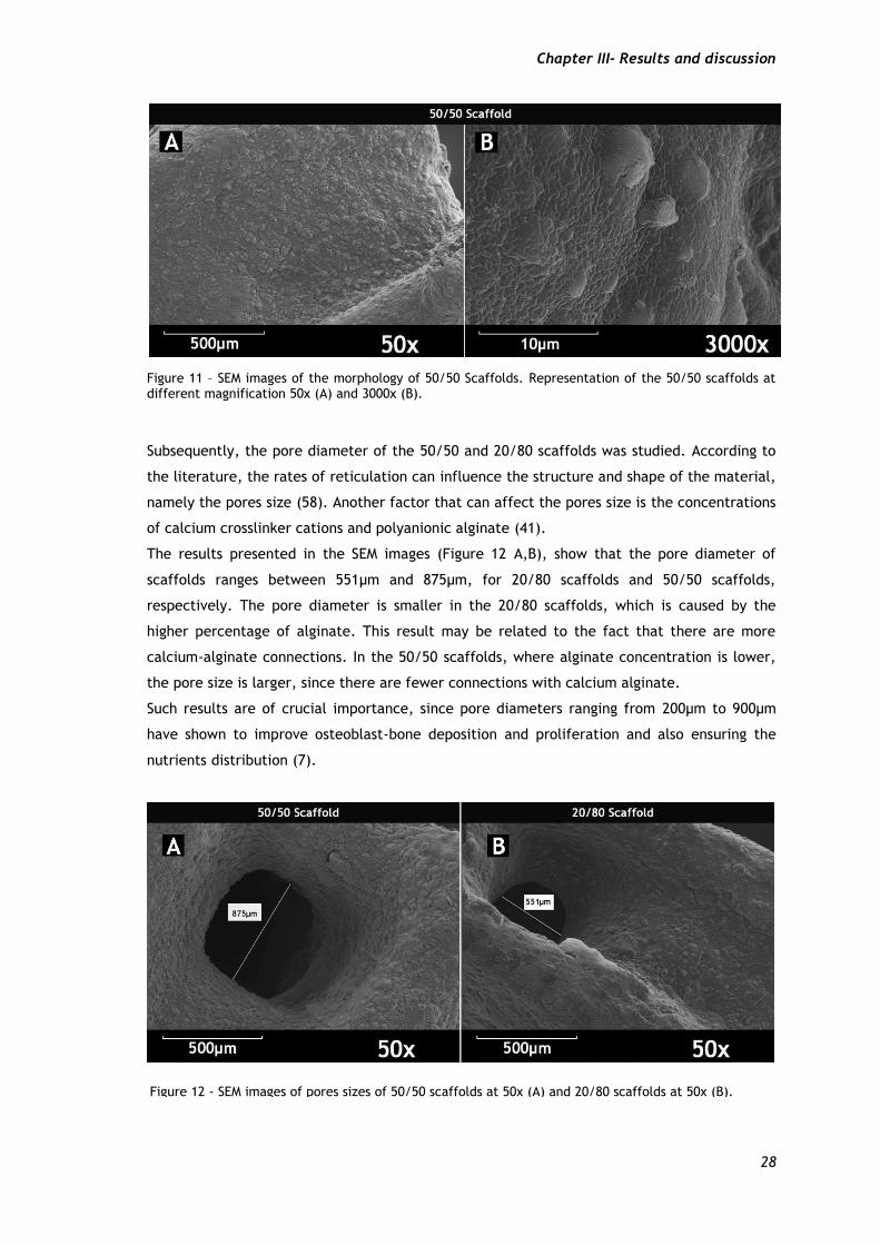
Subsequently, the pore diameter of the 50/50 and 20/80 scaffolds was studied. According to
the literature, the rates of reticulation can influence the structure and shape of the material,
namely the pores size (58). Another factor that can affect the pores size is the concentrations
of calcium crosslinker cations and polyanionic alginate (41).
The results presented in the SEM images (Figure 12 A,B), show that the pore diameter of
scaffolds ranges between 551µm and 875µm, for 20/80 scaffolds and 50/50 scaffolds,
respectively. The pore diameter is smaller in the 20/80 scaffolds, which is caused by the
higher percentage of alginate. This result may be related to the fact that there are more
calcium-alginate connections. In the 50/50 scaffolds, where alginate concentration is lower,
the pore size is larger, since there are fewer connections with calcium alginate.
Such results are of crucial importance, since pore diameters ranging from 200µm to 900µm
have shown to improve osteoblast-bone deposition and proliferation and also ensuring the
nutrients distribution (7).
Figure 12 - SEM images of pores sizes of 50/50 scaffolds at 50x (A) and 20/80 scaffolds at 50x (B).
Chapter III- Results and discussion
29
3.2. Physicochemical Characterization
3.2.1. - Fourier Transform Infrared Spectroscopy (FTIR)
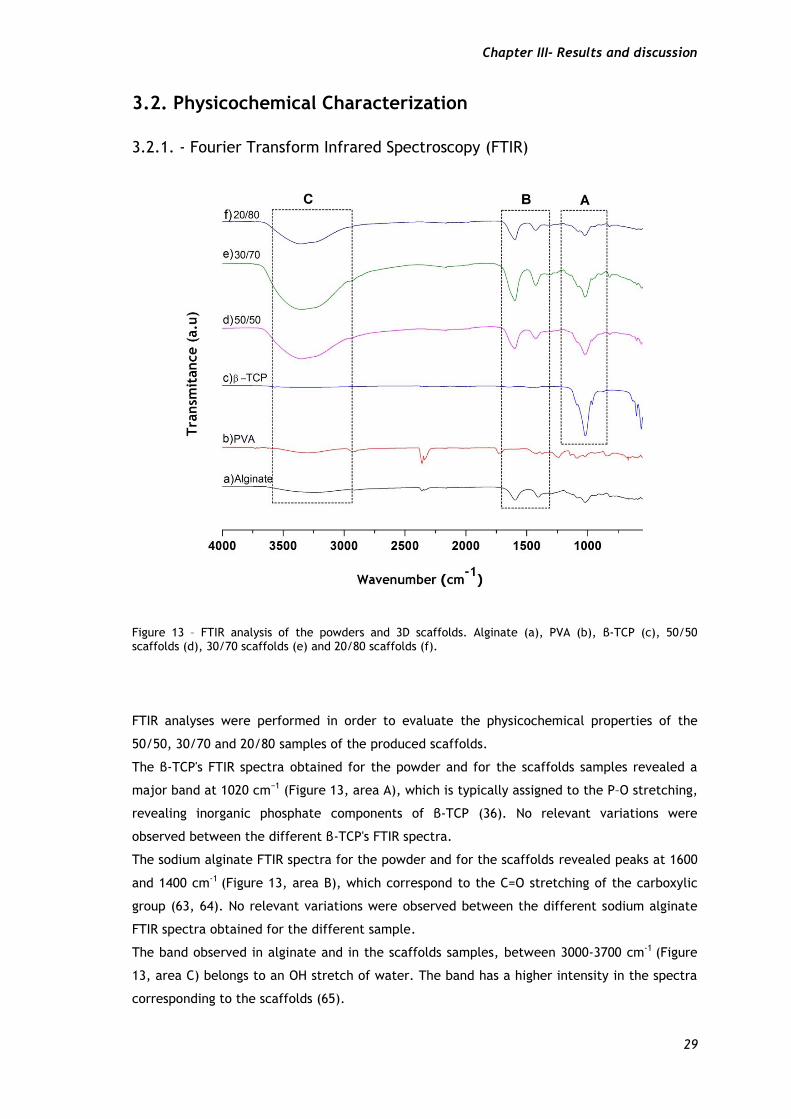
Figure 13 – FTIR analysis of the powders and 3D scaffolds. Alginate (a), PVA (b), β-TCP (c), 50/50 scaffolds (d), 30/70 scaffolds (e) and 20/80 scaffolds (f).
FTIR analyses were performed in order to evaluate the physicochemical properties of the
50/50, 30/70 and 20/80 samples of the produced scaffolds.
The β-TCP's FTIR spectra obtained for the powder and for the scaffolds samples revealed a
major band at 1020 cm−1 (Figure 13, area A), which is typically assigned to the P–O stretching,
revealing inorganic phosphate components of β-TCP (36). No relevant variations were
observed between the different β-TCP's FTIR spectra.
The sodium alginate FTIR spectra for the powder and for the scaffolds revealed peaks at 1600
and 1400 cm-1 (Figure 13, area B), which correspond to the C=O stretching of the carboxylic
group (63, 64). No relevant variations were observed between the different sodium alginate
FTIR spectra obtained for the different sample.
The band observed in alginate and in the scaffolds samples, between 3000-3700 cm-1 (Figure
13, area C) belongs to an OH stretch of water. The band has a higher intensity in the spectra
corresponding to the scaffolds (65).
Chapter III- Results and discussion
30
3.2.2. X-Ray Diffraction (XRD)
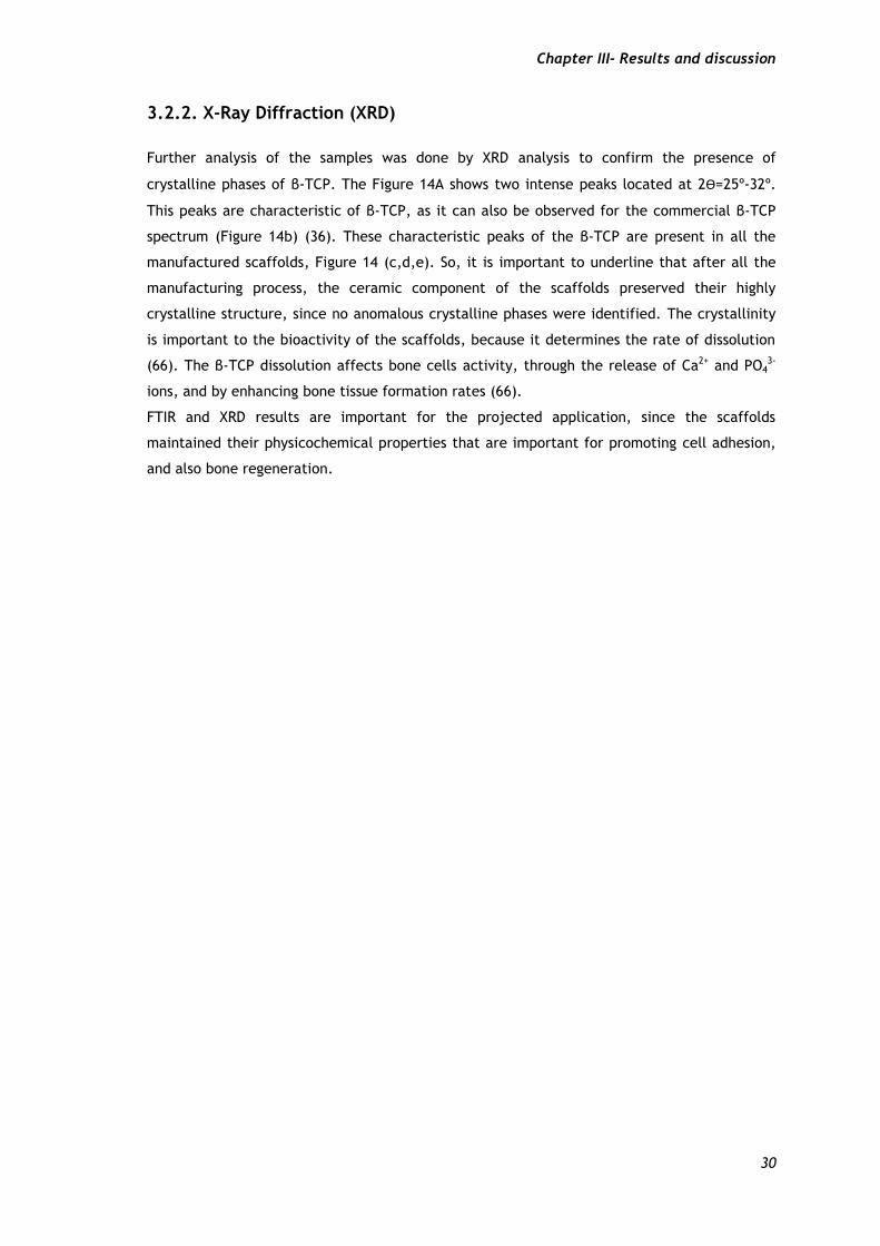
Further analysis of the samples was done by XRD analysis to confirm the presence of
crystalline phases of β-TCP. The Figure 14A shows two intense peaks located at 2Ѳ=25º-32º.
This peaks are characteristic of β-TCP, as it can also be observed for the commercial β-TCP
spectrum (Figure 14b) (36). These characteristic peaks of the β-TCP are present in all the
manufactured scaffolds, Figure 14 (c,d,e). So, it is important to underline that after all the
manufacturing process, the ceramic component of the scaffolds preserved their highly
crystalline structure, since no anomalous crystalline phases were identified. The crystallinity
is important to the bioactivity of the scaffolds, because it determines the rate of dissolution
(66). The β-TCP dissolution affects bone cells activity, through the release of Ca2+ and PO43-
ions, and by enhancing bone tissue formation rates (66).
FTIR and XRD results are important for the projected application, since the scaffolds
maintained their physicochemical properties that are important for promoting cell adhesion,
and also bone regeneration.
Chapter III- Results and discussion
31
Figure 14- X-Ray spectra of the powders and 3D scaffolds. PVA (a), β-TCP (b), 50/50 scaffolds (c), 30/70 scaffolds (d) and 20/80 scaffolds (e), (A), and zoomed XRD spectra in a range between 25º-35º, (B).
Chapter III- Results and discussion
32
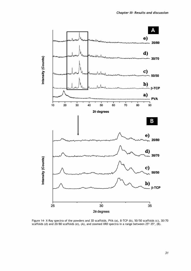
3.2.3. Energy Dispersive Spectroscopy
An energy dispersive spectroscopic (EDS) analysis of the scaffolds was also performed to
characterize the distribution of elemental calcium and phosphorus in the scaffolds. The
results show a slight increase in phosphate and calcium ions in the 50/50 scaffold, followed
by 30/70 and 20/80, respectively. These results are in agreement with the scaffolds β-TCP
composition.
Table 2 - EDS analysis of the produced 3D scaffolds.
3.3. Alginate/β-TCP Composite Scaffolds: Mechanical
Characterization.
The mechanical behavior of the 3D scaffolds was characterized by testing the resistance to
compression and by determining the Young’s modulus tests, Figure 15.
Figure 15 shows that the Young´s modulus and compressive strength of the 50/50 scaffolds
are higher than that of the other samples. A suitable scaffold to be used for bone
regeneration must have both flexibility and resistance to compression (2). Previous studies
have shown that although alginate has several good properties for the application in bone
regeneration, when it is used alone it does not have the mechanical properties suitable for
this biomedical application. (67) To overcome this problem, its addition to a ceramic
material, such as β-TCP, can enhance the mechanical properties of the produced scaffolds
(67). Furthermore, several studies have also reported that one of the main limitations of the
use of bioceramics for bone regeneration are related to their mechanical properties, since
they are brittleness and have a low fatigue resistance (18, 45). Thus, to improve the
mechanical properties of scaffolds, composite systems like bioceramics/polymers can be used
(45). Notwithstanding, it is necessary to find a balance between the different materials.
Scaffolds O Na P Cl Ca
50/50 61.23% 0.50% 7.69% 11.96% 18.62%
30/70 64.18% 0.80% 5.37% 14.37% 15.27%
20/80 68.45% 1.08% 3.50% 13.93% 13.04%
Chapter III- Results and discussion
33
Through the analysis of Figure 15, it is possible to observe that the young´s modulus and
compressive strength is higher for the 50/50 scaffolds when compared to the 30/70 and 20/80
scaffolds. With the increase of β-TCP concentration the Young´s modulus and compressive
strength increases, (Figure 15 A,B).
Thus the results are likely correlated with the previously described, the Young´s modulus and
compressive strength is greater in the 50/50 scaffolds, since these scaffolds have higher
percentage of β-TCP followed by 30/70 and 20/80 respectively. This result is consistent with
a study previously performed by Matsuno et al, where β-TCP was selected to reinforce the
initial strength of an alginate hydrogel (67).
Moreover, the mechanical properties of the scaffolds are linked with the biodegradability rate
(7). Scaffolds with improved mechanical properties have a slower degradation rate,
maintaining their mechanical stability (68). This is important, because, the degradation ratio
must be compatible with the ingrowth of the newly formed bone tissue (34). The 50/50
scaffolds have better mechanical properties than the others, which results in a greater
mechanical stability, maintaining the spaces required for cell ingrowth and matrix
production, during bone formation (7).
Furthermore, comparing the mechanical properties of 50/50 scaffolds with those presented
by the natural cancellous bone, it is possible to observe that these scaffolds have less
compressive strength and greater young´s modulus (34). This suggests that despite having less
compression strength, the properties of the 50/50 scaffolds are improved with the increase in
elasticity.
Figure 15 – Characterization of scaffolds mechanical properties. Young’s Modulus (A) and Compressive Strength of scaffolds (B). 50/50, 30/70 and 20/80 β-TCP/Alginate scaffolds. Statistical analysis was performed using one-way ANOVA with Newman-Keuls test (* p < 0.05).
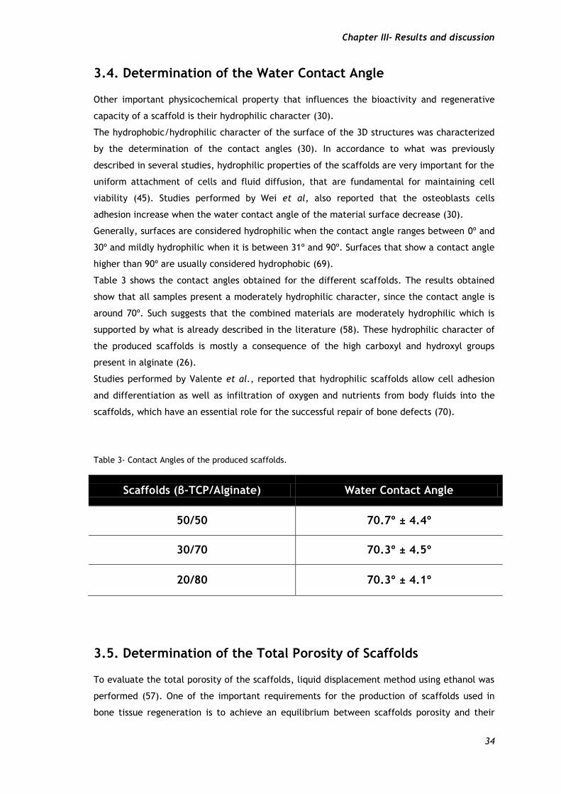
Chapter III- Results and discussion
34
3.4. Determination of the Water Contact Angle
Other important physicochemical property that influences the bioactivity and regenerative
capacity of a scaffold is their hydrophilic character (30).
The hydrophobic/hydrophilic character of the surface of the 3D structures was characterized
by the determination of the contact angles (30). In accordance to what was previously
described in several studies, hydrophilic properties of the scaffolds are very important for the
uniform attachment of cells and fluid diffusion, that are fundamental for maintaining cell
viability (45). Studies performed by Wei et al, also reported that the osteoblasts cells
adhesion increase when the water contact angle of the material surface decrease (30).
Generally, surfaces are considered hydrophilic when the contact angle ranges between 0º and
30º and mildly hydrophilic when it is between 31º and 90º. Surfaces that show a contact angle
higher than 90º are usually considered hydrophobic (69).
Table 3 shows the contact angles obtained for the different scaffolds. The results obtained
show that all samples present a moderately hydrophilic character, since the contact angle is
around 70º. Such suggests that the combined materials are moderately hydrophilic which is
supported by what is already described in the literature (58). These hydrophilic character of
the produced scaffolds is mostly a consequence of the high carboxyl and hydroxyl groups
present in alginate (26).
Studies performed by Valente et al., reported that hydrophilic scaffolds allow cell adhesion
and differentiation as well as infiltration of oxygen and nutrients from body fluids into the
scaffolds, which have an essential role for the successful repair of bone defects (70).
Table 3- Contact Angles of the produced scaffolds.
Scaffolds (β-TCP/Alginate) Water Contact Angle
50/50 70.7º ± 4.4º
30/70 70.3º ± 4.5º
20/80 70.3º ± 4.1º
3.5. Determination of the Total Porosity of Scaffolds
To evaluate the total porosity of the scaffolds, liquid displacement method using ethanol was
performed (57). One of the important requirements for the production of scaffolds used in
bone tissue regeneration is to achieve an equilibrium between scaffolds porosity and their
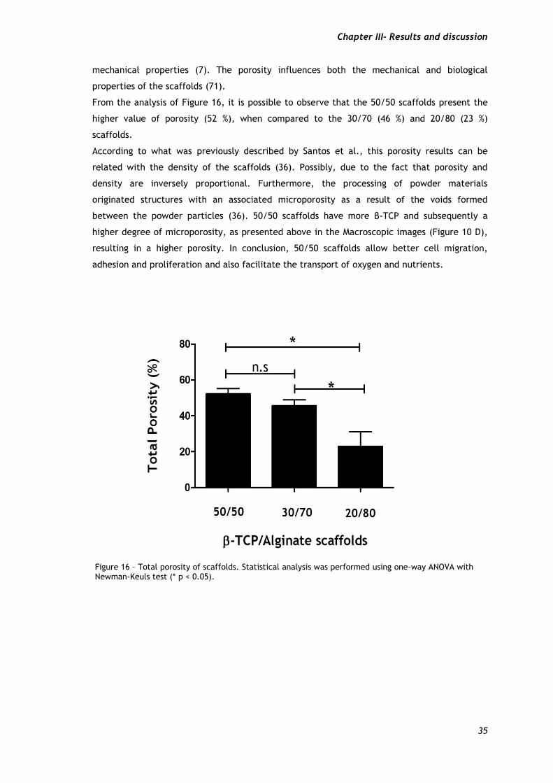
Chapter III- Results and discussion
35
mechanical properties (7). The porosity influences both the mechanical and biological
properties of the scaffolds (71).
From the analysis of Figure 16, it is possible to observe that the 50/50 scaffolds present the
higher value of porosity (52 %), when compared to the 30/70 (46 %) and 20/80 (23 %)
scaffolds.
According to what was previously described by Santos et al., this porosity results can be
related with the density of the scaffolds (36). Possibly, due to the fact that porosity and
density are inversely proportional. Furthermore, the processing of powder materials
originated structures with an associated microporosity as a result of the voids formed
between the powder particles (36). 50/50 scaffolds have more β-TCP and subsequently a
higher degree of microporosity, as presented above in the Macroscopic images (Figure 10 D),
resulting in a higher porosity. In conclusion, 50/50 scaffolds allow better cell migration,
adhesion and proliferation and also facilitate the transport of oxygen and nutrients.
Figure 16 – Total porosity of scaffolds. Statistical analysis was performed using one-way ANOVA with Newman-Keuls test (* p < 0.05).
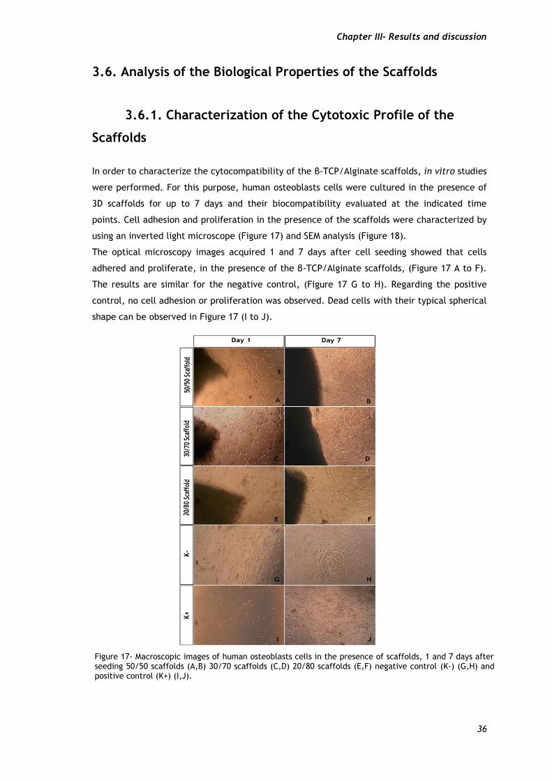
Chapter III- Results and discussion
36
3.6. Analysis of the Biological Properties of the Scaffolds
3.6.1. Characterization of the Cytotoxic Profile of the
Scaffolds
In order to characterize the cytocompatibility of the β-TCP/Alginate scaffolds, in vitro studies
were performed. For this purpose, human osteoblasts cells were cultured in the presence of
3D scaffolds for up to 7 days and their biocompatibility evaluated at the indicated time
points. Cell adhesion and proliferation in the presence of the scaffolds were characterized by
using an inverted light microscope (Figure 17) and SEM analysis (Figure 18).
The optical microscopy images acquired 1 and 7 days after cell seeding showed that cells
adhered and proliferate, in the presence of the β-TCP/Alginate scaffolds, (Figure 17 A to F).
The results are similar for the negative control, (Figure 17 G to H). Regarding the positive
control, no cell adhesion or proliferation was observed. Dead cells with their typical spherical
shape can be observed in Figure 17 (I to J).
Figure 17- Macroscopic images of human osteoblasts cells in the presence of scaffolds, 1 and 7 days after seeding 50/50 scaffolds (A,B) 30/70 scaffolds (C,D) 20/80 scaffolds (E,F) negative control (K-) (G,H) and positive control (K+) (I,J).
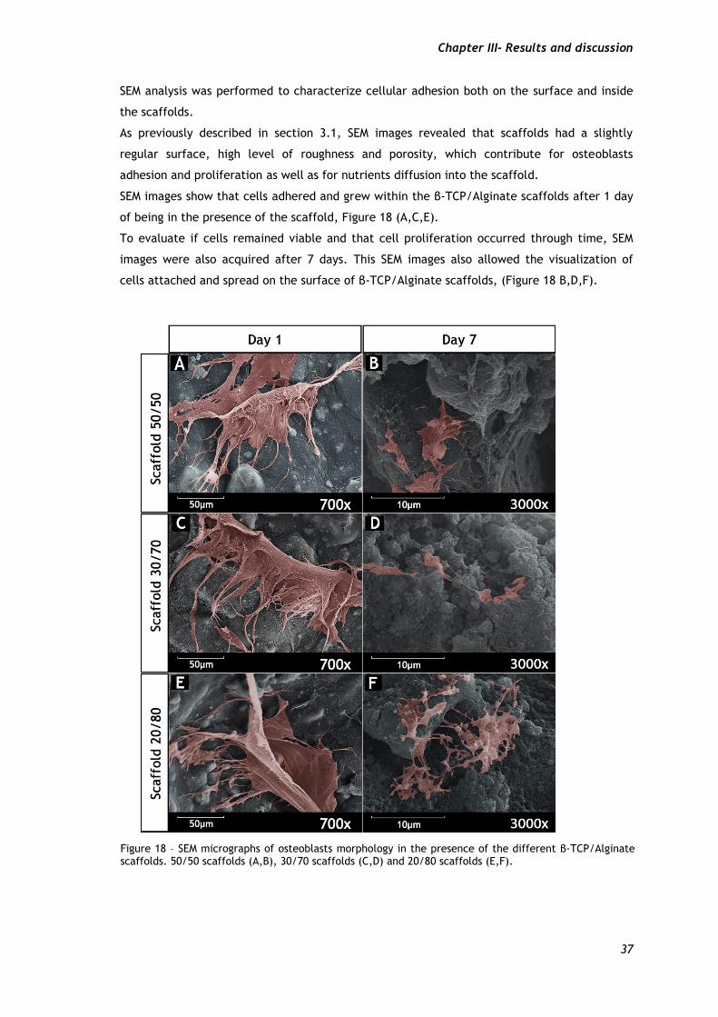
Chapter III- Results and discussion
37
SEM analysis was performed to characterize cellular adhesion both on the surface and inside
the scaffolds.
As previously described in section 3.1, SEM images revealed that scaffolds had a slightly
regular surface, high level of roughness and porosity, which contribute for osteoblasts
adhesion and proliferation as well as for nutrients diffusion into the scaffold.
SEM images show that cells adhered and grew within the β-TCP/Alginate scaffolds after 1 day
of being in the presence of the scaffold, Figure 18 (A,C,E).
To evaluate if cells remained viable and that cell proliferation occurred through time, SEM
images were also acquired after 7 days. This SEM images also allowed the visualization of
cells attached and spread on the surface of β-TCP/Alginate scaffolds, (Figure 18 B,D,F).
Figure 18 – SEM micrographs of osteoblasts morphology in the presence of the different β-TCP/Alginate scaffolds. 50/50 scaffolds (A,B), 30/70 scaffolds (C,D) and 20/80 scaffolds (E,F).
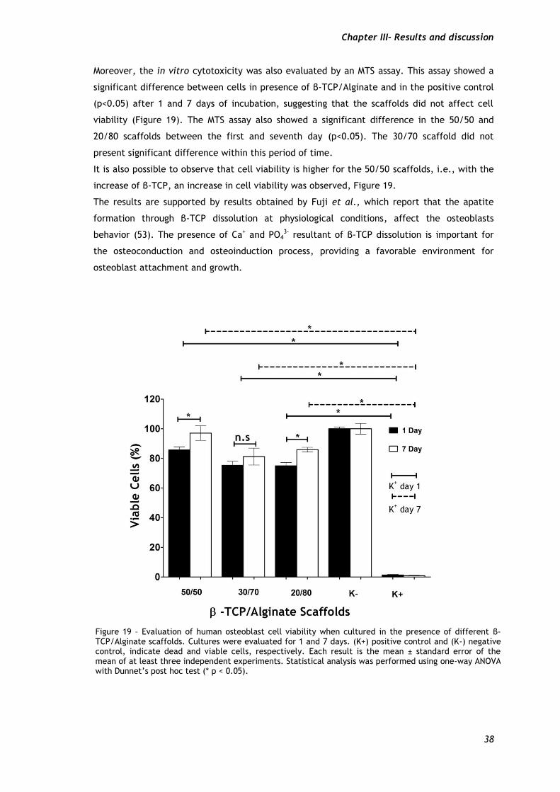
Chapter III- Results and discussion
38
Moreover, the in vitro cytotoxicity was also evaluated by an MTS assay. This assay showed a
significant difference between cells in presence of β-TCP/Alginate and in the positive control
(p<0.05) after 1 and 7 days of incubation, suggesting that the scaffolds did not affect cell
viability (Figure 19). The MTS assay also showed a significant difference in the 50/50 and
20/80 scaffolds between the first and seventh day (p<0.05). The 30/70 scaffold did not
present significant difference within this period of time.
It is also possible to observe that cell viability is higher for the 50/50 scaffolds, i.e., with the
increase of β-TCP, an increase in cell viability was observed, Figure 19.
The results are supported by results obtained by Fuji et al., which report that the apatite
formation through β-TCP dissolution at physiological conditions, affect the osteoblasts
behavior (53). The presence of Ca+ and PO43- resultant of β-TCP dissolution is important for
the osteoconduction and osteoinduction process, providing a favorable environment for
osteoblast attachment and growth.
Figure 19 – Evaluation of human osteoblast cell viability when cultured in the presence of different β-TCP/Alginate scaffolds. Cultures were evaluated for 1 and 7 days. (K+) positive control and (K-) negative control, indicate dead and viable cells, respectively. Each result is the mean ± standard error of the mean of at least three independent experiments. Statistical analysis was performed using one-way ANOVA with Dunnet’s post hoc test (* p < 0.05).
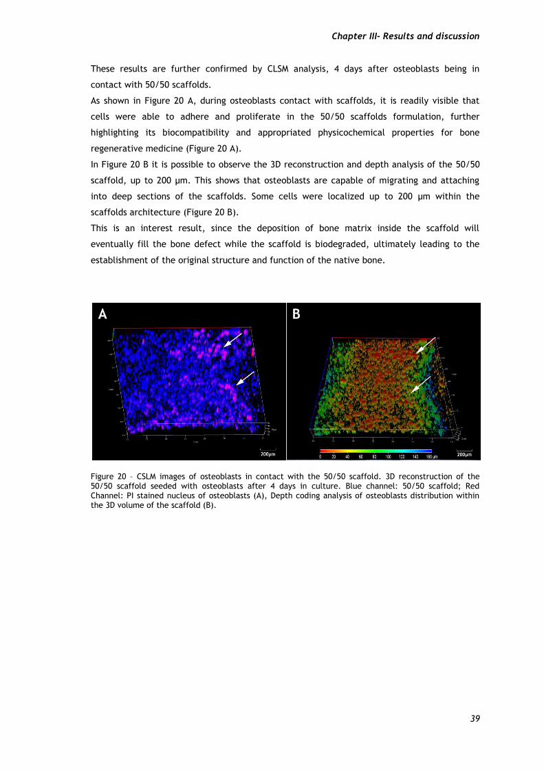
Chapter III- Results and discussion
39
These results are further confirmed by CLSM analysis, 4 days after osteoblasts being in
contact with 50/50 scaffolds.
As shown in Figure 20 A, during osteoblasts contact with scaffolds, it is readily visible that
cells were able to adhere and proliferate in the 50/50 scaffolds formulation, further
highlighting its biocompatibility and appropriated physicochemical properties for bone
regenerative medicine (Figure 20 A).
In Figure 20 B it is possible to observe the 3D reconstruction and depth analysis of the 50/50
scaffold, up to 200 µm. This shows that osteoblasts are capable of migrating and attaching
into deep sections of the scaffolds. Some cells were localized up to 200 µm within the
scaffolds architecture (Figure 20 B).
This is an interest result, since the deposition of bone matrix inside the scaffold will
eventually fill the bone defect while the scaffold is biodegraded, ultimately leading to the
establishment of the original structure and function of the native bone.
Figure 20 – CSLM images of osteoblasts in contact with the 50/50 scaffold. 3D reconstruction of the 50/50 scaffold seeded with osteoblasts after 4 days in culture. Blue channel: 50/50 scaffold; Red Channel: PI stained nucleus of osteoblasts (A), Depth coding analysis of osteoblasts distribution within the 3D volume of the scaffold (B).

Chapter IV – Conclusions and Future Perspectives

Chapter IV- Conclusions and Future Perspectives
41
4. Conclusions and Future Perspectives
Bone defects have been increasing, affecting millions of people worldwide. Temporary and
biodegradable scaffolds are a good choice to overcome this problem. Nowadays, several
techniques are used for the production of scaffolds. Rapid prototyping technologies allow the
construction of 3D models with a well-defined structure and porosity, presenting some
advantages over conventional techniques. Fab@home model is one of the newer printers used
for production of 3D structures to be used in bone tissue regeneration. However, further
studies must be performed in order to increase the suitability of this equipment for this
particular biomedical application. In this study β-TCP/Alginate scaffolds were produced by
rapid prototyping using a Fab@home model. Three different groups of scaffolds were
produced, varying the ratios between the concentrations of β-TCP and Alginate. Respectively,
50/50 % (w/w), 30/70 % (w/w) and 20/80 % (w/w), B-TCP/Alginate scaffolds.
The physicochemical results (FTIR, XRD and EDS) showed, that after all the manufacturing
process all samples have a highly crystalline structure and contain in their composition
calcium and phosphate elements. The produced scaffolds are porous structures with
hydrophilic character, which allows an improved cell adhesion and fluid diffusion within
scaffolds. SEM images show that scaffolds present a rough surface. The in vitro cytotoxic
assays performed show a good cell-scaffold interaction and biocompatibility, enhanced in the
50/50 scaffolds. Confocal scanning laser microscopy shows the osteoblasts distribution within
the 50/50 scaffolds 4 days after osteoblasts be in the presence of scaffolds.
It is possible to conclude that the 50/50 scaffolds present better mechanical and biological
properties than the 30/70 and 20/80 scaffolds, respectively. The results suggest that the
scaffolds with higher β-TCP percentage have better conditions for the osteogenic process and
better mechanical properties than the ones with higher alginate polymer percentage.
Mechanical properties results show that 50/50 scaffolds have more compressive strength and
also Young´s modulus.
In a near future, an enhancement of the mechanical properties would be to optimize the pore
size, in order to improve the mechanical resistance of the scaffolds. Another approach would
be the addition of a ceramic compound such as hydroxyapatite for improving the mechanical
resistance. So, the scaffold will maintain their bioactive property but with a better
resistance. Furthermore, methodologies like the application of a coating of a resistant
material, through techniques such as electro-spinning, can possible be used to improve the
mechanical properties of the produced scaffolds.

Chapter V – Bibliography
Chapter V- Bibliography
43
1. Ralston SH. Structure and metabolism of bone. Medicine. 2005;33(12):58-60.
2. Seeley RR, Stephens TD, Tate P. Anatomia & Fisiologia. 8th ed: Lusociência-Edições
Técnicas e Científicas, Lda.; 2008.
3. Van De Graaff KM. Human Anatomy. 6th ed: The McGraw-Hill Companies, Inc.; 2002.
4. Marieb EN, Hoehn K. Human Anatomy & Physiology. 7th ed: Pearson Education, Inc.;
2007.
5. Alvarez K, Nakajima H. Metallic scaffolds for bone regeneration. Materials.
2009;2(3):790-832.
6. Hsiong SX, Mooney DJ. Regeneration of vascularized bone. Periodontology 2000.
2006;41(1):109-22.
7. Salgado AJ, Coutinho OP, Reis RL. Bone tissue engineering: state of the art and future
trends. Macromolecular bioscience. 2004;4(8):743-65.
8. Mackie E. Osteoblasts: novel roles in orchestration of skeletal architecture. The
international journal of biochemistry & cell biology. 2003;35(9):1301-5.
9. Neve A, Corrado A, Cantatore FP. Osteoblast physiology in normal and pathological
conditions. Cell and tissue research. 2011;343(2):289-302.
10. Mebarek S, Abousalham A, Magne D, Do LD, Bandorowicz-Pikula J, Pikula S, et al.
Phospholipases of mineralization competent cells and matrix vesicles: roles in physiological
and pathological mineralizations. International journal of molecular sciences.
2013;14(3):5036-129.
11. Jayakumar P, Di Silvio L. Osteoblasts in bone tissue engineering. Proceedings of the
institution of mechanical engineers, Part H: journal of engineering in medicine.
2010;224(12):1415-40.
12. Clarke B. Normal bone anatomy and physiology. Clinical journal of the american
society of nephrology. 2008;3(Supplement 3):S131-S9.
13. Jang J, Castano O, Kim H. Electrospun materials as potential platforms for bone tissue
engineering. Advanced drug delivery reviews. 2009;61(12):1065-83.
Chapter V- Bibliography
44
14. Hadjidakis DJ, Androulakis II. Bone remodeling. Annals of the new york academy of
sciences. 2006;1092(1):385-96.
15. Robling AG, Castillo AB, Turner CH. Biomechanical and molecular regulation of bone
remodeling. Annual review of biomedical engineering 2006;8(1):455-98.
16. Raisz LG. Physiology and pathophysiology of bone remodeling. Clinical chemistry.
1999;45(8):1353-8.
17. Chai YC, Carlier A, Bolander J, Roberts SJ, Geris L, Schrooten J, et al. Current views
on calcium phosphate osteogenicity and the translation into effective bone regeneration
strategies. Acta biomaterialia. 2012;8(11):3876–87.
18. Chiara G, Letizia F, Lorenzo F, Edoardo S, Diego S, Stefano S, et al. Nanostructured
biomaterials for tissue engineered bone tissue reconstruction. International journal of
molecular sciences. 2012;13(1):737-57.
19. Williams DF. To engineer is to create: the link between engineering and regeneration.
Trends in biotechnology. 2006;24(1):4-8.
20. Djouad F, Guerit D, Marie M, Toupet K, Jorgensen C, Noel D. Mesenchymal Stem Cells:
New insights into bone regenerative applications. Journal of biomaterials and tissue
engineering. 2012;2(1):14-28.
21. Hutmacher DW, Sittinger M, Risbud MV. Scaffold-based tissue engineering: rationale
for computer-aided design and solid free-form fabrication systems. Trends in biotechnology.
2004;22(7):354-62.
22. Kneser U, Schaefer D, Polykandriotis E, Horch R. Tissue engineering of bone: the
reconstructive surgeon's point of view. Journal of cellular and molecular medicine.
2006;10(1):7-19.
23. Giannoudis PV, Dinopoulos H, Tsiridis E. Bone substitutes: an update. Injury.
2005;36(3):S20-S7.
24. Hutmacher DW. Scaffold design and fabrication technologies for engineering tissues—
state of the art and future perspectives. Journal of biomaterials science, polymer edition.
2001;12(1):107-24.
Chapter V- Bibliography
45
25. Bhat S, Kumar A. Biomaterials and bioengineering tomorrow’s healthcare. Biomatter.
2013;3(2):e24717.
26. Valente J, Valente T, Alves P, Ferreira P, Silva A, Correia I. Alginate based scaffolds
for bone tissue engineering. Materials science and engineering: C. 2012;32:2596–603.
27. Dreifke MB, Ebraheim NA, Jayasuriya AC. Investigation of potential injectable
polymeric biomaterials for bone regeneration. Journal of biomedical materials research part
A. 2013;0.
28. Leong K, Cheah C, Chua C. Solid freeform fabrication of three-dimensional scaffolds
for engineering replacement tissues and organs. Biomaterials. 2003;24(13):2363-78.
29. Williams DF. On the mechanisms of biocompatibility. Biomaterials. 2008;29(20):2941-
53.
30. Chang H, Wang Y. Cell responses to surface and architecture of tissue engineering
scaffolds. Regenerative medicine and tissue engineering—cells and biomaterials,
InTech:2011;569-88.
31. Hollister SJ. Porous scaffold design for tissue engineering. Nature materials.
2005;4(7):518-24.
32. Dhandayuthapani B, Yoshida Y, Maekawa T, Kumar DS. Polymeric scaffolds in tissue
engineering application: a review. International journal of polymer science. 2011;2011:1-19.
33. Yoon H, Kim GH, Koh YH. A Micro-scale surface-structured PCL scaffold fabricated by
a 3D plotter and a chemical blowing agent. Journal of biomaterials science, polymer edition.
2010;21(2):159-70.
34. Hutmacher DW, Schantz JT, Lam CXF, Tan KC, Lim TC. State of the art and future
directions of scaffold‐based bone engineering from a biomaterials perspective. Journal of
tissue engineering and regenerative medicine. 2007;1(4):245-60.
35. Suzuki T, Yamamoto T, Toriyama M, Nishizawa K, Yokogawa Y, Mucalo MR, et al.
Surface instability of calcium phosphate ceramics in tissue culture medium and the effect on
adhesion and growth of anchorage‐dependent animal cells. Journal of biomedical materials
research. 1997;34(4):507-17.
Chapter V- Bibliography
46
36. Santos CF, Silva AP, Lopes L, Pires I, Correia IJ. Design and production of sintered β-
tricalcium phosphate 3D scaffolds for bone tissue regeneration. Materials science and
engineering: C. 2012;32(5):1293-8.
37. Li X, Bian W, Li D, Lian Q, Jin Z. Fabrication of porous beta-tricalcium phosphate with
microchannel and customized geometry based on gel-casting and rapid prototyping.
Proceedings of the institution of mechanical engineers, Part H: journal of engineering in
medicine. 2011;225(3):315-23.
38. Yang Y, Bumgardner J, Cavin R, Carnes D, Ong J. Osteoblast precursor cell
attachment on heat-treated calcium phosphate coatings. Journal of dental research.
2003;82(6):449-53.
39. Christensena BE. Alginates as biomaterials in tissue engineering. Carbohydrate
chemistry: chemical and biological approaches. 2011;37:227-58.
40. LeRoux MA, Guilak F, Setton LA. Compressive and shear properties of alginate gel:
effects of sodium ions and alginate concentration. Journal of biomedical materials research.
1999;47(1):46-53.
41. Daemi H, Barikani M. Synthesis and characterization of calcium alginate
nanoparticles, sodium homopolymannuronate salt and its calcium nanoparticles. Scientia
iranica. 2012;19(6):2023–8.
42. Lantada AD, Morgado PL. Rapid Prototyping for biomedical engineering: current
capabilities and challenges. Annual review of biomedical engineering. 2012;14:73-96.
43. Seitz H, Rieder W, Irsen S, Leukers B, Tille C. Three‐dimensional printing of porous
ceramic scaffolds for bone tissue engineering. Journal of biomedical materials research Part
B: applied biomaterials. 2005;74(2):782-8.
44. Nandakumar A, Barradas A, de Boer J, Moroni L, van Blitterswijk C, Habibovic P.
Combining technologies to create bioactive hybrid scaffolds for bone tissue engineering.
Biomatter. 2013;3(1):1-13.
45. Yeo MG, Kim GH. Preparation and characterization of 3D composite scaffolds based on
rapid-prototyped PCL/β-TCP struts and electrospun PCL coated with collagen and HA for bone
regeneration. Chemistry of materials. 2011;24(5):903-13.
Chapter V- Bibliography
47
46. Stevens B, Yang Y, Mohandas A, Stucker B, Nguyen KT. A review of materials,
fabrication methods, and strategies used to enhance bone regeneration in engineered bone
tissues. Journal of biomedical materials research Part B: applied biomaterials.
2008;85(2):573-82.
47. Ang T, Sultana F, Hutmacher D, Wong YS, Fuh J, Mo X, et al. Fabrication of 3D
chitosan–hydroxyapatite scaffolds using a robotic dispensing system. Materials science and
engineering: C. 2002;20(1):35-42.
48. Wüst S, Müller R, Hofmann S. Controlled positioning of cells in biomaterials—
approaches towards 3D tissue printing. Journal of functional biomaterials. 2011;2(3):119-54.
49. Lixandrão Filho A, Cheung P, Noritomi P, da Silva J, Colangelo N, Kang H, et al.
Construction and adaptation of an open source rapid prototyping machine for biomedical
research purposes-a multinational collaborative development. Innovative developments in
design and manufacturing. 2009:469-73.
50. Kang K, Hockaday L, Butcher J. Quantitative optimization of solid freeform deposition
of aqueous hydrogels. Biofabrication. 2013;5(3):1-13.
51. Cohen DL, Lo W, Tsavaris A, Peng D, Lipson H, Bonassar LJ. Increased mixing improves
hydrogel homogeneity and quality of three-dimensional printed constructs. Tissue engineering
Part C: methods. 2010;17(2):239-48.
52. Vivanco J, Aiyangar A, Araneda A, Ploeg H-L. Mechanical characterization of
injection-molded macro porous bioceramic bone scaffolds. Journal of the mechanical
behavior of biomedical materials. 2012;9:137-52.
53. Fuji T, Anada T, Honda Y, Shiwaku Y, Koike H, Kamakura S, et al. Octacalcium
Phosphate–Precipitated Alginate Scaffold for Bone Regeneration. Tissue engineering Part A.
2009;15(11):3525-35.
54. Wu X, Liu Y, Li X, Wen P, Zhang Y, Long Y, et al. Preparation of aligned porous gelatin
scaffolds by unidirectional freeze-drying method. Acta biomaterialia. 2010;6(3):1167-77.
55. Lacroix D, Chateau A, Ginebra M-P, Planell JA. Micro-finite element models of bone
tissue-engineering scaffolds. Biomaterials. 2006;27(30):5326-34.
Chapter V- Bibliography
48
56. Correia TR, Antunes BP, Castilho PH, Nunes JC, Pessoa de Amorim MT, Escobar IC, et
al. A bi-layer electrospun nanofiber membrane for plasmid DNA recovery from fermentation
broths. Separation and purification technology. 2013;112:20-5.
57. Nie H-L, Zhu L-M. Adsorption of papain with Cibacron Blue F3GA carrying chitosan-
coated nylon affinity membranes. International journal of biological macromolecules.
2007;40(3):261-7.
58. Cho SH, Lim SM, Han DK, Yuk SH, Im GI, Lee JH. Time-dependent alginate/polyvinyl
alcohol hydrogels as injectable cell carriers. Journal of biomaterials science, polymer edition.
2009;20(7-8):863-76.
59. Karageorgiou V, Kaplan D. Porosity of 3D biomaterial scaffolds and osteogenesis.
Biomaterials. 2005;26(27):5474-91.
60. Dos Santos E, Farina M, Soares G, Anselme K. Chemical and topographical influence of
hydroxyapatite and β‐tricalcium phosphate surfaces on human osteoblastic cell behavior.
Journal of biomedical materials research Part A. 2009;89(2):510-20.
61. Navarro M, Aparicio C, Charles-Harris M, Ginebra M, Engel E, Planell J. Development
of a biodegradable composite scaffold for bone tissue engineering: physicochemical,
topographical, mechanical, degradation, and biological properties. Ordered polymeric
nanostructures at surfaces. 200: Springer; 2006:209-31.
62. Lenhert S, Meier M-B, Meyer U, Chi L, Wiesmann HP. Osteoblast alignment, elongation
and migration on grooved polystyrene surfaces patterned by Langmuir–Blodgett lithography.
Biomaterials. 2005;26(5):563-70.
63. Pathak TS, San Kim J, Lee S-J, Baek D-J, Paeng K-J. Preparation of alginic acid and
metal alginate from algae and their comparative study. Journal of polymers and the
environment. 2008;16(3):198-204.
64. Lawrie G, Keen I, Drew B, Chandler-Temple A, Rintoul L, Fredericks P, et al.
Interactions between alginate and chitosan biopolymers characterized using FTIR and XPS.
Biomacromolecules. 2007;8(8):2533-41.
65. Lambert JB, Shurvell HF, Lightner DA, Cooks RG. Organic structural spectroscopy.:
Prentice Hall; 1998.
Chapter V- Bibliography
49
66. Berube P, Yang Y, Carnes DL, Stover RE, Boland EJ, Ong JL. The effect of sputtered
calcium phosphate coatings of different crystallinity on osteoblast differentiation. Journal of
periodontology. 2005;76(10):1697-709.
67. Matsuno T, Hashimoto Y, Adachi S, Omata K, Yoshitaka Y, Ozeki Y, et al. Preparation
of injectable 3D-formed. BETA.-tricalcium phosphate bead/alginate composite for bone tissue
engineering. Dental materials journal. 2008;27(6):827-34.
68. Ghanaati S, Barbeck M, Detsch R, Deisinger U, Hilbig U, Rausch V, et al. The chemical
composition of synthetic bone substitutes influences tissue reactions in vivo: histological and
histomorphometrical analysis of the cellular inflammatory response to hydroxyapatite, beta-
tricalcium phosphate and biphasic calcium phosphate ceramics. Biomedical materials.
2012;7(1):015005.
69. Shalumon K, Anulekha K, Chennazhi K, Tamura H, Nair S, Jayakumar R. Fabrication of
chitosan/poly (caprolactone) nanofibrous scaffold for bone and skin tissue engineering.
International journal of biological macromolecules. 2011;48(4):571-6.
70. Wang C, Meng G, Zhang L, Xiong Z, Liu J. Physical properties and biocompatibility of a
core-sheath structure composite scaffold for bone tissue engineering in vitro. BioMed
Research International. 2012;2012.
71. Tarafder S, Balla VK, Davies NM, Bandyopadhyay A, Bose S. Microwave‐sintered 3D
printed tricalcium phosphate scaffolds for bone tissue engineering. Journal of tissue
engineering and regenerative medicine. 2012.