Understanding biology through structures Course work 2006 Structure Determination and Analysis :...
69
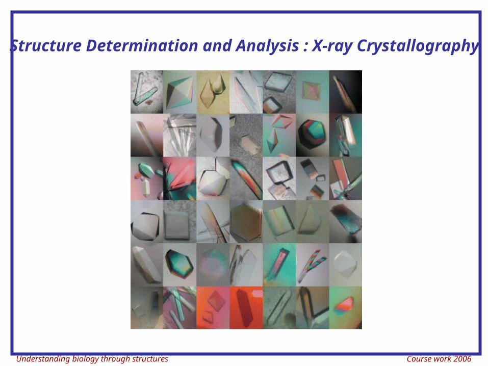
Understanding biology through structures Course work 2006 Structure Determination and Analysis : X-ray Crystallography
-
Upload
cathleen-armstrong -
Category
Documents
-
view
224 -
download
2
Transcript of Understanding biology through structures Course work 2006 Structure Determination and Analysis :...
Understanding biology through structures Course work 2006
Structure Determination and Analysis : X-ray Crystallography
Understanding biology through structures Course work 2006
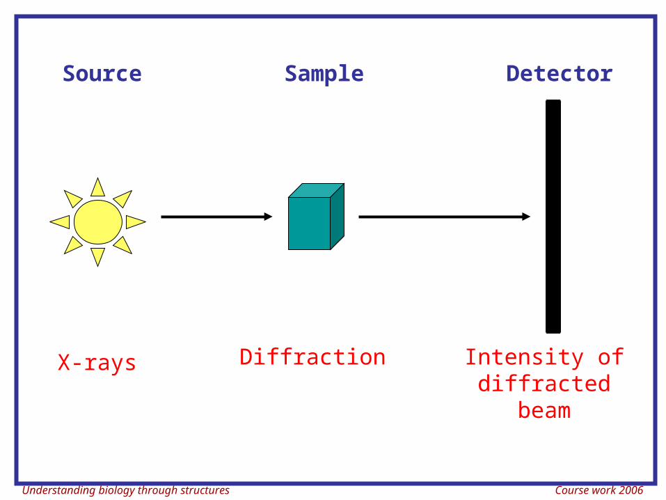
X-rays Intensity of diffracted beam
Diffraction
Source Sample Detector
Understanding biology through structures Course work 2006
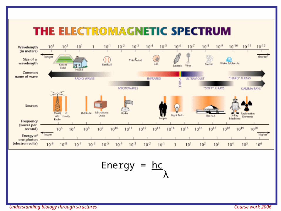
Energy = hc λ
Understanding biology through structures Course work 2006
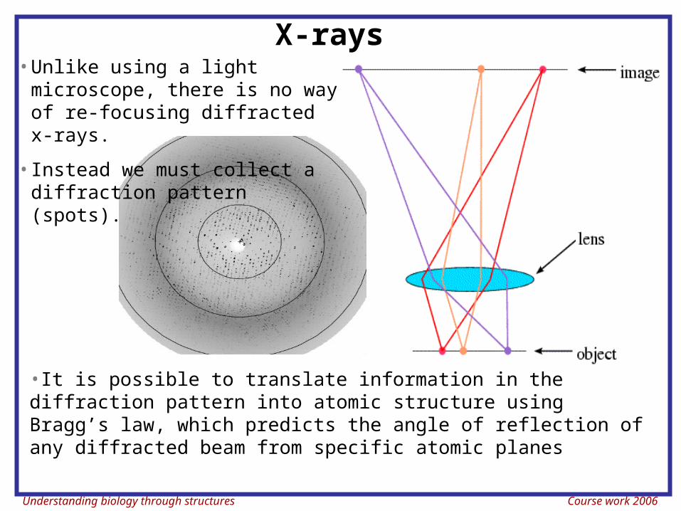
X-rays
•It is possible to translate information in the diffraction pattern into atomic structure using Bragg’s law, which predicts the angle of reflection of any diffracted beam from specific atomic planes
•Unlike using a light microscope, there is no way of re-focusing diffracted x-rays.
•Instead we must collect a diffraction pattern (spots).
Understanding biology through structures Course work 2006
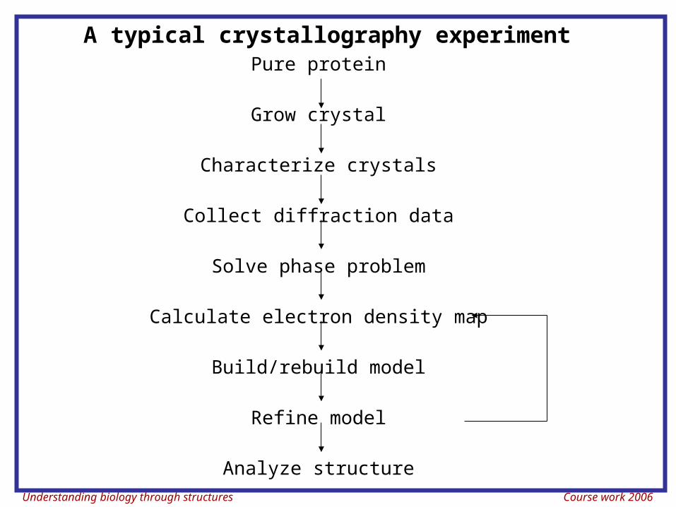
A typical crystallography experimentPure protein
Grow crystal
Characterize crystals
Collect diffraction data
Solve phase problem
Calculate electron density map
Build/rebuild model
Refine model
Analyze structure
Understanding biology through structures Course work 2006
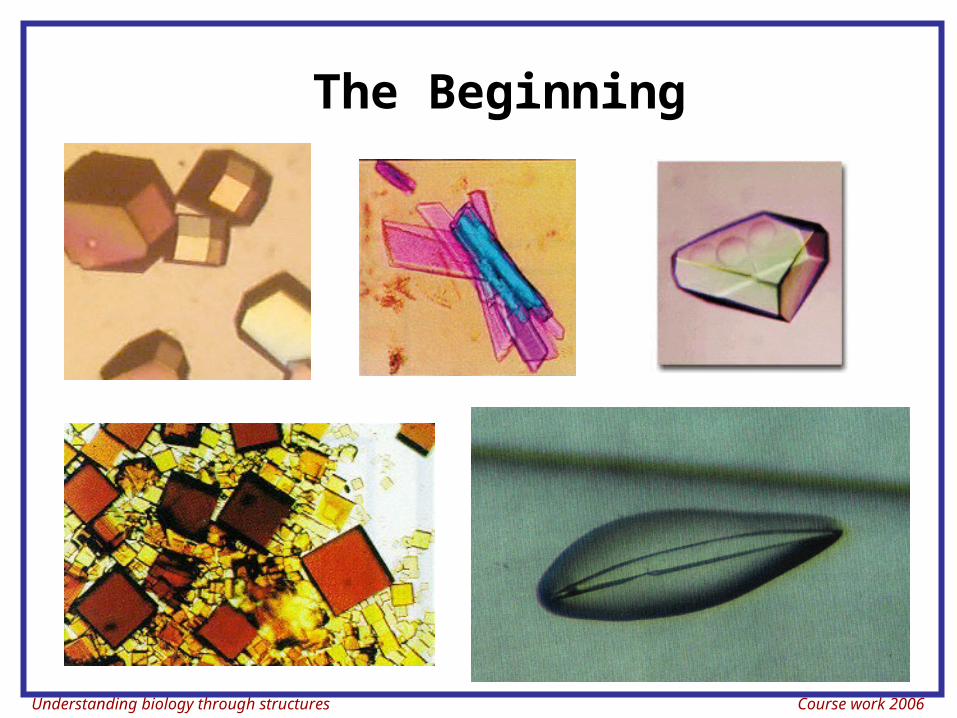
The Beginning
Understanding biology through structures Course work 2006
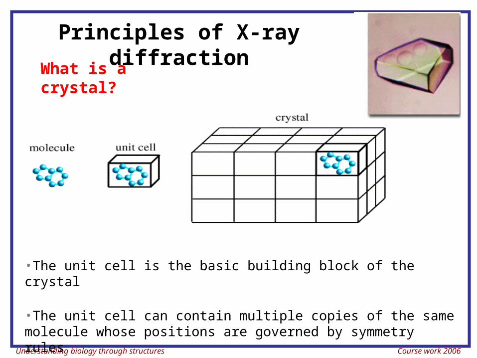
Principles of X-ray diffractionWhat is a
crystal?
•The unit cell is the basic building block of the crystal
•The unit cell can contain multiple copies of the same molecule whose positions are governed by symmetry rules
Understanding biology through structures Course work 2006
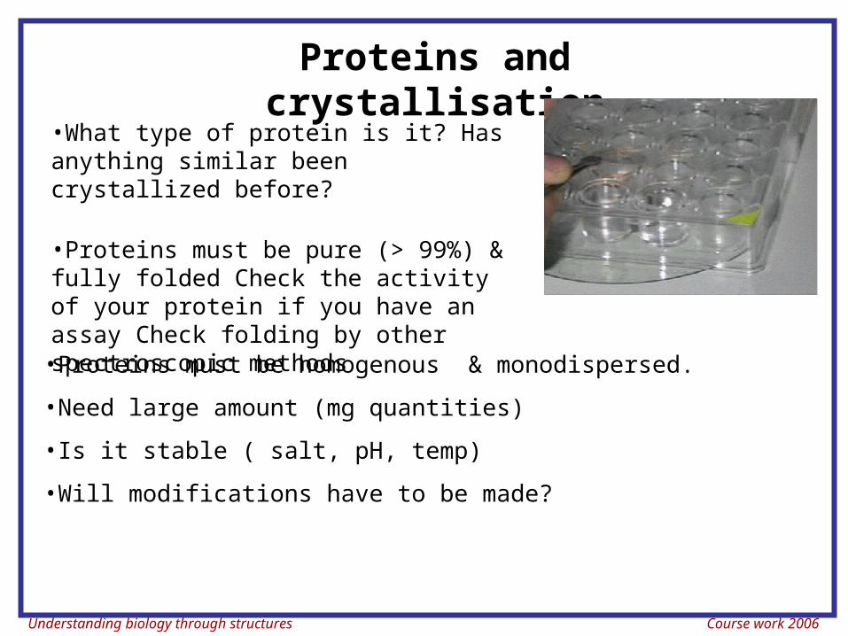
Proteins and crystallisation
•Proteins must be homogenous & monodispersed.
•Need large amount (mg quantities)
•Is it stable ( salt, pH, temp)
•Will modifications have to be made?
•What type of protein is it? Has anything similar been crystallized before?
•Proteins must be pure (> 99%) & fully folded Check the activity of your protein if you have an assay Check folding by other spectroscopic methods
Understanding biology through structures Course work 2006
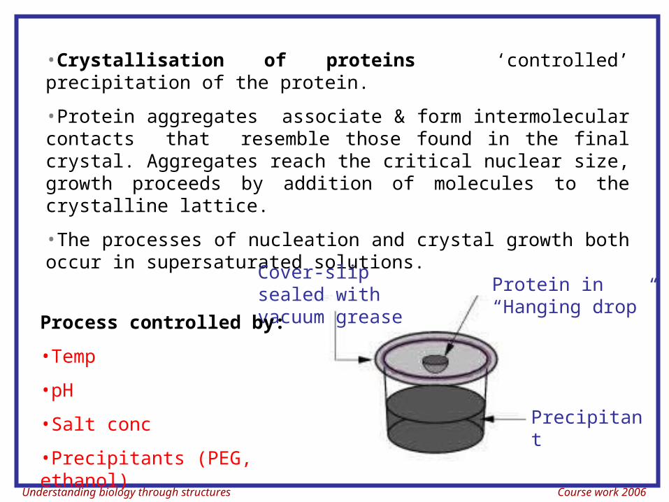
•Crystallisation of proteins ‘controlled’ precipitation of the protein.
•Protein aggregates associate & form intermolecular contacts that resemble those found in the final crystal. Aggregates reach the critical nuclear size, growth proceeds by addition of molecules to the crystalline lattice.
•The processes of nucleation and crystal growth both occur in supersaturated solutions.
Precipitant
Cover-slip sealed with vacuum grease
Protein in “Hanging drop”
Process controlled by:
•Temp
•pH
•Salt conc
•Precipitants (PEG, ethanol)
Understanding biology through structures Course work 2006
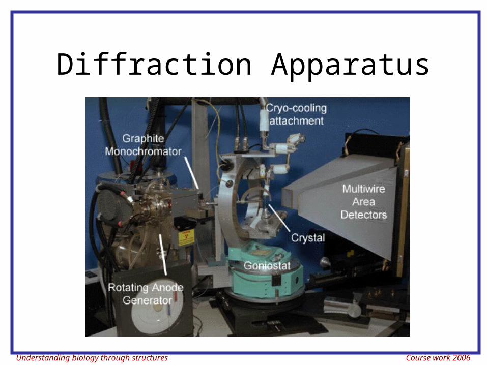
Diffraction Apparatus
Understanding biology through structures Course work 2006
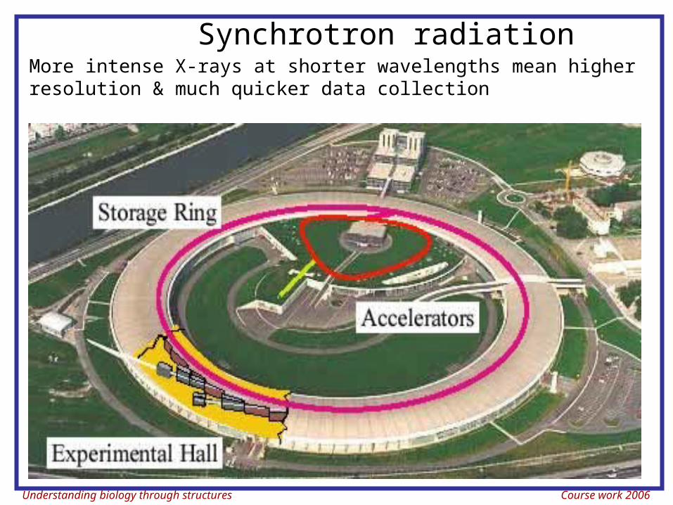
Synchrotron radiationMore intense X-rays at shorter wavelengths mean higher resolution & much quicker data collection
Understanding biology through structures Course work 2006
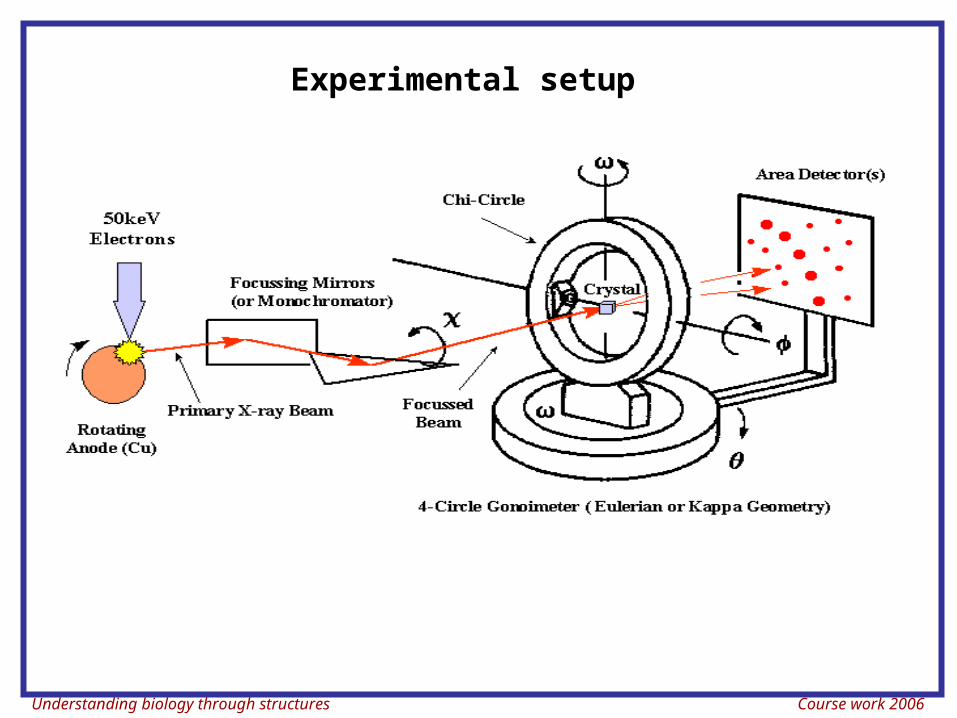
Experimental setup
Understanding biology through structures Course work 2006
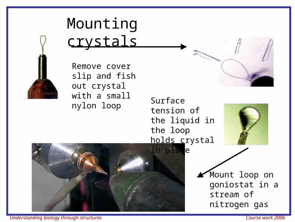
Remove cover slip and fish out crystal with a small nylon loop
Mount loop on goniostat in a stream of nitrogen gas
Surface tension of the liquid in the loop holds crystal in place
Mounting crystals
Understanding biology through structures Course work 2006
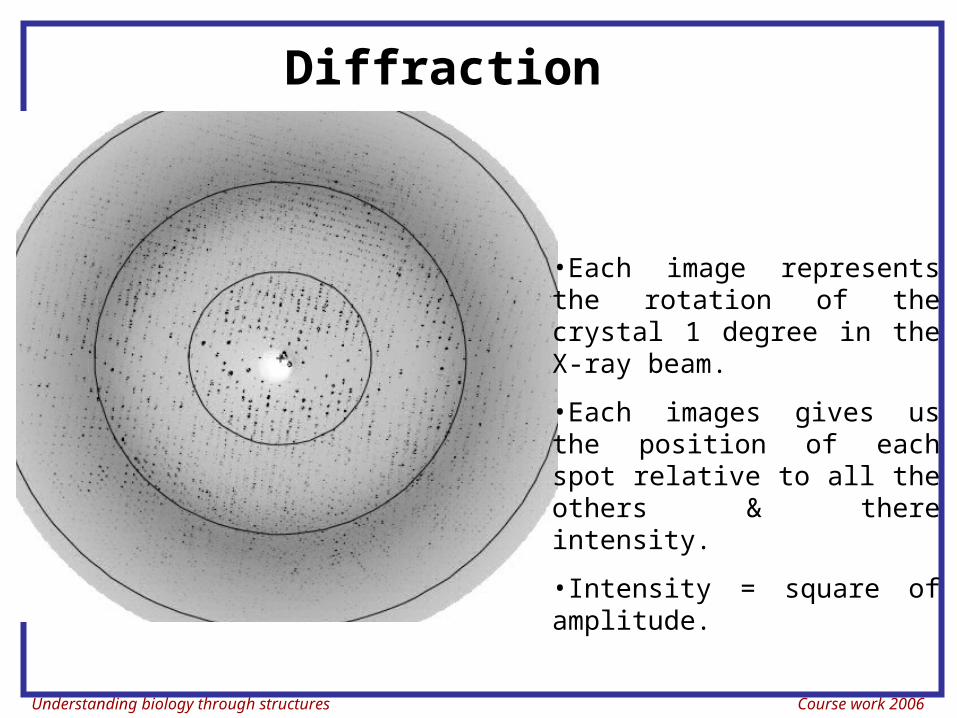
Diffraction
•Each image represents the rotation of the crystal 1 degree in the X-ray beam.
•Each images gives us the position of each spot relative to all the others & there intensity.
•Intensity = square of amplitude.
Understanding biology through structures Course work 2006
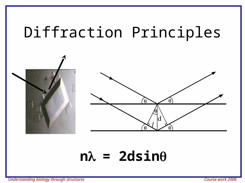
Diffraction Principles
n= 2dsin
Understanding biology through structures Course work 2006
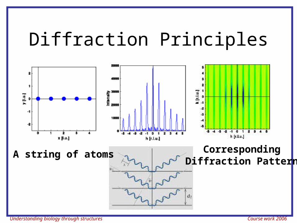
Diffraction Principles
A string of atoms CorrespondingDiffraction Pattern
Understanding biology through structures Course work 2006
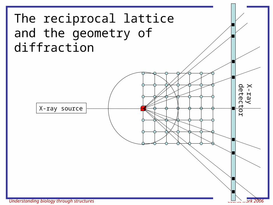
The reciprocal lattice and the geometry of diffraction
X-ray source
X-ra
y
dete
ctor
Understanding biology through structures Course work 2006
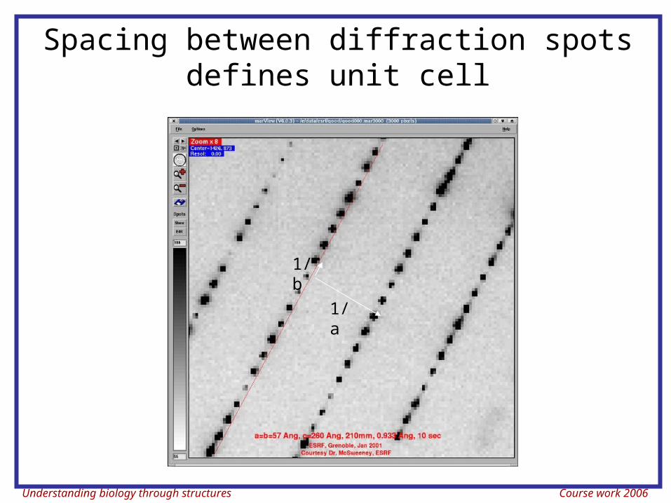
Spacing between diffraction spots defines unit cell
1/a
1/b
Understanding biology through structures Course work 2006
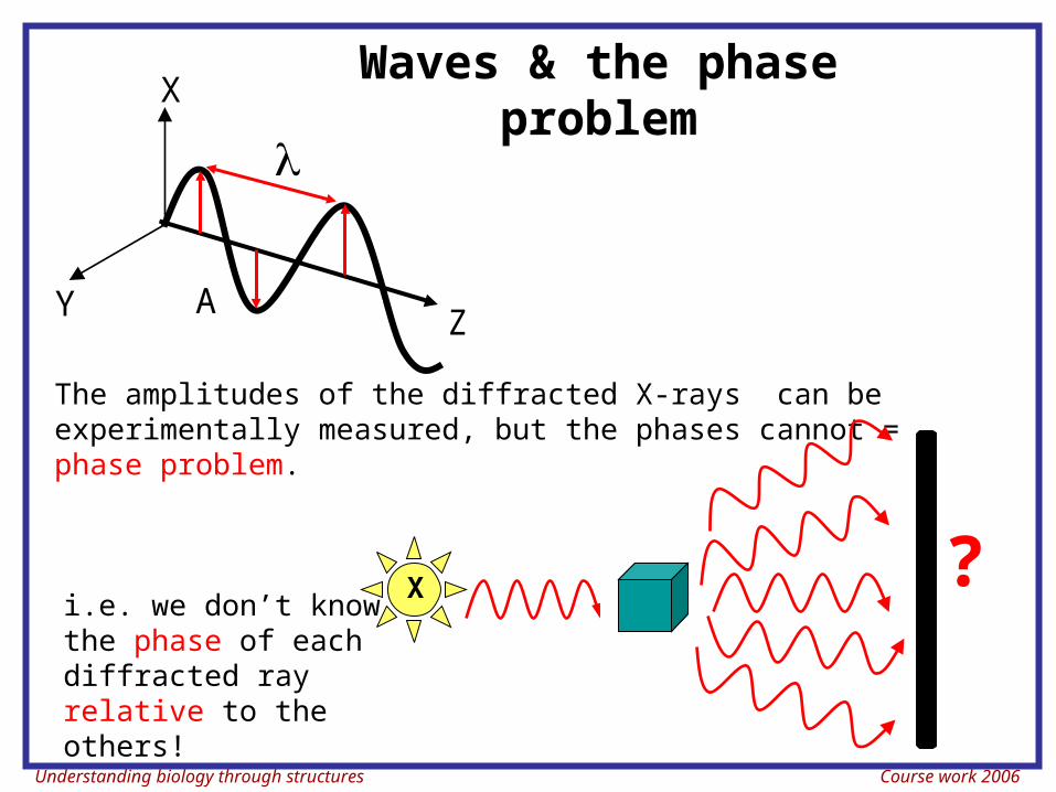
Waves & the phase problem
The amplitudes of the diffracted X-rays can be experimentally measured, but the phases cannot = phase problem.
i.e. we don’t know the phase of each diffracted ray relative to the others!
X ?
A
Z
X
Y
Understanding biology through structures Course work 2006
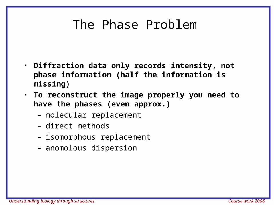
The Phase Problem
• Diffraction data only records intensity, not phase information (half the information is missing)
• To reconstruct the image properly you need to have the phases (even approx.)– molecular replacement– direct methods– isomorphous replacement– anomolous dispersion
Understanding biology through structures Course work 2006
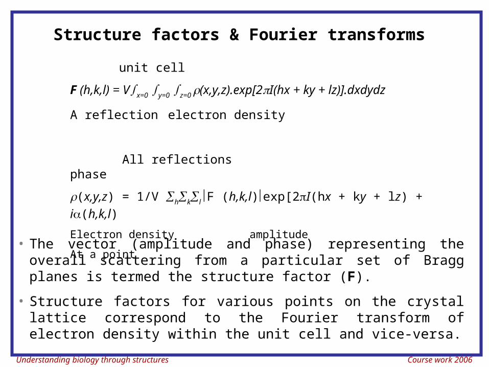
Structure factors & Fourier transforms
unit cell
F (h,k,l) = Vx=0 y=0 z=0 (x,y,z).exp[2I(hx + ky + lz)].dxdydz
A reflection electron density
All reflections phase
(x,y,z) = 1/V hkl F (h,k,l)exp[2I(hx + ky + lz) + i(h,k,l)
Electron density amplitude
At a point • The vector (amplitude and phase) representing the overall
scattering from a particular set of Bragg planes is termed the structure factor (F).
• Structure factors for various points on the crystal lattice correspond to the Fourier transform of electron density within the unit cell and vice-versa.
Understanding biology through structures Course work 2006
F T
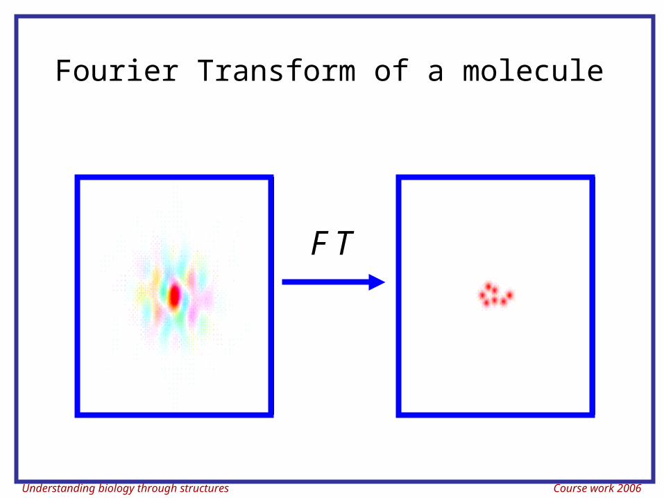
Fourier Transform of a molecule
Understanding biology through structures Course work 2006
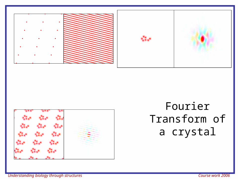
Fourier Transform of a
crystal

Understanding biology through structures Course work 2006
The Phase Problem
• Diffraction data only records intensity, not phase information (half the information is missing)
• To reconstruct the image properly you need to have the phases (even approx.)– molecular replacement– direct methods– isomorphous replacement– anomolous dispersion
Understanding biology through structures Course work 2006
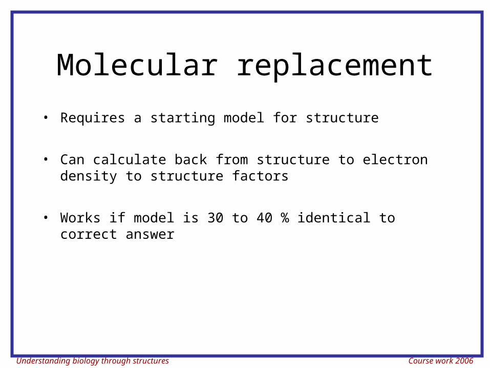
Molecular replacement
• Requires a starting model for structure
• Can calculate back from structure to electron density to structure factors
• Works if model is 30 to 40 % identical to correct answer
Understanding biology through structures Course work 2006
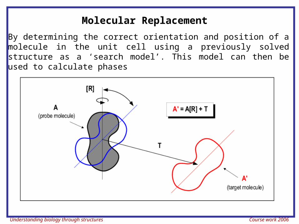
Molecular Replacement
By determining the correct orientation and position of a molecule in the unit cell using a previously solved structure as a ‘search model’. This model can then be used to calculate phases
Understanding biology through structures Course work 2006
Isomorphous replacement (IR)
• Provides indirect estimates of the protein phase angles by observing the interference effects of the intensities on scattered beams by a heavy atom marker.
• All the electrons in the heavy atom will scatter essentially in the same phase.
• We can solve the positions of these heavy atoms because they are few in number and strong in signal.
• Using this estimate we can deduce the positions of the protein atoms and their phases
Understanding biology through structures Course work 2006
Anomalous scattering
• Scattering information of an atom whose absorption frequency is close to the wavelength of the source beam produces phase information
• Resolved anomalous scattering requires intensity measurements at one wavelength
• Multi-wavelength anomalous dispersion, requires intensity measurements at several wavelengths
Understanding biology through structures Course work 2006
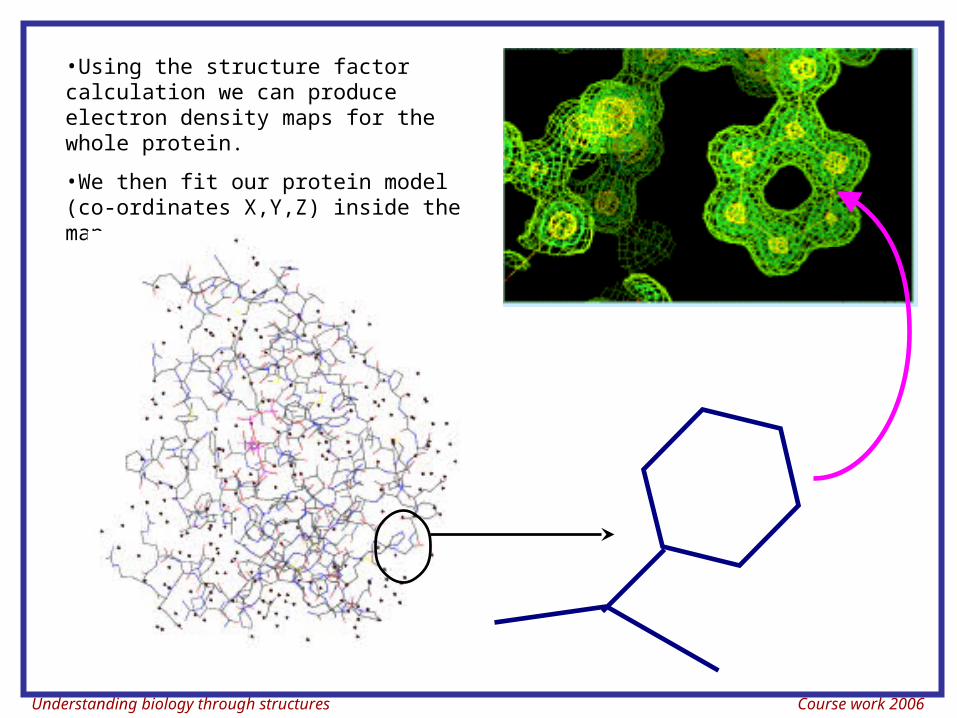
•Using the structure factor calculation we can produce electron density maps for the whole protein.
•We then fit our protein model (co-ordinates X,Y,Z) inside the map.
Understanding biology through structures Course work 2006
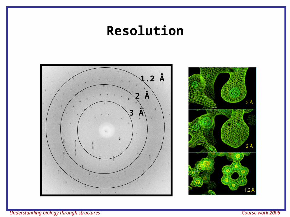
Resolution
1.2 Å
2 Å
3 Å
Understanding biology through structures Course work 2006
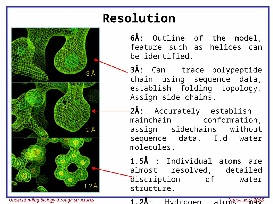
Resolution
6Å: Outline of the model, feature such as helices can be identified.
3Å: Can trace polypeptide chain using sequence data, establish folding topology. Assign side chains.
2Å: Accurately establish mainchain conformation, assign sidechains without sequence data, I.d water molecules.
1.5Å : Individual atoms are almost resolved, detailed discription of water structure.
1.2Å: Hydrogen atoms may become visible.
Understanding biology through structures Course work 2006
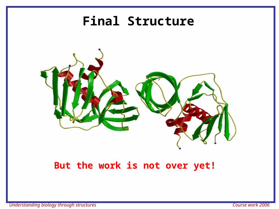
Final Structure
But the work is not over yet!
Understanding biology through structures Course work 2006
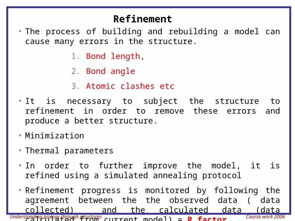
Refinement• The process of building and rebuilding a model can cause
many errors in the structure.
1. Bond length,
2. Bond angle
3. Atomic clashes etc
• It is necessary to subject the structure to refinement in order to remove these errors and produce a better structure.
• Minimization
• Thermal parameters
• In order to further improve the model, it is refined using a simulated annealing protocol
• Refinement progress is monitored by following the agreement between the the observed data ( data collected) and the calculated data (data calculated from current model) = R factor
Understanding biology through structures Course work 2006
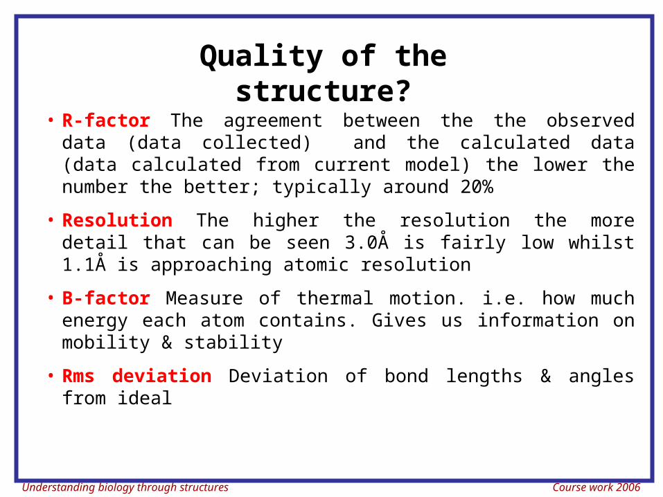
• R-factor The agreement between the the observed data (data collected) and the calculated data (data calculated from current model) the lower the number the better; typically around 20%
• Resolution The higher the resolution the more detail that can be seen 3.0Å is fairly low whilst 1.1Å is approaching atomic resolution
• B-factor Measure of thermal motion. i.e. how much energy each atom contains. Gives us information on mobility & stability
• Rms deviation Deviation of bond lengths & angles from ideal
Quality of the structure?
Understanding biology through structures Course work 2006
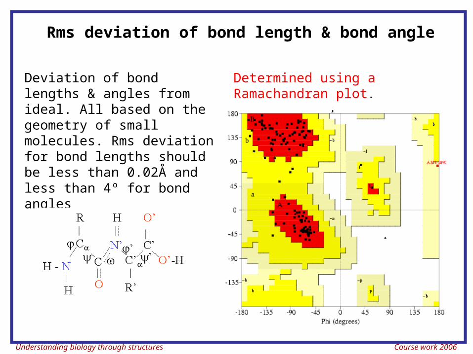
Deviation of bond lengths & angles from ideal. All based on the geometry of small molecules. Rms deviation for bond lengths should be less than 0.02Å and less than 4º for bond angles
Determined using a Ramachandran plot.
Rms deviation of bond length & bond angle
Understanding biology through structures Course work 2006
Absorption of Light
Understanding biology through structures Course work 2006
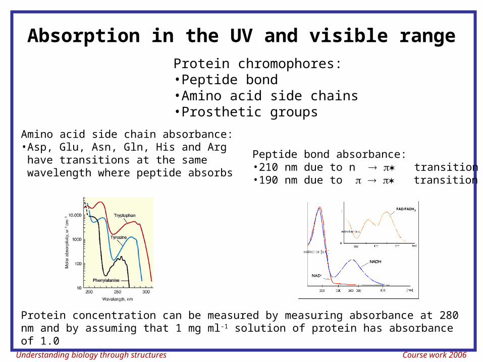
Protein chromophores:•Peptide bond•Amino acid side chains•Prosthetic groups
Peptide bond absorbance:•210 nm due to n transition•190 nm due to transition
Amino acid side chain absorbance:•Asp, Glu, Asn, Gln, His and Arg have transitions at the same wavelength where peptide absorbs
Protein concentration can be measured by measuring absorbance at 280 nm and by assuming that 1 mg ml-1 solution of protein has absorbance of 1.0
Absorption in the UV and visible range
Understanding biology through structures Course work 2006
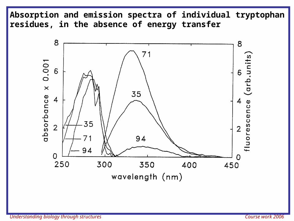
Absorption and emission spectra of individual tryptophan residues, in the absence of energy transfer
Understanding biology through structures Course work 2006
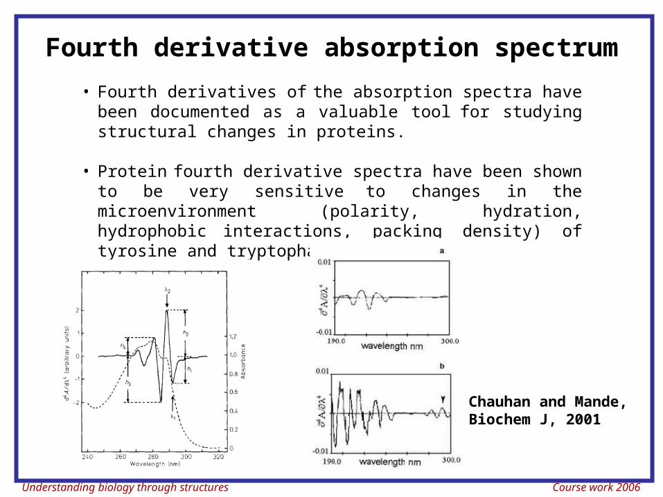
Fourth derivative absorption spectrum
• Fourth derivatives of the absorption spectra have been documented as a valuable tool for studying structural changes in proteins.
• Protein fourth derivative spectra have been shown to be very sensitive to changes in the microenvironment (polarity, hydration, hydrophobic interactions, packing density) of tyrosine and tryptophan residues
Chauhan and Mande, Biochem J, 2001
Understanding biology through structures Course work 2006
Measurements of conformational properties using optical activity
Understanding biology through structures Course work 2006
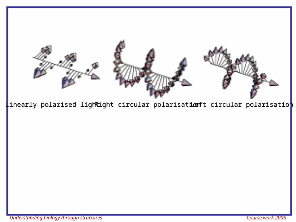
Linearly polarised lightLinearly polarised light Right circular polarisationRight circular polarisation Left circular polarisationLeft circular polarisation
Understanding biology through structures Course work 2006
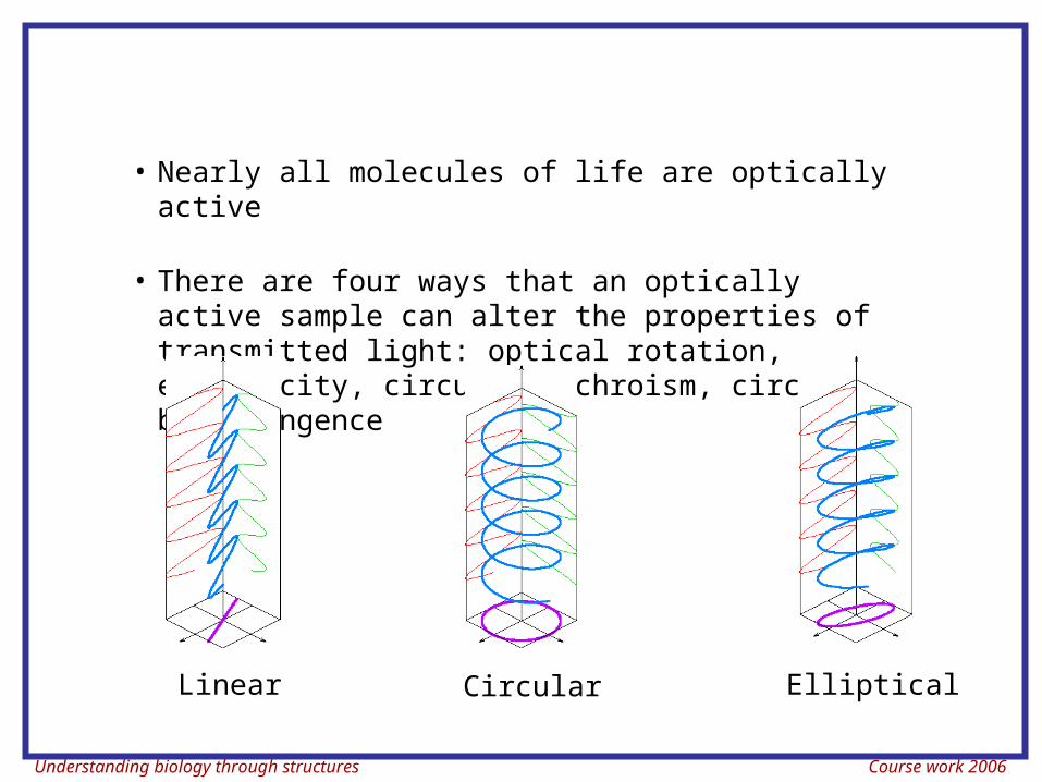
• Nearly all molecules of life are optically active
• There are four ways that an optically active sample can alter the properties of transmitted light: optical rotation, ellipticity, circular dichroism, circular birefringence
Linear Circular Elliptical
Understanding biology through structures Course work 2006
After passing through an optically active absorbing sample, the light is changed in two aspects:
1. The maximal amplitude E is no longer confined to a place, instead it traces an ellipse
Ellipticity = tan-1 (minor/major axis)
2. The orientation of the ellipse is an indication of optical activity. If the sample did not absorb any light, the ellipse would such small axial ratio that it would be equivalent to a plane-polarised light. In this case we will say that the plane polarised light has been rotated.
3. Orientation of the ellipse is the optical rotation. Optical rotation as a function of wavelength is called the optical rotatory dispersion (ORD).
Understanding biology through structures Course work 2006
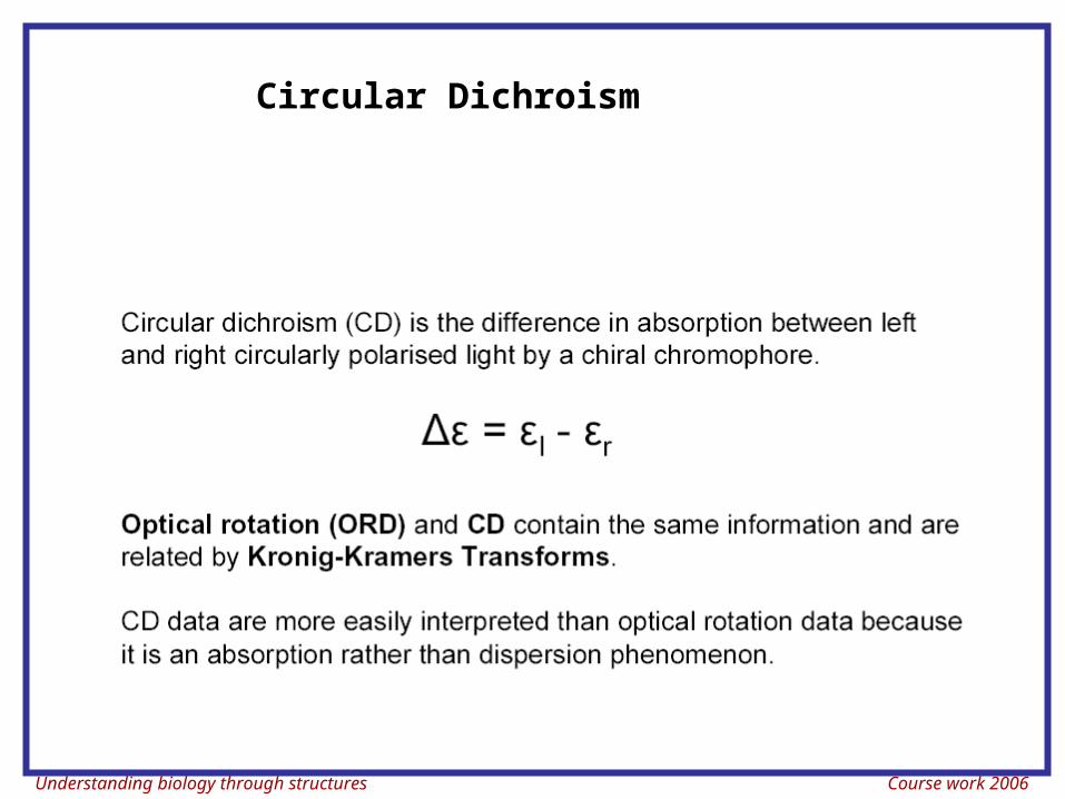
Circular Dichroism
Understanding biology through structures Course work 2006
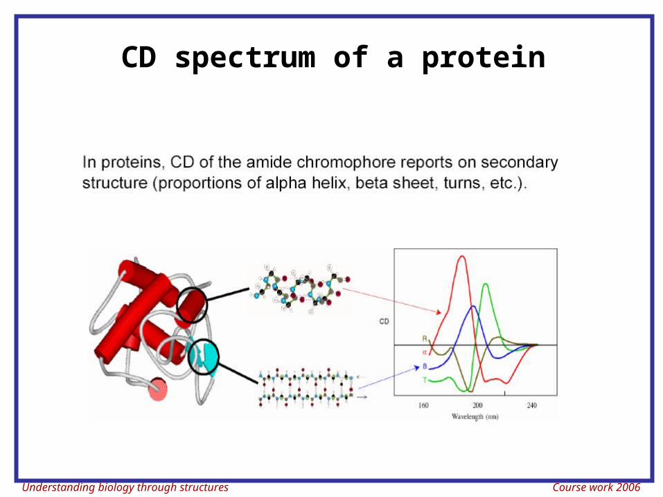
CD spectrum of a protein
Understanding biology through structures Course work 2006
Understanding biology through structures Course work 2006
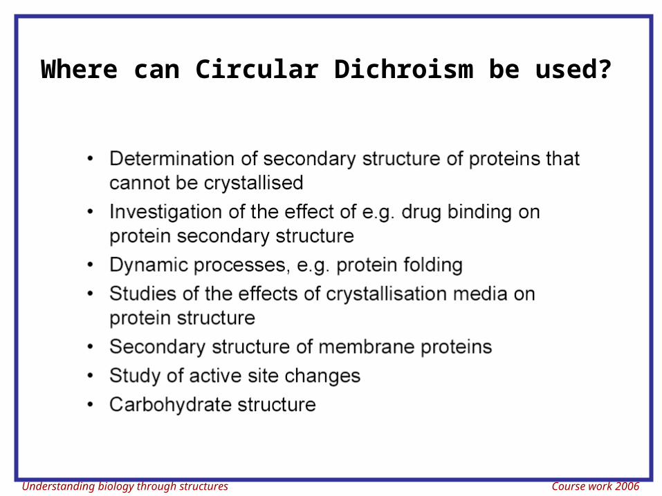
Where can Circular Dichroism be used?
Understanding biology through structures Course work 2006
Measurements of conformational properties using fluorescence
Understanding biology through structures Course work 2006
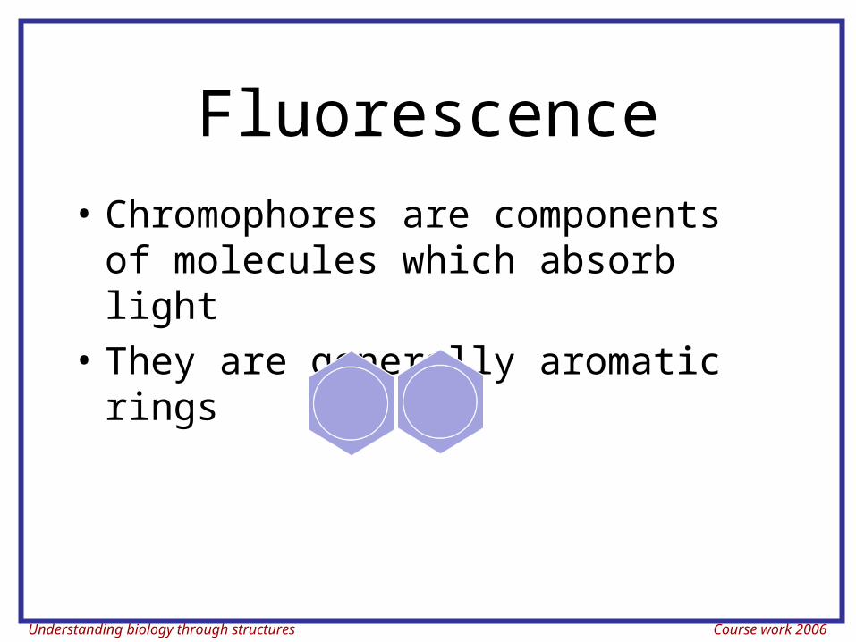
Fluorescence• Chromophores are components of
molecules which absorb light• They are generally aromatic rings
Understanding biology through structures Course work 2006
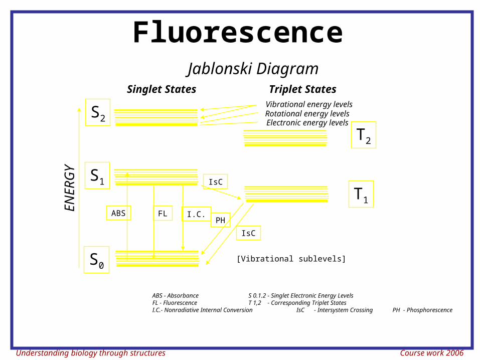
FluorescenceE
NE
RG
Y
S0
S1
S2
T2
T1ABS FL I.C.
ABS - Absorbance S 0.1.2 - Singlet Electronic Energy LevelsFL - Fluorescence T 1,2 - Corresponding Triplet StatesI.C.- Nonradiative Internal Conversion IsC - Intersystem Crossing PH - Phosphorescence
IsC
IsC
PH
[Vibrational sublevels]
Jablonski Diagram
Vibrational energy levelsRotational energy levelsElectronic energy levels
Singlet States Triplet States
Understanding biology through structures Course work 2006
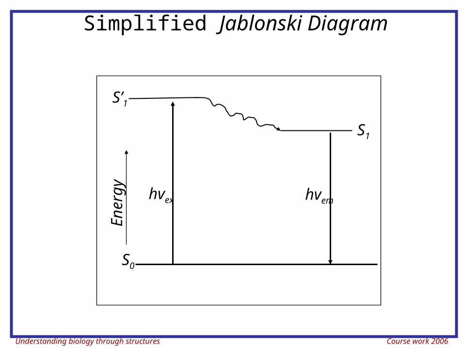
Simplified Jablonski Diagram
S0
S’1En
erg
yS1
hvex hvem
Understanding biology through structures Course work 2006
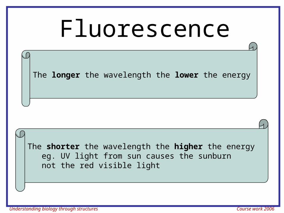
Fluorescence
The longer the wavelength the lower the energy
The shorter the wavelength the higher the energyeg. UV light from sun causes the sunburn not the red visible light
Understanding biology through structures Course work 2006
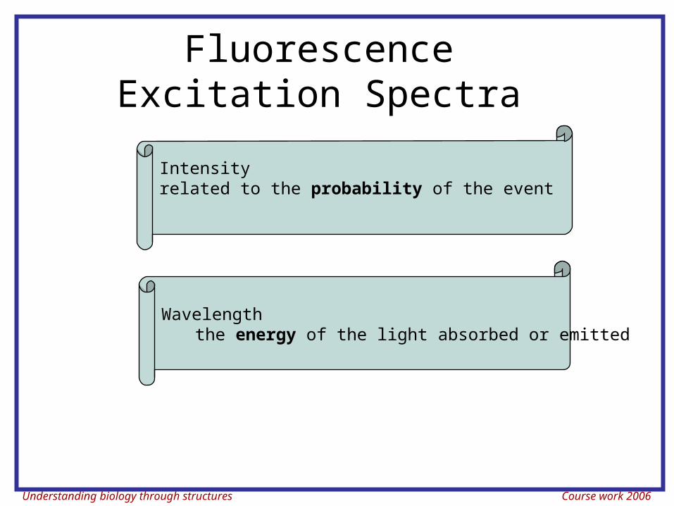
Fluorescence Excitation Spectra
Intensity related to the probability of the event
Wavelengththe energy of the light absorbed or emitted
Understanding biology through structures Course work 2006
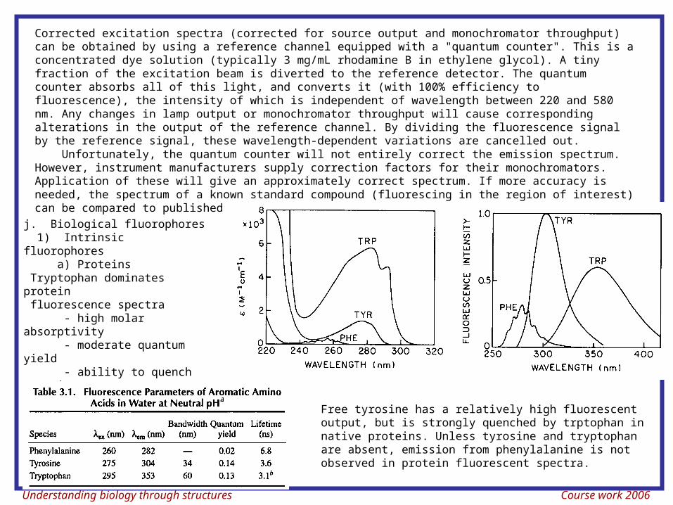
Corrected excitation spectra (corrected for source output and monochromator throughput) can be obtained by using a reference channel equipped with a "quantum counter". This is a concentrated dye solution (typically 3 mg/mL rhodamine B in ethylene glycol). A tiny fraction of the excitation beam is diverted to the reference detector. The quantum counter absorbs all of this light, and converts it (with 100% efficiency to fluorescence), the intensity of which is independent of wavelength between 220 and 580 nm. Any changes in lamp output or monochromator throughput will cause corresponding alterations in the output of the reference channel. By dividing the fluorescence signal by the reference signal, these wavelength-dependent variations are cancelled out. Unfortunately, the quantum counter will not entirely correct the emission spectrum. However, instrument manufacturers supply correction factors for their monochromators. Application of these will give an approximately correct spectrum. If more accuracy is needed, the spectrum of a known standard compound (fluorescing in the region of interest) can be compared to published standards.
j. Biological fluorophores 1) Intrinsic fluorophores a) Proteins Tryptophan dominates protein fluorescence spectra - high molar absorptivity - moderate quantum yield - ability to quench tyrosine and phenylalanine emission by energy transfer.
Free tyrosine has a relatively high fluorescent output, but is strongly quenched by trptophan in native proteins. Unless tyrosine and tryptophan are absent, emission from phenylalanine is not observed in protein fluorescent spectra.
Understanding biology through structures Course work 2006
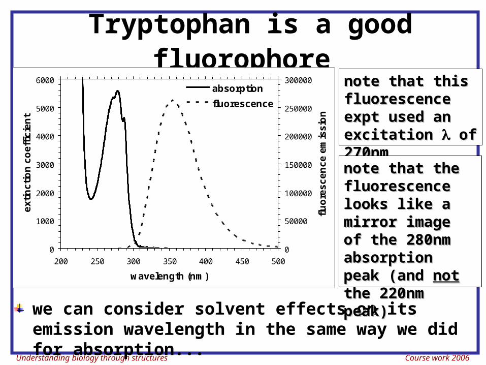
Tryptophan is a good fluorophore
0
1000
2000
3000
4000
5000
6000
200 250 300 350 400 450 500
wavelength (nm)
ex
tin
cti
on
co
eff
icie
nt
0
50000
100000
150000
200000
250000
300000
flu
ore
sc
en
ce
em
iss
ion
absorption
fluorescence
note that this note that this fluorescence fluorescence expt used an expt used an excitation excitation of of 270nm270nm
we can consider solvent effects on its emission wavelength in the same way we did for absorption...
note that the note that the fluorescence fluorescence looks like a looks like a mirror image of mirror image of the 280nm the 280nm absorption absorption peak (and peak (and notnot the 220nm the 220nm peak)peak)
Understanding biology through structures Course work 2006
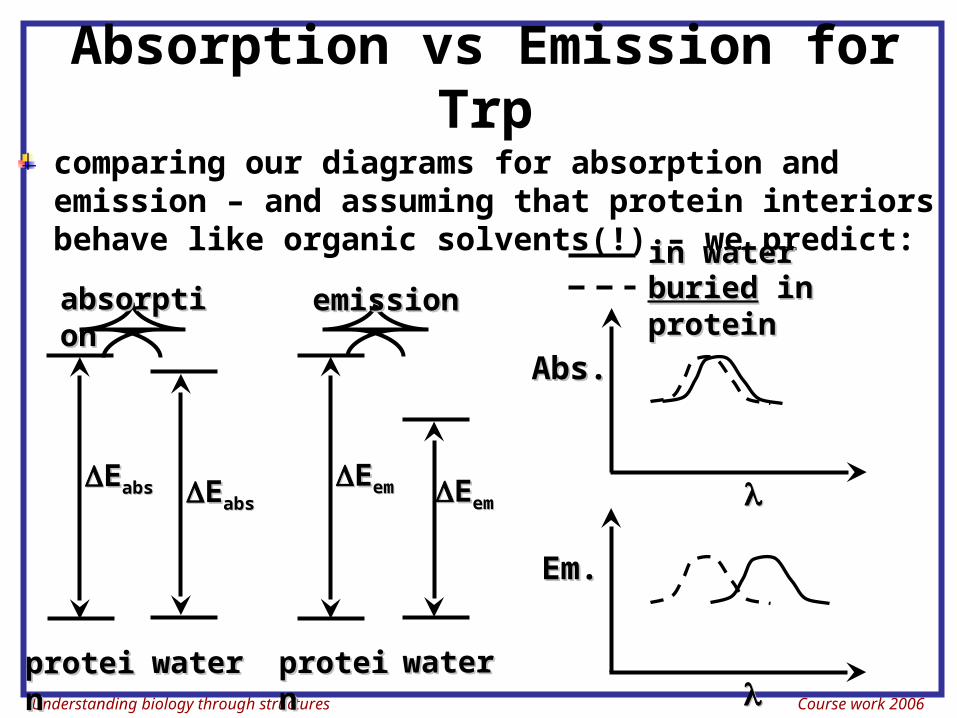
Absorption vs Emission for Trp
comparing our diagrams for absorption and emission – and assuming that protein interiors behave like organic solvents(!) – we predict:
Abs.Abs.
in waterin waterburiedburied in in proteinprotein
EEabsabs EEabsabs
proteiproteinn
waterwater
absorptiabsorptionon
EEemem EEemem
emissionemission
proteiproteinn
waterwater
Em.Em.
Understanding biology through structures Course work 2006
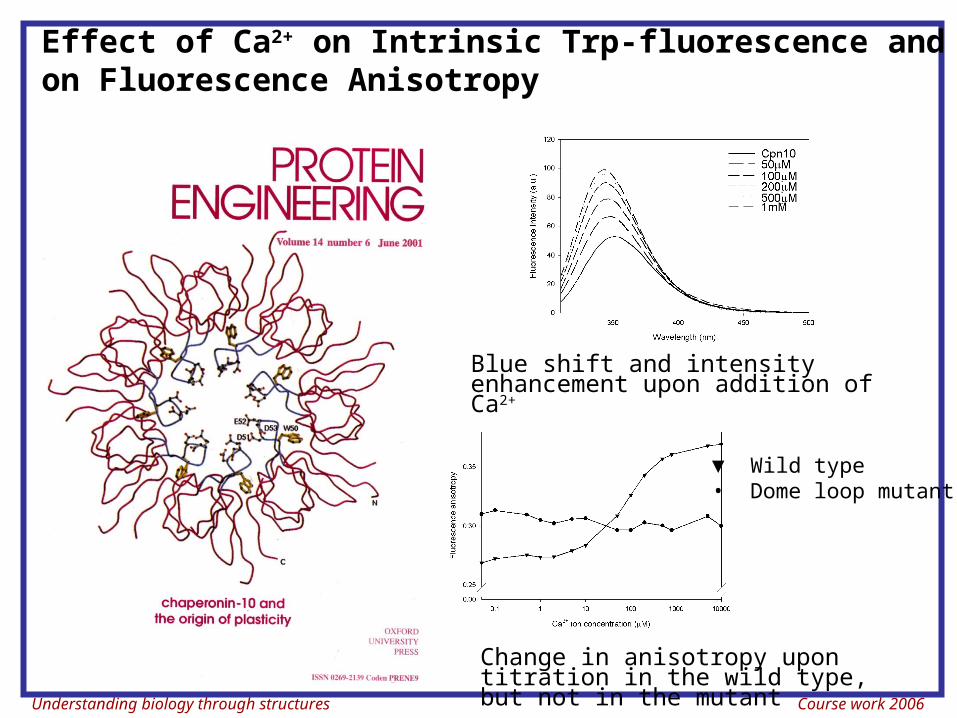
Effect of Ca2+ on Intrinsic Trp-fluorescence and on Fluorescence Anisotropy
▼ Wild type• Dome loop mutant
Blue shift and intensity enhancement upon addition of Ca2+
Change in anisotropy upon titration in the wild type, but not in the mutant
Understanding biology through structures Course work 2006
Raman Scatter• A molecule may undergo a vibrational
transition (not an electronic shift) at exactly the same time as scattering occurs
• This results in a photon emission of a photon differing in energy from the energy of the incident photon by the amount of the above energy - this is Raman scattering.
• The dominant effect in flow cytometry is the stretch of the O-H bonds of water. At 488 nm excitation488 nm excitation this would give emission at 575-595575-595 nm nm
Understanding biology through structures Course work 2006
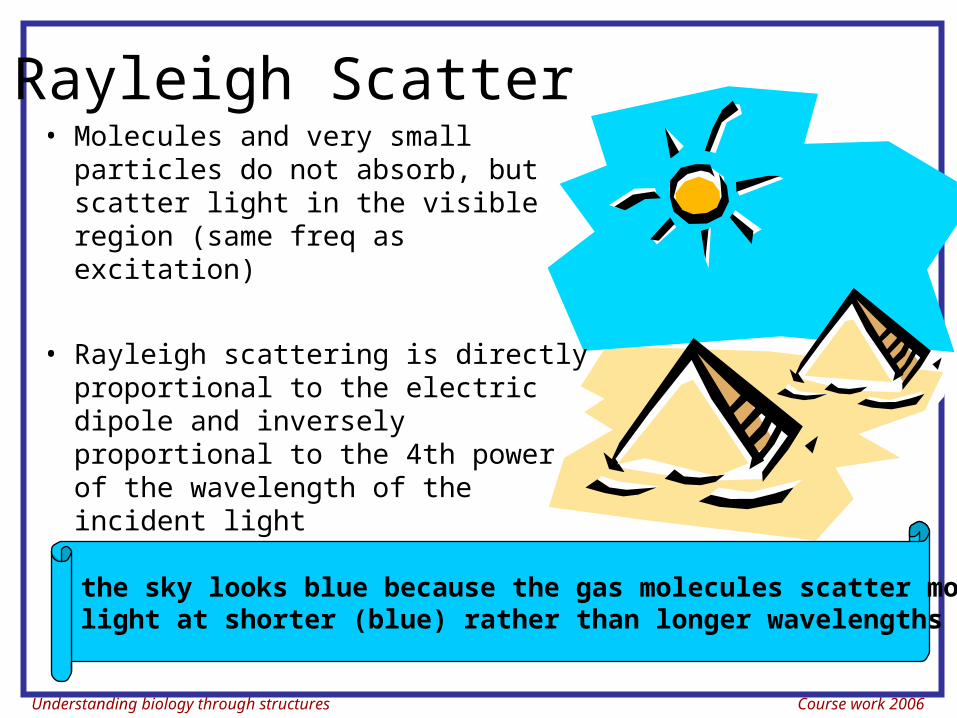
Rayleigh Scatter• Molecules and very small
particles do not absorb, but scatter light in the visible region (same freq as excitation)
• Rayleigh scattering is directly proportional to the electric dipole and inversely proportional to the 4th power of the wavelength of the incident light
the sky looks blue because the gas molecules scatter more light at shorter (blue) rather than longer wavelengths (red)
Understanding biology through structures Course work 2006
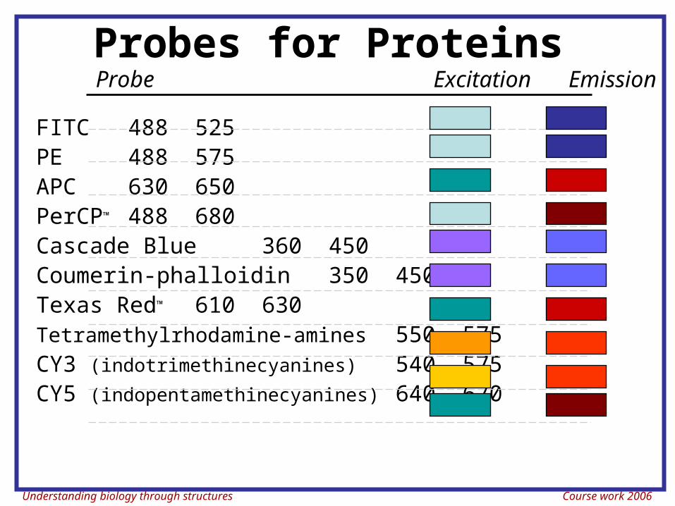
Probes for Proteins
FITC 488 525PE 488 575APC 630 650PerCP™ 488 680Cascade Blue 360 450Coumerin-phalloidin 350 450Texas Red™ 610 630Tetramethylrhodamine-amines 550 575CY3 (indotrimethinecyanines) 540 575CY5 (indopentamethinecyanines) 640 670
Probe Excitation Emission
Understanding biology through structures Course work 2006
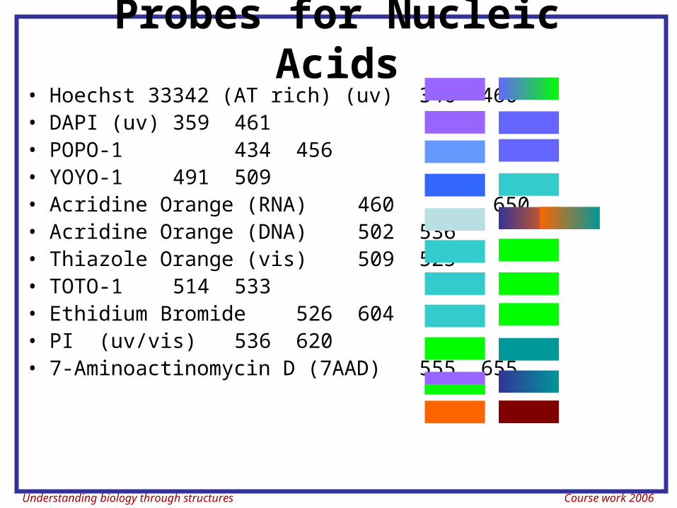
• Hoechst 33342 (AT rich) (uv) 346 460• DAPI (uv) 359 461• POPO-1 434 456• YOYO-1 491 509• Acridine Orange (RNA) 460 650• Acridine Orange (DNA) 502 536• Thiazole Orange (vis) 509 525• TOTO-1 514 533• Ethidium Bromide 526 604• PI (uv/vis) 536 620• 7-Aminoactinomycin D (7AAD) 555 655
Probes for Nucleic Acids
Understanding biology through structures Course work 2006
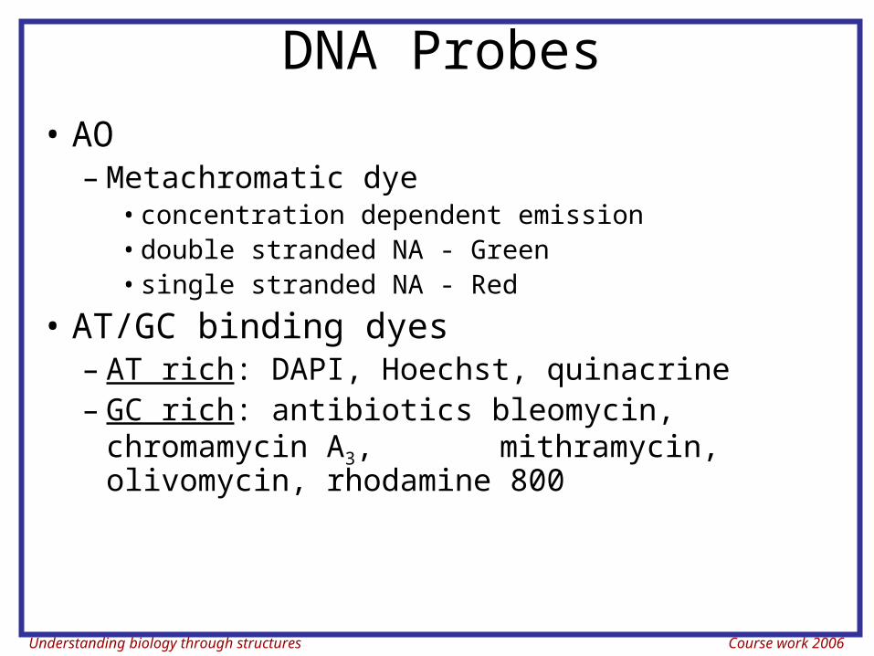
DNA Probes• AO
– Metachromatic dye• concentration dependent emission• double stranded NA - Green• single stranded NA - Red
• AT/GC binding dyes– AT rich: DAPI, Hoechst, quinacrine– GC rich: antibiotics bleomycin, chromamycin
A3, mithramycin, olivomycin, rhodamine 800
Understanding biology through structures Course work 2006
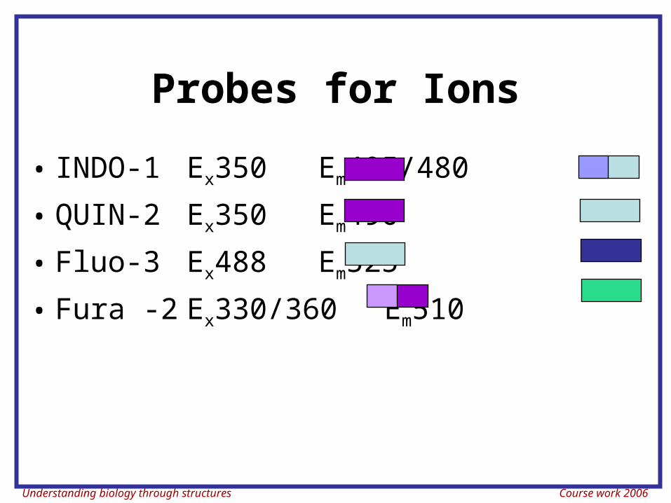
Probes for Ions
• INDO-1 Ex350Em405/480
• QUIN-2 Ex350 Em490
• Fluo-3 Ex488 Em525
• Fura -2 Ex330/360 Em510
Understanding biology through structures Course work 2006
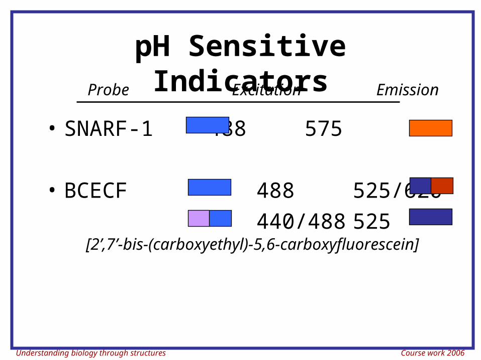
pH Sensitive Indicators
• SNARF-1 488 575
• BCECF 488 525/620
440/488 525[2’,7’-bis-(carboxyethyl)-5,6-carboxyfluorescein]
Probe Excitation Emission
Understanding biology through structures Course work 2006
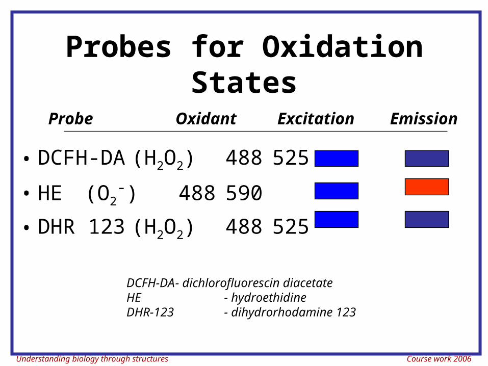
Probes for Oxidation States
• DCFH-DA(H2O2) 488 525
• HE (O2-) 488 590
• DHR 123 (H2O2) 488 525
Probe Oxidant Excitation Emission
DCFH-DA - dichlorofluorescin diacetateHE - hydroethidineDHR-123 - dihydrorhodamine 123
Understanding biology through structures Course work 2006
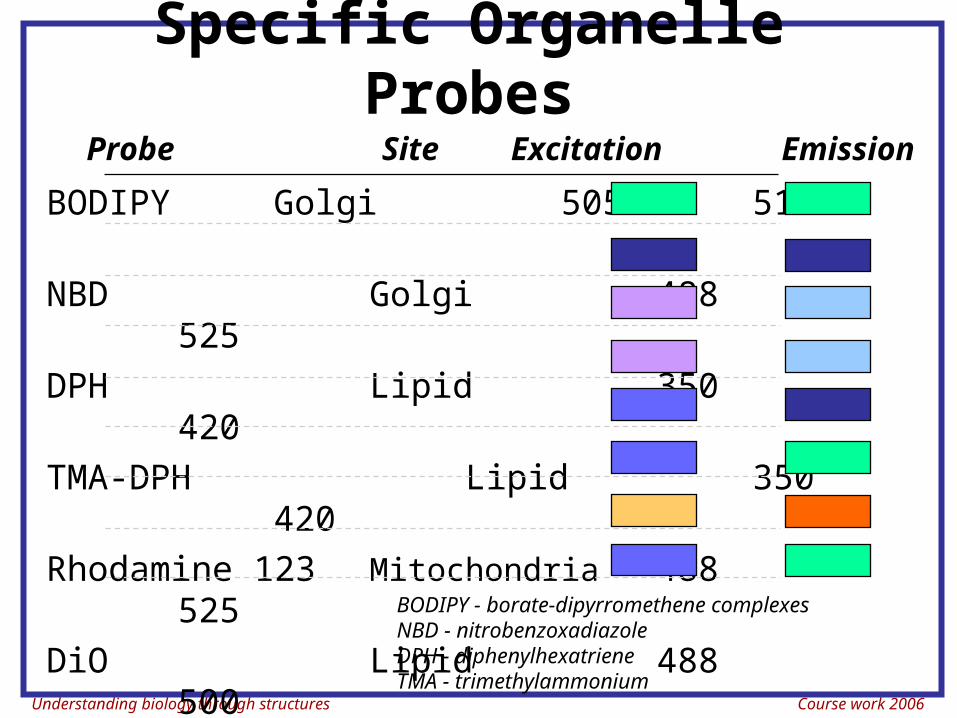
Specific Organelle Probes
BODIPY Golgi 505 511
NBD Golgi 488 525
DPH Lipid 350 420
TMA-DPH Lipid 350 420
Rhodamine 123 Mitochondria 488 525
DiO Lipid 488 500
diI-Cn-(5) Lipid 550 565
diO-Cn-(3) Lipid 488 500
Probe Site Excitation Emission
BODIPY - borate-dipyrromethene complexesNBD - nitrobenzoxadiazoleDPH - diphenylhexatrieneTMA - trimethylammonium
Understanding biology through structures Course work 2006
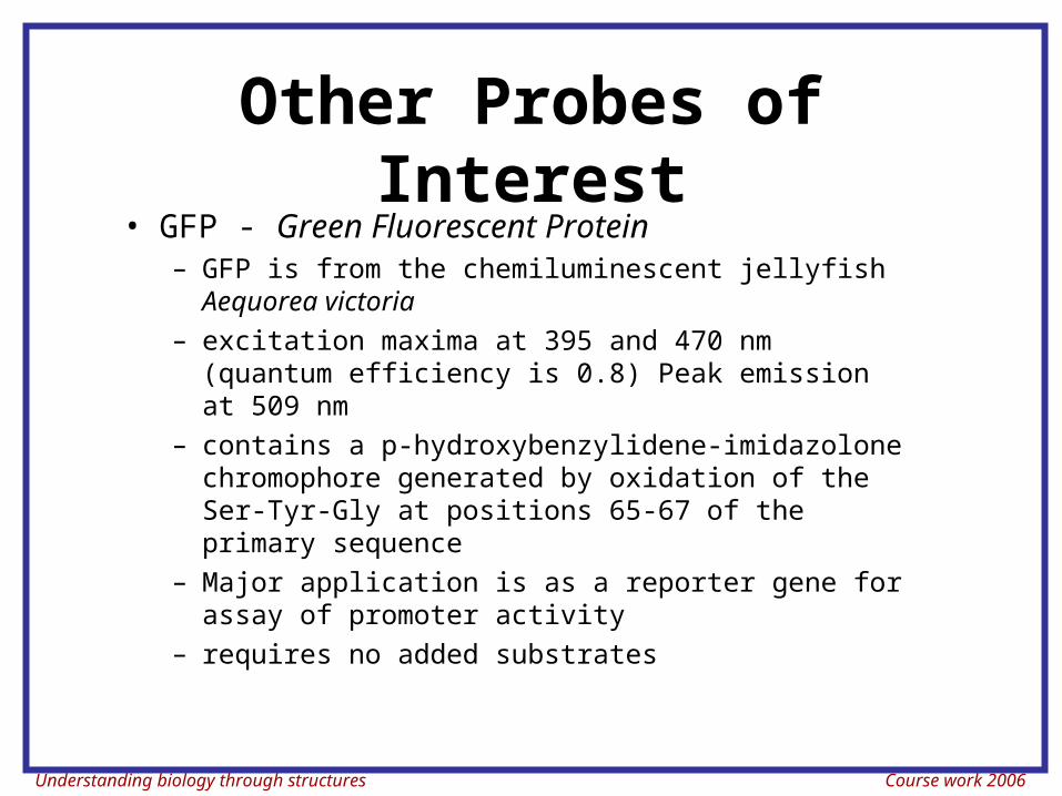
Other Probes of Interest• GFP - Green Fluorescent Protein
– GFP is from the chemiluminescent jellyfish Aequorea victoria
– excitation maxima at 395 and 470 nm (quantum efficiency is 0.8) Peak emission at 509 nm
– contains a p-hydroxybenzylidene-imidazolone chromophore generated by oxidation of the Ser-Tyr-Gly at positions 65-67 of the primary sequence
– Major application is as a reporter gene for assay of promoter activity
– requires no added substrates
Understanding biology through structures Course work 2006
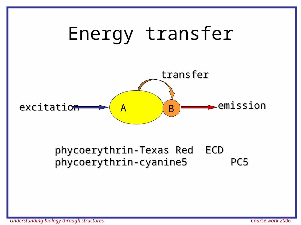
Energy transfer
excitationexcitation emissionemission
transfertransfer
A B
phycoerythrin-Texas Redphycoerythrin-Texas Red ECDECDphycoerythrin-cyanine5phycoerythrin-cyanine5 PC5PC5
Understanding biology through structures Course work 2006
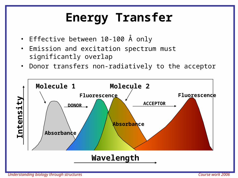
Energy Transfer
• Effective between 10-100 Å only• Emission and excitation spectrum must significantly
overlap• Donor transfers non-radiatively to the acceptor
Inte
nsit
y
Wavelength
Absorbance
DONOR
Absorbance
Fluorescence Fluorescence
ACCEPTOR
Molecule 1 Molecule 2