![18F]Flubatine as a novel α4β2 nicotinic acetylcholine ...](https://static.fdocument.org/doc/165x107/629737326d4e5a451c0d4cae/18fflubatine-as-a-novel-42-nicotinic-acetylcholine-.jpg)
Ultrastructural Distribution of the 7 Nicotinic Acetylcholine Receptor
11
Ultrastructural Distribution of the 7 Nicotinic Acetylcholine Receptor Subunit in Rat Hippocampus Ruth Fabian-Fine, 1,2 Paul Skehel, 2 Mick L. Errington, 2 Heather A. Davies, 1 Emanuele Sher, 3 Michael G. Stewart, 1 and Alan Fine 2,4 1 Department of Biological Sciences, The Open University, Milton Keynes, MK7 6AA, United Kingdom, 2 Division of Neurophysiology, National Institute for Medical Research, Mill Hill, London NW7 1AA, United Kingdom, 3 Lilly Research Centre, Eli Lilly and Co., Erl Wood Manor, Windlesham, Surrey GU20 6PH, United Kingdom, and 4 Department of Physiology and Biophysics, Dalhousie University Faculty of Medicine, Halifax, Nova Scotia B3H 4H7, Canada Acetylcholine (ACh) is an important neurotransmitter in the mammalian brain; it is implicated in arousal, learning, and other cognitive functions. Recent studies indicate that nicotinic re- ceptors contribute to these cholinergic effects, in addition to the established role of muscarinic receptors. In the hippocampus, where cholinergic involvement in learning and memory is par- ticularly well documented, 7 nicotinic acetylcholine receptor subunits (7 nAChRs) are highly expressed, but their precise ultrastructural localization has not been determined. Here, we describe the results of immunogold labeling of serial ultrathin sections through stratum radiatum of area CA1 in the rat. Using both anti-7 nAChR immunolabeling and -bungarotoxin bind- ing, we find that 7 nAChRs are present at nearly all synapses in CA1 stratum radiatum, with immunolabeling present at both presynaptic and postsynaptic elements. Morphological consid- erations and double immunolabeling indicate that GABAergic as well as glutamatergic synapses bear 7 nAChRs, at densi- ties approaching those observed for glutamate receptors in CA1 stratum radiatum. Postsynaptically, 7 nAChRs often are distributed at dendritic spines in a perisynaptic annulus. In the postsynaptic cytoplasm, immunolabeling is associated with spine apparatus and other membranous structures, suggesting that 7 nAChRs may undergo dynamic regulation, with inser- tion into the synapse and subsequent internalization. The wide- spread and substantial expression of 7 nAChRs at synapses in the hippocampus is consistent with an important role in medi- ating and/or modulating synaptic transmission, plasticity, and neurodegeneration. Key words: acetylcholine receptors; nicotine; dendritic spines; postsynaptic density; immuno-electron microscopy; glutamate; GABA; A 1–42 Acetylcholine (ACh) is the major excitatory neurotransmitter in the peripheral nervous system. Ionotropic nicotinic receptors mediate postsynaptic excitatory responses at the neuromuscular junction, and there is evidence that nicotinic receptors may also act presynaptically to modulate acetylcholine release in the pe- riphery (Wessler et al., 1992; Liang and Vizi, 1997). In the mammalian CNS, specific receptors for nicotinic ligands have been recognized for many years (Arimatsu et al., 1978; Dudai and Segal, 1978; Hunt and Schmidt, 1978; Segal et al., 1978), but only recently has evidence begun to emerge for their functional roles, including possible mediation of fast postsynaptic responses at certain brain sites (Zhang et al., 1993; Roerig et al., 1997; Chu et al., 2000) and modulation of release of various transmitters, including glutamate (Vidal and Changeux, 1993; McGehee et al., 1995; Gray et al., 1996), GABA (Lena et al., 1993), ACh (McGe- hee et al., 1995), and dopamine (Rapier et al., 1988). Nicotinic receptors constitute a heterogeneous family of ion channels. In the nervous system, nine different subunits (2–10) and three different subunits (2–4) have been described. Most are as- sumed to form heteropentameric structures, with various combi- nations of and subunits. There is also evidence in heterolo- gous expression systems that some subunits, particularly 7, form homopentamers (Couturier et al., 1990; Schoepfer et al., 1990; Seguela et al., 1993). Nicotinic acetylcholine receptors containing 7 subunits (7 nAChRs) are, along with those containing the 4/2 combination, the most abundant in brain. The distribution of these receptors is specific, with the 7 subunit, which is selec- tively bound by -bungarotoxin (Bgtx) (Chen and Patrick, 1997; Orr-Urtreger et al., 1997), abundant in particular cortical and subcortical areas (Bina et al., 1995) of the mammalian brain; conspicuous among these is the hippocampus (Dominguez del Toro et al., 1994). The 7 subunit appears to participate in numerous important processes, including modulation of release of several neurotransmitters, mediation of postsynaptic excitatory responses, long-term potentiation (LTP), and cognitive f unction (Hunter et al., 1994; Fujii et al., 2000; Mansvelder and McGehee, 2000) (for review, see Role and Berg, 1996; Wonnacott, 1997; Radcliffe et al., 1999; Levin and Rezvani, 2000). Light microscopic immunostaining has revealed the presence of 7 nAChRs in both somatic and dendritic regions in all hip- pocampal areas (Dominguez del Toro et al., 1994). Hippocampal cells in culture were found to exhibit patchy 7 nAChR immu- nolabeling on somata and dendrites, colocalized with presynaptic markers (Barrantes et al., 1995; Zarei et al., 1999), but the identity of the labeled cells was unspecified, nor was it possible at the light microscopic level to establish the presynaptic versus Received Feb. 21, 2001; revised June 13, 2001; accepted June 28, 2001. This work was supported by the Medical Research Council and by grants from the Human Frontier Science Program (A.F.) and the Brain and Behavioural Sciences Research Council (108/BI 11211) (M.G.S.). We thank J.-A. Horne, E. Hirst, and Drs. T. V. P. Bliss, I. Burdett, and I. A. Meinertzhagen for helpful discussion. Correspondence should be addressed to Dr. Ruth Fabian-Fine, Department of Psychology, Dalhousie University, Halifax, Nova Scotia B3H 4J1 Canada. E-mail: [email protected]. Copyright © 2001 Society for Neuroscience 0270-6474/01/217993-11$15.00/0 The Journal of Neuroscience, October 15, 2001, 21(20):7993–8003
Transcript of Ultrastructural Distribution of the 7 Nicotinic Acetylcholine Receptor
Ultrastructural Distribution of the �7 Nicotinic AcetylcholineReceptor Subunit in Rat Hippocampus
Ruth Fabian-Fine,1,2 Paul Skehel,2 Mick L. Errington,2 Heather A. Davies,1 Emanuele Sher,3Michael G. Stewart,1 and Alan Fine2,4
1Department of Biological Sciences, The Open University, Milton Keynes, MK7 6AA, United Kingdom, 2Division ofNeurophysiology, National Institute for Medical Research, Mill Hill, London NW7 1AA, United Kingdom, 3Lilly ResearchCentre, Eli Lilly and Co., Erl Wood Manor, Windlesham, Surrey GU20 6PH, United Kingdom, and 4Department ofPhysiology and Biophysics, Dalhousie University Faculty of Medicine, Halifax, Nova Scotia B3H 4H7, Canada
Acetylcholine (ACh) is an important neurotransmitter in themammalian brain; it is implicated in arousal, learning, and othercognitive functions. Recent studies indicate that nicotinic re-ceptors contribute to these cholinergic effects, in addition to theestablished role of muscarinic receptors. In the hippocampus,where cholinergic involvement in learning and memory is par-ticularly well documented, �7 nicotinic acetylcholine receptorsubunits (�7 nAChRs) are highly expressed, but their preciseultrastructural localization has not been determined. Here, wedescribe the results of immunogold labeling of serial ultrathinsections through stratum radiatum of area CA1 in the rat. Usingboth anti-�7 nAChR immunolabeling and �-bungarotoxin bind-ing, we find that �7 nAChRs are present at nearly all synapsesin CA1 stratum radiatum, with immunolabeling present at bothpresynaptic and postsynaptic elements. Morphological consid-erations and double immunolabeling indicate that GABAergic
as well as glutamatergic synapses bear �7 nAChRs, at densi-ties approaching those observed for glutamate receptors inCA1 stratum radiatum. Postsynaptically, �7 nAChRs often aredistributed at dendritic spines in a perisynaptic annulus. In thepostsynaptic cytoplasm, immunolabeling is associated withspine apparatus and other membranous structures, suggestingthat �7 nAChRs may undergo dynamic regulation, with inser-tion into the synapse and subsequent internalization. The wide-spread and substantial expression of �7 nAChRs at synapses inthe hippocampus is consistent with an important role in medi-ating and/or modulating synaptic transmission, plasticity, andneurodegeneration.
Key words: acetylcholine receptors; nicotine; dendriticspines; postsynaptic density; immuno-electron microscopy;glutamate; GABA; A�1–42
Acetylcholine (ACh) is the major excitatory neurotransmitter inthe peripheral nervous system. Ionotropic nicotinic receptorsmediate postsynaptic excitatory responses at the neuromuscularjunction, and there is evidence that nicotinic receptors may alsoact presynaptically to modulate acetylcholine release in the pe-riphery (Wessler et al., 1992; Liang and Vizi, 1997). In themammalian CNS, specific receptors for nicotinic ligands havebeen recognized for many years (Arimatsu et al., 1978; Dudai andSegal, 1978; Hunt and Schmidt, 1978; Segal et al., 1978), but onlyrecently has evidence begun to emerge for their functional roles,including possible mediation of fast postsynaptic responses atcertain brain sites (Zhang et al., 1993; Roerig et al., 1997; Chu etal., 2000) and modulation of release of various transmitters,including glutamate (Vidal and Changeux, 1993; McGehee et al.,1995; Gray et al., 1996), GABA (Lena et al., 1993), ACh (McGe-hee et al., 1995), and dopamine (Rapier et al., 1988). Nicotinicreceptors constitute a heterogeneous family of ion channels. Inthe nervous system, nine different � subunits (�2–�10) and threedifferent � subunits (�2–�4) have been described. Most are as-
sumed to form heteropentameric structures, with various combi-nations of � and � subunits. There is also evidence in heterolo-gous expression systems that some subunits, particularly �7, formhomopentamers (Couturier et al., 1990; Schoepfer et al., 1990;Seguela et al., 1993). Nicotinic acetylcholine receptors containing�7 subunits (�7 nAChRs) are, along with those containing the�4/�2 combination, the most abundant in brain. The distributionof these receptors is specific, with the �7 subunit, which is selec-tively bound by �-bungarotoxin (�Bgtx) (Chen and Patrick, 1997;Orr-Urtreger et al., 1997), abundant in particular cortical andsubcortical areas (Bina et al., 1995) of the mammalian brain;conspicuous among these is the hippocampus (Dominguez delToro et al., 1994). The �7 subunit appears to participate innumerous important processes, including modulation of releaseof several neurotransmitters, mediation of postsynaptic excitatoryresponses, long-term potentiation (LTP), and cognitive function(Hunter et al., 1994; Fujii et al., 2000; Mansvelder and McGehee,2000) (for review, see Role and Berg, 1996; Wonnacott, 1997;Radcliffe et al., 1999; Levin and Rezvani, 2000).
Light microscopic immunostaining has revealed the presence of�7 nAChRs in both somatic and dendritic regions in all hip-pocampal areas (Dominguez del Toro et al., 1994). Hippocampalcells in culture were found to exhibit patchy �7 nAChR immu-nolabeling on somata and dendrites, colocalized with presynapticmarkers (Barrantes et al., 1995; Zarei et al., 1999), but theidentity of the labeled cells was unspecified, nor was it possible atthe light microscopic level to establish the presynaptic versus
Received Feb. 21, 2001; revised June 13, 2001; accepted June 28, 2001.This work was supported by the Medical Research Council and by grants from the
Human Frontier Science Program (A.F.) and the Brain and Behavioural SciencesResearch Council (108/BI 11211) (M.G.S.). We thank J.-A. Horne, E. Hirst, andDrs. T. V. P. Bliss, I. Burdett, and I. A. Meinertzhagen for helpful discussion.
Correspondence should be addressed to Dr. Ruth Fabian-Fine, Department ofPsychology, Dalhousie University, Halifax, Nova Scotia B3H 4J1 Canada. E-mail:[email protected] © 2001 Society for Neuroscience 0270-6474/01/217993-11$15.00/0
The Journal of Neuroscience, October 15, 2001, 21(20):7993–8003
postsynaptic nature of the labeling. Electron microscopic (EM)analysis of 125I-�Bgtx binding provided evidence for �7 nAChRsat hippocampal synapses (Hunt and Schmidt, 1978), but the largegrain radius and relative insensitivity of the method preventedfirm conclusions about incidence or distribution of the �Bgtxbinding sites. Full understanding of the varied and subtle func-tional roles recently attributed to �7 nAChRs in the hippocampus(Radcliffe et al., 1999) will require high-resolution analysis of thesubcellular distribution of this subunit. To this end, we haveperformed light and electron microscopic immunostaining ofCA1 stratum radiatum, and here report that �7 receptors arehighly abundant at almost all synapses in this region. The inten-sity of the signal suggests that the importance of �7-mediatednicotinic cholinergic signaling may be far greater than is currentlyrecognized.
MATERIALS AND METHODSLight microscopic �7 nAChR immunolabeling. Intact brain preparationswere obtained from two adult male Sprague Dawley rats. Animals wereanesthetized with urethane (1.5 gm/kg, i.p.) and perfused over �20 minwith 200 ml of 4% paraformaldehyde (PFA)/0.3% glutaraldehyde (GA)in PBS (0.1 M, pH 7.2). After dissection the brains were immersed in thesame fixative for 2 hr and embedded in 7% Agarose. Vibratome sections30 �m thick were cut transversely through the brain using a LeicaVT1000S and rinsed in PBS (4 � 10 min). Sections containing thehippocampal formation were then incubated in 1% glycine in PBS toquench residual reactive aldehyde groups, washed in PBS (2 � 10 min),permeabilized in 0.1% saponin (S2149; Sigma, St. Louis, MO)/PBS for15 min, and incubated with the primary monoclonal anti-�7 nAChRantibody Mab 306 (M220, Sigma) (Schoepfer et al., 1990) at 4°C over-night. The antibody was diluted 1:3000 in an incubation medium (IM)consisting of PBS with 1% bovine serum albumin (A4503, Sigma), 5%normal goat serum, 0.5% cold water fish skin gelatin (G7765, Sigma), and0.01% saponin. After incubation, preparations were rinsed thoroughly inPBS. To detect anti-�7 nAChR antibody binding, a secondary goatanti-mouse antibody coupled to the fluorochrome Cy3 (Stratech Scien-tific, Luton, UK; 115-165-003; 1:500 in IM at 4°C overnight) was used.Preparations were then washed in PBS (6 � 1 hr), mounted in Mowiol4–88 (475904, Calbiochem, La Jolla, CA) on glass slides, and examinedusing a Zeiss Axiophot microscope with epifluorescence optics. RedCy3-immunofluorescence was detected using a standard rhodamine filterset. Preparations were immediately photographed on color slide film(Ektachrome 400).
Light microscopic labeling for synaptophysin and �Bgtx. Hippocampalpreparations were obtained from two adult male Sprague Dawley rats.Animals, anesthetized as above, were perfused over �10 min with 200 mlof freshly prepared 4% PFA in PBS. Brains were removed, embedded in7% Agarose, and 20 �m vibratome sections obtained as above. Afterpermeabilization with 0.01% saponin in IM (30 min), sections wereincubated in IM containing a monoclonal anti-synaptophysin antibody(1:120; SY38, 902322; Boehringer Mannheim, Mannheim, Germany)overnight at 4°C. After washing in PBS (5 � 1 hr) and 0.01% saponin inIM, preparations were incubated overnight at 4°C in IM containing asecondary goat anti-mouse antibody coupled to Cy3 (1:500; see above)
and Alexa488-conjugated �Bgtx (1:500; B13422; Molecular Probes). Af-ter thorough rinsing in PBS (4 � 1 hr), slices were mounted in Mowioland examined with a Leica TCS NT confocal microscope using standardfluorescein and rhodamine filter combinations.
Postembedding immunogold labeling for electron microscopy. Freeze-substituted tissue was used to preserve greater immunoreactivity of thetissue and achieve maximum staining intensity. Three adult (all 22months old) CFHB male rats were anesthetized with Sagatal and per-fused through the heart with 50 ml of 0.9% saline followed by 250 ml 0.1M phosphate buffer, pH 7.4, containing 4% paraformaldehyde, 0.05%glutaraldehyde, and 0.2% picric acid (Somogyi and Takagi, 1982). Hip-pocampus was dissected immediately, and �0.5 mm slabs were cut byhand from the dorsal part, soaked in PB for 15 min, and then immersedin 0.125 M triethanolamine hydrochloride in PB for 30 min to quenchunreacted aldehydes. The tissue slabs were cryoprotected over 2 hr inincreasing concentrations of glycerol in PBS to 30% and then impact-frozen on a polished copper mirror at �193°C in a Leica MM80 impactfreezer. Frozen slabs were freeze-substituted in a Leica AFS automaticfreeze substitution system in methanol with 0.5% uranyl acetate for 24 hrat �85°C, rinsed in methanol, and the temperature raised to �50°Cbefore infiltration and embedding in Lowicryl HM 20 (R1034; AgarScientific, Stansted, England). The resin was polymerized at �50°C byexposure to ultraviolet light. Serial ultrathin sections (50 nm) were cutwith a Reichert Ultracut and collected on pioloform-coated single-slotnickel grids. To treat all sections identically and to prevent tearing duringthe labeling procedure, grids were mounted in a grid support plate(16705698; Leica Microsystems, Milton Keynes, UK). Sections werewetted in PBS for 30 min and preincubated in IM for 30 min at roomtemperature. Sections were then incubated with the monoclonal anti-�7nAChR antibody (see above; 1:4000 in IM) overnight at 4°C followed by1 hr at 37°C. After thorough washing in PBS and preincubation in IM (30min), the secondary antibody (goat anti-mouse coupled to 10 nm goldparticles; G7402; Sigma) was applied at a dilution of 1:100 for 4 hr at37°C. Preparations were washed subsequently in IM (10 min) and PBS(3 � 10 min) before final rinsing in double-distilled water.
For the double labeling of (1) glutamate and �7 nAChR, (2) GABAand �7 nAChR, or (3) dual epitopes of �7 nAChR, the followingantibodies were used: (1) rabbit anti-glutamate (1:20,000 in IM; G6642;Sigma) detected with goat anti-rabbit 10 nm gold plus mouse anti-�7nAChR (1:4000 in IM) detected with goat anti-mouse 5 nm gold(EM.GAM5; British Biocell, Cardiff, Wales, UK); (2) rabbit anti-GABA(1:4000 in IM; A2052; Sigma) detected with goat anti-rabbit 10 nm goldplus mouse anti-�7 nAChR (1:4000 in IM) detected with goat anti-mouse5 nm gold, or (3) rabbit anti-�7 nAChR (1:100 in IM; SC5544; SantaCruz Biotechnology, CA) detected with goat anti-rabbit 10 nm gold plusmouse anti-�7 nAChR (1:4000 in IM) detected with goat anti-mouse 5nm gold. All secondary gold-conjugated antibodies were used at a dilu-tion of 1:100 in IM. The sections were contrasted with uranyl acetate (5min) and Reynold’s lead citrate (50 sec) according to standard EMmethods. The preparations were examined using JEOL JEM-100 CXand JEOL JEM-1010 electron microscopes.
For �Bgtx labeling, ultrathin sections were incubated in IM containingbiotin-XX-conjugated �Bgtx (1:300 for 4 hr at room temperature;B-1196; Molecular Probes). After rinsing in PBS, sections were incubatedovernight at 4°C in IM containing mouse anti-biotin antibody (1:250;A-11242; Molecular Probes). For detection of the mouse anti-biotinantibody, a goat anti-mouse antibody coupled to 10 nm gold was used
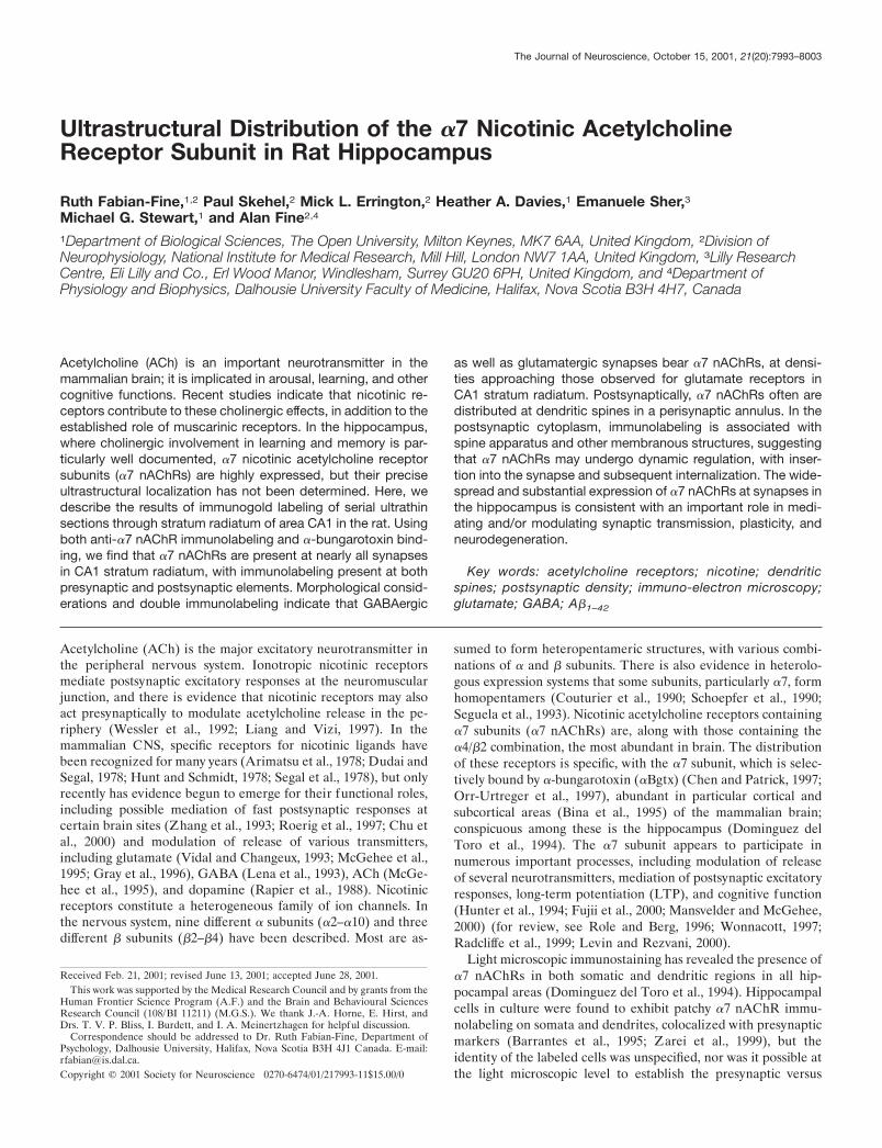
Figure 1. Distribution of �7 nAChR-LIR in rat hippocampus. A, Low magnification micrograph of the hippocampal formation shows that all areasdisplay �7 nAChR-LIR. The strongest immunoreactivity is present in the cell body layers (1, dentate gyrus; 2, CA3; 3, CA1) and in the molecular layerof the dentate gyrus (arrows). B, C, Higher magnification of the CA3 (B) and CA1 (C) regions show that the apical dendrites of the pyramidal cells(arrowheads) display �7 nAChR-LIR. Scale bar: A, 1 mm; B, C, 170 �m.
7994 J. Neurosci., October 15, 2001, 21(20):7993–8003 Fabian-Fine et al. • �7 nAChR Distribution in Rat Hippocampus
(1:100; 4 hr at 37°C; see above). The labeling in control preparations inwhich the biotin-XX-conjugated �Bgtx was omitted did not exceedbackground intensity and revealed no evidence that the biotin bindsunspecifically to synaptic sites.
Mitochondria preparation and Western blot analysis. Mitochondria wereprepared from the hippocampus of four 115 gm male Sprague Dawleyrats, according to standard methods (Løvtrup and Zelander, 1962).Briefly, hippocampus was homogenized on ice in 10 vol of 0.44 M sucrose,10 mM HEPES, and 1 mM MgCl2. The homogenate was then centrifugedat 2000 rpm (400 � g) for 10 min at 4°C in a Beckman JA-20. Thesupernatant was removed, leaving P1, and centrifuged again at 14,000rpm (17,500 � g) for 15 min under the same conditions. The resultingsupernatant S2 was removed, and the pellet was resuspended in ice-coldhomogenization buffer and centrifuged again at 9000 rpm (7000 � g) for15 min to generate a supernatant S3 and a pellet enriched for mitochon-dria. This operation was repeated twice more to wash the mitochondrialfraction. Equivalent amounts of each fraction and the initial homogenatewere analyzed by Western blot analysis. Proteins were separated bySDS-PAGE as described previously (Schagger and von Jagow, 1987)using a 10% separating gel, and transferred to Immobilon-P by electro-blotting. Membranes were blocked in either 2% (w/v) BSA (Fraction V,735086, Boehringer Mannheim), 0.05% (v/v) Nonidet P40 (BDH, PooleUK) in PBS, or 3% nonfat milk powder, 0.05% (v/v) NP40 in PBS.Primary antibodies were applied in the same buffer at dilutions of
1:10,000 for anti-�7 nAChR (M220) and 1:30,000 for anti-Hsp60 (SPA-804; StressGen, Victoria British Columbia, Canada). Immunoreactivitywas detected using HRP-conjugated donkey anti-rabbit (711-035-152;Stratech Scientific, Luton, UK) or anti-mouse (715-035-150; StratechScientific) serum at 1:10,000 and ECL (RPN 2106, Amersham PharmaciaBiotech, Little Chalfont, UK).
Controls. The specificity of the mouse anti-�7 nAChR antibodies usedhere has been described previously (Schoepfer et al., 1990; Dominguezdel Toro et al., 1994). Specificity of antibody binding was confirmed byimmunoblots and by the absence of immunolabeling in preparations fromwhich primary antibodies were omitted. The anti-synaptophysin antibodyused here is well characterized and is known to bind selectively topresynaptic sites (Wiedenmann and Franke 1985). Additional controlswere as described below.
Image analysis and processing. Because sections from all three animalsprocessed for electron microscopy showed similar labeling intensity,randomly selected serial sections from all animals were combined foranalysis. Gold particles at synaptic profiles were counted manually onprinted electron micrographs from 14 series (10,000� magnification),each consisting of three serial sections. For the purpose of this study,structures considered to be synapses satisfied the following criteria: (1)presence of a synaptic cleft; (2) accumulation of synaptic vesicles in thepresynaptic terminal close to the synaptic cleft; and (3) presence of apostsynaptic density in the postsynaptic profile. According to the appear-
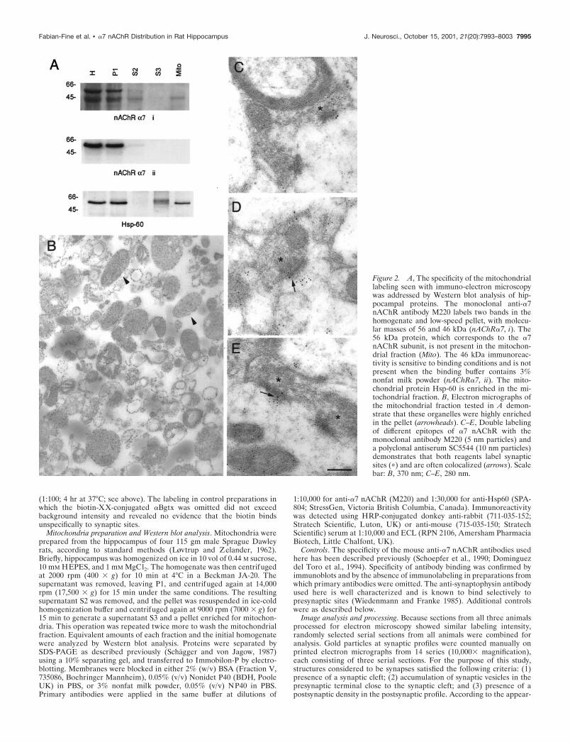
Figure 2. A, The specificity of the mitochondriallabeling seen with immuno-electron microscopywas addressed by Western blot analysis of hip-pocampal proteins. The monoclonal anti-�7nAChR antibody M220 labels two bands in thehomogenate and low-speed pellet, with molecu-lar masses of 56 and 46 kDa (nAChR�7, i). The56 kDa protein, which corresponds to the �7nAChR subunit, is not present in the mitochon-drial fraction (Mito). The 46 kDa immunoreac-tivity is sensitive to binding conditions and is notpresent when the binding buffer contains 3%nonfat milk powder (nAChR�7, ii). The mito-chondrial protein Hsp-60 is enriched in the mi-tochondrial fraction. B, Electron micrographs ofthe mitochondrial fraction tested in A demon-strate that these organelles were highly enrichedin the pellet (arrowheads). C–E, Double labelingof different epitopes of �7 nAChR with themonoclonal antibody M220 (5 nm particles) anda polyclonal antiserum SC5544 (10 nm particles)demonstrates that both reagents label synapticsites (�) and are often colocalized (arrows). Scalebar: B, 370 nm; C–E, 280 nm.
Fabian-Fine et al. • �7 nAChR Distribution in Rat Hippocampus J. Neurosci., October 15, 2001, 21(20):7993–8003 7995
ance of the postsynaptic density, two different synapse types have beendistinguished in the vertebrate CNS (Gray, 1959; Colonnier, 1968). Thefirst type (type 1 or “asymmetrical synapse”) has an extensive postsyn-aptic density and a population of large, round, electron-lucent vesicles inthe presynaptic profile. Type 2 (“symmetrical synapses”) are character-ized by a less conspicuous postsynaptic density and a presynaptic popu-lation of small, pleomorphic, electron-lucent vesicles. It is generallyaccepted that glutamatergic synapses have asymmetric morphology,whereas GABAergic synapses are symmetric. Complete counts of allgold particles at each synapse would require full three-dimensionalreconstruction of each synapse, a prohibitively time-consuming task. Asa useful approximation that largely eliminates false-negative (unlabeled)synapses, we analyzed sets of three serial sections. All synapses inrandomly selected fields of view that were present throughout all threeserial sections were counted. For presentation, micrographs and slideswere digitized at high resolution using a Mustek flatbed scanner or aNikon LS-1000 slide scanner. Contrast and brightness were optimized inAdobe Photoshop 5.0.
Statistical analysis. Gold particle incidence was compared to determinewhether the �7 nAChR immunolabeling over synaptic membranes wassignificantly higher than background labeling. Gold particles werecounted over the synaptic membranes identified in all serial sections. Theactual sampled region was the synaptic cleft plus the adjoining 30-nm-wide presynaptic and postsynaptic zones, because the separation betweena labeled epitope and a gold particle attached to a secondary antibody canextend up to 28 nm (Matsubara et al., 1996). These counts were com-pared with the number of gold particles over equivalent areas of regionswhere no labeling was expected, such as myelin. Because particle countswere not normally distributed, a Welch t test was used for comparisons.The sampled synaptic area for each synapse was estimated by multiplyingthe total sampled length of the synaptic cleft over the three serial sectionsby the section thickness. Linear regression analysis was performed toexamine possible correlation of the number of gold particles per synapsewith the synaptic area.
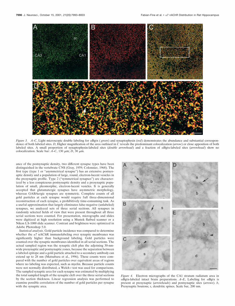
Figure 3. A–C, Light microscopic double labeling for �Bgtx ( green) and synaptophysin (red) demonstrates the abundance and substantial correspon-dence of both labeled sites. D, Higher magnification of the area outlined in C reveals the predominant colocalization (arrow) or close apposition of bothlabeled sites. A small proportion of synaptophysin-labeled sites (double arrowhead) and a fraction of �Bgtx-labeled sites (arrowhead) show nocolocalization. Scale bar: A–C, 130 �m; D, 30 �m.
Figure 4. Electron micrographs of the CA1 stratum radiatum area in�Bgtx-labeled intact brain preparations. A–E, Labeling for �Bgtx ispresent at presynaptic (arrowheads) and postsynaptic sites (arrows). b,Presynaptic boutons; s, dendritic spines. Scale bar, 200 nm.
7996 J. Neurosci., October 15, 2001, 21(20):7993–8003 Fabian-Fine et al. • �7 nAChR Distribution in Rat Hippocampus
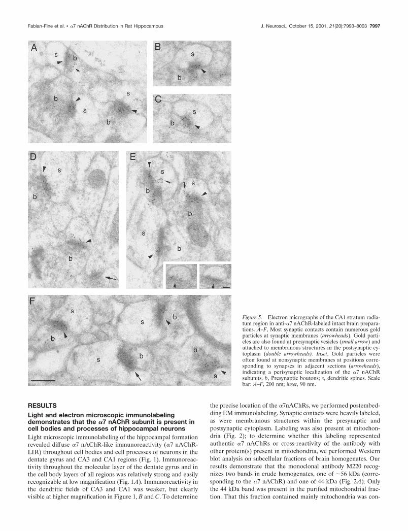
RESULTSLight and electron microscopic immunolabelingdemonstrates that the �7 nAChR subunit is present incell bodies and processes of hippocampal neuronsLight microscopic immunolabeling of the hippocampal formationrevealed diffuse �7 nAChR-like immunoreactivity (�7 nAChR-LIR) throughout cell bodies and cell processes of neurons in thedentate gyrus and CA3 and CA1 regions (Fig. 1). Immunoreac-tivity throughout the molecular layer of the dentate gyrus and inthe cell body layers of all regions was relatively strong and easilyrecognizable at low magnification (Fig. 1A). Immunoreactivity inthe dendritic fields of CA3 and CA1 was weaker, but clearlyvisible at higher magnification in Figure 1, B and C. To determine
the precise location of the �7nAChRs, we performed postembed-ding EM immunolabeling. Synaptic contacts were heavily labeled,as were membranous structures within the presynaptic andpostsynaptic cytoplasm. Labeling was also present at mitochon-dria (Fig. 2); to determine whether this labeling representedauthentic �7 nAChRs or cross-reactivity of the antibody withother protein(s) present in mitochondria, we performed Westernblot analysis on subcellular fractions of brain homogenates. Ourresults demonstrate that the monoclonal antibody M220 recog-nizes two bands in crude homogenates, one of �56 kDa (corre-sponding to the �7 nAChR) and one of 44 kDa (Fig. 2A). Onlythe 44 kDa band was present in the purified mitochondrial frac-tion. That this fraction contained mainly mitochondria was con-
Figure 5. Electron micrographs of the CA1 stratum radia-tum region in anti-�7 nAChR-labeled intact brain prepara-tions. A–F, Most synaptic contacts contain numerous goldparticles at synaptic membranes (arrowheads). Gold parti-cles are also found at presynaptic vesicles (small arrow) andattached to membranous structures in the postsynaptic cy-toplasm (double arrowheads). Inset, Gold particles wereoften found at nonsynaptic membranes at positions corre-sponding to synapses in adjacent sections (arrowheads),indicating a perisynaptic localization of the �7 nAChRsubunits. b, Presynaptic boutons; s, dendritic spines. Scalebar: A–F, 200 nm; inset, 90 nm.
Fabian-Fine et al. • �7 nAChR Distribution in Rat Hippocampus J. Neurosci., October 15, 2001, 21(20):7993–8003 7997
firmed by its enrichment in the mitochondrial marker, Hsp-60(Fig. 2A), and by ultrastructural investigation of the fraction (Fig.2B). We were able to eliminate the 44 kDa band in Western blotsusing 3% nonfat milk powder in the blocking medium (Fig. 2A)(see Materials and Methods). This blocking procedure, however,was not compatible with immuno-electron microscopy. To con-firm the specificity of �7 nAChR immunolabeling, we performedtwo independent tests: (1) double labeling using the monoclonalantibody (M220) and a polyclonal antiserum (SC5544) raisedagainst a larger epitope of the �7 subunit (polyclonal: amino acids367–502; monoclonal: amino acids 380–400); and (2) light andelectron microscopic labeling for �Bgtx. Figure 2C–E demon-strate that both antibodies directed against the �7 subunit labeled
synaptic contacts, frequently colocalizing at both presynaptic andpostsynaptic sites. As shown in Figure 2, D and E (5 nm particles),the monoclonal antibody yielded stronger labeling, and only themonoclonal antibody labeled mitochondria (Fig. 2C–E).
The presence of �7 nAChR at synaptic sites was furtherconfirmed by �Bgtx binding. As demonstrated in Figure 3, lightmicroscopic double labeling for �Bgtx and synaptophysin re-vealed abundant expression of both epitopes in hippocampalsynaptic zones, often in close apposition. In keeping with previousfindings (Hunt and Schmidt, 1979), �Bgtx binding was seen onlyoccasionally throughout neuronal cell bodies; this difference com-pared with the pattern of anti-�7 nAChR immunolabeling mayreflect the post-translational processing required for �Bgtx bind-ing (Chen et al., 1998; Aztiria et al., 2000) but not for binding ofthe monoclonal antibody directed against the peptide epitope.Ultrastructural investigation of hippocampal tissue labeled for�Bgtx revealed widespread presynaptic and postsynaptic labeling(Fig. 4) that, although less intense than the labeling observed withthe anti-�7 nAChR antibody, was qualitatively similar in distri-bution, confirming that the synaptic immunolabeling reflects thepresence of authentic �7 nAChRs.
Most synapses in CA1 stratum radiatum display�7 nAChR-LIRThe ultrastructural investigation of dentate gyrus and CA1 andCA3 regions revealed �7 nAChR-LIR at synaptic sites in allhippocampal areas (Fig. 5). Gold particles were located mainly(1) at the postsynaptic density, (2) within the synaptic cleft, (3) inthe postsynaptic cytoplasm (Figs. 5–7), and (4) at presynapticallylocated vesicles, often located in close proximity to the synaptic
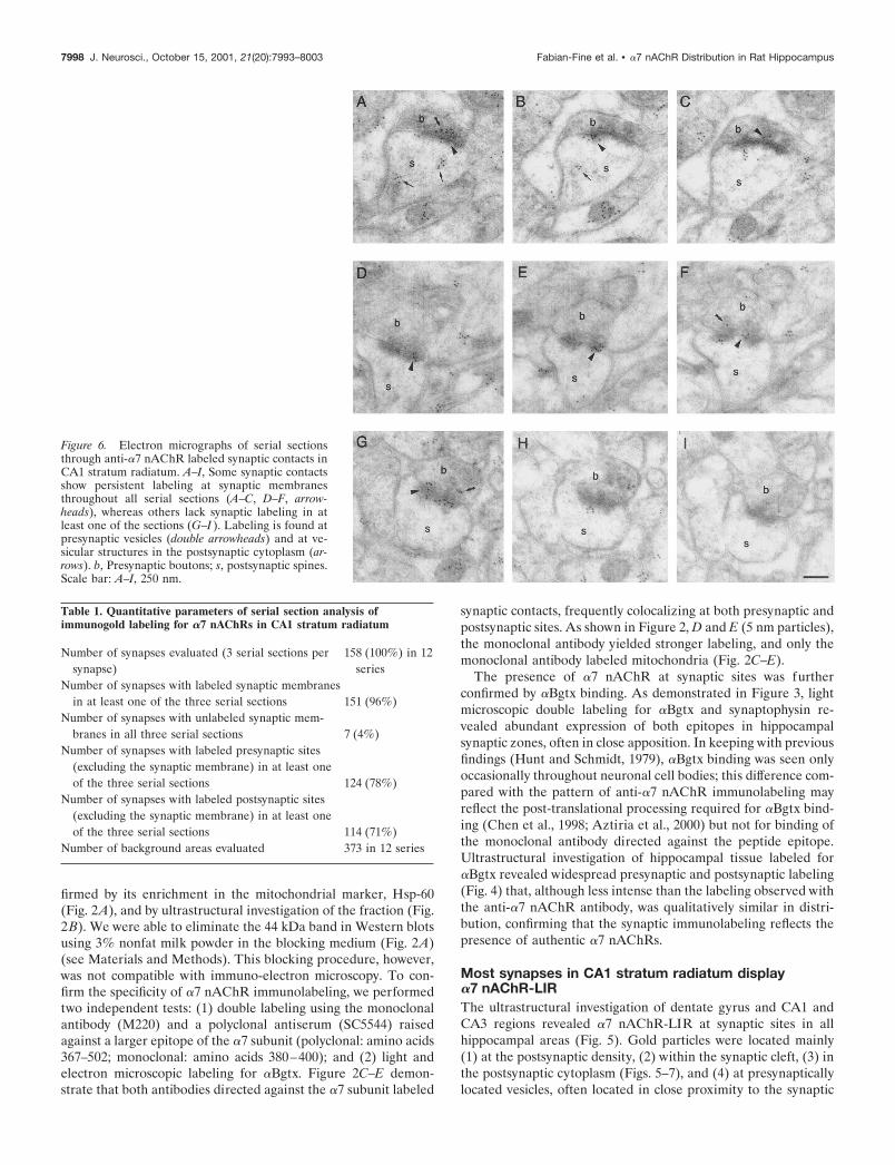
Figure 6. Electron micrographs of serial sectionsthrough anti-�7 nAChR labeled synaptic contacts inCA1 stratum radiatum. A–I, Some synaptic contactsshow persistent labeling at synaptic membranesthroughout all serial sections (A–C, D–F, arrow-heads), whereas others lack synaptic labeling in atleast one of the sections (G–I ). Labeling is found atpresynaptic vesicles (double arrowheads) and at ve-sicular structures in the postsynaptic cytoplasm (ar-rows). b, Presynaptic boutons; s, postsynaptic spines.Scale bar: A–I, 250 nm.
Table 1. Quantitative parameters of serial section analysis ofimmunogold labeling for �7 nAChRs in CA1 stratum radiatum
Number of synapses evaluated (3 serial sections persynapse)
158 (100%) in 12series
Number of synapses with labeled synaptic membranesin at least one of the three serial sections 151 (96%)
Number of synapses with unlabeled synaptic mem-branes in all three serial sections 7 (4%)
Number of synapses with labeled presynaptic sites(excluding the synaptic membrane) in at least oneof the three serial sections 124 (78%)
Number of synapses with labeled postsynaptic sites(excluding the synaptic membrane) in at least oneof the three serial sections 114 (71%)
Number of background areas evaluated 373 in 12 series
7998 J. Neurosci., October 15, 2001, 21(20):7993–8003 Fabian-Fine et al. • �7 nAChR Distribution in Rat Hippocampus
membrane but �30 nm apart from it (indicating that the epitopeis located at the vesicles rather than the synaptic membrane). It isnot clear whether the latter are synaptic vesicles or specializedtransport vesicles. Labeled synapses were present at both den-
dritic spines and shafts. We determined the percentage of syn-apses labeled in the CA1 stratum radiatum region by serial sectionanalysis (Fig. 6, Table 1), because gold particles may not bepresent in every section through a �7 nAChR-LIR synapse, sothat observations on single sections will underestimate the trueincidence of labeled structures. For each of the 158 synapsesevaluated in this study, three serial sections were analyzed. Asynapse was considered immunoreactive for �7 nAChRs if one ofthe three sections showed at least one gold particle within �30 nmof the synaptic cleft to cover the possible separation between alabeled epitope and a gold particle attached to a secondaryantibody (see above). The extent of this separation also renderedit impossible to ascertain whether gold particles within the syn-aptic cleft represented binding to the presynaptic or postsynapticmembrane; therefore, gold particles found within 30 nm of thesynaptic cleft were classed together as labeling at synapticmembranes.
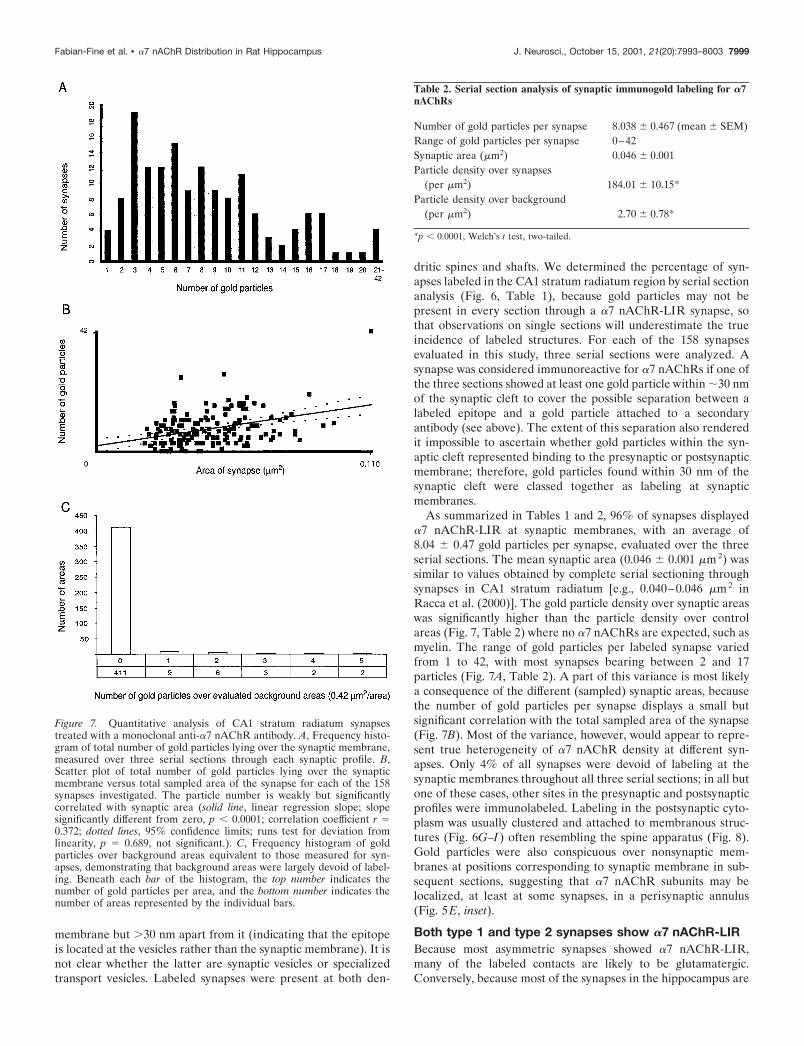
As summarized in Tables 1 and 2, 96% of synapses displayed�7 nAChR-LIR at synaptic membranes, with an average of8.04 � 0.47 gold particles per synapse, evaluated over the threeserial sections. The mean synaptic area (0.046 � 0.001 �m2) wassimilar to values obtained by complete serial sectioning throughsynapses in CA1 stratum radiatum [e.g., 0.040–0.046 �m2 inRacca et al. (2000)]. The gold particle density over synaptic areaswas significantly higher than the particle density over controlareas (Fig. 7, Table 2) where no �7 nAChRs are expected, such asmyelin. The range of gold particles per labeled synapse variedfrom 1 to 42, with most synapses bearing between 2 and 17particles (Fig. 7A, Table 2). A part of this variance is most likelya consequence of the different (sampled) synaptic areas, becausethe number of gold particles per synapse displays a small butsignificant correlation with the total sampled area of the synapse(Fig. 7B). Most of the variance, however, would appear to repre-sent true heterogeneity of �7 nAChR density at different syn-apses. Only 4% of all synapses were devoid of labeling at thesynaptic membranes throughout all three serial sections; in all butone of these cases, other sites in the presynaptic and postsynapticprofiles were immunolabeled. Labeling in the postsynaptic cyto-plasm was usually clustered and attached to membranous struc-tures (Fig. 6G–I) often resembling the spine apparatus (Fig. 8).Gold particles were also conspicuous over nonsynaptic mem-branes at positions corresponding to synaptic membrane in sub-sequent sections, suggesting that �7 nAChR subunits may belocalized, at least at some synapses, in a perisynaptic annulus(Fig. 5E, inset).
Both type 1 and type 2 synapses show �7 nAChR-LIRBecause most asymmetric synapses showed �7 nAChR-LIR,many of the labeled contacts are likely to be glutamatergic.Conversely, because most of the synapses in the hippocampus are
Figure 7. Quantitative analysis of CA1 stratum radiatum synapsestreated with a monoclonal anti-�7 nAChR antibody. A, Frequency histo-gram of total number of gold particles lying over the synaptic membrane,measured over three serial sections through each synaptic profile. B,Scatter plot of total number of gold particles lying over the synapticmembrane versus total sampled area of the synapse for each of the 158synapses investigated. The particle number is weakly but significantlycorrelated with synaptic area (solid line, linear regression slope; slopesignificantly different from zero, p � 0.0001; correlation coefficient r �0.372; dotted lines, 95% confidence limits; runs test for deviation fromlinearity, p � 0.689, not significant.). C, Frequency histogram of goldparticles over background areas equivalent to those measured for syn-apses, demonstrating that background areas were largely devoid of label-ing. Beneath each bar of the histogram, the top number indicates thenumber of gold particles per area, and the bottom number indicates thenumber of areas represented by the individual bars.
Table 2. Serial section analysis of synaptic immunogold labeling for �7nAChRs
Number of gold particles per synapse 8.038 � 0.467 (mean � SEM)Range of gold particles per synapse 0–42Synaptic area (�m2) 0.046 � 0.001Particle density over synapses
(per �m2) 184.01 � 10.15*Particle density over background
(per �m2) 2.70 � 0.78*
*p � 0.0001, Welch’s t test, two-tailed.
Fabian-Fine et al. • �7 nAChR Distribution in Rat Hippocampus J. Neurosci., October 15, 2001, 21(20):7993–8003 7999
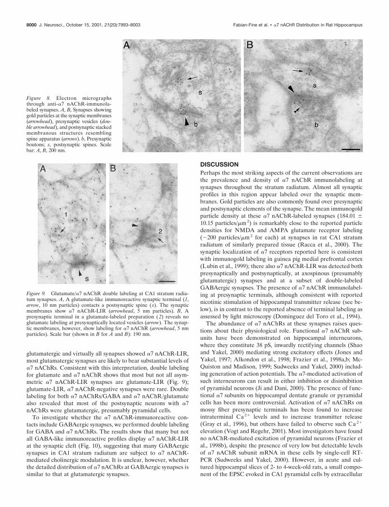
glutamatergic and virtually all synapses showed �7 nAChR-LIR,most glutamatergic synapses are likely to bear substantial levels of�7 nAChRs. Consistent with this interpretation, double labelingfor glutamate and �7 nAChR shows that most but not all asym-metric �7 nAChR-LIR synapses are glutamate-LIR (Fig. 9);glutamate-LIR, �7 nAChR-negative synapses were rare. Doublelabeling for both �7 nAChRs/GABA and �7 nAChR/glutamatealso revealed that most of the postsynaptic neurons with �7nAChRs were glutamatergic, presumably pyramidal cells.
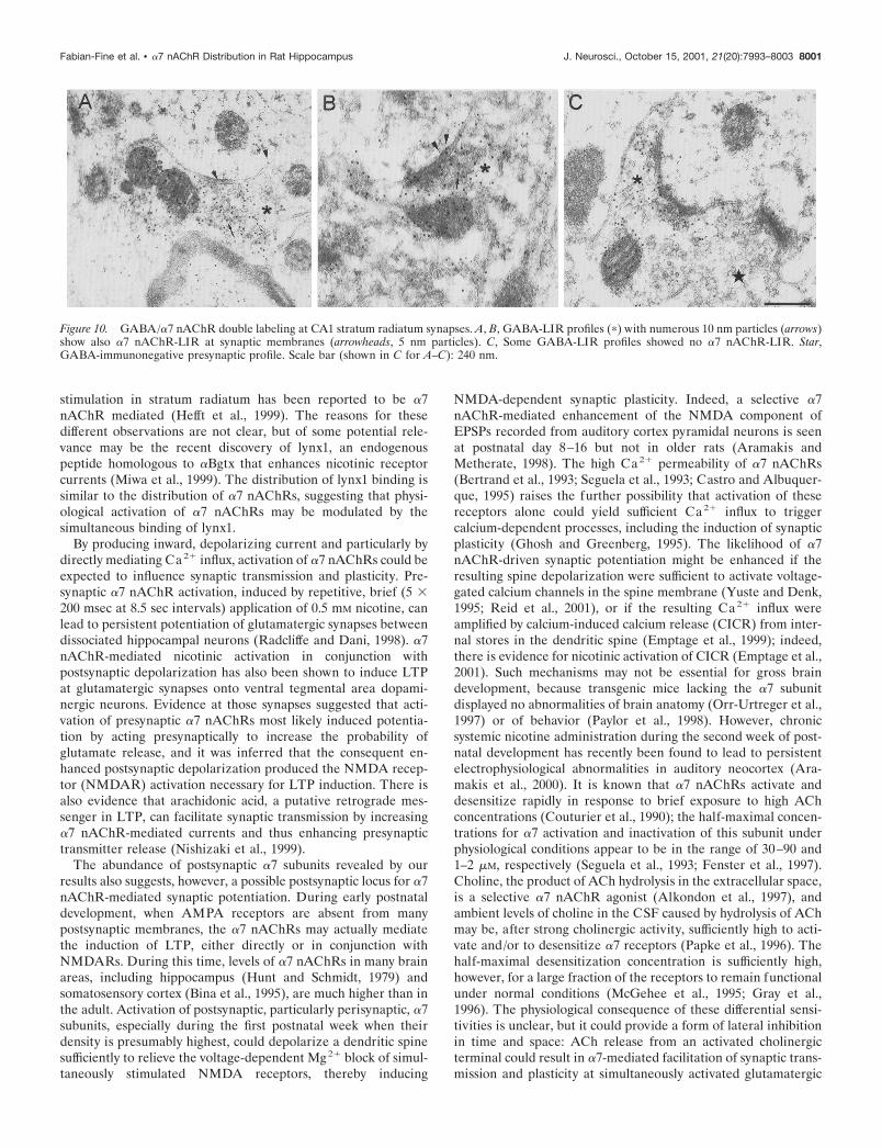
To investigate whether the �7 nAChR-immunoreactive con-tacts include GABAergic synapses, we performed double labelingfor GABA and �7 nAChRs. The results show that many but notall GABA-like immunoreactive profiles display �7 nAChR-LIRat the synaptic cleft (Fig. 10), suggesting that many GABAergicsynapses in CA1 stratum radiatum are subject to �7 nAChR-mediated cholinergic modulation. It is unclear, however, whetherthe detailed distribution of �7 nAChRs at GABAergic synapses issimilar to that at glutamatergic synapses.
DISCUSSIONPerhaps the most striking aspects of the current observations arethe prevalence and density of �7 nAChR immunolabeling atsynapses throughout the stratum radiatum. Almost all synapticprofiles in this region appear labeled over the synaptic mem-branes. Gold particles are also commonly found over presynapticand postsynaptic elements of the synapse. The mean immunogoldparticle density at these �7 nAChR-labeled synapses (184.01 �10.15 particles/�m2) is remarkably close to the reported particledensities for NMDA and AMPA glutamate receptor labeling(�200 particles/�m2 for each) at synapses in rat CA1 stratumradiatum of similarly prepared tissue (Racca et al., 2000). Thesynaptic localization of �7 receptors reported here is consistentwith immunogold labeling in guinea pig medial prefrontal cortex(Lubin et al., 1999); there also �7 nAChR-LIR was detected bothpresynaptically and postsynaptically, at axospinous (presumablyglutamatergic) synapses and at a subset of double-labeledGABAergic synapses. The presence of �7 nAChR immunolabel-ing at presynaptic terminals, although consistent with reportednicotinic stimulation of hippocampal transmitter release (see be-low), is in contrast to the reported absence of terminal labeling asassessed by light microscopy (Dominguez del Toro et al., 1994).
The abundance of �7 nAChRs at these synapses raises ques-tions about their physiological role. Functional �7 nAChR sub-units have been demonstrated on hippocampal interneurons,where they constitute 38 pS, inwardly rectifying channels (Shaoand Yakel, 2000) mediating strong excitatory effects (Jones andYakel, 1997; Alkondon et al., 1998; Frazier et al., 1998a,b; Mc-Quiston and Madison, 1999; Sudweeks and Yakel, 2000) includ-ing generation of action potentials. The �7-mediated activation ofsuch interneurons can result in either inhibition or disinhibitionof pyramidal neurons (Ji and Dani, 2000). The presence of func-tional �7 subunits on hippocampal dentate granule or pyramidalcells has been more controversial. Activation of �7 nAChRs onmossy fiber presynaptic terminals has been found to increaseintraterminal Ca2� levels and to increase transmitter release(Gray et al., 1996), but others have failed to observe such Ca2�
elevation (Vogt and Regehr, 2001). Most investigators have foundno nAChR-mediated excitation of pyramidal neurons (Frazier etal., 1998b), despite the presence of very low but detectable levelsof �7 nAChR subunit mRNA in these cells by single-cell RT-PCR (Sudweeks and Yakel, 2000). However, in acute and cul-tured hippocampal slices of 2- to 4-week-old rats, a small compo-nent of the EPSC evoked in CA1 pyramidal cells by extracellular
Figure 8. Electron micrographsthrough anti-�7 nAChR-immunola-beled synapses. A, B, Synapses showinggold particles at the synaptic membranes(arrowhead), presynaptic vesicles (dou-ble arrowhead), and postsynaptic stackedmembranous structures resemblingspine apparatus (arrows). b, Presynapticboutons; s, postsynaptic spines. Scalebar: A, B, 200 nm.
Figure 9. Glutamate/�7 nAChR double labeling at CA1 stratum radia-tum synapses. A, A glutamate-like immunoreactive synaptic terminal (1,arrow, 10 nm particles) contacts a postsynaptic spine ( s). The synapticmembranes show �7 nAChR-LIR (arrowhead, 5 nm particles). B, Apresynaptic terminal in a glutamate-labeled preparation (2) reveals noglutamate labeling at presynaptically located vesicles (arrow). The synap-tic membranes, however, show labeling for �7 nAChR (arrowhead, 5 nmparticles). Scale bar (shown in B for A and B): 190 nm.
8000 J. Neurosci., October 15, 2001, 21(20):7993–8003 Fabian-Fine et al. • �7 nAChR Distribution in Rat Hippocampus
stimulation in stratum radiatum has been reported to be �7nAChR mediated (Hefft et al., 1999). The reasons for thesedifferent observations are not clear, but of some potential rele-vance may be the recent discovery of lynx1, an endogenouspeptide homologous to �Bgtx that enhances nicotinic receptorcurrents (Miwa et al., 1999). The distribution of lynx1 binding issimilar to the distribution of �7 nAChRs, suggesting that physi-ological activation of �7 nAChRs may be modulated by thesimultaneous binding of lynx1.
By producing inward, depolarizing current and particularly bydirectly mediating Ca2� influx, activation of �7 nAChRs could beexpected to influence synaptic transmission and plasticity. Pre-synaptic �7 nAChR activation, induced by repetitive, brief (5 �200 msec at 8.5 sec intervals) application of 0.5 mM nicotine, canlead to persistent potentiation of glutamatergic synapses betweendissociated hippocampal neurons (Radcliffe and Dani, 1998). �7nAChR-mediated nicotinic activation in conjunction withpostsynaptic depolarization has also been shown to induce LTPat glutamatergic synapses onto ventral tegmental area dopami-nergic neurons. Evidence at those synapses suggested that acti-vation of presynaptic �7 nAChRs most likely induced potentia-tion by acting presynaptically to increase the probability ofglutamate release, and it was inferred that the consequent en-hanced postsynaptic depolarization produced the NMDA recep-tor (NMDAR) activation necessary for LTP induction. There isalso evidence that arachidonic acid, a putative retrograde mes-senger in LTP, can facilitate synaptic transmission by increasing�7 nAChR-mediated currents and thus enhancing presynaptictransmitter release (Nishizaki et al., 1999).
The abundance of postsynaptic �7 subunits revealed by ourresults also suggests, however, a possible postsynaptic locus for �7nAChR-mediated synaptic potentiation. During early postnataldevelopment, when AMPA receptors are absent from manypostsynaptic membranes, the �7 nAChRs may actually mediatethe induction of LTP, either directly or in conjunction withNMDARs. During this time, levels of �7 nAChRs in many brainareas, including hippocampus (Hunt and Schmidt, 1979) andsomatosensory cortex (Bina et al., 1995), are much higher than inthe adult. Activation of postsynaptic, particularly perisynaptic, �7subunits, especially during the first postnatal week when theirdensity is presumably highest, could depolarize a dendritic spinesufficiently to relieve the voltage-dependent Mg2� block of simul-taneously stimulated NMDA receptors, thereby inducing
NMDA-dependent synaptic plasticity. Indeed, a selective �7nAChR-mediated enhancement of the NMDA component ofEPSPs recorded from auditory cortex pyramidal neurons is seenat postnatal day 8–16 but not in older rats (Aramakis andMetherate, 1998). The high Ca2� permeability of �7 nAChRs(Bertrand et al., 1993; Seguela et al., 1993; Castro and Albuquer-que, 1995) raises the further possibility that activation of thesereceptors alone could yield sufficient Ca2� influx to triggercalcium-dependent processes, including the induction of synapticplasticity (Ghosh and Greenberg, 1995). The likelihood of �7nAChR-driven synaptic potentiation might be enhanced if theresulting spine depolarization were sufficient to activate voltage-gated calcium channels in the spine membrane (Yuste and Denk,1995; Reid et al., 2001), or if the resulting Ca2� influx wereamplified by calcium-induced calcium release (CICR) from inter-nal stores in the dendritic spine (Emptage et al., 1999); indeed,there is evidence for nicotinic activation of CICR (Emptage et al.,2001). Such mechanisms may not be essential for gross braindevelopment, because transgenic mice lacking the �7 subunitdisplayed no abnormalities of brain anatomy (Orr-Urtreger et al.,1997) or of behavior (Paylor et al., 1998). However, chronicsystemic nicotine administration during the second week of post-natal development has recently been found to lead to persistentelectrophysiological abnormalities in auditory neocortex (Ara-makis et al., 2000). It is known that �7 nAChRs activate anddesensitize rapidly in response to brief exposure to high AChconcentrations (Couturier et al., 1990); the half-maximal concen-trations for �7 activation and inactivation of this subunit underphysiological conditions appear to be in the range of 30–90 and1–2 �M, respectively (Seguela et al., 1993; Fenster et al., 1997).Choline, the product of ACh hydrolysis in the extracellular space,is a selective �7 nAChR agonist (Alkondon et al., 1997), andambient levels of choline in the CSF caused by hydrolysis of AChmay be, after strong cholinergic activity, sufficiently high to acti-vate and/or to desensitize �7 receptors (Papke et al., 1996). Thehalf-maximal desensitization concentration is sufficiently high,however, for a large fraction of the receptors to remain functionalunder normal conditions (McGehee et al., 1995; Gray et al.,1996). The physiological consequence of these differential sensi-tivities is unclear, but it could provide a form of lateral inhibitionin time and space: ACh release from an activated cholinergicterminal could result in �7-mediated facilitation of synaptic trans-mission and plasticity at simultaneously activated glutamatergic
Figure 10. GABA/�7 nAChR double labeling at CA1 stratum radiatum synapses. A, B, GABA-LIR profiles (�) with numerous 10 nm particles (arrows)show also �7 nAChR-LIR at synaptic membranes (arrowheads, 5 nm particles). C, Some GABA-LIR profiles showed no �7 nAChR-LIR. Star,GABA-immunonegative presynaptic profile. Scale bar (shown in C for A–C): 240 nm.
Fabian-Fine et al. • �7 nAChR Distribution in Rat Hippocampus J. Neurosci., October 15, 2001, 21(20):7993–8003 8001
synapses close to the activated terminal where the local AChconcentration would transiently be high. As a result of diffusion,synapses farther from the cholinergic terminal, or nearby syn-apses activated asynchronously, would experience inactivatingACh concentrations.
In addition to labeling of synaptic membranes, we also ob-served abundant labeling over endoplasmic reticulum and overmembranous structures in the postsynaptic cytoplasm, includingthe spine apparatus (Figs. 5–7). This pattern resembles thereported association of �2 and �4 nAChRs with endoplasmicreticulum and transport vesicles in various neurons, includingneocortical pyramidal cells (Hill et al., 1993; Nakayama et al.,1995). These observations of a large pool of intracellular �7nAChRs suggest that these subunits may be actively internalizedor inserted and that the extracellular, functional receptors maythus be dynamically regulated in response to the ongoing activityof the neuron.
Finally, the near-ubiquitous presence of �7 nAChRs at hip-pocampal synapses described here renders more salient the re-cent report (Wang et al., 2000b) of high-affinity binding of�-amyloid peptide (A�1–42) to �7 nAChRs. Submicromolar con-centrations of soluble A�1–42, like its fibrillar amyloid precipitate,may be neurotoxic (Roher et al., 1996); toxicity can be partiallyblocked by nicotine (Wang et al., 2000a). Furthermore, similarlylow concentrations of A�1–42 can block �7 nAChR-mediatedcurrents in hippocampal interneurons (Pettit et al., 2001). Thusthe widespread distribution of �7 nAChRs in the hippocampusand their interactions with A�1–42 peptide may be importantfactors in early cognitive impairments and later neuronal loss inAlzheimer’s disease.
REFERENCESAlkondon M, Pereira EF, Cortes WS, Maelicke A, Albuquerque EX
(1997) Choline is a selective agonist of alpha7 nicotinic acetylcholinereceptors in the rat brain neurons. Eur J Neurosci 9:2734–2742.
Alkondon M, Pereira EFR, Albuquerque EX (1998) �-Bungarotoxin-and methyllycaconitine-sensitive nicotinic receptors mediate fast syn-aptic transmission in interneurons of rat hippocampal slices. Brain Res810:257–263.
Aramakis VB, Metherate R (1998) Nicotine selectively enhancesNMDA receptor-mediated synaptic transmission during postnatal de-velopment in sensory neocortex. J Neurosci 18:8485–8495.
Aramakis VB, Hsieh CY, Leslie FM, Metherate R (2000) A criticalperiod for nicotine-induced disruption of synaptic development in ratauditory cortex. J Neurosci 20:6106–6116.
Arimatsu Y, Seto A, Amano T (1978) Localization of alpha-bungarotoxin binding sites in mouse brain by light and electron micro-scopic autoradiography. Brain Res 147:165–169.
Aztiria EM, Sogayar MC, Barrantes FJ (2000) Expression of a neuronalnicotinic acetylcholine receptor in insect and mammalian host cellsystems. Neurochem Res 25:171–180.
Barrantes GE, Rogers AT, Lindstrom J, Wonnacott S (1995) alpha-Bungarotoxin binding sites in rat hippocampal and cortical cultures:initial characterisation, colocalisation with alpha 7 subunits and up-regulation by chronic nicotine treatment. Brain Res 672:228–236.
Bertrand D, Galzi JL, Devilliers-Thiery A, Bertrand S, Changeux JP(1993) Mutation at two distinct sites within the channel domain M2alter calcium permeability of the neuronal alpha7 nicotinic receptor.Proc Natl Acad Sci USA 90:6971–6975.
Bina KG, Guzman P, Broide RS, Leslie FM, Smith MA, O’Dowd DK(1995) Localization of alpha 7 nicotinic receptor subunit mRNA andalpha-bungarotoxin binding sites in developing mouse somatosensorythalamocortical system. J Comp Neurol 363:321–332.
Castro NG, Albuquerque EX (1995) �-Bungarotoxin-sensitive hip-pocampal nicotinic receptor channel has a high calcium permeability.Biophys J 68:516–524.
Chen D, Patrick JW (1997) The alpha-bungarotoxin-binding nicotinicacetylcholine receptor from rat brain contains only the alpha7 subunit.J Biol Chem 272:24024–24029.
Chen D, Dang H, Patrick JW (1998) Contributions of N-linked glyco-sylation to the expression of a functional alpha7-nicotinic receptor inXenopus oocytes. J Neurochem 70:349–357.
Chu ZG, Zhou FM, Hablitz JJ (2000) Nicotinic acetylcholine receptor-mediated synaptic potentials in rat neocortex. Brain Res 887:399–405.
Colonnier M (1968) Synaptic patterns on different cell types in thedifferent laminae of the cat visual cortex. An electron microscope study.Brain Res 9:268–287.
Couturier S, Bertrand D, Matter JM, Hernandez MC, Bertrand S, Millar N,Valera S, Barkas T, Ballivet M (1990) A neuronal nicotinic acetylcholinereceptor subunit (alpha 7) is developmentally regulated and forms ahomo-oligomeric channel blocked by alpha-BTX. Neuron 5:847–856.
Dominguez del Toro E, Juiz JM, Peng X, Lindstrom J, Criado M (1994)Immunocytochemical localization of the alpha 7 subunit of the nico-tinic acetylcholine receptor in the rat central nervous system. J CompNeurol 349:325–342.
Dudai Y, Segal M (1978) alpha-Bungarotoxin binding sites in rat hip-pocampus: localization in postsynaptic cells. Brain Res 154:167–171.
Emptage N, Bliss TV, Fine A (1999) Single synaptic events evokeNMDA receptor-mediated release of calcium from internal stores inhippocampal dendritic spines. Neuron 22:115–124.
Emptage N, Reid CA, Fine A (2001) Calcium stores in hippocampalsynaptic boutons mediate short term plasticity, store-operated Ca2�entry and spontaneous transmitter release. Neuron 29:197–208.
Fenster CP, Rains MF, Noerager B, Quick MW, Lester RA (1997)Influence of subunit composition on desensitization of neuronal ace-tylcholine receptors at low concentrations of nicotine. J Neurosci17:5747–5759.
Frazier CJ, Buhler AV, Weiner JL, Dunwiddie TV (1998a) Synapticpotentials mediated via alpha-bungarotoxin-sensitive nicotinic acetyl-choline receptors in rat hippocampal interneurons. J Neurosci18:8228–8235.
Frazier CJ, Rollins YD, Breese CR, Leonard S, Freedman R, DunwiddieTV (1998b) Acetylcholine activates an �-bungarotoxin-sensitive nico-tinic current in rat hippocampal interneurons, but not pyramidal cells.J Neurosci 18:1187–1195.
Fujii S, Ji Z, Sumikawa K (2000) Inactivation of alpha7 ACh receptorsand activation of non-alpha7 ACh receptors both contribute to longterm potentiation induction in the hippocampal CA1 region. NeurosciLett 286:134–138.
Ghosh A, Greenberg ME (1995) Calcium signalling in neurons: molec-ular mechanisms and cellular consequences. Science 268:239–247.
Gray EG (1959) Axo-somatic and axo-dendritic synapses of the cerebralcortex: an electron microscope study. J Anat 93:420–433.
Gray R, Rajan AS, Radcliffe KA, Yakehiro M, Dani JA (1996) Hip-pocampal synaptic transmission enhanced by low concentrations ofnicotine. Nature 383:713–716.
Hefft S, Hulo S, Bertrand D, Muller D (1999) Synaptic transmission atnicotinic acetylcholine receptors in rat hippocampal organotypic cul-tures and slices. J Physiol (Lond) 515:769–776.
Hill JAJ, Zoli M, Bourgeois JP, Changeux JP (1993) Immunocytochem-ical localization of a neuronal nicotinic receptor: the beta 2-subunit.J Neurosci 13:1551–1568.
Hunt S, Schmidt J (1979) The relationship of alpha-bungarotoxin bind-ing activity and cholinergic termination within the rat hippocampus.Neuroscience 4:585–592.
Hunt SP, Schmidt J (1978) The electron microscopic autoradiographiclocalization of alpha-bungarotoxin binding sites within the centralnervous system of the rat. Brain Res 142:152–159.
Hunter BE, de Fiebre CM, Papke RL, Kem WR, Meyer EM (1994) Anovel nicotinic agonist facilitates induction of long-term potentiation inthe rat hippocampus. Neurosci Lett 168:130–134.
Ji D, Dani JA (2000) Inhibition and disinhibition of pyramidal neuronsby activation of nicotinic receptors on hippocampal interneurons.J Neurophysiol 83:2682–2690.
Jones S, Yakel JL (1997) Functional nicotinic ACh receptors on inter-neurons in the rat hippocampus. J Physiol (Lond) 504:603–610.
Lena C, Changeux J-P, Mulle C (1993) Evidence for “preterminal” nic-otinic receptors on GABAergic axons in the rat interpeduncular nu-cleus. J Neurosci 13:2680–2688.
Levin ED, Rezvani AH (2000) Development of nicotinic drug therapyfor cognitive disorders. Eur J Pharmacol 393:141–146.
Liang SD, Vizi ES (1997) Positive feedback modulation of acetylcholinerelease from isolated rat superior cervical ganglion. J Pharmacol ExpTher 280:650–655.
Løvtrup S, Zelander T (1962) Isolation of brain mitochondria. Exp CellRes 27:468–473.
Lubin M, Erisir A, Aoki C (1999) Ultrastructural immunolocalization ofthe �7 nAChR subunit in guinea pig medial prefrontal cortex. Ann NYAcad Sci 868:628–632.
Mansvelder HD, McGehee DS (2000) Long-term potentiation of exci-tatory inputs to brain reward areas by nicotine. Neuron 27:349–357.
Matsubara A, Laake JH, Davanger S, Usami S, Ottersen OP (1996)Organization of AMPA receptor subunits at a glutamate synapse: aquantitative immunogold analysis of hair cell synapses in the rat organof Corti. J Neurosci 16:4457–4467.
McGehee DS, Heath MJ, Gelber S, Devay P, Role LW (1995) Nicotine
8002 J. Neurosci., October 15, 2001, 21(20):7993–8003 Fabian-Fine et al. • �7 nAChR Distribution in Rat Hippocampus
enhancement of fast excitatory synaptic transmission in CNS by pre-synaptic receptors. Science 269:1692–1696.
McQuiston AR, Madison DV (1999) Nicotinic receptor activation ex-cites distinct subtypes of interneurons in the rat hippocampus. J Neu-rosci 19:2887–2896.
Miwa JM, Ibanez-Tallon I, Crabtree GW, Sanchez R, Sali A, Role LW,Heintz N (1999) lynx1, an endogenous toxin-like modulator of nicotinicacetylcholine receptors in the mammalian CNS. Neuron 23:105–114.
Nakayama H, Shioda S, Okuda H, Nakashima T, Nakai Y (1995) Immu-nocytochemical localization of nicotinic acetylcholine receptor in ratcerebral cortex. Brain Res Mol Brain Res 32:321–328.
Nishizaki T, Nomura T, Matsuoka T, Enikolopov G, Sumikawa K (1999)Arachidonic acid induces a long-lasting facilitation of hippocampalsynaptic transmission by modulating PKC activity and nicotinic AChreceptors. Brain Res Mol Brain Res 69:263–272.
Orr-Urtreger A, Goldner FM, Saeki M, Lorenzo I, Goldberg L, De BiasiM, Dani JA, Patrick JW, Beaudet AL (1997) Mice deficient in the �7neuronal nicotinic acetylcholine receptor lack �-bungarotoxin bindingsites and hippocampal fast nicotinic currents. J Neurosci 17:9165–9171.
Papke RL, Bencherif M, Lippiello P (1996) An evaluation of neuronalnicotinic acetylcholine receptor activation by quaternary nitrogen com-pounds indicates that choline is selective for the alpha 7 subtype.Neurosci Lett 213:201–204.
Paylor R, Nguyen M, Crawley JN, Patrick J, Beaudet A, Orr-Urtreger A(1998) Alpha7 nicotinic receptor subunits are not necessary forhippocampal-dependent learning or sensorimotor gating: a behavioralcharacterization of Acra7-deficient mice. Learn Mem 5:302–316.
Pettit DL, Shao Z, Yakel JL (2001) �-Amyloid1–42 peptide directlymodulates nicotinic receptors in the rat hippocampal slice. J Neurosci21:RC120.
Racca C, Stephenson FA, Streit P, Roberts JD, Somogyi P (2000)NMDA receptor content of synapses in stratum radiatum of the hip-pocampal CA1 area. J Neurosci 20:2512–2522.
Radcliffe KA, Dani JA (1998) Nicotinic stimulation produces multipleforms of increased glutamatergic synaptic transmission. J Neurosci18:7075–7083.
Radcliffe KA, Fisher JL, Gray R, Dani JA (1999) Nicotinic modulationof glutamate and GABA synaptic transmission of hippocampal neu-rons. Ann NY Acad Sci 868:591–610.
Rapier C, Lunt GG, Wonnacott S (1988) Stereoselective nicotine-induced release of dopamine from striatal synaptosomes: concentrationdependence and repetitive stimulation. J Neurochem 50:1123–1130.
Reid CA, Fabian-Fine R, Fine A (2001) Postsynaptic calcium transientsevoked by activation of individual hippocampal mossy fiber synapses.J Neurosci 21:2206–2214.
Roerig B, Nelson DA, Katz LC (1997) Fast synaptic signaling by nico-tinic acetylcholine and serotonin 5-HT3 receptors in developing visualcortex. J Neurosci 17:8353–8362.
Roher AE, Chaney MO, Kuo YM, Webster SD, Stine WB, HaverkampLJ, Woods AS, Cotter RJ, Tuohy JM, Krafft GA, Bonnell BS, Em-merling MR (1996) Morphology and toxicity of Abeta-(1–42) dimerderived from neuritic and vascular amyloid deposits of Alzheimer’sdisease. J Biol Chem 271:20631–20635.
Role LW, Berg DK (1996) Nicotinic receptors in the development andmodulation of CNS synapses. Neuron 16:1077–1085.
Schagger H, von Jagow G (1987) Tricine-sodium dodecyl sulfate-polyacrylamide gel electrophoresis for the separation of proteins in therange from 1 to 100 kDa. Anal Biochem 166:368–379.
Schoepfer R, Conroy WG, Whiting P, Gore M, Lindstrom J (1990) Brainalpha-bungarotoxin binding protein cDNAs and mAbs reveal subtypesof this branch of the ligand-gated ion channel gene superfamily. Neuron5:35–48.
Segal M, Dudai Y, Amsterdam A (1978) Distribution of an �-bungaro-toxin-binding cholinergic nicotinic receptor in rat brain. Brain Res148:105–119.
Seguela P, Wadiche J, Dineley-Miller K, Dani JA, Patrick JW (1993)Molecular cloning, functional properties, and distribution of rat brainalpha 7: a nicotinic cation channel highly permeable to calcium. J Neu-rosci 13:596–604.
Shao Z, Yakel JL (2000) Single channel properties of neuronal nicotinicACh receptors in stratum radiatum interneurons of rat hippocampalslices. J Physiol (Lond) 527:507–513.
Somogyi P, Takagi H (1982) A note on the use of picric acid-paraformaldehyde-glutaraldehyde fixative for correlated light and elec-tron microscopic immunocytochemistry. Neuroscience 7:1779–1783.
Sudweeks SN, Yakel JL (2000) Functional and molecular characteriza-tion of neuronal nicotinic ACh receptors in rat CA1 hippocampalneurons. J Physiol (Lond) 527:515–528.
Vidal C, Changeux J-P (1993) Nicotinic acid and muscarinic modula-tions of excitatory synaptic transmission in the rat prefrontal cortex invitro. Neuroscience 56:23–32.
Vogt KE, Regehr WG (2001) Cholinergic modulation of excitatory syn-aptic transmission in the CA3 area of the hippocampus. J Neurosci21:75–83.
Wang HY, Lee DH, D’Andrea MR, Peterson PA, Shank RP, Reitz AB(2000a) beta-Amyloid(1–42) binds to alpha7 nicotinic acetylcholinereceptor with high affinity. Implications for Alzheimer’s disease pa-thology. J Biol Chem 275:5626–5632.
Wang HY, Lee DH, Davis CB, Shank RP (2000b) Amyloid peptideAbeta(1–42) binds selectively and with picomolar affinity to alpha7nicotinic acetylcholine receptors. J Neurochem 75:1155–1161.
Wessler I, Apel C, Garmsen M, Klein A (1992) Effects of nicotinereceptor agonists on acetylcholine release from the isolated motornerve, small intestine and trachea of rats and guinea-pigs. Clin Invest70:182–189.
Wiedenmann B, Franke WW (1985) Identification and localization ofsynaptophysin, an integral membrane glycoprotein of MW 38,000 char-acteristic of presynaptic vesicles. Cell 41:1017–1028.
Wonnacott S (1997) Presynaptic nicotinic ACh receptors. Trends Neu-rosci 20:92–98.
Yuste R, Denk W (1995) Dendritic spines as basic functional units ofneuronal integration. Nature 375:682–684.
Zarei MM, Radcliffe KA, Chen D, Patrick JW, Dani JA (1999) Distri-butions of nicotinic acetylcholine receptor alpha7 and beta2 subunits oncultured hippocampal neurons. Neuroscience 88:755–764.
Zhang M, Wang YT, Vyas DM, Neuman RS, Bieger D (1993) Nicotiniccholinoceptor-mediated excitatory postsynaptic potentials in rat nu-cleus ambiguus. Exp Brain Res 96:83–88.
Fabian-Fine et al. • �7 nAChR Distribution in Rat Hippocampus J. Neurosci., October 15, 2001, 21(20):7993–8003 8003