TYPES OF HEMOGLOBINS & HEMOGLOBINOPATHIES BIOCHEMISTRY DR AMINA TARIQ.
24
TYPES OF HEMOGLOBINS & HEMOGLOBINOPATHIES BIOCHEMISTRY DR AMINA TARIQ
-
Upload
julie-eaton -
Category
Documents
-
view
231 -
download
1
Transcript of TYPES OF HEMOGLOBINS & HEMOGLOBINOPATHIES BIOCHEMISTRY DR AMINA TARIQ.
TYPES OF HEMOGLOBINS & HEMOGLOBINOPATHIESBIOCHEMISTRYDR AMINA TARIQ

Types in humans
In the embryo:Gower 1 (ζ2ε2) Gower 2 (α2ε2) Hemoglobin Portland (ζ2γ2)
In the fetus:Hemoglobin F (α2γ2)

In Adults :
Hemoglobin A (α2β2) - The most common with a normal amount over 95%
Hemoglobin A2 (α2δ2) - δ chain synthesis begins late in the third trimester and in adults, it has a normal range of 1.5-3.5%

Hemoglobin F (α2γ2) - In adults Hemoglobin F is restricted to a limited population of red cells called F-cells. However, the level of Hb F can be elevated in persons with sickle-cell disease.
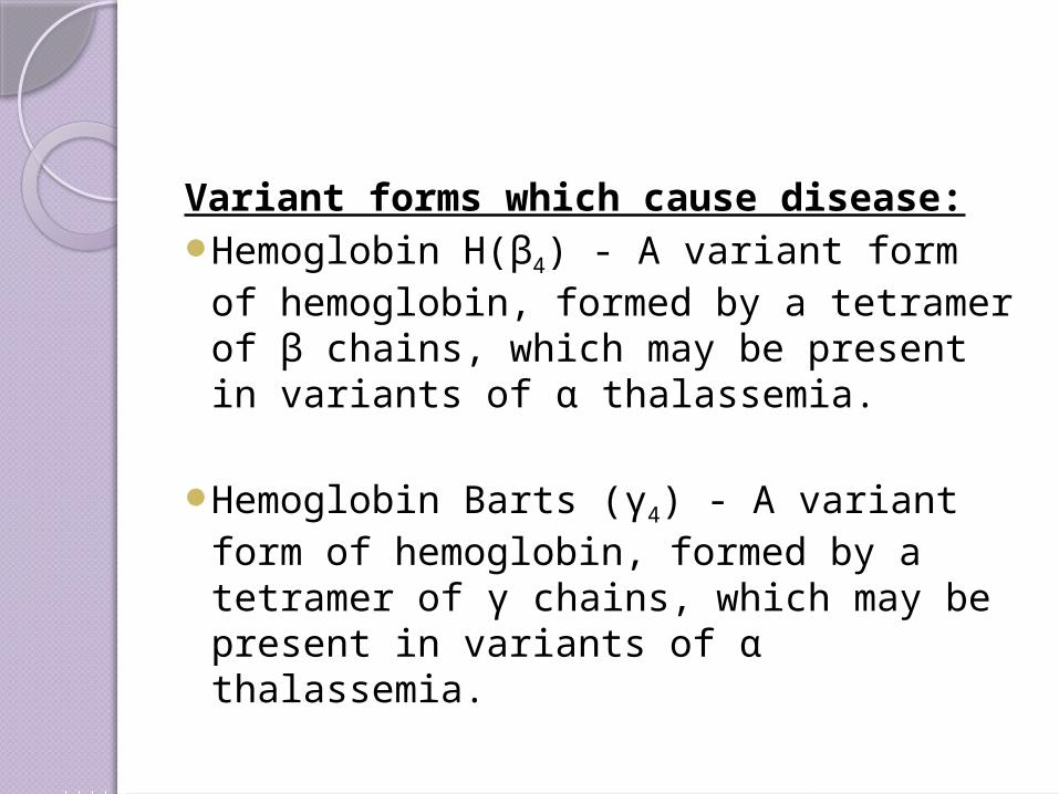
Variant forms which cause disease:Hemoglobin H(β4) - A variant form of
hemoglobin, formed by a tetramer of β chains, which may be present in variants of α thalassemia.
Hemoglobin Barts (γ4) - A variant form of hemoglobin, formed by a tetramer of γ chains, which may be present in variants of α thalassemia.
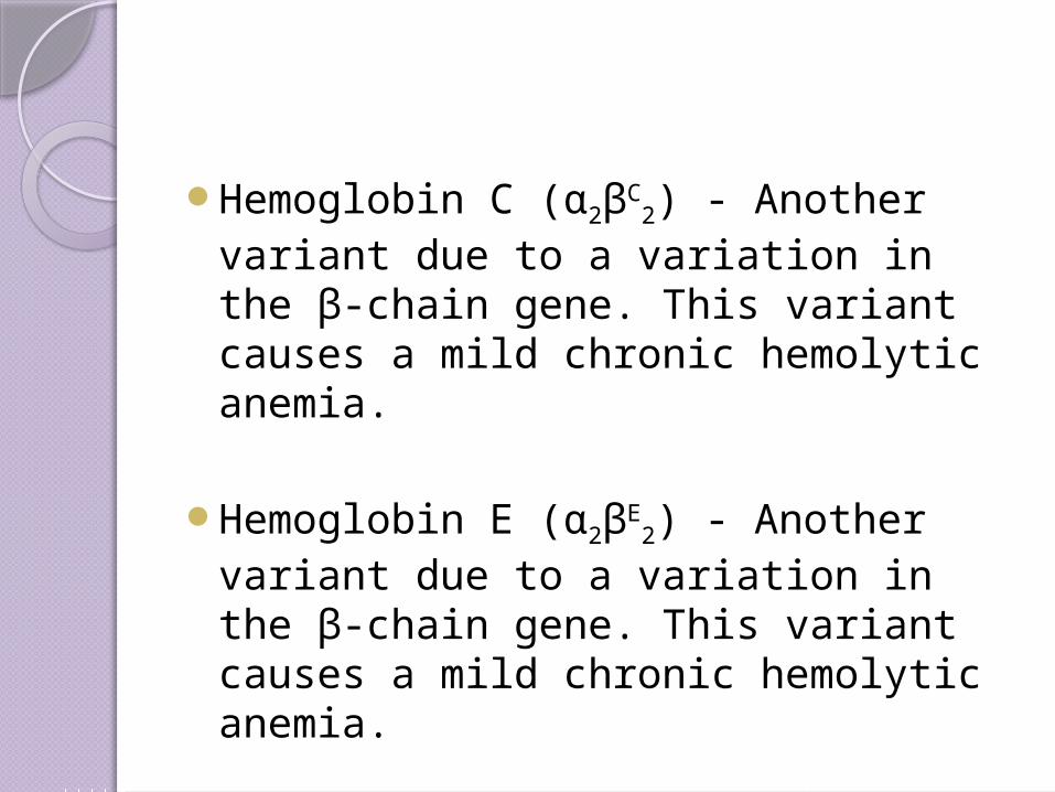
Hemoglobin C (α2βC2) - Another variant
due to a variation in the β-chain gene. This variant causes a mild chronic hemolytic anemia.
Hemoglobin E (α2βE2) - Another variant
due to a variation in the β-chain gene. This variant causes a mild chronic hemolytic anemia.
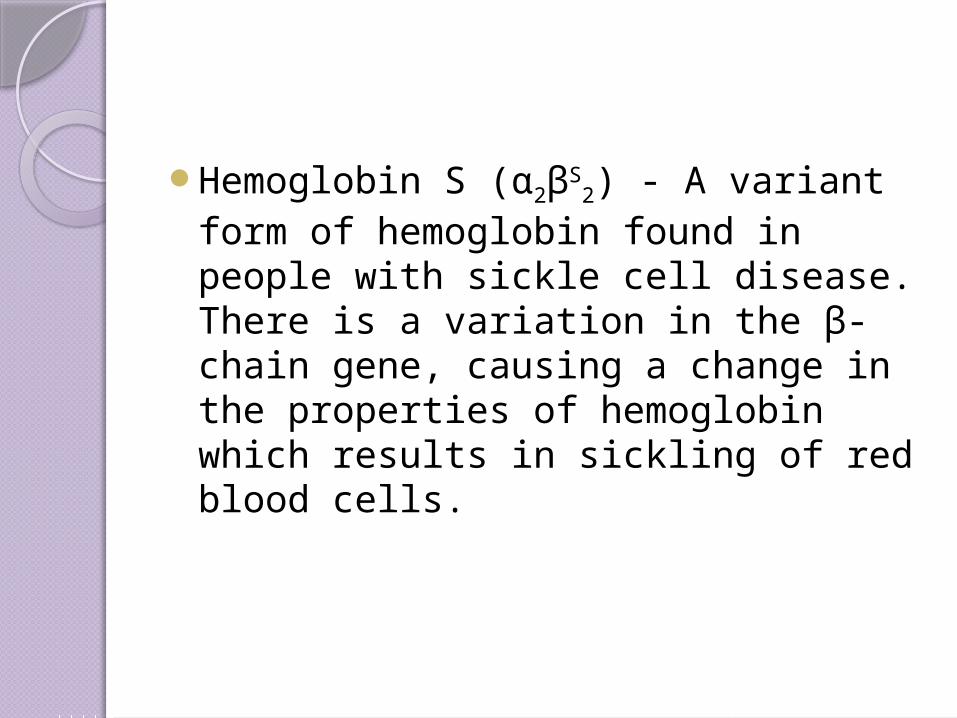
Hemoglobin S (α2βS2) - A variant form
of hemoglobin found in people with sickle cell disease. There is a variation in the β-chain gene, causing a change in the properties of hemoglobin which results in sickling of red blood cells.
Genetics
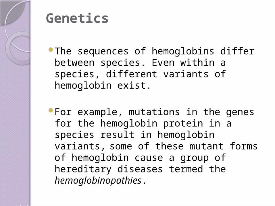
The sequences of hemoglobins differ between species. Even within a species, different variants of hemoglobin exist.
For example, mutations in the genes for the hemoglobin protein in a species result in hemoglobin variants, some of these mutant forms of hemoglobin cause a group of hereditary diseases termed the hemoglobinopathies.

The best known is sickle-cell disease, which was the first human disease whose mechanism was understood at the molecular level.
A separate set of diseases called thalassemias involves underproduction of normal and sometimes abnormal hemoglobins, through problems and mutations in globin gene regulation. All these diseases produce anemia.
The iron ion may either be in the Fe2+ or Fe3+ state, but methemoglobin (Fe3+) cannot bind oxygen.
In binding, oxygen temporarily oxidizes (Fe2+) to (Fe3+), so iron must exist in the +2 oxidation state to bind oxygen. The enzyme methemoglobin reductase reactivates hemoglobin found in the inactive (Fe3+) state by reducing the iron center.
The absorption spectra of oxyhemoglobin and deoxyhemoglobin differ. The oxyhemoglobin has significantly lower absorption of the 660 nm wavelength than deoxyhemoglobin, while at 940 nm its absorption is slightly higher.

This accounts for hemoglobin's red color and deoxyhemoglobin's blue color. This difference is used for measurement of the amount of oxygen in patient's blood by an instrument called pulse oximeter.


Hemoglobin AS - A heterozygous form causing Sickle cell trait with one adult gene and one sickle cell disease gene
Hemoglobin SC disease - Another heterozygous form with one sickle gene and another encoding Hemoglobin C.
Role in diseaseHemoglobin deficiency can be caused
either by decreased amount of hemoglobin molecules as in anemia, or decreased ability of each molecule to bind oxygen at the same partial pressure of oxygen.
Hemoglobinopathies(genetic defects resulting in abnormal structure of the hemoglobin molecule) may cause both.
There is a group of genetic disorders, known as the Porphyrias that are characterized by errors in metabolic pathways of heme synthesis.

To a small extent, hemoglobin A slowly combines with glucose at the terminal valine (an alpha aminoacid) of each β chain. The resulting molecule is often referred to as Hb A1c. As the concentration of glucose in the blood increases, the percentage of Hb A that turns into Hb A1c increases.

In diabetics whose glucose usually runs high, the percent Hb A1c also runs high. Because of the slow rate of Hb A combination with glucose, the Hb A1c percentage is representative of glucose level in the blood averaged over a longer time (the half-life of red blood cells, which is typically 50–55 days).

The levels of glycosylated hemoglobin are tested to monitor the long-term control of the chronic disease of type 2 diabetes mellitus (T2DM). Poor control of T2DM results in high levels of glycosylated hemoglobin in the red blood cells.

The normal reference range is approximately 4 %–5.9 %. Though difficult to obtain, values less than 7 % are recommended for people with Type 2 DM. Levels greater than 9 % are associated with poor control of the glycosylated hemoglobin and levels greater than 12 % are associated with very poor control.
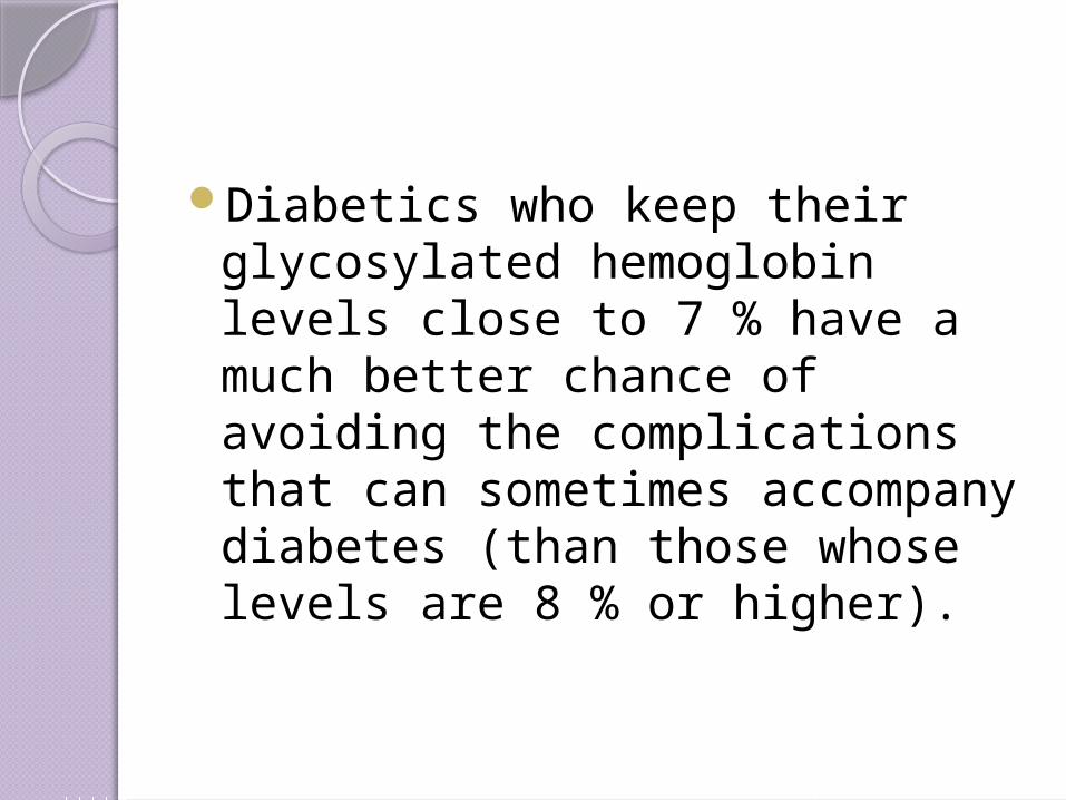
Diabetics who keep their glycosylated hemoglobin levels close to 7 % have a much better chance of avoiding the complications that can sometimes accompany diabetes (than those whose levels are 8 % or higher).

Diagnostic usesHemoglobin concentration
measurement is among the most commonly performed blood tests, usually as part of a complete blood count.
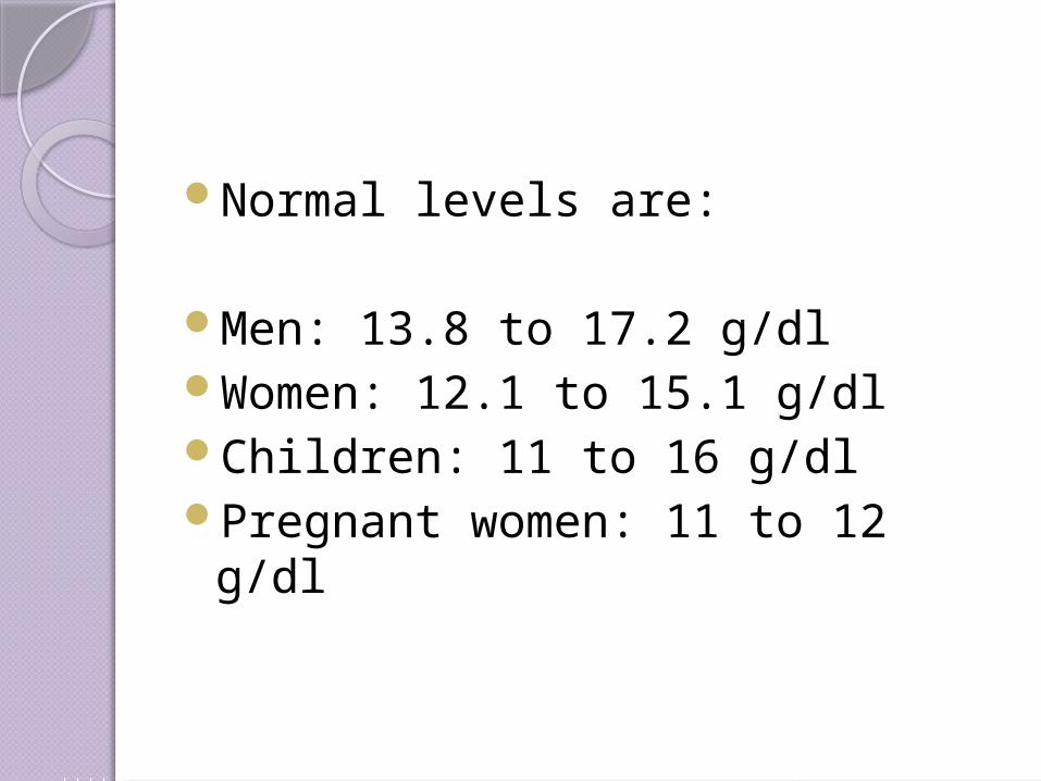
Normal levels are:
Men: 13.8 to 17.2 g/dl Women: 12.1 to 15.1 g/dl Children: 11 to 16 g/dl Pregnant women: 11 to 12 g/dl