
Two Novel MicroRNA Biomarkers Related to β-Cell … · on elevation of blood glucose and reduction...
10
CLINICAL RESEARCH ARTICLE Two Novel MicroRNA Biomarkers Related to b-Cell Damage and Their Potential Values for Early Diagnosis of Type 1 Diabetes Li Liu, 1,2,3 * Jinhua Yan, 1,3 * Haixia Xu, 1,3 Yunxia Zhu, 4 Hua Liang, 1,3 Wen Pan, 1,3 Bin Yao, 1,3 Xiao Han, 4 Jianping Ye, 5 and Jianping Weng 1,3 1 Department of Endocrinology and Metabolism, the Third Affiliated Hospital of Sun Yat-sen University, Guangzhou, Guangdong 510630, China; 2 Department of Endocrinology, Tongji Hospital, Tongji Medical College, Huazhong University of Science and Technology, Wuhan, Hubei 430030, China; 3 Guangdong Provincial Key Laboratory of Diabetology, Guangzhou, Guangdong 510630, China; 4 Key Laboratory of Human Functional Genomics of Jiangsu Province, Biochemistry and Molecular Biology, Nanjing Medical University, Nanjing, Jiangsu 210029, China; and 5 Antioxidant and Gene Regulation Laboratory, Pennington Biomedical Research Center, Louisiana State University, Baton Rouge, Louisiana 70808 Context: New strategies and biomarkers are needed in the early detection of b-cell damage in the progress of type 1 diabetes mellitus (T1DM). Objective: To explore whether serum microRNAs (miRNA) should be served as biomarkers for T1DM. Design, Settings, and Patients: The miRNA profile was established with miRNA microarray in dis- covery phase (six T1DM, six controls). A miRNA-based model for T1DM diagnosis was developed using logistic regression analysis in the training dataset (40 T1DM, 56 controls) and then validated with leave-one-out cross validation and another independent validation dataset (33 T1DM, 29 controls). Main Outcome Measures: Quantitative reverse transcription polymerase chain reaction was applied to confirm the differences of candidate miRNAs between T1DM and controls. Area under the receiver-operating characteristic (ROC) curve (AUC) was used to evaluate diagnostic accuracy. INS-1 cells, streptozotocin-treated mice (n = 4), and nonobese diabetic (NOD) mice (n = 12) were used to evaluate the association of miRNAs with b-cell damage. Results: A miRNA -based model was established in the training dataset with high diagnostic accuracy for T1DM (AUC = 0.817) based on six candidate differential expressed miRNAs identified in discovery phase. The validation dataset showed the model’s satisfactory diagnostic performance (AUC = 0.804). Secretions of miR-1225-5p and miR-320c were significantly increased in streptozotocin- treated mice and INS-1 cells. Noteworthy, the elevation of these two miRNAs was observed before glucose elevation in the progress of diabetes in NOD mice. Conclusions: Two miRNA biomarkers (miR-1225-5p and miR-320c) related to b-cell damage were identified in patients with recent-onset T1DM. The miRNA-based model established in this study exhibited a good performance in diagnosis of T1DM. ( J Clin Endocrinol Metab 103: 1320–1329, 2018) T 1DM is a polygenic disorder characterized by immune-mediated destruction of pancreatic b cells, leading to insulin deficiency (1). Early detection of b-cell damage allowing for intervention to be initiated at an early stage of the disease may be expected to improve therapeutic efficacy. Currently, the diagnosis is dependent ISSN Print 0021-972X ISSN Online 1945-7197 Printed in USA Copyright © 2018 Endocrine Society Received 27 June 2017. Accepted 18 January 2018. First Published Online 23 January 2018 *These authors equally contributed to this study. Abbreviations: AUC, area under the receiver-operating characteristic curve; BMI, body mass index; miRNA, microRNA; NOD, nonobese diabetic; PCR, polymerase chain reaction; qRT, quantitative reverse transcription; ROC, receiver-operating characteristic; STZ, streptozotocin; T1DM, type 1 diabetes mellitus; TUNEL, terminal deoxynucleotidyl transferase mediated nick-end labeling. 1320 https://academic.oup.com/jcem J Clin Endocrinol Metab, April 2018, 103(4):1320–1329 doi: 10.1210/jc.2017-01417 Downloaded from https://academic.oup.com/jcem/article-abstract/103/4/1320/4822207 by Endocrine Society Member Access 1 user on 16 April 2018
Transcript of Two Novel MicroRNA Biomarkers Related to β-Cell … · on elevation of blood glucose and reduction...
C L I N I C A L R E S E A R C H A R T I C L E
Two Novel MicroRNA Biomarkers Related to b-CellDamage and Their Potential Values for Early Diagnosisof Type 1 Diabetes
Li Liu,1,2,3* Jinhua Yan,1,3* Haixia Xu,1,3 Yunxia Zhu,4 Hua Liang,1,3 Wen Pan,1,3
Bin Yao,1,3 Xiao Han,4 Jianping Ye,5 and Jianping Weng1,3
1Department of Endocrinology and Metabolism, the Third Affiliated Hospital of Sun Yat-sen University,Guangzhou, Guangdong 510630, China; 2Department of Endocrinology, Tongji Hospital, Tongji MedicalCollege, Huazhong University of Science and Technology, Wuhan, Hubei 430030, China; 3GuangdongProvincial Key Laboratory of Diabetology, Guangzhou, Guangdong 510630, China; 4Key Laboratory ofHuman Functional Genomics of Jiangsu Province, Biochemistry and Molecular Biology, Nanjing MedicalUniversity, Nanjing, Jiangsu 210029, China; and 5Antioxidant and Gene Regulation Laboratory, PenningtonBiomedical Research Center, Louisiana State University, Baton Rouge, Louisiana 70808
Context: New strategies and biomarkers are needed in the early detection of b-cell damage in theprogress of type 1 diabetes mellitus (T1DM).
Objective: To explorewhether serummicroRNAs (miRNA) should be served as biomarkers for T1DM.
Design, Settings, and Patients: The miRNA profile was established with miRNA microarray in dis-covery phase (six T1DM, six controls). AmiRNA-basedmodel for T1DMdiagnosis was developed usinglogistic regression analysis in the training dataset (40 T1DM, 56 controls) and then validated withleave-one-out cross validation and another independent validation dataset (33 T1DM, 29 controls).
MainOutcomeMeasures:Quantitative reverse transcription polymerase chain reactionwas appliedto confirm the differences of candidate miRNAs between T1DM and controls. Area under thereceiver-operating characteristic (ROC) curve (AUC) was used to evaluate diagnostic accuracy. INS-1cells, streptozotocin-treated mice (n = 4), and nonobese diabetic (NOD) mice (n = 12) were used toevaluate the association of miRNAs with b-cell damage.
Results:AmiRNA -basedmodelwas established in the training datasetwith high diagnostic accuracyfor T1DM (AUC= 0.817) based on six candidate differential expressedmiRNAs identified in discoveryphase. The validation dataset showed the model’s satisfactory diagnostic performance (AUC =0.804). Secretions of miR-1225-5p and miR-320c were significantly increased in streptozotocin-treated mice and INS-1 cells. Noteworthy, the elevation of these two miRNAs was observed beforeglucose elevation in the progress of diabetes in NOD mice.
Conclusions: TwomiRNAbiomarkers (miR-1225-5pandmiR-320c) related tob-cell damagewere identifiedin patients with recent-onset T1DM. The miRNA-based model established in this study exhibited agood performance in diagnosis of T1DM. (J Clin Endocrinol Metab 103: 1320–1329, 2018)
T1DM is a polygenic disorder characterized byimmune-mediated destruction of pancreatic b cells,
leading to insulin deficiency (1). Early detection of b-cell
damage allowing for intervention to be initiated at anearly stage of the disease may be expected to improvetherapeutic efficacy. Currently, the diagnosis is dependent
ISSN Print 0021-972X ISSN Online 1945-7197Printed in USACopyright © 2018 Endocrine SocietyReceived 27 June 2017. Accepted 18 January 2018.First Published Online 23 January 2018
*These authors equally contributed to this study.Abbreviations: AUC, area under the receiver-operating characteristic curve; BMI, bodymass index; miRNA,microRNA; NOD, nonobese diabetic; PCR, polymerase chain reaction;qRT, quantitative reverse transcription; ROC, receiver-operating characteristic; STZ,streptozotocin; T1DM, type 1 diabetes mellitus; TUNEL, terminal deoxynucleotidyltransferase mediated nick-end labeling.
1320 https://academic.oup.com/jcem J Clin Endocrinol Metab, April 2018, 103(4):1320–1329 doi: 10.1210/jc.2017-01417
Downloaded from https://academic.oup.com/jcem/article-abstract/103/4/1320/4822207by Endocrine Society Member Access 1 useron 16 April 2018
on elevation of blood glucose and reduction in plasmainsulin and/or C-peptide. These parameters are good foridentifying b-cell damage, but only useful in establishedtype 1 diabetes mellitus (T1DM). Autoantibodies areconsidered markers of the autoimmune process early inthe disease, but they often do not reflect the degree ofb-cell damage (2). Unmethylated insulin DNA is regardedas a potential parameter for early b-cell damage, but itsuse is limited by the relative low concentration in plasma,and the amount of unmethylated DNA release mightbe affected by the mechanisms causing b-cell death (3).So, there is clearly a need for new methods to identifypatients with early T1DM or individuals on their way todeveloping T1DM.
MicroRNAs (miRNAs) are a class of small noncodingRNAs of approximately 22 nucleotides in length thatposttranscriptionally regulate gene expression in variousbiological processes (4). Tissue-specific miRNAs alreadyserve as noninvasive markers in the study of cancers,immunological diseases, cardiovascular diseases, andtype 2 diabetes mellitus (5–9). miRNAs can be detected inserum, plasma, urine, and tears by polymerase chainreaction (PCR) with high specificity, which makesmiRNAs ideal biomarker candidates for different dis-eases (10). miRNAs play an important role in the regu-lation of b-cell function, including proliferation andapoptosis (11–13). However, it remains unknown ifcirculating miRNAs can be used as a biomarker for earlydiagnosis of b-cell damage in T1DM.
In the current study, we established a miRNA-basedmodel with a high specificity and sensitivity in theevaluation of b-cell damage in vivo and vitro.
Subjects and Methods
Clinical subjects and research designSeventy-nine patients with recent-onset T1DM were
recruited in the Guangdong T1DM Translational MedicineStudy Program between August 2011 and June 2014 (14). Thediagnostic criteria for T1DM are: (1) insulin-dependent duringlong-term follow-up, (2) positive for at least one of followingautoantibodies: antibodies against the 65-kDa isoform of glu-tamate decarboxylase, insulinoma antigen 2, or zinc transporter8, as determined with a radiobinding assay confirmed by theIslet Autoantibody Standardization Program, (3) disease du-ration ,1 year, and (4) fasting and stimulated C-peptide levelsare ,0.6 ng/mL and 0.9 ng/mL, respectively (SupplementalMethods). Age- and sex-matched healthy individuals who arenegative for the three diabetes-associated autoantibodies and afamily history of diabetes were recruited from the physicalexamination center of the Third Affiliated Hospital of Sun Yat-sen University. Individuals with any febrile illness, accompanieddiabetic complications, or other diseases were excluded from thestudy. The study was approved by the Ethics Committee of theThird Affiliated Hospital of Sun Yat-sen University. Writteninformed consent was obtained from adult participants or
from the parents/guardians of individuals younger than age18 years.
miRNA-based screening model for T1DM
Discovery phaseThe human miRNA microarray, including 2006 mature
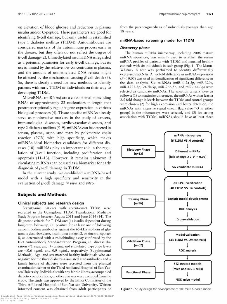
miRNA sequences, was initially used to establish the serummiRNA profiles of patients with T1DM and matched healthycontrols with six individuals in each group (Fig. 1). The Mann-Whitney U test was performed to identify differentiallyexpressed miRNAs. A twofold difference in miRNA expression(P, 0.05) was used in identification of significant difference inthe data analysis. Six miRNAs (miR-642a-3p, miR-320c,miR-1225-5p, let-7b-5p, miR-26b-5p, and miR-144-3p) wereselected as candidate miRNAs. The selection criteria were asfollows: (1) to maximize differences, the miRNAs with at least a2.5-fold change in levels between the T1DMand control groupswere chosen (2) for high expression and better detection, themiRNAs with intensive signal (mean flag value .3 in eithergroup) in the microarrays were selected, and (3) for strongassociation with T1DM, miRNAs should have at least three
Figure 1. Study design for development of the miRNA-based model.
doi: 10.1210/jc.2017-01417 https://academic.oup.com/jcem 1321
Downloaded from https://academic.oup.com/jcem/article-abstract/103/4/1320/4822207by Endocrine Society Member Access 1 useron 16 April 2018
predicated gene targets to be qualified as candidate genes ofT1DM in the T1Dbase (15).
Training phaseThe six candidatemiRNAswere verified in a training cohort,
40 patients with recent-onset T1DM and 56 healthy controls,with their relative expression levels determined by quantitativereverse transcription (qRT)-PCR. Next, a miRNA-based modelwas developed using logistic regression analysis based on thetraining dataset. Receiver-operating characteristic (ROC)curves and leave-one-out cross-validation was used to evaluatethe diagnostic performance of the model.
Validation phaseThe performance of the miRNA-based model was validated
by differentiating T1DM cases from healthy controls in anotherindependent validation cohort including 33 patients withrecent-onset T1DM and 29 healthy controls.
Functional phaseThe association ofb-cell damagewith the identifiedmiRNAs
in the model was confirmed in streptozotocin (STZ)-treatedmice and INS-1 cell as well as in a nonobese diabetic (NOD)mice model of T1DM.
Microarray and qRT-PCRWe used the Agilent human miRNA microarray V19.0
(Agilent Technologies, Santa Clara, CA) in our study. Themicroarray data have been deposited in the NCBI Gene Ex-pression Omnibus (GSE94649, http://www.ncbi.nlm.nih.gov/geo). The qRT-PCR was performed using TaqMan primersaccording to the manufacturer protocol (Applied Biosystems,Foster City, CA) (Supplemental Methods). U6 small nu-clear RNA was chosen as a stable endogenous control (16).The relative expression levels of the miRNAs were calculatedusing the 22DCt method, in which DCt = Ct (target) – Ct(reference) (17).
AnimalsC57BL/6 male mice (n = 8) and female NOD/ShiLtJ (NOD)
mice (n = 12) were purchased from Beijing HFK Bioscience CO.,Ltd. (Beijing, China). Mice were kept under specific pathogen-free conditions in a 12-hour dark, 12-hour light cycle with freeaccess to food and water. C57BL/6 male mice (n = 4) weretreated by intraperitoneal injection of multiple doses (50mg/kg)of STZ (Sigma-Aldrich Inc., St Louis, MO) for 5 consecutivedays. The control mice (n = 4) were injected with the samevolume of vehicle. Mice with fasting blood glucose of11.1 mmol/L or more for 2 consecutive days were considereddiabetic. All procedures with animals complied with the Guidefor the Care and Use of Laboratory Animals and were approvedby the animal care and use committee of Sun Yat-sen University.
Flow cytometry analysisINS-1 cells without mycoplasma contamination were ob-
tained from Key Laboratory of Human Functional Genomics ofJiangsu Province (Nanjing, Jiangsu, China). INS-1 cells werecultured in six-well plates and treated with STZ for 24 hours toinduce apoptosis. Then, the cells were collected after digestionwith 0.25% trypsin and incubatedwith 20mL of binding buffer,5 mL of Annexin V-FITC, and 5 mL of propidium iodide. After
incubation at room temperature in the dark for 15minutes, cellswere analyzed with a flow cytometry for apoptosis. The resultswere calculated using CellQuestTM Pro software (BD Bio-sciences, Mississauga, ON, Canada) and expressed as thepercentage of apoptotic cells among the total cells.
TUNEL staining assayINS-1 cells were cultured on a cover glass in 24-well plates
and treated with STZ for 24 hours to induce apoptosis. Then,the apoptotic cells were stained in a terminal deoxynucleotidyltransferase mediated nick-end labeling (TUNEL) assayaccording to the instruction of the kit manufacturer (In Situ CellDeath Detection Kit; Roche, Basel, Switzerland). Apoptotic cellswere stained by green fluorescence, and all cells were markedwith blue fluorescence using Hoechst. The apoptotic ratio wascalculated as TUNEL-positive cells divided by total cell num-ber. The number of cells was counted in five random fieldsfrom three different slides at 3400 magnification. The averageof TUNEL-positive cells was expressed in percentage inthese fields.
Statistical analysisData are presented as mean 6 standard error or median
(interquartile range) for continuous variables and as n (%) forcategorical variables. Comparisons between two groups wereanalyzed using the Student t, Mann-Whitney U, or x2 test.
Development and evaluation of themiRNA-based model
The candidate miRNAs showing significant differentialexpression between T1DM cases and controls were introducedinto a binary logistic regression model controlling for age, sex,and body mass index (BMI), and step-wise backward selectionwas performed to determinate the best model for discriminatingpatients with T1DM from healthy individuals. ROC curveswere constructed to evaluate the discriminatory performance ofeach miRNA and the model for differentiating T1DM casesfrom healthy controls according to the area under the ROCcurve (AUC) value.
Simulation studies were performed to test the magnitude ofthe bias introduced by model selection. Leave-one-out cross-validation was used to validate the logistic diagnostic modelbased on the training dataset (Supplemental Methods)
SPSS, version 20.0, software (SPSS Inc., Chicago, IL) and R,version 3.0.2, software (R Foundation for Statistical Com-puting, Vienna, Austria) (18) were used for statistical analysis.Statistical significance was defined as a two-tailed P , 0.05.
Results
Identification of candidate miRNAsTo identify the serum miRNAs that could be used in
diagnosis of T1DM, we determined miRNA profiles inthe serum of six patients with recent-onset T1DM andsix healthy individuals using a microarray (see clinicalcharacteristics of subjects in Supplemental Table 1). Adifference was observed in 27 miRNAs between thepatients and controls (fold change $2, P , 0.05; Sup-plemental Table 2), 17 of which increased and 10 of
1322 Liu et al Serum MicroRNAs and Type 1 Diabetes Mellitus J Clin Endocrinol Metab, April 2018, 103(4):1320–1329
Downloaded from https://academic.oup.com/jcem/article-abstract/103/4/1320/4822207by Endocrine Society Member Access 1 useron 16 April 2018
which decreased in patients with T1DM. According tothe further selection criteria in this study (see the Clinicalsubjects and research design section of this article), sixmiRNAs (miR-642a-3p, miR-320c, miR-1225-5p, let-7b-5p, miR-26b-5p, and miR-144-3p) were selected ascandidates for the next round of biomarker study. Pre-dicted target genes are shown in Supplemental Table 3.
Development of a miRNA-based modelTo test the value of candidate miRNAs, we examined
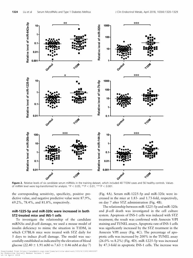
them by qRT-PCR in the training cohort, which included40 patients with recent-onset T1DM and 56 healthycontrols. The age and sex were well matched in the twogroups of this cohort (see clinical characteristics ofsubjects in Table 1). The levels of all the six miRNAswereincreased in patients with T1DM (Fig. 2, all P , 0.05);three miRNAs (miR-642a-3p, miR-320c, andmiR-1225-5p) were consistent with the results of microarray study.
To establish a diagnostic model that could be usedefficiently for identification of patients with T1DM, alogistic regression analysis was applied to the trainingdataset to estimate the accuracy rate in the diagnosis ofT1DM. Specifically, the six candidate miRNAs wereinitially introduced into the model and adjusted for po-tential confounders (age, sex, and BMI). Step-wisebackward selection was performed to determine thebest model as following: logit (P = T1DM) = 0.613 +0.1743miR-320c – 2.1993miR-1225-5p + 0.093 age– 0.213 3 BMI, which saved the independent significantitems for last to optimize the combination and avoidoverfitting (Supplemental Table 4). The model was testedusing a likelihood test and was found to be significant(x2 = 37.75 and P , 0.001).
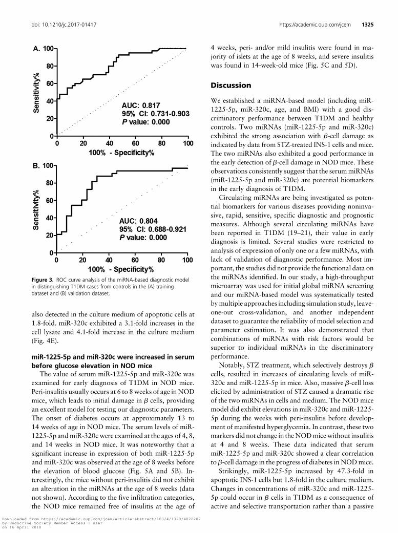
The diagnostic performance of the model was evalu-ated with ROC analysis. As shown in Fig. 3A, the AUCvalue of the model was 0.817 (95% confidence interval,0.731 to 0.903;P, 0.001), whichwas higher than that ofeach individual miRNA (Supplemental Table 5). Theoptimal cutoff point was defined when the Youden indexreached the maximum. The corresponding sensitivity,specificity, positive predictive value, and negative pre-dictive value were 55.0%, 92.9%, 84.6%, and 74.3%,respectively.
A simulation study was performed to evaluate themiRNA-based model for overfitting. A series of empiricalP values for the model’s AUC was obtained; the highestsimulated AUC value with P , 0.001 was 0.801. Inaddition, leave-one-out cross-validation was used toevaluate the discriminatory performance further forT1DM in the training dataset. The resulting error ratewas 18.3%, suggesting that the model had a good sta-bility and reliability.
Validation of the miRNA-based model in anotherindependent dataset
The model was validated in another cohort including33 patients with recent-onset T1DM and 29 healthycontrols (Table 1). The levels of miR-320c andmiR-1225-5p in this validation cohort are reported inSupplemental Fig. 2. The parameters of the model wereapplied to the validation dataset to evaluate the proba-bility of being diagnosed with T1DM. The predictiveprobability was used to construct the ROC curve. Asshown in Fig. 3B, the AUC value of the model was0.804 (95% confidence interval, 0.688 to 0.921; P ,
0.001). When the Youden index reached the maximum,
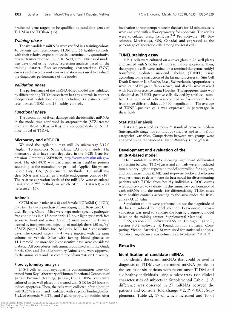
Table 1. Characteristics of Study Subjects in the Training and Validation Datasets
Characteristics
Training Dataset
P Value
Validation Dataset
P ValueT1DM Cases
(n = 40)Healthy Controls
(n = 56)T1DM Cases
(n = 33)Healthy Controls
(n = 29)
Age, y 22.31 6 1.90 20.00 6 1.34 0.308a 21.18 6 1.76 20.90 6 2.14 0.919a
Age group, y 0.802b 0.706b
,15 14 (35.0%) 21 (37.5%) 11 (33.3%) 11 (37.9%)$15 26 (65.0%) 35 (62.5%) 22 (66.7%) 18 (62.1%)
Female, n, % 26 (65.0%) 35 (62.5%) 0.802b 18 (54.5%) 14 (48.3%) 0.622b
BMI, kg/m2 18.75 6 0.46 19.73 6 0.38 0.104a 18.42 6 0.63 21.28 6 0.50 0.001a
HbA1c, % 7.4 (6.4, 9.8) 5.1 (4.9, 5.3) 0.000c 8.9 (7.5, 12.6) 5.1 (4.9, 5.2) 0.000c
Duration, mo 8.54 6 0.74 — — 6.93 6 1.28 — —
Insulin dose, m/kg/d 0.56 6 0.03 — — 0.66 6 0.05 — —
FCP, ng/mL 0.25 (0.09, 0.34) — — 0.28 (0.10, 0.55) — —
PCP2h, ng/mL 0.57 (0.27, 0.80) — — 0.54 (0.29, 0.75) — —
Abbreviations: FCP, fasting serum C-peptide; HbA1c, glycated hemoglobin A1c; PCP2h, 2-h postprandial serum C-peptide.aStudent t test.bx2 test.cMann-Whitney U test.
doi: 10.1210/jc.2017-01417 https://academic.oup.com/jcem 1323
Downloaded from https://academic.oup.com/jcem/article-abstract/103/4/1320/4822207by Endocrine Society Member Access 1 useron 16 April 2018
the corresponding sensitivity, specificity, positive pre-dictive value, and negative predictive value were 87.9%,69.2%, 78.4%, and 81.8%, respectively.
miR-1225-5p and miR-320c were increased in bothSTZ-treated mice and INS-1 cells
To investigate the relationship of the candidatemiRNAs and b-cell damage, we used a mouse model ofinsulin deficiency to mimic the situation in T1DM, inwhich C57BL/6 mice were treated with STZ daily for5 days to induce b-cell damage. The model was suc-cessfully established as indicated by the elevation of bloodglucose (22.406 1.91 mM vs 7.636 0.46 mM at day 7)
(Fig. 4A). Serum miR-1225-5p and miR-320c were in-creased in the mice at 1.83- and 1.73-fold, respectively,on day 7 after STZ administration (Fig. 4B).
The relationship betweenmiR-1225-5p andmiR-320cand b-cell death was investigated in the cell culturesystem. Apoptosis of INS-1 cells was induced with STZtreatment; the result was confirmed with Annexin V/PIstaining and TUNEL assays. Apoptotic rate of INS-1 cellswas significantly increased by the STZ treatment in theAnnexin V/PI assay (Fig. 4C). The percentage of apo-ptotic cells was increased by 200% in the TUNEL assay(26.0% vs 8.2%) (Fig. 4D). miR-1225-5p was increasedby 47.3-fold in apoptotic INS-1 cells. The increase was
Figure 2. Relative levels of six candidate serum miRNAs in the training dataset, which included 40 T1DM cases and 56 healthy controls. Valuesof miRNA level were log-transformed for analysis. *P , 0.05; **P , 0.01; ***P , 0.001.
1324 Liu et al Serum MicroRNAs and Type 1 Diabetes Mellitus J Clin Endocrinol Metab, April 2018, 103(4):1320–1329
Downloaded from https://academic.oup.com/jcem/article-abstract/103/4/1320/4822207by Endocrine Society Member Access 1 useron 16 April 2018
also detected in the culture medium of apoptotic cells at1.8-fold. miR-320c exhibited a 3.1-fold increases in thecell lysate and 4.1-fold increase in the culture medium(Fig. 4E).
miR-1225-5p and miR-320c were increased in serumbefore glucose elevation in NOD mice
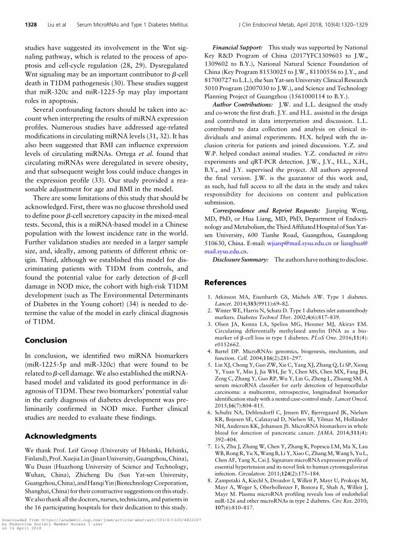
The value of serum miR-1225-5p and miR-320c wasexamined for early diagnosis of T1DM in NOD mice.Peri-insulitis usually occurs at 6 to 8weeks of age inNODmice, which leads to initial damage in b cells, providingan excellent model for testing our diagnostic parameters.The onset of diabetes occurs at approximately 13 to14 weeks of age in NOD mice. The serum levels of miR-1225-5p andmiR-320c were examined at the ages of 4, 8,and 14 weeks in NOD mice. It was noteworthy that asignificant increase in expression of both miR-1225-5pand miR-320c was observed at the age of 8 weeks beforethe elevation of blood glucose (Fig. 5A and 5B). In-terestingly, the mice without peri-insulitis did not exhibitan alteration in the miRNAs at the age of 8 weeks (datanot shown). According to the five infiltration categories,the NOD mice remained free of insulitis at the age of
4 weeks, peri- and/or mild insulitis were found in ma-jority of islets at the age of 8 weeks, and severe insulitiswas found in 14-week-old mice (Fig. 5C and 5D).
Discussion
We established a miRNA-based model (including miR-1225-5p, miR-320c, age, and BMI) with a good dis-criminatory performance between T1DM and healthycontrols. Two miRNAs (miR-1225-5p and miR-320c)exhibited the strong association with b-cell damage asindicated by data from STZ-treated INS-1 cells and mice.The two miRNAs also exhibited a good performance inthe early detection of b-cell damage in NOD mice. Theseobservations consistently suggest that the serummiRNAs(miR-1225-5p and miR-320c) are potential biomarkersin the early diagnosis of T1DM.
Circulating miRNAs are being investigated as poten-tial biomarkers for various diseases providing noninva-sive, rapid, sensitive, specific diagnostic and prognosticmeasures. Although several circulating miRNAs havebeen reported in T1DM (19–21), their value in earlydiagnosis is limited. Several studies were restricted toanalysis of expression of only one or a fewmiRNAs, withlack of validation of diagnostic performance. Most im-portant, the studies did not provide the functional data onthe miRNAs identified. In our study, a high-throughputmicroarray was used for initial global miRNA screeningand our miRNA-based model was systematically testedbymultiple approaches including simulation study, leave-one-out cross-validation, and another independentdataset to guarantee the reliability of model selection andparameter estimation. It was also demonstrated thatcombinations of miRNAs with risk factors would besuperior to individual miRNAs in the discriminatoryperformance.
Notably, STZ treatment, which selectively destroys bcells, resulted in increases of circulating levels of miR-320c and miR-1225-5p in mice. Also, massive b-cell losselicited by administration of STZ caused a dramatic riseof the two miRNAs in cells and medium. The NOD micemodel did exhibit elevations in miR-320c and miR-1225-5p during the weeks with peri-insulitis before develop-ment of manifested hyperglycemia. In contrast, these twomarkers did not change in theNODmicewithout insulitisat 4 and 8 weeks. These data indicated that serummiR-1225-5p and miR-320c showed a clear correlationto b-cell damage in the progress of diabetes in NODmice.
Strikingly, miR-1225-5p increased by 47.3-fold inapoptotic INS-1 cells but 1.8-fold in the culture medium.Changes in concentrations of miR-320c and miR-1225-5p could occur in b cells in T1DM as a consequence ofactive and selective transportation rather than a passive
Figure 3. ROC curve analysis of the miRNA-based diagnostic modelin distinguishing T1DM cases from controls in the (A) trainingdataset and (B) validation dataset.
doi: 10.1210/jc.2017-01417 https://academic.oup.com/jcem 1325
Downloaded from https://academic.oup.com/jcem/article-abstract/103/4/1320/4822207by Endocrine Society Member Access 1 useron 16 April 2018
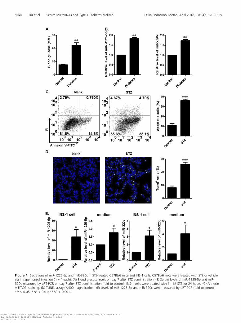
Figure 4. Secretions of miR-1225-5p and miR-320c in STZ-treated C57BL/6 mice and INS-1 cells. C57BL/6 mice were treated with STZ or vehiclevia intraperitoneal injection (n = 4 each). (A) Blood glucose levels on day 7 after STZ administration. (B) Serum levels of miR-1225-5p and miR-320c measured by qRT-PCR on day 7 after STZ administration (fold to control). INS-1 cells were treated with 1 mM STZ for 24 hours. (C) AnnexinV-FITC/PI staining. (D) TUNEL assay (3400 magnification). (E) Levels of miR-1225-5p and miR-320c were measured by qRT-PCR (fold to control).*P , 0.05; **P , 0.01; ***P , 0.001.
1326 Liu et al Serum MicroRNAs and Type 1 Diabetes Mellitus J Clin Endocrinol Metab, April 2018, 103(4):1320–1329
Downloaded from https://academic.oup.com/jcem/article-abstract/103/4/1320/4822207by Endocrine Society Member Access 1 useron 16 April 2018

consequence of cell lysis. Previous studies have indicatedthat miR-320c and miR-1225-5p can be wrapped intoexosomes, which protect miRNAs from degradation byRNAse and serve as vehicles for miRNA secretion intoextracellular space and body fluids (22). Therefore, wespeculate that the increased circulating miRNAs could bereleased from dying b cells via exosomes or other vehi-cles, although we cannot specifically exclude the possi-bility that other cells such as a cells also can releasemiRNAs after STZ treatment.
This article identified miR-320c and miR-1225-5p inthe T1DM serum for evaluation of b-cell activity. Bio-informatics analysis suggests that miR-320c may regulateseveral candidate genes for T1DM, including STAT4,SH2B3, RASGRP1, and CCR7 (23, 24). miR-320c hasalso been studied for its role in the signaling pathway oftransforming growth factor-b, cell cycle, and endocytosis(25, 26). Blume et al. demonstrated that miR-320c me-diates p53 effects in the regulation of cell cycle and ap-optosis (27). With respect to miR-1225-5p, previous
Figure 5. Serum levels of miR-1225-5p and miR-320c in prediabetic NOD mice. (A) Blood glucose and (B) serum levels of miR-1225-5p and miR-320c measured by qRT-PCR at different ages from female NOD mice developed diabetes finally (n = 12). (C) Histological analysis of insulitis ofpancreata from NOD mice at the ages of 4, 8, and 14 weeks (3400 magnification). (D) Insulitis score: 0, no insulitis; 1, peri-insulitis; 2, mildinsulitis (,25% of the infiltrated islet); 3, moderate insulitis (25% to 75% of the infiltrated islet); 4, severe insulitis (.75% islet infiltration).Results are presented as the mean score of n = 5 mice in each group; for each pancreas, an average insulitis score was calculated by adding thescore of each islet and then dividing by the total number of islets counted. N.S., nonsignificant; w, week. *P , 0.05; **P , 0.01; ***P , 0.001.
doi: 10.1210/jc.2017-01417 https://academic.oup.com/jcem 1327
Downloaded from https://academic.oup.com/jcem/article-abstract/103/4/1320/4822207by Endocrine Society Member Access 1 useron 16 April 2018
studies have suggested its involvement in the Wnt sig-naling pathway, which is related to the process of apo-ptosis and cell-cycle regulation (28, 29). DysregulatedWnt signaling may be an important contributor to b-celldeath in T1DM pathogenesis (30). These studies suggestthat miR-320c and miR-1225-5p may play importantroles in apoptosis.
Several confounding factors should be taken into ac-count when interpreting the results of miRNA expressionprofiles. Numerous studies have addressed age-relatedmodifications in circulating miRNA levels (31, 32). It hasalso been suggested that BMI can influence expressionlevels of circulating miRNAs. Ortega et al. found thatcirculating miRNAs were deregulated in severe obesity,and that subsequent weight loss could induce changes inthe expression profile (33). Our study provided a rea-sonable adjustment for age and BMI in the model.
There are some limitations of this study that should beacknowledged. First, there was no glucose threshold usedto define poor b-cell secretory capacity in the mixed-mealtests. Second, this is a miRNA-based model in a Chinesepopulation with the lowest incidence rate in the world.Further validation studies are needed in a larger samplesize, and, ideally, among patients of different ethnic or-igin. Third, although we established this model for dis-criminating patients with T1DM from controls, andfound the potential value for early detection of b-celldamage in NOD mice, the cohort with high-risk T1DMdevelopment (such as The Environmental Determinantsof Diabetes in the Young cohort) (34) is needed to de-termine the value of the model in early clinical diagnosisof T1DM.
Conclusion
In conclusion, we identified two miRNA biomarkers(miR-1225-5p and miR-320c) that were found to berelated to b-cell damage.We also established themiRNA-based model and validated its good performance in di-agnosis of T1DM. These two biomarkers’ potential valuein the early diagnosis of diabetes development was pre-liminarily confirmed in NOD mice. Further clinicalstudies are needed to evaluate these findings.
Acknowledgments
We thank Prof. Leif Groop (University of Helsinki, Helsinki,Finland),Prof.XuejiaLin (JinanUniversity,Guangzhou,China),Wu Duan (Huazhong University of Science and Technology,Wuhan, China), Zhicheng Du (Sun Yat-sen University,Guangzhou,China),andHanqiYin(BiotechnologyCorporation,Shanghai,China) for their constructive suggestionson this study.Wealso thankall the doctors, nurses, technicians, andpatients inthe 16 participating hospitals for their dedication to this study.
Financial Support: This study was supported by NationalKey R&D Program of China (2017YFC1309603 to J.W.,1309602 to B.Y.), National Natural Science Foundation ofChina (Key Program 81530025 to J.W., 81100556 to J.Y., and81700727 to L.L.), the SunYat-senUniversity Clinical Research5010 Program (2007030 to J.W.), and Science and TechnologyPlanning Project of Guangzhou (1561000114 to B.Y.).
Author Contributions: J.W. and L.L. designed the studyand co-wrote the first draft. J.Y. and H.L. assisted in the designand contributed in data interpretation and discussion. L.L.contributed to data collection and analysis on clinical in-dividuals and animal experiments. H.X. helped with the in-clusion criteria for patients and joined discussions. Y.Z. andW.P. helped conduct animal studies. Y.Z. conducted in vitroexperiments and qRT-PCR detection. J.W., J.Y., H.L., X.H.,B.Y., and J.Y. supervised the project. All authors approvedthe final version. J.W. is the guarantor of this work and,as such, had full access to all the data in the study and takesresponsibility for decisions on content and publicationsubmission.
Correspondence and Reprint Requests: Jianping Weng,MD, PhD, or Hua Liang, MD, PhD, Department of Endocri-nology andMetabolism, theThirdAffiliatedHospital of SunYat-sen University, 600 Tianhe Road, Guangzhou, Guangdong510630, China. E-mail: [email protected] or [email protected].
DisclosureSummary: Theauthorshavenothing todisclose.
References
1. Atkinson MA, Eisenbarth GS, Michels AW. Type 1 diabetes.Lancet. 2014;383(9911):69–82.
2. WinterWE, Harris N, Schatz D. Type 1 diabetes islet autoantibodymarkers. Diabetes Technol Ther. 2002;4(6):817–839.
3. Olsen JA, Kenna LA, Spelios MG, Hessner MJ, Akirav EM.Circulating differentially methylated amylin DNA as a bio-marker of b-cell loss in type 1 diabetes. PLoS One. 2016;11(4):e0152662.
4. Bartel DP. MicroRNAs: genomics, biogenesis, mechanism, andfunction. Cell. 2004;116(2):281–297.
5. LinXJ, ChongY, GuoZW,Xie C, YangXJ, ZhangQ, Li SP, XiongY, Yuan Y, Min J, Jia WH, Jie Y, Chen MS, Chen MX, Fang JH,Zeng C, Zhang Y, Guo RP, Wu Y, Lin G, Zheng L, Zhuang SM. Aserum microRNA classifier for early detection of hepatocellularcarcinoma: a multicentre, retrospective, longitudinal biomarkeridentification studywith a nested case-control study.LancetOncol.2015;16(7):804–815.
6. Schultz NA, Dehlendorff C, Jensen BV, Bjerregaard JK, NielsenKR, Bojesen SE, Calatayud D, Nielsen SE, Yilmaz M, HollanderNH, Andersen KK, Johansen JS. MicroRNA biomarkers in wholeblood for detection of pancreatic cancer. JAMA. 2014;311(4):392–404.
7. Li S, Zhu J, Zhang W, Chen Y, Zhang K, Popescu LM, Ma X, LauWB,RongR,YuX,WangB, Li Y,XiaoC,ZhangM,Wang S, YuL,Chen AF, YangX, Cai J. SignaturemicroRNA expression profile ofessential hypertension and its novel link to human cytomegalovirusinfection. Circulation. 2011;124(2):175–184.
8. Zampetaki A, Kiechl S, Drozdov I, Willeit P, Mayr U, Prokopi M,Mayr A, Weger S, Oberhollenzer F, Bonora E, Shah A, Willeit J,Mayr M. Plasma microRNA profiling reveals loss of endothelialmiR-126 and other microRNAs in type 2 diabetes. Circ Res. 2010;107(6):810–817.
1328 Liu et al Serum MicroRNAs and Type 1 Diabetes Mellitus J Clin Endocrinol Metab, April 2018, 103(4):1320–1329
Downloaded from https://academic.oup.com/jcem/article-abstract/103/4/1320/4822207by Endocrine Society Member Access 1 useron 16 April 2018
9. Churov AV, Oleinik EK, Knip M. MicroRNAs in rheumatoidarthritis: altered expression and diagnostic potential. AutoimmunRev. 2015;14(11):1029–1037.
10. Chen X, Ba Y,Ma L, Cai X, Yin Y,Wang K, Guo J, Zhang Y, ChenJ, GuoX, Li Q, Li X,WangW,Zhang Y,Wang J, JiangX, XiangY,XuC, Zheng P, Zhang J, Li R, ZhangH, ShangX,Gong T, NingG,Wang J, Zen K, Zhang J, Zhang CY. Characterization of micro-RNAs in serum: a novel class of biomarkers for diagnosis of cancerand other diseases. Cell Res. 2008;18(10):997–1006.
11. Baroukh N, Ravier MA, Loder MK, Hill EV, Bounacer A,Scharfmann R, Rutter GA, Van Obberghen E. MicroRNA-124aregulates Foxa2 expression and intracellular signaling in pancreaticbeta-cell lines. J Biol Chem. 2007;282(27):19575–19588.
12. Roggli E, Britan A, Gattesco S, Lin-Marq N, Abderrahmani A,Meda P, Regazzi R. Involvement of microRNAs in the cytotoxiceffects exerted by proinflammatory cytokines on pancreatic beta-cells. Diabetes. 2010;59(4):978–986.
13. Poy MN, Eliasson L, Krutzfeldt J, Kuwajima S, Ma X, MacdonaldPE, Pfeffer S, Tuschl T, Rajewsky N, Rorsman P, Stoffel M. Apancreatic islet-specific microRNA regulates insulin secretion.Nature. 2004;432(7014):226–230.
14. Liu L, Yang D, Zhang Y, Lin S, Zheng X, Lin S, Chen L, Zhang X,Li L, Liang G, Yao B, Yan J, Weng J. Glycaemic control and itsassociated factors in Chinese adults with type 1 diabetes mellitus.Diabetes Metab Res Rev. 2015;31(8):803–810.
15. Burren OS, Adlem EC, Achuthan P, Christensen M, Coulson RM,Todd JA. T1DBase: update 2011, organization and presentation oflarge-scale data sets for type 1 diabetes research.Nucleic Acids Res.2011;39(Database issue):D997–D1001.
16. Starkey Lewis PJ, Dear J, Platt V, Simpson KJ, Craig DG, AntoineDJ, FrenchNS, DhaunN,WebbDJ, Costello EM,Neoptolemos JP,Moggs J, Goldring CE, Park BK. Circulating microRNAs as po-tential markers of human drug-induced liver injury. Hepatology.2011;54(5):1767–1776.
17. Zuo Z, Calin GA, de Paula HM, Medeiros LJ, Fernandez MH,Shimizu M, Garcia-Manero G, Bueso-Ramos CE. CirculatingmicroRNAs let-7a and miR-16 predict progression-free survivaland overall survival in patients with myelodysplastic syndrome.Blood. 2011;118(2):413–415.
18. R Core Team. (2015). R: a language and environment for statisticalcomputing. Vienna, Austria: R Foundation for Statistical Com-puting. Available at: http://www/R-project.org/. Accessed 24 Jan-uary 2018.
19. Nielsen LB, Wang C, Sorensen K, Bang-Berthelsen CH, Hansen L,Andersen ML, Hougaard P, Juul A, Zhang CY, Pociot F, Mor-tensen HB. Circulating levels of microRNA from children withnewly diagnosed type 1 diabetes and healthy controls: evidence thatmiR-25 associates to residual beta-cell function and glycaemiccontrol during disease progression. Exp Diabetes Res. 2012;2012:896362.
20. Samandari N, Mirza AH, Nielsen LB, Kaur S, Hougaard P,Fredheim S, Mortensen HB, Pociot F. Circulating microRNA levelspredict residual beta cell function and glycaemic control in childrenwith type 1 diabetes mellitus. Diabetologia. 2017;60(2):354–363.
21. Erener S, Marwaha A, Tan R, Panagiotopoulos C, Kieffer TJ.Profiling of circulating microRNAs in children with recent onset oftype 1 diabetes. JCI Insight. 2017;2(4):e89656.
22. Tokuhisa M, Ichikawa Y, Kosaka N, Ochiya T, Yashiro M,Hirakawa K, Kosaka T, Makino H, Akiyama H, Kunisaki C,Endo I. Exosomal miRNAs from peritoneum lavage fluid as po-tential prognostic biomarkers of peritoneal metastasis in gastriccancer. PLoS One. 2015;10(7):e0130472.
23. Huang W, Sherman BT, Lempicki RA. Bioinformatics enrichmenttools: paths toward the comprehensive functional analysis of largegene lists. Nucleic Acids Res. 2009;37(1):1–13.
24. Huang W, Sherman BT, Lempicki RA. Systematic and integrativeanalysis of large gene lists using DAVID bioinformatics resources.Nat Protoc. 2009;4(1):44–57.
25. Wang X, Wu J, Lin Y, Zhu Y, Xu X, Xu X, Liang Z, Li S, Hu Z,Zheng X, Xie L. MicroRNA-320c inhibits tumorous behaviors ofbladder cancer by targeting cyclin-dependent kinase 6. J Exp ClinCancer Res. 2014;33(1):69.
26. Delic D, Eisele C, Schmid R, Baum P,Wiech F, GerlM, Zimdahl H,Pullen SS, Urquhart R. Urinary exosomal miRNA signature in typeII diabetic nephropathy patients. PLoS One. 2016;11(3):e0150154.
27. Blume CJ, Hotz-Wagenblatt A, Hullein J, Sellner L, Jethwa A, StolzT, Slabicki M, Lee K, Sharathchandra A, Benner A, Dietrich S,Oakes CC, Dreger P, te Raa D, Kater AP, Jauch A,Merkel O, OrenM,Hielscher T, Zenz T. p53-dependent non-codingRNAnetworksin chronic lymphocytic leukemia. Leukemia. 2015;29(10):2015–2023.
28. HamamR,Ali AM,AlsalehKA,KassemM,AlfayezM,AldahmashA, AlajezNM.microRNA expression profiling on individual breastcancer patients identifies novel panel of circulating microRNA forearly detection. Sci Rep. 2016;6(1):25997.
29. Fisher JN, Terao M, Fratelli M, Kurosaki M, Paroni G, Zanetti A,Gianni M, Bolis M, Lupi M, Tsykin A, Goodall GJ, Garattini E.MicroRNA networks regulated by all-trans retinoic acid andlapatinib control the growth, survival and motility of breast cancercells. Oncotarget. 2015;6(15):13176–13200.
30. Liu JL, Kaddour N, Chowdhury S, Li Q, Gao ZH. Role of CCN5(WNT1 inducible signaling pathway protein 2) in pancreatic islets.J Diabetes. 2017;9(5):462–474.
31. Olivieri F, Spazzafumo L, Santini G, Lazzarini R, Albertini MC,Rippo MR, Galeazzi R, Abbatecola AM, Marcheselli F, Monti D,Ostan R, Cevenini E, Antonicelli R, Franceschi C, Procopio AD.Age-related differences in the expression of circulatingmicroRNAs:miR-21 as a new circulating marker of inflammaging.Mech AgeingDev. 2012;133(11-12):675–685.
32. Olivieri F, Lazzarini R, Recchioni R, Marcheselli F, Rippo MR, DiNuzzo S, Albertini MC, Graciotti L, Babini L, Mariotti S, Spada G,Abbatecola AM, Antonicelli R, Franceschi C, Procopio AD. MiR-146a as marker of senescence-associated pro-inflammatory statusin cells involved in vascular remodelling. Age (Dordr). 2013;35(4):1157–1172.
33. Ortega FJ,Mercader JM, Catalan V,Moreno-Navarrete JM, PueyoN, Sabater M, Gomez-Ambrosi J, Anglada R, Fernandez-FormosoJA, Ricart W, Fruhbeck G, Fernandez-Real JM. Targeting thecirculatingmicroRNA signature of obesity.ClinChem. 2013;59(5):781–792.
34. Group TS; TEDDY Study Group. The Environmental Determi-nants of Diabetes in the Young (TEDDY) study: study design.Pediatr Diabetes. 2007;8(5):286–298.
doi: 10.1210/jc.2017-01417 https://academic.oup.com/jcem 1329
Downloaded from https://academic.oup.com/jcem/article-abstract/103/4/1320/4822207by Endocrine Society Member Access 1 useron 16 April 2018