Tungsten Carbide-Cobalt Nanoparticles Induce Reactive ... › media › 2535 ›...
9
Tungsten Carbide-Cobalt Nanoparticles Induce Reactive Oxygen Species, AKT, ERK, AP-1, NF-κB, VEGF, and Angiogenesis Ling-Zhi Liu 1 & Min Ding 2 & Jenny Z. Zheng 3 & Yingxue Zhu 1 & Bruce A. Fenderson 1 & Bingyun Li 4 & Jing J. Yu 3 & Bing-Hua Jiang 1 Received: 21 January 2015 /Accepted: 6 April 2015 # Springer Science+Business Media New York 2015 Abstract Powder mixtures of tungsten carbide and metallic cobalt (WC-Co) are widely used in various products. Nano- particles are engineered structures with at least one dimension of 100 nm or smaller. WC-Co is known to be associated with lung injury and diseases. Angiogenesis is a key process during vasculature, carcinogenesis, recovery of injury, and inflamma- tory diseases. However, the cellular effects of WC-Co nano- particles on angiogenesis remain to be elucidated. In this study, we investigated angiogenic response and relative mech- anisms after exposure to WC-Co nanoparticles. Our results showed that WC-Co nanoparticles at 5 μg/cm 2 induced ROS production which activated AKT and ERK1/2 signaling path- ways in lung epithelial cells by reactive oxygen species (ROS) staining and immunoblotting; WC-Co treatment also in- creased transcriptional activation of AP-1, NF-κB, and VEGF by reporter assay. Further studies demonstrated that ROS are upstream molecules of AKT and ERK signaling pathways; the activation of AP-1, NF-κB, and VEGF was through ROS generation, AKT and ERK1/2 activation. In addition, WC- Co nanoparticles affected the cells to induce angiogenesis by chicken chorioallantoic membrane (CAM) assay. These re- sults illustrate that exposure to WC-Co nanoparticles induces angiogenic response by activating ROS, AKT, and ERK1/2 signaling pathways and the downstream molecules and eluci- date the potential molecular mechanisms during this process. This information may be useful for preventing potential dam- age from nanoparticle exposure in the future. Keywords Carbide-cobalt nanoparticles . Reactive oxygen species . Signaling pathways . VEGF . Angiogenesis Abbreviations WC-Co Tungsten carbide and metallic cobalt ROS Reactive oxygen species CAM Chicken chorioallantoic membrane PBS Phosphate-buffered saline DCFH-DA 2′,7′-Dichlorofluorescein diacetate H 2 O 2 Hydrogen peroxide AP-1 Activator protein-1 NF-κB Nuclear factor kappa B VEGF Vascular endothelial growth factor ERK Extracellular signal regulated kinase Introduction Nanotechnology is one of the fastest growing emerging tech- nologies and has a wide range of applications such as integrat- ed sensors, semiconductors, medical imaging, drug delivery, structural materials, sunscreens, cosmetics, and coatings [1]. Nanoparticles have at least one dimension of 100 nm or small- er. This small size results in properties which may * Ling-Zhi Liu [email protected] * Bing-Hua Jiang [email protected] 1 Department of Pathology, Anatomy and Cell Biology, Thomas Jefferson University, Philadelphia, PA, USA 2 Health Effects Laboratory Division, National Institute for Occupational Safety and Health, Morgantown, WV, USA 3 Mary Babb Randolph Cancer Center, Department of Biochemistry, West Virginia University, Morgantown, WV, USA 4 Department of Orthopedics, West Virginia University, Morgantown, WV, USA Biol Trace Elem Res DOI 10.1007/s12011-015-0331-6
Transcript of Tungsten Carbide-Cobalt Nanoparticles Induce Reactive ... › media › 2535 ›...
Tungsten Carbide-Cobalt Nanoparticles Induce Reactive OxygenSpecies, AKT, ERK, AP-1, NF-κB, VEGF, and Angiogenesis
Ling-Zhi Liu1& Min Ding2 & Jenny Z. Zheng3 & Yingxue Zhu1
& Bruce A. Fenderson1&
Bingyun Li4 & Jing J. Yu3& Bing-Hua Jiang1
Received: 21 January 2015 /Accepted: 6 April 2015# Springer Science+Business Media New York 2015
Abstract Powder mixtures of tungsten carbide and metalliccobalt (WC-Co) are widely used in various products. Nano-particles are engineered structures with at least one dimensionof 100 nm or smaller. WC-Co is known to be associated withlung injury and diseases. Angiogenesis is a key process duringvasculature, carcinogenesis, recovery of injury, and inflamma-tory diseases. However, the cellular effects of WC-Co nano-particles on angiogenesis remain to be elucidated. In thisstudy, we investigated angiogenic response and relative mech-anisms after exposure to WC-Co nanoparticles. Our resultsshowed that WC-Co nanoparticles at 5 μg/cm2 induced ROSproduction which activated AKT and ERK1/2 signaling path-ways in lung epithelial cells by reactive oxygen species (ROS)staining and immunoblotting; WC-Co treatment also in-creased transcriptional activation of AP-1, NF-κB, and VEGFby reporter assay. Further studies demonstrated that ROS areupstreammolecules of AKTand ERK signaling pathways; theactivation of AP-1, NF-κB, and VEGF was through ROS
generation, AKT and ERK1/2 activation. In addition, WC-Co nanoparticles affected the cells to induce angiogenesis bychicken chorioallantoic membrane (CAM) assay. These re-sults illustrate that exposure to WC-Co nanoparticles inducesangiogenic response by activating ROS, AKT, and ERK1/2signaling pathways and the downstream molecules and eluci-date the potential molecular mechanisms during this process.This information may be useful for preventing potential dam-age from nanoparticle exposure in the future.
Keywords Carbide-cobalt nanoparticles . Reactive oxygenspecies . Signaling pathways . VEGF . Angiogenesis
AbbreviationsWC-Co Tungsten carbide and metallic cobaltROS Reactive oxygen speciesCAM Chicken chorioallantoic membranePBS Phosphate-buffered salineDCFH-DA 2′,7′-Dichlorofluorescein diacetateH2O2 Hydrogen peroxideAP-1 Activator protein-1NF-κB Nuclear factor kappa BVEGF Vascular endothelial growth factorERK Extracellular signal regulated kinase
Introduction
Nanotechnology is one of the fastest growing emerging tech-nologies and has a wide range of applications such as integrat-ed sensors, semiconductors, medical imaging, drug delivery,structural materials, sunscreens, cosmetics, and coatings [1].Nanoparticles have at least one dimension of 100 nm or small-er. This small size results in properties which may
* Ling-Zhi [email protected]
* Bing-Hua [email protected]
1 Department of Pathology, Anatomy and Cell Biology, ThomasJefferson University, Philadelphia, PA, USA
2 Health Effects Laboratory Division, National Institute forOccupational Safety and Health, Morgantown, WV, USA
3 Mary Babb Randolph Cancer Center, Department of Biochemistry,West Virginia University, Morgantown, WV, USA
4 Department of Orthopedics, West Virginia University,Morgantown, WV, USA
Biol Trace Elem ResDOI 10.1007/s12011-015-0331-6

substantially differ from particles of the same compositionin the micrometer scale. Intensive researches focused onthe development of nanotechnology could lead to an in-creasing risk of exposure to nanoparticles over the nextfew years. There are increasing public concerns over theadverse health effects of exposure to nanoparticles andpossible environmental impact of this emerging nanotech-nology. Recent evidence has indicated that nanoparticlesexhibit a higher deposition in all regions of the respiratorytract when compared to the fine particles, and that expo-sure to nanoparticles may induce cytotoxicity, pulmonaryinflammation, and/or other adverse effects in the cells andthe lung [2–5]. Once deposited in the airways, nanoparti-cles may migrate more rapidly to the interstitium andlymph nodes of the lung than the fine particles [6]. How-ever, the mechanisms involved in potential bioactivity ofnanoparticles leading to the adverse effects are poorlyunderstood.
Hard metal or cemented carbide consists of a powdermixture by 80 to 90 % of tungsten carbide (WC) and 5 to10 % of metallic cobalt (Co). It has been demonstratedthat the inhalation of hard metal particles may cause aninterstitial pulmonary disease and lung cancer, the mech-anism of which involves an interaction between Co andWC particles, the generation of reactive oxygen species,and oxidant-induced DNA-damage [7–12]. WC-Co expo-sure leads to hard metal pulmonary disease, giant cellinterstitial pneumonia, and lung fibrosis [13–15], andmay be related with lung carcinoma [16]. A recent studyindicates that WC-Co particles in the nano-size range canbe internalized by lung epithelial cells, suggesting thatinternalization may play a key role in the enhanced tox-icity of nano-WC-Co particles over micro-WC-Co parti-cles [17]. However, the effect of WC-Co on angiogenesisresponse remains to be elucidated. In this study, we ana-lyze the effects of WC-Co nanoparticles in regulating theproduction of reactive oxygen species (ROS), the tran-scriptional activation of AP-1, NF-κB, and VEGF, theactivation of AKT or extracellular signal regulated kinase(ERK), and the induction of angiogenesis.
Materials and Methods
Cell Culture
BEAS-2B, the normalized human bronchial epithelial cellsfrom the lungs, were cultured in DMEM supplemented with10 % FBS and 100 units/ml penicillin and 100 μg/ml strepto-mycin and cultured at 37 °C in a 5 % CO2 incubator. Humanlung adenocarcinoma cells A549 were cultured in RPMI 1640supplemented with 10 % FBS and antibiotics under the samecondition.
Adenovirus
The construction of dominant-negative AKT (AKT-K179M)and GFP has been described previously [18]. Recombinantadenoviruses were made using the AdEasy system [19]. Viraltiters were determined using the BD Adeno-X™ Rapid TiterKit (BD Biosciences Clontech, Mountain View, CA) accord-ing to the user manual.
Angiogenesis Using the CAM Assay
Fertilized white Leghorn chicken eggs were incubated at37 °C with 70 % humidity for 9 days. An artificial air sacwas created over a region containing small blood vesselsin the chicken chorioallantoic membrane (CAM) as de-scribed [18]. To test angiogenesis, the cells weresuspended in serum-free medium containing 50 %Matrigel (BD Biosciences, Bedford, MA). Aliquots (3×10⁶ cells, 30 μl) of the mixture were then applied onto theCAM of 9-day-old embryos. The area around the im-planted Matrigel was photographed 4 days after the im-plantation, and the number of blood vessels was obtainedby counting the branching of blood vessels. The experi-ments were performed using 8~10 chicken embryos foreach treatment.
Intracellular Hydrogen Peroxide (H2O2) Detection
BEAS-2B cells were seeded on coverslips in a 6-wellplate and cultured overnight. After treated without (thecontrol) or with 5 μg/cm2 fine- or nano-sized WC-Cofor the indicated period time, the cells were incubatedwith 5 μM 2′,7′-dichlorofluorescein diacetate (DCFH-DA) for 15 min. The cells were washed twice withphosphate-buffered saline (PBS) and fixed with 10 %buffered formalin for 10 min. DCFH-DA diffusesthrough the cell membrane and is rapidly oxidized tohighly fluorescent dichlorofluorescein in the presenceof H2O2. The fluorescent images were captured usinga fluorescence microscope. For the treatments with cat-alase, LY294002, or U0126, cells were pretreated withthese reagents for 30 min. Then, the particles wereadded, and cells were treated as above.
Transient Transfection and Luciferase Assay
BEAS-2B cells were seeded in 12-well plates and culturedto 70 % confluence. The cells were transiently transfectedwith activator protein-1 (AP-1), nuclear factor kappa B(NF-κB), or vascular endothelial growth factor (VEGF)reporter plasmids and pCMV-β-galactosidase (β-gal)using Lipofectamine (Invitrogen, Carlsbad, CA) accord-ing to the manufacturer’s instructions. After transfection,
Liu et al.

the cells were cultured overnight, followed by differenttreatments. Cells were then washed and lysed with report-er lysis buffer (Promega, Madison, WI). The luciferase(Luc) activities of the cell extracts were determined usingthe luciferase assay system (Promega, Madison, WI). Therelative luciferase activity (defined as reporter activity)was calculated as the ratio of Luc/β-gal activity and nor-malized to the control.
Western Blotting
The cells were lysed in RIPA buffer supplemented withDTT and proteinase inhibitors. The cellular lysates wereclarified by centrifugation at 15,000×g for 10 min. Al-iquots of the protein extracts were resolved in SDS/polyacrylamide gels and transferred to nitrocellulose
membranes. Membranes were blocked with 5 % nonfatdry milk in 1× PBS buffer containing 0.05 % Tween 20and incubated with antibodies against phospho-AKT, to-tal AKT, phospho-ERK1/2 (Cell Signaling Technology,Beverly, MA), and total ERK2 (Santa Cruz Biotechnol-ogy, Santa Cruz, CA). Specific protein signals were de-tected by incubating with horseradish peroxidase-conjugated antibodies and visualizing with a chemilumi-nescence reagent (Pierce Biotechnology, Rockford, IL).
Statistical Analysis
The data represent mean±SD from three independentexperiments except where indicated. Statistical analysiswas performed by Student t test at a significant level(p<0.05).
Control Fine Nano
0 min 15 min 30 min 1 h 2 h
a
b
Fig. 1 Nano-sized tungsten carbide-cobalt (WC-Co) increased ROSproduction. a BEAS-2B cells were seeded onto coverslips in 6-wellplate and incubated at 37 °C for 24 h. The cells were cultured in freshserum-free mediumwith fine-sized or nano-sizedWC-Co at 5 μg/cm2 for30 min. DCFH-DA (5 μM) was added and incubated for 15 min. Then,the cells were washed and fixed. The images were captured with afluorescence microscope (upper panel). The corresponding bright fieldmicrographs are shown in the bottom panel. Bar: 200 μm. b BEAS-2B
cells were seeded onto a glass slip in the 6-well plate and incubated at37 °C for 24 h. The cells were then cultured in serum-free medium withnano-size of WC-Co at 5 μg/cm2 for the time as indicated. DCFH-DA(5 μM) was added and incubated with the cells for 15 min. Then, the cellswere washed and fixed, and the images were captured as above. Thefluorescent images were captured using a confocal fluorescencemicroscope (upper panel). The corresponding micrographs are shownin the bottom panel. Bar: 200 μm
WC-Co Nanoparticles Induce Angiogenesis
Results
WC-Co Nanoparticles Induced Reactive Oxygen Species(ROS) Production
ROS include superoxide, hydrogen peroxide (H2O2), and hy-droxyl radical. ROS have been demonstrated to be involved inregulating cell metabolism, apoptosis, proliferation, tumor an-giogenesis, and progression, and are also secondary messen-gers [20–22]. To study whether nanoparticles of WC-Co canstimulate ROS generation, BEAS-2B cells were seeded on thecoverslips, and cultured overnight, then treated with solvent,fine particles, or nano particles. The production of ROS wasdetected by DCFH-DA staining as previously described [23,24]. The nano-sized WC-Co at 5 μg/cm2 induced high levelsof ROS production in the cells, while the treatment of cellswith the solvent or the fine size WC-Co at 5 μg/cm2 did notincrease ROS generation (Fig. 1A). The ROS production wasobserved 15 min after WC-Co nanoparticle treatment andgradually increased up to 2 h (Fig. 1B). These results indicatedthat WC-Co nanoparticles at 5 μg/cm2 strongly induced ROSproduction in BEAS-2B cells.
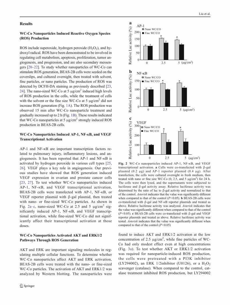
WC-Co Nanoparticles Induced AP-1, NF-κB, and VEGFTranscriptional Activation
AP-1 and NF-κB are important transcription factors re-lated to pulmonary injury, inflammatory lesions, and an-giogenesis. It has been reported that AP-1 and NF-κB isactivated by hydrogen peroxide in various cell types [25,26]. VEGF plays a key role in angiogenesis. Our previ-ous studies have showed that ROS generation inducedVEGF expression in ovarian and prostate cancer cells[23, 27]. To test whether WC-Co nanoparticles inducedAP-1, NF-κB, and VEGF transcriptional activation,BEAS-2B cells were transfected with AP-1, NF-κB, orVEGF reporter plasmid with β-gal plasmid, then treatedwith nano- or fine-sized WC-Co particles. As shown inFig. 2a–c, nano-sized WC-Co at 2.5 and 5 μg/cm2 sig-nificantly induced AP-1, NF-κB, and VEGF transcrip-tional activation, while fine-sized WC-Co did not signif-icantly affect their transcriptional activation at thosedoses.
WC-Co Nanoparticles Activated AKTand ERK1/2Pathways Through ROS Generation
AKT and ERK are important signaling molecules in reg-ulating multiple cellular functions. To determine whetherWC-Co nanoparticles affect AKT and ERK activation,BEAS-2B cells were treated with the nano- or fine-sizedWC-Co particles. The activation of AKT and ERK1/2 wasanalyzed by Western blotting. The nanoparticles were
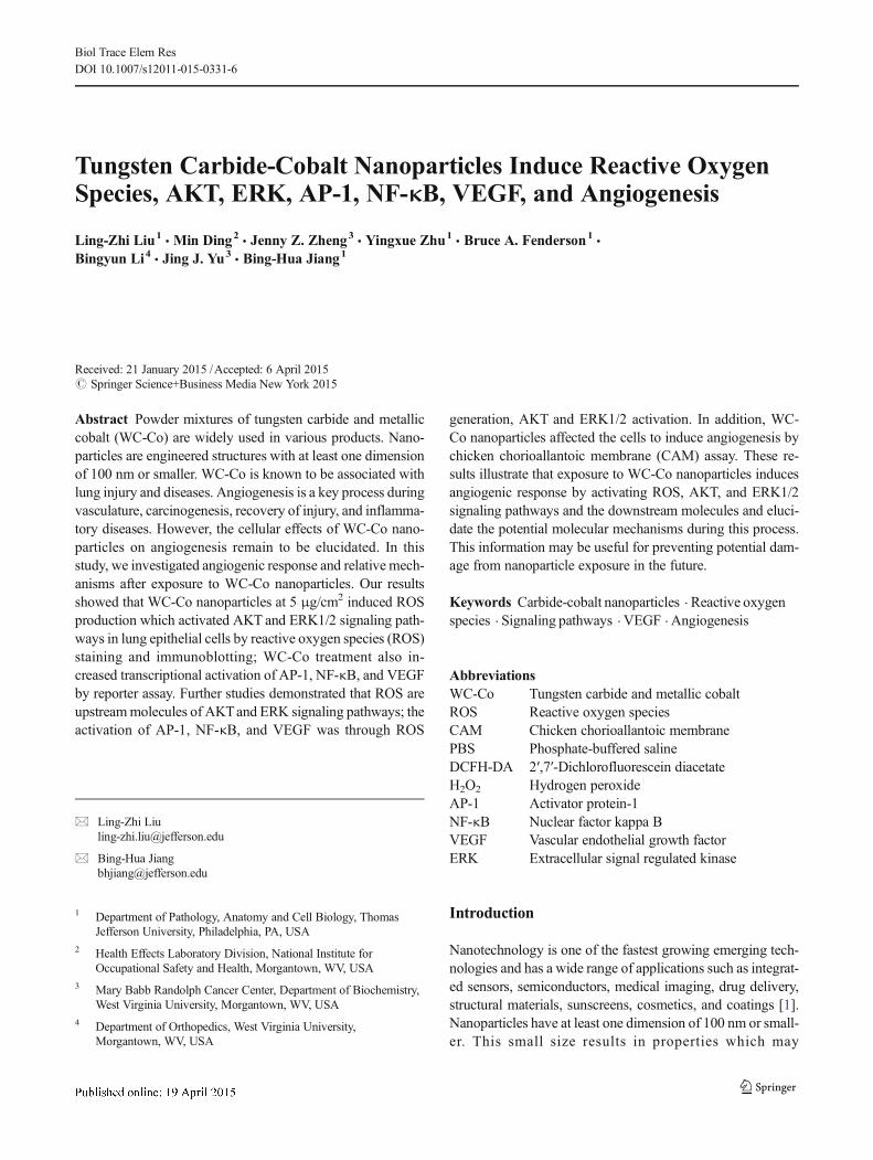
found to induce AKT and ERK1/2 activation at the lowconcentration of 2.5 μg/cm2, while fine particles of WC-Co had only modest effect even at high concentrations(Fig. 3a). To test whether AKT or ERK1/2 activationwas required for nanoparticle-induced ROS production,the cells were pretreated with a PI3K inhibitor(LY294002), an ERK 1/2inhibitor (U0126), or a H2O2
scavenger (catalase). When compared to the control, cat-alase treatment inhibited ROS production, but LY294002
0 2.5 5 ( g/cm2)0
100
200
300
* *Nano WC/CO
Fine WC/CO
VEGFc
0 2.5 5 ( g/cm2)0
50
100
150 * *
Nano WC/CO
Fine WC/CO
b
( g/cm2)
AP-1
* *
0
50
100
150
200Nano WC/CO
Fine WC/CO
0 2.5 5
a
NF- B
Fig. 2 WC-Co nanoparticles induced AP-1, NF-κB, and VEGFtranscriptional activation. a Cells were co-transfected with β-galplasmid (0.2 μg) and AP-1 reporter plasmid (0.4 μg). Aftertransfection, the cells were cultured overnight in fresh medium, thentreated with nano or fine size WC-Co (0, 2.5, and 5 μg/cm2) for 24 h.The cells were then lysed, and the supernatants were subjected toluciferase and β-gal activity assay. Relative luciferase activity wasdetermined by the ratio of luc to β-gal activity and normalized to thatof the control. Asterisk indicates that the value was significantly differentwhen compared to that of the control (P<0.05). b BEAS-2B cells wereco-transfected with β-gal and NF-κB reporter plasmids and treated asabove. Relative luciferase activity was analyzed. Asterisk indicates thatthe value was significantly different when compared to that of the control(P<0.05). c BEAS-2B cells were co-transfected with β-gal and VEGFreporter plasmids and treated as above. Relative luciferase activity wastested. Asterisk indicates that the value was significantly different whencompared to that of the control (P<0.05)
Liu et al.
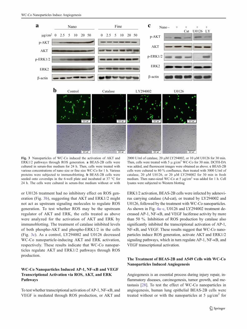
or U0126 treatment had no inhibitory effect on ROS gen-eration (Fig. 3b), suggesting that AKT and ERK1/2 mightnot act as upstream signaling molecules to regulate ROSgeneration. To test whether ROS may be the upstreamregulator of AKT and ERK, the cells treated as abovewere analyzed for the activation of AKT and ERK byimmunoblotting. The treatment of catalase inhibited levelsof both phospho-AKT and phospho-ERK1/2 in the cells(Fig. 3c). As a control, LY294002 and U0126 decreasedWC-Co nanoparticle-inducing AKT and ERK activation,respectively. These results indicate that WC-Co nanopar-ticles regulate AKT and ERK1/2 pathways through ROSproduction.
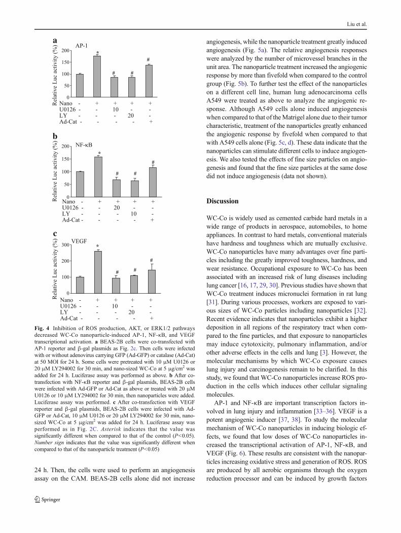
WC-Co Nanoparticles Induced AP-1, NF-κB and VEGFTranscriptional Activation via ROS, AKT, and ERKPathways
To test whether transcriptional activation of AP-1, NF-κB, andVEGF is mediated through ROS production, or AKT and
ERK1/2 activation, BEAS-2B cells were infected by adenovi-rus carrying catalase (Ad-cat), or treated by LY294002 andU0126, followed by the treatment with WC-Co nanoparticles.As shown in Fig. 4a–c, U0126 and LY294002 treatment de-creased AP-1, NF-κB, and VEGF luciferase activity by morethan 50 %. Inhibition of ROS production by catalase alsosignificantly inhibited the transcriptional activation of AP-1,NF-κB, and VEGF. These results suggest that WC-Co nano-particles induce ROS generation, activate AKT and ERK1/2signaling pathways, which in turn regulate AP-1, NF-κB, andVEGF transcriptional activation.
The Treatment of BEAS-2B and A549 Cells with WC-CoNanoparticles Induced Angiogenesis
Angiogenesis is an essential process during injury repair, in-flammatory diseases, carcinogenesis, tumor growth, and me-tastasis [28]. To test the effect of WC-Co nanoparticles inangiogenesis, human lung epithelial BEAS-2B cells weretreated without or with the nanoparticles at 5 μg/cm2 for
g/cm2 0 2.5 5 10 20 50 0 2.5 5 10 20 50
FineNano
p-AKT
p-ERK1/2
AKT
ERK2
a
-actin
Control Catalase LY294002 U0126b
p-AKT
p-ERK1/2
Nano - + + + +Cat U0126 LY
AKT
ERK2
c
-actin
Fig. 3 Nanoparticles of WC-Co induced the activation of AKT andERK1/2 pathways through ROS generation. a BEAS-2B cells werecultured in serum-free medium for 24 h. Then, cells were treated withvarious concentrations of nano size or fine size WC-Co for 1 h. Variousproteins were subjected to immunoblotting. b BEAS-2B cells wereseeded onto coverslips in the 6-well plate and incubated at 37 °C for24 h. The cells were cultured in serum-free medium without or with
2000 U/ml of catalase, 20 μM LY294002, or 10 μM U0126 for 30 min.Then, cells were treated with 5 μ g/cm2 WC-Co for 30 min. DCFH-DAwas added, and fluorescent images were obtained as above. c BEAS-2Bcells were cultured to 80 % confluence, then treated with 3000 U/ml ofcatalase, 20 μM U0126, or 20 μM LY294002 for 30 min in freshmedium. Then nano-sized WC-Co at 5 μg/cm2 was added for 1 h. Celllysates were subjected to Western blotting
WC-Co Nanoparticles Induce Angiogenesis
24 h. Then, the cells were used to perform an angiogenesisassay on the CAM. BEAS-2B cells alone did not increase
angiogenesis, while the nanoparticle treatment greatly inducedangiogenesis (Fig. 5a). The relative angiogenesis responseswere analyzed by the number of microvessel branches in theunit area. The nanoparticle treatment increased the angiogenicresponse by more than fivefold when compared to the controlgroup (Fig. 5b). To further test the effect of the nanoparticleson a different cell line, human lung adenocarcinoma cellsA549 were treated as above to analyze the angiogenic re-sponse. Although A549 cells alone induced angiogenesiswhen compared to that of theMatrigel alone due to their tumorcharacteristic, treatment of the nanoparticles greatly enhancedthe angiogenic response by fivefold when compared to thatwith A549 cells alone (Fig. 5c, d). These data indicate that thenanoparticles can stimulate different cells to induce angiogen-esis. We also tested the effects of fine size particles on angio-genesis and found that the fine size particles at the same dosedid not induce angiogenesis (data not shown).
Discussion
WC-Co is widely used as cemented carbide hard metals in awide range of products in aerospace, automobiles, to homeappliances. In contrast to hard metals, conventional materialshave hardness and toughness which are mutually exclusive.WC-Co nanoparticles have many advantages over fine parti-cles including the greatly improved toughness, hardness, andwear resistance. Occupational exposure to WC-Co has beenassociated with an increased risk of lung diseases includinglung cancer [16, 17, 29, 30]. Previous studies have shown thatWC-Co treatment induces micronuclei formation in rat lung[31]. During various processes, workers are exposed to vari-ous sizes of WC-Co particles including nanoparticles [32].Recent evidence indicates that nanoparticles exhibit a higherdeposition in all regions of the respiratory tract when com-pared to the fine particles, and that exposure to nanoparticlesmay induce cytotoxicity, pulmonary inflammation, and/orother adverse effects in the cells and lung [3]. However, themolecular mechanisms by which WC-Co exposure causeslung injury and carcinogenesis remain to be clarified. In thisstudy, we found that WC-Co nanoparticles increase ROS pro-duction in the cells which induces other cellular signalingmolecules.
AP-1 and NF-κB are important transcription factors in-volved in lung injury and inflammation [33–36]. VEGF is apotent angiogenic inducer [37, 38]. To study the molecularmechanism of WC-Co nanoparticles in inducing biologic ef-fects, we found that low doses of WC-Co nanoparticles in-creased the transcriptional activation of AP-1, NF-κB, andVEGF (Fig. 6). These results are consistent with the nanopar-ticles increasing oxidative stress and generation of ROS. ROSare produced by all aerobic organisms through the oxygenreduction processor and can be induced by growth factors
0
50
100
150
200
Nano -U0126 - - 20 - -LY - - - 10 -Ad-Cat - - - - +
NF- B
*
# #
#
b
Nano -U0126 - - 10 - -LY - - - 20 -Ad-Cat - - - - +
AP-1
*
#
#
#
0
50
100
150
200
a
Nano -
+ + + +
+ + + +
+ + + +U0126 - - 10 - -LY - - - 20 -Ad-Cat - - - - +
VEGF*
# ##
0
100
200
300
c
Fig. 4 Inhibition of ROS production, AKT, or ERK1/2 pathwaysdecreased WC-Co nanoparticle-induced AP-1, NF-κB, and VEGFtranscriptional activation. a BEAS-2B cells were co-transfected withAP-1 reporter and β-gal plasmids as Fig. 2c. Then cells were infectedwith or without adenovirus carrying GFP (Ad-GFP) or catalase (Ad-Cat)at 50 MOI for 24 h. Some cells were pretreated with 10 μM U0126 or20 μM LY294002 for 30 min, and nano-sized WC-Co at 5 μg/cm2 wasadded for 24 h. Luciferase assay was performed as above. b After co-transfection with NF-κB reporter and β-gal plasmids, BEAS-2B cellswere infected with Ad-GFP or Ad-Cat as above or treated with 20 μMU0126 or 10 μM LY294002 for 30 min, then nanoparticles were added.Luciferase assay was performed. c After co-transfection with VEGFreporter and β-gal plasmids, BEAS-2B cells were infected with Ad-GFP or Ad-Cat, 10 μM U0126 or 20 μM LY294002 for 30 min, nano-sized WC-Co at 5 μg/cm2 was added for 24 h. Luciferase assay wasperformed as in Fig. 2C. Asterisk indicates that the value wassignificantly different when compared to that of the control (P<0.05).Number sign indicates that the value was significantly different whencompared to that of the nanoparticle treatment (P<0.05)
Liu et al.

and cytokines [23, 39]. Our results showed that nanoparticles,but not fine particles of WC-Co at 5 μg/cm2 stimulate ROSproduction. As we expected, catalase inhibited ROS produc-tion, indicating that H2O2 is one of the species of ROS in-duced by nano-sized WC-Co. Growing evidence has shown
that H2O2 may act as a second messenger to regulate variouscellular functions and signaling pathways including ERK,phosphatidylinositol 3-kinase (PI3K), small GTPases, NF-κB,and nitrogen oxide (NO) pathways [40–44]. Previous reportsindicate that PI3K and ERK may regulate ROS production[45, 46]. We found that the inhibitors of PI3K and ERK1/2,LY294002, and U0126 did not affect ROS generation. In con-trast, treatment with catalase inhibited both AKTand ERK1/2.These results suggest that ROS act as upstream inducers ofAKT and ERK1/2 in the cells. This finding is consistent withprevious studies in a different cell system showing that H2O2
was the upstream regulator of AKT and ERK [47, 48]. Wefurther showed that ROS, AKT, and ERK1/2 are upstreamregulators of WC-Co nanoparticle-induced AP-1, NF-κB,and VEGF transcriptional activation.
Angiogenesis is critical in the injury repair, inflammation,tumor development, and neovascularization [49, 50]. Giventhe role of AP-1, NF-κB, and VEGF in regulating angiogen-esis, to determine whether WC-Co nanoparticles can induceangiogenesis, we demonstrated that treatment of BEAS-2 cellswith WC-Co nanoparticles at low dose induced angiogenesis,while equal dose of fine-sized WC-Co did not have a strongeffect on angiogenesis. Similar results were obtained using
CAM Control Nano WC-Coa b
0
100
200
300
400
500
600**
Rel
ativ
e an
gio
gen
esis
(%
) BEAS-2B
CAM Nano WC-Coc
A549
**
Rel
ativ
e an
gio
gen
esis
(%
)
d
0
100
200
300
400
500
600
CAM Control Nano WC-Co
CAM Control Nano WC-Co
Control
Fig. 5 BEAS-2B and A549 cells treated with nanoparticles of WC-Coinduced angiogenesis in the chicken chorioallantoic membrane (CAM)model. a BEAS-2B cells were treated without or with 5 μg/cm2 of nanoWC-Co for 24 h, then trypsinized, resuspended in serum-free medium(3×10⁷/ml, 0.1 ml), and mixed in 1:1 ratio with Matrigel (CollaborativeBiomedical Products, Bedford, MA). Aliquots of the mixture were thenapplied to the CAMof 9-day-old embryos. After 96-h incubation, the areaaround the implanted Matrigel was photographed with a Nikon digitalcamera, and the numbers of newly formed vessels were counted. Assaysfor each treatment were carried out using 10 embryos per experiment.Representative photos generated from CAM alone or BEAS-2B cells
treated without or with WC-Co (upper panel—low magnification;bottom panel—higher magnification). Bar: 2 mm. b The number ofblood vessels was obtained by counting the branching of blood vesselsand normalizing to the control (without WC-Co nanoparticles treatment)as relative angiogenesis. The data represent the mean±SD of the relativeangiogenesis from 8 different embryos. Asterisks indicate that the valuewas significantly different when compared to that of the control(P<0.01). c Representative photos generated from CAM alone or A549cells treated without or with WC-Co. Similar experiment was taken byusing A549 cells, and angiogenesis was assayed as above. d Relativeangiogenesis induced by A549 cells treated with WC-Co nanoparticles
WC-Co nanoparticles
ROS generation
AKT and ERK1/2 pathways
AP-1, NF- B and VEGF
Angiogenesis
Fig. 6 The diagram summarizes our findings:WC-Co nanoparticles leadto ROS generation, which activates AKTand ERK1/2 pathways to induceangiogenesis via AP-1, NF-κB, and VEGF
WC-Co Nanoparticles Induce Angiogenesis

A549 cells, indicating that WC-Co nanoparticles enhancedA549 cells-induced angiogenesis. These results are consistentwith the induction of transcriptional activation of AP-1,NF-κB, and VEGF in this study. These data elucidate cellulareffects of WC-Co nanoparticles and potential molecularmechanisms involved in the response to WC-Co nanoparticleexposure.
Since more and more nanotechnologies are developed andvarious sizes of WC-Co were produced during the process ofgenerating the hard metal, it is important to understand thebiological effect of nanoparticles of WC-Co on inducing hu-man diseases such as lung cancer, chronic pulmonary obstruc-tive diseases, and bronchitis. Angiogenesis is well known tobe closely associated with carcinogenesis, inflammatory dis-eases, and chronic pulmonary diseases including bronchitis. Inthis study, we found that nanoparticles of WC-Co inducedROS generation then increased the transcriptional activitiesof AP-1, NF-κB, and VEGF through activating AKT andERK1/2 pathways, thus inducing angiogenesis. This studywill afford useful information for preventing potential damagefrom nanoparticle exposure in the future.
Conclusions
These results illustrate that exposure to WC-Co nanoparticlesinduces angiogenic response by activating ROS, AKT, andERK1/2 signaling pathways and the downstream molecules,including AP-1, NF-κB, and VEGF. This information may beuseful for preventing potential damage from nanoparticle ex-posure in the future.
Acknowledgments This work was supported by R01HL091456,R01ES020868, and R21CA175975 from National Institutes of Health.
References
1. Roco MC (2004) Science and technology integration for increasedhuman potential and societal outcomes. Ann NYAcad Sci 1013:1–16
2. Renwick LC, Brown D, Clouter A, Donaldson K (2004) Increasedinflammation and altered macrophage chemotactic responsescaused by two ultrafine particle types. Occup Environ Med 61:442–447
3. Gradon L, Orlicki D, Podgorski A (2000) Deposition and retentionof ultrafine aerosol particles in the human respiratory system.Normal and pathological cases. Int J Occup Saf Ergon 6:189–207
4. Inoue K, Takano H, Yanagisawa R, Hirano S, Sakurai M, ShimadaA, Yoshikawa T (2006) Effects of airway exposure to nanoparticleson lung inflammation induced by bacterial endotoxin in mice.Environ Health Perspect 114:1325–1330
5. Roberts RA, Shen T, Allen IC, Hasan W, DeSimone JM, Ting JP(2013) Analysis of the murine immune response to pulmonary de-livery of precisely fabricated nano- and microscale particles. PLoSOne 8:e62115
6. Oberdorster G, Ferin J, Lehnert BE (1994) Correlation betweenparticle size, in vivo particle persistence, and lung injury. EnvironHealth Perspect 102(Suppl 5):173–179
7. Tanaka T, Kojima I, Ohse T, Ingelfinger JR, Adler S, Fujita T,Nangaku M (2005) Cobalt promotes angiogenesis via hypoxia-inducible factor and protects tubulointerstitium in the remnant kid-ney model. Lab Invest 85:1292–1307
8. Moulin JJ, Wild P, Romazini S, Lasfargues G, Peltier A, Bozec C,Deguerry P, Pellet F, Pedrix A (1998) Lung cancer risk in hard-metal workers. Am J Epidemiol 148:241–248
9. Lison D, Carbonnelle P, Mollo L, Lauwerys R, Fubini B (1995)Physicochemical mechanism of the interaction between cobalt met-al and carbide particles to generate toxic activated oxygen species.Chem Res Toxicol 8:600–606
10. Wild P, Perdrix A, Romazini S, Moulin JJ, Pellet F (2000) Lungcancer mortality in a site producing hard metals. Occup EnvironMed 57:568–573
11. Rinna A,Magdolenova Z, Hudecova A,KruszewskiM, RefsnesM,Dusinska M (2015) Effect of silver nanoparticles on mitogen-activated protein kinases activation: role of reactive oxygen speciesand implication in DNA damage. Mutagenesis 30:59–66
12. Durga M, Nathiya S, Rajasekar A, Devasena T (2014) Effects ofultrafine petrol exhaust particles on cytotoxicity, oxidative stressgeneration, DNA damage and inflammation in human A549 lungcells and murine RAW 264.7 macrophages. Environ ToxicolPharmacol 38:518–530
13. Nakamura Y, Nishizaka Y, Ariyasu R, Okamoto N, Yoshida M,Taki M, Nagano H, Hanaoka K, Nakagawa K, Yoshimura C,Wakayama T, Amitani R (2014) Hard metal lung disease diagnosedon a transbronchial lung biopsy following recurrent contact derma-titis. Intern Med 53:139–143
14. Kaneko Y, Kikuchi N, Ishii Y, Kawabata Y, Moriyama H, TeradaM, Suzuki E, Kobayashi M, Watanabe K, Hizawa N (2010) Upperlobe-dominant pulmonary fibrosis showing deposits of hard metalcomponent in the fibrotic lesions. Intern Med 49:2143–2145
15. Tanaka J, Moriyama H, Terada M, Takada T, Suzuki E, Narita I,Kawabata Y, Yamaguchi T, Hebisawa A, Sakai F, Arakawa H(2014) An observational study of giant cell interstitial pneumoniaand lung fibrosis in hard metal lung disease. BMJ Open 4:e004407
16. Wild P, Bourgkard E, Paris C (2009) Lung cancer and exposure tometals: the epidemiological evidence. Methods Mol Biol 472:139–167
17. Armstead AL, Arena CB, Li B (2014) Exploring the potential roleof tungsten carbide cobalt (WC-Co) nanoparticle internalization inobserved toxicity toward lung epithelial cells in vitro. Toxicol ApplPharmacol 278:1–8
18. Jiang BH, Zheng JZ, AokiM, Vogt PK (2000) Phosphatidylinositol3-kinase signaling mediates angiogenesis and expression of vascu-lar endothelial growth factor in endothelial cells. Proc Natl Acad SciU S A 97:1749–1753
19. He TC, Zhou S, da Costa LT, Yu J, Kinzler KW, Vogelstein B(1998) A simplified system for generating recombinant adenovi-ruses. Proc Natl Acad Sci U S A 95:2509–2514
20. Bauer G (2000) Reactive oxygen and nitrogen species: efficient,selective, and interactive signals during intercellular induction ofapoptosis. Anticancer Res 20:4115–4139
21. Forman HJ, Fukuto JM, Torres M (2004) Redox signaling: thiolchemistry defines which reactive oxygen and nitrogen species canact as second messengers. Am J Physiol Cell Physiol 287:C246–C256
22. Schieber M, Chandel NS (2014) ROS function in redox signalingand oxidative stress. Curr Biol 24:R453–R462
23. Liu LZ, Hu XW, Xia C, He J, Zhou Q, Shi X, Fang J, Jiang BH(2006) Reactive oxygen species regulate epidermal growth factor-induced vascular endothelial growth factor and hypoxia-inducible
Liu et al.
factor-1alpha expression through activation of AKT and P70S6K1in human ovarian cancer cells. Free Radic Biol Med 41:1521–1533
24. Carpenter RL, Jiang Y, Jing Y, He J, Rojanasakul Y, Liu LZ, JiangBH (2011) Arsenite induces cell transformation by reactive oxygenspecies, AKT, ERK1/2, and p70S6K1. Biochem Biophys ResCommun 414:533–538
25. Hsu TC, Young MR, Cmarik J, Colburn NH (2000) Activator pro-tein 1 (AP-1)- and nuclear factor kappaB (NF-kappaB)-dependenttranscriptional events in carcinogenesis. Free Radic Biol Med 28:1338–1348
26. Manduteanu I, Dragomir E, Voinea M, Capraru M, Simionescu M(2007) Enoxaparin reduces H2O2-induced activation of human en-dothelial cells by a mechanism involving cell adhesion moleculesand nuclear transcription factors. Pharmacology 79:154–162
27. Zhou Q, Liu LZ, Fu B, Hu X, Shi X, Fang J, Jiang BH (2007)Reactive oxygen species regulate insulin-induced VEGF and HIF-1alpha expression through the activation of p70S6K1 in humanprostate cancer cells. Carcinogenesis 28:28–37
28. Folkman J (1974) Tumor angiogenesis. Adv Cancer Res 19:331–358
29. Armstead AL, Minarchick VC, Porter DW, Nurkiewicz TR, Li B(2015) Acute inflammatory responses of nanoparticles in an intra-tracheal instillation rat model. PLoS One 10, e0118778
30. Dunlop P, Muller NL, Wilson J, Flint J, Churg A (2005) Hard metallung disease: high resolution CT and histologic correlation of theinitial findings and demonstration of interval improvement. JThorac Imaging 20:301–304
31. De Boeck M, Hoet P, Lombaert N, Nemery B, Kirsch-Volders M,Lison D (2003) In vivo genotoxicity of hard metal dust: inductionof micronuclei in rat type II epithelial lung cells. Carcinogenesis 24:1793–1800
32. Stefaniak AB, Virji MA, Day GA (2009) Characterization of expo-sures among cemented tungsten carbide workers. Part I: size-fractionated exposures to airborne cobalt and tungsten particles. JExpo Sci Environ Epidemiol 19:475–491
33. Alvira CM (2014) Nuclear factor-kappa-B signaling in lung devel-opment and disease: one pathway, numerous functions. BirthDefects Res A Clin Mol Teratol 100:202–216
34. Chopra M, Reuben JS, Sharma AC (2009) Acute lung injury: apo-ptosis and signaling mechanisms. Exp Biol Med (Maywood) 234:361–371
35. Lee IT, Yang CM (2013) Inflammatory signalings involved in air-way and pulmonary diseases. Mediators Inflamm 2013:791231
36. Verstrepen L, Beyaert R (2014) Receptor proximal kinases in NF-kappaB signaling as potential therapeutic targets in cancer and in-flammation. Biochem Pharmacol 92:519–529
37. Shibuya M (2015) VEGF-VEGFR system as a target for suppress-ing inflammation and other diseases. EndocrMetab Immune DisordDrug Targets
38. Connolly DT, Heuvelman DM, Nelson R, Olander JV, Eppley BL,Delfino JJ, Siegel NR, Leimgruber RM, Feder J (1989) Tumorvascular permeability factor stimulates endothelial cell growth andangiogenesis. J Clin Invest 84:1470–1478
39. Yang L, Liu G, Lin Z,Wang Y, He H, Liu T, KampDW (2014) Pro-inflammatory response and oxidative stress induced by specificcomponents in ambient particulate matter in human bronchial epi-thelial cells. Environ Toxicol
40. Bae YS, Sung JY, Kim OS, Kim YJ, Hur KC, Kazlauskas A, RheeSG (2000) Platelet-derived growth factor-induced H (2) O (2) pro-duction requires the activation of phosphatidylinositol 3-kinase. JBiol Chem 275:10527–10531
41. Finkel T (1998) Oxygen radicals and signaling. Curr Opin Cell Biol10:248–253
42. May JM, de Haen C (1979) The insulin-like effect of hydrogenperoxide on pathways of lipid synthesis in rat adipocytes. J BiolChem 254:9017–9021
43. Schreck R, AlbermannK, Baeuerle PA (1992) Nuclear factor kappaB: an oxidative stress-responsive transcription factor of eukaryoticcells (a review). Free Radic Res Commun 17:221–237
44. Vercellotti GM, Severson SP, Duane P, Moldow CF (1991)Hydrogen peroxide alters signal transduction in human endothelialcells. J Lab Clin Med 117:15–24
45. Baumer AT, Ten Freyhaus H, Sauer H, Wartenberg M, Kappert K,Schnabel P, Konkol C, Hescheler J, Vantler M, Rosenkranz S(2008) Phosphatidylinositol 3-kinase-dependent membrane recruit-ment of Rac-1 and p47phox is critical for alpha-platelet-derivedgrowth factor receptor-induced production of reactive oxygen spe-cies. J Biol Chem 283:7864–7876
46. Zhuang S, Kinsey G, Yan Y, Han J, Schnellmann R (2008) ERKactivation mediates mitochondrial dysfunction and necrosis in-duced by hydrogen peroxide in renal proximal tubular cells. JPharmacol Exp Ther 325:732–740
47. Tournier C, Thomas G, Pierre J, Jacquemin C, Pierre M, Saunier B(1997) Mediation by arachidonic acid metabolites of the H2O2-induced stimulation of mitogen-activated protein kinases (extracel-lular-signal-regulated kinase and c-Jun NH2-terminal kinase). Eur JBiochem 244:587–595
48. Ushio-FukaiM, Alexander RW, AkersM, Yin Q, Fujio Y,Walsh K,Griendling KK (1999) Reactive oxygen species mediate the activa-tion of Akt/protein kinase B by angiotensin II in vascular smoothmuscle cells. J Biol Chem 274:22699–22704
49. Folkman J (1995)Angiogenesis in cancer, vascular, rheumatoid andother disease. Nat Med 1:27–31
50. RisauW (1997)Mechanisms of angiogenesis. Nature 386:671–674
WC-Co Nanoparticles Induce Angiogenesis