
Tu1794 Differences in Amygdala Functional Connectivity Between Erosive (ERD) and Non-Erosive Reflux...
1
AGA Abstracts neurons. In contrast to control rats, UK14304 decreased the amplitude of IPSC in 12 of 17 rats that underwent homotypic stress (2hrs restrain, 20min forced swim, 40min cold @4°C and 2hrs water avoidance) for 5 days in a concentration-dependent manner (91±1%, 78±1% and 53±9% of baseline amplitude in response to perfusion with 0.1, 1 and 3μM, respectively; P<0.05 vs control). Pretreatment with CRF had no additional inhibitory action on the UK14304-induced inhibition of GABAergic currents. Miniature strain gauges were applied to the anterior corpus of anaesthetized rats to record gastric motility. In control rats, DMV microinjections of the indirect sympathomimetic tyramine (4.5nmoles/60nl) reduced gastric motility to 50±10% of baseline (N=10); conversely, microinjections of tyramine in the brainstem of homotypic stress rats decreased gastric motility to 7±5% of baseline (N=2). These data suggest that homotypic stress induces an upregulation of catecholaminergic, including α2-mediated, responses in vagal neurocircuits resulting in an exaggerated vagally- mediated gastroinhibition. Supported by DK55530 and DK78364 Tu1793 Elevated Sympathetic Activity Induces Anxiety-Like Behavior in Rats Subjected to Neonatal Inflammation John H. Winston, Sushil K. Sarna Background: Clinical evidence strongly supports a role for adverse early life experiences (AELE) as etiological factors In Functional Dyspepsia (FD) patients. AELEs affect central visceral circuits that underlie behavioral and autonomic responses to interoceptive and stressful events. FD patients demonstrate elevated sympathetic activity at baseline and in response to stress. The mechanisms underlying this dysfunction and how it contributes to symptom generation remain unknown. We investigated these questions in a rat model of FD-like gastric hypersensitivity (GHS) that was induced by neonatal colonic inflammation, where we demonstrated that increased serum norepinephrine contributed to gastric hypersen- sitivity through up-regulation of NGF expression in the fundus. Aims: To measure: the effects of either sympathectomy or adrenalectomy on GHS and NGF expression in the fundus, changes in gene expression in sympathetic circuits that correlate with increased GHS, and physiological and behavioral correlates of sympathetic activity. Results: Sympathetic ablation by systemic treatment with 6-OH dopamine significantly reduced GHS in FD-like rats vs. pre-treatment baseline (p<0.05) and significantly decreased NGF protein expression in the fundus (p<0.05). Adrenalectomy produced no significant effect on GHS in FD-like rats vs. pre-treatment baseline or sham operated FD-like rats and had no significant effect on NGF expression in the fundus. Tyrosine hydroxylase mRNA expression in Celiac ganglia was elevated 2.5-fold (p<0.05) in FD-like vs. Ctr. rats. This increase was significantly attenuated in rats treated with RU486 as neonates, which prevents the acquisition of GHS. Tyrosine hydroxylase protein expression in celiac ganglia was increased 1.3-fold (p<0.05) in FD-like vs. Ctr. rats. This increase was significantly reduced in rats treated with RU486 as neonates. In the paraventricular nucleus, glucocorticoid receptor expression was significantly decreased (25%, p<0.05), also prevented by neonatal treatment with RU486. Heart rate variability sympathetic-vagal balance was two-fold greater in FD-like rats vs. controls (p>0.05). FD-like rats spent significantly less time in the open arm of the elevated plus maze vs. controls (p<0.05). Chronic stress significantly reduced open arm time of control rats, but was without significant effect in FD-like rats. Conclusion: These findings are consistent with increased basal peripheral sympathetic activity mediated in part by increased TH expression. Higher anxiety in FD-like rats may be related to excess central sympathetic activity. Lack of an increase in anxiety in response to chronic stress in FD-like rats suggests central dysregulation of sympathetic outflow. Supported in part by NIDDK Grant 5R01DK088796. Tu1794 Differences in Amygdala Functional Connectivity Between Erosive (ERD) and Non-Erosive Reflux Disease (NERD) Patients During Esophageal Acid Exposure Mark Kern, Arash Babaei, Robert M. Siwiec, Erica A. Samuel, Ling Mei, Patrick Sanvanson, Reza Shaker Functional connectivity magnetic resonance imaging (fc-MRI) studies have shown significant differences in correlations of brain signals in a number of limbic structures like thalamus, insula and cingulate between erosive and non-erosive reflux disease patients when stimulated by esophageal acid exposure. However, many important brain structures directly associated with emotional and visceral processes have not been systematically studied in these patient groups. Among these structures is amygdala which has a key function in regulating fear, anxiety, motivation, goal-directed behavior, attention, social behavior and autonomic func- tion that makes it a reasonable target for esophageal acid induced effects. Aims: To character- ize and compare the functional connectivity between the fc-MRI signals from amygdala to the rest of the brain in NERD and ERD patients during esophageal buffer and acid stimulation. Methods: In 24 heartburn patients (12 NERD, 6 female, age 46±5 years without esophageal erosions and 12 ERD, 6 female, age 32±11 years with healed erosive esophagitis), fc-MRI data were gathered during two 9-minute scans. In the first scan, buffer was perfused in the distal esophagus at 1 ml/min. In the second scan, 0.1N HCl was perfused. Fc was quantified by the correlation coefficient (CC) between average fc-MRI time series (seeds) in two spherical volumes centered in the right and left amygdala and time series of all fc-MRI volume elements (voxels) across the entire brain. The amygdala seeds were centered at the locations identified by the MNI atlas. Group analysis was performed using unpaired t-tests (with cluster size Monte Carlo simulation to correct for multiple comparison) of CC to test amygdala connectiv- ity differences across NERD and ERD patients. Results: Between group comparisons showed significant differences in amygdala connectivity during acid but not buffer infusion when comparing ERD to NERD patients. These differences were characterized by larger CC values in ERD patients for right amygdala connection to the right superior temporal gyrus and anterior insula (Figure 1A), right middle frontal gyrus as well as right posterior insula (p<0.001 corrected). Significant left amygdala connectivity differences were also seen during acid perfusion for correlations to the right insula (Figure 1B). Conclusions: During esopha- geal acid exposure, fc analysis of the amygdala with a number of brain regions shows greater levels of correlation for erosive compared to non-erosive reflux disease patients. The regions S-844 AGA Abstracts with augmented connectivity include the insula, temporal and frontal gyri. These regions play important roles in interoception and visceral pain networks; therefore, our results may imply a differential effect of erosive versus non-erosive esophageal disorders on the connectivity of the amygdala with these brain networks. Tu1795 Transnasal Esophageal Intubation Per Se Affects the Introceptive but Not the Default Mode Network Mark Kern, Arash Babaei, Robert M. Siwiec, Erica A. Samuel, Ling Mei, Reza Shaker Recent whole brain studies indicate the presence of an esophageal catheter without any infusion or stimulation may alter amygdala functional connectivity (fc) and influence the results of comparative studies. Whether this influence involves other brain regions utilizing different analytical techniques is currently unknown. Aim: To characterize the effect of esophageal intubation per se on fc within two commonly studied networks, namely interocep- tive (ICN) and default mode (DMN) networks, in health and disease. Methods: In 12 NERD patients (6 female, age 46±5 years) and 14 healthy subjects (7 female, age 27±4 years), fc- MRI time series data were gathered in two 9-minute scans, one scan without trans-nasal esophageal intubation and one scan with intubation. Regions of interest (ROI) were defined by stereotaxic coordinates of previously published networks and average fc-MRI time series were calculated for all time series within spherical regions centered at the defined coordinates. ROI sphere diameters were either 0.5 or 1.0 cm depending on the size of the anatomical regions wherein the spheres were positioned. Correlation coefficients (CC) were calculated between all ROI within DMN and ICN networks. Group analysis was performed using unpaired t-tests (corrected for multiple comparisons by False Discovery Rate (FDR) methods) between NERD and healthy subjects for each network by comparing normalized CC values. Results: In both groups there were no significant differences found in DMN fc between scans without and with the catheter. In contrast, significant differences were found in ICN fc when comparing the no catheter to catheter-present condition in both groups. In the efferent arm of the ICN of controls, fc differences were in the left posterior hippocampus - right perigenual anterior cingulate connection (p<0.002, corrected for multiple comparisons) wherein average CC values showed a decrease in connectivity after intubation. In NERD, efferent ICN fc differences were in the left amygdala - right ventral anterior insula connection (p<0.002) again showing decreased connectivity after intubation (Figure 1). In the afferent ICN, no intubation effect was found in the control group when comparing rest to intubated connectivity, however; in theseqnum=" NERD patients, significant differences in the left ventral anterior insula - right ventral middle insula connections were detected (p<0.0005). Conclusions: Intubation of the esophagus per se does not uniformly affect the connectivity of different brain networks. For example it has a significant effect on connectivity of ICN but not DMN network in both NERD patients and healthy controls. These findings indicate that the effect of the presence of an esophageal catheter per se on functional connectivity needs to be taken into account to avoid spurious results from studies involving esopha- geal intubation.
Transcript of Tu1794 Differences in Amygdala Functional Connectivity Between Erosive (ERD) and Non-Erosive Reflux...
AG
AA
bst
ract
sneurons. In contrast to control rats, UK14304 decreased the amplitude of IPSC in 12 of 17rats that underwent homotypic stress (2hrs restrain, 20min forced swim, 40min cold @4°Cand 2hrs water avoidance) for 5 days in a concentration-dependent manner (91±1%, 78±1%and 53±9% of baseline amplitude in response to perfusion with 0.1, 1 and 3μM, respectively;P<0.05 vs control). Pretreatment with CRF had no additional inhibitory action on theUK14304-induced inhibition of GABAergic currents. Miniature strain gauges were appliedto the anterior corpus of anaesthetized rats to record gastric motility. In control rats, DMVmicroinjections of the indirect sympathomimetic tyramine (4.5nmoles/60nl) reduced gastricmotility to 50±10% of baseline (N=10); conversely, microinjections of tyramine in thebrainstem of homotypic stress rats decreased gastric motility to 7±5% of baseline (N=2).These data suggest that homotypic stress induces an upregulation of catecholaminergic,including α2-mediated, responses in vagal neurocircuits resulting in an exaggerated vagally-mediated gastroinhibition. Supported by DK55530 and DK78364
Tu1793
Elevated Sympathetic Activity Induces Anxiety-Like Behavior in RatsSubjected to Neonatal InflammationJohn H. Winston, Sushil K. Sarna
Background: Clinical evidence strongly supports a role for adverse early life experiences(AELE) as etiological factors In Functional Dyspepsia (FD) patients. AELEs affect centralvisceral circuits that underlie behavioral and autonomic responses to interoceptive andstressful events. FD patients demonstrate elevated sympathetic activity at baseline and inresponse to stress. The mechanisms underlying this dysfunction and how it contributes tosymptom generation remain unknown. We investigated these questions in a rat model ofFD-like gastric hypersensitivity (GHS) that was induced by neonatal colonic inflammation,where we demonstrated that increased serum norepinephrine contributed to gastric hypersen-sitivity through up-regulation of NGF expression in the fundus. Aims: To measure: theeffects of either sympathectomy or adrenalectomy on GHS and NGF expression in thefundus, changes in gene expression in sympathetic circuits that correlate with increasedGHS, and physiological and behavioral correlates of sympathetic activity. Results: Sympatheticablation by systemic treatment with 6-OH dopamine significantly reduced GHS in FD-likerats vs. pre-treatment baseline (p<0.05) and significantly decreased NGF protein expressionin the fundus (p<0.05). Adrenalectomy produced no significant effect on GHS in FD-likerats vs. pre-treatment baseline or sham operated FD-like rats and had no significant effecton NGF expression in the fundus. Tyrosine hydroxylase mRNA expression in Celiac gangliawas elevated 2.5-fold (p<0.05) in FD-like vs. Ctr. rats. This increase was significantlyattenuated in rats treated with RU486 as neonates, which prevents the acquisition of GHS.Tyrosine hydroxylase protein expression in celiac ganglia was increased 1.3-fold (p<0.05)in FD-like vs. Ctr. rats. This increase was significantly reduced in rats treated with RU486 asneonates. In the paraventricular nucleus, glucocorticoid receptor expression was significantlydecreased (25%, p<0.05), also prevented by neonatal treatment with RU486. Heart ratevariability sympathetic-vagal balance was two-fold greater in FD-like rats vs. controls(p>0.05). FD-like rats spent significantly less time in the open arm of the elevated plusmaze vs. controls (p<0.05). Chronic stress significantly reduced open arm time of controlrats, but was without significant effect in FD-like rats. Conclusion: These findings areconsistent with increased basal peripheral sympathetic activity mediated in part by increasedTH expression. Higher anxiety in FD-like rats may be related to excess central sympatheticactivity. Lack of an increase in anxiety in response to chronic stress in FD-like rats suggestscentral dysregulation of sympathetic outflow. Supported in part by NIDDK Grant5R01DK088796.
Tu1794
Differences in Amygdala Functional Connectivity Between Erosive (ERD) andNon-Erosive Reflux Disease (NERD) Patients During Esophageal AcidExposureMark Kern, Arash Babaei, Robert M. Siwiec, Erica A. Samuel, Ling Mei, PatrickSanvanson, Reza Shaker
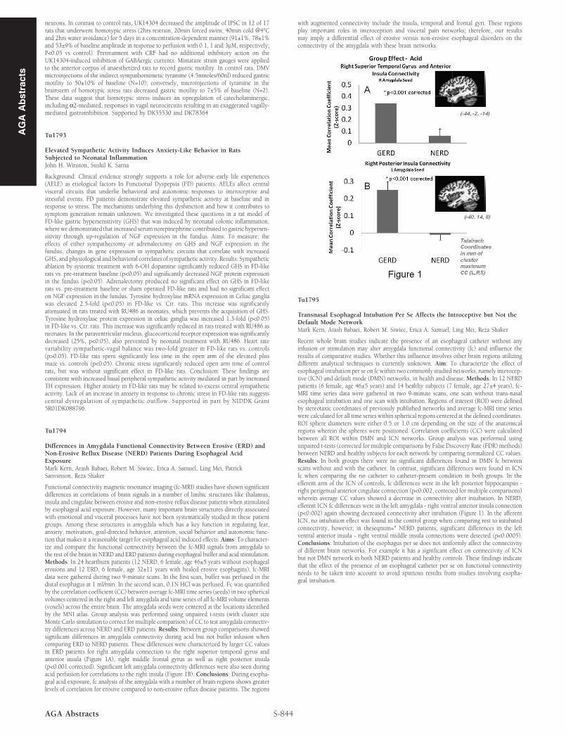
Functional connectivity magnetic resonance imaging (fc-MRI) studies have shown significantdifferences in correlations of brain signals in a number of limbic structures like thalamus,insula and cingulate between erosive and non-erosive reflux disease patients when stimulatedby esophageal acid exposure. However, many important brain structures directly associatedwith emotional and visceral processes have not been systematically studied in these patientgroups. Among these structures is amygdala which has a key function in regulating fear,anxiety, motivation, goal-directed behavior, attention, social behavior and autonomic func-tion that makes it a reasonable target for esophageal acid induced effects. Aims: To character-ize and compare the functional connectivity between the fc-MRI signals from amygdala tothe rest of the brain in NERD and ERD patients during esophageal buffer and acid stimulation.Methods: In 24 heartburn patients (12 NERD, 6 female, age 46±5 years without esophagealerosions and 12 ERD, 6 female, age 32±11 years with healed erosive esophagitis), fc-MRIdata were gathered during two 9-minute scans. In the first scan, buffer was perfused in thedistal esophagus at 1 ml/min. In the second scan, 0.1N HCl was perfused. Fc was quantifiedby the correlation coefficient (CC) between average fc-MRI time series (seeds) in two sphericalvolumes centered in the right and left amygdala and time series of all fc-MRI volume elements(voxels) across the entire brain. The amygdala seeds were centered at the locations identifiedby the MNI atlas. Group analysis was performed using unpaired t-tests (with cluster sizeMonte Carlo simulation to correct for multiple comparison) of CC to test amygdala connectiv-ity differences across NERD and ERD patients. Results: Between group comparisons showedsignificant differences in amygdala connectivity during acid but not buffer infusion whencomparing ERD to NERD patients. These differences were characterized by larger CC valuesin ERD patients for right amygdala connection to the right superior temporal gyrus andanterior insula (Figure 1A), right middle frontal gyrus as well as right posterior insula(p<0.001 corrected). Significant left amygdala connectivity differences were also seen duringacid perfusion for correlations to the right insula (Figure 1B). Conclusions: During esopha-geal acid exposure, fc analysis of the amygdala with a number of brain regions shows greaterlevels of correlation for erosive compared to non-erosive reflux disease patients. The regions
S-844AGA Abstracts
with augmented connectivity include the insula, temporal and frontal gyri. These regionsplay important roles in interoception and visceral pain networks; therefore, our resultsmay imply a differential effect of erosive versus non-erosive esophageal disorders on theconnectivity of the amygdala with these brain networks.
Tu1795
Transnasal Esophageal Intubation Per Se Affects the Introceptive but Not theDefault Mode NetworkMark Kern, Arash Babaei, Robert M. Siwiec, Erica A. Samuel, Ling Mei, Reza Shaker
Recent whole brain studies indicate the presence of an esophageal catheter without anyinfusion or stimulation may alter amygdala functional connectivity (fc) and influence theresults of comparative studies. Whether this influence involves other brain regions utilizingdifferent analytical techniques is currently unknown. Aim: To characterize the effect ofesophageal intubation per se on fc within two commonly studied networks, namely interocep-tive (ICN) and default mode (DMN) networks, in health and disease. Methods: In 12 NERDpatients (6 female, age 46±5 years) and 14 healthy subjects (7 female, age 27±4 years), fc-MRI time series data were gathered in two 9-minute scans, one scan without trans-nasalesophageal intubation and one scan with intubation. Regions of interest (ROI) were definedby stereotaxic coordinates of previously published networks and average fc-MRI time serieswere calculated for all time series within spherical regions centered at the defined coordinates.ROI sphere diameters were either 0.5 or 1.0 cm depending on the size of the anatomicalregions wherein the spheres were positioned. Correlation coefficients (CC) were calculatedbetween all ROI within DMN and ICN networks. Group analysis was performed usingunpaired t-tests (corrected for multiple comparisons by False Discovery Rate (FDR) methods)between NERD and healthy subjects for each network by comparing normalized CC values.Results: In both groups there were no significant differences found in DMN fc betweenscans without and with the catheter. In contrast, significant differences were found in ICNfc when comparing the no catheter to catheter-present condition in both groups. In theefferent arm of the ICN of controls, fc differences were in the left posterior hippocampus -right perigenual anterior cingulate connection (p<0.002, corrected for multiple comparisons)wherein average CC values showed a decrease in connectivity after intubation. In NERD,efferent ICN fc differences were in the left amygdala - right ventral anterior insula connection(p<0.002) again showing decreased connectivity after intubation (Figure 1). In the afferentICN, no intubation effect was found in the control group when comparing rest to intubatedconnectivity, however; in theseqnum=" NERD patients, significant differences in the leftventral anterior insula - right ventral middle insula connections were detected (p<0.0005).Conclusions: Intubation of the esophagus per se does not uniformly affect the connectivityof different brain networks. For example it has a significant effect on connectivity of ICNbut not DMN network in both NERD patients and healthy controls. These findings indicatethat the effect of the presence of an esophageal catheter per se on functional connectivityneeds to be taken into account to avoid spurious results from studies involving esopha-geal intubation.


![Extensions of a theorem of Erd}os on nonhamiltonian graphs · Erd}os [4] re ned the bound in terms of the minimum degree of the graph: This paper started at SQUARES meeting of the](https://static.fdocument.org/doc/165x107/5f1c3f09e658c3063c4c478d/extensions-of-a-theorem-of-erdos-on-nonhamiltonian-graphs-erdos-4-re-ned-the.jpg)