TNF-α AND MICROGLIAL HORMETIC INVOLVEMENT IN NEUROLOGICAL ... · TNF-α AND MICROGLIAL HORMETIC...
25
TNF-α AND MICROGLIAL HORMETIC INVOLVEMENT IN NEUROLOGICAL HEALTH & MIGRAINE Richard P. Kraig Department of Neurology, The University of Chicago Medical Center, Chicago, IL Heidi M. Mitchell Department of Neurology, The University of Chicago Medical Center, Chicago, IL Barbara Christie-Pope Department of Biology, Cornell College, Mt. Vernon, IA Phillip E. Kunkler Department of Neurology, The University of Chicago Medical Center, Chicago, IL David M. White Department of Neurology, The University of Chicago Medical Center, Chicago, IL Ya-Ping Tang Department of Psychiatry, The University of Chicago Medical Center, Chicago, IL George Langan Department of Surgery, The University of Chicago Medical Center, Chicago, IL Environmental enrichment, i.e., increased intellectual, social, and physical activity makes brain more resilient to subsequent neurological disease. The mechanisms for this effect remain incompletely defined, but evidence shows tumor necrosis factor-alpha (TNF- α) is involved. TNF-α, at acutely high levels, possesses the intrinsic capacity to enhance injury associated with neurological disease. Conversely, the effect of TNF-α at low-levels is nutritive over time, consistent with physiological conditioning hormesis. Evidence shows that neural activity triggers low-level pro-inflammatory signaling involving TNF-α. This low-level TNF-α signaling alters gene expression, resulting in an enhanced resilience to disease. Brain-immune signaling may become maladaptive when increased activity is chronic without sufficient periods of reduced activity necessary for nutritive adaptation. Such tonically increased activity may explain, for example, the transformation of episodic to chronic migraine with related increased susceptibility to spreading depression, the most likely underlying cause of this malady. Thus, TNF-α, whose function is to alter gene expres- sion, and its principal cellular source, microglia, seem powerfully positioned to orches- trate hormetic immune signaling that establishes the phenotype of neurological health and disease from brain activity. Keywords: migraine, spreading depression, neuro-immune, cytokine, excitotoxicity, environmental enrichment Dose-Response, 8:389–413, 2010 Formerly Nonlinearity in Biology, Toxicology, and Medicine Copyright © 2010 University of Massachusetts ISSN: 1559-3258 DOI: 10.2203/dose-response.09-056.Kraig Address correspondence to Richard P. Kraig, Department of Neurology; MC2030, The University of Chicago Medical Center, 5841 South Maryland Avenue; Chicago, IL 60637-1470, Phone: 773-702-0802; Fax: 773-702-5175, E-mail: [email protected] 389
Transcript of TNF-α AND MICROGLIAL HORMETIC INVOLVEMENT IN NEUROLOGICAL ... · TNF-α AND MICROGLIAL HORMETIC...
TNF-α AND MICROGLIAL HORMETIC INVOLVEMENT IN NEUROLOGICALHEALTH & MIGRAINE
Richard P. Kraig � Department of Neurology, The University of Chicago MedicalCenter, Chicago, IL
Heidi M. Mitchell � Department of Neurology, The University of ChicagoMedical Center, Chicago, IL
Barbara Christie-Pope � Department of Biology, Cornell College, Mt. Vernon,IA
Phillip E. Kunkler � Department of Neurology, The University of ChicagoMedical Center, Chicago, IL
David M. White � Department of Neurology, The University of Chicago MedicalCenter, Chicago, IL
Ya-Ping Tang � Department of Psychiatry, The University of Chicago MedicalCenter, Chicago, IL
George Langan � Department of Surgery, The University of Chicago MedicalCenter, Chicago, IL
� Environmental enrichment, i.e., increased intellectual, social, and physical activitymakes brain more resilient to subsequent neurological disease. The mechanisms for thiseffect remain incompletely defined, but evidence shows tumor necrosis factor-alpha (TNF-α) is involved. TNF-α, at acutely high levels, possesses the intrinsic capacity to enhanceinjury associated with neurological disease. Conversely, the effect of TNF-α at low-levels isnutritive over time, consistent with physiological conditioning hormesis. Evidence showsthat neural activity triggers low-level pro-inflammatory signaling involving TNF-α. Thislow-level TNF-α signaling alters gene expression, resulting in an enhanced resilience todisease. Brain-immune signaling may become maladaptive when increased activity ischronic without sufficient periods of reduced activity necessary for nutritive adaptation.Such tonically increased activity may explain, for example, the transformation of episodicto chronic migraine with related increased susceptibility to spreading depression, the mostlikely underlying cause of this malady. Thus, TNF-α, whose function is to alter gene expres-sion, and its principal cellular source, microglia, seem powerfully positioned to orches-trate hormetic immune signaling that establishes the phenotype of neurological healthand disease from brain activity.
Keywords: migraine, spreading depression, neuro-immune, cytokine, excitotoxicity, environmentalenrichment
Dose-Response, 8:389–413, 2010Formerly Nonlinearity in Biology, Toxicology, and MedicineCopyright © 2010 University of MassachusettsISSN: 1559-3258DOI: 10.2203/dose-response.09-056.Kraig
Address correspondence to Richard P. Kraig, Department of Neurology; MC2030, TheUniversity of Chicago Medical Center, 5841 South Maryland Avenue; Chicago, IL 60637-1470,Phone: 773-702-0802; Fax: 773-702-5175, E-mail: [email protected]
389
INTRODUCTION
Brain is unique among organ structures in that it exists to predict(Llinás 2002), a capacity that requires constant “reprogramming.”Importantly, reprogramming increases the brain’s resistance to neurolog-ical disease. Neural reprogramming is classically evidenced by Hebbiansynaptic plasticity, and extends to environmental enrichment (EE; i.e.,increased intellectual, social, and physical activity), which is well-known tobe protective (for review see van Praag et al. 2000; Will et al. 2004).
Reprogramming is also an inherent capacity of the immune system(Waldmann 2002; Graca and Waldmann 2006; Waldmann et al. 2008). Inaddition, immune system reprogramming may be a well-conservedprocess by which ischemic preconditioning stimuli applied to brain promptsubsequent neuroprotection (Marsh et. al. 2009).
Our focus is to illustrate how immune stimuli within brain, associatedwith neural activity, similarly reprogram gene function to create the phe-notype of increased neurological health through interactive signalingbetween neurons and glia. Increasing evidence indicates that neural andimmune systems interact in response to increased activity to establish thephenotype of enhanced brain health. This activity-dependent cooperativereprogramming may be driven by tumor necrosis factor-alpha (TNF-α),an innate immunity cytokine that emanates from microglia under normalcircumstances (Hulse et al. 2008).
Classically, microglia, principal immune signaling cells within brain,have been recognized for their powerful destructive capacities toenhance brain injury via mechanisms that include increased expressionof TNF-α (Chao and Hu 1994; Dawson et al. 1996; Barone et al. 1997;Meistrell et al. 1997; Aggarwal et al. 2001; Zou and Crews 2005).Accordingly, why would the brain use lethal signaling systems to enhanceits strength? Developing evidence indicates the answer lies in the old say-ing “…what doesn’t kill you makes you stronger …” (Hadley 2003). Thisnotion termed “physiological conditioning hormesis” (Calabrese et al.2007) is increasingly recognized as a well-conserved capacity of biologicalsystems, including brain (Mattson et al. 2002; Mattson 2008a, 2008b).Mounting evidence indicates that TNF-α and microglia can be neuropro-tective (Cheng et al. 1994; Wilde et al. 2000; Hallenbeck 2002; Stoll et al.2002; Streit 2005, 2006; Turrin and Rivest 2006; Carson et al. 2007;Hanisch and Kettenmann 2007; Sriram and O’Callaghan 2007; Hulse, etal. 2008; Salmina 2009).
Hormesis is a dose-response pattern involving low-level stimulationand high-level inhibition (Calabrese and Baldwin 2003). Hormesis is seenwith neuroprotective treatments for stroke and brain trauma (Calabrese2008) and also may play a role in the brain’s intrinsic capacity to protectitself against injury. Importantly, such irritative signaling that initiatesneuroprotection requires time to develop.
R. P. Kraig and others
390
We present original data and related literature to support and extendthe suggestion that brain uses TNF-α-dependent signaling (Arumugam etal. 2006) from microglia to initiate subsequent adaptive changes that pro-vide the neuroprotection consistent with physiological conditioninghormesis (Calabrese et al. 2007). Furthermore, we propose that this nutri-tive scenario may become maladaptive if initiating stimuli (i.e., neuralactivity) rise to a frequency that precludes sufficient recovery periodsneeded for adequate adaptive responses. We suggest the latter may exem-plify the transformation of episodic to chronic migraine with relatedincreased susceptibility to spreading depression, the most likely underly-ing cause for this headache disorder (Lauritzen and Kraig 2005). Similarconsiderations are likely to apply to other neurodegenerative disorders,including temporal lobe epilepsy.
ADAPTIVE POTENTIAL OF ENHANCED BRAIN ACTIVITY
Rationale for study of environmental enrichment-based neuroprotection
The mechanisms of endogenous neuroprotection are commonlystudied in reference to “ischemic tolerance,” a phenomenon where alethal stimulus brought below threshold imparts neuroprotection againstsubsequent injury (Kirino 2002; Dirnagl et al. 2003, Dirnagl et al. 2009).As noted above, such neuroprotection most often includes the study ofstimuli applied to brain. Deciphering the means by which biologicallydriven mechanisms mitigate brain injury will lead to improved treatmentstrategies that may result in fewer negative sequelae and greater physio-logical effects (Kraig and Kunkler 2002). The notion that treatmentsshould follow naturally evolved pathways has considerable merit. Ourinterest in defining the mechanisms of EE-based neuroprotectionextends beyond traditional ischemic tolerance studies. This emphasis hastwo distinct advantages. First, defining mechanisms will provide impor-tant insights for new acute treatment strategies derived from stimuli withno potential for confounding effects associated with lethal stimuli.Second, detailed knowledge of underlying EE mechanisms will establishthe physiological bases (and therefore rationale) for neurological preven-tative health strategies designed to lessen the impact of neurological dis-eases. The latter would empower patients and healthcare providers withevidence-based strategies to improve their health before the onset ofbrain disease.
Clinical evidence for the environmental enrichment benefits
Considerable clinical evidence indicates that moderately increasedexercise lessens the severity of neurodegenerative disease. For example,Chen and coworkers (2005) noted that higher levels of physical activitymay lower the risk of Parkinson’s disease development in men prone to
TNF-α and microglial hormesis from brain activity
391
develop this disorder. Curiously, women in the study showed no benefit.Rovio and coworkers (2005) also noted that physical activity at midlifecorrelated with decreased risk of dementia and development ofAlzheimer’s disease in later life. Van Gelder and coworkers reported asimilar positive impact of physical activity begun later in life (2004).
Importantly to our thesis, Rovio and colleagues (2005) observed thegreatest reduction in disease development, due to increased physicalactivity, in individuals with a genetic marker for Alzheimer’s disease,APOE4. This finding begins to illustrate the concepts put forward bySilverman (2004) in a recent article entitled, “Rethinking GeneticDeterminism.” Silverman notes inadequacies of the traditional assump-tions that “…the gene is deterministic in gene expression and can there-fore predict disease propensities.” He suggests instead that more atten-tion should be given to how “environment” alters genetic propensity toinfluence phenotype. While genetic determinism may be valid in specificcircumstances (e.g., lethal genetic changes), our research focus is greatlyinfluenced by the therapeutic potential of Silverman’s suggestion, whichwe believe EE-based neuroprotection powerfully exemplifies.
A second caveat of EE-based neuroprotection is whether physicalactivity alone is sufficient to induce the protection. For example, exerciseis sufficient to retard development of cardiovascular disease (Thompsonet al. 2003). However, physical activity alone may not be adequate for neu-roprotection from EE (Sturman et al. 2005). Instead, learning (van Praaget al. 2000) may be the key component by which EE (i.e., increased intel-lectual, physical, and social activity) triggers neuroprotection.
Experimental evidence for environmental enrichment neuroprotection
Evidence from experimental animal studies supports the neuropro-tective capacity of EE (Will et al. 2004). EE initiated after brain trauma(Wagner et al. 2002) or stroke (Dahlqvist et al. 2004) enhances cognitivefunction involving the hippocampus when compared to non-enrichedanimals. EE also slows cognitive decline in ageing rodents (Kempermannet al. 2002).
EE is also effective in enhancing brain function after injury to devel-oping brain. Early enrichment in perinatal rats a day afterhypoxia/ischemia results in partial recovery of memory deficits (Pereiraet al. 2008). However, this effect was only seen in females. A commonproblem in studies of EE is so-called dominant male effect that stressesother enriched male animals (see below). This prompted many toinclude only females in enrichment studies. Alternatively, when EE startstwo weeks after neonatal ischemia, male rats show significant improve-ment compared to non-enriched counterparts (Pereira et al. 2007).Furthermore, postnatal EE can ameliorate behavioral effects of fetal alco-hol syndrome (Hannigan et al. 2007) and the consequences of other neu-
R. P. Kraig and others
392
rological disease models (Nithianantharajah and Hannan 2006).Accordingly, EE and experiential interventions may reduce or amelioratethe neuronal and non-neuronal abnormalities caused by injury to thedeveloping brain (Dong and Greenough 2004). Remarkably, the positiveeffects of EE are seen when EE is initiated long after perinatal injury (i.e.,after weaning).
EE is also effective when initiated before the onset of neurological dis-ease. For example, EE reduces temporal lobe seizures in adult rats andreduces cognitive deficits compared to non-enriched counterparts(Young et al. 1999).
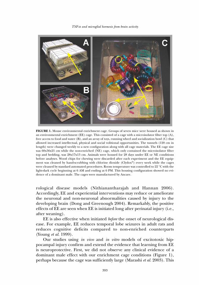
Our studies using in vivo and in vitro models of excitotoxic hip-pocampal injury confirm and extend the evidence that learning from EEis neuroprotective. First, we did not observe any clinical evidence of adominant male effect with our enrichment cage conditions (Figure 1),perhaps because the cage was sufficiently large (Marashi et al. 2003). This
TNF-α and microglial hormesis from brain activity
393
FIGURE 1. Mouse environmental enrichment cage. Groups of seven mice were housed as shown inan environmental enrichment (EE) cage. This consisted of a cage with a microisolator filter top (A),free access to food and water (B), and an array of toys, running wheel and socialization bowl (C) thatallowed increased intellectual, physical and social volitional opportunities. The tunnels (120 cm inlength) were changed weekly to a new configuration along with all cage materials. The EE cage sizewas 60x56x21 cm while the non-enriched (NE) cage, which only contained the microisolator filtertop and bedding, was 28x17x13 cm. Animals were housed for 28 days under EE or NE conditionsbefore analyses. Wood chips for chewing were discarded after each experiment and the EE equip-ment was cleaned by hand-scrubbing with chlorine dioxide (Clydox®) every week while the cageswere cleaned by standard automated procedures. Room temperature was controlled to 22 °C with thelight-dark cycle beginning at 6 AM and ending at 6 PM. This housing configuration showed no evi-dence of a dominant male. The cages were manufactured by Ancare.
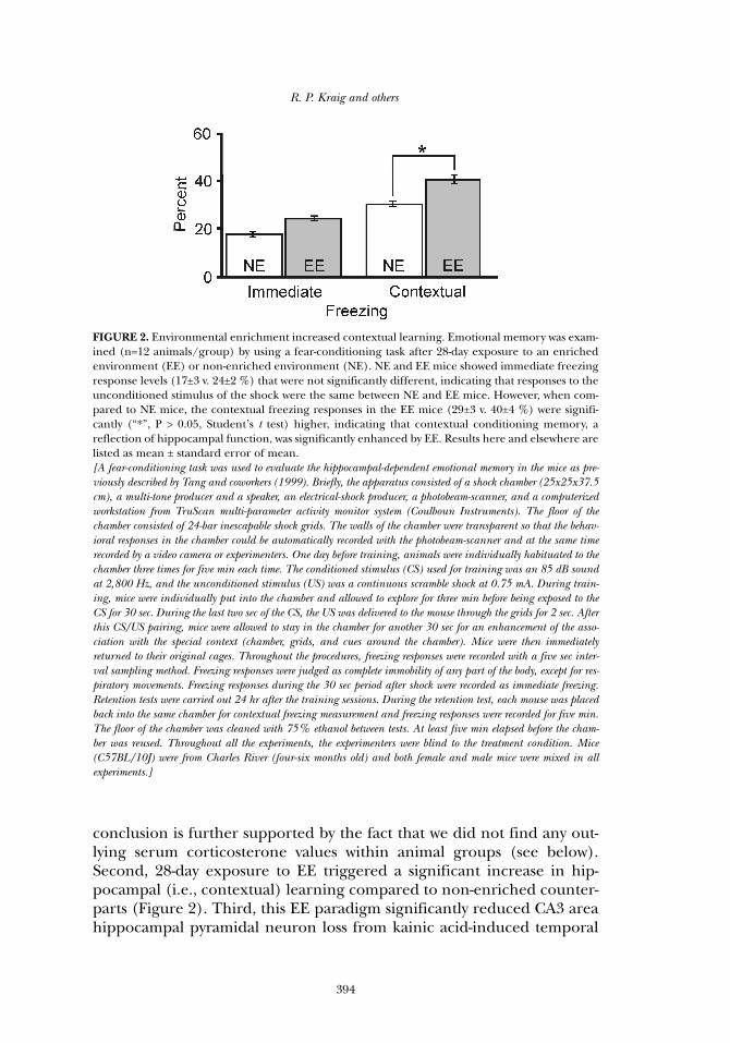
conclusion is further supported by the fact that we did not find any out-lying serum corticosterone values within animal groups (see below).Second, 28-day exposure to EE triggered a significant increase in hip-pocampal (i.e., contextual) learning compared to non-enriched counter-parts (Figure 2). Third, this EE paradigm significantly reduced CA3 areahippocampal pyramidal neuron loss from kainic acid-induced temporal
R. P. Kraig and others
394
FIGURE 2. Environmental enrichment increased contextual learning. Emotional memory was exam-ined (n=12 animals/group) by using a fear-conditioning task after 28-day exposure to an enrichedenvironment (EE) or non-enriched environment (NE). NE and EE mice showed immediate freezingresponse levels (17±3 v. 24±2 %) that were not significantly different, indicating that responses to theunconditioned stimulus of the shock were the same between NE and EE mice. However, when com-pared to NE mice, the contextual freezing responses in the EE mice (29±3 v. 40±4 %) were signifi-cantly (“*”, P > 0.05, Student’s t test) higher, indicating that contextual conditioning memory, areflection of hippocampal function, was significantly enhanced by EE. Results here and elsewhere arelisted as mean ± standard error of mean. [A fear-conditioning task was used to evaluate the hippocampal-dependent emotional memory in the mice as pre-viously described by Tang and coworkers (1999). Briefly, the apparatus consisted of a shock chamber (25x25x37.5cm), a multi-tone producer and a speaker, an electrical-shock producer, a photobeam-scanner, and a computerizedworkstation from TruScan multi-parameter activity monitor system (Coulboun Instruments). The floor of thechamber consisted of 24-bar inescapable shock grids. The walls of the chamber were transparent so that the behav-ioral responses in the chamber could be automatically recorded with the photobeam-scanner and at the same timerecorded by a video camera or experimenters. One day before training, animals were individually habituated to thechamber three times for five min each time. The conditioned stimulus (CS) used for training was an 85 dB soundat 2,800 Hz, and the unconditioned stimulus (US) was a continuous scramble shock at 0.75 mA. During train-ing, mice were individually put into the chamber and allowed to explore for three min before being exposed to theCS for 30 sec. During the last two sec of the CS, the US was delivered to the mouse through the grids for 2 sec. Afterthis CS/US pairing, mice were allowed to stay in the chamber for another 30 sec for an enhancement of the asso-ciation with the special context (chamber, grids, and cues around the chamber). Mice were then immediatelyreturned to their original cages. Throughout the procedures, freezing responses were recorded with a five sec inter-val sampling method. Freezing responses were judged as complete immobility of any part of the body, except for res-piratory movements. Freezing responses during the 30 sec period after shock were recorded as immediate freezing.Retention tests were carried out 24 hr after the training sessions. During the retention test, each mouse was placedback into the same chamber for contextual freezing measurement and freezing responses were recorded for five min.The floor of the chamber was cleaned with 75% ethanol between tests. At least five min elapsed before the cham-ber was reused. Throughout all the experiments, the experimenters were blind to the treatment condition. Mice(C57BL/10J) were from Charles River (four-six months old) and both female and male mice were mixed in allexperiments.]

lobe epilepsy, and significantly improved animal survival compared tonon-enriched counterparts (Figure 3).
Initially, serum corticosterone levels were measured to probe forpotential stress reaction from exposure to EE. Our results indicate whatmay be an important signaling pattern associated with increased learningfrom EE. We, like others (Kempermann et al. 2002), found that morning
TNF-α and microglial hormesis from brain activity
395
FIGURE 3. Environmental enrichment protected hippocampus against excitotoxic injury. A well-established model of temporal lobe epilepsy (TLE) was used to determine whether an enriched envi-ronment (EE) evoked hippocampal neuroprotection compared to a non-enriched environment(NE). (A) Representative Fluoro-Jade images show caudal CA3 area hippocampal pyramidal neurondeath (arrow) four days after kainic acid injections in NE compared to EE mice. (B) Quantificationof neurons lost (n=5/group; white) as well as number of surviving animals (n=28/group; gray)showed that EE triggered a significant (“*”, P < 0.05, Student’s t test) reduction in the number of neu-rons lost and a significant (P<0.01) increase in the percentage of surviving animals. Specific valueswere 117±17 v. 23±16 neurons lost and 45±8 v. 77±6 % surviving mice (Kraig et al. 2004). [TLE was induced by a subcutaneous injection of freshly prepared kainic acid (30 mg/kg (Calbiochem) dissolvedin phosphate buffered saline (PBS; at 1mg/mL; Avanzini et al. 1998)) in C57BL/10J mice. Sham controlsreceived similar volume subcutaneous injections of PBS. Animals were briefly restrained in a holding device forinjections. After injections, animals were placed in individual cages without lids for continuous observation.Seizure severity was quantitated (every 20 min during continuous monitoring) for the next four hrs according toa scale proposed by Racine (1972) and modified to include death (Hulse et al. 2004). This rating was used toensure that all animals included for study reached the same general level of seizure severity. Those that did notwere excluded from study. In the event that mice experienced continuous seizures for 20 min, they would have beeneuthanized. No animals had to be removed or euthanized in this study. At the conclusion of observation periods,animals were kept in individual cages with standard housing until their brains were serially sectioned (40 µm)through the hippocampus. TLE injury was measured using Fluoro-Jade staining (Schmued and Hopkins 2000)to count dying neurons using stereological techniques (Howard and Reed 1998; West 1999) for the CA3 area ofhippocampus (from sections taken every 240 µm).]
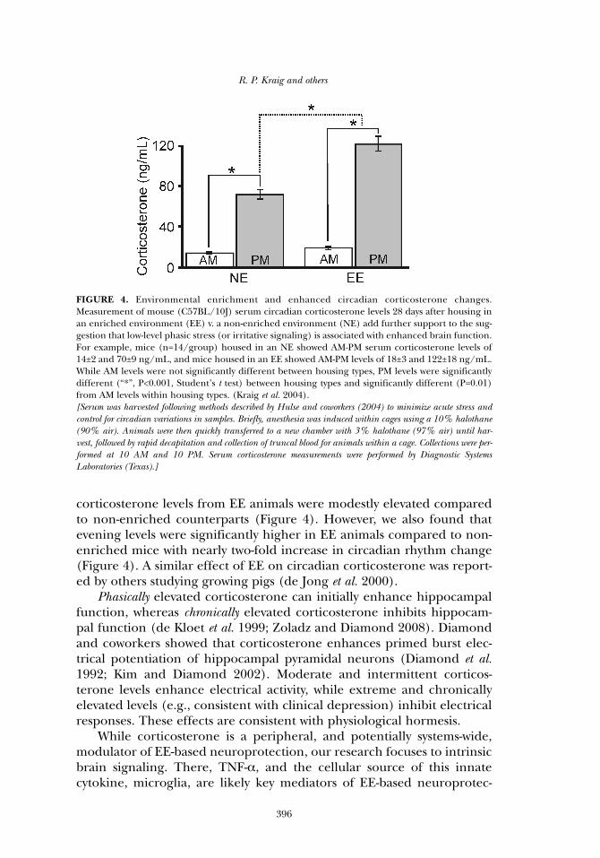
corticosterone levels from EE animals were modestly elevated comparedto non-enriched counterparts (Figure 4). However, we also found thatevening levels were significantly higher in EE animals compared to non-enriched mice with nearly two-fold increase in circadian rhythm change(Figure 4). A similar effect of EE on circadian corticosterone was report-ed by others studying growing pigs (de Jong et al. 2000).
Phasically elevated corticosterone can initially enhance hippocampalfunction, whereas chronically elevated corticosterone inhibits hippocam-pal function (de Kloet et al. 1999; Zoladz and Diamond 2008). Diamondand coworkers showed that corticosterone enhances primed burst elec-trical potentiation of hippocampal pyramidal neurons (Diamond et al.1992; Kim and Diamond 2002). Moderate and intermittent corticos-terone levels enhance electrical activity, while extreme and chronicallyelevated levels (e.g., consistent with clinical depression) inhibit electricalresponses. These effects are consistent with physiological hormesis.
While corticosterone is a peripheral, and potentially systems-wide,modulator of EE-based neuroprotection, our research focuses to intrinsicbrain signaling. There, TNF-α, and the cellular source of this innatecytokine, microglia, are likely key mediators of EE-based neuroprotec-
R. P. Kraig and others
396
FIGURE 4. Environmental enrichment and enhanced circadian corticosterone changes.Measurement of mouse (C57BL/10J) serum circadian corticosterone levels 28 days after housing inan enriched environment (EE) v. a non-enriched environment (NE) add further support to the sug-gestion that low-level phasic stress (or irritative signaling) is associated with enhanced brain function.For example, mice (n=14/group) housed in an NE showed AM-PM serum corticosterone levels of14±2 and 70±9 ng/mL, and mice housed in an EE showed AM-PM levels of 18±3 and 122±18 ng/mL.While AM levels were not significantly different between housing types, PM levels were significantlydifferent (“*”, P<0.001, Student’s t test) between housing types and significantly different (P=0.01)from AM levels within housing types. (Kraig et al. 2004). [Serum was harvested following methods described by Hulse and coworkers (2004) to minimize acute stress andcontrol for circadian variations in samples. Briefly, anesthesia was induced within cages using a 10% halothane(90% air). Animals were then quickly transferred to a new chamber with 3% halothane (97% air) until har-vest, followed by rapid decapitation and collection of truncal blood for animals within a cage. Collections were per-formed at 10 AM and 10 PM. Serum corticosterone measurements were performed by Diagnostic SystemsLaboratories (Texas).]
tion. This focus stems from data indicating that learning from EE initiatesneuroprotective and hormetic TNF-α signaling from microglia.
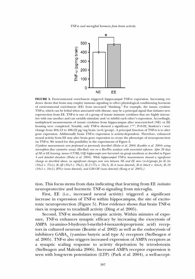
First, EE (i.e., increased neural activity) triggered a significantincrease in expression of TNF-α within hippocampus, the site of excito-toxic neuroprotection (Figure 5). Prior evidence shows that brain TNF-αrises in response to treadmill activity (Ding et al. 2005).
Second, TNF-α modulates synaptic activity. Within minutes of expo-sure, TNF-α enhances synaptic efficacy by increasing the exocytosis ofAMPA (α-amino-3-hydroxy-5-methyl-4-isoxazoleproprionic acid) recep-tors in cultured neurons (Beattie et al. 2002) as well as the endocytosis ofinhibitory GABAA (γ-amino butyric acid type A) receptors (Stellwagen etal. 2005). TNF-α also triggers increased expression of AMPA receptors asa synaptic scaling response to activity deprivation by tetrodotoxin(Stellwagen and Malenka 2006). Increased AMPA receptor expression isseen with long-term potentiation (LTP) (Park et al. 2004), a well-accept-
TNF-α and microglial hormesis from brain activity
397
FIGURE 5. Environmental enrichment triggered hippocampal TNF-α expression. Increasing evi-dence shows that brain may employ immune signaling to effect physiological conditioning hormesisof environmental enrichment (EE) from increased “thinking.” For example, the innate cytokineTNF-α, which can be lethal when associated with disease, may be a principal signal that initiates neu-roprotection from EE. TNF-α is one of a group of innate immune cytokines that are highly interac-tive with one another and can variably stimulate and/or inhibit each other’s expression. Accordingly,multiplexed measurements of innate cytokines from hippocampus after non-enriched (NE) or EEhousing were completed. Notably, only TNF-α showed a significant (“*”, P<0.02, Student’s t test)change from 404±12 to 488±22 pg/mg brain (n=6/group). A principal function of TNF-α is to altergene expression. Additionally brain TNF-α expression is activity-dependent. Therefore, enhancedneural activity from EE may alter brain gene expression to create the phenotype of neuroprotectionvia TNF-α. We tested for this possibility in the experiments of Figure 6. [Cytokine measurements were performed as previously described (Hulse et al. 2004; Kunkler et al. 2004) usingmicrosphere flow cytometric assays (Bio-Rad) run on a Bio-Plex analyzer with associated software. After 28 daysof NE or EE housing, mouse (C57BL/10J) hippocampi were harvested via group anesthesia as described in Figure4 and detailed elsewhere (Hulse et al. 2004). While hippocampal TNF-α measurements showed a significantchange as described above, no significant changes were seen between NE and EE mice (n=6/group) for IL-1α(34±2 v. 37±1), IL-1β (34±3 v. 39±1), IL-2 (77± v. 78±3), IL-4 (none detected), IL-6 (66±4 v. 64±4), IL-10(10±1 v. 10±1), IFN-γ (none detected), and GM-CSF (none detected) (Kraig et al. 2003).]
R. P. Kraig and others
398
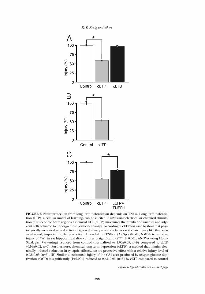
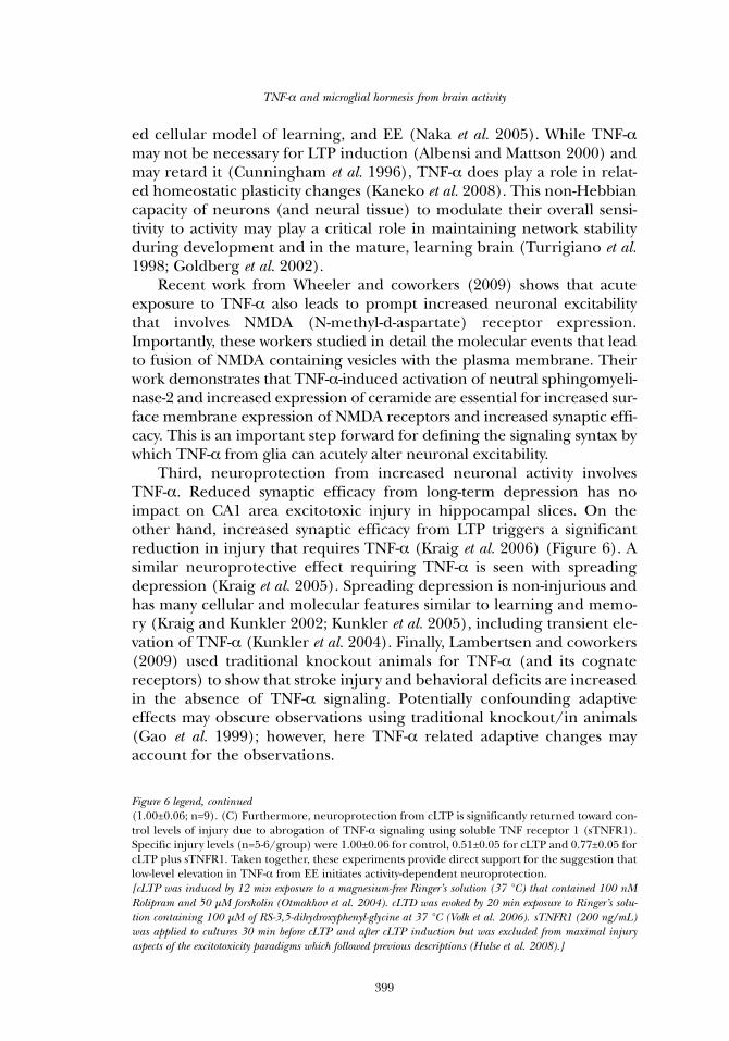
FIGURE 6. Neuroprotection from long-term potentiation depends on TNF-α. Long-term potentia-tion (LTP), a cellular model of learning, can be elicited in vitro using electrical or chemical stimula-tion of susceptible brain regions. Chemical LTP (cLTP) maximizes the number of synapses and adja-cent cells activated to undergo these plasticity changes. Accordingly, cLTP was used to show that phys-iologically increased neural activity triggered neuroprotection from excitotoxic injury like that seenin vivo and, importantly, the protection depended on TNF-α. (A) Specifically, NMDA irreversibleinjury of CA1 in rat hippocampal slice cultures is significantly (“*”, P<0.001, ANOVA using Holm-Sidak post hoc testing) reduced from control (normalized to 1.00±0.05, n=9) compared to cLTP(0.59±0.02, n=6). Furthermore, chemical long-term depression (cLTD), a method that mimics elec-trically induced reduction in synaptic efficacy, has no protective effect with a relative injury level of0.95±0.05 (n=5). (B) Similarly, excitotoxic injury of the CA1 area produced by oxygen glucose dep-rivation (OGD) is significantly (P<0.001) reduced to 0.53±0.05 (n=6) by cLTP compared to control
Figure 6 legend continued on next page
TNF-α and microglial hormesis from brain activity
399
ed cellular model of learning, and EE (Naka et al. 2005). While TNF-αmay not be necessary for LTP induction (Albensi and Mattson 2000) andmay retard it (Cunningham et al. 1996), TNF-α does play a role in relat-ed homeostatic plasticity changes (Kaneko et al. 2008). This non-Hebbiancapacity of neurons (and neural tissue) to modulate their overall sensi-tivity to activity may play a critical role in maintaining network stabilityduring development and in the mature, learning brain (Turrigiano et al.1998; Goldberg et al. 2002).
Recent work from Wheeler and coworkers (2009) shows that acuteexposure to TNF-α also leads to prompt increased neuronal excitabilitythat involves NMDA (N-methyl-d-aspartate) receptor expression.Importantly, these workers studied in detail the molecular events that leadto fusion of NMDA containing vesicles with the plasma membrane. Theirwork demonstrates that TNF-α-induced activation of neutral sphingomyeli-nase-2 and increased expression of ceramide are essential for increased sur-face membrane expression of NMDA receptors and increased synaptic effi-cacy. This is an important step forward for defining the signaling syntax bywhich TNF-α from glia can acutely alter neuronal excitability.
Third, neuroprotection from increased neuronal activity involvesTNF-α. Reduced synaptic efficacy from long-term depression has noimpact on CA1 area excitotoxic injury in hippocampal slices. On theother hand, increased synaptic efficacy from LTP triggers a significantreduction in injury that requires TNF-α (Kraig et al. 2006) (Figure 6). Asimilar neuroprotective effect requiring TNF-α is seen with spreadingdepression (Kraig et al. 2005). Spreading depression is non-injurious andhas many cellular and molecular features similar to learning and memo-ry (Kraig and Kunkler 2002; Kunkler et al. 2005), including transient ele-vation of TNF-α (Kunkler et al. 2004). Finally, Lambertsen and coworkers(2009) used traditional knockout animals for TNF-α (and its cognatereceptors) to show that stroke injury and behavioral deficits are increasedin the absence of TNF-α signaling. Potentially confounding adaptiveeffects may obscure observations using traditional knockout/in animals(Gao et al. 1999); however, here TNF-α related adaptive changes mayaccount for the observations.
Figure 6 legend, continued(1.00±0.06; n=9). (C) Furthermore, neuroprotection from cLTP is significantly returned toward con-trol levels of injury due to abrogation of TNF-α signaling using soluble TNF receptor 1 (sTNFR1).Specific injury levels (n=5-6/group) were 1.00±0.06 for control, 0.51±0.05 for cLTP and 0.77±0.05 forcLTP plus sTNFR1. Taken together, these experiments provide direct support for the suggestion thatlow-level elevation in TNF-α from EE initiates activity-dependent neuroprotection. [cLTP was induced by 12 min exposure to a magnesium-free Ringer’s solution (37 °C) that contained 100 nMRolipram and 50 µM forskolin (Otmakhov et al. 2004). cLTD was evoked by 20 min exposure to Ringer’s solu-tion containing 100 µM of RS-3,5-dihydroxyphenyl-glycine at 37 °C (Volk et al. 2006). sTNFR1 (200 ng/mL)was applied to cultures 30 min before cLTP and after cLTP induction but was excluded from maximal injuryaspects of the excitotoxicity paradigms which followed previous descriptions (Hulse et al. 2008).]
R. P. Kraig and others
400
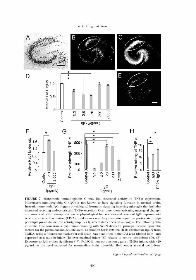
FIGURE 7. Monomeric immunoglobin G may link neuronal activity to TNF-α expression.Monomeric immunoglobin G (IgG) is not known to have signaling function in normal brain.Instead, monomeric IgG triggers physiological hormetic signaling involving microglia that includesincreased recycling endocytosis and TNF-α secretion. Over time, these activating microglial changesare associated with neuroprotection at physiological but not elevated levels of IgG. E-prostanoidreceptor subtype 2 activation (EP2A), used as an exemplary paracrine signal proportionate to hip-pocampal pyramidal neuron activity, amplifies IgG-mediated effects on microglia. The following dataillustrate these conclusions. (A) Immunostaining with NeuN shows the principal neuron cytoarchi-tecture for the pyramidal and dentate areas. Calibration bar is 250 µm. (B-D) Excitotoxic injury fromNMDA, using a fluorescent marker for cell death, was quantified in the CA1 area (dotted lines) andexpressed as a ratio in injury (B) over maximal injury (C) relative to control conditions (D). (E)Exposure to IgG evokes significant (“*”, P<0.001) neuroprotection against NMDA injury, with ~20µg/mL as the level expected for mammalian brain interstitial fluid under normal conditions.
Figure 7 legend continued on next page
Fourth, monomeric immunoglobin G (IgG) may help interlink neu-ronal activity to TNF-α production (Hulse et al. 2008) (Figure 7). EE acti-vates microglia to a pro-inflammatory state (Ziv et al. 2006). While allbrain cell types plus adjacent endothelial cells are capable of releasingTNF-α, microglia are recognized as the likely predominant source of thispro-inflammatory cytokine, especially in response to disease (Hopkinsand Rothwell, 1995; Allan and Rothwell, 2001). Microglia are also likelythe predominant, if not sole, source of TNF-α under physiological condi-tions including EE (Hulse et al. 2008). Monomeric IgG is commonlyregarded as a quiescent signaling molecule poised only to initiate poten-tially injurious inflammatory reactions in response to disease via immunecomplex formation and associated phagocytosis and TNF-α production.Instead, IgG signaling within non-injured brain results in neuroprotec-tion when it follows a physiological conditioning hormesis dose-responsepattern and takes time to develop. This IgG-mediated neuroprotectioninvolves enhanced microglial recycling endocytosis and TNF-α produc-tion—changes that are quickly evident after IgG exposure. Importantly,minocycline, known for its anti-inflammatory and neuroprotective effectswhen administered after the onset of brain disease, abrogates IgG-medi-ated neuroprotection and related microglial enhanced recycling endocy-tosis and TNF-α production. Furthermore, E-prostanoid receptor subtype2 activation by prostaglandin E2, which is released by hippocampalpyramidal neurons proportionate to their electrical activity, amplifiesIgG-mediated effects on microglia. This suggests paracrine mediatorsreleased from neurons, due to their increased activity, can affectmicroglial TNF-α release via amplification of recycling endocytosis. Theseresults confirm and extend evidence indicating that microglial pro-inflammatory activation over time is nutritive. Furthermore, they begin toillustrate specific immune signaling mechanisms by which neurons andmicroglia interact to enhance brain function.
Adaptive nature of activity-dependent neuroprotection
Neuroprotection from EE requires time to develop. This suggests thatEE, like other preconditioning stimuli (Barone et al. 1998; Nishio et al.2000), requires new protein synthesis. The predominant effect of TNF-αis to alter gene expression (Abbas and Lichtman 2003). Thus, TNF-α iswell positioned to orchestrate the activity-dependent structural and func-
TNF-α and microglial hormesis from brain activity
401
Figure 7 legend, continuedMicroglial recycling endocytosis (F) and TNF-α production (G) also respond to IgG in a physiologi-cal hormetic dose-response pattern. (H) Finally, neuroprotection from IgG is amplified by co-incu-bation of IgG with Butaprost, an EP2 receptor agonist, which acted as a surrogate of prostaglandinE2 release from increased neuronal activity. This amplification was removed by abrogation of TNF-αsignaling via sTNFR1 inclusion (not shown). Results adapted with permission from Hulse et al. 2008.
tional adaptation necessary for EE-based neuroprotection. However, theprecise nature of these adaptive processes and their relation to TNF-αand microglial activation remain undefined. Nonetheless, evidence from
R. P. Kraig and others
402
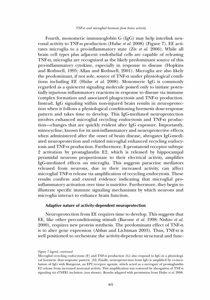
FIGURE 8. Sufficient and phasic signaling of activity-dependent neuroprotection. (A) Brain vitalityand neural activity (i.e., the degree and duration of brain cellular depolarization) follow a hormeticdose response relationship. At the extremes, severely reduced (e.g., coma) and excessive activity(e.g., stroke) are lethal. On the other hand, increased brain cell activity from environmental enrich-ment (EE) or long-term potentiation, a cellular model of learning, is maximally nutritive. Episodicspreading depression, the underlying cause of migraine, can be neuroprotective. However, whenspreading depression becomes too frequent, episodic migraine may be transformed into chronicmigraine because periods for adaptive signaling are too short. (B) The principal tenets of physio-logical conditioning hormesis are illustrated as the need for an initial irritative stimulus (1) whichover time results in a period of increased vitality via anabolic structural and functional changes (2).Repetitive phasic episodes of initiating stimuli (3) further enhance brain vitality (4). However, whenthe frequency of initiating stimuli precludes adequate periods of adaptation (5), maladaptive struc-tural and functional changes ensue that include the development of disease (e.g., chronic migraine).In the extreme (6), initiating stimuli are so overwhelming that associated irritative signals (i.e.,microglial activation and production of TNF-α and oxidants) only enhance neurodegeneration.
physical activity, dietary restriction, and ageing research provide provoca-tive clues.
Radak and colleagues (2008) review evidence that moderate physicalactivity improves general health by triggering adaptive responses to reac-tive oxygen species (ROS). They note that physical activity extremes (i.e.,inactivity and excessive activity) lead to deterioration in health, consistentwith the basic tenet of hormesis. Exercise must be sufficiently high to ini-tiate “stress” so that adaptive responses will occur. Importantly, exerciseneeds to be intermittent with sufficient periods of rest to allow for thedevelopment of adaptive responses associated with improved muscle(and organismal) health. We suggest this requirement not only applies tobrain and EE (Figure 8), but may also be involved in the maladaptiveeffects of excessively increased brain activity (see below). The authorspoint to literature (Gomez-Cabrera et al. 2005) indicating that ROS inhi-bition via exogenous scavengers abrogates the positive effects of physicalexercise, a result now extended to humans (Ristow et al. 2009).
Dietary restriction may also improve organismal health. Schulz andcoworkers (2007) show that glucose restriction increases the lifespan ofCaenorhabditis elegans. This caloric restriction paradigm triggers increasedROS formation, and importantly, increased ROS scavenging via height-ened catalase activity. Similar to physical activity, exogenous antioxidantsand vitamins that scavenge ROS may reduce the worm lifespan initiallyincreased by dietary restriction.
The Mattson laboratory emphasizes that these peripheral precondi-tioning strategies for exercise and dietary restriction hormesis involvingROS are likely to apply to the ageing brain (Arumugam et al. 2006). Thissuggestion is supported by data showing that NMDA receptor activationenhances neuronal antioxidant defenses (Papadia et al. 2008).
Modestly increased ROS production and the pronounced impact ofintrinsic antioxidants on neurological preventative health may be linked toupstream microglial activation that includes production of TNF-α and oxi-dants. Several lines of evidence support this suggestion. First, TNF-α stimu-lates glucose uptake into astrocytes (Yu et al. 1995; Véga et al. 2002), whichin turn provide high-energy substrates to neurons (Barros and Deitmer2009). Second, learning increases brain oxidative metabolism (Heiss et al.1992). Third, microglia are activated by synaptic activity (Ziv et al. 2006;Hulse et al. 2008) and activated microglia release ROS (Block et al. 2007;Innamorato et al. 2009), which can prompt increased generation of neuraltissue antioxidants. Fourth, oxidant stress plus antioxidant responses canmodulate synthesis of proteins involved in activity-dependent neuroprotec-tion, most likely via the “translational switch,” eukaryotic translational initi-ation factor 2α (Costa-Mattiolo et al. 2009; Tan et al. 2009).
Fifth, Radak and colleagues (2008) emphasize that not only mustexercise be sufficiently stressful, but adequate rest periods are also essen-
TNF-α and microglial hormesis from brain activity
403
tial to improve health. While sufficiently increased brain activity (i.e., EE)is necessary to initiate improved health, sleep may provide the essentialrest period required for adaptive changes. Vyazovskiy and colleagues(2008) note that while the wake state is associated with potentiation ofsynaptic function, sleep reverses this to depotentiation. This scenario isschematized in Figure 8. Importantly, although synaptic potentiation fallswith sleep, protein synthesis rises (Ramm and Smith, 1990). Seeminglyparadoxical to our overall thesis involving neural activity and TNF-α, hip-pocampal TNF-α mRNA rises with sleep (Cearley et al. 2003). However,TNF-α changes associated with LTP in the wake state may be localized toonly the synaptic regions activated by learning and thus changes wouldnot be detectable in whole brain regions used for the circadian mRNAmeasurements. Finally, brain catalase activity (and possibly the activitiesof other antioxidants which collectively can influence protein synthesis),shows phasic behavior (Sani et al. 2006).
In summary, mounting evidence indicates that physiologicallyincreased neural activity begets increased neuronal excitability via mech-anisms that include TNF-α from microglia (Figure 9). When thisincreased neural activity is sufficiently phasic, brain becomes moreresilient. In contrast, heightened neural activity and associated glial acti-vation, without adequate periods for adaptation, may leave brain moresusceptible to disease (see below).
MALADAPTIVE POTENTIAL OF ENHANCED BRAIN ACTIVITY
Considerations of brain physiological conditioning hormesis havelargely focused on the nourishing and restorative capacities of this sig-naling response pattern. However, examination of mechanisms by whichincreased brain activity can become maladaptive may help explain selectbrain diseases. For example, spreading depression is a non injurious andtransient perturbation of brain that, like EE, triggers microglial activation(Caggiano and Kraig 1996), increased TNF-α (Kunkler et al. 2004)(Figure 10), and neuroprotection that depends on TNF-α (Kraig et al.2005). Spreading depression is also the most likely underlying cause forepisodic migraine aura and pain (Moskowitz et al. 1993; Kunkler andKraig 2003; Lauritzen and Kraig 2005).
Reduced inhibitory synaptic function plays a key role in the mecha-nisms of spreading depression. Our work, and that from the Somjen lab-oratory (for review see Kunkler et al. 2005), show that a synchronousreduction in synaptic inhibitory drive occurring over a sufficiently largebrain volume initiates spreading depression (Figure 10). In addition, asspreading depression subsides, inhibitory synaptic function is last torecover. We speculate that as spreading depression (i.e., migraine)becomes more frequent, the lack of sufficient periods for compensatoryadaptive recovery leads to increased brain excitability from reduced inhi-
R. P. Kraig and others
404
bition, which may account for the transformation of episodic to chronicmigraine (Figure 8).
Chronic migraine is a prevalent healthcare burden (Lipton et al.2004) whose pathogenesis remains incompletely defined. Evidence sug-gests that chronic migraine occurs with central sensitization of sensorypathways that involves increased expression of pro-inflammatory media-tors and alterations in the periaqueductal gray (Aurora 2009). However,increased migraine frequency also correlates with the transformation ofepisodic migraine to chronic migraine (Silberstein and Olesen 2005),suggesting the frequency of spreading depression may be an initiatingcause of chronic migraine pain. Accordingly, central sensitization of sen-sory pathways and alterations of the periaqueductal gray may be “down-
TNF-α and microglial hormesis from brain activity
405
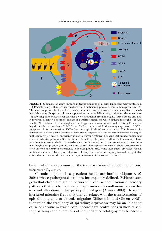
FIGURE 9. Schematic of neuro-immune initiating signaling of activity-dependent neuroprotection.(1) Physiologically enhanced neuronal activity, if sufficiently phasic, becomes neuroprotective. (2)This nutritive process begins with activity-dependent release of neuronal paracrine mediators includ-ing high energy phosphates, glutamate, potassium and especially prostaglandins, which can enhance(3) recycling endocytosis associated with TNF-α production from microglia. Astrocytes are also like-ly involved in activity-dependent release of paracrine mediators, which activate microglia. (4) As aresult, TNF-α released from microglia further triggers an increase in neuronal activity by (5) increas-ing the surface expression of NMDA and AMPA receptors while decreasing expression of GABAreceptors. (6) At the same time, TNF-α from microglia likely influence astrocytes. The choreographybetween this neuron-glial interactive behavior from heightened neuronal activity involves two impor-tant tenets. First, it must be sufficiently robust to trigger “irritative” signaling that initiates subsequentanabolic adaptive processes. Second, it must be sufficiently phasic to allow for homeostatic plasticprocesses to return activity levels toward normal. Furthermore, but in contrast to a return toward nor-mal, heightened physiological activity must be sufficiently phasic to allow anabolic processes suffi-cient time to build a stronger resilience to neurological disease. While these latter “processes” remainundefined, evidence from physical activity, dietary restriction, and ageing research suggest thatantioxidant defenses and anabolism in response to oxidant stress may be involved.
R. P. Kraig and others
406
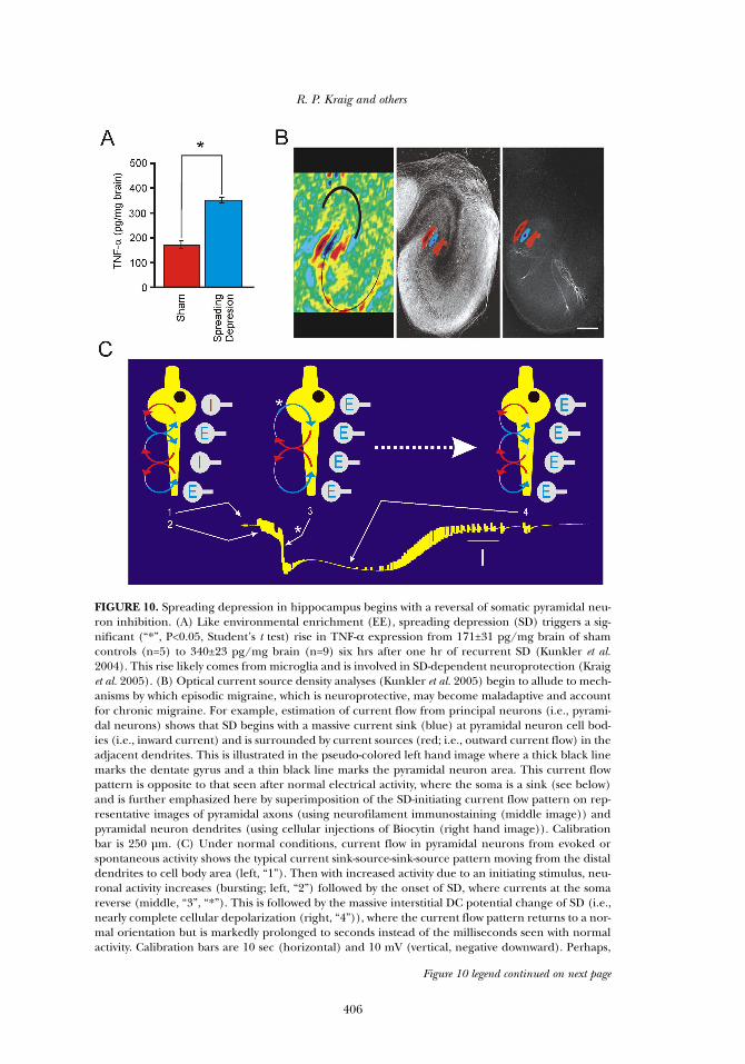
FIGURE 10. Spreading depression in hippocampus begins with a reversal of somatic pyramidal neu-ron inhibition. (A) Like environmental enrichment (EE), spreading depression (SD) triggers a sig-nificant (“*”, P<0.05, Student’s t test) rise in TNF-α expression from 171±31 pg/mg brain of shamcontrols (n=5) to 340±23 pg/mg brain (n=9) six hrs after one hr of recurrent SD (Kunkler et al.2004). This rise likely comes from microglia and is involved in SD-dependent neuroprotection (Kraiget al. 2005). (B) Optical current source density analyses (Kunkler et al. 2005) begin to allude to mech-anisms by which episodic migraine, which is neuroprotective, may become maladaptive and accountfor chronic migraine. For example, estimation of current flow from principal neurons (i.e., pyrami-dal neurons) shows that SD begins with a massive current sink (blue) at pyramidal neuron cell bod-ies (i.e., inward current) and is surrounded by current sources (red; i.e., outward current flow) in theadjacent dendrites. This is illustrated in the pseudo-colored left hand image where a thick black linemarks the dentate gyrus and a thin black line marks the pyramidal neuron area. This current flowpattern is opposite to that seen after normal electrical activity, where the soma is a sink (see below)and is further emphasized here by superimposition of the SD-initiating current flow pattern on rep-resentative images of pyramidal axons (using neurofilament immunostaining (middle image)) andpyramidal neuron dendrites (using cellular injections of Biocytin (right hand image)). Calibrationbar is 250 µm. (C) Under normal conditions, current flow in pyramidal neurons from evoked orspontaneous activity shows the typical current sink-source-sink-source pattern moving from the distaldendrites to cell body area (left, “1”). Then with increased activity due to an initiating stimulus, neu-ronal activity increases (bursting; left, “2”) followed by the onset of SD, where currents at the somareverse (middle, “3”, “*”). This is followed by the massive interstitial DC potential change of SD (i.e.,nearly complete cellular depolarization (right, “4”)), where the current flow pattern returns to a nor-mal orientation but is markedly prolonged to seconds instead of the milliseconds seen with normalactivity. Calibration bars are 10 sec (horizontal) and 10 mV (vertical, negative downward). Perhaps,
Figure 10 legend continued on next page
stream” signaling phenomena of chronic migraine while recurrentspreading depression is the “upstream” neural signaling causal change.
Spreading depression initiates pro-inflammatory changes. Astrocytes(Kraig et al. 1991) and microglia (Caggiano and Kraig 1996) show reac-tive changes for weeks after spreading depression. Importantly, spreadingdepression also triggers increased TNF-α production (Kunkler et al.2004) and microglial activation (Caggiano and Kraig 1996) which wouldinclude increased production of tissue oxidants. Together these factors,as well as release of other factors such as brain derived neurotrophic fac-tor from microglia (Coull et al. 2005), can reduce neural networkinhibitory synaptic drive, which would promote recurrent spreadingdepression (i.e., chronic migraine). Evidence supports this maladaptivepotential of physiological conditioning hormesis. Repetitive spreadingdepression triggers a selective suppression of inhibitory function (Krugeret al. 1996) and cortical hyperexcitability in migraineurs stems fromreduced inhibition (Palmer et al. 2000). Accordingly, microglial activationfrom increased brain activity that occurs without sufficient time to permitadequate adaptation, may initiate maladaptive consequences to brain.
SUMMARY
Physiological conditioning hormesis characterizes the signalingresponse patterns by which increased brain activity from EE generatesenhanced resilience to brain disease. Two basic tenets of hormesisrequire that initiating irritative, but not injurious, stimuli are sufficientlyrobust and that adequate periods of recovery from stressful initiatingstimuli occur to allow adequate time for adaptive processes to take effect.Microglia, because of their activation from neural activity and their asso-ciated production of TNF-α and oxidant irritants, are well-positioned toorchestrate hormetic immune signaling that establishes the phenotype ofneurological health and disease from brain activity.
ACKNOWLEDGEMENT
This work was supported by grants from the National Institute ofNeurological Disorders and Stroke (NS-19108), the National Institute ofChild Health and Human Disorders (5 PO1 HD 09402), the MigraineResearch Foundation and the White Foundation. BC-P was supported bya Campbell McConnell Fellowship from Cornell College. Ms. Marcia P.Kraig assisted in the preparation and maintenance of culture systems and
TNF-α and microglial hormesis from brain activity
407
Figure 10 legend, continuedwhen SD becomes too frequent, tonic somatic inhibition is enhanced by the lack of adaptation to ini-tiating irritative stimuli from microglia that include TNF-α and oxidants, which can reduce or reverseinhibitory synaptic drive. As a result, pyramidal neuron excitability is enhanced and brain becomesmore susceptible to SD, resulting in recurrent SD (i.e., chronic migraine). Optical current sourcedensity images were adapted with permission from Kunkler et al. 2005.
in the mouse enrichment studies. We thank Yelena Grinberg for readingand commenting on a final version of the manuscript.
REFERENCES
Abbas AK and Lichtman AH. 2003. Cellular and Molecular Immunology. Saunders, PhiladelphiaAggarwal BB, Samanta A, and Feldmann M. 2001. TNFα. In: Openheim JJ and Feldmann M (eds),
Cytokine Reference, Volume 1: Ligands, pp 413-434. Academic Press, San DiegoAlbensi BC and Mattson MP. 2000. Evidence for the involvement of TNF and NF B in hippocampal
synaptic plasticity. Synapse 35:151-159Allan SM and Rothwell NJ. 2001. Cytokines and acute neurodegeneration. Nat Rev Neurosci 2:734-
744Arumugam TV, Gleichmann M, Tang SC, and Mattson MP. 2006. Hormesis/preconditioning mech-
anisms, the nervous system and aging. Ageing Res Rev 5:165-78 Aurora SK. 2009. Spectrum of Illness: understanding biological patterns and relationships in chron-
ic migraine. Neurology 72: (Suppl. 1) S8-S13Avanzini G, Moshé SL, Schwartzkroin PA, Engel Jr J. 1998. Animal models of localization-related
epilepsy. In: Engel Jr J, Pedley TA (eds), Epilepsy: A Comprehensive Textbook, pp 271-310.Lippincott-Raven, Philadelphia
Barone FC, Arvin B, White RF, Miller A, Webb CL, Willette RN, Lysko PG, and Feuerstein GZ. 1997.Tumor necrosis factor-alpha. A mediator of focal ischemia brain injury. Stroke 28:1233-1244
Barone FC, White RF, Spera PA, Ellison J, Currie RW, Wang X, and Feuerstein GZ. 1998. Ischemicpreconditioning and brain tolerance: temporal histological and functional outcomes, proteinsynthesis requirement, and interleukin-1 receptor antagonist and early gene expression. Stroke29:1937-1950
Barros LF and Deitmer JW. 2009. Glucose and lactate supply to the synapse. Brain Res Revdoi:10.1016/ j.brainresrev.2009.10.002 .
Beattie EC, Stellwagen D, Morishita W, Bresnahan JC, Ha BK, Von Zastrow M, Beattie MS, andMalenka RC. 2002. Control of synaptic strength by glial TNFα. Science 295:2282-2285
Block ML, Zecca L, and Hong JS. 2007. Microglia-mediated neurotoxicity: uncovering the molecularmechanisms. Nat Rev Neurosci 8:57-69
Caggiano AO and Kraig RP. 1996. Eicosanoids & nitric oxide influence induction of reactive gliosisfrom spreading depression in microglia but not astrocytes. J Comp Neurol 369:93-108
Calabrese EJ. 2008. Drug therapies for stroke and traumatic brain injury often display U-shaped doseresponses: occurrence, mechanisms, and clinical implications. Crit Rev Toxicol 38:557-577
Calabrese EJ, Bachmann KA, Bailer AJ, Bolger PM, Borak J, Cai L, Cedergreen N, Cherian MG,Chiueh CC, Clarkson TW, Cook RR, Diamond DM, Doolittle DJ, Dorato MA, Duke SO,Feinendegen L, Gardner DE, Hart RW, Hastings KL, Hayes AW, Hoffmann GR, Ives JA,Jaworowski Z, Johnson TE, Jonas WB, Kaminski NE, Keller JG, Klaunig JE, Knudsen TB,Kozumbo WJ, Lettieri T, Liu SZ, Maisseu A, Maynard KI, Masoro EJ, McClellan RO, MehendaleHM, Mothersill C, Newlin DB, Nigg HN, Oehme FW, Phalen RF, Philbert MA, Rattan SI, RiviereJE, Rodricks J, Sapolsky RM, Scott BR, Seymour C, Sinclair DA, Smith-Sonneborn J, Snow ET,Spear L, Stevenson DE, Thomas Y, Tubiana M, Williams GM, and Mattson MP. 2007. Biologicalstress response terminology: Integrating the concepts of adaptive response and preconditioningstress within a hormetic dose-response framework. Toxicol Appl Pharmacol 222:122-128
Calabrese EJ and Baldwin LA. 2003. Hormesis: The dose-response revolution. Annu Rev PharmacolToxicol 43:175-197
Carson MJ, Bilousova TV, Puntambekar SS, Melchior B, Doose JM, and Ethell IM. 2007. A rose by anyother name? The potential consequences of microglial heterogeneity during CNS health anddisease. Neurotherapeutics 4:571-579
Cearley C, Churchill L, and Krueger JM. 2003. Time of day differences in IL1beta and TNFalphamRNA levels in specific regions of the rat brain. Neurosci Lett 352:61-63
Chao CC and Hu SS. 1994. Tumor necrosis factor-alpha potentiates glutamate neurotoxicity inhuman fetal brain cell cultures. Dev Neurosci 16:172-179
Chen H, Zhang SM, Schwarzschild MA, Hernán MA, and Ascherio A. 2005. Physical activity and therisk of Parkinson disease. Neurology 64:664-669
R. P. Kraig and others
408
Cheng B, Christakos S, and Mattson MP. 1994. Tumor necrosis factors protect neurons against meta-bolic-excitotoxic insults and promote maintenance of calcium homeostasis. Neuron 12:139-153
Costa-Mattioli M, Sossin WS, Klann E, and Sonenberg N. 2009. Translational control of long-lastingsynaptic plasticity and memory. Neuron 61:10-26
Coull J, Beggs S, Boudreau D, Boivin D, Tsuda M, Inoue K, Gravel C, Salter MW, and DeKoninck YD.2005. BDNF from microglia causes the shift in neuronal anion gradient underlying neuropath-ic pain. Nature 438:923-935
Cunningham AJ, Murray CA, O’Neill LAJ, Lynch MA, and O’Connor JJ. 1996. Interleukin-1β (IL-1β)and tumor necrosis factor (TNF) inhibit long-term potentiation in the rat dentate gyrus in vitro.Neurosci Lett 203:17-20
Dahlqvist P, Rönnback A, Bergstrom SA, Söderstrom I, and Olsson T. 2004. Environmental enrich-ment reverses learning impairment in the Morris water maze after focal cerebral ischemia inrats. Eur J Neurosci 19:2288-2298
Dawson DA, Martin D, and Hallenbeck JM. 1996. Inhibition of tumor necrosis factor-alpha reducesfocal cerebral ischemic injury in the spontaneously hypertensive rat. Neurosci Lett 218:41-44
de Jong IC, Prelle IT, van de Burgwal JA, Lambooij E, Korte SM, Blokhuis HJ, and Koolhaas JM. 2000.Effects of environmental enrichment on behavioral responses to novelty, learning and memory,and the circadian rhythm in cortisol in growing pigs. Physiol Behav 68:571-578
de Kloet ER, Oitzl MS, and Joëls M. 1999. Stress and cognition: are corticosteroids good or bad guys?Trends Neurosci 22:422-426
Diamond DM, Bennett MC, Fleshner M, and Rose GM. 1992. Inverted-U relationship between thelevel of peripheral corticosterone and the magnitude of hippocampal primed burst potentia-tion. Hippocampus 2:421-430
Ding YH, Young CN, Luan X, Li J, Rafols JA, Clark JC, McAllister JP, and Ding Y. 2005. Exercise pre-conditioning ameliorates inflammatory injury in ischemic rats during reperfusion. ActaNeuropathol 109:237-246
Dirnagl U, Simon RP, and Hallenbeck JM. 2003. Ischemic tolerance and endogenous neuroprotec-tion. Trends in Neurosci 26:248-254
Dirnagl U, Becker K, and Meisel A. 2009. Preconditioning and tolerance against cerebral ischaemia:from experimental strategies to clinical use. Lancet Neurology 8:398-412
Dong WK and Greenough WT. 2004. Plasticity of nonneuronal brain tissue: roles in developmentaldisorders. Mental Ret Dev Disabil Res Rev 10:85-90
Gao X, Kemper A, and Popko B. 1999. Advanced transgenic and gene-targeting approaches.Neurochem Res 24:1181-1188
Goldberg J, Holthoff K, and Yuste R. 2002. A problem with Hebb and local spikes. Trends Neurosci25:433-435
Gomez-Cabrera MC, Borrás C, Pallardó FV, Sastre J, Ji LL, and Viña J. 2005. Decreasing xanthine oxi-dase-mediated oxidative stress prevents useful cellular adaptations to exercise in rats. J Physiol567:113-120
Graca L and Waldmann H. 2006. Reprogramming the immune system using antibodies. MethodsMol Biol 333:247-268
Hadley C. 2003. What doesn’t kill you makes you stronger. EMBO Reports 4:924-926Hallenbeck JM. 2002. The many faces of tumor necrosis factor in stroke. Nat Med 8:1363-1368Hanisch UK and Kettenmann H. 2007. Microglia: active sensor and versatile effector cells in the nor-
mal and pathologic brain. Nat Neurosci 10:1387-1394Hannigan JH, O’Leary-Moore SK, and Berman RF. 2007. Postnatal environmental or experiential
amelioration of neurobehavioral effects of perinatal alcohol exposure in rats. NeurosciBioBehav Rev 31:202-211
Heiss WD, Pawlik G, Holthoff V, Kessler J, and Szelies B. 1992. PET correlates of normal andimpaired memory functions. Cerebrovasc Brain Metab Rev 4:1-27
Hopkins SJ and Rothwell NJ. 1995. Cytokines and the nervous system I: Expression and recognition.Trends Neurosci 18:83-88
Howard CV and Reed MG. 1998. Unbiased Stereology – Three Dimensional Measurement inMicroscopy. Stereology. BIOS Scientific Publishers, Oxford
Hulse RE, Kunkler PE, Fedynyshyn J, and Kraig RP. 2004. Optimization of multiplexed bead-basedcytokine immunoassays for rat serum and brain tissue. J Neurosci Methods 136:87-98
TNF-α and microglial hormesis from brain activity
409
Hulse RE, Swenson WG, Kunkler PE, White DM and Kraig RP. 2008. Monomeric IgG is neuropro-tective via enhancing microglial recycling endocytosis and TNF-alpha. J Neurosci 28:12199-12211
Innamorato NG, Lastres-Becker I, and Cuadrado A. 2009. Role of microglial redox balance in mod-ulation of neuroinflammation. Curr Opin Neurol 22:308-314
Kaneko M, Stellwagen D, Malenka RC, and Stryker MP. 2008. Tumor necrosis factor-α mediates onecomponent of competitive, experience-dependent plasticity in developing visual cortex.Neuron 58:673-680
Kempermann G, Gast D, and Gage FH. 2002. Neuroplasticity in old age: Sustained five-fold induc-tion of hippocampal neurogenesis by long-term environmental enrichment. Ann Neurol52:135-143
Kim JJ and Diamond DM. 2002. The stressed hippocampus, synaptic plasticity and lost memories. NatRev Neurosci 3:453-462
Kirino T. 2002. Ischemic tolerance. J Cereb Blood Flow Met 22:1283-1296Kraig RP, Dong LM, Thisted R, and Jaeger CB. 1991. Recurrent spreading depression increases
immunohistochemical evidence of glial fibrillary acidic protein. J Neurosci 11:2187-2198Kraig RP and Kunkler PE. 2002. Spreading depression - a teleological means for self-protection from
brain ischemia. In: Chan P (ed), Cerebrovascular Disease (22nd Princeton ResearchConference), pp 142-157. Cambridge University Press, New York
Kraig RP, Hulse RE, Kunkler PE, and Langan G. 2003. Environmental enrichment (EE) may be neu-roprotective by modulating synaptic activity (SA) via pro- & anti-inflammatory mediators (IMs).Soc Neurosci 29: Prog # 737.15
Kraig RP, Hulse RE, Kunkler PE, Nakajima A, and Tang Y. 2004. Increased learning and diurnal cor-ticosterone changes may trigger environmental enrichment (EE)-based neuroprotection (NP).Soc Neurosci 30: Prog #681.16
Kraig RP, Hulse RE and Kunkler PE. 2005. Spreading depression (SD)-induced neuroprotectiondepends on TNF-α. Soc Neurosci 31: Prog # 97.12
Kraig RP, Hulse RE, and Kunkler PE. 2006. LTP-induced neuroprotection depends on TNF-α. SocNeurosci 32: Prog #87.4
Kruger H, Luhmann HJ, and Heinemann U. 1996. Repetitive spreading depression causes selectivesuppression of GABAergic function. Neuroreport 7:2733-2736
Kunkler PE and Kraig RP. 2003. Hippocampal spreading depression bilaterally activates the caudaltrigeminal nucleus in rodents. Hippocampus 13:835-844
Kunkler PE, Hulse RE, and Kraig RP. 2004. Multiplexed cytokine protein expression profiles fromspreading depression in hippocampal organotypic cultures. J Cereb Blood Flow Metab 24:829-839
Kunkler PE, Hulse RE, Schmitt MW, Nicholson C, and Kraig RP. 2005. Optical current source densi-ty analysis in hippocampal organotypic culture shows that spreading depression occurs withuniquely reversing currents. J Neurosci 25:3952-3961
Lambertsen KL, Clausen BH, Babcock AA, Gregersen R, Fenger C, Nielsen HH, Haugaard LS,Wirenfeldt M, Nielsen M, Dagnaes-Hansen F, Bluethmann H, Faergeman NJ, Meldgaard M,Deierborg T, and Finsen B. 2009. Microglia protect neurons against ischemia by synthesis oftumor necrosis factor. J Neurosci 29:1319-1330
Lauritzen MC and Kraig RP. 2005. Spreading depression and migraine: In: Olesen J, Goadsby P,Ramadan N, Tfelt-Hansen Welch KMA (eds), The Headaches, 3rd Edition, pp 269-276.Lippincott-Raven, Philadelphia
Llinás RR. 2002. i Of The Vortex. MIT Press, Westwood, MA Lipton RB, Bigal ME, Steiner TJ, Silberstein SD, and Olesen J. 2004. Classification of primary
headaches. Neurology 63:427-435 Marashi V, Barnekow A, Ossendorf E, and Sachser N. 2003. Effects of different forms of environ-
mental enrichment on behavioral, endocrinological, and immunological parameters in malemice. Horm Behav 43:281-292
Marsh BJ, Williams-Karnesky RL, and Stenzel-Poore MP. 2009. Toll-like receptor signaling in endoge-nous neuroprotection and stroke. Neuroscience 158:1007-1020
Mattson MP, Chan SL, and Duan W. 2002. Modification of brain aging and neurodegenerative dis-orders by genes, diet, and behavior. Physiol Rev 82:637-672
Mattson MP. 2008a. Hormesis defined. Ageing Res Rev 7:1-7
R. P. Kraig and others
410
Mattson MP. 2008b. Hormesis and disease resistance: activation of cellular stress response pathways.Hum Exp Toxicol 27:155-162
Meistrell ME 3rd, Botchkina GI, Wang H, Di Santo E, Cockroft KM, Bloom O, Vishnubhakat JM,Ghezzi P, and Tracey KJ. 1997. Tumor necrosis factor alpha is a brain damaging cytokine in cere-bral ischemia. Shock 8:341-3348
Moskowitz M, Nozaki K, and Kraig RP. 1993. Neocortical spreading depression provokes the expres-sion of c-fos protein-like immunoreactivity with trigeminal nucleus caudalis via trigeminovascu-lar mechanisms. J Neurosci 13:1167-1177
Naka F, Narita N, Okado N, and Narita M. 2005. Modification of AMPA receptor properties follow-ing environmental enrichment. Brain & Dev 27:275-278
Nishio S, Yunoki M, Chen ZF, Anzivino M, and Lee KS. 2000. Ischemic tolerance in the rat neocor-tex following hypothermic preconditioning. J Neurosurg 93:845-851
Nithianantharajah J and Hannan AJ. 2006. Enriched environments, experience-dependent plasticityand disorders of the nervous system. Nat Rev Neurosci 7:697-709
Otmakhov N, Khibnik L, Otmakhova N, Carpenter S, Riahi S, Asrican B, and Lisman J. 2004.Forskolin-induced LTP in the CA1 hippocampal region is NMDA receptor dependent. JNeurophysiol 91:1955-1962
Palmer JE, Chronicle EP, Rolan P, and Mulleners WM. 2000. Cortical hyperexcitability is corticalunder-inhibition: evidence from a novel functional test of migraine patients. Cephalgia 20:525-532
Papadia S, Soriano FX, Léveillé F, Martel MA, Dakin KA, Hansen HH, Kaindl A, Sifringer M, FowlerJ, Stefovska V, McKenzie G, Craigon M, Corriveau R, Ghazal P, Horsburgh K, Yankner BA, WyllieDJ, Ikonomidou C, and Hardingham GE. 2008. Synaptic NMDA receptor activity boosts intrin-sic antioxidant defenses. Nat Neurosci 11:476-487
Park M, Penick EC, Edwards JG, Kauer KA, and Ehlers MD. 2004. Recycling endosomes supply AMPAreceptors for LTP. Science 305:1972-1975
Pereira LO, Arteni NS, Petersen RC, da Rocha AP, Achaval M, and Netto CA. 2007. Effects of dailyenvironmental enrichment on memory deficits and brain injury following neonatal hypoxia-ischemia in the rat. Neurobiol Learn Mem 87:101-108
Pereira LO, Strapasson AC, Nabinger PM, Achaval M, and Netto CA. 2008. Early enriched housingresults in partial recovery of memory deficits in female, but not in male rats after neonatalhypoxia-ischemia. Brain Res. 1218:257-266
Racine RJ. 1972. Modification of seizure activity by electrical stimulation. II. Motor seizure.Electroenceph Clin Neurophysiol 32:281-94
Radak Z, Chung HY, Koltai E, Taylor AW, and Goto S. 2008. Exercise, oxidative stress and hormesis.Ageing Res Rev 7:34-42
Ramm P and Smith CT. 1990. Rates of cerebral protein synthesis are linked to slow wave sleep in therat. Physiol Behav 48:749-753
Ristow M, Zarse K, Oberbach A, Kloting N, Birringer M, Kiehntopf M, Stumvoll M, Kahn CR, andBluher M. 2009. Antioxidants prevent health-promoting effects of physical exercise in humans.Proc Natl Acad Sci USA 106:8665-8670
Rovio S, Kåreholt I, Helkala EL, Viitanen M, Winblad B, Tuomilehto J, Soininen H, Nissinen A, andKivipelto M. 2005. Leisure-time physical activity at midlife and the risk of dementia andAlzheimer’s disease. Lancet Neurol 4:705-711
Salmina AB. 2009. Neuron-glia interactions as therapeutic targets in neurodegeneration. JAlzheimers Dis 16:485-502
Sani M, Sebai H, Gadacha W, Boughattas NA, Reinberg A, and Mossadok BA. 2006. Catalase activityand rhythmic patterns in mouse brain, kidney and liver. Comp Biochem Physiol B Biochem MolBiol 145:331-337
Schmued LC and Hopkins KJ. 2000. Fluoro-Jade B: a high affinity fluorescent marker for the local-ization of neuronal degeneration. Brain Res 874: 123-130
Schulz TJ, Zarse K, Voigt A, Urban N, Birringer M, and Ristow M. 2007. Glucose restriction extendsCaenorhabditis elegans life span by inducing mitochondrial respiration and increasing oxidativestress. Cell Met 6:280-293
Silberstein SD and Olesen J. 2005. Chronic migraines. In: Olesen J, Goadsby P, Ramadan N, Tfelt-Hansen, and Welch KMA (eds), The Headaches, 3rd Edition, pp 613-617. Lippincott-Raven,Philadelphia
Silverman PH. 2004. Rethinking genetic determinism. The Scientist 18:32-33
TNF-α and microglial hormesis from brain activity
411
Sriram K and O’Callaghan JP. 2007. Divergent roles for tumor necrosis factor-α in the brain. JNeuroimmune Pharmacol 2:140-153
Stellwagen D, Beattie EC, Seo JY, and Malenka RC. 2005. Differential regulation of AMPA receptorand GABA receptor trafficking by tumor necrosis factor alpha. J Neurosci 25:3219-3228
Stellwagen D and Malenka RC. 2006. Synaptic scaling mediated by glial TNF-alpha. Nature 440:1054-1059
Stoll G, Jander S, and Schroeter M. 2002. Detrimental and beneficial effects of injury-induced inflam-mation and cytokine expression in the nervous system. Adv Exp Med Biol 513:87-113
Streit WJ. 2005. Microglia and neuroprotection: implications for Alzheimer’s disease. Brain Res BrainRes Rev. 48:234-239
Streit WJ. 2006. Microglial senescence: does the brain’s immune system have an expiration date?Trends Neurosci 29:506-510
Sturman MT, Morris MC, Mendes de Leon CF, Bienias JL, Wilson RS, and Evans DA. 2005. Physicalactivity, cognitive activity, and cognitive decline in a biracial community population. ArchNeurol 62:1750-1754
Tan S, Somia N, Maher P, and Schubert D. 2009. Regulation of antioxidant metabolism by translationinitiation factor 2alpha. J Cell Biol 152:997-1006
Tang YP, Shimizu E, Dube GR, Rampon C, Kerchner GA, Zhuo M, Liu G, and Tsien JZ. 1999. Geneticenhancement of learning and memory in mice. Nature 401: 63-69
Thompson PD, Buchner D, Pina IL, Balady GJ, Williams MA, Marcus BH, Berra K, Blair SN, Costa F,Franklin B, Fletcher GF, Gordon NF, Pate RR, Rodriguez BL, Yancey AK, and Wenger NK. 2003.Exercise and physical activity in the prevention and treatment of atherosclerotic cardiovasculardisease: a statement from the Council on Clinical Cardiology (Subcommittee on Exercise,Rehabilitation, and Prevention) and the Council on Nutrition, Physical Activity, and Metabolism(Subcommittee on Physical Activity). Circulation 107:3109-3116
Turrigiano GG, Leslie KR, Desai NS, Rutherford LC, and Nelson SB. 1998. Activity-dependent scal-ing of quantal amplitude in neocortical neurons. Nature 391:892-896
Turrin NP and Rivest S. 2006. Molecular and cellular immune mediators of neuroprotection. MolNeurobiol 34:221-242
van Gelder BM, Tijhuis MA, Kalmijn S, Giampaoli S, Nissinen A, and Kromhout D. 2004. Physicalactivity in relation to cognitive decline in elderly men: the FINE Study. Neurology 63:2316-2321
van Praag H, Kempermann G, and Gage FH. 2000. Neural consequences of environmental enrich-ment. Nat Rev Neurosci 1:191-198
Véga C, Pellerin L, Dantzer R, and Magistretti PJ. 2002. Long-term modulation of glucose utilizationby IL-1 alpha and TNF-alpha in astrocytes: Na+ pump activity as a potential target via distinct sig-naling mechanisms. Glia 39:10-18
Volk LJ, Daly CA, and Huber KM. 2006. Differential roles for group 1 mGluR subtypes in inductionand expression of chemically induced hippocampal long-term depression. J Neurophysiol95:2427-2438
Vyazovskiy VV, Cirelli C, Pfister-Genskow M, Faraguna U, and Tononi G. 2008. Molecular and elec-trophysiological evidence for net synaptic potentiation in wake and depression in sleep. NatNeurosci 11:200-208
Wagner AK, Kline AE, Sokoloski J, Zafonte RD, Capulong E, and Dixon CE. 2002. Intervention withenvironmental enrichment after experimental brain trauma enhances cognitive recovery inmale but not female rats. Neurosci Lett 334:165-168
Waldmann H. 2002. Reprogramming the immune system. Immunol Rev 185:227-235Waldmann H, Adams E, and Cobbold S. 2008. Reprogramming the immune system: co-receptor
blockade as a paradigm for harnessing tolerance mechanisms. Immnol Rev 223:361-370West MJ. 1999. Stereological methods for estimating the total number of neurons and synapses: issues
of precision and bias. Trends Neurosci 22:51-61Wheeler D, Knapp E, Bandaru VVR, Wang Y, Knorr D, Poirier C, Mattson MP, Geiger JD, and
Haughey NJ. 2009. Tumor necrosis factor-α-induced neutral sphingomyelinase-2 modulatessynaptic plasticity by controlling the membrane insertion of NMDA receptors. J Neurochem109:1237-1249
Wilde GJC, Pringle AK, Sundstrom LE, Mann DA, and Ianotti F. 2000. Attenuation and augmenta-tion of ischaemia-related neuronal death by tumour necrosis factor-α in vitro. Eur J Neurosci12:3863-3870
R. P. Kraig and others
412
Will B, Galani R, Kelche C, and Rosenzweig MR. 2004. Recovery from brain injury in animals: rela-tive efficacy of environmental enrichment, physical exercise or formal training (1990-2002).Prog Brain Res 72:167-182
Young D, Lawlor PA, Leone P, Dragunow M, and During MJ. 1999. Environmental enrichmentinhibits spontaneous apoptosis, prevents seizures and is neuroprotective. Nat Med 5:448-453
Yu N, Maciejewski-Lenoir D, Bloom FE, and Magistretti PJ. 1995. Tumor necrosis factor-alpha andinterleukin-1 alpha enhance glucose utilization by astrocytes: involvement of phospholipase A2.Mol Pharmacol 48:550-558
Ziv Y, Ron N, Butovsky O, Landa G, Sudai E, Greenberg N, Cohen H, Kipnis J, and Schwartz M. 2006.Immune cells contribute to the maintenance of neurogenesis and spatial learning abilities inadulthood. Nat Neurosci 9:268-275
Zoladz P and Diamond DM. 2008. Linear and non-linear dose-response functions reveal a hormeticrelationship between stress and learning. Dose-Response 7:132-148
Zou JY and Crews FT. 2005. TNFα potentiates glutamate neurotoxicity by inhibiting glutamate uptakein organotypic brain slice cultures: neuroprotection mediated by NF B inhibition. Brain Res1034:11-24
TNF-α and microglial hormesis from brain activity
413