Título artículo / Títol article: Toward an Understanding ... · 59 to their use as an...
12
Título artículo / Títol article: Toward an Understanding of the Growth of Ag Filaments on α-Ag2WO4 and Their Photoluminescent Properties: A Combined Experimental and Theoretical Study Autores / Autors Longo, E. ; Volanti, Diogo P. ; Longo, V.M. ; Gracia Edo, Lourdes ; Nogueira, Içamira C. ; Almeida, M.A.P. ; Pinheiro, Antonio N. ; Ferrer, Mateus M. ; Cavalcante, Laécio S. ; Andrés Bort, Juan Revista: The Journal of Physical Chemistry C (2013) vol. 118, no 2 Versión / Versió: Postprint del autor Cita bibliográfica / Cita bibliogràfica (ISO 690): Longo, E., et al. Toward an Understanding of the Growth of Ag Filaments on α-Ag2WO4 and Their Photoluminescent Properties: A Combined Experimental and Theoretical Study). The Journal of Physical Chemistry A, 2013, vol. 118, no 2, p. 1229– 1239. url Repositori UJI: http://hdl.handle.net/10234/85730
Transcript of Título artículo / Títol article: Toward an Understanding ... · 59 to their use as an...

Título artículo / Títol article: Toward an Understanding of the Growth of Ag Filaments on α-Ag2WO4 and Their Photoluminescent Properties: A Combined Experimental and Theoretical Study
Autores / Autors Longo, E. ; Volanti, Diogo P. ; Longo, V.M. ; Gracia Edo, Lourdes ; Nogueira, Içamira C. ; Almeida, M.A.P. ; Pinheiro, Antonio N. ; Ferrer, Mateus M. ; Cavalcante, Laécio S. ; Andrés Bort, Juan
Revista: The Journal of Physical Chemistry C (2013) vol. 118, no 2
Versión / Versió: Postprint del autor
Cita bibliográfica / Cita bibliogràfica (ISO 690):
Longo, E., et al. Toward an Understanding of the Growth of Ag Filaments on α-Ag2WO4 and Their Photoluminescent Properties: A Combined Experimental and Theoretical Study). The Journal of Physical Chemistry A, 2013, vol. 118, no 2, p. 1229–1239.
url Repositori UJI: http://hdl.handle.net/10234/85730

1 Toward an Understanding of the Growth of Ag Filaments on2 α‑Ag2WO4 and their Photoluminescent Properties: A Combined3 Experimental and Theoretical Study4 Elson Longo,† Diogo P. Volanti,‡,* Valeria M. Longo,† Lourdes Gracia,§ Icamira C. Nogueira,⊥
5 Marcio A. P. Almeida,† Antonio N. Pinheiro,⊥ Mateus M. Ferrer,⊥ Laecio S. Cavalcante,∥
6 and Juan Andres§
7†Instituto de Química, UNESP−Universidade Estadual Paulista, R. Francisco Degni, 55, Araraquara, 14800-900, Brazil
8‡Departamento de Química e Ciencias Ambientais, UNESP−Universidade Estadual Paulista, R. Cristovao Colombo, 2265, Sao Jose
9 do Rio Preto, 15054-000, Brazil
10§Departament de Química Física i Analítica, UJI−Universitat Jaume I, Av. de Vicent Sos Baynat, s/n, Castello de la Plana, 12071,
11 Spain
12⊥Departamento de Química, UFSCar−Universidade Federal de Sao Carlos, Rod. Washington Luis, km 235, Sao Carlos, 13565-905,
13 Brazil
14∥Departamento de Química, UESPI−Universidade Estadual do Piauí, R. Joao Cabral, 2231, Teresina, 64002-150, Brazil
15 *S Supporting Information
16 ABSTRACT: A combined experimental and theoretical study17 was conducted on the structure and electronic properties of α-18 Ag2WO4 to clarify the nucleation and growth processes of Ag19 filaments on α-Ag2WO4 crystals induced by electron beam20 irradiation under electron microscopy. X-ray diffraction with21 Rietveld analysis, micro-Raman and Fourier-transform infrared22 spectroscopy were used to analyze the structural order/23 disorder of α-Ag2WO4 crystals. These complementary24 techniques indicated that the microwave-assisted hydrothermal25 method employed in the synthesis of α-Ag2WO4 crystals leads26 to the freezing of distorted [WO6] and [AgOy] (y = 2, 4, 6 and27 7) clusters as the constituent polyhedra of α-Ag2WO4. On the28 basis of the theoretical and experimental results, we provide a complete assignment of the structure of α-Ag2WO4 and describe29 the relationship among the disorder, nucleation growth, rate of Ag formation, and photoluminescence behavior before and after30 the irradiation of the accelerated electron beam. Density functional theory (DFT) studies indicated significant changes in the31 order−disorder of the initial α-Ag2WO4electronic structure, with a decrease in the band gap value from 3.55 to 2.72 eV. The first32 stages of the electron irradiation on α-Ag2WO4 crystal were investigated by DFT calculations, and we have derived a mechanism33 to describe the formation and growth of Ag filaments during the electronic excitation of the [AgO2] cluster.
1. INTRODUCTION
34 Nanoparticle growth mechanisms have received much attention35 in recent years; controlling the size and morphologies of36 nanostructures is important for technological application. In37 this context, in situ electron microscopy constitutes an elegant38 technique that uncovers dynamic processes in the growth of39 nanocrystals. Recent technological advancements, in conjunc-40 tion with high-resolution imaging, provide a new opportunity41 to view nanoscale processes. These advancements have been42 made possible as a result of the expansion and diffusion of43 transmission electron microscopy (TEM) heating holders for in44 situ electron microscopy.1−5 Several studies have reported using45 the TEM heating holder to monitor the crystal growth of46 different nanomaterials, such as bismuth,6 germanium,7
47 indium−arsenide,8 and vanadium oxide,9 and in situ liquid
48TEM has been employed to understand the growth process of49copper and lead sulfide nanostructures.10−12 The greatest50impact of this new method of investigating the stages of crystal51growth is the possibility to observe the step-by-step evolution52of the crystal at the nanoscale.13,14
53The preparation and characterization of noble metal54nanoparticles is an interdisciplinary subject and has attracted55much attention due to the fundamental and applied scientific56value of nanometer-sized metals.15−20 Among these metals, Ag57nanoparticles possess unique properties with a wide range of58applications, from surface-enhanced Raman spectroscopy21,22
Received: August 15, 2013Revised: November 17, 2013
Article
pubs.acs.org/JPCC
© XXXX American Chemical Society A dx.doi.org/10.1021/jp408167v | J. Phys. Chem. C XXXX, XXX, XXX−XXX
kbg00 | ACSJCA | JCA10.0.1465/W Unicode | research.3f (R3.6.i4 HF01:4180 | 2.0 alpha 39) 2013/10/21 02:46:00 | PROD-JCAVA | rq_3084185 | 12/31/2013 17:18:45 | 11 | JCA-DEFAULT

59 to their use as an antibacterial agent.23−29 Over the past decade,60 Ag nanocrystals of myriad shapes have been synthesized using61 various methods.18,30−36 In this context, an emerging trend in62 nanotechnology is the creation of new nanomaterials and the63 exploration of their novel physical and chemical proper-64 ties.37−39 Often, newly identified nanomaterials bring to light65 previously undiscovered phenomena. One example, which66 changed the direction of noble metal research, is the first real-67 time, in situ nucleation and growth of Ag filaments on α-68 Ag2WO4 crystals, driven by an accelerated electron beam from69 an electronic microscope under high vacuum.40
70 In the present paper, a combined experimental and71 theoretical study was conducted on the structural arrangement72 that leads to the interesting growth process of Ag filaments on73 the α-Ag2WO4 crystal surface induced by field-emission74 scanning electron microscopy (FESEM) and complementary75 transmission electron microscopy (TEM) with selected-area76 diffraction (SAD) characterization; the photoluminescence77 (PL) enhancement of the Ag filaments was also investigated.78 X-ray diffraction (XRD) with Rietveld refinement, micro-79 Raman (MR) spectroscopy and Fourier transform infrared80 spectroscopy (FTIR) were used to analyze the structural order81 and disorder conditions of the α-Ag2WO4 structure prior to the82 Ag growth. The shape evolution and growth process of the α-83 Ag2WO4 crystals synthesized using a microwave-assisted84 hydrothermal (MAH) method at different temperatures was85 analyzed. The first stages of the Ag formation on α-Ag2WO486 crystal provoked by electron irradiation were simulated by first-87 principles calculations based on density functional theory88 (DFT). The order−disorder structural conditions of the growth89 evolution and photoluminescence (PL) enhancement were90 inferred based on the theoretical results.
2. EXPERIMENTAL SECTION91 2.1. Synthesis of α-Ag2WO4. The typical α-Ag2WO4 crystal92 synthesis procedure was followed: 1 × 10⁻³ mol sodium tungstate93 dihydrate (Na2WO4·2H2O, 99.5% purity, Sigma-Aldrich) and 2 × 10⁻³
94 mol silver nitrate (AgNO3, 99.8% purity, Sigma-Aldrich) were95 separately dissolved in test tubes containing 50 mL deionized water.96 Before the addition of the salts, 0.5 g sodium dodecyl sulfate (SDS)97 (C12H25SO4Na, 99% purity, Sigma-Aldrich) was dissolved in both of98 the tubes. The 100-mL combined suspension was transferred into a99 fluorinated ethylene propylene (PTFE) autoclave without stirring. The100 autoclave was then sealed and placed in a microwave-aided device for101 hydrothermal synthesis.41 α-Ag2WO4 samples were prepared at102 different temperatures (100, 120, 140, and 160 °C) for 1 h. The α-103 Ag2WO4 crystals were obtained as light beige, fine powder. The104 precipitates were collected and washed several times with acetone and105 dried at room temperature for 6 h.106 2.2. Characterizations. The samples were characterized by XRD107 using a D/Max-2500PC diffractometer (Rigaku, Japan) with Cu Kα108 radiation (λ = 1.5406 Å) in the 2θ range from 10° to 70° in the normal109 routine with a scanning velocity of 2°/min and from 10° to 110° with110 a scanning velocity of 1°/min in the Rietveld routine, both with a step111 of 0.02°. MR measurements were recorded using a LabRAM HR 800112 mm model (Horiba, Jobin-Yvon, France). High-resolution Raman113 spectra were recorded with a He−Ne laser at 632.81 nm (model CCD114 DU420AOE325) operating at 25−1000 cm−1 and keeping its115 maximum output power at 6 mW. A 50-μm lens was used to prevent116 sample overheating. FTIR spectra were recorded from 250 to 1000117 cm−1 using KBr pellets and a Bomem-Michelson spectrophotometer in118 transmittance mode (model MB-102). UV−vis spectra were recorded119 using a Varian spectrophotometer (model Cary 5G) in diffuse120 reflectance mode. The shapes and sizes of the α-Ag2WO4 microcrystals121 were observed with a field-emission scanning electron microscope122 (model Inspect F50, FEI Company, Hillsboro, OR) operating at 10
123kV. TEM analyses were performed with a CM200-Philips microscope124operating at 200 kV. The structural characterization of the samples was125estimated using SAD. Specimens for TEM images were obtained by126drying droplets of as-prepared samples from an acetone dispersion that127had been sonicated for 10 min and deposited on 300-mesh Cu grids.128PL measurements were performed with a Monospec 27 mono-129chromator (Thermal Jarrel Ash) coupled to an R446 photomultiplier130(Hamamatsu Photonics, Japan). A krypton-ion laser (Coherent Innova13190K; λ = 350.7 nm) was used as the excitation source; its maximum132output power was maintained at 500 mW. The laser beam was passed133through an optical chopper, and its maximum power on the sample134was maintained at 40 mW. PL measurements were performed at room135temperature.1362.3. Theoretical Calculation. Calculations for α-Ag2WO4 crystal137were performed with a CRYSTAL09 program package.42,43 Tungsten138was described by a large-core ECP, derived by Hay and Wadt, and139modified by Cora et al.44 Silver and oxygen centers were described140using HAYWSC-311d31G and O (6-31d1G) basis sets, respectively,141which were taken from the Crystal Web site.45 Becke’s three-parameter142hybrid nonlocal exchange functional,46 was used in combination with a143Lee−Yang−Parr gradient-corrected correlation functional (B3LYP).47
144The diagonalization of the Fock matrix was performed at adequate k-145points grids in the reciprocal space. The thresholds controlling the146accuracy of the calculation of the Coulomb and exchange integrals147were set to 10−8 (ITOL1 to ITOL4) and 10−14 (ITOL5), and the148percent of Fock/Kohn−Sham matrices mixing was set to 40 (IPMIX =14940).42 The band structure and the density of states (DOS) projected150on atoms and orbitals of bulk α-Ag2WO4 were constructed along the151appropriate high-symmetry directions of the corresponding irreducible152Brillouin zone. To take into account the negative charged system, we153inserted two additional electrons in the Ag atom of the [AgO2]154clusters, and in order to simulate properly this electron excess an all-155electron basis set (9766-3114d1G),45 was used to describe this Ag156center.
3. RESULTS AND DISCUSSION
157 f1The XRD patterns (Figure 1) indicate that all prepared α-158Ag2WO4 crystals have an orthorhombic structure without any159deleterious phases and belong to the space group Pn2n, with a160C2v
10 symmetry.48 These crystals have sharp and well-defined
Figure 1. XRD patterns of α-Ag2WO4 microcrystals prepared at (a)100, (b) 120, (c) 140, and (d) 160 °C for 1 h by the MAH method.The vertical lines indicate the position and relative intensity of the datafrom ICSD No. 4165 for the α-Ag2WO4 phase.
The Journal of Physical Chemistry C Article
dx.doi.org/10.1021/jp408167v | J. Phys. Chem. C XXXX, XXX, XXX−XXXB
161 diffraction peaks, which indicate a structural order and162 crystallinity at long-range. However, it is difficult to confirm163 the existence of Ag nanoparticles in these crystals based on164 XRD measurements.49 Moreover, all diffraction peaks are in165 close agreement with the inorganic crystal structure database166 (ICSD) (No. 416525) and the literature.50 The Rietveld167 analysis also corroborates these results (Figures S1 andS2 and168 Tables S1−S5, Supporting Information).169 The MR spectra of the α-Ag2WO4 crystals synthesized using
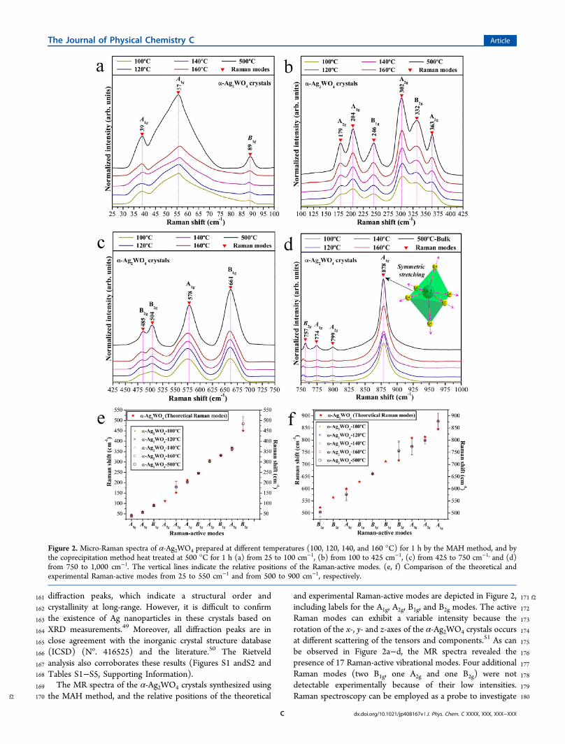
f2 170 the MAH method, and the relative positions of the theoretical
171 f2and experimental Raman-active modes are depicted in Figure 2,172including labels for the A1g, A2g, B1g, and B2g modes. The active173Raman modes can exhibit a variable intensity because the174rotation of the x-, y- and z-axes of the α-Ag2WO4 crystals occurs175at different scattering of the tensors and components.51 As can176be observed in Figure 2a−d, the MR spectra revealed the177presence of 17 Raman-active vibrational modes. Four additional178Raman modes (two B1g, one A2g and one B2g) were not179detectable experimentally because of their low intensities.180Raman spectroscopy can be employed as a probe to investigate
Figure 2. Micro-Raman spectra of α-Ag2WO4 prepared at different temperatures (100, 120, 140, and 160 °C) for 1 h by the MAH method, and bythe coprecipitation method heat treated at 500 °C for 1 h (a) from 25 to 100 cm−1, (b) from 100 to 425 cm−1, (c) from 425 to 750 cm−1, and (d)from 750 to 1,000 cm−1. The vertical lines indicate the relative positions of the Raman-active modes. (e, f) Comparison of the theoretical andexperimental Raman-active modes from 25 to 550 cm−1 and from 500 to 900 cm−1, respectively.
The Journal of Physical Chemistry C Article
dx.doi.org/10.1021/jp408167v | J. Phys. Chem. C XXXX, XXX, XXX−XXXC
181 the degree of structural order−disorder at short-range in the182 materials.52,53 Therefore, 17 well-defined Raman-active vibra-183 tional modes can be observed for typical α-Ag2WO4 crystals,184 indicating a high degree of short-range structural order in the185 lattice. However, this behavior was not observed in the α-186 Ag2WO4 crystals synthesized by the MAH method, particularly187 in the sample treated at 100 °C. It is notable that the MR188 spectra of the synthesized crystals exhibited broad vibrational189 modes, indicating structural disorder at short-range. In addition,190 the disorder increased with the temperature treatment. This191 characteristic can be related to the very rapid kinetics of the192 MAH synthetic conditions. The MR spectrum of the sample193 treated at 100 °C did not present well-defined Raman peaks194 due to major short-range structural disorder.195 Another interesting and important feature is the more196 pronounced structural local order presented by the lattice in the197 form of [WO6] clusters (see Figure 2d), as opposed to the198 lattice modifier assigned to [AgOy] (y = 7, 6, 4 and 2) clusters199 (Figure 2a−c). Specifically, the α-Ag2WO4 crystals prepared by200 the MAH method presented more well-defined Raman-active201 vibrational modes related to the symmetric stretching of (←202 O←W→O→) bonds of the octahedral [WO6] clusters than for203 the external vibrational modes of the distorted [AgOy] (y = 7, 6,204 4 and 2) clusters. The theoretical Raman-active modes were205 calculated through the atomic positions and lattice parameters206 for the optimized α-Ag2WO4 crystals and are illustrated in207 Figure 2e and f and presented in Table S6. There is good208 agreement among the Raman-active modes of the α-Ag2WO4209 crystals obtained in our samples, the first-principles calculation210 and the previously reported results from Turkovic et al.51 The211 two B1g, one A2g and one B2g modes that were not212 experimentally observed (Figure 2e,f) were predicted by the213 first-principles calculation, suggesting that their intensity may214 be too low in the Raman spectrum. The slight variations in the215 positions of the typical vibrational modes of our sample when216 compared with those reported in the literature can be attributed217 to the preparation method, average crystal size, distortions of218 the (O−Ag−O)/(O−W−O) bonds at short-range and/or219 intermolecular forces between the [AgOy]−[WO6]−[AgOy]220 clusters at intermediate range. Moreover, our theoretical221 calculations do not consider the nonharmonic contribution to222 the crystal-lattice vibration phonons.
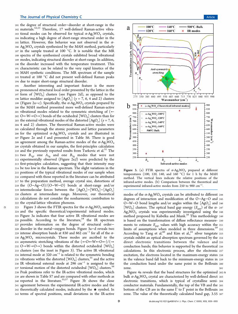
f3 223 Figure 3 shows the FTIR spectra for the α-Ag2WO4 samples224 and the specific theoretical/experimental infrared modes.225 Figure 3a indicates that four active IR vibrational modes are226 possible. According to the literature,54 the IR spectrum227 provides information on the degree of structural order−228 disorder in the metal−oxygen bonds. Figure 3a−d reveals two229 intense absorption bands at 830 and 862 cm−1 for all of the α-230 Ag2WO4 microcrystals. These modes are ascribed to the231 asymmetric stretching vibrations of the (←O←W←O←)/(→232 O→W→O→) bonds within the distorted octahedral [WO6]233 clusters (see the inset in Figure 3). The active IR vibrational234 internal mode at 320 cm−1 is related to the symmetric bending235 vibrations within the distorted [WO6] clusters,
55 and the active236 IR vibrational external mode at 296 cm−1 is assigned to the237 torsional motion of the distorted octahedral [WO6] clusters.
56
238 Peak positions refer to the IR-active vibrational modes, which239 are shown in Table S7 and are compared with other methods as240 reported in the literature.55,57 Figure 3b shows the close241 agreement between the experimental IR-active modes and the242 theoretically calculated modes, indicated by the ★ symbol. In243 terms of spectral positions, small deviations in the IR-active
244modes of the α-Ag2WO4 crystals can be attributed to different245degrees of interaction and modification of the O−Ag−O and246O−W−O bond lengths and/or angles within the [AgOy] and247[WO6] clusters. The optical band gap energy (Egap) of the α-248Ag2WO4 crystals was experimentally investigated using the249method proposed by Kubelka and Munk.58 This methodology250is based on the transformation of diffuse reflectance measure-251ments to estimate Egap values with high accuracy within the252limits of assumptions when modeled in three dimensions.59
253According to Tang et al.60 and Kim et al.,61 silver tungstate254crystals exhibit an optical absorption spectrum governed by the255direct electronic transitions between the valence and256conduction bands; this behavior is supported by the theoretical257calculations. In this electronic process, after the electronic258excitation, the electrons located in the maximum-energy states259in the valence band fall back to the minimum-energy states in260the conduction band under the same point in the Brillouin261zone.262 f4Figure 4a reveals that the band structures for the optimized263bulk α-Ag2WO4 crystal are characterized by well-defined direct264electronic transitions, which is typical of crystalline semi-265conductor materials. Fundamentally, the top of the VB and the266bottom of the CB are in the same Γ to Γ point in the Brillouin267zone. The value of the theoretically calculated band gap, 3.55
Figure 3. (a) FTIR spectra of α-Ag2WO4 prepared at differenttemperatures (100, 120, 140, and 160 °C) for 1 h by the MAHmethod. The vertical lines indicate the relative positions of theinfrared-active modes. (b) Comparison between the theoretical andexperimental infrared-active modes from 250 to 900 cm−1.
The Journal of Physical Chemistry C Article
dx.doi.org/10.1021/jp408167v | J. Phys. Chem. C XXXX, XXX, XXX−XXXD

268 eV, was slightly higher than the experimental value of ∼3.2 eV,269 estimated by UV−vis spectroscopy, for all samples synthesized270 by the MAH method. This overestimation of the predicted271 band gap can be due to the fact that it is calculated from the272 difference between the bottom of the conduction band (CB)273 and the top of the valence band (VB) within the Kohn−Sham274 formalism. However, it must be considered that the theoretical275 calculations estimate the optimized structure with a minimum276 energy, and the experimental structure derived from the MAH277 treatment conserves freezing distortions at short and278 intermediate range, which are not the most stable structures.279 Moreover, the band structure of this complicated network280 arrangement of [WO6] and [AgOy] clusters is basically281 determined by the W 5d orbitals in the conduction band282 (CB) and a valence band (VB) derived mostly from hybridized283 O 2p and Ag 4d orbitals. This reduction in the experimentally284 optical band gap value is most likely linked to distortions of the285 [WO6]/[AgOy] that are favorable to the formation of286 intermediate energy levels (photogenerated electron−hole287 pairs) between the VB and CB.288 To simulate the electron absorption process based on the α-289 Ag2WO4 structure, theoretical calculations were performed290 while taking into account the incorporation of two electrons in291 the structure. An analysis of the α-Ag2WO4 structure shows that292 the [AgO2] cluster presents the largest value of positive293 Mulliken charge for the Ag moiety, with only two adjacent294 oxygen anions. Therefore, this cluster was the most appropriate295 candidate to receive the external electron beam irradiation; in a
296sense, we inserted two additional electrons in the Ag atom of297the [AgO2] cluster as a starting point and atomic positions were298optimized. The resulting geometry is presented in Table S5b.299Figure 4b depicts the band structure for optimized neutral bulk300α-Ag2WO4 crystal charged with two electrons on the [AgO2]301clusters. An analysis of the band structure and the DOS (Figure302S3a) for the charged system points out that intermediate levels303are formed in the vicinity of the CB, which are composed of 5s304orbitals of [AgO2]. Therefore, new energy levels are created in305the forbidden region of the band gap, leading to a disordered306structure (Figure S3b). The Fermi level is now located at307approximately 3.0 eV, considering the VB maximum at the308zero-energy level, and the presence of these electron traps309reduces the band gap energy to 2.72 eV, becoming indirect310from the U point to the Γ point (Figure 4b, Figure S3).311 f5Figure 5 shows the DOS projected on the 4d, 5d, and 2p312orbitals of Ag, W and O atoms, respectively, for neutral α-313Ag2WO4. The DOS structure of this complex network314arrangement can be analyzed in terms of orbitals contribution315of the atoms that form [WO6] and [AgOy] (y = 7, 6, 4 and 2)316polyhedra. Figure 5a and b show that the projected DOS on the317orbitals of the Ag1 and Ag2 atoms, coordinated by seven318oxygens ([AgO7]), are basically derived from the 4dxz orbital of319the valence band. The same relationship occurs with the Ag3320atom, coordinated by six oxygens ([AgO6]), as depicted in321Figure 5c. When the coordination changes to four ([AgO4]), as322in the Ag4 and Ag5 atoms, the major contribution is derived323from the 4dxz and 4dz2 orbitals (see Figure 5d,e). Finally, in the324bicoordinated Ag6 atom ([AgO2]) the VB is mostly composed325by 4dxz orbital (see Figure 5f). The projected DOS on the W326atom is basically determined by the 5d orbitals in the327conduction band (CB) with more important role of 5dz2328orbitals (Figure 5g). The valence band (VB) is primarily329derived from hybridized O 2p (Figure 5h) and Ag 4d orbitals.330A study of the growth of Ag on the α-Ag2WO4 surface as a331function of exposure time to a scanning electron microscope332under an accelerating voltage of 10 kV was carried out. The333onset of Ag nanoparticle nuclei on the surface of the α-Ag2WO4334 f6crystals was observed by the FESEM images (Figure 6) as soon335as the samples began to be analyzed. This behavior was336observed for all the samples synthesized at different temper-337atures, namely, 100, 120, 140, and 160 °C (Figure 6, parts a, c, e338and g, respectively). After 5 min of irradiation, the growth of339the initial particles of Ag and the onset of new nuclei growth340were observed in all the samples (100, 120, 140, and 160 °C)341(Figure 6, parts b, d, f, and h, respectively). It is important to342emphasize that the sample prepared at 160 °C (Figure 6h) has343a higher number of Ag nuclei as well as a higher absorption of344existing particles. This behavior was also demonstrated in our345previous work.40 In this way, the most organized sample (160346°C) favors the nucleation of metallic Ag nanorods. However,347the growth process occurs preferentially in samples where the348nucleation is smaller. Table S9 (of the Supporting Information)349presents the calculated values of the surface energy for (001),350(100), (010), (011), (101) and (110) facets of α-Ag2WO4. The351surface (100) is the most stable facet, with the higher352percentage of the relaxing process. If charged α-Ag2WO4353structure is focused on the plane (100) compared to the354equilibrium geometry, it can be see an approaching of Ag4 and355Ag5 centers (from 4.0 to3.26 Å) when the system is charged in356the vicinity of Ag6 atoms. In addition, Ag6−O distance357increases from 2.34 to2.54 Å showing that this situation favors358an accumulation of Ag atoms along the most stable (100)
Figure 4. Band structures for optimized bulk α-Ag2WO4 crystal (a) inthe neutral state and (b) charged with two electrons on the [AgO2]clusters.
The Journal of Physical Chemistry C Article
dx.doi.org/10.1021/jp408167v | J. Phys. Chem. C XXXX, XXX, XXX−XXXE

359 surface. Therefore, the ab initio calculations indicate that the360 absorption of electrons leads to the disordered structure that361 facilitates Ag nucleation. It is possible that a more ordered362 structure has a more homogeneous surface and thus facilitates a363 more homogeneous nucleation; a more detailed study of this364 system is necessary to test this possibility. The TEM images of365 all samples and the structural electron diffraction (SAD) details366 of the growth process of Ag on the α-Ag2WO4 crystals are367 shown in Figures S4 and S5 and Table S8 in the Supporting368 Information.
369 f7Figure 7 shows the PL spectra recorded at room temperature370for the α-Ag2WO4 samples, excited by a 350.7 nm line of a371krypton ion laser, before and after irradiation by an accelerated372electron beam. The PL spectral profiles show typical behavior373for multiphonon or multilevel processes, i.e., a solid system374where relaxation occurs by several pathways, which involve the375participation of numerous energy states within the band gap.62
376It is generally assumed that the blue-green emission of377tungstate is due to the charge-transfer transitions within the378[WO4]
2‑ complex in ordered systems,63−67 or complex cluster379vacancies in the former,68−70 and/or modified lattice.70 It is
Figure 5. DOS projected on the 4d, 5d, and 2p orbitals of (a−f) Ag, (g) W, and (h) O atoms, respectively.
The Journal of Physical Chemistry C Article
dx.doi.org/10.1021/jp408167v | J. Phys. Chem. C XXXX, XXX, XXX−XXXF

380 well-known that the physical and chemical properties of381 materials are strongly correlated with structural factors,382 primarily the structural order−disorder in the lattice. The383 materials can be described in terms of the packing of the384 constituent clusters, which can be considered as the structural385 motifs. A specific feature of tungstates with a scheelite structure386 is the existence of [WO6] and [AgOy] clusters in a crystal387 lattice.71 This orthorhombic structure can also be understood in388 terms of a network of [WO6] clusters, linked by strong bonds389 [...W−O−W...] between the neighboring clusters, whose390 internal vibration spectra provide information on the structure391 and order−disorder effects in the crystal lattices.72,73 Breaking
392the symmetry of these clusters through distortions, breathings393and tilts creates a large number of different structures with394different material properties; this phenomenon can be related395to local (short), intermediate and long-range structural order−396disorder. Therefore, for α-Ag2WO4, the material properties can397be primarily associated with the constituent clusters, and the398disparity or mismatch of both clusters can induce structural399order−disorder effects, which significantly influence the400luminescence properties of the tungstates.74−76
401Disorder in materials can be manifested in many ways;402examples include vibrational, spin and orientation disorder403(referenced to a periodic lattice) and topological disorder.404Topological disorder is the type of disorder associated with405glassy and amorphous solid structures in which the structure406cannot be defined in terms of a periodic lattice. PL is a powerful407probe of certain aspects of short-range (2−5 Å) and medium-408range order (5−20 Å), such as clusters where the degree of409local order is such that structurally inequivalent sites can be410distinguished due to their different types of electronic411transitions which are linked to a specific structural arrange-412ment.62
413In Figure 7a, the maximum blue PL emission peak is414centered at 449 nm for all the samples; however, another415diffuse emission in the red region peaking from 621 to 640 nm416was also observed. The nucleation−dissolution−recrystalliza-417tion mechanism favored by the MAH process can be seen as an418order−disorder−order process of nature and gives rise to a419nonclassical crystallization process.77 Using density functional420calculations, Ghazi et al.78 noted that growth is an order−421disorder−order pattern of cyclic nature. Between two ordered422clusters, growth proceeds via disordered clusters, and global423order emerges suddenly with the addition of only one or two424atoms. In this sense, the different intensities in the emission425profiles can be attributed to slight differences in the defect426densities linked to the distorted clusters and complex vacancies427generated by the MAH heat treatment. The first emission peak428can be related to distorted [WO6] octahedra that are, in the429case of our samples, more ordered, in accordance with the MR430and IR spectral data and previous reports.63−67 The emission in431the red spectrum region is most likely linked to the [AgOy]432clusters that form complex vacancies, inducing more disorder433and deeper defects in the forbidden band gap. However, other434factors may also be involved, such as the degree of aggregation435and the orientation between particles, the variations in the436particle size distribution, the morphology of the particles and437surface defects. All these factors have an influence on the438intensity of the PL emission.439The PL profile of irradiated α-Ag2WO4 depicted in Figure 7b440is quite different from the nonirradiated PL profile (Figure 7a).441No blue shift is observed in the emission maximum, indicating442that the [WO6] clusters are unchanged by the irradiation443(Figure 7c,f). However, the profile of the PL emission in the444red region of the spectra is changed. As discussed above, these445changes can be attributed to the [AgOy] clusters becoming446more disordered by the Ag metallic growth on the surface. This447process generates complex vacancies of VAg′ and VO
z (where VOz
448= VOx , VO
• , VO••), but it is clear that the [WO6] surface clusters
449should also be slightly disordered as a result of Ag migration450and Ag nanorod formation. As predicted by first-principles451calculations, in the disordered structure the electronic levels are452significantly affected by the inclusion of electrons, as it is453observed in the band gap structure, favoring the red emission.454The samples prepared at 100 (Figure 7c), 120 (Figure 7d), and
Figure 6. Initial FESEM images of the α-Ag2WO4 samples obtained bythe MAH method at (a) 100, (c) 120, (e) 140 and (d) 160 °C. After 5min, microscopy analyses of the same samples were recorded: (b) 100,(d) 120, (f) 140, and (h) 160 °C. (Scale bar = 400 nm in parts a−f and200 nm in parts g and h.)
The Journal of Physical Chemistry C Article
dx.doi.org/10.1021/jp408167v | J. Phys. Chem. C XXXX, XXX, XXX−XXXG

455 140 °C (Figure 7c) show intermediate PL intensities. The456 maximum enhancement in PL emission after irradiation was457 observed in the sample heat-treated at 160 °C (Figure 7f). This458 sample presents the highest degree of structural order prior to459 irradiation, as discussed in the MR and FTIR analysis, and the460 highest nucleation rate after irradiation, as observed by FESEM461 (Figure 6h). The more abundant and homogeneous nucleation462 favored by the more ordered structure results in more VAg′ and463 VO
z complex vacancies and a larger effect on the PL464 enhancement.
4. CONCLUSIONS
465 In this study, α-Ag2WO4 particles were successfully synthesized466 by an MAH method; XRD patterns and Rietveld analysis467 confirmed the orthorhombic structure obtained. The physical/468 chemical properties and the corresponding performance of the469 α-Ag2WO4 crystals are closely related to the crystal structure,470 and in the present case, the local electric fields or polarized471 fields in the distorted metal−oxygen polyhedra, namely [WO6]
472and [AgOy] (y = 7, 6, 4, and 2). MR and FTIR spectroscopy473indicate that the MAH method employed in the synthesis of α-474Ag2WO4 crystals leads to the freezing of distorted [WO6] and475[AgOy] clusters. An external electron beam irradiation induces476the formation and crystal growth of Ag filaments on the α-477Ag2WO4 crystal and a PL enhancement. This finding is478reshaping our understanding of these molecular processes,479revealing previously hidden subtleties. A theoretical inves-480tigation using density functional theory (DFT) was carried out481to understand the introduction of electrons into the α-Ag2WO4
482lattice. The results indicate that the electron-induced growth483process of Ag on α-Ag2WO4 crystal is closely connected with484the structural and electronic properties of the [AgO2] cluster;485this process results in a drastic increase of the structural and486electronic disorder, as evidenced by the decrease in the band487gap from 3.55 to 2.72 eV. Finally, no blue shift of the emission488maximum was observed, indicating that the [WO6] clusters489were unchanged by the irradiation; however, changes were490observed in the red region of the PL profile. These changes
Figure 7. PL spectra recorded at room temperature of the α-Ag2WO4 crystals obtained by the MAH method at 100, 120, 140, and 160 °C, excited bya 350.7 nm line of a krypton ion laser (a) before and (b) after irradiation by an accelerated electron beam. For better visualization, we show thespectrum of each individual sample: (c) 100, (d) 120, (e) 140, and (f) 160 °C irradiated (red) and nonirradiated (blue).
The Journal of Physical Chemistry C Article
dx.doi.org/10.1021/jp408167v | J. Phys. Chem. C XXXX, XXX, XXX−XXXH
491 were attributed to unstable [AgOy] clusters that became492 disordered by the growth of metallic Ag on the surface, leading493 to complex vacancies. First-principles calculations predicted494 that this process would lead to a disordered structure with deep495 defects inserted in the band gap, favoring the red emission. The496 results of this research provide fundamental insight into the PL497 properties of α-Ag2WO4 crystals, the electron-induced synthesis498 of Ag/α-Ag2WO4 and its relationship with the morphology by499 controlling surface/bulk defects. We believe that this process500 may also be applicable for controlling other properties such as501 microbial activity and photodegradation.
502 ■ ASSOCIATED CONTENT503 *S Supporting Information504 Figures and tables of Rietveld refinement analyses of the α-505 Ag2WO4 crystal, DFT analysis. and TEM analysis with the506 corresponding SAD characterization. This material is available507 free of charge via the Internet at http://pubs.acs.org.
508 ■ AUTHOR INFORMATION509 Corresponding Author510 *E-mail: (D.P.V.) [email protected].
511 Notes512 The authors declare no competing financial interest.
513 ■ ACKNOWLEDGMENTS514 This work is financially supported by the National Council for515 Scientific and Technological Development (CNPq), Sao Paulo516 Research Foundation (FAPESP), Prometeo/2009/053 (Gen-517 eralitat Valenciana) and Ministerio de Economia y Compet-518 itividad (Spain), CTQ2012-36253-C03-02, and the Spanish−519 Brazilian program (PHB2009-0065-PC) for their financial520 support. TEM facilities were provided by LME-IQ-UNESP.
521 ■ REFERENCES(1)522 Kamino, T.; Sasaki, K.; Saka, H. High Resolution Electron
523 Microscopy in Situ Observation of Dynamic Behavior of Grain524 Boundaries and Interfaces at Very High Temperatures. Microsc.525 Microanal 1997, 3, 393−408.
(2)526 Saka, H.; Kamino, T.; Arai, S.; Sasaki, K. In Situ Heating527 Transmission Electron Microscopy. MRS Bull. 2008, 33, 93−100.
(3)528 Yaguchi, T.; Kanemura, T.; Shimizu, T.; Imamura, D.; Watabe,529 A.; Kamino, T. Development of a Technique for in Situ High530 Temperature Tem Observation of Catalysts in a Highly Moisturized531 Air Atmosphere. J. Electron Microsc. 2012, 61, 199−206.
(4)532 Parent, L. R.; Robinson, D. B.; Woehl, T. J.; Ristenpart, W. D.;533 Evans, J. E.; Browning, N. D.; Arslan, I. Direct in Situ Observation of534 Nanoparticle Synthesis in a Liquid Crystal Surfactant Template. ACS535 Nano 2012, 6, 3589−3596.
(5)536 Nam, S.-W.; Chung, H.-S.; Lo, Y. C.; Qi, L.; Li, J.; Lu, Y.;537 Johnson, A. T. C.; Jung, Y.; Nukala, P.; Agarwal, R. Electrical Wind538 Force-Driven and Dislocation-Templated Amorphization in Phase-539 Change Nanowires. Science 2012, 336, 1561−1566.
(6)540 Xin, H. L.; Zheng, H. In Situ Observation of Oscillatory Growth541 of Bismuth Nanoparticles. Nano Lett. 2012, 12, 1470−1474.
(7)542 Dayeh, S. A.; Picraux, S. T. Direct Observation of Nanoscale Size543 Effects in Ge Semiconductor Nanowire Growth. Nano Lett. 2010, 10,544 4032−4039.
(8)545 Mandl, B.; Stangl, J.; Hilner, E.; Zakharov, A. A.; Hillerich, K.;546 Dey, A. W.; Samuelson, L.; Bauer, G.; Deppert, K.; Mikkelsen, A.547 Growth Mechanism of Self-Catalyzed Group III-V Nanowires. Nano548 Lett. 2010, 10, 4443−4449.
(9)549 Sohn, J. I.; Joo, H. J.; Porter, A. E.; Choi, C.-J.; Kim, K.; Kang, D.550 J.; Welland, M. E. Direct Observation of the Structural Component of
551the Metal-Insulator Phase Transition and Growth Habits of Epitaxially552Grown VO2 Nanowires. Nano Lett. 2007, 7, 1570−1574.
(10) 553Radisic, A.; Ross, F. M.; Searson, P. C. In Situ Study of the554Growth Kinetics of Individual Island Electrodeposition of Copper. J.555Phys. Chem. B 2006, 110, 7862−7868.
(11) 556Williamson, M. J.; Tromp, R. M.; Vereecken, P. M.; Hull, R.;557Ross, F. M. Dynamic Microscopy of Nanoscale Cluster Growth at the558Solid-Liquid Interface. Nat. Mater. 2003, 2, 532−536.
(12) 559Evans, J. E.; Jungjohann, K. L.; Browning, N. D.; Arslan, I.560Controlled Growth of Nanoparticles from Solution with in Situ Liquid561Transmission Electron Microscopy. Nano Lett. 2011, 11, 2809−2813.
(13) 562Yuk, J. M.; Park, J.; Ercius, P.; Kim, K.; Hellebusch, D. J.;563Crommie, M. F.; Lee, J. Y.; Zettl, A.; Alivisatos, A. P. High-Resolution564Em of Colloidal Nanocrystal Growth Using Graphene Liquid Cells.565Science 2012, 336, 61−64.
(14) 566Park, J.; Zheng, H. M.; Lee, W. C.; Geissler, P. L.; Rabani, E.;567Alivisatos, A. P. Direct Observation of Nanoparticle Superlattice568Formation by Using Liquid Cell Transmission Electron Microscopy.569ACS Nano 2012, 6, 2078−2085.
(15) 570Burda, C.; Chen, X. B.; Narayanan, R.; El-Sayed, M. A.571Chemistry and Properties of Nanocrystals of Different Shapes. Chem.572Rev. 2005, 105, 1025−1102.
(16) 573Xia, Y.; Xiong, Y.; Lim, B.; Skrabalak, Sara E. Shape-Controlled574Synthesis of Metal Nanocrystals: Simple Chemistry Meets Complex575Physics? Angew. Chem., Int. Ed. 2009, 48, 60−103.
(17) 576Murphy, C. J.; Gole, A. M.; Stone, J. W.; Sisco, P. N.; Alkilany,577A. M.; Goldsmith, E. C.; Baxter, S. C. Gold Nanoparticles in Biology:578Beyond Toxicity to Cellular Imaging. Acc. Chem. Res. 2008, 41, 1721−5791730.
(18) 580Rycenga, M.; Cobley, C. M.; Zeng, J.; Li, W. Y.; Moran, C. H.;581Zhang, Q.; Qin, D.; Xia, Y. N. Controlling the Synthesis and Assembly582of Silver Nanostructures for Plasmonic Applications. Chem. Rev. 2011,583111, 3669−3712.
(19) 584Lim, B.; Jiang, M.; Camargo, P. H. C.; Cho, E. C.; Tao, J.; Lu,585X.; Zhu, Y.; Xia, Y. Pd-Pt Bimetallic Nanodendrites with High Activity586for Oxygen Reduction. Science 2009, 324, 1302−1305.
(20) 587Anker, J. N.; Hall, W. P.; Lyandres, O.; Shah, N. C.; Zhao, J.;588Van Duyne, R. P. Biosensing with Plasmonic Nanosensors. Nat. Mater.5892008, 7, 442−453.
(21) 590Hutter, E.; Fendler, J. H. Exploitation of Localized Surface591Plasmon Resonance. Adv. Mater. 2004, 16, 1685−1706.
(22) 592Hao, E.; Schatz, G. C. Electromagnetic Fields around Silver593Nanoparticles and Dimers. J. Chem. Phys. 2004, 120, 357−366.
(23) 594Liu, J.; Sonshine, D. A.; Shervani, S.; Hurt, R. H. Controlled595Release of Biologically Active Silver from Nanosilver Surfaces. ACS596Nano 2010, 4, 6903−6913.
(24) 597Gunawan, C.; Teoh, W. Y.; Marquis, C. P.; Lifia, J.; Amal, R.598Reversible Antimicrobial Photoswitching in Nanosilver. Small 2009, 5,599341−344.
(25) 600Kittler, S.; Greulich, C.; Diendorf, J.; Koeller, M.; Epple, M.601Toxicity of Silver Nanoparticles Increases During Storage Because of602Slow Dissolution under Release of Silver Ions. Chem. Mater. 2010, 22,6034548−4554.
(26) 604Sotiriou, G. A.; Teleki, A.; Camenzind, A.; Krumeich, F.; Meyer,605A.; Panke, S.; Pratsinis, S. E. Nanosilver on Nanostructured Silica:606Antibacterial Activity and Ag Surface Area. Chem. Eng. J. 2011, 170,607547−554.
(27) 608Christopher, P.; Xin, H.; Linic, S. Visible-Light-Enhanced609Catalytic Oxidation Reactions on Plasmonic Silver Nanostructures.610Nat. Chem. 2011, 3, 467−472.
(28) 611Kumar, A.; Vemula, P. K.; Ajayan, P. M.; John, G. Silver-612Nanoparticle-Embedded Antimicrobial Paints Based on Vegetable Oil.613Nat. Mater. 2008, 7, 236−241.
(29) 614Chernousova, S.; Epple, M. Silver as Antibacterial Agent: Ion,615Nanoparticle, and Metal. Angew. Chem., Int. Ed. 2013, 52, 1636−1653.
(30) 616Jin, R. C.; Cao, Y. W.; Mirkin, C. A.; Kelly, K. L.; Schatz, G. C.;617Zheng, J. G. Photoinduced Conversion of Silver Nanospheres to618Nanoprisms. Science 2001, 294, 1901−1903.
The Journal of Physical Chemistry C Article
dx.doi.org/10.1021/jp408167v | J. Phys. Chem. C XXXX, XXX, XXX−XXXI

(31)619 Jana, N. R.; Gearheart, L.; Murphy, C. J. Wet Chemical620 Synthesis of Silver Nanorods and Nanowires of Controllable Aspect621 Ratio. Chem. Commun. 2001, 617−618.
(32)622 Tao, A.; Sinsermsuksakul, P.; Yang, P. Polyhedral Silver623 Nanocrystals with Distinct Scattering Signatures. Angew. Chem., Int.624 Ed. 2006, 45, 4597−4601.
(33)625 Pietrobon, B.; Kitaev, V. Photochemical Synthesis of626 Monodisperse Size-Controlled Silver Decahedral Nanoparticles and627 Their Remarkable Optical Properties. Chem. Mater. 2008, 20, 5186−628 5190.
(34)629 Zhang, J.; Li, S.; Wu, J.; Schatz, G. C.; Mirkin, C. A. Plasmon-630 Mediated Synthesis of Silver Triangular Bipyramids. Angew. Chem., Int.631 Ed. 2009, 48, 7787−7791.
(35)632 Coskun, S.; Aksoy, B.; Unalan, H. E. Polyol Synthesis of Silver633 Nanowires: An Extensive Parametric Study. Cryst. Growth Des. 2011,634 11, 4963−4969.
(36)635 Lee, J.; Lee, P.; Lee, H.; Lee, D.; Lee, S. S.; Ko, S. H. Very Long636 Ag Nanowire Synthesis and Its Application in a Highly Transparent,637 Conductive and Flexible Metal Electrode Touch Panel. Nanoscale638 2012, 4, 6408−6414.
(37)639 Ariga, K.; Vinu, A.; Yamauchi, Y.; Ji, Q. M.; Hill, J. P.640 Nanoarchitectonics for Mesoporous Materials. Bull. Chem. Soc. Jpn.641 2012, 85, 1−32.
(38)642 Xiang, Q.; Yu, J.; Jaroniec, M. Graphene-Based Semiconductor643 Photocatalysts. Chem. Soc. Rev. 2012, 41, 782−796.
(39)644 Saha, K.; Agasti, S. S.; Kim, C.; Li, X.; Rotello, V. M. Gold645 Nanoparticles in Chemical and Biological Sensing. Chem. Rev. 2012,646 112, 2739−2779.
(40)647 Longo, E.; Cavalcante, L. S.; Volanti, D. P.; Gouveia, A. F.;648 Longo, V. M.; Varela, J. A.; Orlandi, M. O.; Andres, J. Direct in Situ649 Observation of the Electron-Driven Synthesis of Ag Filaments on α-650 Ag2WO4 Crystals. Sci. Rep. 2013, 3.
(41)651 Longo da Silva, E.; Arana Varela, J.; de Araujo Almeida, D. K.;652 Paschoalini Volanti, D. Microwave Aided Device for Hydrothermal653 Synthesis of Nanostructured Oxides, Particularly Obtaining Particles of654 Metal Oxides, Comprises Container, in which Hydrothermal Reaction655 Takes Place, and Lid for Container. BR200815393-A2, 07 Dec 2010.
(42)656 Dovesi, R.; Saunders, V. R. R. C.; Orlando, R.; Zicovich-Wilson,657 C. M.; Pascale, F.; Civalleri, B.; Doll, K.; Harrison, N. M.; Bush, I. J.;658 D’Arco, P.; Llunell, M., Crystal09 User’s Manual. University of Torino:659 Torino, Italy, 2009.
(43)660 Dovesi, R.; Orlando, R.; Civalleri, B.; Roetti, C.; Saunders, V. R.;661 Zicovich-Wilson, C. M. Crystal: A Computational Tool for the Ab662 Initio Study of the Electronic Properties of Crystals. Z. Kristallogr.663 2005, 220, 571−573.
(44)664 Cora, F.; Patel, A.; Harrison, N. M.; Dovesi, R.; Catlow, C. R. A.665 An Ab Initio Hartree-Fock Study of the Cubic and Tetragonal Phases666 of Bulk Tungsten Trioxide. J. Am. Chem. Soc. 1996, 118, 12174−667 12182.
(45)668 Pileni, M. P. Supracrystals of Inorganic Nanocrystals: An Open669 Challenge for New Physical Properties. Acc. Chem. Res. 2008, 41,670 1799−1809.
(46)671 Becke, A. D. Density-Functional Thermochemistry. 3. The Role672 of Exact Exchange. J. Chem. Phys. 1993, 98, 5648−5652.
(47)673 Lee, C. T.; Yang, W. T.; Parr, R. G. Development of the Colle-674 Salvetti Correlation-Energy Formula into a Functional of the Electron-675 Density. Phys. Rev. B 1988, 37, 785−789.
(48)676 Skarstad, P. M.; Geller, S. (W4O16)8- Polyion in High-677 Temperature Modification of Silver Tungstate. Mater. Res. Bull.678 1975, 10, 791−799.
(49)679 Wang, P.; Huang, B.; Qin, X.; Zhang, X.; Dai, Y.; Whangbo, M.-680 H. Ag/AgBr/WO3 Center Dot H2O: Visible-Light Photocatalyst for681 Bacteria Destruction. Inorg. Chem. 2009, 48, 10697−10702.
(50)682 Stone, D.; Liu, J.; Singh, D. P.; Muratore, C.; Voevodin, A. A.;683 Mishra, S.; Rebholz, C.; Ge, Q.; Aouadi, S. M. Layered Atomic684 Structures of Double Oxides for Low Shear Strength at High685 Temperatures. Scr. Mater. 2010, 62, 735−738.
(51) 686Turkovic, A.; Fox, D. L.; Scott, J. F.; Geller, S.; Ruse, G. F. High-687Temperature Raman-Spectroscopy of Silver Tetra-Tungstate,688Ag8W4O16. Mater. Res. Bull. 1977, 12, 189−195.
(52) 689Marques, A. P. A.; Motta, F. V.; Leite, E. R.; Pizani, P. S.; Varela,690J. A.; Longo, E.; de Melo, D. M. A. Evolution of Photoluminescence as691a Function of the Structural Order or Disorder in CaMoO4
692Nanopowders. J. Appl. Phys. 2008, 104.(53) 693Pereira, P. F. S.; de Moura, A. P.; Nogueira, I. C.; Lima, M. V. S.;
694Longo, E.; de Sousa Filho, P. C.; Serra, O. A.; Nassar, E. J.; Rosa, I. L.695V. Study of the Annealing Temperature Effect on the Structural and696Luminescent Properties of SrWO4:Eu Phosphors Prepared by a Non-697Hydrolytic Sol-Gel Process. J. Alloys Compd. 2012, 526, 11−21.
(54) 698Cavalcante, L. S.; Batista, F. M. C.; Almeida, M. A. P.; Rabelo, A.699C.; Nogueira, I. C.; Batista, N. C.; Varela, J. A.; Santos, M. R. M. C.;700Longo, E.; Siu Li, M. Structural Refinement, Growth Process,701Photoluminescence and Photocatalytic Properties of (Ba1‑xPr2x/3)WO4
702Crystals Synthesized by the Coprecipitation Method. RSC Adv. 2012,7032, 6438−6454.
(55) 704Clark, G. M.; Doyle, W. P. Infra-Red Spectra of Anhydrous705Molybdates and Tungstates. Spectrochim. Acta 1966, 22, 1441.
(56) 706Siriwong, P.; Thongtem, T.; Phuruangrat, A.; Thongtem, S.707Hydrothermal Synthesis, Characterization, and Optical Properties of708Wolframite ZnWO4 Nanorods. CrystEngComm 2011, 13, 1564−1569.
(57) 709McKechnie, J. S.; Turner, L. D. S.; Vincent, C. A.; Bonino, F.;710Lazzari, M.; Rivolta, B. Silver Monotungstates, Ditungstates and711Tetratungstates. J. Inorg. Nucl. Chem. 1979, 41, 177−179.
(58) 712Kubelka, P.; Munk, F. Ein Beitrag Zur Optik Der Farbanstriche.713Z. Tech. Phys. 1931, 12, 593−601.
(59) 714Myrick, M. L.; Simcock, M. N.; Baranowski, M.; Brooke, H.;715Morgan, S. L.; McCutcheon, J. N. The Kubelka-Munk Diffuse716Reflectance Formula Revisited. Appl. Spectrosc. Rev. 2011, 46, 140−717165.
(60) 718Tang, J. W.; Ye, J. H. Correlation of Crystal Structures and719Electronic Structures and Photocatalytic Properties of the W-720Containing Oxides. J. Mater. Chem. 2005, 15, 4246−4251.
(61) 721Kim, D. W.; Cho, I.-S.; Lee, S.; Bae, S.-T.; Shin, S. S.; Han, G. S.;722Jung, H. S.; Hong, K. S. Photophysical and Photocatalytic Properties of723Ag2M2O7 (M = Mo, W). J. Am. Ceram. Soc. 2010, 93, 3867−3872.
(62) 724Longo, V. M.; Cavalcante, L. S.; Paris, E. C.; Sczancoski, J. C.;725Pizani, P. S.; Li, M. S.; Andres, J.; Longo, E.; Varela, J. A. Hierarchical726Assembly of CaMoO4 Nano-Octahedrons and Their Photolumines-727cence Properties. J. Phys. Chem. C 2011, 115, 5207−5219.
(63) 728Yin, Y.; Gao, Y.; Sun, Y.; Zhou, B.; Ma, L.; Wu, X.; Zhang, X.729Synthesis and Photoluminescent Properties of Camoo4 Nanostruc-730tures at Room Temperature. Mater. Lett. 2010, 64, 602−604.
(64) 731Groenink, J. A.; Hakfoort, C.; Blasse, G. Luminescence of732Calcium Molybdate. Phys Status Solidi A 1979, 54, 329−336.
(65) 733Ryu, J. H.; Yoon, J. W.; Lim, C. S.; Oh, W. C.; Shim, K. B.734Microwave-Assisted Synthesis of Camoo4 Nano-Powders by a Citrate735Complex Method and Its Photoluminescence Property. J. Alloys736Compd. 2005, 390, 245−249.
(66) 737Campos, A. B.; Simoes, A. Z.; Longo, E.; Varela, J. A.; Longo, V.738M.; de Figueiredo, A. T.; de Vicente, F. S.; Hernandes, A. C.739Mechanisms Behind Blue, Green, and Red Photoluminescence740Emissions in CaWO4 and CaMoO4 Powders. Appl. Phys. Lett. 2007,74191.
(67) 742Xu, C.; Zou, D.; Guo, H.; Jie, F.; Ying, T. Luminescence743Properties of Hierarchical CaMoO4 Microspheres Derived by Ionic744Liquid-Assisted Process. J. Lumin. 2009, 129, 474−477.
(68) 745Liu, T.; Chen, J.; Yan, F. Optical Polarized Properties Related to746the Oxygen Vacancy in the CaMoO4 Crystal. J. Lumin. 2009, 129,747101−104.
(69) 748Pu, C.; Liu, T.; Zhang, Q. Study of the Electronic Structures of749CaMoO4 Crystal Related to Oxygen Vacancy. Phys. Status Solidi B7502008, 245, 1586−1589.
(70) 751Longo, V. M.; de Figueiredo, A. T.; Campos, A. B.; Espinosa, J.752W. M.; Hernandes, A. C.; Taft, C. A.; Sambrano, J. R.; Varela, J. A.;753Longo, E. Different Origins of Green-Light Photoluminescence
The Journal of Physical Chemistry C Article
dx.doi.org/10.1021/jp408167v | J. Phys. Chem. C XXXX, XXX, XXX−XXXJ

754 Emission in Structurally Ordered and Disordered Powders of Calcium755 Molybdate. J. Phys. Chem. A 2008, 112, 8920−8928.
(71)756 Wyckoff, R. W. G., Crystal Structures. Wiley: New York, 1948;757 Vol. II.
(72)758 Phuruangrat, A.; Thongtem, T.; Thongtem, S. Synthesis of Lead759 Molybdate and Lead Tungstate Via Microwave Irradiation Method. J.760 Cryst. Growth 2009, 311, 4076−4081.
(73)761 Phuruangrat, A.; Thongtem, T.; Thongtem, S. Barium762 Molybdate and Barium Tungstate Nanocrystals Synthesized by a763 Cyclic Microwave Irradiation. J. Phys. Chem. Solids 2009, 70, 955−959.
(74)764 Itoh, M.; Fujita, M. Optical Properties of Scheelite and Raspite765 Pbwo4 Crystals. Phys. Rev. B 2000, 62, 12825−12830.
(75)766 Nikl, M.; Bohacek, P.; Mihokova, E.; Kobayashi, M.; Ishii, M.;767 Usuki, Y.; Babin, V.; Stolovich, A.; Zazubovich, S.; Bacci, M. Excitonic768 Emission of Scheelite Tungstates AWO4 (A = Pb, Ca, Ba, Sr). J. Lumin.769 2000, 87−9, 1136−1139.
(76)770 Gracia, L.; Longo, V. M.; Cavalcante, L. S.; Beltran, A.; Avansi,771 W.; Li, M. S.; Mastelaro, V. R.; Varela, J. A.; Longo, E.; Andres, J.772 Presence of Excited Electronic State in CaWO4 Crystals Provoked by a773 Tetrahedral Distortion: An Experimental and Theoretical Investiga-774 tion. J. Appl. Phys. 2011, 110.
(77)775 Luo, Z. J.; Li, H. M.; Shu, H. M.; Wang, K.; Xia, J. X.; Yan, Y. S.776 Synthesis of BaMoO4 Nestlike Nanostructures under a New Growth777 Mechanism. Cryst. Growth Des. 2008, 8, 2275−2281.
(78)778 Ghazi, S. M.; Zorriasatein, S.; Kanhere, D. G. Building Clusters779 Atom-by-Atom: From Local Order to Global Order. J. Phys. Chem. A780 2009, 113, 2659−2662.
The Journal of Physical Chemistry C Article
dx.doi.org/10.1021/jp408167v | J. Phys. Chem. C XXXX, XXX, XXX−XXXK


![Synthesis, antibacterial activity and interaction of DNA ... · derivados del ácido cinámico presentan actividad biológica frente a cepas fúngicas y parasitarias [15,16], sin](https://static.fdocument.org/doc/165x107/5d66d00e88c99356368b65d4/synthesis-antibacterial-activity-and-interaction-of-dna-derivados-del-acido.jpg)