Thyroid Autoimmune Diseases: Mechanism of development of Autoimmune endocrine disease: Two factors...
21
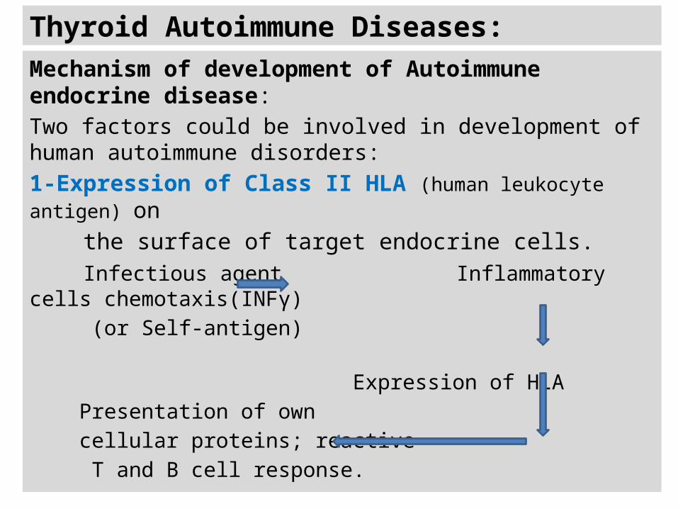
Thyroid Autoimmune Diseases: Mechanism of development of Autoimmune endocrine disease: Two factors could be involved in development of human autoimmune disorders: 1-Expression of Class II HLA (human leukocyte antigen) on the surface of target endocrine cells. Infectious agent Inflammatory cells chemotaxis(INFγ) (or Self-antigen) Expression of HLA Presentation of own cellular proteins; reactive T and B cell response.
-
Upload
dinah-morrison -
Category
Documents
-
view
268 -
download
1
Transcript of Thyroid Autoimmune Diseases: Mechanism of development of Autoimmune endocrine disease: Two factors...
Thyroid Autoimmune Diseases:
Mechanism of development of Autoimmune endocrine disease:Two factors could be involved in development of human autoimmune disorders: 1-Expression of Class II HLA (human leukocyte antigen) on the surface of target endocrine cells. Infectious agent Inflammatory cells chemotaxis(INFγ) (or Self-antigen) Expression of HLA Presentation of own cellular proteins; reactive T and B cell response.
N
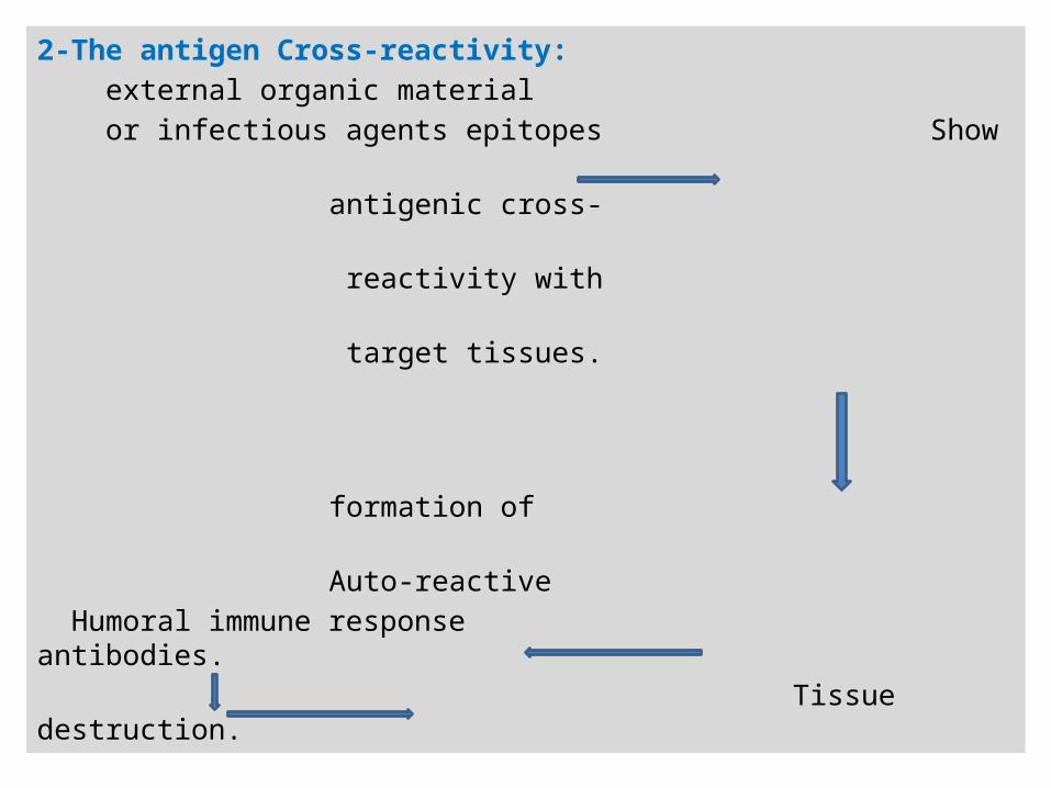
2-The antigen Cross-reactivity: external organic material or infectious agents epitopes Show antigenic cross- reactivity with target tissues.
formation of Auto-reactive Humoral immune response antibodies. Tissue destruction.
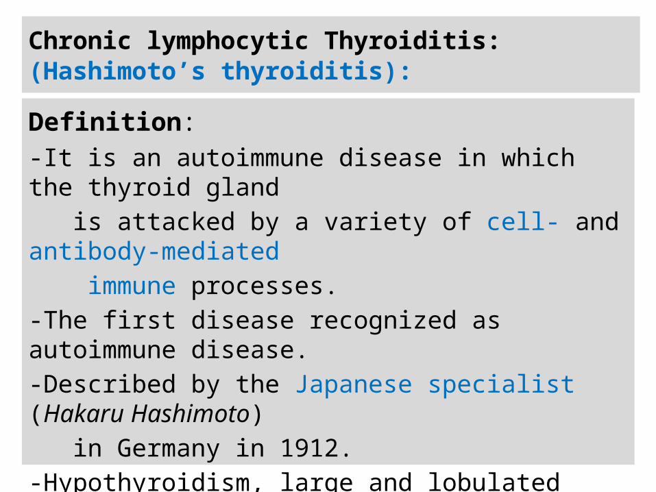
Chronic lymphocytic Thyroiditis:(Hashimoto’s thyroiditis):
Definition: -It is an autoimmune disease in which the thyroid gland is attacked by a variety of cell- and antibody-mediated immune processes. -The first disease recognized as autoimmune disease.-Described by the Japanese specialist (Hakaru Hashimoto) in Germany in 1912.-Hypothyroidism, large and lobulated thyroid gland.-Enlargement of thyroid due to lymphocytic infiltration and fibrosis.
n
General considerations: -Family history of thyroid disease and HLA gene polymorphism (DR4, DR5).-CTLA-4 (Cytotoxic T-Lymphocyte gene A-4) Polymorphism; results in reduced negative regulation of T-lymphocytes.-Studies on Monozygotic twins show: Anti-Thyroid Antibodies (Gene homology of 80%).
Other Risk factors: -Infectious agents : Human Herpes Virus-6 (A, and B).-Chromosomal disorders: Turner ,Klinefelter’s, and Down’s Syndrome.- Pollutants (Tobacco smoke).
n
-Most common in middle-aged and starts in adulthood. - 5-10 times more common in woman than in men.
-Associated with other autoimmune diseases such as: Systemic lupus erythematosus, dermatitis, and scleroderma.
Major Immunologic features of Hashimoto’s thyroiditis:
1-The lymphocytic infiltration of the thyroid gland.
2-The Antibodies against thyroid antigens are present.
3-The cellular sensitization to thyroid antigens.
n
Pathogenesis: -Expression of MHC Class II-self epitope complex on the thyroid cuboidal cell surface. -Thyroid cell-CD4+ Lymphocyte interaction. -Cytokines production, and chemotaxis of CTL and Macrophage.
-CTL-Thyroid cell interaction; Loss of T lymphocyte suppressor function due to CTL gene A mutation. -Killing of target cell by CTL; Apoptosis.
-Engulfment of cellular peptide by macrophage; Ag presentation.
n
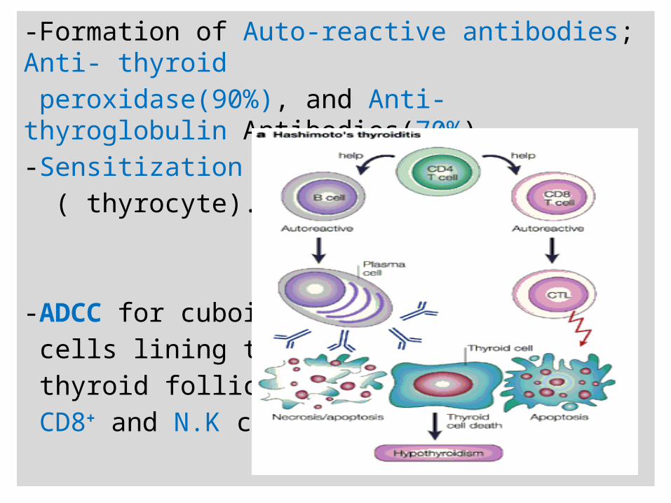
-Formation of Auto-reactive antibodies; Anti- thyroid peroxidase(90%), and Anti-thyroglobulin Antibodies(70%).-Sensitization of Thyroid tissue ( thyrocyte).
-ADCC for cuboidal cells lining the thyroid follicles by CD8+ and N.K cells.
n
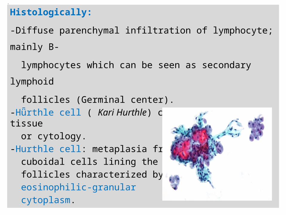
Histologically:
-Diffuse parenchymal infiltration of lymphocyte; mainly B-
lymphocytes which can be seen as secondary lymphoid
follicles (Germinal center).-Hǖrthle cell ( Kari Hurthle) could be seen in tissue or cytology. -Hurthle cell: metaplasia from cuboidal cells lining the thyroid follicles characterized by eosinophilic-granular cytoplasm.
N
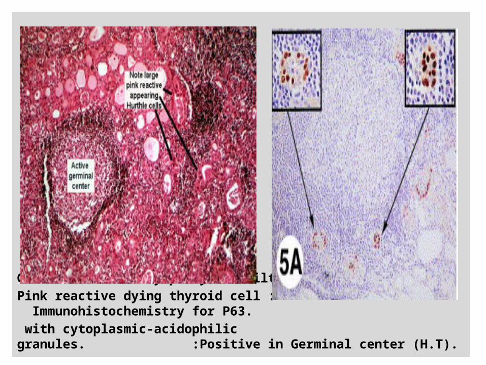
Germinal center: lymphocyte infiltrate.Pink reactive dying thyroid cell : Immunohistochemistry for P63. with cytoplasmic-acidophilic granules. :Positive in Germinal center (H.T).
Clinical Features of Chronic thyroiditis:
Primary stage: -Clinical hyperthyroidism due to inflammatory breakdown of thyroid follicles ( Silent, Painless inflammation).
Late stage:-Hypothyroidism due to progressive destruction of thyroid tissue and cellular malfunction.
-The most common outcome of Hashimoto’s disease is the hypothyroidism.
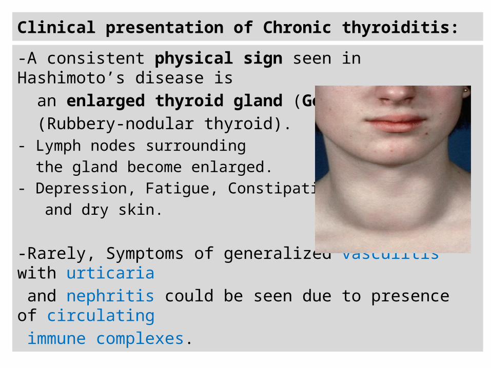
Clinical presentation of Chronic thyroiditis:
-A consistent physical sign seen in Hashimoto’s disease is an enlarged thyroid gland (Goiter). (Rubbery-nodular thyroid). - Lymph nodes surrounding the gland become enlarged. - Depression, Fatigue, Constipation, and dry skin.
-Rarely, Symptoms of generalized vasculitis with urticaria and nephritis could be seen due to presence of circulating immune complexes.
Differential diagnosis of Chronic thyroiditis:
-The hallmark of the diagnosis of this disease is the presence of circulating Autoantibodies:1-Anti-thyroglobulin antibodies. (Antibodies titer)2-Anti-thyroid peroxidase antibodies.(Antibodies titer).
-These antibodies show a sensitivity of 90% and detected by: 1-Immunofluorescence assay (Colloid aspiration). 2-ELISA. 3-Agglutination assay.
-In Seronegative patients , autoantibodies are localized in intrathyroidal lymphoid follicles.
Graves’ Disease: Definition:
-It is an autoimmune disease where the thyroid is
activated by anti-TSH receptor autoantibodies to produce
excessive amount of thyroid hormones.
-The most common cause of hyperthyroidism (60-90% of
all cases).
-It has a powerful hereditary component, affects up to 2%
of the female population, and is between five and ten
times as common in females as in males.
n
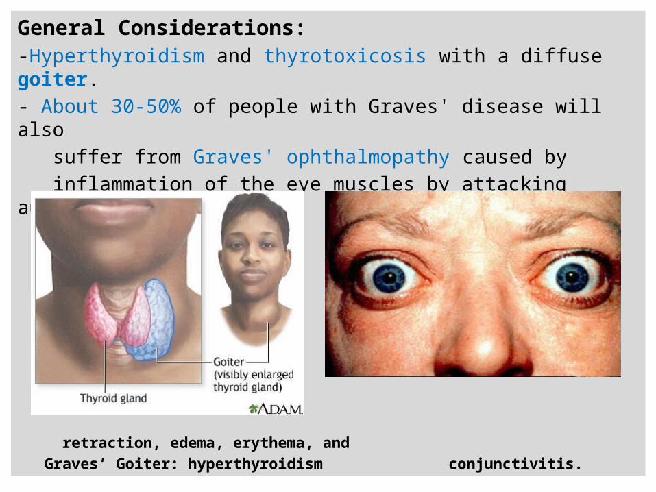
General Considerations: -Hyperthyroidism and thyrotoxicosis with a diffuse goiter. - About 30-50% of people with Graves' disease will also suffer from Graves' ophthalmopathy caused by inflammation of the eye muscles by attacking autoantibodies.
Exophthalmos: upper eyelid retraction, edema, erythema, and Graves’ Goiter: hyperthyroidism conjunctivitis.
N
-Specific cross-reactivity between some microbes (Viruses;
Coxsackievirus, and bacteria; Yersinia enterocolitica) and
TSH-Receptor of thyroid follicular cells.
-Strong association with DR3, DQα , and DQβ genotype of
MHC II haplotypes.
-Family History:
The disease is associated with different types of generalized
autoimmune susceptibility; such as Hashimoto’s disease and
antibodies to gastric intrinsic factors.
N
Clinical presentation:
-Goiter, exophthalmos(30-50%), muscle weakness, weight loss,
diarrhea and frequent defecation, hyperactivity ,tachycardia,
hair loss, and Oligomenorrhea.
Major Immunologic features of Graves’ disease:
1-Antibodies against thyroid antigen (TSH-R) are present in
70-100% of Graves’ patients; that stimulate thyroid cell
function.
2-Class II HLA expression on the surface of thyroid cells.
3-Associated autoimmune ophthalmopathy.
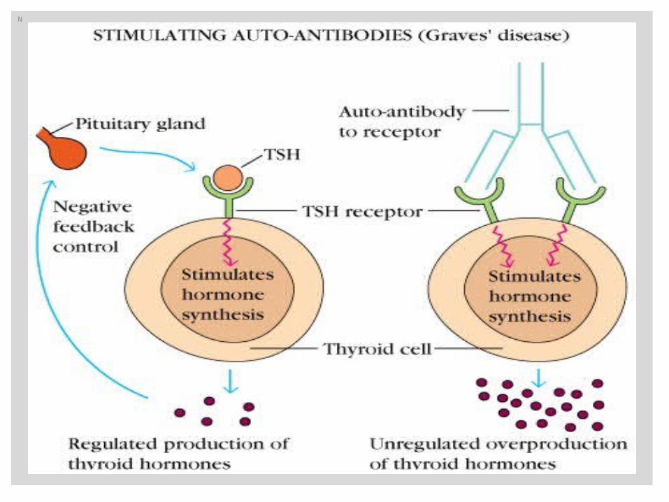
Graves’ disease Pathogenesis :
-Autoantibodies present against TSH-receptor: 1- Thyroid-stimulating immunoglobulins (TSI): Activate TSH-receptor; elevated thyroid hormones.2- Thyroid growth immunoglobulins( TGI) : Growth of thyroid follicles. 3-Thyroid binding-inhibiting immunoglobulins (TBII) : Inhibits TSH binding.-No Cellular immune response; Histology show no destruction of thyroid tissues.-Colloid suspension show lymphocytic infiltration: CD4, CD8, and B lymphocytes.
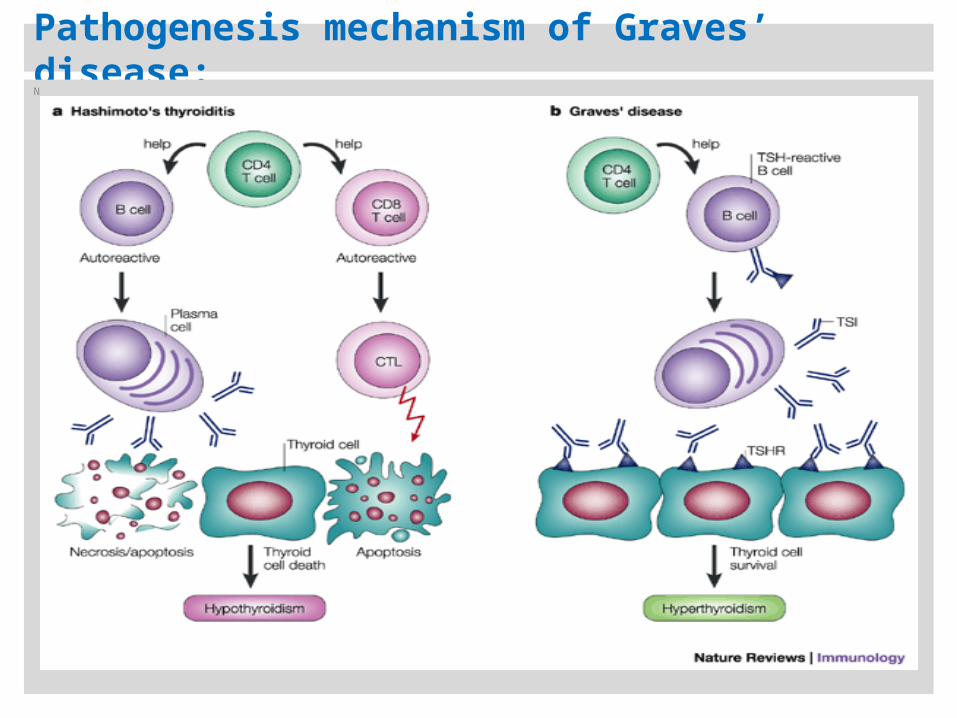
Pathogenesis mechanism of Graves’ disease: N
Diagnosis of Graves’ disease: -Clinically: Signs and symptoms of hyperthyroidism. -Radiology: Increased uptake of radioactive iodine.
-Serology: A-Elevated Total and free T4, and T3. B-Identification of Anti-thyroid antibodies in patient’s sera: 1-Antibodies that activate cellular cAMP ; Thyroid stimulating Immunoglobulin (TSI). 2-Thyroid growth stimulating immunoglobulins (TGI). 3-Antibodies that displace the binding of TSH from its receptor (TBII).
N
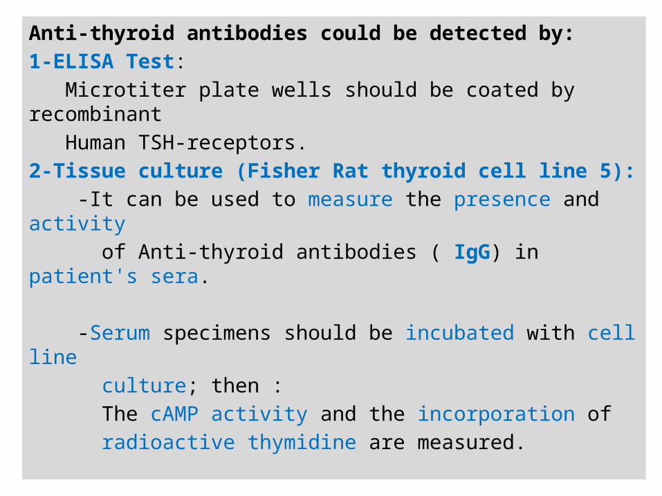
Anti-thyroid antibodies could be detected by:1-ELISA Test: Microtiter plate wells should be coated by recombinant Human TSH-receptors.2-Tissue culture (Fisher Rat thyroid cell line 5): -It can be used to measure the presence and activity of Anti-thyroid antibodies ( IgG) in patient's sera. -Serum specimens should be incubated with cell line culture; then : The cAMP activity and the incorporation of radioactive thymidine are measured.
n
N