Thesis Final 061710 esubmit - etd.library.vanderbilt.edu · tsA58: 5’- CCT CTG AGC TAT TCC AGA...
14
134 CHAPTER V LOSS OF TIE1 PROMOTES ANTI-ATHEROGENIC eNOS SYNTHESIS AND DOWNREGULATES NF-κB ACTIVATION Introduction Previous studies have shown that activation of Tie1 inhibits the function of Tie2 to maintain endothelial quiescence (Kwak et al., 1999; Papapetropoulos et al., 2000; Harfouche et al., 2002). Increased Tie1 expression has also been found in human tissue samples from patients with autoimmune inflammatory conditions such as rheumatoid arthritis and osteoarthritis (Shahrara et al., 2002). Further, overexpression of Tie1 in human umbilical vein endothelial cells increased the transcriptional message of inflammatory markers such as VCAM-1, ICAM-1 and E-selectin in a manner that was blocked by a p85 inhibitor (Chan et al., 2008). Suppression of Tie1 expression with siRNA downregulated expression of not just these markers, but also of TLR2 and IL- 1β inflammatory molecules (Chan & Sukhatme, 2009), suggesting a new pro- inflammatory role for Tie1. While Tie1 is pervasively expressed in the endothelia of developing embryos, post- natal expression is severely diminished in most organs except for the lungs (Taichman et al., 2002). Pulmonary Tie1 expression was dramatically increased during the period correlating with increased angiogenic activity of the maturing lung. In partially denuded carotid rat arteries where activated endothelial cells are re-populating the injury sites, Tie1 expression was also found to be upregulated (Fujikawa et al., 1999). Furthermore,
Transcript of Thesis Final 061710 esubmit - etd.library.vanderbilt.edu · tsA58: 5’- CCT CTG AGC TAT TCC AGA...
134
CHAPTER V
LOSS OF TIE1 PROMOTES ANTI-ATHEROGENIC eNOS SYNTHESIS AND
DOWNREGULATES NF-κB ACTIVATION
Introduction
Previous studies have shown that activation of Tie1 inhibits the function of Tie2 to
maintain endothelial quiescence (Kwak et al., 1999; Papapetropoulos et al., 2000;
Harfouche et al., 2002). Increased Tie1 expression has also been found in human tissue
samples from patients with autoimmune inflammatory conditions such as rheumatoid
arthritis and osteoarthritis (Shahrara et al., 2002). Further, overexpression of Tie1 in
human umbilical vein endothelial cells increased the transcriptional message of
inflammatory markers such as VCAM-1, ICAM-1 and E-selectin in a manner that was
blocked by a p85 inhibitor (Chan et al., 2008). Suppression of Tie1 expression with
siRNA downregulated expression of not just these markers, but also of TLR2 and IL-
1β inflammatory molecules (Chan & Sukhatme, 2009), suggesting a new pro-
inflammatory role for Tie1.
While Tie1 is pervasively expressed in the endothelia of developing embryos, post-
natal expression is severely diminished in most organs except for the lungs (Taichman et
al., 2002). Pulmonary Tie1 expression was dramatically increased during the period
correlating with increased angiogenic activity of the maturing lung. In partially denuded
carotid rat arteries where activated endothelial cells are re-populating the injury sites,
Tie1 expression was also found to be upregulated (Fujikawa et al., 1999). Furthermore,
135
Tie1 expression is increased in epithelial tumor cells of various organs, consistent with a
role in cellular activation (Cance et al., 1993; Lin et al., 1999; Tseng et al., 2001; Yang et
al., 2003; Ito et al., 2004; Uruno et al., 2004). These studies suggest that Tie1 expression
may promote endothelial cell activation. In the presence of either protein kinase C (PKC)
(Yabkowitz et al., 1997), vascular endothelial growth factor (VEGF) (Tsiamis et al.,
2002), phorbol 12-myristate 13 acetate (PMA) (Marron et al., 2000) or tumor necrosis
factor-α (TNF-α) (Yabkowitz et al., 1999), Tie1 is cleaved releasing its extracellular
domain and generating a membrane bound receptor fragment comprising the intracellular
and transmembrane domains. The cleaved intracellular product persists in the cytosol for
several hours (Marron et al., 2000), and has been demonstrated to bind signaling partners
such as Shp2 (Marron et al., 2000). The prolonged existence of the cleaved Tie1
intracellular fragment in the cytosol and its association with secondary messengers
suggest that this Tie1 endodomain may have further intracellular signaling functions
(Marron et al., 2000). However, the resultant effect of this proteolytic processing on Tie1
mediated signaling has not been studied.
One of the most important molecules investigated for its response to flow is nitric
oxide. The intracellular level of this potent vasodilator is regulated by eNOS (Uematsu et
al., 1995). Levels of eNOS dramatically increases with onset of laminar flow and
quantities remain high even after 24 hours (Kuchan & Frangos, 1994; Corson et al.,
1996). Nitric oxide has anti-inflammatory properties that regulate the nf-κB transcription
factor (Hajra et al., 2000). Disturbed flow conditions activate nf-κB resulting in
downregulation of eNOS expression. In contrast, laminar flow with high shear stress
increases expression of eNOS, which suppresses nf-κB activity (Davis et al., 2001; Davis
136
et al., 2004). As many studies have shown eNOS production is a very sensitive assay of
the effect of shear stress on endothelial cells (Kuchan & Frangos, 1994) and regulation of
nitric oxide release is an important process in endothelial function.
Tie1 expression and activity is also mediated by shear stress. Laminar flow with high
shear stress decreased Tie1 protein levels in vitro (Chen-Konak et al., 2003).
Interestingly, Chen-Konak et al showed that following 6 hours of pre-conditioning,
alterations in shear stress magnitude resulted in a decrease of Tie1 levels. Conversely,
Porat et al showed that disturbed flow conditions in vitro upregulates Tie1 promoter
activity (Porat et al., 2004). Also, bovine aortic endothelial cells exposed to shear stress
generated higher levels of the cleaved Tie1 intracellular fragment when compared to
static flow controls (Chen-Konak et al., 2003). Taken together, these studies suggest that
Tie1 modulates endothelial cell activation, and Tie1 expression and activity is regulated
by shear stress. However, an effect of shear stress on Tie1 mediated endothelial cell
activation has not been studied.
Experimental Procedures
Genotyping
At three weeks of age, tail samples from offspring were digested in 100 mM Tris pH
8.5, 5 mM EDTA, 0.2% sodium dodecyl sulfate, 200 mM NaCl, 100 µg/ml of Proteinase
K overnight at 55oC. Mice were genotyped by polymerase chain reaction with
REDExtract-N-Amp PCR Reaction Mix (Sigma) using the following primers,
137
cre: 5’- TCC GGG CTG CCA CGA CCA A -3’ and 5’- GGC GCG GCA ACA CCA
TTT TT -3’;
tie1: 5’- TGC CCC CCC TTC CAG AGA CTT CC -3’ and 5’- GCA AAG AGG ATC
CCC ACC AGA CCA TAC T –3’ and 5’- GGG GAT GTG CTG CAA GGC GAT TAA
G -3’;
tie-flox: 5’- TCG GGC GCG TTC AGA GTG GTA T -3’ and 5’- ATG CCT GTT CTA
TTT ATT TTT CCA G -3’
tsA58: 5’- CCT CTG AGC TAT TCC AGA AGT AGT G -3’ and 5’- TTA GAG CTT
TAA ATC TCT GTA GGT AG -3’
Animal Breeding and Tamoxifen Injections
SCL-ERT-Cre mice (Gothert et al., 2004) were a kind gift from Dr. Gothert. Tie1flox/
flox:SCL-ERT-Cre mice were bred in our laboratory. All mice used in this study were bred
onto a pure C57BL/6 background and were maintained in microisolator cages on a rodent
chow diet (Purina Mills Inc) and autoclaved water ad libitum. Animal care and
experimental procedures were performed according to the regulations of Vanderbilt
University’s Animal Care and Use Committee.
Generation of Tie1 mutant alleles
To generate Tie1flox/flox mice a loxP site and a neomycin resistance cassette were
introduced within the first intron of Tie1, and another loxP site just upstream of the
minimal promoter region. This strategic placement of loxP sites would allow excision of
the Tie1 minimal promoter and exon 1 upon Cre activation (Qu et al., 2010).
138
Protein Isolation and Western Blotting
Tissue samples or cells were lysed on ice using a lysis buffer (50mM Tris, 50mM
NaCl, 1% Triton X-100, 1mM EGTA, 1mM Na3VO4, 5mM NaF, 1x β-glycerophosphate)
supplemented with Complete protease inhibitor cocktail (Roche). After centrifugation at
12000 rpm at 4oC, the supernatant was collected. Protein concentrations were determined
with Protein Assay Reagent (Bio-Rad Laboratories). The nuclear pellet was rinsed three
times in lysis buffer and incubated at 100oC with 2x sample buffer to extract nuclear
proteins. Lysates were resolved by SDS-PAGE electrophoresis and transferred onto
Hybond ECL nitrocellulose membranes (GE Healthcare). Blots were blocked with 5%
non-fat milk and probed with rabbit antibodies to Tie1 (sc342), NOS3 (sc654), p50
(sc114), sp3 (sc644) (Santa Cruz Biotechnology) and mouse antibody to β-actin (Sigma
cat# A5316) overnight at 4oC. After washing with 0.1% Tween-20 supplemented
phosphate buffered saline (PBST), blots were incubated with secondary antibodies IRDye
800CW goat anti-rabbit IgG and IRDye 680CW goat anti-mouse IgG for 1 hour at room
temperature with gentle agitation. Blots were washed and scanned on the Odyssey
Infrared Imaging System (LI-COR Biosciences) and densitometry analyses were
performed using Odyssey Software.
Mouse Aortic Endothelial Cell Isolation (MAEC)
The Immorto mouse is a transgenic mouse generated by the introduction of
thermolabile SV40 T Ag, tsA58(Jat et al., 1991). In this model, the simian virus 40
(SV40) large tumor antigen of a temperature-sensitive strain (tsA58) is fused with the
139
major histocompatibility complex promoter H-2Kb. Expression of the promoter and the
large T antigen protein is only evident when immorto mouse-derived cells are cultured at
a permissive temperature (33oC). The addition of IFN-γ was also used to further enhance
promoter activity(Jat et al., 1991). A 4 to 8 week-old immorto mouse was anesthetized
with isoflurane and sacrificed by cervical dislocation. The lumbar aorta was punctured
and 5ml of Hanks Balanced Salt Solution (HBSS) was perfused through the left ventricle
to flush out blood. The heart, lung and aorta bloc was excised and transferred to a dish
containing HBSS and antibiotics. The heart and lungs were removed. The rest of the aorta
was incubated at 37oC for 15 minutes with filtered 10mg/ml collagenase Type II
(Worthington Chemicals) and 1x antibiotics dissolved in dispase (Roche). The adventitia
was gently removed and the remaining intima layer comprising the endothelium and
internal elastic lamina was cut open longitudinally and incubated at 37oC for 30 minutes
in filtered 20mg/ml collagenase Type II solution and 1x antibiotics dissolved in dispase.
The aorta was dissociated by pipetting several times in the dissolving solution and
filtered through a 100µm sterile cell strainer (Fisher Scientific). Culture media (MCDB
131 [Gibco], 10% FBS [Hyclone], 10U/ml heparin [Sigma], 2.75nM hydrocortisone
[Sigma] and 0.2% Bovine Brain Extract [Hammond Celltech], 10U/ml recombinant
murine IFN-γ [Peprotech], 1x antibiotics [Gibco]) was added to the cell suspension and
centrifuged at 375xg for 10 minutes. The cell mixture was then plated onto a fibronectin
coated culture dish and propagated at 33oC in a mixture of 5% carbon dioxide and 95%
oxygen. MAECs were isolated from wild-type and Tie1flox/flox:SCL-ERT-Cre immorto
mice.
140
4-hydroxytamoxifen Induction of Cre Activation and Immunocytochemistry
Mouse aortic endothelial cells from Tie1flox/flox:SCL-ERT-Cre mice were cultured at
37oC in complete media (as described above) without IFN-γ and treated with 5µM of 4-
hydroxytamoxifen (Sigma) every two days for six days. To ascertain ERT-Cre nuclear
localization, 4-hydroxytamoxifen treated cells were rinsed once with HBSS, fixed with
cold 100% methanol for 3 minutes and rinsed twice with HBSS. Cells were blocked with
5% goat serum, incubated overnight at 4oC with Cre antibody (Abcam, AB24608), rinsed
and immunostained with Alexa 488 conjugated goat anti-rabbit secondary antibody (BD
Biosciences).
Shear Stress Experiments
Shear stress experiments were performed using a custom cone-and-plate shear stress
viscometer design. An inverted servo motor (ElectroCraft, USA) is attached to a
plexiglass cone with 0.5o angle. A Motomatic II motor controller (Reliance Electric,
ElectroCraft, USA) regulates velocity in laminar flow, and a digital function generator
(Instek, San Diego, CA, USA) is used to produce a sinusoidal waveform modulating
oscillatory flow. A digital oscilloscope (Tektronix, USA) is used in parallel to monitor
the output magnitude and waveform. The servomotor and cone are lowered onto the
culture dish by a step controlled base stand. Laminar shear is attained with unidirectional
motion of the cone while oscillatory shear is achieved by bidirectional motion at ± 5
dynes/cm2. Shear stress experiments are performed for 24 hours in complete media in a
sterile, 5% CO2 incubator at 37oC.
141
RNA Extraction
RNA from tissue or cells was isolated using TriReagent (Molecular Research Center,
Cincinnati, OH, USA) following the manufacturer’s instructions. RNA concentration and
purity were analyzed on a NanoVue spectrophotometer (Thermo Scientific). Samples
with 260/280 ratio > 1.8 (1.9-2.1 preferrably) and 260/230 ratio > 1.5 were deemed of
sufficient quality for qRT-PCR. 2 µg of total RNA was reverse-transcribed into cDNA
using SuperScript III First-Strand System (Invitrogen) in 20 µl as per manufacturer’s
instructions.
Quantitative real-time Polymerase Chain Reaction (qRT-PCR)
All qRT-PCR was performed using iQ SYBR Green Supermix (Bio-Rad) on the iQ5
Real Time PCR Detection System (Bio-Rad). Experiments were performed in triplicate
using glyceraldehyde-3-phosphate dehydrogenase as the housekeeping gene to normalize
the data. The relative change in gene expression was determined using the critical
threshold (Ct) and the equation Fold Induction = 2^(-ΔΔ Ct) (Schmittgen & Livak, 2008).
Primers used were as follows, Tie1: 5'-GTGCCACCATTTTGACACTG-3' and 5'-
CAGGCACAGCAGGTTGTAGA-3'; glyceraldehyde-3-phosphate dehydrogenase: 5'-
CACTGGCATGGCCTTCCGTG-3' and 5'-AGGAAATGAGCTTGACAAAG-3
Statistical Analyses
All statistical differences in this study were determined by a 2-tailed, unpaired,
student’s t-test with 95% confidence intervals, using Prism 4.0 software (Graphpad Inc).
142
Results
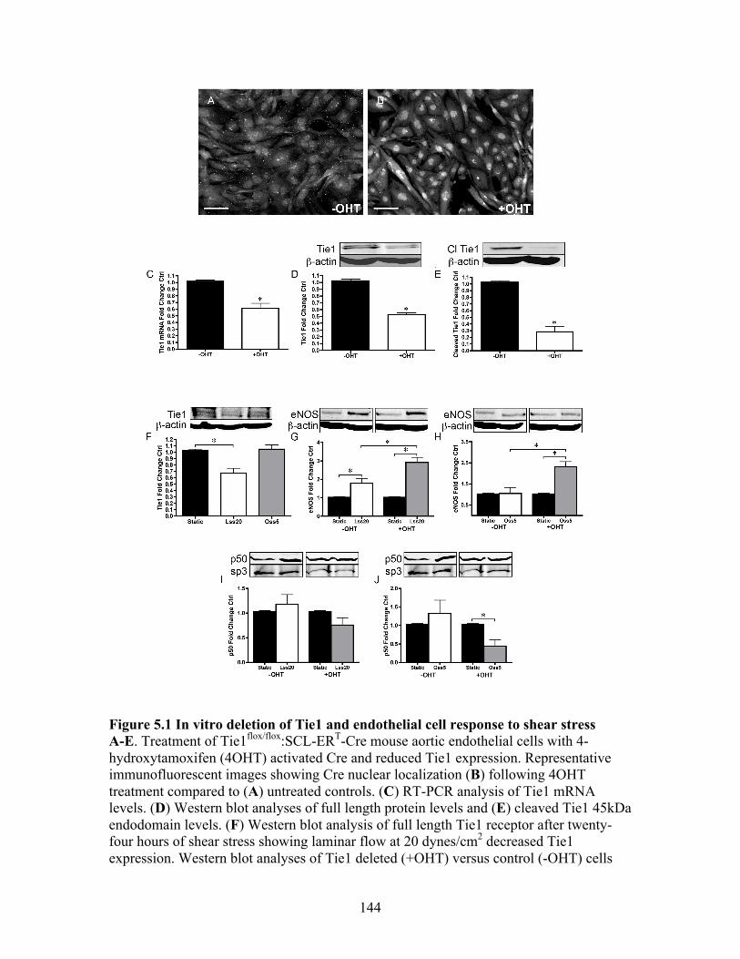
4-hydroxytamoxifen treatment activates Cre-mediated Tie1 deletion
To determine the effect of Tie1 deletion in vitro, we isolated endothelial cells from
homozygous Tie1flox/flox:SCL-ERT-Cre immorto mice, allowing for the use of 4-
hydroxytamoxifen (4OHT) to activate Cre transcription and hence induce Tie1 deletion.
To ascertain activation of Cre in vitro we immunostained 4OHT treated Tie1flox/flox:SCL-
ERT-Cre MAECs using Cre antibody. Increased nuclear staining indicates successful
translocation of Cre (Figure 5A, B). qRT-PCR and western blot analyses of full-length
Tie1 indicate 4OHT administration resulted in 41% and 45% reduction of Tie1
expression respectively (Figure 5C, D). Additionally, the cleaved Tie1 intracellular
fragment (McCarthy et al., 1999; Marron et al., 2000; Marron et al., 2007) was reduced
by 72% (Figure 5E).
Tie1 attenuation augments eNOS expression and decreases p50 nuclear translocation
We exposed the MAECs to laminar or oscillatory flow for 24 hours to investigate the
effect of shear stress on Tie1 in vitro. In these MAECs, laminar flow at 20 dynes/cm2
decreased Tie1 receptor expression while oscillatory flow had no effect on Tie1
expression (Figure 5F). Laminar flow also increased eNOS protein levels in
Tie1flox/flox:SCL-ERT-Cre MAECs as expected (Kuchan & Frangos, 1994). Interestingly,
after tamoxifen-induced deletion of Tie1, laminar flow further augmented eNOS
expression (Figure 5G). Also, oscillatory flow now increased eNOS levels when
compared either with cells grown under static conditions or with cells not exposed to
143
tamoxifen (Figure 5H). Laminar flow with Tie1 deletion had no significant effect on p50
nuclear localization (Figure 5I). Under oscillatory flow conditions, however, we found
that Tie1 deletion led to a 56% reduction of p50 nuclear translocation (p<0.01) (Figure
5J). In conclusion, these results suggest that Tie1 expression may be regulated by shear
stress and Tie1 may play a pro-inflammatory role acting via nf-κB signaling in promoting
the progression of atherosclerosis.
144
Figure 5.1 In vitro deletion of Tie1 and endothelial cell response to shear stress A-E. Treatment of Tie1flox/flox:SCL-ERT-Cre mouse aortic endothelial cells with 4-hydroxytamoxifen (4OHT) activated Cre and reduced Tie1 expression. Representative immunofluorescent images showing Cre nuclear localization (B) following 4OHT treatment compared to (A) untreated controls. (C) RT-PCR analysis of Tie1 mRNA levels. (D) Western blot analyses of full length protein levels and (E) cleaved Tie1 45kDa endodomain levels. (F) Western blot analysis of full length Tie1 receptor after twenty-four hours of shear stress showing laminar flow at 20 dynes/cm2 decreased Tie1 expression. Western blot analyses of Tie1 deleted (+OHT) versus control (-OHT) cells
145
comparing (G) laminar shear and (H) oscillatory shear induced eNOS expression. Western blot analyses of Tie1 deleted (+OHT) versus control (-OHT) cells comparing (I) laminar shear and (J) oscillatory shear induced p50 nuclear localization (normalized to sp3). (*p<0.05; White bar represents 100µm scale; Cl Tie1, cleaved Tie1; Lss20, laminar shear 20 dynes/cm2; Oss5, oscillatory shear 1Hz 5 dynes/cm2; OHT, 4-hydroxytamoxifen)
146
Discussion
We achieved a 50% deletion of Tie1 in vitro, concordant with in vivo tamoxifen
induced deletion in Tie1flox/flox:SCL-ERT-Cre mice. We surmise that Cre expression under
the SCL promoter may not be sufficient for extensive deletion of bi-allelic floxed-Tie1.
Marron et al previously reported cleavage of Tie1 with phorbol ester or VEGF (Marron et
al., 2007) while Chen-Konak and colleagues reported changes in levels of Tie1
intracellular fragment after brief alterations in shear stress (Chen-Konak et al., 2003).
Upon administration of 4OHT, we also noted a concomitant decrease in levels of cleaved
Tie1 intracellular fragment. While deletion of Tie1 resulted in a 50% reduction in full-
length Tie1 receptor, the cleaved Tie1 fragment was decreased by 74%.
Our in vitro experiments in murine aortic endothelial cells showed that twenty-four
hours of laminar flow at 20 dynes/cm2 suppressed Tie1 expression and increased eNOS
levels. Using bovine aortic endothelial cells, Chen-Konak et al found that brief in vitro
application of 10 dyne/cm2 shear stress temporarily decreased Tie1 but expression levels
returned to baseline after 2 hours (Chen-Konak et al., 2003). An octameric negative shear
stress response element (nSSRE) was also found that downregulates Tie1 expression
(Chen-Konak et al., 2003). Shear stress response elements are targets of the nf-κB
transcription factor, which in turn are regulated by shear stress (Gimbrone et al., 1999).
Tie1 was upregulated with a step increase (15 dyne/cm2) and downregulated with a step
decrease (5 dyne/cm2) in shear stress. Tie1 promoter activity in HUVECs was found
augmented by non-laminar flow in an in vitro step flow system(Porat et al., 2004).
Similar to the effect of adding PMA, VEGF or TNF-α (Yabkowitz et al., 1999), and also
mirroring our results, Chen-Konak found that shear stress induced cleavage of Tie1.
147
We also showed that genetic deletion of Tie1 in vitro augmented shear stress induced
eNOS expression and reduced p50 levels. Recent studies have shown that siRNA
silencing of Tie1 in vitro reduced expression of inflammatory markers (Chan &
Sukhatme, 2009) while overexpression studies demonstrated complementary findings
(Chan et al., 2008), suggesting a pro-inflammatory for Tie1. These findings together
support our in vivo data and together suggest a novel pro-inflammatory role for Tie1 and
a potential mechanism for Tie1 in atherosclerosis progression via the nf-κB pathway.
![Development of Testes and Expression of β-catenin in ... · low semen quality [5]. ... GAPDH, sense: 5’-TGG AGT CTA CTG GCG TCT TC-3’, anti-sense: 5’-ITC ACA CCC ATC ACA AAC](https://static.fdocument.org/doc/165x107/5d2cd5bd88c993136e8b4e7d/development-of-testes-and-expression-of-catenin-in-low-semen-quality.jpg)