Therapeutics Butein Sensitizes Human Hepatoma Cells to...
14
Research Article Butein Sensitizes Human Hepatoma Cells to TRAIL-Induced Apoptosis via Extracellular Signal-Regulated Kinase/Sp1– Dependent DR5 Upregulation and NF-κB Inactivation Dong-Oh Moon 1 , Mun-Ock Kim 2 , Yung Hyun Choi 3 , and Gi-Young Kim 1 Abstract Tumor necrosis factor–related apoptosis-inducing ligand (TRAIL) induces cell death in various types of cancer cells but has little or no effect on normal cells. Human hepatoma cells are resistant to TRAIL-induced apoptosis. Although butein is known to mediate anticancer, anti-inflammatory, and antioxidant activities, little is known about the mechanism of butein in terms of TRAIL-induced apoptosis of human hepatoma cells. In this study, we determined that butein enhances TRAIL-induced apoptosis in hepatoma cells through upregulation of DR5. Luciferase analysis showed that a 5′-flanking region containing four Sp1-binding sites within the DR5 promoter was enhanced by butein (-305/-300). Electrophoretic mobility shift assays and chromatin immunoprecipitation studies were used to analyze the elevation of Sp1 binding to DR5 promoter sites by butein. Point mutations of the Sp1-binding site also attenuated promoter activity. Furthermore, pre- treatment of the blocking chimeric antibody and small interfering RNA for DR5 significantly suppressed TRAIL-mediated apoptosis by butein in Hep3B cells. Butein also stimulated extracellular signal-regulated kinase (ERK) activation, and the ERK inhibitor PD98059 blocked butein-induced DR5 expression and sup- pressed binding of Sp1 to the DR5 promoter. Additionally, generation of reactive oxygen species had no effect on cell viability, although pretreatment with N-acetyl-L-cysteine or glutathione inhibited combined treatment- induced reactive oxygen species. Indeed, butein repressed the TRAIL-mediated activation of NF-κB and decreased its transcriptional activity. Our results suggest that butein could sensitize certain human hepatoma cells to TRAIL-induced apoptosis through stimulating its death signaling and by repressing the survival func- tion in these cells. Mol Cancer Ther; 9(6); 1583–95. ©2010 AACR. Introduction Tumor necrosis factor (TNF) family members have lim- ited use in anticancer therapy because they cause severe cytotoxicity to normal cells (1). However, for a couple of decades, TNF-related apoptosis-inducing ligand (TRAIL) has been thought to be a new candidate for anticancer therapy because TRAIL selectively suppresses tumor growth in vivo and in vitro but has little or no effect on normal cells (2, 3). TRAIL can bind to two death receptors (DR), DR4 and DR5, which contain a cytoplasmic functional death domain. After TRAIL binds to DRs, TRAIL triggers cell death through at least two fundamen- tal apoptotic pathways, referred to as the extrinsic path- way and the intrinsic pathway, depending on the cell type (4). TRAIL-induced apoptosis initiated by the extrin- sic pathway involves DR engagement, death-inducing signaling complex formation, and proteolytic activation of caspase-8 (5). Proteolytic caspase-8 further activates Bid, which, in turn, translocates to the mitochondria and activates the intrinsic pathway (6). Activated caspase-8 is released into the cytoplasm and induces a pro- tease cascade that activates effector caspases such as cas- pase-3 and caspase-7 (7). DR4 and DR5 not only give the apoptosis signal through the Fas-associated death do- main and caspase-8 but also activate NF-κB, which regu- lates the expression of survival factors such as members of the inhibitor of apoptosis (IAP) family (IAP1, IAP2, and XIAP) and Bcl-xL. Many studies have attempted to find new anticancer agents, which could augment apoptosis induced by TRAIL, because some cancer cells are resistant to the apoptotic effects of TRAIL (8–10). The mechanism un- derlying the augmentation of TRAIL-induced apoptosis is largely related to their ability to upregulate the ex- pression level of TRAIL receptors (i.e., DR4 and Authors' Affiliations: 1 Laboratory of Immunobiology, Department of Marine Life Sciences, Jeju National University, Jeju, Republic of Korea; 2 Molecular Cancer Research Center, Korea Research Institute of Bioscience and Biotechnology, Ochang, Republic of Korea; and 3 Department of Biochemistry, Dongeui University College of Oriental Medicine, Busan, Republic of Korea Corresponding Authors: Gi-Young Kim, Laboratory of Immunobiology, Department of Marine Life Science, Jeju National University, Jeju 690- 756, Republic of Korea. Phone: 82-64-754-3427; Fax: 82-64-756-3493. E-mail: [email protected] or Yung Hyun Choi, Department of Biochemistry, Dongeui University College of Oriental Medicine, Busan 614-051, Republic of Korea. Phone: 82-51-850-7413; Fax: 82-51-853- 4036. E-mail: [email protected] doi: 10.1158/1535-7163.MCT-09-0942 ©2010 American Association for Cancer Research. Molecular Cancer Therapeutics www.aacrjournals.org 1583 on August 12, 2019. © 2010 American Association for Cancer Research. mct.aacrjournals.org Downloaded from Published OnlineFirst June 1, 2010; DOI: 10.1158/1535-7163.MCT-09-0942
Transcript of Therapeutics Butein Sensitizes Human Hepatoma Cells to...

Published OnlineFirst June 1, 2010; DOI: 10.1158/1535-7163.MCT-09-0942
Research Article Molecular
CancerTherapeutics
Butein Sensitizes Human Hepatoma Cells to TRAIL-InducedApoptosis via Extracellular Signal-Regulated Kinase/Sp1–Dependent DR5 Upregulation and NF-κB Inactivation
Dong-Oh Moon1, Mun-Ock Kim2, Yung Hyun Choi3, and Gi-Young Kim1
Abstract
Authors' AMarine Life2MoleculaBioscienc3DepartmeMedicine, B
CorresponDepartmen756, RepubE-mail: imBiochemist614-051, R4036. E-ma
doi: 10.115
©2010 Am
www.aacr
Down
Tumor necrosis factor–related apoptosis-inducing ligand (TRAIL) induces cell death in various types ofcancer cells but has little or no effect on normal cells. Human hepatoma cells are resistant to TRAIL-inducedapoptosis. Although butein is known to mediate anticancer, anti-inflammatory, and antioxidant activities,little is known about the mechanism of butein in terms of TRAIL-induced apoptosis of human hepatoma cells.In this study, we determined that butein enhances TRAIL-induced apoptosis in hepatoma cells throughupregulation of DR5. Luciferase analysis showed that a 5′-flanking region containing four Sp1-binding siteswithin the DR5 promoter was enhanced by butein (−305/−300). Electrophoretic mobility shift assays andchromatin immunoprecipitation studies were used to analyze the elevation of Sp1 binding to DR5 promotersites by butein. Point mutations of the Sp1-binding site also attenuated promoter activity. Furthermore, pre-treatment of the blocking chimeric antibody and small interfering RNA for DR5 significantly suppressedTRAIL-mediated apoptosis by butein in Hep3B cells. Butein also stimulated extracellular signal-regulatedkinase (ERK) activation, and the ERK inhibitor PD98059 blocked butein-induced DR5 expression and sup-pressed binding of Sp1 to the DR5 promoter. Additionally, generation of reactive oxygen species had no effecton cell viability, although pretreatment with N-acetyl-L-cysteine or glutathione inhibited combined treatment-induced reactive oxygen species. Indeed, butein repressed the TRAIL-mediated activation of NF-κB anddecreased its transcriptional activity. Our results suggest that butein could sensitize certain human hepatomacells to TRAIL-induced apoptosis through stimulating its death signaling and by repressing the survival func-tion in these cells. Mol Cancer Ther; 9(6); 1583–95. ©2010 AACR.
Introduction
Tumor necrosis factor (TNF) family members have lim-ited use in anticancer therapy because they cause severecytotoxicity to normal cells (1). However, for a couple ofdecades, TNF-related apoptosis-inducing ligand (TRAIL)has been thought to be a new candidate for anticancertherapy because TRAIL selectively suppresses tumorgrowth in vivo and in vitro but has little or no effect onnormal cells (2, 3). TRAIL can bind to two death receptors(DR), DR4 and DR5, which contain a cytoplasmic
ffiliations: 1Laboratory of Immunobiology, Department ofSciences, Jeju National University, Jeju, Republic of Korea;r Cancer Research Center, Korea Research Institute ofe and Biotechnology, Ochang, Republic of Korea; andnt of Biochemistry, Dongeui University College of Orientalusan, Republic of Korea
ding Authors: Gi-Young Kim, Laboratory of Immunobiology,t of Marine Life Science, Jeju National University, Jeju 690-lic of Korea. Phone: 82-64-754-3427; Fax: [email protected] or Yung Hyun Choi, Department ofry, Dongeui University College of Oriental Medicine, Busanepublic of Korea. Phone: 82-51-850-7413; Fax: 82-51-853-il: [email protected]
8/1535-7163.MCT-09-0942
erican Association for Cancer Research.
journals.org
on August 12, 2019mct.aacrjournals.org loaded from
functional death domain. After TRAIL binds to DRs,TRAIL triggers cell death through at least two fundamen-tal apoptotic pathways, referred to as the extrinsic path-way and the intrinsic pathway, depending on the celltype (4). TRAIL-induced apoptosis initiated by the extrin-sic pathway involves DR engagement, death-inducingsignaling complex formation, and proteolytic activationof caspase-8 (5). Proteolytic caspase-8 further activatesBid, which, in turn, translocates to the mitochondriaand activates the intrinsic pathway (6). Activatedcaspase-8 is released into the cytoplasm and induces a pro-tease cascade that activates effector caspases such as cas-pase-3 and caspase-7 (7). DR4 and DR5 not only give theapoptosis signal through the Fas-associated death do-main and caspase-8 but also activate NF-κB, which regu-lates the expression of survival factors such asmembers ofthe inhibitor of apoptosis (IAP) family (IAP1, IAP2, andXIAP) and Bcl-xL.Many studies have attempted to find new anticancer
agents, which could augment apoptosis induced byTRAIL, because some cancer cells are resistant to theapoptotic effects of TRAIL (8–10). The mechanism un-derlying the augmentation of TRAIL-induced apoptosisis largely related to their ability to upregulate the ex-pression level of TRAIL receptors (i.e., DR4 and
1583
. © 2010 American Association for Cancer Research.
Moon et al.
1584
Published OnlineFirst June 1, 2010; DOI: 10.1158/1535-7163.MCT-09-0942
DR5). Therefore, agents that upregulate the expressionof DR4 and/or DR5 may have potential for clinicalmanagement of cancer. Because DR4 and DR5 can beregulated by either a p53-dependent or a p53-indepen-dent mechanism (11–13), it is possible that augmenta-tion of TRAIL-induced apoptosis by anticancer agentsmay be either p53 dependent or p53 independent.Natural products have played a highly significant role
over the years in the discovery of new drugs. This is par-ticularly evident in the treatment of cancers and infec-tious diseases in which more than 60% and 75% ofdrugs, respectively, are of natural origin (14). Therefore,a natural product with strong synergistic activity withTRAIL but minimal toxicity could be a new tool for can-cer therapy. Butein is one such agent that has been iden-tified from numerous plants, including the stem bark ofSemecarpus anacardium, the heartwood ofDalbergia odorifera,and the traditional Chinese and Tibetan medicinal herbsCaragana jubata and Rhus verniciflua Stokes. Previousstudies suggested that butein sensitizes apoptosis inhuman promyelocytic leukemia cells and B16 melanomacells (15, 16). In the in vitro experiment, butein also sup-pressed the proliferation of many human cancers, includ-ing breast carcinoma, colon carcinoma, osteosarcoma,and hepatic stellate cells (17–20). But the anticancermechanisms of butein in TRAIL-mediated apoptosis arelittle understood.In this study, we showed DR5 expression and NF-κB–
mediated mechanism underlying butein-sensitized apo-ptosis induced by TRAIL in human hepatoma cancer cells.
Materials and Methods
ReagentsAntibodies against Sp1, nucleolin, p50, p65, Bid,
caspase-3, caspase-8, caspase-9, poly(ADP-ribose) poly-merase (PARP), DR4, DR5, IAP-1, IAP-2, XIAP, Bcl-2,Bcl-xL, Bax, and Bad were purchased from Santa CruzBiotechnology. Antibodies against c-Jun NH2-terminal ki-nase (JNK), phospho-JNK, extracellular signal-regulatedkinase (ERK), phospho-ERK, p38, and phospho-p38 werepurchased from Cell Signaling. The antibody againstβ-actin was purchased from Sigma. Peroxidase-labeleddonkey anti-rabbit and sheep anti-mouse immunoglobulin,recombinant human TRAIL/Apo2 ligand (the nontagged19-kDa protein, amino acids 114–281), and TNF-α werepurchased from KOMA Biotechnology. Antibodies forFas and PP2A were purchased from Upstate Biotechno-logy. The blocking antibody against DR5 was purchasedfrom R&D Systems. 6-Carboxy-2′,7′-dichlorofluoresceindiacetate (H2DCFDA) and 3,3′-dihexyloxacarbocyanineiodide (DiOC6) were purchased from Molecular Probes.Glutathione, N-acetyl-L-cysteine (NAC), pyrrolidinedithiocarbamate (PDTC), MG132, and PS341 were pur-chased from Sigma. Butein was also purchased fromSigma and dissolved in DMSO (vehicle). PD98059,SP600125, SB239063, z-VAD-fmk, and z-IETD-fmk werepurchased from Calbiochem.
Mol Cancer Ther; 9(6) June 2010
on August 12, 2019mct.aacrjournals.org Downloaded from
Cell cultureHuman hepatoma cell lines Hep3B and HepG2 were
cultured in DMEM (Life Technologies). PC3, HCT116,and U937 cells were cultured in RPMI 1640 (Life Techno-logies) supplemented with 10% fetal bovine serum andantibiotics (Sigma). MTT assays were used to determinecell viability.
Flow cytometric analysisCells were fixed with 1 unit/mL of RNase A (DNase
free) and 10 μg/mL of propidium iodide (Sigma) over-night at room temperature in the dark. A FACSCaliburflow cytometer (Becton Dickinson) was used to analyzethe level of apoptotic cells containing sub-G1 DNA con-tent. For Annexin V and DiOC6 staining, live cells wereincubated with Annexin V (R&D Systems) and DiOC6.
DNA fragmentation assayCells were lysed in a buffer containing 10 mmol/L Tris
(pH 7.4), 150 mmol/L NaCl, 5 mmol/L EDTA, and 0.5%Triton X-100 for 30 minutes on ice. Lysates were vortexedand cleared by centrifugation at 10,000 × g for 20 min-utes. Fragmented DNA in the supernatant was extractedwith an equal volume of phenol/chloroform/isoamylalcohol mixture (25:24:1) and analyzed.
Western blot analysisTotal cell extracts were prepared using PRO-PREP pro-
tein extraction solution (iNtRON Biotechnology). Thepreparation of cytoplasmic and nuclear extracts was con-ducted using NE-PER nuclear and cytosolic extractionreagents (Pierce). Total cell extracts were separated onpolyacrylamide gels, and then standard procedures wereused to transfer them to the nitrocellulose membranes.The membranes were developed using an enhancedchemiluminescence reagent (Amersham).
Measurement of reactive oxygen speciesCells were plated at a density of 5 × 10⁴, allowed to
attach for 24 hours, and exposed to 5 mmol/L NACalone, 5 mmol/L glutathione alone, 10 μmol/L buteinalone, or NAC or glutathione plus butein for 1 hour.The cells were stained with 10 μmol/L H2DCFDA for10 minutes at 37°C, and flow cytometry was used todetermine the fluorescence intensity.
In vitro caspase activity assayA caspase activation kit was used according to the
manufacturer's protocol (R&D Systems) to measure theactivity of caspase-like protease.
Reverse transcription-PCR analysisTotal RNAwas extracted using the Trizol reagent (Invitro-
gen), andreverse transcription-PCR(RT-PCR)wasconducted.The sense primer 5′-GTCTGCTCTGATCACCCAAC-3′and the antisense primer 5′-CTGCAACTGTGACTCC-TATG-3′ were used to amplify human DR5 mRNA.For glyceraldehyde-3-phosphate dehydrogenase, the
Molecular Cancer Therapeutics
. © 2010 American Association for Cancer Research.

Butein-induced TRAIL Sensitization
Published OnlineFirst June 1, 2010; DOI: 10.1158/1535-7163.MCT-09-0942
sense primer 5′-CGTCTTCACCATGGAGA-3′ and theantisense primer 5′-CGGCCATCACGCCCACAGTTT-3′were used.
Analysis of surface DR5 expressionIndirect staining with primary rabbit anti-human DR4
or DR5 followed by FITC-conjugated IgG was used toanalyze cells for the surface expression of DR4 andDR5. Flow cytometry was used to analyze the expres-sions of these DRs.
Luciferase assaysThe pDR5/SacI plasmid [containing DR5 promoter
sequence (−2,500/+30)] and pDR5/−605 [containingDR5 promoter sequence (−605/+3)] were gifts from Dr.T. Sakai (Kyoto Prefectural University of Medicine, Kyoto,Japan). To analyze the promoter regions responsible forbutein, the reporter constructs containing single (mSp1-1and mSp1-2), double (mSp1-3 and mSp1-4), or triple(mSp1-5 and mSp1-6) point mutations at putative Sp1-binding sites of the DR5 promoter were used. Thedetailed procedure to generate these mutants has beenpreviously described (21).
Small interfering RNAThe 25-nucleotide small interfering RNA (siRNA) du-
plexes used in this study were purchased from SantaCruz Biotechnology. The cells were transfected with siRNAoligonucleotides using FuGENE 6 Transfection Reagentaccording to the manufacturer's recommendations.
Electrophoretic mobility shift assayDNA-protein binding assays were carried out with
a nuclear extract. Synthetic complementary Sp1 (5′-ATTCGATCGGGGCGGGGCGAGC-3′) and NF-κB(5′-AGTTGAGGGGACTTTCCCAGGC-3′) binding oligo-nucleotides (Santa Cruz Biotechnology) were 3′-biotiny-lated using the biotin 3′-end DNA labeling kit (Pierce)according to the manufacturer's instructions.
Chromatin immunoprecipitation assayThe chromatin immunoprecipitation (ChIP) assay was
done using the EZ-Chip assay kit according to themanufacturer's protocol (Upstate Biotechnology). Theprimers used for the amplification of the Sp1-binding siteof DR5 promoter region were 5′-GCCAGGGCGAAG-GTTA-3′ (sense) and 5′-GGGCATCGTCGGTGTAT-3′(antisense; 276-bp DNA product; ref. 22).
Statistical analysisAll data were derived from at least three independent
experiments. The images were visualized with Chemi-Smart 2000 (Vilber Lourmat). Images were capturedusing Chemi-Capt (Vilber Lourmat) and transported intoPhotoshop. Scion Imaging software (23) was used toquantify. Statistical analyses were conducted usingSigmaPlot software (version 6.0). Values were presentedas mean ± SD. Significant differences between the groups
www.aacrjournals.org
on August 12, 2019mct.aacrjournals.org Downloaded from
were determined using the unpaired Student's t test.Statistical significance was regarded at P < 0.05.
Results
Butein sensitizes TRAIL-induced apoptosisregardless of cell type specificity through activationof caspaseAntiproliferative activity of TRAIL was first analyzed
in four human cancer cell lines: Hep3B, HepG2, HCT116,and U937. Treatment with 100 ng/mL TRAIL inducedlimited inhibition of cell proliferation (<20%) at 24 hours,suggesting that these cells are resistant to the apoptoticeffects of TRAIL (Fig. 1A). Next, we examined the anti-proliferative effects of butein alone or in combinationwith TRAIL in these cells. Butein alone did not signifi-cantly induce any morphologic signs of cell death up to5 μmol/L, although the cellular activity to reduce MTTformazan was slightly decreased at this concentration.However, cell proliferation was significantly reduced bytreatment with a combination of butein and TRAIL whenthe concentration of TRAIL (100 ng/mL) was fixed.These results show that treatment with a combinationof butein and TRAIL effectively inhibits cell proliferationin TRAIL-resistant hepatoma cells in a cell type–nonspecificmanner. Next, we investigated whether the combinationtreatment is dependent on apoptosis. As shown in Fig. 1B,the treatment of Hep3B cells with a combination of5 μmol/L butein and 100 ng/mL TRAIL for 24 hours sig-nificantly increased the accumulation of sub-G1 phasecells (top) and Annexin V staining (middle), whereastreatment with butein or TRAIL alone slightly increased.Pretreatment with a pan-caspase inhibitor z-VAD-fmksignificantly blocked the accumulation of sub-G1 phasecell populations and Annexin V induced by treatmentwith butein and TRAIL. When phase-contrast microscopywas used to examine the change of cell morphology,the cells treated with a combination of 5 μmol/L buteinand 100 ng/mL TRAIL for 24 hours displayed decreasedcell numbers with some apoptotic shrinkage (bottom)compared with the untreated control cells (Fig. 1B). Fur-thermore, DNA fragmentation analysis also showed atypical ladder pattern of internucleosomal DNA frag-mentation in hepatoma cells treated with a combinationof butein and TRAIL but not in cells treated with buteinor TRAIL alone (Fig. 1C). These apoptosis phenomenawere also completely blocked by the pretreatment withz-VAD-fmk. Taken together, these results suggest thatbutein stimulates TRAIL-induced apoptosis through cas-pase activation but is not cell specific.
Treatment with a combination of butein and TRAILactivates extrinsic and intrinsic pathwaysHepatoma cells had been shown to be resistant to
TRAIL because of insufficient activation of the extrinsicpathway and, more importantly, because of the block-ing of death signaling at the mitochondrial level (4).Therefore, Western blot analyses of proapoptotic and
Mol Cancer Ther; 9(6) June 2010 1585
. © 2010 American Association for Cancer Research.

Moon et al.
1586
Published OnlineFirst June 1, 2010; DOI: 10.1158/1535-7163.MCT-09-0942
antiapoptotic factors were done; it was presumed thatthe inhibitory factors in mitochondria were suppressedby treatment with a combination of butein and TRAIL.As shown in Fig. 2A, time-dependent cleavage of cas-
Mol Cancer Ther; 9(6) June 2010
on August 12, 2019mct.aacrjournals.org Downloaded from
pase-8 and Bid was observed by treatment with thecombination of butein and TRAIL. In contrast, little orno change was observed in cells treated with a singleagent. Similarly, caspase-9, caspase-3, and PARP were
Mole
. © 2010 American Associatio
Figure 1. Butein (BUE) sensitizesTRAIL-induced cell deathregardless of cell type specificity.A, effect of treatment with acombination of butein and TRAILon cell viability. Human hepatomaHep3B and HepG2, human coloncancer HCT116, and humanleukemia U937 cells were treatedwith butein for 30 min at theindicated concentrations andfurther treated with or without100 ng/mL TRAIL for 24 h. MTTassay assessed cellular viability.B, effect of treatment with acombination of butein and TRAILon cell death and apoptosis.Hep3B cells treated with 5 μmol/Lbutein alone, 100 ng/mL TRAILalone, or TRAIL + butein (T+B) for24 h. To examine the effect of theinhibition of pan-caspase, Hep3Bcells were pretreated with 25 μmol/Lz-VAD-fmk (z-VAD) for 30 min andfurther treated with butein + TRAILfor 24 h (T+B+z-VAD). Flowcytometry analyzed the DNAcontent (top) and Annexin V+
(middle) of the cells. Morphology ofcells (bottom) was examined underlight microscopy. Magnification,×400. C, effect of treatment with acombination of butein and TRAILon DNA fragmentation. Aftertreatment of Hep3B and HepG2cells as indicated for 24 h,fragmented DNAs were extractedfrom the treated cells and analyzedon 1.5% agarose gel. Data areexpressed as overall mean ± SDfrom three independentexperiments. Statisticalsignificance was determined byStudent's t test. *, P < 0.05versus vehicle control.
cular Cancer Therapeutics
n for Cancer Research.
Butein-induced TRAIL Sensitization
Published OnlineFirst June 1, 2010; DOI: 10.1158/1535-7163.MCT-09-0942
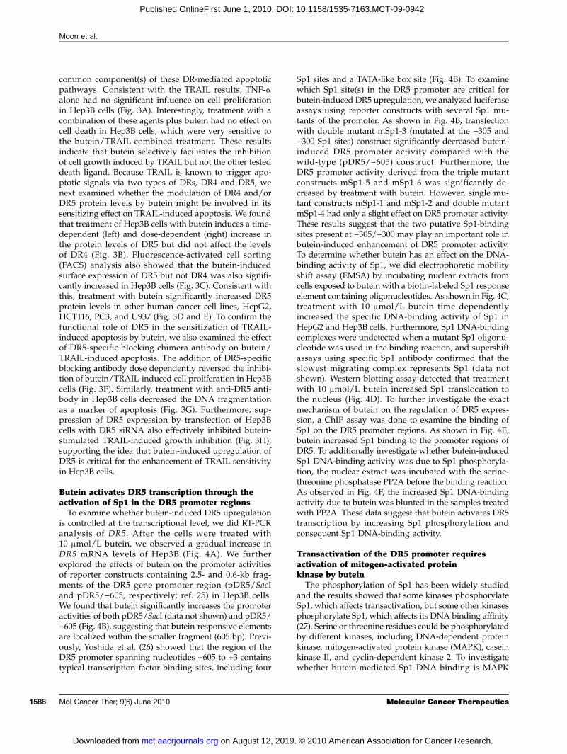
also activated after the combined treatment. Next, apo-ptotic events in the mitochondria were evaluated bymeasuring the mitochondrial membrane potential usingDiOC6. Marked reduction in the mitochondrial mem-brane potential had occurred in cells treated with bu-tein plus TRAIL (Fig. 2B). In addition, this processwas accompanied by the release of cytochrome c fromthe mitochondria into the cytosol (Fig. 2C). However,pretreatment with caspase-8 inhibitor z-IETD-fmk nor-malized mitochondrial membrane potential and com-pletely blocked the release of cytochrome c intocytosol (Fig. 2C). Furthermore, these caspasesactivity was significantly increased by the combinedtreatment (Fig. 2D). The combined treatment also de-
www.aacrjournals.org
on August 12, 2019mct.aacrjournals.org Downloaded from
graded the expression of Bcl-2, XIAP, IAP-1, and IAP-2. In contrast, the level of Bad expression was upregu-lated by the combined treatment (Fig. 2E). Theseresults indicated that treatment with a combination ofbutein and TRAIL reduces the expression of multipleproteins associated with cell survival through the ex-trinsic and intrinsic apoptotic signal pathway.
DR5 upregulation is important for butein-stimulated TRAIL-induced apoptosisAs the TNF superfamily members share similar
protein structures and DR-mediated apoptotic signalingpathways (24), we next examined whether butein sensi-tizes TNF-α–mediated apoptosis, possibly targeting the
Figure 2. Treatment with a combination of butein and/or TRAIL activates apoptotic signal via the extrinsic and intrinsic pathways. A and E, effect oftreatment with a combination of butein and TRAIL on levels of antiapoptotic and proapoptotic protein. Hep3B and HepG2 cells were treated with 100 ng/mLTRAIL alone, 5 μmol/L butein alone, or a combination of both for the indicated time points. Cell extracts were prepared for Western blotting for caspase-8,Bid, caspase-9, caspase-3, PARP, IAPs, Bcl-2, Bcl-xL, Bax, and Bad. B, effect of treatment with a combination of butein and TRAIL on mitochondrialmembrane potential. Hep3B cells were pretreated with caspase-8 inhibitor, z-IETD-fmk, and then the cells were treated with 100 ng/mL TRAIL alone,5 μmol/L butein alone, or a combination of both for 24 h. Mitochondrial membrane potential was measured by flow cytometry using DiOC6 dye. C, thetranslocation of cytochrome c was analyzed by Western blot analysis. D, effect of treatment with a combination of butein and TRAIL on caspase activity.Relative caspase activity was determined by the manufacturer's protocol. Columns, mean from three independent experiments; bars, SD. Statisticalsignificance was determined by Student's t test. *, P < 0.05 versus vehicle control.
Mol Cancer Ther; 9(6) June 2010 1587
. © 2010 American Association for Cancer Research.
Moon et al.
1588
Published OnlineFirst June 1, 2010; DOI: 10.1158/1535-7163.MCT-09-0942
common component(s) of these DR-mediated apoptoticpathways. Consistent with the TRAIL results, TNF-αalone had no significant influence on cell proliferationin Hep3B cells (Fig. 3A). Interestingly, treatment with acombination of these agents plus butein had no effect oncell death in Hep3B cells, which were very sensitive tothe butein/TRAIL-combined treatment. These resultsindicate that butein selectively facilitates the inhibitionof cell growth induced by TRAIL but not the other testeddeath ligand. Because TRAIL is known to trigger apo-ptotic signals via two types of DRs, DR4 and DR5, wenext examined whether the modulation of DR4 and/orDR5 protein levels by butein might be involved in itssensitizing effect on TRAIL-induced apoptosis. We foundthat treatment of Hep3B cells with butein induces a time-dependent (left) and dose-dependent (right) increase inthe protein levels of DR5 but did not affect the levelsof DR4 (Fig. 3B). Fluorescence-activated cell sorting(FACS) analysis also showed that the butein-inducedsurface expression of DR5 but not DR4 was also signifi-cantly increased in Hep3B cells (Fig. 3C). Consistent withthis, treatment with butein significantly increased DR5protein levels in other human cancer cell lines, HepG2,HCT116, PC3, and U937 (Fig. 3D and E). To confirm thefunctional role of DR5 in the sensitization of TRAIL-induced apoptosis by butein, we also examined the effectof DR5-specific blocking chimera antibody on butein/TRAIL-induced apoptosis. The addition of DR5-specificblocking antibody dose dependently reversed the inhibi-tion of butein/TRAIL-induced cell proliferation in Hep3Bcells (Fig. 3F). Similarly, treatment with anti-DR5 anti-body in Hep3B cells decreased the DNA fragmentationas a marker of apoptosis (Fig. 3G). Furthermore, sup-pression of DR5 expression by transfection of Hep3Bcells with DR5 siRNA also effectively inhibited butein-stimulated TRAIL-induced growth inhibition (Fig. 3H),supporting the idea that butein-induced upregulation ofDR5 is critical for the enhancement of TRAIL sensitivityin Hep3B cells.
Butein activates DR5 transcription through theactivation of Sp1 in the DR5 promoter regionsTo examine whether butein-induced DR5 upregulation
is controlled at the transcriptional level, we did RT-PCRanalysis of DR5. After the cells were treated with10 μmol/L butein, we observed a gradual increase inDR5 mRNA levels of Hep3B (Fig. 4A). We furtherexplored the effects of butein on the promoter activitiesof reporter constructs containing 2.5- and 0.6-kb frag-ments of the DR5 gene promoter region (pDR5/SacIand pDR5/−605, respectively; ref. 25) in Hep3B cells.We found that butein significantly increases the promoteractivities of both pDR5/SacI (data not shown) and pDR5/−605 (Fig. 4B), suggesting that butein-responsive elementsare localized within the smaller fragment (605 bp). Previ-ously, Yoshida et al. (26) showed that the region of theDR5 promoter spanning nucleotides −605 to +3 containstypical transcription factor binding sites, including four
Mol Cancer Ther; 9(6) June 2010
on August 12, 2019mct.aacrjournals.org Downloaded from
Sp1 sites and a TATA-like box site (Fig. 4B). To examinewhich Sp1 site(s) in the DR5 promoter are critical forbutein-induced DR5 upregulation, we analyzed luciferaseassays using reporter constructs with several Sp1 mu-tants of the promoter. As shown in Fig. 4B, transfectionwith double mutant mSp1-3 (mutated at the −305 and−300 Sp1 sites) construct significantly decreased butein-induced DR5 promoter activity compared with thewild-type (pDR5/−605) construct. Furthermore, theDR5 promoter activity derived from the triple mutantconstructs mSp1-5 and mSp1-6 was significantly de-creased by treatment with butein. However, single mu-tant constructs mSp1-1 and mSp1-2 and double mutantmSp1-4 had only a slight effect on DR5 promoter activity.These results suggest that the two putative Sp1-bindingsites present at −305/−300 may play an important role inbutein-induced enhancement of DR5 promoter activity.To determine whether butein has an effect on the DNA-binding activity of Sp1, we did electrophoretic mobilityshift assay (EMSA) by incubating nuclear extracts fromcells exposed to butein with a biotin-labeled Sp1 responseelement containing oligonucleotides. As shown in Fig. 4C,treatment with 10 μmol/L butein time dependentlyincreased the specific DNA-binding activity of Sp1 inHepG2 and Hep3B cells. Furthermore, Sp1 DNA-bindingcomplexes were undetected when a mutant Sp1 oligonu-cleotide was used in the binding reaction, and supershiftassays using specific Sp1 antibody confirmed that theslowest migrating complex represents Sp1 (data notshown). Western blotting assay detected that treatmentwith 10 μmol/L butein increased Sp1 translocation tothe nucleus (Fig. 4D). To further investigate the exactmechanism of butein on the regulation of DR5 expres-sion, a ChIP assay was done to examine the binding ofSp1 on the DR5 promoter regions. As shown in Fig. 4E,butein increased Sp1 binding to the promoter regions ofDR5. To additionally investigate whether butein-inducedSp1 DNA-binding activity was due to Sp1 phosphoryla-tion, the nuclear extract was incubated with the serine-threonine phosphatase PP2A before the binding reaction.As observed in Fig. 4F, the increased Sp1 DNA-bindingactivity due to butein was blunted in the samples treatedwith PP2A. These data suggest that butein activates DR5transcription by increasing Sp1 phosphorylation andconsequent Sp1 DNA-binding activity.
Transactivation of the DR5 promoter requiresactivation of mitogen-activated proteinkinase by buteinThe phosphorylation of Sp1 has been widely studied
and the results showed that some kinases phosphorylateSp1, which affects transactivation, but some other kinasesphosphorylate Sp1, which affects its DNA binding affinity(27). Serine or threonine residues could be phosphorylatedby different kinases, including DNA-dependent proteinkinase, mitogen-activated protein kinase (MAPK), caseinkinase II, and cyclin-dependent kinase 2. To investigatewhether butein-mediated Sp1 DNA binding is MAPK
Molecular Cancer Therapeutics
. © 2010 American Association for Cancer Research.

Butein-induced TRAIL Sensitization
Published OnlineFirst June 1, 2010; DOI: 10.1158/1535-7163.MCT-09-0942
Figure 3. Butein increases DR5 but not DR4 levels in hepatoma cell lines. A, effect of butein on TNF-α–mediated cell death. Hep3B cells were pretreatedwith the indicated concentration of butein for 30 min and further treated with the indicated concentrations of TNF-α for 24 h. Cellular viability wasanalyzed by MTT. B, butein-induced DR5 upregulation in Hep3B cells. Hep3B cells were treated with 10 μmol/L butein for the indicated time and Westernblot analysis of DR5, DR4, and β-actin as a loading control was conducted. C, effect of butein on the surface expression levels of DR5 and DR4.Hep3B cells were incubated with or without 10 μmol/L butein for 24 h, and flow cytometry was used to analyze the surface expression of DR5 and DR4.X axis, fluorescence intensity; Y axis, relative number of cells. Black histograms, isotype control; black line, treated cells with butein; gray histograms,untreated cells. D, butein-induced DR5 upregulation in other types of cancer cells. Cells were treated with up to 20 μmol/L butein for 24 h, and cell extractswere prepared for Western blot analysis of DR5. E, surface DR5 expression. Flow cytometry was used to analyze the surface expression of DR5. Blackhistograms, isotype control; black line, treated cells with butein; gray histograms, untreated cells. F, effect of DR5-specific blocking chimera antibodyon butein/TRAIL-induced apoptosis. Hep3B cells were pretreated with or without 5 μmol/L butein for 30 min followed by treatment with or without 100 ng/mL TRAIL for 24 h in the presence of the indicated concentrations of DR5-specific blocking chimera antibody. G, fragmented DNA was extracted andanalyzed on 1.5% agarose gel containing ethidium bromide. H, effect of siRNA DR5 on cell death. Hep3B cells were transfected with siRNA duplexesagainst DR5 mRNA. Twenty-four hours after the transfection, cells were treated with 5 μmol/L butein and 100 ng/mL TRAIL for 24 h. Cellular viabilitywas determined by MTT assay. Columns, mean from three independent experiments; bars, SD. Statistical significance was determined by Student'st test. *, P < 0.05 versus vehicle control.
Mol Cancer Ther; 9(6) June 2010www.aacrjournals.org 1589
on August 12, 2019. © 2010 American Association for Cancer Research. mct.aacrjournals.org Downloaded from

Moon et al.
1590
Published OnlineFirst June 1, 2010; DOI: 10.1158/1535-7163.MCT-09-0942
Figure 4. Butein activates transcription of DR5 through Sp1 activation at the −305/−300 region. A, effect of butein on DR5 mRNA levels. Hep3B cells weretreated with up to 10 μmol/L butein for 24 h, total RNA was isolated, and RT-PCR analysis of DR5 and glyceraldehyde-3-phosphate dehydrogenase(GAPDH) was done. B, effect of butein on DR5 promoter activity. Schematic structures (top) of the DR5 promoter constructs used to measure luciferaseactivity. Mutations were introduced into the Sp1 consensus sites, Hep3B cells were transfected with the reporter constructs, and lysates from cellstreated with or without butein were assayed for luciferase activity. C, upregulation of Sp1 DNA binding activity at promoter regions of DR5 by butein. Cellswere incubated with 10 μmol/L butein for the indicated time. Sp1 DNA binding activity was analyzed by LightShift chemiluminescent EMSA kit asdescribed in Materials and Methods. D, butein activates Sp1 translocation to the nucleus. Cytosolic and nucleus proteins were prepared, and Westernblotting analysis of Sp1 was conducted. E, in vivo binding of Sp1 on DR5 promoter regions by butein. ChIP assay was done using antibodies against Sp1 inHep3B and HepG2 cells. Negative controls were done using antibody against rabbit IgG. F, Sp1 phosphorylation by butein. Nuclear extracts preparedfrom untreated Hep3B cells (lane 1), treated with 10 μmol/L butein (lanes 2–6), and treated with indicated concentration of PP2A (lanes 3–6) wereincubated with a biotin end-labeled Sp1-binding sequence oligonucleotide.
Mol Cancer Ther; 9(6) June 2010 Molecular Cancer Therapeutics
on August 12, 2019. © 2010 American Association for Cancer Research. mct.aacrjournals.org Downloaded from
Butein-induced TRAIL Sensitization
Published OnlineFirst June 1, 2010; DOI: 10.1158/1535-7163.MCT-09-0942
dependent, Western blotting analysis was used to test thechange in phosphorylation of JNK, ERK, and p38. Asshown in Fig. 5A, 10 μmol/L butein time dependentlyincreased the phosphorylation of JNK, ERK, and p38.ERK, JNK1/2, and p38 inhibition abrogated butein-mediatedSp1-binding activity (Fig. 5B). Inhibition of ERK, JNK1/2,and p38 inhibited Sp1 translocation to the nucleus(Fig. 5C). However, only inhibition of ERK by PD98059significantly blocked butein-induced DR5 upregulationand inhibited cleavage of caspase-3 (Fig. 5D) and induc-tion of sub-G1 population induced by treatment withbutein plus TRAIL (Fig. 5E). FACS analysis also showedthat PD98059 significantly suppresses the combinedtreatment-induced surface expression of DR5 in Hep3Bcells (Fig. 5F). Moreover, pretreatment of 20 μmol/LPD98059 for 1 hour reduced DR5 promoter (pDR5/−605) activity induced by butein (Fig. 5G). Additionally,we investigated whether reactive oxygen species (ROS)are involved in butein-induced apoptosis in hepatomacells treated with 5 μmol/L butein. H2DCFDA-basedFACS detection revealed that intracellular ROS levelsslightly increased following treatment with butein(Fig. 5H). The butein-induced increases in ROS levelswere completely blocked by pretreatment with antioxi-dants, NAC, and glutathione. We next tested whetherscavenging of ROS attenuates the growth inhibitioninduced by the treatment of the combination of buteinand TRAIL. Pretreatment with NAC and glutathionedid not block the antiproliferative effects induced bythe combination of butein and TRAIL (Fig. 5H). Theseresults suggest that ERK may be involved in butein-induced DR5 expression via Sp1 activation.
Butein inhibits TRAIL-mediated NF-κB activationBecause TRAIL can trigger both death and survival
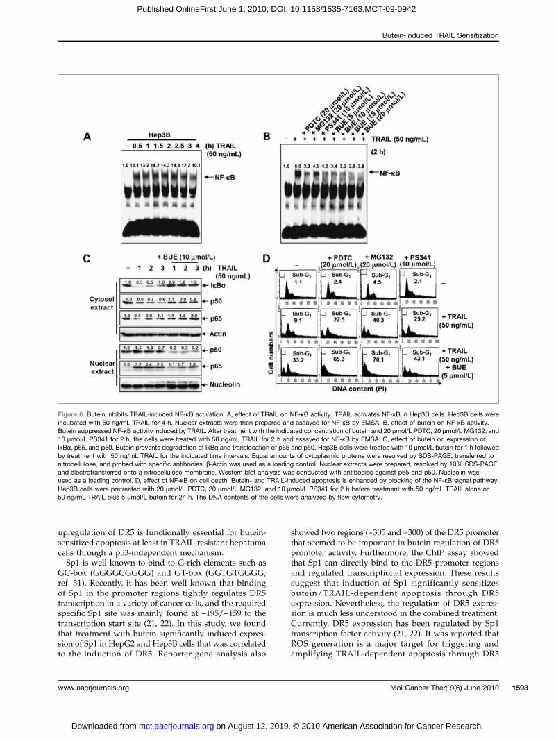
signaling (e.g., NF-κB), thus regulating the expressionof several factors involved in cell survival, we examinedthe effect of butein on TRAIL-mediated NF-κB by EMSAassay. As shown in Fig. 6A, 50 ng/mL TRAIL induced astrong increase in binding of NF-κB within 0.5 hour andwas sustained for 3 hours. Pretreatment with buteininhibited TRAIL-induced NF-κB activation in a dose-dependent manner (Fig. 6B). Especially, at a dose of10 μmol/L, butein significantly inhibited NF-κB activa-tion induced by TRAIL as much as NF-κB inhibitors[PDTC, MG-132, and bortezomib (PS341)] in Hep3B cells(Fig. 6B). It is well known that NF-κB activation byTRAIL is mediated by phosphorylation and degradationof IκBα. As shown in Fig. 6C, butein blocked TRAIL-dependent IκBα degradation. Butein also repressednuclear translocation of p65 and p50 induced by TRAIL.To further confirm the role of NF-κB in butein/TRAIL-induced apoptosis, cells were pretreated for 30 minuteswith PDTC, MG132, and PS341. The cells were then trea-ted with butein and TRAIL for an additional 24 hours,and flow cytometry was used to assess the sub-G1 cellpopulations. As shown in Fig. 6D, cells incubated withTRAIL or PDTC, MG132, and PS341 alone resulted in a
www.aacrjournals.org
on August 12, 2019mct.aacrjournals.org Downloaded from
slight increase in the sub-G1 percentage. However, treat-ment with a combination of the NF-κB inhibitors andTRAIL or TRAIL/butein markedly increased the accu-mulation of sub-G1 phase cells. In addition, as observedby MTT assay (data not shown), butein-mediated re-pressed cell viability is enhanced by blocking of theNF-κB signal pathway. These results suggest that buteintreatment is likely to inhibit the NF-κB pathway and thatthis pathway is likely involved in apoptosis.
Discussion
Although TRAIL is thought to be a highly promisingcandidate for cancer treatment, use of TRAIL has majorlimits in clinical applications because of resistanceagainst TRAIL in a variety of cancer cells (2, 3). Never-theless, many scientists have reported that conventionalcytotoxic drugs such as antioxidants and novel molecular-targeted agents or irradiation markedly sensitize TRAIL-induced apoptosis in TRAIL-resistant cancer cells (8–10).Thus, efforts to identify agents that activate DRs or blockantiapoptotic effectors may improve anticancer therapeu-tic design. Here, we provide evidence that butein is capableof triggering TRAIL-induced apoptosis at least in TRAIL-resistant hepatoma cells.DRs have a highly conserved extracellular region
containing cysteine-rich repeats and a conserved intracel-lular region of 80 amino acids termed the death domain(28). The domain is essential for the rising death signal,resulting in the activation of caspase-8 (29). Therefore, ithas been thought that regulation of DRs causes effectivedeath of cancer cells through direct binding with TRAIL.Some scientists reported that a few chemicals or proteinssensitize TRAIL-induced apoptosis through overexpres-sion of DR4 or DR5 (11–13). In this study, we also foundthat butein mainly increases the expression of DR5 at thetranscriptional level but maintains the expression of DR4.Additionally, for the functional study of DR5, DR5-specific blocking chimeric antibody and transient knock-down of DR5 expression by siRNA duplexes significantlyinhibited the growth-inhibitory effects induced by treat-ment with a combination of butein and TRAIL in Hep3Bcells. Nevertheless, we cannot rule out the possibility thatthe upregulation of DR5 is not the only way for sensitizingto TRAIL because knockdown of DR5 caused only amodest decrease in TRAIL-induced apoptosis despitethe almost complete loss of DR5 expression (8). Recently,it is well known that the tumor suppressor gene p53 is amain regulator for the expression of genes engaged inapoptosis (30). Many scientists have reported that over-expression of p53 transactivates DR4 and DR5 gene-induced DNA damage–inducing agents, suggesting thatDR4 and DR5 definitely are important regulators in p53-dependent apoptosis (11, 13). Nevertheless, there hasbeen conflicting evidence about the involvement of p53in regulating the expression of DR4 and DR5 until now(17). In this study, butein induced the expression of DR5,regardless of p53 status. These results may imply that
Mol Cancer Ther; 9(6) June 2010 1591
. © 2010 American Association for Cancer Research.

Moon et al.
1592
Published OnlineFirst June 1, 2010; DOI: 10.1158/1535-7163.MCT-09-0942
Figure 5. Butein-induced Sp1 translocation is mediated by ERK. A, effects of butein on phosphorylation of MAPKs. Hep3B cells were treated with 10 μmol/Lbutein for the indicated times. Equal amounts of cell lysates were resolved on SDS-polyacrylamide gels, transferred to nitrocellulose membranes, andprobed with antibodies against phospho-JNK (p-JNK), JNK, phospho-ERK (p-ERK), ERK, phospho-p38 (p-p38), and p38. B and C, regulation of Sp1 activityby MAPK inhibitors. MAPKs are involved in butein-upregulated Sp1 DNA binding. Hep3B cells were stimulated with 10 μmol/L butein for 24 h afterpretreatment with 20 μmol/L PD98059, 20 μmol/L SP600125, and 10 μmol/L SB203580 for 1 h. Then, Sp1 DNA binding activity and nuclear translocationwere analyzed by EMSA (B) and Western blot analysis (C). D, effects of MAPKs on DR5 and caspsase-3 induced by butein and TRAIL. Hep3B cellswere stimulated with 5 μmol/L butein and 100 ng/mL TRAIL for 24 h after pretreatment with 20 μmol/L PD98059, 20 μmol/L SP600125, and 10 μmol/LSB203580 for 1 h. Then, DR5 and caspase-3 were analyzed by Western blot analysis. E, flow cytometric analysis of sub-G1 phase by combined treatmentwith butein (10 μmol/L) and TRAIL (100 ng/mL). After combined treatment for 24 h, cells were stained with propidium iodide (PI) and analyzed by flowcytometry. F, effect of PD98059 on butein-induced DR5 expression. Cells were incubated with or without 10 μmol/L butein for 24 h, and flow cytometry wasused to analyze the surface expression of DR5. X axis, fluorescence intensity; Y axis, relative number of cells. Black histograms, isotype control;gray histograms, treated cells with butein. G, effect of PD98059 on DR5 promoter assay. PD98059 inhibits DR5 luciferase activity induced by butein. pDR5/−605 was transfected into Hep3B cells, which were then pretreated with 20 μmol/L PD98059 for 1 h and further treated with 10 μmol/L buteinfor 24 h, lysed, and assayed for luciferase activity. H, effect of ROS on butein/TRAIL-mediated apoptosis. Hep3B cells were pretreated with 5 μmol/L NACand glutathione for 30 min and further treated with 5 μmol/L butein + 100 ng/mL TRAIL for 24 h. Intracellular H2O2 and the DNA contents of thecells were analyzed by flow cytometry. PD, PD98059; SP, SP600125; SB, SB203580; T, TNF-α; B, butein.
Mol Cancer Ther; 9(6) June 2010 Molecular Cancer Therapeutics
on August 12, 2019. © 2010 American Association for Cancer Research. mct.aacrjournals.org Downloaded from
Butein-induced TRAIL Sensitization
Published OnlineFirst June 1, 2010; DOI: 10.1158/1535-7163.MCT-09-0942
upregulation of DR5 is functionally essential for butein-sensitized apoptosis at least in TRAIL-resistant hepatomacells through a p53-independent mechanism.Sp1 is well known to bind to G-rich elements such as
GC-box (GGGGCGGGG) and GT-box (GGTGTGGGG;ref. 31). Recently, it has been well known that bindingof Sp1 in the promoter regions tightly regulates DR5transcription in a variety of cancer cells, and the requiredspecific Sp1 site was mainly found at −195/−159 to thetranscription start site (21, 22). In this study, we foundthat treatment with butein significantly induced expres-sion of Sp1 in HepG2 and Hep3B cells that was correlatedto the induction of DR5. Reporter gene analysis also
www.aacrjournals.org
on August 12, 2019mct.aacrjournals.org Downloaded from
showed two regions (−305 and −300) of the DR5 promoterthat seemed to be important in butein regulation of DR5promoter activity. Furthermore, the ChIP assay showedthat Sp1 can directly bind to the DR5 promoter regionsand regulated transcriptional expression. These resultssuggest that induction of Sp1 significantly sensitizesbutein/TRAIL-dependent apoptosis through DR5expression. Nevertheless, the regulation of DR5 expres-sion is much less understood in the combined treatment.Currently, DR5 expression has been regulated by Sp1transcription factor activity (21, 22). It was reported thatROS generation is a major target for triggering andamplifying TRAIL-dependent apoptosis through DR5
Figure 6. Butein inhibits TRAIL-induced NF-κB activation. A, effect of TRAIL on NF-κB activity. TRAIL activates NF-κB in Hep3B cells. Hep3B cells wereincubated with 50 ng/mL TRAIL for 4 h. Nuclear extracts were then prepared and assayed for NF-κB by EMSA. B, effect of butein on NF-κB activity.Butein suppressed NF-κB activity induced by TRAIL. After treatment with the indicated concentration of butein and 20 μmol/L PDTC, 20 μmol/L MG132, and10 μmol/L PS341 for 2 h, the cells were treated with 50 ng/mL TRAIL for 2 h and assayed for NF-κB by EMSA. C, effect of butein on expression ofIκBα, p65, and p50. Butein prevents degradation of IκBα and translocation of p65 and p50. Hep3B cells were treated with 10 μmol/L butein for 1 h followedby treatment with 50 ng/mL TRAIL for the indicated time intervals. Equal amounts of cytoplasmic proteins were resolved by SDS-PAGE, transferred tonitrocellulose, and probed with specific antibodies. β-Actin was used as a loading control. Nuclear extracts were prepared, resolved by 10% SDS-PAGE,and electrotransferred onto a nitrocellulose membrane. Western blot analysis was conducted with antibodies against p65 and p50. Nucleolin wasused as a loading control. D, effect of NF-κB on cell death. Butein- and TRAIL-induced apoptosis is enhanced by blocking of the NF-κB signal pathway.Hep3B cells were pretreated with 20 μmol/L PDTC, 20 μmol/L MG132, and 10 μmol/L PS341 for 2 h before treatment with 50 ng/mL TRAIL alone or50 ng/mL TRAIL plus 5 μmol/L butein for 24 h. The DNA contents of the cells were analyzed by flow cytometry.
Mol Cancer Ther; 9(6) June 2010 1593
. © 2010 American Association for Cancer Research.

Moon et al.
1594
Published OnlineFirst June 1, 2010; DOI: 10.1158/1535-7163.MCT-09-0942
(32, 33). JNK directly is activated by ROS and stimulatesDR5 expression (25, 34). Therefore, we hypothesized thatthe combined treatment sensitizes the ROS-JNK-Sp1 signalcascades to activate DR5 expression; however, pretreat-ment of NAC or glutathione has no significant effect oncell growth inhibition and DR5 promoter activity. Never-theless, our previous study reported that high doses ofbutein (<40 μmol/L) increase G2-M phase arrest and cellgrowth inhibition through the ROS-JNK pathway (35).However, low doses of butein (<10 μmol/L) induced G1
phase arrest without cell growth inhibition in a ROS-independent manner. Only the ERK inhibitor PD98059significantly blocked butein-mediated Sp1 activationand cell death. The results indicate that Sp1 is a butein-regulated transcription factor, and ERK is an intermediarykinase in this relationship. Additionally, JNK could becorrelated with DR5 expression via Sp1 activation be-cause it is well known that pretreatment with SP600125moderately decreased DR5 expression and cell death(8, 25, 34). We did further study to elucidate if JNK andp38 have influence on the DR5 promoter activity.SP600125 itself significantly increased promoter activityof DR5, but SP600125 evidently downregulated its activityin the presence of butein (data not shown). These dataindicated that the JNK signal pathway may be also relatedto DR5 regulation in butein-induced apoptosis. Never-theless, we cannot rule out the possibility of EKR inhibi-tion by 20 μmol/L SP600125 because SP600125 quiteselectively blocks JNK at nanomolar concentrations. Ad-ditionally, although SB203580 suppressed the expressionof Sp1 and proapoptotic proteins, we cannot find any dif-ferences of DR5 promoter activity in the presence ofSB203580. Therefore, more prudent experiments will beneeded for the effects of p38 on TRAIL-induced apoptosis.The NF-κB transcription factor family consists of several
structurally related proteins such as c-Rel, Rel-A (p60),Rel-B, p50/p105, and p52/p100, which form homodimersor heterodimers with each other and regulate the expres-sion of a number of genes (36). TRAIL induced additionalbinding activity of NF-κB composed of a Rel-A/p50heterodimer in these cells. Ravi et al. (37) have recentlyreported that Rel-A−/− mouse fibroblasts are highlysensitive to TRAIL-induced apoptosis and that anti-CD40–mediated activation of NF-κB, including Rel-A,effectively blocked TRAIL-induced apoptosis. Therefore,NF-κB composed of Rel-A/p50 seems to play a key rolein the resistance of Hep3B and HepG2 cells to TRAIL-
Mol Cancer Ther; 9(6) June 2010
on August 12, 2019mct.aacrjournals.org Downloaded from
induced apoptosis. Indeed, pretreatment with butein in-hibited TRAIL-mediated activation of Rel-A/p50 NF-κBthat was sensitized to TRAIL-induced apoptosis by bu-tein. Similar observations have been reported that buteinsuppressed activation of Rel-A/p50 NF-κB induced byTNF-α (38). Taken together, these results indicate that bu-tein could possibly sensitize to TRAIL-induced apoptosisby inhibiting the activation of NF-κB, including Rel-A. Incontrast, recently important findings showed that theactivation status of NF-κB is not sufficient to determinethe fate of a cell with respect to TRAIL-induced apoptosisin hepatocellular carcinoma (10). Braeuer et al. (39) alsoreported that constitutively activated NF-κB, but not in-duced NF-κB, leads to TRAIL resistance by upregulationof XIAP in human cancer cells. Therefore, the effects ofNF-κB will be investigated in TRAIL resistance. In addi-tion, suppression of NF-κB activity by butein may also beinvolved in the stimulation of caspase-8 activity becausethe NF-κB–induced products, IAP-1, IAP-2, and XIAP,are known to cooperatively block caspase-8 activity (40).In summary,we showed that butein sensitizes apoptosis
in TRAIL-resistant cells via upregulation of DR5 expres-sion. We also showed that DR5 expression is tightly regu-lated by Sp1 activity in the promoter regions of the gene.However, p53 is not necessary for butein-induced DR5 ex-pression and triggers apoptosis in TRAIL-stimulated cells.We also found that the JNK inhibitor SP600125 partiallysuppresses Sp1 and DR5 expression and cell death; how-ever, the ERK inhibitor PD98059 significantly abrogatedthe effect. Consequently, butein p53 independently upre-gulates DR5 expression through the ERK and Sp1 mecha-nism and sensitizes apoptosis in TRAIL-resistant cells.
Disclosure of Potential Conflicts of Interest
No potential conflicts of interest were disclosed.
Grant Support
Basic Science Research Program through the National Research Foun-dation funded by the Ministry of Education, Science and Technology (No.2009-0068626; G-Y. Kim).
The costs of publication of this article were defrayed in part by thepayment of page charges. This article must therefore be hereby markedadvertisement in accordance with 18 U.S.C. Section 1734 solely to indicatethis fact.
Received 10/12/2009; revised 03/24/2010; accepted 03/24/2010;published OnlineFirst 06/01/2010.
References
1. Havell EA, Fiers W, North RJ. The antitumor function of tumor necro-sis factor (TNF), I. Therapeutic action of TNF against an establishedmurine sarcoma is indirect, immunologically dependent, and limitedby severe toxicity. J Exp Med 1998;167:1067–85.
2. Ashkenazi A, Pai RC, Fong S, et al. Safety and antitumor activity ofrecombinant soluble Apo2 ligand. J Clin Invest 1999;104:155–62.
3. Walczak H, Miller RE, Ariail K, et al. Tumoricidal activity of tumor
necrosis factor-related apoptosis-inducing ligand in vivo. Nat Med1999;5:157–63.
4. Almasan A, Ashkenazi A. Apo2L/TRAIL: apoptosis signaling, biology,and potential for cancer therapy. Cytokine Growth Factor Rev 2003;3–4:337–48.
5. Bodmer JL, Holler N, Reynard S, et al. TRAIL receptor-2 signals ap-optosis through FADD and caspase-8. Nat Cell Biol 2000;2:241–3.
Molecular Cancer Therapeutics
. © 2010 American Association for Cancer Research.
Butein-induced TRAIL Sensitization
Published OnlineFirst June 1, 2010; DOI: 10.1158/1535-7163.MCT-09-0942
6. Li H, Zhu H, Xu C, Yuan J. Cleavage of BID by caspase-8 mediatesthe mitochondria damage in the Fas pathway of apoptosis. Cell1998;94:491–501.
7. Srivastava RK. TRAIL/Apo-2L: mechanisms and clinical applicationsin cancer. Neoplasia 2001;3:535–46.
8. Inoue S, Twiddy D, Dyer MJ, Cohen GM. Upregulation of TRAIL-R2 isnot involved in HDACi mediated sensitization to TRAIL-induced ap-optosis. Cell Death Differ 2006;13:2160–2.
9. Koschny R, Ganten TM, Sykora J, et al. TRAIL/bortezomib cotreat-ment is potentially hepatotoxic but induces cancer-specific apopto-sis within a therapeutic window. Hepatology 2007;45:649–58.
10. Ganten TM, Koschny R, Haas TL, et al. Proteasome inhibition sensi-tizes hepatocellular carcinoma cells, but not human hepatocytes, toTRAIL. Hepatology 2005;42:588–59.
11. Wu GS, Burns TF, McDonald ER III, et al. KILLER/DR5 is a DNAdamage-inducible p53-regulated death receptor gene. Nat Genet1997;17:141–3.
12. Sheikh MS, Burns TF, Huang Y, et al. p53-dependent and -independentregulation of the death receptor KILLER/DR5 gene expression inresponse to genotoxic stress and tumor necrosis factor. Cancer Res1998;58:1593–8.
13. Guan B, Yue P, Clayman GL, Sun SY. Evidence that the death recep-tor DR4 is a DNA damage-inducible, p53-regulated gene. J Cell Phy-siol 2001;188:98–105.
14. Newman DJ, Cragg GM, Snader KM. Natural products as sources ofnew drugs over the period 1981-2002. J Nat Prod 2003;66:1022–37.
15. Kim NY, Pae HO, Oh GS, et al. Butein, a plant polyphenol, inducesapoptosis concomitant with increased caspase-3 activity, decreasedBcl-2 expression and increased Bax expression in HL-60 cells. Phar-macol Toxicol 2001;88:261–6.
16. Iwashita K, Kobori M, Yamaki K. Flavonoids inhibit cell growth andinduce apoptosis in B16 melanoma 4A5 cells. Biosci Biotechnol Bio-chem 2000;64:1813–20.
17. Wang Y, Chan FL, Chen S, Leung LK. The plant polyphenol buteininhibits testosterone-induced proliferation in breast cancer cells ex-pressing aromatase. Life Sci 2005;77:39–51.
18. Yit CC, Das NP. Cytotoxic effect of butein on human colon adeno-carcinoma cell proliferation. Cancer Lett 1994;82:65–72.
19. Jang HS, Kook SH, Son YO, et al. Flavonoids purified from Rhusverniciflua Stokes actively inhibit cell growth and induce apoptosisin human osteosarcoma cells. Biochim Biophys Acta 2005;1726:309–16.
20. Lee SH, Seo GS, Kim HS, Woo SW, Ko G, Sohn DH. 2′,4′,6′-Tris(methoxymethoxy) chalcone attenuates hepatic stellate cell prolifer-ation by a heme oxygenase-dependent pathway. Biochem Pharma-col 2006;72:1322–33.
21. Kim YH, Park JW, Lee JY, Kwon TK. Sodium butyrate sensitizesTRAIL-mediated apoptosis by induction of transcription from theDR5 gene promoter through Sp1 sites in colon cancer cells. Carci-nogenesis 2004;25:1813–20.
22. Sun M, Zhang J, Liu S, Liu Y, Zheng D. Sp1 is involved in 8-chloro-adenosine-upregulated death receptor 5 expression in human hepa-toma cells. Oncol Rep 2008;19:177–85.
23. http://www.scioncorp.com.
www.aacrjournals.org
on August 12, 2019mct.aacrjournals.org Downloaded from
24. Locksley RM, Killeen N, Lenardo MJ. The TNF and TNF receptorsuperfamilies: integrating mammalian biology. Cell 2001;104:487–501.
25. Shen HM, Liu ZG. JNK signaling pathway is a key modulator in celldeath mediated by reactive oxygen and nitrogen species. Free RadicBiol Med 2006;40:928–39.
26. Yoshida T, Maeda A, Tani N, Sakai T. Promoter structure and tran-scription initiation sites of the human death receptor 5/TRAIL-R2gene. FEBS Lett 2001;507:381–5.
27. Chu S, Ferro TJ. Sp1, regulation of gene expression by phosphory-lation. Gene 2005;348:1–11.
28. de Lamirande E, Gagnon C. Involvement of the extracellular signalregulated protein kinase pathway in human sperm function; involve-ment of the superoxide anion. Mol Hum Reprod 2002;8:124–35.
29. Nagata S. Apoptosis by death factor. Cell 1997;88:355–65.30. Thorburn A. Death receptor-induced cell killing. Cell Signal 2004;16:
139–44.31. Black AR, Black JD, Azizkhan-Clifford J. Sp1 and Krüppel-like factor
family of transcription factors in cell growth regulation and cancer.J Cell Physiol 2001;188:143–60.
32. Jung EM, Lim JH, Lee TJ, Park JW, Choi KS, Kwon TK. Curcuminsensitizes tumor necrosis factor-related apoptosis-inducing ligand(TRAIL)-induced apoptosis through reactive oxygen species-mediated upregulation of death receptor 5 (DR5). Carcinogenesis2005;26:1905–13.
33. Kim H, Kim EH, Eom YW, et al. Sulforaphane sensitizes tumor necro-sis factor-related apoptosis-inducing ligand (TRAIL)-resistant hepa-toma cells to TRAIL-induced apoptosis through reactive oxygenspecies-mediated up-regulation of DR5. Cancer Res 2006;66:1740–50.
34. Higuchi H, Grambihler A, Canbay A, Bronk SF, Gores GJ. Bile acidsup-regulate death receptor 5/TRAIL-receptor 2 expression via ac-Jun N-terminal kinase-dependent pathway involving Sp1. J BiolChem 2004;279:51–60.
35. Moon DO, Kim MO, Choi YH, Hyun JW, Chang WY, Kim GY. Buteininduces G2/M phase arrest and apoptosis in human hepatoma can-cer cells through ROS generation. Cancer Lett 2010;299:204–13.
36. Barkett M, Gilmore TD. Control of apoptosis by Rel/NF-κB transcrip-tion factors. Oncogene 1999;18:6910–24.
37. Ravi R, Bedi GC, Engstrom LW, et al. Regulation of death receptorexpression and TRAIL/Apo2L-induced apoptosis by NF-κB. Nat CellBiol 2001;3:409–16.
38. Pandey MK, Sandur SK, Sung B, Sethi G, Kunnumakkara AB,Aggarwal BB. Butein, a tetrahydroxychalcone, inhibits nuclear factor(NF)-κB and NF-κB-regulated gene expression through direct inhibi-tion of IκBα kinase β on cysteine 179 residue. J Biol Chem 2007;282:17340–50.
39. Braeuer SJ, Büneker C, Mohr A, Zwacka RM. Constitutively activatednuclear factor-κB, but not induced NF-κB, leads to TRAIL resistanceby up-regulation of X-linked inhibitor of apoptosis protein in humancancer cells. Mol Cancer Res 2006;4:715–28.
40. Wang CY, Mayo MW, Korneluk RG, Goeddel DV, Baldwin AS, Jr. NF-κBantiapoptosis: induction of TRAF1 and TRAF2 and c-IAP1 and c-IAP2to suppress caspase-8 activation. Science 1998;281:1680–3.
Mol Cancer Ther; 9(6) June 2010 1595
. © 2010 American Association for Cancer Research.

2010;9:1583-1595. Published OnlineFirst June 1, 2010.Mol Cancer Ther Dong-Oh Moon, Mun-Ock Kim, Yung Hyun Choi, et al.
B InactivationκDependent DR5 Upregulation and NF-−Apoptosis via Extracellular Signal-Regulated Kinase/Sp1
Butein Sensitizes Human Hepatoma Cells to TRAIL-Induced
Updated version
10.1158/1535-7163.MCT-09-0942doi:
Access the most recent version of this article at:
Cited articles
http://mct.aacrjournals.org/content/9/6/1583.full#ref-list-1
This article cites 39 articles, 6 of which you can access for free at:
Citing articles
http://mct.aacrjournals.org/content/9/6/1583.full#related-urls
This article has been cited by 1 HighWire-hosted articles. Access the articles at:
E-mail alerts related to this article or journal.Sign up to receive free email-alerts
Subscriptions
Reprints and
To order reprints of this article or to subscribe to the journal, contact the AACR Publications
Permissions
Rightslink site. Click on "Request Permissions" which will take you to the Copyright Clearance Center's (CCC)
.http://mct.aacrjournals.org/content/9/6/1583To request permission to re-use all or part of this article, use this link
on August 12, 2019. © 2010 American Association for Cancer Research. mct.aacrjournals.org Downloaded from
Published OnlineFirst June 1, 2010; DOI: 10.1158/1535-7163.MCT-09-0942